


Abstract
Background: Snijders Blok–Fisher syndrome (SNIBFIS; OMIM #618604) is a rare neurodevelopmental disorder associated with heterozygous POU3F3 variants and characterized by developmental delay, prominent speech and language impairment, behavioral abnormalities, and dysmorphic features. Case Presentation: We report a Chinese boy evaluated at 7 years and 9 months for longstanding developmental and language delay. He walked independently at 18 months, began using meaningful single words at approximately 24 months, and remained unable to produce complete sentences at 7 years of age. Wechsler intelligence testing yielded a full-scale IQ of 57. Clinical evaluation documented attention-deficit/hyperactivity disorder and autism spectrum disorder, with a Childhood Autism Rating Scale score of 38. Additional findings included sleep-onset difficulty, low-set ears, an open-mouth appearance, a single transverse palmar crease, and left hyperopia. He had no history of seizures, and electroencephalography was normal. Trio whole-exome sequencing identified a heterozygous POU3F3 nonsense variant, NM_006236.3:c.1225A>T [p.(Lys409Ter)], absent from both parental blood samples and reported as de novo by the diagnostic laboratory. The variant was not identified in the published literature or publicly accessible variant resources searched through 8 September 2026. Conclusions: This case adds to the reported POU3F3 variant spectrum and illustrates the importance of considering the unusual behavior of truncating alleles in this intronless gene.
1. Introduction
POU3F3 (POU class 3 homeobox 3), also known as BRN1, encodes a class III POU-domain transcription factor involved in central nervous system development. A role for POU3F3 in human neurodevelopmental disease was first suggested by a de novo 2q12.1 deletion encompassing POU3F3 and MRPS9 [1]. The association was molecularly delineated in 2019, when Snijders Blok and colleagues described 19 individuals with heterozygous POU3F3 variants and a recognizable neurodevelopmental phenotype [2]. The disorder is now termed Snijders Blok–Fisher syndrome (SNIBFIS; OMIM #618604).
The core phenotype includes developmental delay and/or intellectual disability, with speech and language impairment being particularly prominent. Hypotonia, autism or autistic features, attention and behavioral problems, sleep disturbance, low-set or cupped ears, ophthalmological abnormalities, hearing loss, and skeletal findings have also been reported [2,3]. In the largest published genotype–phenotype analysis to date, Rossi et al. analyzed data from 43 affected individuals. Among 27 individuals carrying truncating variants, 26/27 had developmental delay and/or intellectual disability, all 27 had speech delay or impairment, 15/27 had autism or autistic features, and 24/27 had cupped and/or low-set ears; epilepsy was reported in only 1/27 [3].
POU3F3 is a single-exon gene, and truncating variants may escape classical exon-junction-dependent nonsense-mediated decay. Functional analyses of selected truncations have shown expressed proteins with altered subcellular localization, reduced transcriptional activity and impaired dimerization [2]. ClinGen has assigned POU3F3 a haploinsufficiency score of 0, indicating that simple dosage loss is not an established disease mechanism [4]. Subsequent reports have broadened the clinical and molecular spectrum, including prenatally identified truncating variants, epileptic presentations associated with missense variation, 5′ untranslated region variants, and paternal gonosomal mosaicism [5,6,7,8].
Here, we report a child with SNIBFIS in whom trio-WES identified the POU3F3 c.1225A>T [p.(Lys409Ter)] variant, reported as de novo by the diagnostic laboratory.
2. Case Presentation
2.1. Perinatal and Developmental History
The proband was a Han Chinese boy born at 40 weeks of gestation to nonconsanguineous parents, both aged 29 years at his birth. The pregnancy was uneventful, and routine prenatal ultrasonography showed no obvious abnormalities. He was delivered vaginally, with a birth weight of 3150 g, length of 49 cm, and head circumference of 35 cm. Apgar scores were 10 at both 1 and 5 min, and there were no significant neonatal complications.
His mother first became concerned about his development at around 6 months of age because head control was still unstable. He rolled over at 8 months, sat independently at 10 months, and walked independently at 18 months. He began using meaningful single words at around 24 months and two-word phrases at around 30 months. By about 4 years of age, he was able to communicate toileting needs.
At 7 years of age, his expressive language remained markedly limited, and he was still unable to produce complete sentences. His speech was also unclear. Poor eye contact, limited response to name, reduced joint attention, and difficulty interacting with peers had been noted since early childhood. Because he had difficulty adapting to kindergarten, his family sought further developmental-behavioral evaluation before primary-school entry. The major developmental, clinical, and diagnostic milestones are summarized in Table 1. At that time, his height was 118.1 cm, weight 19.6 kg, BMI 14.1 kg/m2, and head circumference 50.5 cm.
Table 1.
Clinical and diagnostic timeline of the proband.
2.2. Neurodevelopmental and Behavioral Assessment
At a developmental-behavioral evaluation in July 2026, the child had obvious learning difficulties and was easily distracted and restless during structured activities. He had difficulty following instructions and rules. His caregivers also reported frequent temper outbursts, fidgeting, poor persistence with tasks, and a tendency to run around in public places unless closely supervised.
Wechsler intelligence testing at approximately 7 years and 9 months showed a full-scale IQ of 57. The specific test edition and index scores were not documented. Given his marked language impairment and inattention, the FSIQ was interpreted cautiously. ADHD had been diagnosed clinically. DSM-5 symptom counts were 8/9 for inattention and 8/9 for hyperactivity/impulsivity. Mean SNAP-IV item scores were 2.6 for inattention, 2.1 for hyperactivity/impulsivity, and 2.3 for oppositional symptoms. The local scale version was not recorded.
Autism spectrum disorder was also recorded in the clinical diagnosis. The CARS score was 38; the scale version was not recorded. Poor eye contact, limited response to name and joint attention, and reduced reciprocal interaction with peers were observed. Repeated play with the same toy was the main restricted/repetitive behavior documented. His caregivers also reported hair-eating behavior and marked difficulty falling asleep, often after midnight. No ADOS-2 or ADI-R assessment was available.
2.3. Physical Examination and Ancillary Investigations
Physical examination showed low-set ears, an open-mouth appearance, and a single transverse palmar crease. He was alert but restless during the examination and had difficulty remaining seated and following instructions.
He had no history of seizures. Electroencephalography performed on 22 July 2026 was within normal limits. Hearing assessment was normal. Ophthalmological examination showed normal vision in the right eye and hyperopia in the left eye. Echocardiography and renal and urinary tract ultrasonography were unremarkable. A left hand and wrist radiograph assessed using the TW3-RUS method showed a bone age of approximately 6–7 years.
Brain MRI was attempted but could not be completed because he remained unable to keep still despite sedation. Adequate structural brain images were therefore not available.
2.4. Genetic Testing and Variant Interpretation
Trio whole-exome sequencing (WES) was performed on peripheral-blood DNA from the proband and both parents using the BGI DNBSEQ-T7 platform and GenCap exome capture probes (V7.0; approximately 55 Mb). Reads were processed with Cutadapt v1.16, Sentieon v202010.01 and ANNOVAR (16 April 2018); CNVkit was used for copy-number analysis. The reference genome was GRCh37/hg19. Mean sequencing depths were 168.67×, 121.09× and 146.95× in the proband, father and mother, respectively, with >99% of target bases covered at ≥20×.
WES identified a POU3F3 nonsense variant, NM_006236.3:c.1225A>T [p.(Lys409Ter)] (GRCh37 chr2:105473193 A>T), called heterozygous in the proband. At this site, 33 of 112 reads supported the alternate allele (29.5%); parental coverage was 112× and 102×, and the variant was not reported in either parent. SNP-based kinship coefficients were 0.2878 (proband–father) and 0.2836 (proband–mother), consistent with first-degree relationships in the laboratory analysis. The laboratory reported the variant as de novo and classified it as pathogenic under ACMG/AMP criteria [9]. The supporting evidence and its interpretation are summarized in Table 2. No separate Sanger confirmation was documented, and no additional clinically relevant pathogenic or likely pathogenic copy-number variant was reported.
Table 2.
Evidence supporting the laboratory classification of p.(Lys409Ter).
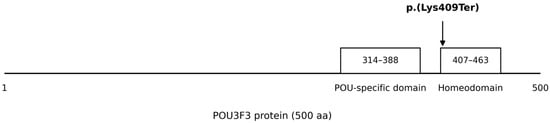
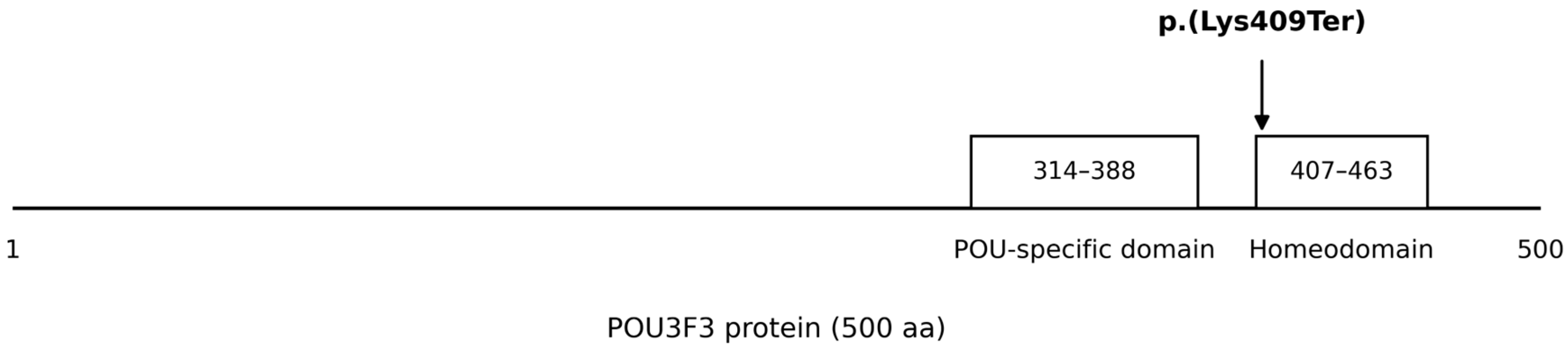
The predicted stop at Lys409 lies near the start of the POU homeodomain (Figure 1) and removes 92 residues from the canonical 500-amino-acid protein. Its specific effect on protein expression or function was not tested.
Figure 1.
Schematic representation of the POU3F3 protein and the location of the p.(Lys409Ter) variant. The canonical POU3F3 protein contains 500 amino acids. The POU-specific domain and homeodomain are indicated according to UniProt entry P20264 [12]. The p.(Lys409Ter) variant is located near the N-terminal portion of the homeodomain.
A targeted search of PubMed, ClinVar, and the Global Variome shared LOVD database performed on 8 September 2026 did not identify the NM_006236.3:c.1225A>T [p.(Lys409Ter)] variant in the published literature or these publicly accessible resources [13,14]. We therefore considered the variant previously unreported, while recognizing that unpublished or non-publicly accessible observations could not be excluded. Detailed search terms are provided in Supplementary Table S1.
2.5. Short-Term Follow-Up
At the initial evaluation, structured speech-language therapy had not yet been started. Details of other rehabilitation interventions or medication changes after diagnosis were not available.
At follow-up on 2 September 2026, at 7 years and 10 months of age, the child had entered Grade 1 but required adult assistance to attend school. No seizures had occurred during the follow-up period.
3. Discussion
The developmental and behavioral profile was broadly consistent with the truncating-variant subgroup reported by Rossi et al. [3] (Table 3). Developmental and speech impairment are prominent features of the disorder, while autistic features, behavioral problems, sleep disturbance, characteristic ear morphology, and visual abnormalities are also reported [2,3]. In this child, expressive-language impairment was particularly marked: motor milestones were delayed but ultimately acquired, whereas complete sentences were still absent at 7 years of age.
Table 3.
Comparison of the present case with reported features of individuals carrying truncating POU3F3 variants.
Because POU3F3 is intronless, truncating variants may escape conventional nonsense-mediated decay. In the functional study by Snijders Blok et al., selected truncations produced detectable abnormal proteins with altered subcellular localization, markedly reduced transcriptional activation and impaired mutant–mutant and mutant–wild-type interactions [2]. ClinGen has not established simple haploinsufficiency as the disease mechanism [4]. The present stop lies near the start of the homeodomain, between previously studied truncating variants p.(Ile400SerfsTer16) and p.(Cys428Ter) [2,12]. Large ASD sequencing studies have also implicated genes involved in transcriptional regulation among rare-variant risk genes [15,16].
Management remains supportive and phenotype directed, with attention to developmental and behavioral needs and surveillance for neurological, ophthalmological, audiological, sleep, and growth-related problems [3]. Genetic counseling is also important because most reported cases are de novo, although paternal gonosomal mosaicism has been described [8].
Limitations include incomplete psychometric test details, unavailable adaptive-function data, unsuccessful brain MRI and short follow-up. The p.(Lys409Ter) allele has not undergone orthogonal or functional validation, and site-specific population coverage metrics were unavailable. The novelty search was restricted to published and publicly accessible sources.
This case report was prepared with reference to the CARE reporting principles [17].
4. Conclusions
We describe a child with Snijders Blok–Fisher syndrome and a previously unreported POU3F3 p.(Lys409Ter) variant identified by trio-WES. The phenotype agrees with reported truncating-variant cases. Existing functional studies indicate that expressed POU3F3 truncations can disrupt transcription-factor activity without establishing conventional haploinsufficiency as the sole mechanism. Our findings add p.(Lys409Ter) to the reported POU3F3 truncating alleles, although its specific functional effects remain to be determined.
Supplementary Materials
The following supporting information can be downloaded at https://www.mdpi.com/article/10.3390/genes17111361/s1, Supplementary Table S1: Targeted search strategy for the novelty assessment of the POU3F3 c.1225A>T [p.(Lys409Ter)] variant.
Author Contributions
Conceptualization, L.Y. and M.C.; investigation, L.Y.; data curation, L.Y.; methodology, L.Y. and M.C.; visualization, L.Y.; writing—original draft preparation, L.Y.; writing—review and editing, L.Y. and M.C.; supervision, M.C. All authors have read and agreed to the published version of the manuscript.
Funding
This research received no external funding.
Institutional Review Board Statement
The study was approved by the Ethics Committee of Tongji Hospital, Tongji Medical College, Huazhong University of Science and Technology (Approval Code: TJ-IRB202412149; Approval Date: 25 December 2024).
Informed Consent Statement
Written informed consent for publication of the patient’s de-identified clinical and genetic information was obtained from the patient’s legal guardian.
Data Availability Statement
The clinical and genetic data underlying this report are not publicly available because they contain potentially identifiable information from a minor. De-identified data may be available from the corresponding author on reasonable request, subject to institutional requirements and the scope of the guardian’s consent.
Conflicts of Interest
The authors declare no conflicts of interest.
References
- Dheedene, A.; Maes, M.; Vergult, S.; Menten, B. A de novo POU3F3 deletion in a boy with intellectual disability and dysmorphic features. Mol. Syndromol. 2014, 5, 32–35. [Google Scholar] [CrossRef] [Scilit] [PubMed]
- Snijders Blok, L.; Kleefstra, T.; Venselaar, H.; Maas, S.; Kroes, H.Y.; Lachmeijer, A.M.A.; van Gassen, K.L.I.; Firth, H.V.; Tomkins, S.; Bodek, S.; et al. De novo variants disturbing the transactivation capacity of POU3F3 cause a characteristic neurodevelopmental disorder. Am. J. Hum. Genet. 2019, 105, 403–412. [Google Scholar] [CrossRef] [Scilit] [PubMed]
- Rossi, A.; Snijders Blok, L.; Neuser, S.; Klöckner, C.; Platzer, K.; Faivre, L.O.; Weigand, H.; Dentici, M.L.; Tartaglia, M.; Niceta, M.; et al. POU3F3-related disorder: Defining the phenotype and expanding the molecular spectrum. Clin. Genet. 2023, 104, 186–197. [Google Scholar] [CrossRef] [Scilit] [PubMed]
- Clinical Genome Resource (ClinGen). POU3F3 Dosage Sensitivity Curation, Evaluated 14 January 2025. Available online: https://search.clinicalgenome.org/kb/gene-dosage/POU3F3 (accessed on 19 October 2026).
- Zhang, H.; Linpeng, S.; Teng, Y.; Peng, C.; Liang, D.; Li, Z.; Wu, L. A de novo heterozygous POU3F3 genotype for the p.(Q214*) variant in a fetus with transient isolated bilateral mild ventriculomegaly: A case report and review of the literature. Front. Pediatr. 2023, 11, 1177137. [Google Scholar] [CrossRef] [Scilit] [PubMed]
- Terrone, G.; Cirillo, E.; Ripoli, F.; Votta, S.; Fiorile, M.F.; Aiello, S.; Paolella, C.; Andolfo, I.; Russo, R.; Bravaccio, C. A novel pathogenic variant causing POU3F3-related neurodevelopmental disorder in a child presenting with infantile epileptic spasms syndrome: Expanding the epileptic phenotype. Seizure 2025, 131, 22–25. [Google Scholar] [CrossRef] [Scilit] [PubMed]
- Hayashi, T.; Miyamoto, S.; Endo, Y.; Shimizu, K.; Ohkubo, Y.; Komatsu, K.; Furukawa, S.; Nakashima, M.; Fukuda, T.; Ogata, T.; et al. Identification of 5′ untranslated region variants in genes involved in neurodevelopmental disorders. J. Hum. Genet. 2026, 71, 353–361. [Google Scholar] [CrossRef] [Scilit] [PubMed]
- Barragán-Muñoz, A.; Trilla, P.; Sanchez, A.; Potrony, M.; Madrigal, I.; Rodriguez-Revenga, L.; Alvarez-Mora, M.I. Case Report: First case of paternal mosaicism in Snijders Blok-Fisher syndrome. Front. Genet. 2026, 17, 1892974. [Google Scholar] [CrossRef] [Scilit] [PubMed]
- Richards, S.; Aziz, N.; Bale, S.; Bick, D.; Das, S.; Gastier-Foster, J.; Grody, W.W.; Hegde, M.; Lyon, E.; Spector, E.; et al. Standards and guidelines for the interpretation of sequence variants: A joint consensus recommendation of the American College of Medical Genetics and Genomics and the Association for Molecular Pathology. Genet. Med. 2015, 17, 405–424. [Google Scholar] [CrossRef] [Scilit] [PubMed]
- Abou Tayoun, A.N.; Pesaran, T.; DiStefano, M.T.; Oza, A.; Rehm, H.L.; Biesecker, L.G.; Harrison, S.M. Recommendations for interpreting the loss of function PVS1 ACMG/AMP variant criterion. Hum. Mutat. 2018, 39, 1517–1524. [Google Scholar] [CrossRef] [Scilit] [PubMed]
- ClinGen Sequence Variant Interpretation Working Group. PM2—Recommendations for Population Absence or Rarity Evidence, Version 1.0, September 2020. Available online: https://www.clinicalgenome.org/site/assets/files/5182/pm2_-_svi_recommendation_-_approved_sept2020.pdf (accessed on 19 October 2026).
- UniProt Consortium. POU3F3—POU Domain, Class 3, Transcription Factor 3—Homo sapiens (UniProtKB P20264). Available online: https://www.uniprot.org/uniprotkb/P20264/entry (accessed on 8 September 2026).
- National Center for Biotechnology Information (NCBI). ClinVar. Available online: https://www.ncbi.nlm.nih.gov/clinvar/ (accessed on 8 September 2026).
- Global Variome shared LOVD. Unique Variants in the POU3F3 Gene. Available online: https://databases.lovd.nl/shared/variants/POU3F3/unique (accessed on 8 September 2026).
- Satterstrom, F.K.; Kosmicki, J.A.; Wang, J.; Breen, M.S.; De Rubeis, S.; An, J.-Y.; Peng, M.; Collins, R.; Grove, J.; Klei, L.; et al. Large-scale exome sequencing study implicates both developmental and functional changes in the neurobiology of autism. Cell 2020, 180, 568–584.e23. [Google Scholar] [CrossRef] [Scilit] [PubMed]
- Fu, J.M.; Satterstrom, F.K.; Peng, M.; Brand, H.; Collins, R.L.; Dong, S.; Wamsley, B.; Klei, L.; Wang, L.; Hao, S.P.; et al. Rare coding variation provides insight into the genetic architecture and phenotypic context of autism. Nat. Genet. 2022, 54, 1320–1331. [Google Scholar] [CrossRef] [Scilit] [PubMed]
- Gagnier, J.J.; Kienle, G.; Altman, D.G.; Moher, D.; Sox, H.; Riley, D.; CARE Group. The CARE guidelines: Consensus-based clinical case report guideline development. J. Clin. Epidemiol. 2014, 67, 46–51. [Google Scholar] [CrossRef] [Scilit] [PubMed]

Disclaimer/Publisher’s Note: The statements, opinions and data contained in all publications are solely those of the individual author(s) and contributor(s) and not of MDPI and/or the editor(s). MDPI and/or the editor(s) disclaim responsibility for any injury to people or property resulting from any ideas, methods, instructions or products referred to in the content. |
© 2026 by the authors. Licensee MDPI, Basel, Switzerland. This article is an open access article distributed under the terms and conditions of the Creative Commons Attribution (CC BY) license.