


Abstract
Background/Objectives: Overgrowth syndromes (OGSs) are a heterogeneous group of rare conditions marked by excessive growth and variable neurodevelopmental and systemic features. They are broadly classified into generalized overgrowth (GOG) and segmental overgrowth (SOG), which differ in clinical presentation and genetic mechanisms. However, many cases remain genetically unresolved, especially in underrepresented populations. Methods: We analyzed a binational cohort of patients from Poland and Lithuania with suspected OGSs, using detailed clinical evaluation and exome sequencing (ES) from blood-derived DNA. Variants were interpreted using current clinical guidelines, and phenotypes were compared between the GOG and SOG groups. Results: Patients with GOG more often showed developmental delay, intellectual disability, neurological issues, and joint laxity, while vascular malformations were more common in patients with SOG. Dysmorphic features appeared earlier in the SOG group, but became more pronounced over time in the GOG group. Genetic analysis led to the identification of definitive diagnoses in 25% of individuals. Pathogenic variants were identified in the genes involved in chromatin regulation and epigenetic control (DNMT3A, EED, KMT5B, CHD3, and NSD1), along with novel variants expanding the known spectrum. Conclusions: These findings provide valuable insights into the genetic architecture of OGSs in underrepresented Central and North-Eastern European populations, supporting the development of region-specific diagnostic and surveillance strategies.
1. Introduction
Overgrowth syndromes (OGSs) are a highly heterogeneous group of rare disorders united by a shared clinical hallmark-excessive growth. This umbrella term encompasses conditions characterized by either generalized overgrowth (GOG) or segmental overgrowth (SOG). In most cases, abnormal growth patterns are detectable prenatally, although postnatal onset has also been reported [1]. GOG is defined by a proportional increase in height, weight, and/or head circumference by at least two standard deviations (SD) above the mean for age- and sex-matched peers [2]. In contrast, SOG refers to disproportionate enlargement of a specific body region compared to the contralateral anatomical counterpart [3].
Although individually rare, OGSs collectively affect approximately 1 in 10,000 individuals [2] and are associated with several comorbidities, including an increased predisposition to pediatric neoplasia [4]. Advances in next-generation sequencing and multi-omics technologies have significantly improved diagnostic yield and expanded our understanding of the molecular basis of these conditions. Nevertheless, a considerable proportion of clinically diagnosed OGS cases remain genetically unresolved. A detection rate of approximately 30 to 50% has been reported [5,6]. This diagnostic gap limits the implementation of precision medicine approaches, particularly in syndromes with tumor predisposition, where timely molecular confirmation is critical for risk stratification and management.
The genetic architecture of OGSs is complex and reflects interactions between germline and somatic variants, epigenetic regulation, and dysregulated cellular growth pathways. A significant subset of generalized overgrowth disorders arises from constitutional (germline) variants causing monogenic conditions, including Sotos syndrome, Weaver syndrome, Primrose syndrome, Malan syndrome, and Cantu syndrome [3,7]. These disorders are typically inherited in an autosomal dominant manner and are caused by pathogenic variants in genes such as NSD1, EZH2, ZBTB20, NFIX, and ABCC9 [8,9,10,11,12]. Despite being associated with clinically distinct phenotypes, these genes share common functional characteristics, as most encode transcriptional or epigenetic regulators involved in gene expression during embryonic and early postnatal development. Disruption of these pathways commonly results in macrocephaly, somatic overgrowth, neurodevelopmental impairment, and craniofacial dysmorphism—core features of GOG.
In contrast, SOG is most often associated with postzygotic somatic mosaic variants arising during embryogenesis and leading to a patchy distribution of affected tissues. This mechanism explains the regional nature of the phenotype and, in many cases, the absence of significant intellectual disability. Key genes implicated in these disorders include PIK3CA, AKT1, and PTEN, which are central components of the PI3K/AKT/mTOR signaling pathway—a critical regulator of cellular proliferation, growth, and survival [13,14]. Activating variants in PIK3CA underlie the PIK3CA-Related Overgrowth Spectrum [15], while somatic variants in AKT1 cause Proteus syndrome [16]. Germline or mosaic PTEN variants are associated with disorders such as Cowden syndrome [17]. Together, these conditions highlight the pivotal role of dysregulated growth signaling in the pathogenesis of SOG.
While monogenic variants represent a major causative mechanism in the pathogenesis of specific syndromes, structural genomic alterations also contribute to overgrowth phenotypes. Chromosomal conditions often associated with generalized overgrowth include translocation 2q37, 3q13.31 deletion, 4p16.3 duplication, 5q35.3 deletion, and others [7]. In addition, submicroscopic copy number variants identified in affected individuals suggest that subtle genomic imbalances may account for a subset of currently unclassified OGS cases [18,19,20]. These findings underscore the importance of comprehensive genomic approaches, such as chromosomal microarray and genome sequencing, in diagnostic evaluation.
Epigenetic dysregulation represents another major pathogenic mechanism, particularly in OGSs associated with neurodevelopmental disorders and intellectual disability. Pathogenic variants in genes involved in chromatin remodeling and histone modification, such as CHD8, DNMT3A, EED, EZH2, HIST1H1E, NSD1, SETD2, CDKN1C, and SUZ12, disrupt transcriptional control during development and contribute to overgrowth phenotypes [3,21]. In addition to sequence-level pathogenic variants, disturbances in genomic imprinting are significant in the pathogenesis of certain syndromes. For example, aberrant imprinting at chromosome 11p15 is a key molecular mechanism underlying Beckwith–Wiedemann syndrome and contributes to both somatic overgrowth and tumor predisposition [1]. Collectively, these findings highlight the convergence of genetic and epigenetic mechanisms driving abnormal growth.
Despite rapid progress in genomic medicine, global reference databases remain unbalanced in their population representation. Polish, Lithuanian, and other North-Eastern European populations are significantly underrepresented in large-scale genomic resources, such as gnomAD, the 1000 Genomes Project, and the UK Biobank. As a result, allele frequency estimates and variant classifications are often derived predominantly from Western European ancestry cohorts, limiting their interpretability in Central and Eastern European populations [22,23]. Recent studies have demonstrated the presence of population-specific and medically relevant variants in Polish and Lithuanian cohorts that are absent or underrepresented in broader European datasets [24,25]. This disparity represents the need for regionally focused genomic studies to improve variant interpretation and clinical applicability.
To address this gap, we established a binational collaboration between Lithuania and Poland—the Genetic Background of Overgrowth Syndromes in Polish and Lithuanian Populations: Basis for Rapid Genetic Testing to Prevent Neoplasias (GOSPL) project. This study presents the first comprehensive genomic analysis of OGSs in Polish and Lithuanian populations, reporting diagnostic outcomes, novel variants, and population-specific findings in affected individuals.
2. Materials and Methods
2.1. Study Cohort and Controls
Patients were recruited from Vilnius University Santaros Klinikos (Lithuania), the Medical University of Bialystok Clinical Hospital (Poland), and additional Clinical Genetics Centers in Poland. A total of 120 unrelated individuals of European ancestry with suspected overgrowth syndromes were included. The patients were classified into generalized overgrowth (GOG) and segmental overgrowth (SOG) groups based on clinical evaluation. Inclusion criteria comprised either segmental overgrowth or at least two of three anthropometric measurements (height, weight, and head circumference) above +2 SD. Clinical data were collected through a standardized clinical questionnaire on pregnancy history, prenatal and postnatal development, anthropometric measurements, dysmorphic features, and other clinical characteristics. Phenotypic assessment was performed by clinical geneticists. The control data used for this study were provided by the Data Access Committee of the Biobank Lab, Department of Oncobiology and Epigenetics, University of Lodz, in the European Genome–Phenome Archive database (accessed at https://ega-archive.org, accessed on 15 November 2024, accession number: EGAD50000000130) and consisted of 412 controls.
2.2. Statistical Analysis
The statistical analysis was conducted using Microsoft Excel and RStudio (v. 2026.05.0) with the R Commander package. For continuous variables, the normality assumption was checked using the Shapiro–Wilk test. For normally distributed variables, Student’s t-test was performed to compare means; otherwise, the Mann–Whitney U (Wilcoxon) test was used to compare medians. Categorical variables were compared with Chi-square or Fisher’s exact test based on the expected frequencies. Differences were deemed to be statistically significant when the p-value < 0.05.
2.3. Sequencing and Annotation
Genomic DNA samples from the Lithuanian cohort (n = 33) and Polish cohort (n = 87) underwent exome sequencing (ES) performed using CeGaT GmbH (Tübingen, Germany) on the Illumina NovaSeq 6000 platform (Illumina, San Francisco, CA, USA) and Twist enrichment kit (Twist Bioscience, San Francisco, CA, USA). All variant call files (VCFs) delivered by the sequencing provider were mapped or lifted over to the hg19/GRCh37 reference genome, using CrossMap (v. 0.7.0) software where necessary. Quality control and variant filtering were performed using an in-house pipeline with GATK (v. 4.0.5.1) and/or BCFTools (v. 1.10) software, applying a minimum coverage threshold of 20×. All filtered variant call files (VCFs) were subsequently annotated using ANNOVAR (version dated: 2025-03-02).
2.4. Variant Analysis
Following variant annotation, the variants were screened against ClinVar (accessed 14 January 2026) to identify previously reported pathogenic or likely pathogenic (P/LP) variants. These variants were manually reviewed to assess the supporting evidence and their consistency with the patients’ clinical phenotypes.
The variants not classified as P/LP in ClinVar underwent further prioritization. The variants with a minor allele frequency (MAF) > 1% in gnomAD or detected in the control dataset (n = 412; Section 2.1) were excluded from further consideration. The missense variants were prioritized using SIFT (<0.05), PolyPhen-2 HDIV (>0.5), and CADD (Phred > 15), requiring all three thresholds to be met. The splice-site variants were evaluated using available splicing predictions, including dbscSNV ADA and RF scores. The stop-gain and frameshift variants were assessed based on their predicted molecular consequences and potential loss of function, supported by CADD, GERP++, and ALoFT predictions where applicable.
Candidate variants were further prioritized according to established gene–disease associations, expected inheritance patterns, and concordance with the patients’ clinical phenotypes. The prioritized variants were subsequently classified according to ACMG/AMP guidelines, considering population frequency, molecular consequences, available functional and segregation evidence, and computational predictions (PP3 where appropriate). De novo occurrence was considered when parental data were available. Final classifications were assigned following manual review of the available evidence.
3. Results
3.1. Cohort Characteristics
The research cohort included 120 unrelated, non-consanguineous individuals of Lithuanian and Polish descent, comprising 50 females (41.7%) and 70 males (58.3%), with a median age of 63 years. Based on clinical assessment, the participants were classified into two groups: generalized overgrowth (GOG, 72/120, 60.0%) and segmental overgrowth (SOG, 48/120, 40.0%). Clinical and demographic characteristics, as well as p-values, are summarized in Appendix A, Table A1. Developmental delay in the neonatal period was significantly more frequent in the GOG group (68.1%) compared to the SOG group (27.1%, p < 0.001). Similarly, intellectual disability was more prevalent in the GOG group (65.3% vs. 14.6%, p < 0.001), as were other neurological disorders (51.4% vs. 31.3%, p = 0.029) and joint laxity (36.1% vs. 18.8%, p = 0.040). Overgrowth parameters were also more commonly observed in the GOG group, including height > +2 SD (72.2% vs. 35.4%, p < 0.001), weight > +2 SD (66.7% vs. 27.1%, p < 0.001), and head circumference > +2 SD (52.8% vs. 18.8%, p < 0.001). In addition, morphological anomalies at the time of evaluation were more frequent in the GOG group, affecting the craniofacial region overall (80.6% vs. 58.3%, p = 0.008), as well as specific areas, such as the eyes (68.1% vs. 39.6%, p = 0.002), mouth (68.1% vs. 31.3%, p < 0.001), and ears (63.9% vs. 37.5%, p = 0.005). In contrast, dysmorphic features at the neonatal period were significantly more prevalent in the SOG group (22.2% vs. 47.9%, p = 0.003), as were vascular abnormalities (13.9% vs. 31.3%, p = 0.022). Family history of hemihypertrophy was also more commonly reported in the SOG group compared to the GOG group (1.4% vs. 10.4%, p = 0.037).
3.2. Diagnostic Yield
Genetic analysis led to the identification of definitive diagnoses in 25% (30/120) of individuals. A summary of all diagnoses established in this cohort is provided in Table 1. The identified conditions were separated into two groups. The majority of diagnoses (60.0%, n = 18) were overgrowth spectrum disorders, while 12 spanned multiple disease categories, including neurodevelopmental, endocrine, hereditary cancer disorders, and skeletal conditions. Out of the identified OGSs, 17 were associated with GOG and one with SOG. The diagnostic yield was 24% in the GOG group and 2% in the SOG group, respectively. The most diagnosed overgrowth condition was Sotos syndrome (five cases), with Tatton–Brown–Rahman syndrome coming a close second (four cases).
Table 1.
The spectrum of the diagnosed disorders in the study cohort. The data are presented as the number of cases. MIM number—Online Mendelian Inheritance in Man database number, AD—autosomal dominant, XLR—X-linked recessive, GOG—generalized overgrowth, SOG—segmental overgrowth, OGSs—overgrowth syndromes, het—heterozygous, hem—hemizygous, P—pathogenic, LP—likely pathogenic, and VUS—variant of uncertain significance. Summary information for each section of the table is shown in bold.
3.3. Novel Genetic Variants
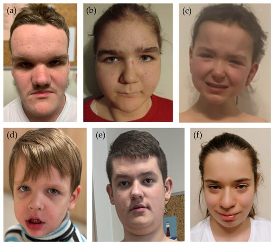
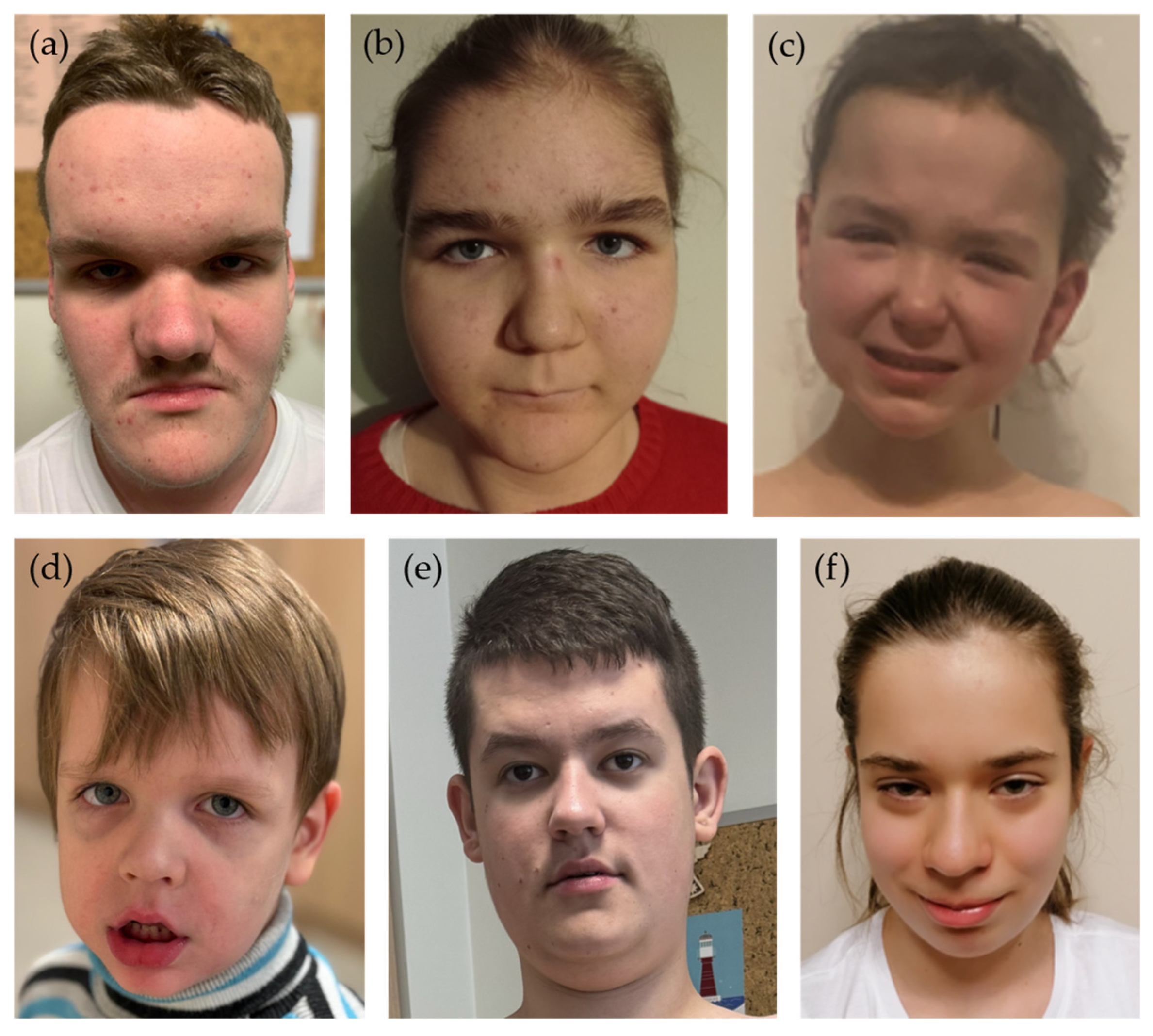
A total of eight novel variants, previously unreported in population databases or the literature, were identified across eight genes in the studied cohort (see Table 2). The variants were detected in genes previously associated with either the overgrowth spectrum (EED and NFIX) or neurodevelopmental/other syndromic disorders (BRWD3, CHD3, DNMT3A, KMT5B, NUS1, and TCF20). Five variants were predicted to result in loss of function, including frameshift, nonsense, and splice-site alterations, while the other three were missense changes. According to ACMG criteria, five variants were classified as pathogenic, and three as likely pathogenic. Phenotypes/clinical features of these patients were consistent with the associated diagnoses. Photographs of the available patients with novel variants are presented in Figure 1. Each gene was affected in a single patient, with no recurrent variants observed. None of the detected variants were present in the control group.
Table 2.
The novel variants identified in the study cohort. The variants are described according to HGVS nomenclature. ACMG classification was assigned according to current guidelines. ACMG—American College of Medical Genetics and Genomics classification, LP—likely pathogenic, VUS—variant of uncertain significance, AD—autosomal dominant, XLR—X-linked recessive, het—heterozygous, and hem—hemizygous.
Figure 1.
Photographs of patients with novel genetic variants: (a) BRWD3:c.151_152insG, (b) DNMT3A:c.1711G>C, (c) EED:c.706A>G, (d) NFIX: c.1001C>A, (e) NUS1:c.662T>A, and (f) TCF20:c.3119del.
4. Discussion
4.1. Phenotypic Findings
The phenotypic findings in our cohort were largely consistent with the existing literature. Developmental delay in the neonatal period, intellectual disability, and neurological manifestations were significantly more prevalent in the generalized overgrowth (GOG) group. Intellectual disability of varying severity is a well-recognized feature across most generalized overgrowth syndromes, although borderline or low-normal cognitive function has also been reported [7]. In contrast, intellectual disability is less commonly associated with segmental overgrowth (SOG), as these conditions are not typically classified among overgrowth–intellectual disability (OGID) syndromes, which are also known to be associated with neurological manifestations, such as epilepsy [26]. Vascular malformations, a hallmark feature of segmental overgrowth disorders, were more frequently observed in the SOG group, in line with previous reports [27]. Similarly, joint laxity was more prevalent in the GOG cohort, which is consistent with its known association with syndromes such as Sotos, Weaver, and Tatton–Brown–Rahman syndromes. Notably, the presence of joint laxity in combination with tall stature may lead to clinical misclassification, for example, as Marfan syndrome [27].
Differences were observed in the presence of dysmorphic features during the neonatal period, with a higher prevalence in the SOG group. Segmental overgrowth may be present at birth in conditions such as PIK3CA-related overgrowth spectrum disorders [28], while in others, such as Proteus syndrome, it may develop later [29]. One possible explanation is that asymmetry, if present, is more readily apparent in the neonatal period than other phenotypic features. Although dysmorphic features can occur in both groups, they tend to become more pronounced and clinically recognizable in GOG syndromes as the phenotype evolves. At the time of evaluation, the distribution of phenotypic features aligned more closely with established clinical expectations, with the GOG group demonstrating a significantly higher prevalence of craniofacial anomalies, including abnormalities of the head, face, eyes, mouth, and ears. Many generalized overgrowth syndromes are characterized by distinctive and recognizable features, such as dolichocephaly and a pointed chin in Sotos syndrome, prominent horizontal eyebrows in Tatton–Brown–Rahman syndrome, macroglossia in Beckwith–Wiedemann syndrome, and preauricular pits or tags in Simpson–Golabi–Behmel syndrome [30].
Unexpectedly, familial hemihyperplasia was observed more frequently in the SOG group. This finding contrasts with the existing literature, which indicates that most segmental overgrowth conditions, including PIK3CA-related overgrowth spectrum and Proteus syndrome, arise sporadically due to postzygotic variants and are, therefore, associated with a low recurrence risk [27]. Nevertheless, rare familial cases have been reported and may account for this observation [31].
4.2. Diagnostic Yield
Recent studies report that the diagnostic yield of exome sequencing (ES) in heterogeneous rare disease cohorts ranges from approximately 30–40% [32,33]. In the present study, the lower overall diagnostic yield of 25% likely reflects methodological and cohort-specific factors. Thus, the true diagnostic rate in our cohort may be underestimated.
In the GOG cohort, the diagnostic yield for OGS conditions reached 24% (17/72), while detection rates of 30–50% in similar cohorts have been reported [5,6]. Beckwith–Wiedemann syndrome is the most common genetic overgrowth syndrome, with an estimated prevalence of approximately 1 in 10,340 individuals [29], followed by Sotos syndrome (1 in 14,000) [10]. While the relatively high number of Sotos syndrome cases (n = 5) identified in our GOG cohort is consistent with this epidemiological data, the absence of Beckwith–Wiedemann syndrome (BWS) cases suggests a potential underrepresentation. This discrepancy may be explained by two factors. First, the molecular heterogeneity of BWS, which frequently involves epigenetic alterations or mosaic changes that are not reliably detected by standard ES approaches. Second, patient selection: BWS has a recognizable clinical phenotype and is typically investigated through first-line testing strategies, such as methylation analysis and single-gene testing. Consequently, the patients with a clinically suspected or already established diagnosis of BWS were generally not referred for ES and were, therefore, not included in this study, which enrolled only patients without a prior molecular diagnosis. In contrast, Tatton–Brown–Rahman syndrome emerged as the second most frequent diagnosis in our cohort (n = 4), despite being considered an ultra-rare condition with a reported prevalence of less than 1 in 1,000,000 [34,35].
Only one SOG patient received a diagnosis through the ES approach (diagnostic yield of 2%, 1/48). A key limitation in this case is the exclusive use of blood-derived gDNA, which significantly reduces sensitivity for detecting somatic mosaic variants. This is particularly relevant in the context of overgrowth disorders, where mosaicism plays a major pathogenic role, such as in SOG. Previous studies have demonstrated that diagnostic yield can be substantially improved through tiered testing strategies incorporating affected tissues, especially in patients with clinical features suggestive of PIK3CA-related overgrowth spectrum and associated vascular anomalies [36].
4.3. Novel Genetic Variants
Several novel variants were identified in our cohort, expanding the mutational spectrum of the associated disorders. The identified novel variants predominantly cluster into two major functional categories: epigenetic regulators involved in chromatin remodeling (DNMT3A, EED, KMT5B, and CHD3) and genes associated with transcriptional and neurodevelopmental processes (TCF20, NFIX, and BRWD3), with additional variants affecting cellular pathways, such as protein glycosylation (NUS1).
The BRWD3 gene variant c.151_152insG is predicted to introduce a frameshift early in the coding sequence, leading to a premature termination codon and likely resulting in either a truncated protein or nonsense-mediated mRNA decay. This finding supports the notion that truncating variants represent a common pathogenic mechanism in X-linked intellectual developmental disorder 93 [37,38]. High expression of BRWD3 in the brain explains the neurodevelopmental phenotype. Additionally, it has been suggested that BRWD3 pathogenic variants may disrupt intracellular signaling pathways affecting cellular proliferation, which may explain the overgrowth features observed in affected individuals, such as macrocephaly and obesity [39].
The CHD3 gene variant c.3253-1G>T affects the splice acceptor site of exon 25 and is predicted to result in aberrant splicing, potentially leading to exon skipping or activation of a cryptic splice site and the production of a truncated or functionally altered protein. Exon 25 lies within the central region of the gene, which encodes part of the ATP-dependent helicase domain of the CHD3 protein. CHD3 variants also disrupt transcriptional programs controlling cell fate and growth, which may underlie overgrowth and macrocephaly [40]. Although the helicase domain has been proposed as a mutational hotspot [41,42], ClinVar reports only eight pathogenic or likely pathogenic splice-site variants in CHD3, with a single variant (c.2343+2T>C) in the helicase region and none reported between exons 24–25.
The identified DNMT3A missense gene variant NM_175629.2:c.1711G>C is located within the ADD domain, a key regulatory region that mediates histone recognition and proper targeting of the methyltransferase to chromatin [43]. The ADD domain, together with the upstream PWWP domain and downstream methyltransferase catalytic domain, is a well-known mutational hotspot, where most missense variants cluster [44]. The variant c.1711G>C was also investigated in a functional study by Huang et al., which demonstrated reduced DNMT3A methyltransferase activity [45].
The EED gene variant c.706A>G, detected in a patient with Cohen–Gibson syndrome, is predicted to result in a missense substitution within this WD40 repeat region, which is critical for PRC2 assembly and function [46]. Previously reported pathogenic EED variants are predominantly missense changes clustering within the WD40 domain, highlighting this region as a functional hotspot. Only 13 pathogenic/likely pathogenic EED variants have been reported in the ClinVar database to date. Cohen–Gibson syndrome is closely related to Weaver syndrome, which is caused by variants in EZH2, another core PRC2 component, highlighting a shared pathogenic mechanism involving the disruption of epigenetic repression [47].
The detected KMT5B variant c.913T>C results in a missense change located inside the canonical SET catalytic domain, suggesting a strong likelihood of disrupting enzymatic activity and chromatin regulation. Missense variants were previously reported to also occur outside well-characterized domains, but are still thought to impair protein function through the disruption of regulatory regions or protein–protein interactions [48]. Even though pathogenic variants in KMT5B are associated with an autosomal dominant neurodevelopmental disorder 51, a tendency for taller stature has also been observed in these patients; thus, there have been considerations to classify this pathology as overgrowth syndrome [49].
Previously reported NFIX pathogenic missense variants associated with overgrowth phenotypes tend to cluster in the N-terminal DNA-binding domain (exon 2), whereas truncating variants and splice-site changes distributed across exons 6–10 are more often associated with the severe Marshall–Smith phenotype [50]. Our reported variant c.1001C>A in a patient with Malan syndrome phenotype lies outside the canonical missense hotspot in exon 2 but falls within exon 7, a region where truncating and splice-site variants associated with Marshall–Smith syndrome have been reported. Notably, this stop-gain variant is located within the Marshall–Smith mutational hotspot yet results in a Malan syndrome phenotype, suggesting that factors beyond variant location and type, such as the fate of the mutant transcript or the specific position of the premature stop codon, may contribute to phenotypic outcome.
The NUS1 gene variant c.662T>A, located in exon 3, is predicted to result in early loss of function via nonsense-mediated mRNA decay. Previously reported NUS1 variants include a heterogeneous spectrum of missense, frameshift, nonsense, and splice-site changes, distributed throughout the gene, rather than confined to a single hotspot [51]. Notably, both truncating and missense variants across the NUS1 coding sequence have been associated with overlapping neurodevelopmental phenotypes, and no clear genotype–phenotype correlation has been established, supporting a shared pathogenic mechanism related to impaired dolichol biosynthesis rather than domain-specific effects [52].
The TCF20 gene variant c.3119del is predicted to result in a frameshift and premature termination codon, leading to loss of normal protein function, most likely through nonsense-mediated decay. This supports a haploinsufficiency mechanism, which is well established for TCF20, a gene shown to be highly intolerant to loss-of-function variation [53]. Compared to previously reported variants, TCF20 changes predominantly consist of loss-of-function variants, including nonsense and frameshift changes distributed throughout the gene, with relatively few missense variants described [53].
To summarize, our analysis further highlights epigenetic regulation as a major dysregulated biological process in individuals with overgrowth and intellectual disability. Previous studies have shown that variants in epigenetic regulator genes (NSD1, EZH2, DNMT3A, CHD8, HIST1H1E, and EED) account for up to 44% of OGID cases [6]. Consistent with this, our findings demonstrate that variants in genes directly involved in epigenetic regulation comprised 50.0% (15/30) of diagnoses, including NSD1, DNMT3A, EZH2, EED, CHD3, GNAS, and KMT5B.
5. Conclusions
Our findings highlight distinct phenotypic patterns between GOG and SOG, and underscore the central role of epigenetic regulation and growth signaling pathways in OGS pathogenesis. This study expands the molecular spectrum of overgrowth syndromes and emphasizes the importance of incorporating tissue-specific testing strategies to improve diagnostic yield. Furthermore, it provides valuable insights into the genetic architecture of OGSs in underrepresented Central and North-Eastern European populations, supporting the development of region-specific diagnostic and surveillance strategies.
Author Contributions
Writing—original draft preparation: K.Š.-J.; writing—review and editing: K.B., A.M., B.A., M.M. and N.W.-K.; genetic data analysis: K.B., B.A., M.M., N.W.-K. and A.T.; clinical data acquisition: K.Š.-J., A.M., B.S.-J. and B.P.; control group: B.M. and J.M.-M.; supervision and conceptualization: R.P. and A.U. Renata Posmyk and Algirdas Utkus contributed equally as coordinators/last authors to this work. All authors have read and agreed to the published version of the manuscript.
Funding
This study was funded by a grant from the Polish National Science Centre (grant number NCN/1/DA/21/001/1106 (DAINA-2 grant call entitled) and by the Research Council of Lithuania (grant number S-LL-21-5).
Institutional Review Board Statement
This study was conducted in accordance with the Declaration of Helsinki (2013 revision) and was approved by the local Bioethics Committees (protocol no. 2022/5-1435-907, approved on 10 May 2022, Lithuania; APK.002.352.2021, Poland, approved on 24 June 2021).
Informed Consent Statement
Written informed consent was obtained from all participants or their legal guardians.
Data Availability Statement
The data that support the findings of this study are available on request from the corresponding author. The data are not publicly available due to privacy or ethical restrictions.
Acknowledgments
We thank the clinicians from Poland and Lithuania, who contributed to gathering the study cohort, and the families for their participation. We are especially grateful to Polish research collaborators Karolina Śledzińska, Jolanta Wierzba, Tadeusz Kałużewski, Dorota Wicher, Karolina Biel, Aleksander Jamsheer, Magdalena Badura-Stronka, Anna Kłosowska, Monika Kowalczyk-Rusak, Piotr Iwanowski, Monika Cichoń-Kotek, Anna Marczak-Hałupka, Klaudia Berk, Joanna Karwowska, Magdalena Niemira, and Anna Zeller and Lithuanian research collaborators Birutė Burnytė and Loreta Cimbalistienė.
Conflicts of Interest
A.T. is an employee of MyEngene Canada Inc., a commercial company operating in the field of pharmacogenomics and precision medicine. MyEngene Canada Inc. had no role in the design of this study, collection or analysis of the data, interpretation of the results, preparation of this manuscript, or decision to submit this manuscript for publication. The other authors declare no conflicts of interest. The funders had no role in the design of this study; in the collection, analyses, or interpretation of data; in the writing of this manuscript; or in the decision to publish the results.
Appendix A
Table A1.
Clinical characteristics and phenotype summary. GOG—generalized overgrowth, SOG—segmental overgrowth, NC—natural conception, IVF—in vitro fertilization, wg—gestational weeks, OFC—occipitofrontal circumference, SD—standard deviation, and IQR—interquartile range. * In 1st or 2nd degree relatives. Title for each section of the table and statistically significant results (p-value < 0.05) are shown in bold.
References
- Brioude, F.; Toutain, A.; Giabicani, E.; Cottereau, E.; Cormier-Daire, V.; Netchine, I. Overgrowth Syndromes-Clinical and Molecular Aspects and Tumour Risk. Nat. Rev. Endocrinol. 2019, 15, 299–311. [Google Scholar] [CrossRef] [Scilit] [PubMed]
- Neri, G.; Moscarda, M. Overgrowth Syndromes: A Classification. Endocr. Dev. 2009, 14, 53–60. [Google Scholar] [CrossRef] [Scilit] [PubMed]
- Burkardt, D.D.; Tatton-Brown, K.; Dobyns, W.; Graham, J.M. Approach to Overgrowth Syndromes in the Genome Era. Am. J. Med. Genet. C Semin. Med. Genet. 2019, 181, 483–490. [Google Scholar] [CrossRef] [Scilit] [PubMed]
- Klein, S.D.; Nisbet, A.; Kalish, J.M. Overgrowth Syndromes, Diagnosis and Management. Curr. Opin. Pediatr. 2023, 35, 620–630. [Google Scholar] [CrossRef] [Scilit] [PubMed]
- Kim, G.J.; Vasco de Albuquerque Albuquerque, E.; Rezende, R.C.; De Polli Cellin, L.; Santillan Vasconez, A.M.; Krepischi, A.C.V.; Santana, L.; Lerario, A.M.; de Souza, V.; Scalco, R.; et al. Exome Sequencing of Patients with Syndromic Tall Stature Reveals Four Novel Candidate Genes. Endocr. Connect. 2025, 14, e250137. [Google Scholar] [CrossRef] [Scilit] [PubMed]
- Tatton-Brown, K.; Loveday, C.; Yost, S.; Clarke, M.; Ramsay, E.; Zachariou, A.; Elliott, A.; Wylie, H.; Ardissone, A.; Rittinger, O.; et al. Mutations in Epigenetic Regulation Genes Are a Major Cause of Overgrowth with Intellectual Disability. Am. J. Hum. Genet. 2017, 100, 725–736. [Google Scholar] [CrossRef] [Scilit] [PubMed]
- Kamien, B.; Ronan, A.; Poke, G.; Sinnerbrink, I.; Baynam, G.; Ward, M.; Gibson, W.T.; Dudding-Byth, T.; Scott, R.J. A Clinical Review of Generalized Overgrowth Syndromes in the Era of Massively Parallel Sequencing. Mol. Syndr. 2018, 9, 70–82. [Google Scholar] [CrossRef] [Scilit] [PubMed]
- Arora, V.; Ferreira, C.R.; Dua Puri, R.; Verma, I.C. Primrose Syndrome. In GeneReviews®; Adam, M.P., Bick, S., Mirzaa, G.M., Pagon, R.A., Wallace, S.E., Amemiya, A., Eds.; University of Washington, Seattle: Seattle, WA, USA, 1993. [Google Scholar]
- Grange, D.K.; Nichols, C.G.; Singh, G.K.; Guilliams, K.; Aggarwal, M. Cantú Syndrome. In GeneReviews®; Adam, M.P., Bick, S., Mirzaa, G.M., Pagon, R.A., Wallace, S.E., Amemiya, A., Eds.; University of Washington, Seattle: Seattle, WA, USA, 1993. [Google Scholar]
- Ocansey, S.; Cole, T.R.; Rahman, N.; Tatton-Brown, K. Sotos Syndrome. In GeneReviews®; Adam, M.P., Bick, S., Mirzaa, G.M., Pagon, R.A., Wallace, S.E., Amemiya, A., Eds.; University of Washington, Seattle: Seattle, WA, USA, 1993. [Google Scholar]
- Ocansey, S.; Tatton-Brown, K. EZH2-Related Overgrowth. In GeneReviews®; Adam, M.P., Bick, S., Mirzaa, G.M., Pagon, R.A., Wallace, S.E., Amemiya, A., Eds.; University of Washington, Seattle: Seattle, WA, USA, 1993. [Google Scholar]
- Priolo, M. NFIX-Related Malan Syndrome. In GeneReviews®; Adam, M.P., Bick, S., Mirzaa, G.M., Pagon, R.A., Wallace, S.E., Amemiya, A., Eds.; University of Washington, Seattle: Seattle, WA, USA, 1993. [Google Scholar]
- Akgumus, G.; Chang, F.; Li, M.M. Overgrowth Syndromes Caused by Somatic Variants in the Phosphatidylinositol 3-Kinase/AKT/Mammalian Target of Rapamycin Pathway. J. Mol. Diagn. 2017, 19, 487–497. [Google Scholar] [CrossRef] [Scilit] [PubMed]
- Tatton-Brown, K.; Weksberg, R. Molecular Mechanisms of Childhood Overgrowth. Am. J. Med. Genet. C Semin. Med. Genet. 2013, 163, 71–75. [Google Scholar] [CrossRef] [Scilit]
- Mirzaa, G.; Graham, J.M.; Keppler-Noreuil, K. PIK3CA-Related Overgrowth Spectrum. In GeneReviews®; Adam, M.P., Bick, S., Mirzaa, G.M., Pagon, R.A., Wallace, S.E., Amemiya, A., Eds.; University of Washington, Seattle: Seattle, WA, USA, 1993. [Google Scholar]
- Biesecker, L.G.; Sapp, J.C. Proteus Syndrome. In GeneReviews®; Adam, M.P., Bick, S., Mirzaa, G.M., Pagon, R.A., Wallace, S.E., Amemiya, A., Eds.; University of Washington, Seattle: Seattle, WA, USA, 1993. [Google Scholar]
- Yehia, L.; Eng, C. PTEN Hamartoma Tumor Syndrome. In GeneReviews®; Adam, M.P., Bick, S., Mirzaa, G.M., Pagon, R.A., Wallace, S.E., Amemiya, A., Eds.; University of Washington, Seattle: Seattle, WA, USA, 1993. [Google Scholar]
- Kurotaki, N.; Imaizumi, K.; Harada, N.; Masuno, M.; Kondoh, T.; Nagai, T.; Ohashi, H.; Naritomi, K.; Tsukahara, M.; Makita, Y.; et al. Haploinsufficiency of NSD1 Causes Sotos Syndrome. Nat. Genet. 2002, 30, 365–366. [Google Scholar] [CrossRef] [Scilit] [PubMed]
- Malan, V.; Chevallier, S.; Soler, G.; Coubes, C.; Lacombe, D.; Pasquier, L.; Soulier, J.; Morichon-Delvallez, N.; Turleau, C.; Munnich, A.; et al. Array-Based Comparative Genomic Hybridization Identifies a High Frequency of Copy Number Variations in Patients with Syndromic Overgrowth. Eur. J. Hum. Genet. 2010, 18, 227–232. [Google Scholar] [CrossRef] [Scilit] [PubMed]
- Sparago, A.; Cerrato, F.; Vernucci, M.; Ferrero, G.B.; Silengo, M.C.; Riccio, A. Microdeletions in the Human H19 DMR Result in Loss of IGF2 Imprinting and Beckwith-Wiedemann Syndrome. Nat. Genet. 2004, 36, 958–960. [Google Scholar] [CrossRef] [Scilit] [PubMed]
- Cohen, A.S.; Gibson, W.T. EED-Associated Overgrowth in a Second Male Patient. J. Hum. Genet. 2016, 61, 831–834. [Google Scholar] [CrossRef] [Scilit] [PubMed]
- Fatumo, S.; Chikowore, T.; Choudhury, A.; Ayub, M.; Martin, A.R.; Kuchenbaecker, K. A Roadmap to Increase Diversity in Genomic Studies. Nat. Med. 2022, 28, 243–250. [Google Scholar] [CrossRef] [Scilit]
- Gudmundsson, S.; Singer-Berk, M.; Watts, N.A.; Phu, W.; Goodrich, J.K.; Solomonson, M.; Rehm, H.L.; MacArthur, D.G.; O’Donnell-Luria, A. Variant Interpretation Using Population Databases: Lessons from gnomAD. Hum. Mutat. 2022, 43, 1012–1030. [Google Scholar] [CrossRef] [Scilit] [PubMed]
- Kaja, E.; Lejman, A.; Sielski, D.; Sypniewski, M.; Gambin, T.; Dawidziuk, M.; Suchocki, T.; Golik, P.; Wojtaszewska, M.; Mroczek, M.; et al. The Thousand Polish Genomes-A Database of Polish Variant Allele Frequencies. Int. J. Mol. Sci. 2022, 23, 4532. [Google Scholar] [CrossRef] [Scilit] [PubMed]
- Urnikyte, A.; Molyte, A.; Kučinskas, V. Genome-Wide Landscape of North-Eastern European Populations: A View from Lithuania. Genes 2021, 12, 1730. [Google Scholar] [CrossRef] [Scilit] [PubMed]
- Delagrammatikas, K.G.; Kit, M.; Church, E.; Diehl, S.E.; Hunter, K.; Tatton-Brown, J.; Kiernan, C.G. Epilepsy and Overgrowth–Intellectual Disability Syndromes: A Patient Organization Perspective on Collaborating to Accelerate Pathways to Treatment. Ther. Adv. Rare Dis. 2024, 5, 26330040241254123. [Google Scholar] [CrossRef] [Scilit] [PubMed]
- Manor, L. Overgrowth Syndromes—Evaluation, Diagnosis, and Management. Front. Pediatr. 2020, 8, 574857. [Google Scholar] [CrossRef] [Scilit] [PubMed]
- PIK3CA-Related Overgrowth Spectrum—Symptoms, Causes, Treatment|NORD. Available online: https://rarediseases.org/rare-diseases/pik3ca-related-overgrowth-spectrum/ (accessed on 9 May 2026).
- Mussa, A.; Carli, D.; Cardaropoli, S.; Ferrero, G.B.; Resta, N. Lateralized and Segmental Overgrowth in Children. Cancers 2021, 13, 6166. [Google Scholar] [CrossRef] [Scilit] [PubMed]
- Home-OMIM-(OMIM.ORG). Available online: https://omim.org/ (accessed on 24 March 2026).
- Heilstedt, H.A.; Bacino, C.A. A Case of Familial Isolated Hemihyperplasia. BMC Med. Genet. 2004, 5, 1. [Google Scholar] [CrossRef] [Scilit] [PubMed][Green Version]
- Albuquerque, A.L.B.; dos Santos, G.G.; Sadok, S.H.; Antonello, B.B.; de Jesus, L.M.; de Carvalho, M.E.A.; Mutarelli, A.; Ribeiro, P.V.Z. Diagnostic Yield of Genome Sequencing Versus Exome Sequencing in Pediatric Patients with Rare Phenotypes: A Systematic Review and Meta-Analysis. Am. J. Med. Genet. Part A 2025, 197, e64146. [Google Scholar] [CrossRef] [Scilit]
- Andersen, P.F.; Ek, J.; Karstensen, H.G.; Bak, M.; Grønborg, S.W.; Hove, H.B.; Diness, B.; Hjortshøj, T.D.; Hammer, T.B.; Høi-Hansen, C.; et al. Diagnostic Yield of Whole Exome Sequencing in a Cohort of 825 Patients. Eur. J. Med. Genet. 2025, 78, 105043. [Google Scholar] [CrossRef] [Scilit] [PubMed]
- Orphanet: Tatton-Brown-Rahman Syndrome. Available online: https://www.orpha.net/en/disease/detail/404443 (accessed on 24 March 2026).
- Ostrowski, P.J.; Tatton-Brown, K. Tatton-Brown-Rahman Syndrome. In GeneReviews®; Adam, M.P., Bick, S., Mirzaa, G.M., Pagon, R.A., Wallace, S.E., Amemiya, A., Eds.; University of Washington, Seattle: Seattle, WA, USA, 1993. [Google Scholar]
- Green, T.E.; Garza, D.; Brown, N.J.; de Silva, M.G.; Bennett, M.F.; Tubb, C.; Phillips, R.J.; MacGregor, D.; Robertson, S.J.; Bekhor, P.; et al. Improving Genetic Diagnostic Yield in a Large Cohort of Children with Rare Vascular Anomalies or PIK3CA-Related Overgrowth Spectrum. Genet. Med. Open 2023, 2, 100837. [Google Scholar] [CrossRef] [Scilit] [PubMed]
- Ostrowski, P.J.; Zachariou, A.; Loveday, C.; Baralle, D.; Blair, E.; Douzgou, S.; Field, M.; Foster, A.; Kyle, C.; Lachlan, K.; et al. Null Variants and Deletions in BRWD3 Cause an X-Linked Syndrome of Mild-Moderate Intellectual Disability, Macrocephaly, and Obesity: A Series of 17 Patients. Am. J. Med. Genet. C Semin. Med. Genet. 2019, 181, 638–643. [Google Scholar] [CrossRef] [Scilit] [PubMed]
- Tenorio, J.; Alarcón, P.; Arias, P.; Ramos, F.J.; Campistol, J.; Climent, S.; García-Miñaur, S.; Dapía, I.; Hernández, A.; Nevado, J.; et al. MRX93 Syndrome (BRWD3 Gene): Five New Patients with Novel Mutations. Clin. Genet. 2019, 95, 726–731. [Google Scholar] [CrossRef] [Scilit] [PubMed]
- Field, M.; Tarpey, P.S.; Smith, R.; Edkins, S.; O’Meara, S.; Stevens, C.; Tofts, C.; Teague, J.; Butler, A.; Dicks, E.; et al. Mutations in the BRWD3 Gene Cause X-Linked Mental Retardation Associated with Macrocephaly. Am. J. Hum. Genet. 2007, 81, 367–374. [Google Scholar] [CrossRef] [Scilit] [PubMed][Green Version]
- Mitchell, Z.H.; den Hoed, J.; Claassen, W.; Demurtas, M.; Deelen, L.; Campeau, P.M.; Liu, K.; Fisher, S.E.; Trizzino, M. The NuRD Component CHD3 Promotes BMP Signalling during Cranial Neural Crest Cell Specification. EMBO Rep. 2025, 26, 4723–4741. [Google Scholar] [CrossRef] [Scilit] [PubMed]
- Drivas, T.G.; Li, D.; Nair, D.; Alaimo, J.T.; Alders, M.; Altmüller, J.; Barakat, T.S.; Bebin, E.M.; Bertsch, N.L.; Blackburn, P.R.; et al. A Second Cohort of CHD3 Patients Expands the Molecular Mechanisms Known to Cause Snijders Blok-Campeau Syndrome. Eur. J. Hum. Genet. 2020, 28, 1422–1431. [Google Scholar] [CrossRef] [Scilit] [PubMed]
- van der Spek, J.; den Hoed, J.; Snijders Blok, L.; Dingemans, A.J.M.; Schijven, D.; Nellaker, C.; Venselaar, H.; Astuti, G.D.N.; Barakat, T.S.; Bebin, E.M.; et al. Inherited Variants in CHD3 Show Variable Expressivity in Snijders Blok-Campeau Syndrome. Genet. Med. 2022, 24, 1283–1296. [Google Scholar] [CrossRef] [Scilit] [PubMed]
- Zhang, Y.; Jurkowska, R.; Soeroes, S.; Rajavelu, A.; Dhayalan, A.; Bock, I.; Rathert, P.; Brandt, O.; Reinhardt, R.; Fischle, W.; et al. Chromatin Methylation Activity of Dnmt3a and Dnmt3a/3L Is Guided by Interaction of the ADD Domain with the Histone H3 Tail. Nucleic Acids Res. 2010, 38, 4246–4253. [Google Scholar] [CrossRef] [Scilit] [PubMed]
- Tatton-Brown, K.; Zachariou, A.; Loveday, C.; Renwick, A.; Mahamdallie, S.; Aksglaede, L.; Baralle, D.; Barge-Schaapveld, D.; Blyth, M.; Bouma, M.; et al. The Tatton-Brown-Rahman Syndrome: A Clinical Study of 55 Individuals with de Novo Constitutive DNMT3A Variants. Wellcome Open Res. 2018, 3, 46. [Google Scholar] [CrossRef] [Scilit] [PubMed]
- Huang, Y.-H.; Chen, C.-W.; Sundaramurthy, V.; Słabicki, M.; Hao, D.; Watson, C.J.; Tovy, A.; Reyes, J.M.; Dakhova, O.; Crovetti, B.R.; et al. Systematic Profiling of DNMT3A Variants Reveals Protein Instability Mediated by the DCAF8 E3 Ubiquitin Ligase Adaptor. Cancer Discov. 2022, 12, 220–235. [Google Scholar] [CrossRef] [Scilit] [PubMed]
- Margueron, R.; Justin, N.; Ohno, K.; Sharpe, M.L.; Son, J.; Drury, W.J.; Voigt, P.; Martin, S.; Taylor, W.R.; De Marco, V.; et al. Role of the Polycomb Protein Eed in the Propagation of Repressive Histone Marks. Nature 2009, 461, 762–767. [Google Scholar] [CrossRef] [Scilit] [PubMed]
- Cohen, A.S.A.; Tuysuz, B.; Shen, Y.; Bhalla, S.K.; Jones, S.J.M.; Gibson, W.T. A Novel Mutation in EED Associated with Overgrowth. J. Hum. Genet. 2015, 60, 339–342. [Google Scholar] [CrossRef] [Scilit] [PubMed]
- Politano, D.; Borgatti, R.; Borgonovi, G.; Cistaro, A.; Danesino, C.; Fania, P.; Garghetti, G.; Guala, A.; Orlando, I.; Schiera, I.G.; et al. Bridging Genotype to Phenotype in KMT5B-Related Syndrome: Evidence from RNA-Seq, 18FDG-PET, Clinical Deep Phenotyping in Two New Cases, and a Literature Review. Genes 2025, 16, 1174. [Google Scholar] [CrossRef] [Scilit] [PubMed]
- Hulen, J.; Kenny, D.; Black, R.; Hallgren, J.; Hammond, K.G.; Bredahl, E.C.; Wickramasekara, R.N.; Abel, P.W.; Stessman, H.A.F. KMT5B Is Required for Early Motor Development. Front. Genet. 2022, 13, 901228. [Google Scholar] [CrossRef] [Scilit] [PubMed]
- Martinez, F.; Marín-Reina, P.; Sanchis-Calvo, A.; Perez-Aytés, A.; Oltra, S.; Roselló, M.; Mayo, S.; Monfort, S.; Pantoja, J.; Orellana, C. Novel Mutations of NFIX Gene Causing Marshall-Smith Syndrome or Sotos-like Syndrome: One Gene, Two Phenotypes. Pediatr. Res. 2015, 78, 533–539. [Google Scholar] [CrossRef] [Scilit] [PubMed]
- Ding, F.; Pan, J.; Ji, S.; Zhang, Y.; Hou, J.; Shi, N.; Liu, H. Case Report: De Novo Variant of the NUS1 Gene Associated with Developmental Delay and Autism Spectrum Disorders in a Chinese Family. Front. Pediatr. 2025, 13, 1557103. [Google Scholar] [CrossRef] [Scilit] [PubMed]
- Brooker, S.M.; Novelli, M.; Coukos, R.; Prakash, N.; Kamel, W.A.; Amengual-Gual, M.; Anheim, M.; Barcia, G.; Bardakjian, T.; Baur, F.; et al. The Spectrum of Neurologic Phenotypes Associated with NUS1 Pathogenic Variants: A Comprehensive Case Series. Ann. Neurol. 2025, 98, 561–572. [Google Scholar] [CrossRef] [Scilit] [PubMed]
- Vetrini, F.; McKee, S.; Rosenfeld, J.A.; Suri, M.; Lewis, A.M.; Nugent, K.M.; Roeder, E.; Littlejohn, R.O.; Holder, S.; Zhu, W.; et al. De Novo and Inherited TCF20 Pathogenic Variants Are Associated with Intellectual Disability, Dysmorphic Features, Hypotonia, and Neurological Impairments with Similarities to Smith-Magenis Syndrome. Genome Med. 2019, 11, 12. [Google Scholar] [CrossRef] [Scilit] [PubMed]

Disclaimer/Publisher’s Note: The statements, opinions and data contained in all publications are solely those of the individual author(s) and contributor(s) and not of MDPI and/or the editor(s). MDPI and/or the editor(s) disclaim responsibility for any injury to people or property resulting from any ideas, methods, instructions or products referred to in the content. |
© 2026 by the authors. Licensee MDPI, Basel, Switzerland. This article is an open access article distributed under the terms and conditions of the Creative Commons Attribution (CC BY) license.