


Abstract
Retinoic acid receptors (RARs)—RARα, RARβ, and RARγ, encoded by RARA, RARB, and RARG—are ligand-regulated transcription factors that convert retinoid availability into cell-type-specific programs controlling development and tissue homeostasis. Disease-associated RAR alterations are usually reviewed separately as leukemia fusions, inherited developmental variants, treatment-resistance mutations, or pathway abnormalities. Here, these alteration classes are integrated within a common domain–mechanism–phenotype framework that links receptor architecture, chromatin regulation, and protein quality control to clinical presentation and therapeutic response. Attention is given to PML::RARA and variant RARA fusions in acute promyelocytic leukemia, germline RARB variants in neurodevelopmental and ocular disease, and RARG-rearranged acute myeloid leukemia. AF-1 and ligand-binding-domain variants, defective receptor turnover, and therapeutic strategies involving retinoids, arsenic trioxide, epigenetic modulation, and targeted protein degradation are also evaluated. The evidence indicates that disease phenotype and treatment response emerge from the interplay among the affected receptor domain, fusion partner, chromatin context, and capacity for protein degradation. Distinguishing established drivers from treatment-emergent lesions, biomarkers, and variants of uncertain significance is essential for accurate diagnosis and precision therapy.
1. Introduction
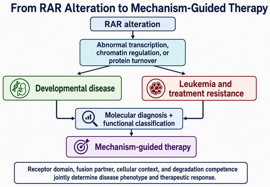
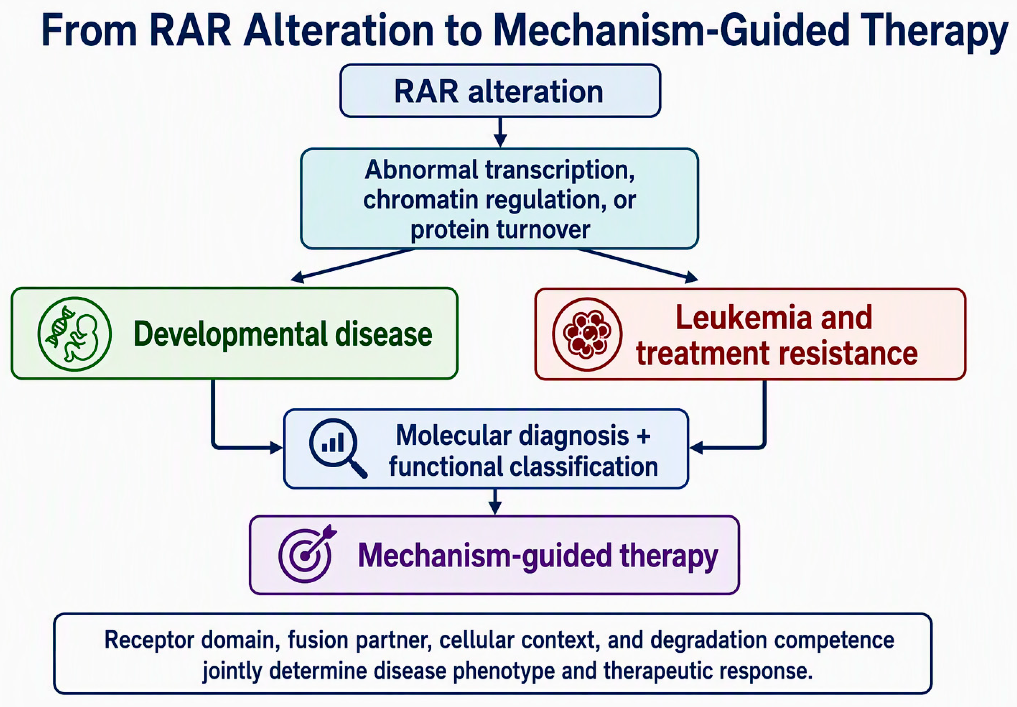
Retinoic acid (RA), the principal bioactive metabolite of vitamin A, controls development and adult tissue homeostasis through tightly regulated synthesis, intracellular transport, receptor activation, and CYP26-mediated clearance [1,2,3,4,5]. RARα, RARβ, and RARγ form heterodimers with retinoid X receptors (RXRs), bind retinoic acid response elements, and recruit corepressor or coactivator complexes according to ligand state, receptor sequence, chromatin accessibility, and post-translational regulation. Although the three receptors share a conserved modular architecture, their expression patterns and disease associations are not interchangeable. RARA is recurrently rearranged in acute promyelocytic leukemia (APL); RARB is linked to a genetically and mechanistically heterogeneous neurodevelopmental and ocular syndrome and can also participate in rare leukemia-associated translocations; and RARG rearrangements define an aggressive APL-like acute myeloid leukemia (AML) subgroup [6,7,8,9,10,11,12] (see Figure 1). Recent reports of additional germline, somatic, and regulatory variants across the RAR family are expanding the scientific landscape and creating new opportunities for future research into their biological significance, disease associations, and clinical relevance [13,14,15,16].
Figure 1.
Graphical abstract. RAR-family alterations can disrupt transcription, chromatin regulation, or protein turnover, leading to developmental disease or leukemia and treatment resistance. Molecular diagnosis and functional classification provide the basis for mechanism-guided therapy.
This review addresses three gaps that are usually considered separately. First, it connects receptor-domain biology and protein homeostasis to variant-specific disease mechanisms [1,2,17,18,19,20,21,22,23,24,25,26,27,28]. Second, it compares RARA, RARB, and RARG alterations within a single clinically oriented framework without conflating oncogenic fusions, inherited variants, treatment-emergent resistance lesions, and expression abnormalities [6,7,8,9,10,11,12,13,14,15,16,29,30,31,32,33,34,35,36,37,38,39,40,41,42,43,44,45]. Third, it evaluates whether therapeutic response is better predicted by alteration class, fusion architecture, and degradation competence than by receptor identity alone [20,23,24,25,26,27,28,45,46,47]. Evidence is therefore discussed hierarchically, distinguishing established disease drivers from mechanistically supported candidates and variants of uncertain significance [48].
2. Receptor Architecture and a Framework for Variant Interpretation
RAR proteins contain an intrinsically disordered N-terminal A/B region harboring ligand-independent activation function 1 (AF-1), a highly conserved C-region DNA-binding domain with two zinc fingers, a flexible D-region hinge, and an E-region ligand-binding domain (LBD) that contains activation function 2 (AF-2) [1,2]. The LBD integrates ligand recognition, heterodimerization, corepressor release, coactivator recruitment, and helix-12 positioning, whereas AF-1 provides a phosphorylation-sensitive surface for cell-context-dependent transcriptional control. This architecture explains why distinct alteration classes produce different phenotypes. LBD variants may reduce ligand affinity, stabilize repression, disrupt coactivator docking, or create dominant-negative behavior; fusion proteins usually preserve RAR DNA-binding and ligand-response modules while replacing the N terminus with an oligomerization or chromatin-regulatory partner; and variants in disordered regulatory regions may alter phosphorylation, turnover, or transcriptional amplitude [1,2,6,16]. Each variant should therefore be reported with its reference transcript, HGVS coding and protein notation, inheritance or somatic origin, affected receptor domain, and level of functional and clinical evidence. Standardized reporting prevents transcript and protein-numbering ambiguities, enables reliable comparison across studies and databases, and supports reproducible assessment of pathogenicity and clinical relevance.
2.1. Ligand Receptor Chromatin Signaling
Cellular RA concentration is shaped by retinol and retinaldehyde metabolism, cellular retinoic acid-binding proteins, and CYP26 enzymes that restrict the amplitude and duration of receptor activation [3,4,5]. In the nucleus, unliganded RAR–RXR complexes can recruit NCoR/SMRT-containing corepressor assemblies and histone deacetylases. Ligand binding reorganizes the LBD, destabilizes corepressor interactions, promotes coactivator recruitment, and enables chromatin remodeling and transcriptional activation [17]. These events are strongly context-dependent: pre-existing chromatin accessibility, lineage-defining transcription factors, enhancer topology, and intracellular ligand routing determine which RA-responsive genes are engaged [1,2,3,4,5,17,18]. Pathogenic variants can therefore compromise signaling even when DNA binding is retained, because altered coregulator exchange or chromatin residence may be sufficient to convert a regulated receptor into a constitutive repressor or aberrant activator.
2.2. Activation Turnover Coupling and Post-Translational Control
RAR transcriptional output is coupled with receptor renewal. Ligand-induced activation can expose regulatory surfaces that are phosphorylated, ubiquitinated, SUMOylated, or recognized by proteasomal and autophagic quality-control systems [19,20,21,22]. Phosphorylation within AF-1 can influence transcriptional competence and receptor stability [21,22], while SUMO–ubiquitin crosstalk may either stabilize receptor intermediates or create degradation signals [19]. In neural stem cells, sequential SUMOylation, ubiquitination, engagement of the VCP/p97 segregase, and proteasomal degradation contribute to receptor turnover; disruption of this pathway in glioma stem-like cells permits accumulation of transcriptionally inactive, modified receptors [19]. In APL, the same general principle becomes therapeutically decisive: ATRA targets the RARα moiety of PML::RARA, whereas arsenic binding to the PML moiety promotes PML nuclear-body reassembly, SUMO-dependent ubiquitination, and proteolytic clearance [23,24,25,26]. Autophagic pathways can provide parallel or complementary routes of receptor disposal, demonstrating that receptor abundance is an active regulatory variable rather than a passive consequence of transcription [20].
2.3. From Variant Class to Cellular Phenotype
Disease mechanisms can be organized along three axes. The first is altered transcriptional state: variants may enforce repression, exaggerate ligand-dependent activation, or reduce receptor output. The second is altered protein homeostasis: changes in phosphorylation, ubiquitination, SUMOylation, neddylation, deubiquitination, or autophagic recognition can prolong or shorten receptor persistence [19,20,24,25,26,27,28]. The third is altered genomic organization: rearrangements introduce oligomerization domains, new chromatin contacts, or partner-specific regulatory activities that cannot be predicted from the retained RAR sequence alone [6,12,23,29,30]. These axes frequently intersect. For example, a fusion can both recruit abnormal corepressor complexes and resist turnover, whereas a germline LBD variant can simultaneously alter ligand responsiveness, transcriptional amplitude, and developmental patterning. Functional classification should therefore integrate biochemical assays, transcriptomic effects, protein stability, cellular differentiation, in vivo modeling, and clinical phenotype rather than relying on sequence prediction alone.
2.4. Literature Search and Evidence Classification
This narrative review was informed by searches of PubMed, Crossref, and ClinVar and by backward searching of the reference lists of relevant primary studies and authoritative reviews; the literature was reassessed through September 2026. Searches combined RARA, RARB, and RARG with terms relating to retinoic acid receptor variants, gene fusions, inherited disorders, cancer, treatment resistance, knockout and knock-in models, post-translational regulation, protein turnover, and functional characterization. Primary clinical and mechanistic studies were prioritized for variant-level and therapeutic claims, whereas reviews were used principally for biological context. Studies were evaluated according to alteration origin, recurrence, segregation, receptor domain, functional evidence, and therapeutic relevance. The review focuses on clinically or mechanistically substantiated human RAR alterations rather than attempting to catalogue every database-only entry. Candidate variants, expression associations, and variants of uncertain significance are identified explicitly when causal evidence is incomplete. Because ClinVar and cancer-variant resources are continuously updated and may contain duplicate or conflicting submissions, transcript-level verification and periodic database re-screening remain necessary.
2.5. Evidence from RAR Knockout and Knock-In Mouse Models
Genetic mouse models have been indispensable for defining receptor-specific and partly redundant RAR functions in development and tissue homeostasis [1,2,3]. Conventional loss-of-function studies show that deletion of a single receptor often produces developmental or organ-specific phenotypes rather than spontaneous cancer, partly because the remaining RAR isotypes can compensate [1,2]. Accordingly, these knockout phenotypes should not be interpreted as direct evidence that the corresponding human gene is a tumor suppressor. Knock-in models carrying pathogenic RARB p.Arg387Cys or p.Leu402Pro reproduce ocular, motor, cognitive, and striatal abnormalities, strengthening variant-level causality for RARB-related disorder [31]. Experimental expression of leukemia-associated RARA or RARG fusions supports differentiation arrest and cooperation with additional oncogenic lesions [6,12,23,30], although not all of these studies used germline knock-in models.
3. RARA Alterations: APL, Variant Rearrangements, and Resistance
Table 1 separates primary leukemia-driving alterations from secondary treatment-emergent lesions. RARA fusions such as PML::RARA initiate leukemia by imposing abnormal transcriptional repression, disrupting nuclear organization, and blocking differentiation [6,23,37]. By contrast, point mutations arising within an established PML::RARA-positive clone modify treatment response rather than initiate disease; these secondary mutations may reduce retinoid binding or strengthen basal repression [39,40]. Mutations affecting the PML portion can additionally impair treatment-induced degradation of the fusion protein [25].
Table 1.
Principal disease-associated alterations across the RAR family. Principal disease-associated alterations across the RAR family, categorized by evidence level (ranging from definitive drivers like PML::RARA and RARG fusions to strong or limited germline/somatic variants) and their corresponding therapeutic responses.
PML::RARA, generated by t(15;17), remains the defining lesion of classic APL and the clearest example of a disease created by structural alteration of an RAR. The fusion combines RARA-mediated DNA recognition and ligand response with PML-dependent oligomerization and nuclear-body disruption, producing an abnormal transcriptional repressor that arrests promyelocyte differentiation [6,23,37]. ATRA and arsenic trioxide are unusually effective because they target complementary portions of the fusion and promote differentiation and oncoprotein clearance [23,37]. Nevertheless, point mutations in the RARA LBD of PML::RARA can emerge under treatment and contribute to relapses [39,40]. Early clinical work identified substitutions including Leu290Val, Arg394Trp, and Met413Thr in relapse specimens [39]. Later structural and functional studies showed that resistance mutations are heterogeneous: some directly impair retinoid binding, whereas a clustered subset can preserve ligand responsiveness yet increase basal repression [40]. Rare RARA partners—including ZBTB16/PLZF, NPM1, NUMA1, STAT5B, PRKAR1A, FIP1L1, BCOR, IRF2BP2, TBL1XR1, and others—create biologically distinct leukemias [29,32,33,34,35,36,38]. Their response to ATRA or arsenic cannot be inferred from morphology alone; i.e., ZBTB16::RARA and STAT5B::RARA are notable for resistance, whereas selected other RARA fusions have shown variable sensitivity [32,33,35,36,38]. Rapid molecular identification of the exact fusion is therefore essential. Outside leukemia, a de novo germline RARA p.Arg276Trp variant was reported in a child with syndromic chorioretinal coloboma and multisystem anomalies; the affected residue is critical for RA interaction [13]. This observation supports biological plausibility but remains a limited gene–disease association compared with RARB-related disorder.
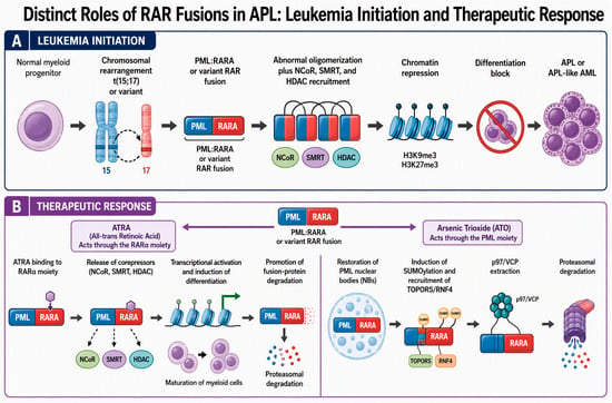
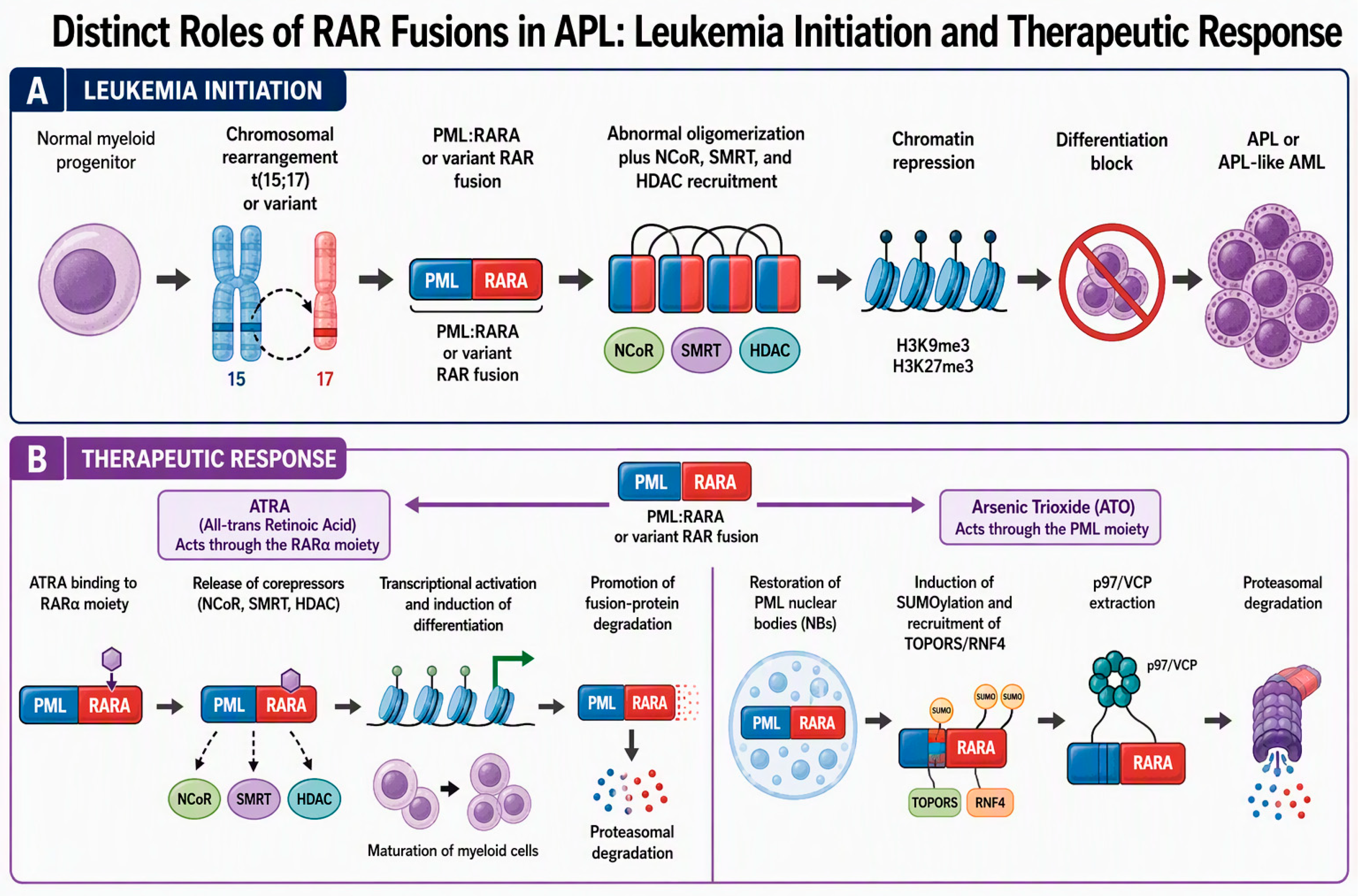
Mechanistically, PML::RARA is more than a high-affinity transcriptional repressor. Fusion-driven oligomerization broadens DNA occupancy, recruits abnormal chromatin complexes, disrupts PML nuclear bodies, and perturbs stress-responsive tumor-suppressive functions. Therapeutic response depends on dismantling this composite state. ATRA promotes corepressor release, differentiation, and degradation through the RARα moiety; arsenic binds a cysteine-rich pocket in PML, induces nuclear-body phase transition, stimulates SUMOylation and SUMO-targeted ubiquitination, and recruits proteasomal machinery [23,24,25,37]. Recent work has added further regulatory layers. Neddylation can stabilize an aberrant phase-separated state, whereas pharmacological deneddylation restores functional PML bodies [28]. The deubiquitinase YOD1 protects PML::RARA from destruction, and its inhibition enhances oncoprotein degradation in preclinical models [27]. Resistance can arise through RARA LBD substitutions that impair ligand binding or strengthen basal repression, and through PML substitutions that interrupt arsenic-triggered SUMO1/SUMO2/3, TOPORS, RNF4, and p97 signaling [25,40]. These findings place proteostasis, chromatin regulation, and nuclear-body organization within one disease mechanism and suggest that the relevant therapeutic endpoint is elimination of the pathogenic protein state rather than receptor occupancy alone (see Figure 2).
Figure 2.
Leukemia initiation and therapeutic response in APL. RAR-family gene fusions function as primary leukemia-driving alterations by enforcing abnormal chromatin repression and blocking differentiation. In classic APL, ATRA and arsenic trioxide target complementary domains of PML::RARA and promote differentiation and oncoprotein elimination [6,23,24,26,37].
4. RARB-Related Disorder: Germline Variants and Clinical Heterogeneity
RARB variants cause syndromic microphthalmia 12 (MCOPS12), increasingly termed RARB-related disorder [7,8,41]. The initial 2013 report established two mechanistic routes: biallelic truncating variants produced loss of receptor activity, whereas recurrent de novo substitutions at Arg387 produced ligand-dependent gain-of-function [7]. Subsequent cohorts broadened the phenotype beyond microphthalmia, pulmonary hypoplasia, diaphragmatic hernia, and cardiac defects to include global developmental delay, hypotonia, progressive spasticity or dystonia, feeding difficulty, brain abnormalities, and variable ocular involvement [8,41]. In 2023, functional analysis of 25 additional individuals carrying 17 novel pathogenic or likely pathogenic variants showed that dominant variants may act through either gain-of-function or dominant-negative mechanisms [8]. Accordingly, absence of microphthalmia does not exclude the diagnosis. Recent reports also expand the allelic spectrum to frameshift and noncoding regulatory variants; however, regulatory variants require direct functional support and careful exclusion of alternative explanations [42,43]. Two 2026 mouse models carrying p.Arg387Cys and p.Leu402Pro reproduced ocular, motor, and cognitive abnormalities and implicated striatal dysfunction, strengthening the mechanistic connection between altered receptor signaling and movement disorder [31].
The cellular effects of RARB variants are best understood as quantitative disturbances of a developmental transcriptional network. RARβ normally participates in ligand-sensitive repression and activation together with RXR, corepressors, coactivators, and chromatin-modifying complexes. Recurrent Arg387 substitutions alter the LBD and can produce ligand-dependent gain-of-function, whereas p.Leu402Pro and other dominant variants can interfere with wild-type receptor activity. Biallelic truncating alleles reduce signaling output, demonstrating that both excessive and insufficient RARβ activity can disrupt development [7,8,31,41]. The resulting phenotype reflects tissue timing and dosage: ocular morphogenesis, pulmonary and diaphragmatic development, motor-system maturation, and striatal function appear especially vulnerable. Distinct from inherited RARB-related disorder, recurrent TBL1XR1::RARB rearrangements have been described in pediatric APL-like leukemia lacking RARA rearrangement, extending fusion-driven leukemogenesis to a second RAR family member [10]. This separation between germline dosage disorders and somatic fusion disease should be explicit in variant interpretation and counseling (see Table 2).
Table 2.
Representative RARB alterations and associated phenotypes.
5. Molecular Determinants of Therapeutic Resistance in RARG-Rearranged AML Versus Classical PML::RARA-Positive APL
RARG-rearranged acute myeloid leukemia (AML) is a rare yet clinically critical disease entity that frequently mimics acute promyelocytic leukemia (APL) in morphology, immune phenotype, and associated coagulopathy, while characteristically lacking the canonical PML::RARA fusion [9]. Reported fusion partners include CPSF6, NUP98, HNRNPC, HNRNPM, PML, and NPM1, occasionally exhibiting complex or tripartite structural architectures [9,30,44]. Clinical data from international cohorts demonstrate a characteristically poor response to all-trans retinoic acid (ATRA) and arsenic trioxide (ATO), high early mortality, and adverse overall survival, supporting its classification as a distinct AML subgroup rather than a conventional APL variant [9].
A direct comparison between classical PML::RARA-positive APL and RARG-rearranged AML highlights three interdependent molecular dimensions governing therapeutic response (see Figure 3):
Figure 3.
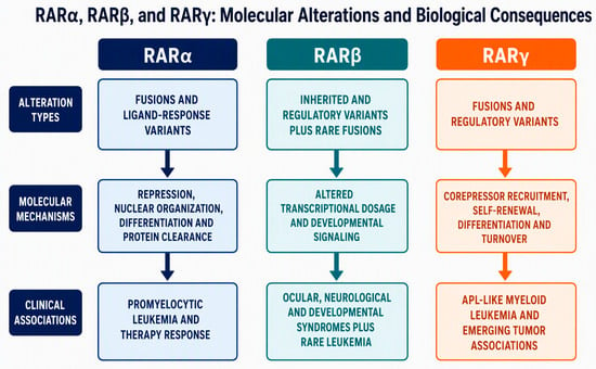
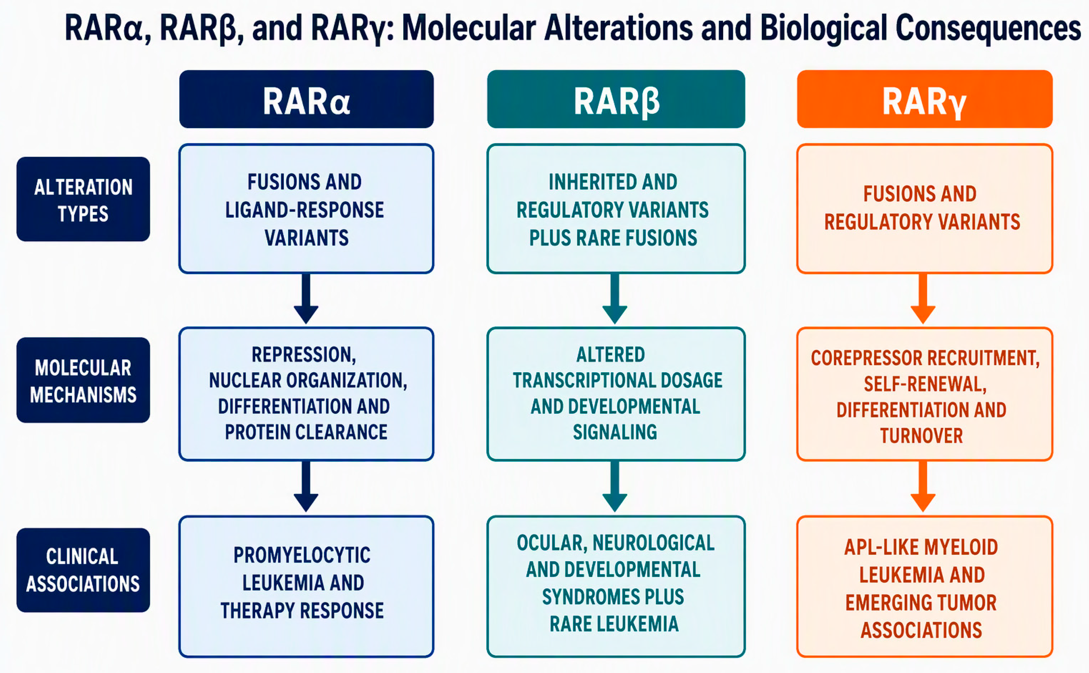
Genotype–mechanism–phenotype relationships across the RAR family. RARA alterations are dominated by leukemia-driving fusions and treatment-response variants [6,13,23,29,32,33,34,35,36,37,38,39,40]; RARB alterations primarily cause inherited ocular, neurological, and developmental syndromes but also include rare leukemia-associated fusions [7,8,10,31,41,42,43]; and RARG alterations include APL-like AML fusions and emerging regulatory or tumor-associated changes [9,11,12,14,15,16,30,44,45].
- Fusion-Partner Identity and Oligomerization Architecture
- ○
- Classical APL (PML::RARA): The PML moiety drives constitutive homo-oligomerization and localizes the fusion protein to nuclear bodies, creating a high-affinity platform for corepressor complex binding [6,23].
- ○
- RARG-Rearranged AML: Diverse fusion partners (such as CPSF6 or NUP98) substitute for PML. These partners dictate distinct subcellular localization patterns, higher-order structural constraints, and novel protein–protein interactions [12,30]. For instance, CPSF6::RARG has been shown to physically engage histone deacetylase 3 (HDAC3) and cooperate directly with recurrent RAS-pathway mutations to expand preleukemic myeloid progenitors and drive differentiation arrest [30].
- Retained Receptor Domains and Structural Constraints
- ○
- Classical APL (PML::RARA): Retains functional retinoic acid-binding domains of RARα, allowing pharmacological doses of ATRA to induce a conformational switch that triggers corepressor release, coactivator recruitment, and eventual degradation of the fusion protein [21].
- ○
- RARG-Rearranged AML: Truncations or altered fusion topologies frequently disrupt regions required for productive, ligand-induced activation [29,30]. The structural constraints imposed by non-PML fusion partners alter the ligand-binding pocket or stereo-orientation of the receptor, preventing proper conformational remodeling upon ATRA exposure [29].
- Corepressor Recruitment and Differential Drug Sensitivity
- ○
- ATRA Resistance: Unlike PML::RARA, which undergoes ATRA-induced degradation [21,22], RARG fusions maintain aberrant corepressor retention or fail to undergo efficient ligand-dependent clearance, blunting transcriptional reactivation of target genes like BCL2 and ATF3 [12].
- ○
- Arsenic Trioxide (ATO) Resistance: The classic PML moiety contains critical cysteine residues that bind arsenic trioxide, triggering targeted SUMOylation, degradation, and therapeutic clearance of the fusion protein [24,25,26]. Because RARG-rearranged AML partners entirely lack this specific PML arsenic-binding motif, ATO is fundamentally ineffective [9,11,12].
Because empirical ATRA administration is standard practice when APL is initially suspected in the clinic, rapid diagnostic exclusion of PML::RARA coupled with RNA-based fusion detection is vital to avoid futile retinoid exposure and promptly redirect therapy toward AML-directed regimens [9,44,49].
6. RAR Pathway Dysregulation in Solid Tumors: Limited Evidence for Recurrent Coding Drivers
Somatic coding variants, altered receptor expression, promoter methylation, noncoding regulation, and defective receptor turnover have been reported across various solid tumors, but these observations represent distinct biological and evidentiary categories [5,14,15,16,19]. In most settings, evidence for broad RAR-pathway dysregulation is considerably stronger than evidence for recurrent pathogenic coding mutations [5,14]. For instance, reduced RARγ expression in prostate cancer has been linked to a miR-96 regulatory axis and altered androgen-receptor signaling [14], whereas context-dependent RARγ activity has been implicated in other tumor models [15]. In breast cancer, receptor expression, RARβ2 promoter methylation, histone deacetylation, and the intracellular routing of retinoic acid via CRABP2 or FABP5 function as notable determinants of retinoid responsiveness [5]. Furthermore, glioblastoma illustrates how protein-quality-control defects such as the accumulation of SUMOylated and ubiquitinated retinoid receptors due to impaired VCP-dependent processing and proteasomal turnover can uncouple receptor abundance from true transcriptional competence [19].
Critically, these diverse alterations should not be conflated with Mendelian pathogenic variants or leukemia-defining fusions [5]. Each molecular change must instead be appropriately classified as a recurrent disease driver, treatment-resistance mutation, predictive biomarker, epigenetic modification, experimentally supported candidate, or variant of uncertain significance [48].
Extending this evaluative framework, a recent study in Genes characterized rare cancer-associated substitutions mapping to the N-terminal AF-1 phospho-regulatory region of RARγ [16]. Focusing on variants affecting or flanking the conserved Ser77/Ser79 regulatory module, this analysis highlighted potential disruptions to phosphorylation-dependent transcriptional control [16]. Earlier biochemical investigations established that ligand-activated RARγ2 and RARα1 undergo regulated ubiquitin–proteasome-dependent degradation modulated by phosphorylation and RXR hetero dimerization [21]. Specifically, mutation of these phospho-acceptor serines blocks retinoic acid-induced ubiquitination and degradation, leading to receptor accumulation, while disruption of PI3K/Akt-dependent signaling alters turnover and transcriptional activity [21,22].
While these historical cellular models provide a strong mechanistic precedent for testing whether cancer-associated AF-1 substitutions alter RARγ stability or downstream signaling, the clinical relevance of these rare variants remains unproven [16,21]. Because they currently lack sufficient clinical recurrence, familial segregation, and functional validation in vivo, they should be strictly regarded as mechanistically plausible structural modifiers rather than established cancer drivers [16]. Prospective functional studies comparing wild-type RARγ against each variant across ligand-response curves, phosphorylation states, half-lives, and chromatin occupancy profiles are required to establish their true biological impact [16].
7. Diagnostic and Functional Evaluation
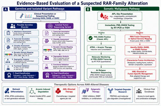
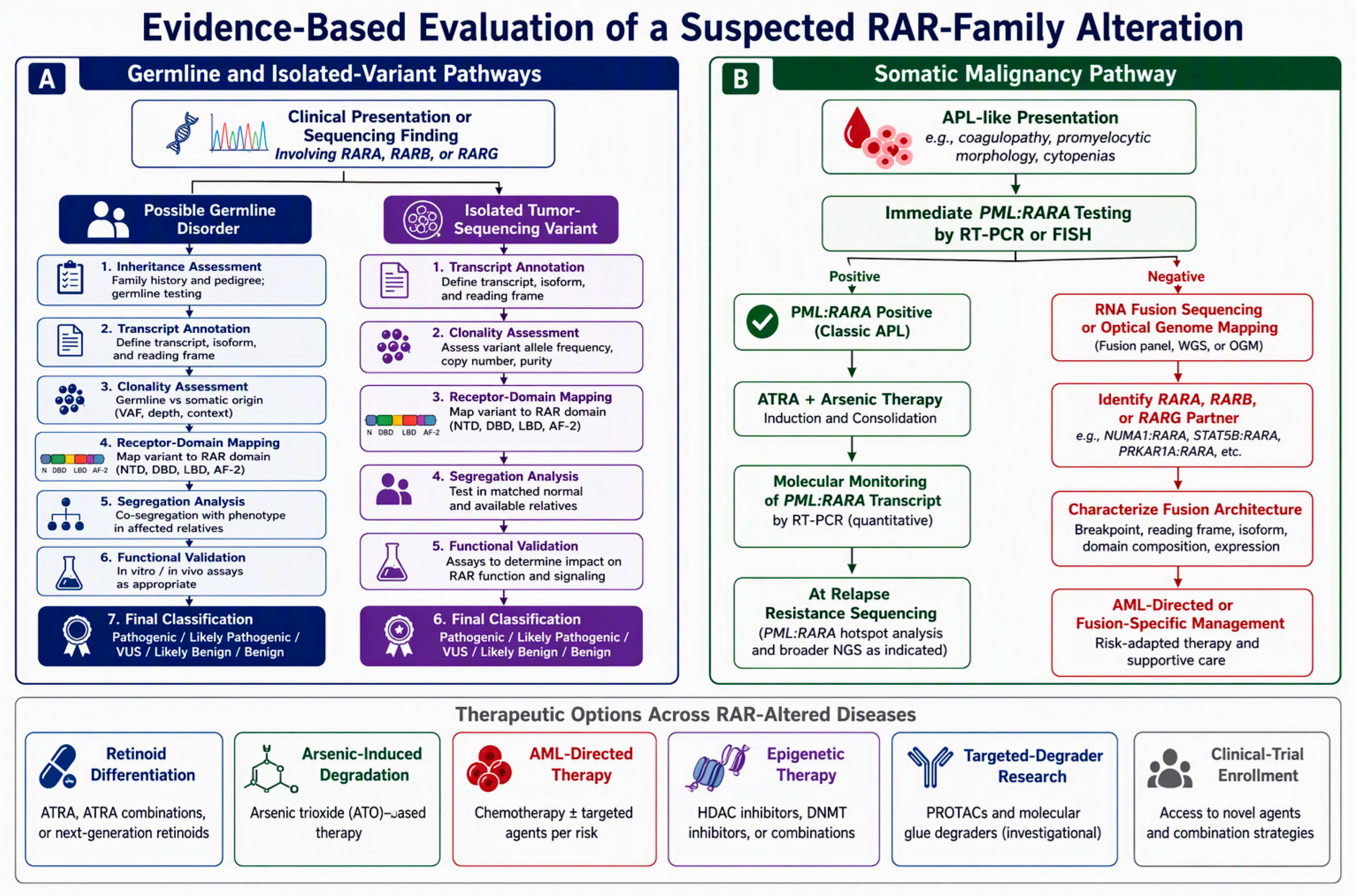
The diagnostic workflow should follow the biological alteration under consideration. In an APL-like emergency, morphology and coagulopathy warrant immediate supportive management and commonly prompt empiric ATRA while rapid PML::RARA testing is performed [35,37,49]. A negative result should trigger assays capable of identifying cryptic and variant rearrangements, including RARA, RARB, and RARG break-apart testing and RNA-based fusion sequencing [9,10,11,29,35,44,49]. Complete fusion architecture matters because partner identity, retained domains, and tripartite transcripts influence mechanism and treatment response [9,12,29,30]. For suspected RARB-related disorder, trio exome or genome sequencing should be interpreted together with inheritance, copy-number analysis, transcript-specific HGVS annotation, ClinVar or ClinGen evidence, and detailed phenotype concordance [7,8,31,41,42,43]. Functional assays are especially important because pathogenic RARB variants can increase, decrease, or dominantly interfere with transcriptional activity [7,8,31,41,42,43]. A rigorous assay panel should examine basal and ligand-stimulated reporter activity, dose response, nuclear localization, DNA binding, corepressor and coactivator exchange, protein stability, ubiquitination, and dominant-negative behavior. For somatic variants, database presence alone does not establish causality. Interpretation should additionally consider clonality, recurrence, treatment timing, co-mutations, and whether the alteration occurs in the native receptor or a fusion allele, applying established oncogenicity criteria and seeking orthogonal functional and clinical support [48]. Multi-omic methods, including long-read or RNA sequencing, chromatin profiling, quantitative proteomics, single-cell analysis, and patient-derived models, may help connect a sequence change to disease mechanism; however, treatment decisions should remain anchored to current disease-specific clinical guidance rather than inferred from receptor identity alone (see Figure 4, Table 3).
Figure 4.
Evidence-based evaluation of a suspected RAR-family alteration. The workflow distinguishes germline developmental disease, isolated tumor-sequencing variants, and APL or APL-like malignancy, integrating inheritance or clonality, transcript-aware annotation, fusion resolution, mechanism-appropriate functional validation, and alteration-specific therapeutic interpretation [7,8,9,10,11,12,29,30,31,35,39,40,41,42,43,44,45,48,49].
Table 3.
Diagnostic and therapeutic implications of major RAR alterations.
8. Therapeutic Implications
RAR alterations demonstrate why precision therapy must be mechanism-based. In classic APL, ATRA and arsenic trioxide act on complementary fusion-protein modules and converge on differentiation, PML nuclear-body restoration, and oncoprotein clearance [23,24,25,26,37]. Resistance mutations can reveal the disrupted step: RARA LBD variants may impair ligand response, whereas PML substitutions may block SUMO-dependent recruitment of TOPORS, RNF4, p97, and the proteasome [25,40]. Experimental inhibition of the deubiquitinase YOD1 or neddylation further illustrates how manipulation of protein quality control can destabilize PML::RARA or restore productive nuclear-body organization in preclinical models [27,28]. In contrast, RARG-rearranged AML requires AML-directed treatment; response-adapted transplantation or clinical-trial enrollment may be appropriate, but isolated responses to hypomethylating agents do not define a standard [9,12,45]. No disease-modifying retinoid therapy has been validated for inherited RARB-related disorder, and the coexistence of gain-of-function, dominant-negative, and loss-of-function mechanisms argues against nonspecific retinoid supplementation [7,8,31,41,42,43]. Chaperone-mediated autophagy has been experimentally activated by small molecules that stabilize the interaction between RARα and its corepressor NCoR1 in a retinal-degeneration model [20]. More broadly, tumor-directed targeted-protein-degradation platforms and a preclinical RARα PROTAC delivered with exosome-like nanoparticles provide complementary proof of principle for selective protein elimination and targeted delivery [46,47]. Translation to RAR-associated disease will require isotype-selective ligands, an appropriate E3-ligase context, productive ternary-complex geometry, access to the relevant tissue, and biomarkers that distinguish degradation from transcriptional antagonism [46,47]. Fusion-selective degradation and disruption of abnormal corepressor complexes remain promising future objectives, but their therapeutic value has not yet been established for RAR-associated disease.
9. Critical Challenges and Future Directions
Several barriers currently limit translation. First, nomenclature remains inconsistent across legacy cytogenetics, slash notation, transcript isoforms, and protein numbering. HGNC recommends double-colon notation for fusion genes [50]; accordingly, this review adopts that convention throughout, while retaining legacy slash or hyphen notation only when reproducing historical article titles or established protein names. Second, when reported variants lack matched normal DNA, segregation data, recurrence, or assays performed in the relevant cell type, their classification is constrained under established oncogenicity criteria [48]. Third, standard transcriptional reporters capture only one dimension of receptor biology. Studies of RAR and PML::RARA regulation demonstrate that phosphorylation, SUMOylation, ubiquitination, VCP/p97-dependent processing, and proteasomal turnover can materially alter receptor or fusion-protein abundance and activity [19,21,22,25,26]. Chromatin context, nuclear-body organization, phase separation, and lineage-dependent transcriptional programs represent additional mechanistic layers that require dedicated assays [12,17,18,24,28,30]. Fourth, clinical evidence for rare RAR-family fusion leukemias remains limited to case reports, small series, and one modest international RARG-rearranged cohort, restricting robust treatment comparisons [9,10,11,44,45]. International registries should therefore link genotype, complete fusion architecture, co-mutations, treatment, molecular response, and outcome. Functionally, saturation mutagenesis of receptor domains, quantitative proteostasis assays, CRISPR-engineered disease models, patient-derived organoids, and single-cell multi-omics could generate variant-effect maps that are directly useful for clinical classification. Biomarker development should include receptor abundance, modification state, corepressor dependence, degradation competence, and downstream transcriptional endotypes. Finally, targeted-degradation strategies must preserve the normal developmental and homeostatic functions of the RAR family. Tumor-directed targeted protein degrader delivery can improve tissue selectivity and reduce on-target, off-tissue effects, while a recent preclinical RARα-targeting PROTAC delivered in exosome-like nanoparticles provides proof of principle for combining receptor degradation with targeted delivery [46,47]. However, fusion-selective RAR degradation and the use of tissue-restricted E3 ligases remain important future objectives rather than established strategies.
10. Limitations
This narrative review is limited by the heterogeneity of the available evidence. The literature includes clinical cohorts, small case series, individual reports, experimental models, and pathway-level studies that do not provide equivalent support for causality or treatment response. Several RAR alterations are too rare for robust genotype–phenotype estimates, and publication bias may favor unusual positive findings over negative functional or clinical observations. Direct comparison among variants is further constrained by differences in ligand concentration, cellular background, transcript isoform, assay design, and measured endpoint. The solid-tumor literature is especially heterogeneous and often supports pathway dysregulation rather than recurrent pathogenic coding alterations. Database classifications and newly reported variants may change as additional segregation, recurrence, structural, and therapeutic data become available. Because this article is a narrative rather than a systematic review, the search and study-selection process was designed to identify clinically and mechanistically substantiated RAR alterations rather than to provide an exhaustive evidence synthesis. These limitations reinforce the need for standardized annotation, shared registries, transparent evidence grading, and orthogonal functional validation.
11. Conclusions
Disease-associated RAR alterations fall into four clinically distinct evidence tiers: established leukemia-driving fusions, validated germline RARB disorders, treatment-emergent resistance mutations, and candidate regulatory or solid-tumor alterations that still require independent validation. Their effects are shaped by receptor domain, fusion partner, chromatin context, post-translational regulation, and protein-quality-control capacity. This framework explains why sequence conservation does not imply clinical equivalence and why retinoid responsiveness cannot be inferred from morphology or receptor identity alone. The therapeutic success of differentiation and degradation in classic APL provides a compelling mechanistic precedent, but extension to other RAR-associated diseases requires alteration-specific evidence, complete molecular characterization, quantitative functional assays, and preservation of physiological RAR signaling. Standardized annotation and international genotype–phenotype registries will be essential for moving rare observations from plausible mechanisms to clinically actionable classification.
Funding
This research received no external funding.
Institutional Review Board Statement
Not applicable.
Informed Consent Statement
Not applicable.
Data Availability Statement
No new data were created or analyzed in this review. Data sharing is not applicable to this article.
Acknowledgments
This review is dedicated to the memory of Pierre Chambon (1931–2026), a visionary scientific leader and defining figure in modern molecular biology and nuclear receptor research, whose pioneering discoveries established fundamental concepts in retinoid receptor biology. During the preparation of this manuscript, the author used generative artificial intelligence tools for language editing, structural organization, and formatting assistance. The author reviewed and edited all generated material and takes full responsibility for the accuracy, interpretation, originality, and integrity of the published content.
Conflicts of Interest
The author declare no conflicts of interest.
References
- Di Masi, A.; Leboffe, L.; De Marinis, E.; Pagano, F.; Cicconi, L.; Rochette-Egly, C.; Lo-Coco, F.; Ascenzi, P.; Nervi, C. Retinoic acid receptors: From molecular mechanisms to cancer therapy. Mol. Asp. Med. 2015, 41, 1–115. [Google Scholar] [CrossRef] [Scilit] [PubMed]
- Petkovich, M.; Chambon, P. Retinoic acid receptors at 35 years. J. Mol. Endocrinol. 2022, 69, T13–T24. [Google Scholar] [CrossRef] [Scilit] [PubMed]
- Ghyselinck, N.B.; Duester, G. Retinoic acid signaling pathways. Development 2019, 146, dev167502. [Google Scholar] [CrossRef] [Scilit] [PubMed]
- Isoherranen, N.; Wen, Y.W. The interplay between retinoic acid binding proteins and retinoic acid degrading enzymes in modulating retinoic acid concentrations. Curr. Top. Dev. Biol. 2025, 161, 167–200. [Google Scholar] [CrossRef] [Scilit] [PubMed]
- De Angelis, M.T.; Dattilo, V.; Vono, N.; Lise, V.; Reda, M.; De Marco, C.; Rizzuto, A.; Trapasso, F.; Viglietto, G.; Santamaria, G. Retinoic acid in health and disease. Signal Transduct. Target. Ther. 2026, 11, 400. [Google Scholar] [CrossRef] [Scilit] [PubMed]
- Melnick, A.; Licht, J.D. Deconstructing a disease: RARα, its fusion partners, and their roles in the pathogenesis of acute promyelocytic leukemia. Blood 1999, 93, 3167–3215. [Google Scholar] [CrossRef] [Scilit] [PubMed]
- Srour, M.; Chitayat, D.; Caron, V.; Chassaing, N.; Bitoun, P.; Patry, L.; Cordier, M.-P.; Capo-Chichi, J.-M.; Francannet, C.; Calvas, P.; et al. Recessive and dominant mutations in retinoic acid receptor beta in cases with microphthalmia and diaphragmatic hernia. Am. J. Hum. Genet. 2013, 93, 765–772. [Google Scholar] [CrossRef] [Scilit] [PubMed]
- Caron, V.; Chassaing, N.; Ragge, N.; Boschann, F.; Ngu, A.M.-H.; Meloche, E.; Chorfi, S.; Lakhani, S.A.; Ji, W.; Steiner, L.; et al. Clinical and functional heterogeneity associated with the disruption of retinoic acid receptor beta. Genet. Med. 2023, 25, 100856. [Google Scholar] [CrossRef] [Scilit] [PubMed]
- Zhu, H.H.; Qin, Y.Z.; Zhang, Z.L.; Liu, Y.-J.; Wen, L.-J.; You, M.J.; Zhang, C.; Such, E.; Luo, H.; Yuan, H.-J.; et al. A global study for acute myeloid leukemia with RARG rearrangement. Blood Adv. 2023, 7, 2972–2982. [Google Scholar] [CrossRef] [Scilit] [PubMed]
- Osumi, T.; Tsujimoto, S.I.; Tamura, M.; Uchiyama, M.; Nakabayashi, K.; Okamura, K.; Yoshida, M.; Tomizawa, D.; Watanabe, A.; Takahashi, H.; et al. Recurrent RARB translocations in acute promyelocytic leukemia lacking RARA translocation. Cancer Res. 2018, 78, 4452–4458. [Google Scholar] [CrossRef] [Scilit] [PubMed]
- Conserva, M.R.; Redavid, I.; Anelli, L.; Zagaria, A.; Specchia, G.; Albano, F. RARG gene dysregulation in acute myeloid leukemia. Front. Mol. Biosci. 2019, 6, 114. [Google Scholar] [CrossRef] [Scilit] [PubMed]
- Wang, F.; Zhao, L.; Tan, Y.; Cen, X.; Gao, H.; Jiang, H.; Liu, Y.; Li, Y.; Zhang, T.; Zhao, C.; et al. Oncogenic role of RARG rearrangements in acute myeloid leukemia resembling acute promyelocytic leukemia. Nat. Commun. 2025, 16, 617. [Google Scholar] [CrossRef] [Scilit] [PubMed]
- Jakubiuk-Tomaszuk, A.; Murcia Pienkowski, V.; Zietkiewicz, S.; Rydzanicz, M.; Kosińska, J.; Stawiński, P.; Szumiński, M.; Płoski, R. Syndromic chorioretinal coloboma associated with heterozygous de novo RARA mutation affecting an amino acid critical for retinoic acid interaction. Clin. Genet. 2019, 96, 371–375. [Google Scholar] [CrossRef] [Scilit] [PubMed]
- Long, M.D.; Singh, P.K.; Russell, J.R.; Llimos, G.; Rosario, S.; Rizvi, A.; Berg, P.R.v.D.; Kirk, J.; Sucheston-Campbell, L.E.; Smiraglia, D.J.; et al. The miR-96 and RARγ signaling axis governs androgen signaling and prostate cancer progression. Oncogene 2019, 38, 421–444. [Google Scholar] [CrossRef] [Scilit] [PubMed]
- Guo, S.; Han, H.; Ding, B.; Zhang, W.; Qin, L. The role of RARG in solid tumor progression and therapeutic potential. Discov. Oncol. 2026, 17, 493. [Google Scholar] [CrossRef] [Scilit] [PubMed]
- Kopf, E. Rare Cancer-Associated Mutations May Affect the AF1 Phosphoregulatory Domain of RARγ. Genes 2026, 17, 1119. [Google Scholar] [CrossRef] [Scilit] [PubMed]
- Tambones, I.; Sagar, A.; Vankova, P.; Loginov, D.; Carivenc, C.; Rochel, N.; Bourguet, W.; Man, P.; Bernadó, P.; le Maire, A. New structural insights into the control of the retinoic acid receptors RAR/RXR by DNA, ligands, and transcriptional coregulators. Nucleic Acids Res. 2025, 53, gkaf967. [Google Scholar] [CrossRef] [Scilit] [PubMed]
- Pattison, J.M.; Melo, S.P.; Piekos, S.N.; Torkelson, J.L.; Bashkirova, E.; Mumbach, M.R.; Rajasingh, C.; Zhen, H.H.; Li, L.; Liaw, E.; et al. Retinoic acid and BMP4 cooperate with p63 to alter chromatin dynamics during surface epithelial commitment. Nat. Genet. 2018, 50, 1658–1665. [Google Scholar] [CrossRef] [Scilit] [PubMed]
- Rodriguez, V.; Bailey, R.; Larion, M.; Gilbert, M.R. Retinoid receptor turnover mediated by sumoylation, ubiquitination and the valosin-containing protein is disrupted in glioblastoma. Sci. Rep. 2019, 9, 16250. [Google Scholar] [CrossRef] [Scilit] [PubMed]
- Gomez-Sintes, R.; Xin, Q.; Jimenez-Loygorri, J.I.; McCabe, M.; Diaz, A.; Garner, T.P.; Cotto-Rios, X.M.; Wu, Y.; Dong, S.; Reynolds, C.A.; et al. Targeting retinoic acid receptor alpha-corepressor interaction activates chaperone-mediated autophagy and protects against retinal degeneration. Nat. Commun. 2022, 13, 4220. [Google Scholar] [CrossRef] [Scilit] [PubMed]
- Kopf, E.; Plassat, J.-L.; Vivat, V.; de Thé, H.; Chambon, P.; Rochette-Egly, C. Dimerization with retinoid X receptors and phosphorylation modulate the retinoic acid-induced degradation of retinoic acid receptors α and γ through the ubiquitin-proteasome pathway. J. Biol. Chem. 2000, 275, 33280–33288. [Google Scholar] [CrossRef] [Scilit] [PubMed]
- Giannì, M.; Kopf, E.; Bastien, J.; Oulad-Abdelghani, M.; Garattini, E.; Chambon, P.; Rochette-Egly, C. Down-regulation of the phosphatidylinositol 3-kinase/Akt pathway is involved in retinoic acid-induced phosphorylation, degradation, and transcriptional activity of retinoic acid receptor γ2. J. Biol. Chem. 2002, 277, 24859–24862. [Google Scholar] [CrossRef] [Scilit] [PubMed]
- Wu, H.C.; Laplantine, E.; Esnault, C.; Niwa-Kawakita, M.; Zhang, Y.; Bastos, V.D.A.; Geoffroy, M.-C.; de Thé, H. PML-driven sumoylation of PML::RARA-bound repressors drives hematopoietic progenitor immortalization. J. Exp. Med. 2026, 223, e20252333. [Google Scholar] [CrossRef] [Scilit] [PubMed]
- Bercier, P.; Wang, Q.Q.; Zang, N.; Zhang, J.; Yang, C.; Maimaitiyiming, Y.; Abou-Ghali, M.; Berthier, C.; Wu, C.; Niwa-Kawakita, M.; et al. Structural basis of PML-RARA oncoprotein targeting by arsenic unravels a cysteine rheostat controlling PML body assembly and function. Cancer Discov. 2023, 13, 2548–2565. [Google Scholar] [CrossRef] [Scilit] [PubMed]
- Jaffray, E.G.; Tatham, M.H.; Mojsa, B.; Plechanovová, A.; Rojas-Fernandez, A.; Liu, J.C.; Mailand, N.; Ibrahim, A.F.; Ball, G.; Porter, I.M.; et al. PML mutants from arsenic-resistant patients reveal SUMO1-TOPORS and SUMO2/3-RNF4 degradation pathways. J. Cell Biol. 2025, 224, e202407133. [Google Scholar] [CrossRef] [Scilit]
- Jaffray, E.G.; Tatham, M.H.; Mojsa, B.; Liczmanska, M.; Rojas-Fernandez, A.; Yin, Y.; Ball, G.; Hay, R.T. The p97/VCP segregase is essential for arsenic-induced degradation of PML and PML-RARA. J. Cell Biol. 2023, 222, e202201027. [Google Scholar] [CrossRef] [Scilit] [PubMed]
- Shao, X.; Chen, Y.; Wang, W.; Du, W.; Zhang, X.; Cai, M.; Bing, S.; Cao, J.; Xu, X.; Yang, B.; et al. Blockade of deubiquitinase YOD1 degrades oncogenic PML/RARα and eradicates acute promyelocytic leukemia cells. Acta Pharm. Sin. B 2022, 12, 1856–1870. [Google Scholar] [CrossRef] [Scilit] [PubMed]
- Shao, X.; Chen, Y.; Xu, A.; Xiang, D.; Wang, W.; Du, W.; Huang, Y.; Zhang, X.; Cai, M.; Xia, Z.; et al. Deneddylation of PML/RARα reconstructs functional PML nuclear bodies via orchestrating phase separation to eradicate APL. Cell Death Differ. 2022, 29, 1654–1668. [Google Scholar] [CrossRef] [Scilit] [PubMed]
- Wu, H.C.; de Thé, H. Tripartite RAR fusions explain RA resistance. Blood 2024, 144, 1461–1462. [Google Scholar] [CrossRef] [Scilit] [PubMed]
- Liu, T.; Wang, T.; Qi, L.; Liu, Y.; Shan, M.; Wang, F.; Fang, Y.; Liu, S.; Wen, L.; Chen, S.; et al. CPSF6-RARγ interacts with histone deacetylase 3 to promote myeloid transformation in RARG-fusion acute myeloid leukemia. Nat. Commun. 2025, 16, 616. [Google Scholar] [CrossRef] [Scilit] [PubMed]
- Zinter, N.; Shah, D.; Artot, V.; Lemmetti, N.; Semaan, H.; Soret, R.; Pilon, N.; Nassif, C.; Birling, M.-C.; Raja-Ravi-Shankar, A.; et al. Compromised striatal structure and function in mouse models of RARB-related disorder. Brain 2026. Online ahead of print. [Google Scholar] [CrossRef] [Scilit] [PubMed]
- Mozziconacci, M.J.; Liberatore, C.; Brunel, V.; Grignani, F.; Arnoulet, C.; Ferrucci, P.F.; Fernandez, F.; Sainty, D.; Pelicci, P.G.; Birg, F.; et al. In vitro response to all-trans retinoic acid of acute promyelocytic leukemias with nonreciprocal PML/RARA or RARA/PML fusion genes. Genes Chromosomes Cancer 1998, 22, 241–250. [Google Scholar] [CrossRef] [Scilit]
- Menezes, J.; Acquadro, F.; Perez-Pons de la Villa, C.; García-Sánchez, F.; Álvarez, S.; Cigudosa, J.C. FIP1L1/RARA with breakpoint at FIP1L1 intron 13: A variant translocation in acute promyelocytic leukemia. Haematologica 2011, 96, 1565–1566. [Google Scholar] [CrossRef] [Scilit] [PubMed]
- Yin, C.C.; Jain, N.; Mehrotra, M.; Zhagn, J.; Protopopov, A.; Zuo, Z.; Pemmaraju, N.; DiNardo, C.; Hirsch-Ginsberg, C.; Wang, S.A.; et al. Identification of a cryptic IRF2BP2-RARA fusion in acute promyelocytic leukemia. J. Natl. Compr. Canc. Netw. 2015, 13, 19–22. [Google Scholar] [CrossRef] [Scilit] [PubMed]
- Wen, L.; Xu, Y.; Yao, L.; Wang, N.; Wang, Q.; Liu, T.; Pan, J.; Cen, J.; Zhou, H.; Miao, M.; et al. Clinical and molecular features of acute promyelocytic leukemia with variant retinoid acid receptor fusions. Haematologica 2019, 104, e195–e199. [Google Scholar] [CrossRef] [Scilit] [PubMed]
- Peterson, J.F.; He, R.R.; Nayer, H.; Cuevo, R.S.; Smadbeck, J.B.; Vasmatzis, G.; Greipp, P.T.; Ketterling, R.P.; Hoppman, N.L.; Baughn, L.B. Characterization of a rarely reported STAT5B/RARA gene fusion in a young adult with newly diagnosed acute promyelocytic leukemia with resistance to ATRA therapy. Cancer Genet. 2019, 237, 51–54. [Google Scholar] [CrossRef] [Scilit] [PubMed]
- Liquori, A.; Ibañez, M.; Sargas, C.; Sanz, M.A.; Barragán, E.; Cervera, J. Acute promyelocytic leukemia: A constellation of molecular events around a single PML-RARA fusion gene. Cancers 2020, 12, 624. [Google Scholar] [CrossRef] [Scilit] [PubMed]
- Liu, G.; Liu, L.; Di Bartolo, D.; Li, K.Y.; Li, X. Acute promyelocytic leukemia with rare genetic aberrations: A report of three cases. Genes 2023, 14, 46. [Google Scholar] [CrossRef] [Scilit] [PubMed]
- Ding, W.; Li, Y.P.; Nobile, L.M.; Grills, G.; Carrera, I.; Paietta, E.; Tallman, M.S.; Wiernik, P.H.; Gallagher, R.E. Leukemic cellular retinoic acid resistance and missense mutations in the PML-RARalpha fusion gene after relapse of acute promyelocytic leukemia from treatment with all-trans retinoic acid and intensive chemotherapy. Blood 1998, 92, 1172–1183. [Google Scholar] [CrossRef] [Scilit] [PubMed]
- Barbosa, G.M.; Yuan, H.; Tambones, I.; Stote, R.H.; Wu, H.C.; De Thé, H.; Le Maire, A.; Geoffroy, M.-C. ATRA resistance conferred by a clustered subset of hotspot PML::RARA mutations enhancing basal repression rather than precluding transactivation. Haematologica 2026. Early View. [Google Scholar] [CrossRef] [Scilit] [PubMed]
- Srour, M.; Caron, V.; Pearson, T.; Nielsen, S.B.; Lévesque, S.; Delrue, M.-A.; Becker, T.A.; Hamdan, F.F.; Kibar, Z.; Sattler, S.G.; et al. Gain-of-function mutations in RARB cause intellectual disability with progressive motor impairment. Hum. Mutat. 2016, 37, 786–793. [Google Scholar] [CrossRef] [Scilit] [PubMed]
- Replogle, M.R.; Thompson, S.; Reis, L.M.; Semina, E.V. A de novo noncoding RARB variant associated with complex microphthalmia alters a putative regulatory element. Hum. Mutat. 2024, 2024, 6619280. [Google Scholar] [CrossRef] [Scilit] [PubMed]
- Koyanagi, Y.; Morikawa-Anzai, H.; Yoshida, T.; Tominaga, M.; Abe, Y.; Kosaki, R.; Matsubara, K.; Fukami, M.; Nishina, S. Clinical features of syndromic microphthalmia in two novel RARB variants. Hum. Genome Var. 2026, 13, 11. [Google Scholar] [CrossRef] [Scilit] [PubMed]
- Tao, S.; Song, L.; Deng, Y.; Chen, Y.; Shi, Y.; Gan, Y.; Deng, Z.; Ding, B.; He, Z.; Wang, C.; et al. Acute myeloid leukemia with NUP98-RARG gene fusion similar to acute promyelocytic leukemia: Case report and literature review. Onco Targets Ther. 2020, 13, 10559–10566. [Google Scholar] [CrossRef] [Scilit] [PubMed]
- Wei, Z.; Shao, L.; Xu, S.; Zhang, X.; Wang, L.; Qin, P.; Song, Q.; Hou, M.; Shi, Y. Case report: Successful therapy with azacitidine for acute myeloid leukemia with NUP98::RARG resembling acute promyelocytic leukemia. Front. Oncol. 2024, 14, 1460557. [Google Scholar] [CrossRef] [Scilit] [PubMed]
- Venkatesan, J.; Murugan, D.; Lakshminarayanan, K.; Smith, A.R.; Thirumalaiswamy, H.V.; Kandhasamy, H.; Zender, B.; Zheng, G.; Rangasamy, L. Powering up targeted protein degradation through active and passive tumour-targeting strategies: Current and future scopes. Pharmacol. Ther. 2024, 263, 108725. [Google Scholar] [CrossRef] [Scilit] [PubMed]
- Zhu, G.; Zhao, Y.; Chen, M.; Lu, S.; Xu, L.; Chen, G.; Zeng, L.; Chen, J. Synthesis of RARα-PROTAC and its delivery via Polygonatum sibiricum exosome-like nanoparticles for the treatment of colorectal cancer. Mater. Today Bio 2026, 40, 103651. [Google Scholar] [CrossRef] [Scilit] [PubMed]
- Horak, P.; Griffith, M.; Danos, A.M.; Pitel, B.A.; Madhavan, S.; Liu, X.; Chow, C.; Williams, H.; Carmody, L.; Barrow-Laing, L.; et al. Standards for the classification of pathogenicity of somatic variants in cancer (oncogenicity): Joint recommendations of ClinGen, CGC, and VICC. Genet. Med. 2022, 24, 986–998. [Google Scholar] [CrossRef] [Scilit] [PubMed]
- George, G.V.; Elsadawi, M.; Evans, A.G.; Ali, S.; Zhang, B.; Iqbal, M.A. Utilization of RT-PCR, and optical genome mapping in acute promyelocytic leukemia with cryptic PML::RARA rearrangement: A case discussion and systematic literature review. Genes 2025, 16, 7. [Google Scholar] [CrossRef] [Scilit] [PubMed]
- Bruford, E.A.; Antonescu, C.R.; Carroll, A.J.; Chinnaiyan, A.; Cree, I.A.; Cross, N.C.P.; Dalgleish, R.; Gale, R.P.; Harrison, C.J.; Hastings, R.J.; et al. HUGO Gene Nomenclature Committee (HGNC) recommendations for the designation of gene fusions. Leukemia 2021, 35, 3040–3043. [Google Scholar] [CrossRef] [Scilit] [PubMed]

Disclaimer/Publisher’s Note: The statements, opinions and data contained in all publications are solely those of the individual author(s) and contributor(s) and not of MDPI and/or the editor(s). MDPI and/or the editor(s) disclaim responsibility for any injury to people or property resulting from any ideas, methods, instructions or products referred to in the content. |
© 2026 by the author. Licensee MDPI, Basel, Switzerland. This article is an open access article distributed under the terms and conditions of the Creative Commons Attribution (CC BY) license.