


Abstract
Liquid biopsy has emerged as one of the most promising approaches in precision oncology by enabling minimally invasive, real-time molecular characterization of tumors. Despite growing evidence, its clinical utility in urogenital cancers remains insufficiently defined. Differences in tumor biology, assay sensitivity and availability of tumor-derived material limit the assessment of its diagnostic, prognostic and predictive value across individual malignancies. This review summarizes the biological basis of liquid biopsy and evaluates its current and emerging clinical applications in prostate, bladder, renal cell, testicular, and penile cancers. Particular emphasis is placed on its potential for molecular tumor characterization, risk stratification, treatment selection, monitoring of therapeutic response and resistance, and detection of minimal residual disease. The current evidence indicates that liquid biopsy provides complementary information to tissue biopsy, particularly by enabling repeated sampling and longitudinal monitoring of tumor evolution. Its clinical value appears most relevant when tissue acquisition is challenging or dynamic molecular assessment is required. However, important limitations include low levels of tumor-derived material in selected malignancies, methodological heterogeneity, and insufficient prospective validation. Liquid biopsy should currently be considered a complementary rather than a replacement approach to tissue biopsy. Further technological advances, methodological standardization, and prospective clinical validation may facilitate its broader integration into clinical practice and support more precise diagnosis, treatment monitoring, and personalized management of urogenital cancers.
1. Introduction
Urogenital cancers, including prostate cancer (PCa), bladder cancer (BCa), and renal cell carcinoma (RCC), account for a substantial proportion of cancer incidence and mortality worldwide [1,2]. Despite significant advances in imaging, pathology, and systemic therapies, clinical management remains challenging because of marked molecular heterogeneity, disease evolution during treatment, and the emergence of therapy-resistant clones [3,4,5]. Consequently, precision oncology has increasingly focused on the identification of molecular biomarkers that enable individualized treatment selection, prognostic stratification, and longitudinal disease monitoring [3,4].
Although tissue biopsy remains the cornerstone of molecular diagnostics, it provides only a static snapshot of a biologically dynamic disease [5,6]. Furthermore, tissue sampling is invasive and often fails to capture spatial and temporal heterogeneity, particularly in metastatic settings. These limitations have stimulated growing interest in liquid biopsy, a minimally invasive approach based on the analysis of tumor-derived material released into biological fluids, including circulating tumor DNA (ctDNA), circulating cell-free DNA (cfDNA), circulating tumor cells (CTCs), extracellular vesicles, and circulating microRNAs (miRNAs) [5,6,7].
Recent technological advances, particularly in next-generation sequencing (NGS) and digital droplet PCR (ddPCR), have substantially improved the sensitivity of liquid biopsy assays, enabling the detection of clinically relevant genomic alterations at very low allele frequencies [8,9]. As a result, liquid biopsy has emerged as a promising tool for early cancer detection, treatment monitoring, identification of resistance-associated mutations, and assessment of minimal residual disease (MRD) [10,11].
Among urogenital malignancies, PCa represents one of the most extensively studied models for liquid biopsy implementation. Molecular alterations involving androgen receptor splice variant 7 (AR-V7), breast cancer 1/2 (BRCA1/2), ataxia-telangiectasia mutated (ATM), and phosphatase and tensin homolog (PTEN) have demonstrated significant clinical value in predicting treatment response, identifying resistance to androgen receptor-targeted therapies, and guiding the use of poly(ADP-ribose) polymerase (PARP) inhibitors in advanced disease [12,13,14,15]. Similarly, in BCa biomarkers such as fibroblast growth factor receptor 3 (FGFR3), telomerase reverse transcriptase (TERT) promoter mutations, and tumor protein p53 (TP53) alterations have shown potential utility for non-invasive diagnosis, surveillance, and recurrence monitoring using plasma- and urine-based assays [5,6].
In RCC, liquid biopsy applications remain more challenging because circulating tumor DNA is frequently present at lower concentrations than in other solid tumors [16]. Nevertheless, increasing evidence supports the relevance of genomic alterations involving von Hippel–Lindau (VHL), polybromo 1 (PBRM1), BRCA1 associated protein 1 (BAP1), and SET domain containing 2 (SETD2), which are central drivers of RCC pathogenesis and may serve as biomarkers for disease characterization and therapeutic stratification [17,18,19]. Beyond these common malignancies, emerging liquid biopsy approaches are also being investigated in rare genitourinary tumors, including testicular and penile cancers, where novel biomarkers such as circulating miR-371a-3p have demonstrated encouraging diagnostic performance [17,19].
Despite considerable progress, several challenges continue to limit the widespread clinical implementation of liquid biopsy, including analytical variability, assay standardization, low biomarker abundance in selected tumor types, and the need for prospective validation studies. Nevertheless, ongoing advances in sequencing technologies and multi-omics approaches are expected to further expand the role of liquid biopsy in precision oncology [6,10].
Therefore, this review summarizes the biological basis of liquid biopsy and discusses the clinical significance of molecular and genetic biomarkers in urogenital cancers. Particular emphasis is placed on their applications in early detection, treatment monitoring, resistance mutation profiling, and MRD assessment, as well as on cancer-specific biomarkers in PCa, BCa, RCC, and selected rare genitourinary malignancies [6,10].
2. Biological Basis of Liquid Biopsy
2.1. Cell-Free DNA
cfDNA refers to extracellular DNA fragments present in biological fluids, predominantly originating from physiological cell turnover, including apoptosis and necrosis of normal cells [6]. In cancer patients, a proportion of cfDNA is derived from tumor cells and is referred to as ctDNA [6,17]. Although total cfDNA levels may increase in association with cancer, their interpretation is not tumor-specific, as they can also be influenced by tissue injury, systemic inflammation, treatment-related toxicity, and other host-related processes [20,21]. Therefore, total cfDNA concentration should not be interpreted as a direct surrogate of tumor burden unless the tumor origin or tumor fraction of the analyzed DNA is demonstrated.
Analysis of cfDNA encompasses several approaches, including quantification of total DNA concentration, assessment of fragment size distribution, detection of tumor-specific genetic alterations, and evaluation of epigenetic signatures such as DNA methylation patterns [17,20,21]. Advances in sequencing technologies have enabled increasingly detailed characterization of cfDNA, providing information that may complement conventional tissue-based analyses [20,21].
Recent studies have demonstrated that genome-wide cfDNA fragmentation profiles differ significantly between cancer patients and healthy individuals, suggesting their potential utility as non-invasive biomarkers for early cancer detection [17,20]. Similarly, cfDNA methylation analysis has emerged as a promising approach for cancer detection and risk stratification, including in early-stage disease and settings characterized by low levels of tumor-derived DNA [20,21]. cfDNA-based approaches are also being investigated as tools for cancer screening, risk stratification and disease monitoring within precision oncology frameworks [20,21].
2.2. Circulating Tumor DNA
CtDNA represents fragmented DNA released into the bloodstream by tumor cells through apoptosis, necrosis, and active secretion mechanisms [17,20]. ctDNA constitutes a variable fraction of total cfDNA and carries tumor-specific genetic and epigenetic alterations, including point mutations, copy number variations, chromosomal rearrangements, and methylation patterns [17,20]. Because ctDNA can provide information on tumor-specific molecular alterations, it has emerged as an important biomarker in precision oncology, particularly molecular characterization and longitudinal disease monitoring [17].
The concentration of ctDNA correlates with tumor burden, disease stage and metastatic dissemination [17,20]. Advanced analytical technologies such as ddPCR and next-generation sequencing (NGS) allow highly sensitive detection of low-frequency tumor-specific variants, support the investigation of ctDNA for treatment monitoring, identification of resistance mutations, and MRD detection [20]. Furthermore, serial ctDNA assessment may enable real-time monitoring of tumor evolution and may identify molecular relapse before radiological progression becomes apparent [20].
2.3. Circulating Tumor Cells
CTCs are intact malignant cells that detach from primary tumors or metastatic lesions and enter the bloodstream. As viable cellular entities, CTCs provide a unique source of biological material that can be used for phenotypic, genomic, transcriptomic, and functional analyses, offering insights into tumor evolution and metastatic progression. Their presence in peripheral blood is considered a key step in the metastatic cascade and has been associated with poor prognosis and unfavorable clinical outcomes in several solid malignancies, including PCa and BCa [22,23].
Unlike cell-free biomarkers, CTCs enable direct investigation of living tumor cells, allowing the assessment of cellular morphology, gene expression profiles, and protein biomarkers at the single-cell level. Molecular characterization of CTCs can reveal clinically relevant alterations, facilitate the identification of therapeutic targets, and provide information regarding mechanisms of treatment resistance and disease progression [22].
Despite their considerable clinical potential, the implementation of CTC-based analyses remains challenging. CTCs are extremely rare, often occurring at frequencies of only a few cells per billion blood cells, making their detection and isolation technically demanding. Furthermore, biological heterogeneity among CTC populations and the lack of standardized analytical methods continue to limit their widespread integration into routine clinical practice [22,23].
2.4. Exosomes
Exosomes are nanosized extracellular vesicles, typically ranging from 30 to 150 nm in diameter, that are secreted by both normal and malignant cells into the extracellular environment. They contain a diverse molecular cargo, including DNA, RNA, proteins, lipids, and metabolites, which reflects the biological and molecular characteristics of their cells of origin. Through the transfer of these bioactive molecules, exosomes mediate intercellular communication and play a crucial role in regulating physiological and pathological processes [24,25].
In cancer, tumor-derived exosomes actively contribute to multiple aspects of disease progression, including tumor growth, angiogenesis, immune modulation, invasion, and metastatic niche formation. By influencing both local and distant cellular environments, exosomes facilitate the establishment of conditions favorable for tumor dissemination and therapeutic resistance [24,25].
Compared with ctDNA, exosomal cargo is enclosed within a lipid bilayer membrane, which protects nucleic acids and proteins from enzymatic degradation and enhances their stability in biological fluids. This characteristic improves the reliability of biomarker detection and makes exosomes attractive candidates for liquid biopsy applications. Consequently, exosome-based analyses are increasingly being investigated for cancer diagnosis, prognostic assessment, treatment monitoring, and the identification of clinically relevant molecular alterations [24,25].
2.5. MicroRNAs
MiRNAs are small non-coding RNA molecules, typically 18–25 nucleotides in length, that regulate gene expression at the post-transcriptional level through the inhibition of messenger RNA (mRNA) translation or promotion of mRNA degradation. As key regulators of numerous cellular pathways, miRNAs are involved in essential biological processes including cell proliferation, differentiation, apoptosis, and immune responses. Dysregulation of miRNA expression has been implicated in multiple hallmarks of cancer, contributing to tumor initiation, progression, invasion, metastasis, and therapeutic resistance [26,27].
Circulating miRNAs can be detected in various biological fluids, including plasma, serum, urine, and saliva, either freely circulating or encapsulated within extracellular vesicles such as exosomes. Notably, their remarkable stability in biological fluids, despite the presence of endogenous RNases, is attributed to their association with protein complexes and vesicular structures that protect them from degradation. This stability, together with their tissue-specific expression patterns, makes circulating miRNAs attractive candidates for non-invasive cancer biomarkers [27].
Numerous studies have demonstrated the diagnostic, prognostic, and predictive value of circulating miRNAs across a variety of malignancies. In genitourinary cancers, specific miRNA signatures have been associated with disease detection, tumor aggressiveness, recurrence risk, and treatment response. Recent evidence suggests that exosomal miRNAs may complement or, in selected clinical settings, even outperform ctDNA for the detection of recurrence and minimal residual disease, particularly in prostate cancer. Consequently, miRNA-based liquid biopsy approaches are increasingly being explored as promising tools for precision oncology and personalized cancer management [26,27,28].
A comparison of the origins, key advantages, and analytical limitations of all the aforementioned liquid biopsy components is provided in Table 1.
Table 1.
Characteristics of the main components utilized in liquid biopsy [17,20,21,22,23,25,29,30,31,32,33].
3. Clinical Applications in Precision Oncology
The integration of liquid biopsy into precision oncology has significantly expanded the possibilities for individualized cancer management. By enabling minimally invasive and dynamic assessment of tumor-derived biomarkers, liquid biopsy provides clinically relevant information throughout the entire disease course. Current applications include early cancer detection, treatment monitoring, identification of resistance-associated alterations, and detection of MRD, all of which contribute to more personalized therapeutic decision-making [5,6,15]. The clinical applications of liquid biopsy in precision oncology are illustrated in Figure 1.
Figure 1.
The clinical applications of liquid biopsy in precision oncology [5,6,15,19].
3.1. Early Detection
One of the most promising applications of liquid biopsy is the early detection of cancer. Conventional diagnostic methods often fail to identify malignancies at their earliest stages, when treatment is most effective. The analysis of ctDNA, cfDNA fragmentation patterns, DNA methylation profiles, exosomal cargo, and circulating miRNAs has demonstrated considerable potential for detecting tumor-associated molecular alterations before clinical symptoms become apparent [16].
In urogenital cancers, urine- and plasma-based biomarkers have shown encouraging results for the early detection of bladder cancer, prostate cancer, and renal cell carcinoma. In particular, the identification of tumor-specific mutations and epigenetic alterations in cfDNA and ctDNA may improve diagnostic sensitivity while reducing the need for invasive procedures [16,17].
3.2. Treatment Monitoring
Liquid biopsy enables real-time monitoring of treatment response through serial assessment of tumor-derived biomarkers during therapy [5,34]. Changes in ctDNA levels frequently correlate with treatment efficacy and may precede radiological evidence of response or progression. Consequently, liquid biopsy offers the possibility of evaluating therapeutic effectiveness earlier than conventional imaging methods [5].
In patients receiving targeted therapies, hormonal treatment, immunotherapy, or chemotherapy, dynamic changes in ctDNA concentrations may provide valuable information regarding tumor burden and disease evolution. Similar applications have been described for CTC enumeration and exosomal biomarker analysis, further supporting the role of liquid biopsy in longitudinal patient monitoring [5,21,34].
3.3. Identification of Resistance Mutations
Acquired resistance remains a major challenge in cancer treatment. Under therapeutic pressure, tumor cells may accumulate new genomic alterations that promote disease progression and treatment failure. Liquid biopsy allows repeated molecular profiling during treatment and facilitates the identification of emerging resistance-associated mutations without the need for repeated tissue biopsies [6,7].
In PCa, liquid biopsy has been successfully applied to detect alterations associated with resistance to androgen receptor-targeted therapies, including AR-V7 expression and mutations affecting DNA damage repair pathways [12,13]. Similarly, genomic alterations identified through ctDNA analysis may reveal mechanisms of resistance in BCa and RCC, potentially supporting treatment selection and therapeutic adaptation [12,13,35].
3.4. Minimal Residual Disease
MRD refers to the persistence of microscopic cancer burden following curative-intent treatment despite the absence of detectable disease on conventional imaging. The detection of MRD has emerged as one of the most clinically relevant applications of liquid biopsy [15].
Highly sensitive ctDNA assays can identify residual tumor-derived genetic alterations after surgery or systemic therapy, often preceding radiological recurrence by several months [15]. In BCa, ctDNA and urinary tumor DNA analyses have demonstrated the ability to predict recurrence following radical cystectomy, highlighting their potential utility in postoperative surveillance and risk stratification [11]. The clinical implementation of MRD testing achieved a landmark milestone in May 2026 with the FDA approval of adjuvant atezolizumab guided by the tumor-informed Signatera CDx companion diagnostic [36].
The incorporation of MRD assessment into clinical decision-making may facilitate earlier therapeutic intervention, improve patient selection for adjuvant treatment, and contribute to the development of personalized surveillance strategies [15]. While prospective clinical utility is firmly established in specific urothelial cancer settings, ongoing studies continue to evaluate its broader integration across precision oncology (discussed in detail in Section 9).
4. Prostate Cancer
PCa remains one of the leading causes of cancer-related morbidity and mortality among men worldwide and continues to represent a major clinical challenge despite substantial advances in diagnostics and systemic therapy. In the United States alone, more than 313,000 new cases are projected to be diagnosed in 2025 [2]. Its presentation encompases both indolent localized tumors and highly aggressive metastatic disease with propensity for bone involvement, characterized by therapeutic resistance and unfavorable clinical outcomes [37]. Although conventional clinicopathological parameters, including prostate-specific antigen (PSA) levels, Gleason Grade Group, and radiographic staging, remain fundamental for diagnosis and risk stratification, they provide limited insight into the dynamic molecular alterations that drive disease progression and treatment resistance, particularly in metastatic castration-resistant prostate cancer (mCRPC) [5].
The development of resistance to androgen deprivation therapy and androgen receptor signaling inhibitors is accompanied by progressive genomic evolution, clonal selection, and activation of alternative oncogenic pathways. Consequently, there is increasing demand for minimally invasive approaches capable of real-time molecular profiling and longitudinal disease monitoring. Recent advances in molecular profiling technologies have identified several clinically relevant biomarkers detectable through liquid biopsy platforms in PCa. Among the most extensively investigated are androgen receptor splice variant 7 (AR-V7), homologous recombination repair (HRR) gene alterations involving BRCA1, BRCA2, and ATM, as well as phosphatase and tensin homolog deleted on chromosome ten (PTEN) loss associated with dysregulation of the phosphoinositide 3-kinase (PI3K)/AKT signaling pathway [38]. These biomarkers demonstrate substantial predictive, prognostic, and therapeutic significance and increasingly contribute to treatment stratification, resistance monitoring, and the implementation of personalized therapeutic strategies in advanced prostate cancer.
4.1. Phosphatase and Tensin Homolog Deleted on Chromosome Ten
PTEN functions as a central negative regulator of the PI3K/AKT/mTOR pathway, controlling cellular proliferation, survival, and metabolic adaptation. PTEN deficiency holds established prognostic significance in prostate cancer [39,40] with loss identified in approximately 20% of primary tumors and serving as a key driver in castration-resistant PCa [41].
Regarding liquid biopsy applications, functional PTEN protein can be selectively packaged into exosomes and secreted into the tumor microenvironment [42]. Extracellular vesicle-mediated delivery of PTEN to deficient cells suppresses PI3K/AKT signaling, reducing cyclin D1 expression [43], increasing p27 levels and inducing G1 cell cycle arrest [44]. Clinically, PTEN loss serves as a key predictive biomarker guiding patient selection for AKT inhibitors, such as ipatasertib, although population-specific genetic heterogeneity and ethnic variations in therapeutic tolerance warrant consideration [44].
4.2. BRCA1/2
BRCA1 and BRCA2 are tumor suppressor genes essential for homologous recombination repair (HRR) of DNA double-strand breaks, with BRCA2 mediating RAD51-dependent repair through its C-terminal RAD51-binding domain and BRC repeat motifs [45]. While BRCA mutations are classically associated with hereditary breast and ovarian cancer syndromes, mounting evidence implicates these alterations in prostate cancer pathogenesis, where biallelic inactivation of BRCA2, BRCA1, or ATM has been identified in up to 20% of affected individuals, with germline pathogenic HRR alterations reported in roughly 12–13% of men with metastatic disease [46]. BRCA2 is consistently the most frequently altered gene within this pathway, and its deficiency is associated with a more aggressive clinical phenotype, higher rates of biochemical recurrence, and inferior overall survival compared with BRCA-wildtype disease [47]. Beyond its prognostic significance, the BRCA status carries substantial predictive value, identifying patients likely to benefit from PARP inhibitor therapy [48] and platinum-based chemotherapy [49], which has driven guideline recommendations for universal germline and somatic testing in men with metastatic PCa. Tissue biopsy remains the diagnostic standard for somatic testing; however, its utility is constrained by tumor heterogeneity, the predominance of bone metastases requiring DNA-degrading decalcification protocols, and the frequent use of archival formalin-fixed paraffin-embedded specimens of variable quality, all of which contribute to appreciable test failure rates [50]. Liquid biopsy has emerged as a minimally invasive complementary strategy that circumvents many of these limitations while capturing real-time tumor heterogeneity and clonal evolution. Studies evaluating plasma-based genomic profiling have demonstrated high concordance with tissue-based BRCA1/2 detection in advanced disease. For example, Chi et al. reported that plasma ctDNA profiling reliably identified clinically actionable BRCA1, BRCA2, and ATM alterations in patients screened for the PROfound trial, highlighting its utility in selecting candidates for PARP inhibitor therapy [51]. Similarly, Tukachinsky et al. showed that comprehensive genomic profiling of ctDNA could detect BRCA alterations together with additional resistance-associated mutations, providing valuable information for treatment stratification and disease monitoring [52]. However, not all studies have reached the same conclusions regarding the diagnostic value of BRCA mutations. Domrazek et al., in a meta-analysis of prostate cancer biomarkers, suggested that although BRCA mutations play an important role in hereditary cancer susceptibility and therapeutic decision-making, their relatively low prevalence limits their usefulness as stand-alone diagnostic biomarkers [53]. Collectively, current evidence indicates that the greatest clinical value of BRCA assessment in liquid biopsy lies in guiding targeted therapy, monitoring tumor evolution, and supporting personalized treatment strategies in advanced urogenital malignancies.
4.3. Androgen Receptor Variant 7
Androgen receptor splice variant 7 (AR-V7) is the predominant molecular driver of resistance to androgen deprivation therapy (ADT) and androgen receptor signaling inhibitors (ARSIs) in castration-resistant prostate cancer (CRPC) [54]. Lacking the ligand-binding domain, AR-V7 functions as a constitutively active transcription factor that sustains androgen-independent AR signaling [54].
Clinically, AR-V7 expression is uncommon in hormone-sensitive prostate cancer but becomes increasingly prevalent during progression to metastatic CRPC, particularly following exposure to ARSIs such as enzalutamide and abiraterone. Detection of AR-V7 in CTCs, using validated immunocytochemical or RNA-based assays, has consistently been associated with reduced PSA response rates, shorter progression-free survival (PFS), and inferior overall survival (OS). In the landmark study by Antonarakis and colleagues, followed by the prospective validation by Scher et al., nuclear AR-V7 expression in CTCs was established as a predictive biomarker identifying patients unlikely to benefit from ARSIs but more likely to respond to taxane chemotherapy, supporting its use for therapeutic stratification in metastatic CRPC [55]. These findings have been reinforced by meta-analyses confirming that AR-V7-positive patients experience significantly worse outcomes on ARSIs but derive greater survival benefit from taxane-based chemotherapy [56]. Recent evidence from the PRESIDE trial further demonstrated that combining AR-V7 detection with circulating tumor DNA analysis improves molecular stratification of patients with enzalutamide-resistant disease [57]. Nevertheless, despite robust evidence supporting its prognostic and predictive significance, AR-V7 has not yet been incorporated into routine clinical decision-making, largely owing to the absence of assay standardization and prospective trials demonstrating clinical utility [57].
4.4. The Ataxia Telangiectasia Mutated
Ataxia telangiectasia mutated (ATM) is a central regulator of the DNA damage response, coordinating double-strand break repair and cell cycle checkpoints [58]. Germline pathogenic ATM variants occur in 0.5–1% of the population, conferring a 4.4-fold increased lifetime risk of prostate cancer [59].
In prostate cancer, ATM alterations are enriched in aggressive disease, occurring in approximately 5–8% of CRPC and are more frequent in metastatic and lethal tumors [60]. Large cohort analyses confirm a higher prevalence of germline ATM variants in metastatic versus non-aggressive tumors [61]. Notably, ATM-mutated tumors exhibit distinct genomic features and significantly lower PARP inhibitor responsiveness compared with BRCA2-deficient cancers [62]. PARP inhibitor monotherapy demonstrates limited clinical benefit in ATM-mutated subgroups. Consequently, serial ctDNA profiling provides a crucial strategy to monitor emerging resistance and guide alternative biomarker-driven approaches, such as ATR inhibition [63].
4.5. Extracellular Vesicles
Extracellular vesicles (EVs) have emerged as a promising liquid biopsy platform in prostate cancer owing to their ability to protect and transport tumor-derived nucleic acids, proteins, and metabolites that reflect the molecular landscape of the originating tumor [64]. EV cargo, including PCA3, TMPRSS2:ERG, miR-21, miR-141, miR-375, survivin, and AR-V7 transcripts, has demonstrated diagnostic, prognostic, and predictive value, while EV-derived DNA exhibits high concordance with tumor tissue, supporting its utility for longitudinal genomic profiling [65]. Among EV-based assays, the ExoDx Prostate (IntelliScore) is currently the only clinically validated test, demonstrating approximately 90% sensitivity and an 89% negative predictive value for clinically significant prostate cancer and earning incorporation into the NCCN guidelines [66].
Urine and blood are the principal biofluids investigated for EV in PCa, each offering distinct biological and analytical advantages. Urine is favored because of its close anatomical relationship with the prostate, non-invasive collection, and enrichment in prostate-derived EVs, especially following digital rectal examination (DRE), which increases the release of prostatic secretions into the urinary tract and enhances biomarker sensitivity [65]. Urinary EVs contain prostate-specific molecules, including PSA, PSMA, PAP, TMPRSS2, and TGM4, although their analysis is complicated by substantial inter- and intra-individual variability, uromodulin interference, microbial contamination, and differences in urine concentration and composition [67]. Blood-derived EVs provide complementary information, particularly in metastatic disease, where they may capture the molecular landscape of distant lesions. However, plasma EV analysis is technically challenging because of co-isolation with abundant lipoproteins, platelets, cell-free DNA, and other circulating particles that can confound downstream analyses [68,69]. Consequently, pre-analytical variables including sample collection, anticoagulant selection, processing time, storage conditions, and isolation methodology have a profound impact on EV yield and composition, underscoring the need for standardized protocols, such as the updated MISEV2023 guidelines and SPREC recommendations, to improve reproducibility and facilitate clinical translation [70].
Despite these advances, widespread clinical implementation of EV biomarkers remains limited by methodological heterogeneity, including variability in pre-analytical handling, isolation techniques, cargo characterization, normalization strategies, and the lack of standardized multi-center validation, all of which continue to challenge reproducibility and cross-study comparability [71].
5. Bladder Cancer
BCa is one of the most common cancers of the urinary tract, with transitional cell carcinoma accounting for many cases [72]. The disease is characterized by clinical and molecular heterogeneity. It is divided into non-muscle-invasive bladder cancer (NMIBC) and the more aggressive muscle-invasive bladder cancer (MIBC). About 70–75% of patients are initially diagnosed with NMIBC, but some tumors progress to an invasive form, which has a poorer prognosis [73].
BCa has a very high recurrence rate. Recurrence occurs in 50–70% of patients with NMIBC after transurethral resection of the tumor, requiring long-term monitoring [73]. Cystoscopy is the primary monitoring method, allowing direct assessment of the bladder mucosa. However, this method is invasive, expensive, and associated with patient discomfort. Urine cytology is a supplemental diagnostic tool, which is highly specific but has limited sensitivity in low grade tumors [74].
Recently liquid biopsy and urinary biomarkers are becoming more and more important. Bladder cancer is suitable for this type of diagnosis because cancer cells, DNA, RNA, proteins and exosomes are directly released into the urine. Urine analysis allows the detection of molecular changes, such as TERT, FGFR3, and TP53 mutations, as well as monitoring of treatment response and disease recurrence. The development of NGS technologies increases the potential of urinary biomarkers as non-invasive tools supporting precision oncology in bladder cancer [75].
5.1. Fibroblast Growth Factor Receptor 3
FGFR3 is a receptor tyrosine kinase (RTK) regulating cell proliferation, differentiation, migration and survival [76]. Ligand binding activates downstream signaling pathways that, when dysregulated, drive bladder carcinogenesis [77]. Activating point mutations (e.g., S249C, Y373C, R248C, G370C) lead to ligand-independent activation of the receptor [78].
FGFR3 mutations are especially enriched in low-grade papillary non-muscle-invasive bladder cancer (NMIBC) compared with muscle-invasive tumors [79]. A study by Ascione et al. [80] described that the FGFR3 mutations can appear in more than half of the patients with NMIBC, which makes them one of the most important molecular biomarkers of this group of cancers. Although associated with lower genomic instability and favorable prognosis, the high recurrence rate of NMIBC underscores their importance as diagnostic and surveillance biomarkers [80].
Molecular profiling of FGFR3 via liquid biopsy (ctDNA/utDNA NGS) enables non-invasive monitoring and early detection of emerging resistance, although liquid biopsy platforms exhibit constrained sensitivity for detecting FGFR3 gene fusions [78]. In contrast to liquid biopsy approaches, the therascreen FGFR RGQ reverse transcription polymerase chain reaction (RT-PCR) Kit is an FDA-approved companion diagnostic for the detection of selected FGFR3 point mutations in exons seven and 10 and FGFR3-TACC3 fusions in urothelial cancer using RNA extracted from formalin-fixed, paraffin-embedded (FFPE) tumor tissue [81]. Therefore, the therascreen FGFR RGQ RT-PCR Kit represents a tissue-based companion diagnostic rather than a liquid biopsy assay [81].
The development of FGFR inhibitors has led to the introduction of new treatment options for patients with advanced or metastatic bladder cancer with FGFR3 alterations. Currently, therapies targeted on FGFR are one of the most important development directions in modern uro-oncology, especially in patients who do not respond to standard chemotherapy or immunotherapy [82]. What is more, there is still the problem of acquired resistance to FGFR inhibitors, which is a result of secondary receptor mutations, activation of alternative signaling pathways or tumor heterogeneity [83]. Despite these limitations, FGFR3 remains one of the best known and most promising molecular targets in BCa [80].
5.2. Telomerase Reverse Transcriptase
Telomerase is important in maintaining telomere length by adding repetitive nucleotide sequences to the ends of chromosomes. Its’ catalytic subunit is TERT, which has a highly limited activity in healthy somatic cells. However, TERT reactivation is one of the mechanisms which enables cancer cells to proliferate without restriction and avoid aging. In BCa, TERT promoter mutations are the most common molecular alterations, appearing in about 60–80% of patients. The most frequently observed hotspot mutations are C228T and C250T, leading to increased TERT expression and telomerase activation [73].
TERT promoter mutations appear very early in the BCa carcinogenesis. These changes are present in both low- and high-grade tumors, which suggests that they appear at an early stage in the development of urothelial carcinoma. Studies by Allora et al. [72] showed a high frequency of mutations regardless of the stage of the disease, which highlights their potential diagnostic value even in the early stages of carcinogenesis.
TERT promoter mutations can be detected in urine samples. Cancer cells and free tumor DNA are released into the urine, which allows them to be detected using non-invasive methods such as liquid biopsy. Many studies have showed that TERT mutation analysis in urine is characterized by high specificity and good sensitivity compared to conventional urine cytology. Sensitivity for mutation detection in primary bladder cancer cases ranged from about 52% to 82%, and specificity reached as high as 99% [84]. Moreover, there are some promising reports suggesting that TERT promoter mutations can be detected in urine even years before clinical diagnosis of bladder cancer. In a study based on the Golestan Cohort Study, the presence of mutations was detected up to 10 years before cancer diagnosis, with very high test specificity [84]. This shows that the TERT mutation testing can be useful for high-risk groups and in early disease detection [85].
TERT promoter mutation analysis is also useful in monitoring bladder cancer recurrence. Due to the high recurrence rate, patients require regular cystoscopic monitoring, which is an invasive and expensive procedure. Persistence of TERT mutations in urine after treatment is associated with an increased risk of cancer recurrence. In the study by Descotes et al. [86], a positive TERT mutation after treatment increased the risk of disease relapse by more than five-fold. In addition, the mutation was also detected in some patients with negative cystoscopy results, showing the potential value of this method as an adjunct to standard monitoring [86].
Currently, TERT promoter mutations are one of the most promising molecular biomarkers for BCa [85]. Their high incidence, early occurrence, and the possibility of non-invasive detection in urine make them an attractive diagnostic and prognostic tool in the context of precision oncology [84].
5.3. TP53
TP53 mutations represent one of the most common molecular abnormalities in bladder cancer (BCa), predominantly enriched in muscle-invasive (MIBC) and high-grade tumors [35].
Associated with pronounced genomic instability, TP53 alterations induce an aggressive molecular phenotype linked to increased progression risk, metastasis and adverse clinical outcomes [35].
Liquid biopsy enables non-invasive detection of TP53 gene mutations in both plasma ctDNA and urinary DNA (utDNA) [35]. Serial ctDNA profiling facilitates MRD assessment, therapy response monitoring and early relapse identification prior to radiological evidence. Post-radical cystectomy ctDNA positivity strongly correlates with disease recurrence, whereas reduction or elimination of ctDNA during neoadjuvant chemotherapy or immunotherapy predicts a favorable response. In the study by Lindskrog et al. [35], ctDNA allowed detection of disease recurrence with a sensitivity of 94% and a specificity of 98%, and changes in ctDNA levels appeared several months before radiological changes [35].
5.4. RNA Biomarkers
RNA biomarkers include various classes of RNA molecules, such as mRNA, miRNA, and long non-coding RNA (lncRNA). Unlike genomic changes, RNA biomarkers reflect the dynamic biological activity of cancer cells, delivering information on proliferation, invasiveness, angiogenesis, and response to treatment [30]. The ability to detect these molecules in urine, which is in direct contact with bladder tumors, is important for diagnostic purposes. This makes urinary RNA analysis an attractive, non-invasive tool [74,87].
There are several diagnostic tests based on the analysis of urinary mRNA expression. One of them is the Xpert Bladder Cancer Monitor, which analyzes the expression of five genes associated with BCa (ABL proto-oncogene (ABL1), corticotropin-releasing hormone (CRH), insulin-like growth factor 2 (IGF2), uroplakin 1B (UPK1B), and annexin A10 (ANXA10)). This test is highly sensitive in detecting bladder cancer recurrence, especially in high-risk tumors [88]. Another example is the Cxbladder test, which uses the expression of genes associated with neoplastic processes and can support the diagnosis of patients with hematuria and in monitoring the disease after treatment [30]. Compared to conventional urine cytology, mRNA tests often have higher sensitivity, especially in low-grade tumors [88].
MiRNAs are involved in the regulation of numerous biological processes and their unnormal expression leads to carcinogenesis. There are many MiRNAs with potential diagnostic and prognostic significance, including miR-21, miR-145, miR-126, and the miR-200 family. These molecules show high urinary stability, particularly when transported in exosomes, which enhances their clinical utility [89]. In turn, lncRNAs, molecules with a length over 200 nucleotides that do not encode proteins, participate in chromatin remodeling, transcription regulation, and control of mRNA stability [90]. Abnormal expression of numerous lncRNAs, such as HOX transcript antisense RNA (HOTAIR), metastasis-associated lung adenocarcinoma transcript 1 (MALAT1), and taurine-upregulated gene 1 (TUG1), is associated with cancer cell proliferation, invasiveness, and resistance to treatment [91].
Despite their clinical potential, RNA biomarkers still have some limitations. Analytical results depend on the quality of the urine sample, method of the RNA isolation and on the used analytical methods. There is also a problem of the lack of complete standardization of laboratory procedures and the biological heterogeneity of BCa [30,87].
5.5. Protein Biomarkers
The best-known protein markers include nuclear matrix protein 22 (NMP22), bladder tumor antigen (BTA), and cytokeratin 19 fragment 21-1 (CYFRA21-1) [74]. These markers are potential tools to help in the diagnosis and monitoring of BCa recurrence [73]. NMP22 reflects increased tumor cell death and release of nuclear matrix proteins into the urine, while BTA detects complement proteins produced by tumor cells [74]. CYFRA21-1 is a fragment of cytokeratin 19 present in the urine of patients with urothelial cancer. Despite their high diagnostic sensitivity, their clinical use is still limited, as they can give false positive results due to urinary tract infections, inflammation or hematuria [74].
6. Renal Cell Carcinoma
Clear cell renal cell carcinoma (ccRCC) is driven by recurrent alterations, including tumor suppressor genes on the short arm of chromosome 3 (3p). Mutations of VHL, PBRM1, BAP1, or SETD2 in this locus biologically shape the molecular landscape of the tumor. In effect, these specific genetic alterations subsequently orchestrate disease initiation, progression, and intratumoral heterogeneity. While tumor biopsy remains the gold standard for diagnosis and prognosis [92], detecting these drivers in biological fluids is actively being investigated.
6.1. Von Hippel–Lindau
Pathogenesis of ccRCC is frequently initiated by VHL mutations (80–90% prevalence), biologically driving tumor progression via HIF-1α/2α accumulation [92,93]. For liquid biopsy, circulating tumor DNA (ctDNA) extracted from plasma is the primary analyte [94,95]. Analytically, droplet digital PCR (ddPCR) is valid and sensitive enough to detect plasma-derived VHL variants, despite the technical limitation of inherently low ctDNA shedding in renal tumors [95]. Clinically, VHL detection is utilized for minimal residual disease (MRD) detection and patient monitoring [94,95]. Its clinical validity lies in being the genetic hallmark of ccRCC and predicting the response to VEGF-directed therapies and HIF-2α inhibitors (belzutifan), providing utility in real-time therapeutic tracking [95,96]. Currently, VHL liquid biopsy relies on retrospective data, lacking prospective validation and formal regulatory approval for routine clinical guidelines.
6.2. Polybromo 1
PBRM1 mutations occur in ~40% of ccRCCs, biologically promoting genomic instability and enhanced hypoxia responses [97,98,99]. Utilizing plasma-derived ctDNA, next-generation sequencing (NGS) panels demonstrate analytical validity by successfully detecting PBRM1 variants, though technical performance is hindered by low ctDNA shedding [100,101]. In the clinical setting, it enables real-time monitoring of clonal evolution and resistance [100]. Clinically valid as a predictive biomarker, PBRM1 loss correlates with improved outcomes on anti-PD-1/PD-L1 therapies, offering clinical utility for immunotherapy stratification [102,103]. However, its application is limited by conflicting retrospective cohort results and low shedding profiles, meaning it currently lacks prospective validation and regulatory guideline status.
6.3. Breast Cancer 1 Associated Protein 1
BAP1 mutations characterize an aggressive ccRCC subtype (~15% prevalence) driven by chromatin dysregulation and associated with poor prognosis [103,104,105,106]. Targeted NGS of plasma ctDNA provides analytical validity for detection, but technical performance suffers from limited ctDNA concentrations. Clinically, tracking BAP1 monitors high-risk progression and early recurrence [104]. Its clinical validity is established as a prognostic hallmark that predicts high sensitivity to mTOR inhibitors over anti-angiogenic therapies [104], providing potential clinical utility in guiding treatment for high-risk patients. Like other ccRCC biomarkers, low ctDNA shedding limits broad application, and testing remains retrospectively validated without prospective regulatory approval.
6.4. SET Domain Containing 2
SETD2 inactivation (10–12% of ccRCCs) biologically impairs DNA repair, driving chromosomal instability and early metastasis. Analyzed via NGS from plasma ctDNA, establishing robust analytical validity and technical performance is severely limited by the exceedingly low ctDNA fraction in ccRCC. In clinical settings, it is targeted for monitoring high-risk disease. Clinically valid, SETD2 loss correlates with advanced stage and poor treatment response. While showing potential clinical utility for stratifying aggressive metastatic phenotypes, the extreme limitation of low ctDNA shedding restricts its use to a retrospectively evaluated biomarker lacking regulatory guidelines [107,108].
7. Testicular Cancer
TCa is the most common solid malignancy in men aged 15–44 years, accounting for 1–2% of all male cancers across age groups. Approximately 95% of cases are classified as testicular germ cell tumors (TGCTs), which are divided into two histological and clinical subtypes: seminomas and non-seminomas [109]. The incidence of TCa has doubled over the past four decades. Its development is associated with developmental abnormalities, particularly cryptorchidism, which increases the risk by approximately fourfold [110,111].
Trans-scrotal ultrasound is the first-line imaging modality in the evaluation of suspected testicular cancer when clinical examination raises concern. In combination with physical examination, it represents the standard initial diagnostic approach. Further work-up should include serum tumor markers, including alpha-fetoprotein (AFP), human chorionic gonadotropin (HCG), and lactate dehydrogenase (LDH), prior to any intervention, including orchiectomy [110]. Their clinical utility is limited by relatively low sensitivity at initial diagnosis (approximately 50%), false-positive elevations in benign and malignant conditions, and normal values in early-stage disease as well as in certain histological subtypes such as teratoma and seminoma [112]. Tissue biopsy is typically avoided due to the high risk of tumor seeding. Consequently, there is increasing interest in liquid biopsy approaches as minimally invasive tools for evaluating tumors that are difficult or unsafe to sample directly [17].
7.1. MicroRNAs
Most studies in liquid biopsy and molecular and genetic biomarker research in testicular cancer focus specifically on TGCTs. MiRNAs have shown superior performance compared with conventional serum tumor markers in diagnosis, monitoring, and relapse prediction. Their stability in circulation and relatively low detection costs have driven research into their use in cancer diagnosis and follow-up [113]. Among them, miR-371a-3p is the most consistently validated biomarker, with diagnostic sensitivity and specificity exceeding 90% in TGCTs [18]. According to Sequeira et al. (2022), droplet digital PCR-based quantification of hsa-miR-371a-3p achieved a sensitivity of 94% and specificity of 100% in TGCTs, outperforming classical serum tumor markers (61.5% sensitivity), while also eliminating the need for preamplification and normalization steps compared with real-time quantitative PCR [114].
7.2. Cell-Free DNA
Krasic et al. (2022) performed a systematic review of cfDNA in TGCTs, showing higher sensitivity than conventional serum tumor markers but lower performance than miR-371a-3p, with comparable specificity [115]. cfDNA methylation analysis showed particular promise in detecting teratoma. However, the authors highlighted key translational challenges, including the low mutational burden of TGCTTs and low circulating cfDNA levels in plasma, which limit the applicability of NGS-based approaches [115].
8. Penile Cancer
Penile cancer is a rare malignant neoplasm arising from penile tissues. The majority of cases are squamous cell carcinomas, frequently associated with human papillomavirus (HPV) infection [116]. Conventional squamous cell carcinoma represents the predominant histological subtype of invasive penile malignancies, accounting for approximately 95% of cases. Penile cancer constitutes about 0.5% of all male cancers and nearly 2% of malignancies of the male genital tract [117].
8.1. Human Papillomavirus DNA
In current clinical practice, no validated biomarkers are routinely used for penile carcinoma. Although HPV detection has been extensively studied, no established screening programs incorporating circulating or serological markers in high-risk populations are currently implemented [118]. Liquid biopsy, particularly circulating tumor HPV DNA analysis using ddPCR and NGS, has emerged as a highly sensitive and specific minimally invasive tool for detection, monitoring, MRD assessment, and early recurrence identification in HPV-related cancers [119]. In HPV-driven malignancies, ctDNA-based approaches show a high diagnostic performance, with reported sensitivity and specificity reaching 95.6% and 100%, respectively, in oropharyngeal and anal cancers, and strong concordance with the treatment response [120]. A systematic review and meta-analysis by Elasifer et al. (2023) confirmed high diagnostic specificity of circulating HPV DNA (0.96 in cervical and 1.0 in anal cancer) and prognostic value in HPV-associated anogenital cancers, although no penile cancer studies were identified [121].
8.2. Circulating Tumor DNA
According to Patel et al. (2025), tumor-informed ctDNA monitoring in penile squamous cell carcinoma and primary urethral cancer showed strong correlation with treatment response, with positive ctDNA associated with disease progression (OR = 36.75; 95%CI: 4.51–299.52) and negative results indicating disease stability (OR = 0.027; 95%CI: 0.0033–0.222) [122]. Higher ctDNA mutational burden was also significantly associated with lymph node metastases in penile squamous cell carcinoma, as pN+ patients demonstrated a greater number of detected variants compared with pN- cases (20.6 vs. 17.0), while ctDNA concentration alone was not discriminatory [123].
8.3. Circulating Protein and Immune Biomarkers
Mo et al. reported that circulating C-C motif chemokine ligand 20 (CCL20) levels were significantly elevated in penile carcinoma and showed good diagnostic performance (sensitivity 72.4%; specificity 93.5%), with association to poorer outcomes [124]. According to Glombik et al. (2026), soluble immune checkpoint proteins demonstrated limited predictive value for lymph node metastases due to low sensitivity, although elevated levels of indoleamine-2,3-dioxygenase (IDO), T-cell immunoglobulin and mucin domain 3 (TIM-3), cluster of differentiation (CD) 80 and cytotoxic T-lymphocyte-associated protein 4 (CTLA-4) reflected systemic immunosuppression in penile cancer patients [125]. A comprehensive evaluation of liquid biopsy biomarkers in urogenital cancers across biological, technical, and clinical levels is presented in Table 2.
Table 2.
Comprehensive systematic evaluation of liquid biopsy biomarkers in urogenital cancers across biological, technical, and clinical levels.
9. Discussion
The data for this review were identified through searches of PubMed, Scopus, and Google Scholar using keywords related to liquid biopsy, ctDNA, CTCs, molecular and genetic biomarkers, MRD, and urogenital cancers. The search covered publications from 2001 to 2026, with emphasis on recent advances from 2020 to 2026. Inclusion criteria prioritized clinical trials, cohort studies, systematic reviews, meta-analyses, guidelines, and consensus statements addressing the diagnostic, prognostic, predictive, and treatment-monitoring applications of liquid biopsy. Seminal earlier studies were retained where necessary to provide the foundation for current biomarker research. Studies with insufficient methodological or clinical detail, inadequate biomarker validation, or publications not in English were excluded.
Liquid biopsy has become an increasingly important clinical tool for treatment selection, molecular residual disease detection, and treatment monitoring [87,135]. Compared with tissue biopsy, it enables minimally invasive, repeated sampling and real-time assessment of tumor evolution [29,136].
Liquid biopsy profiling has emerged as a leading approach in cancer diagnostics, providing several advantages over conventional tissue biopsy methods [137]. Because blood interacts with most tumors, blood-based assays are the most commonly used; however, other biological fluids, including urine, pleural effusions, mucus, and cerebrospinal fluid, may also serve as valuable sources of tumor-derived material [19]. Biomarkers obtained from blood and urine include cfDNA, CTCs, RNA, and exosomes. These approaches enable non-invasive tumor characterization and molecular profiling, although their clinical implementation may be challenging due to target dilution, technically demanding isolation methods, and strict sample handling requirements [137,138].
According to Botticelli et al. (2026), concordance between tissue and liquid biopsy was observed in 49% of patients with advanced solid tumors, whereas 35% and 16% of clinically actionable alterations were detected exclusively in tissue or liquid biopsy, respectively [139]. Patients with concordant findings who received tailored therapy achieved improved outcomes compared with standard care, including a median overall survival of 11.05 versus 7.7 months (HR = 0.74; 95% CI, 0.51–1.07) and a median progression-free survival of 4.93 versus 2.8 months (HR = 0.55; 95% CI, 0.40–0.76). The best outcomes were observed in the combined tissue-plus-liquid biopsy group, with overall survival and progression-free survival of 11.05 and 4.93 months, respectively, compared with 9.93 and 3.06 months in the tissue-only group and 4.05 and 2.07 months in the liquid-only group [139].
The strongest evidence for the clinical utility of liquid biopsy has been reported in BCa and PCa. In BCa, urinary tumor DNA assays achieved sensitivities of 80–94% and specificities of 85–95%, outperforming urine cytology and enabling tumor detection years before clinical diagnosis. In upper tract urothelial carcinoma, urinary methylation and mutation analyses demonstrated sensitivities of approximately 80% and specificities of up to 95% [135]. Although ctDNA levels in kidney cancer are relatively low, methylation-based plasma assays and cfMeDIP-Seq approaches have shown promising diagnostic performance, reaching near-100% sensitivity in preliminary studies. In PCa, urinary RNA-, miRNA-, and metabolite-based biomarkers achieved sensitivities and specificities ranging from 70% to 95%, while multigene assays such as MyProstateScore2 and utLIFE-PC improved risk stratification beyond prostate-specific antigen (PSA) testing alone [135].
The clinical utility of liquid biopsy is further supported by recent regulatory advances. In May 2026, the FDA approved atezolizumab and atezolizumab plus hyaluronidase-tqjs as adjuvant therapy for adults with muscle-invasive bladder cancer following cystectomy who have ctDNA-detected MRD, as determined by an FDA-authorized test [36]. This approval provides an important example of the transition of ctDNA from a prognostic biomarker to a clinically actionable tool for treatment selection. However, it does not constitute a general endorsement of substituting other ctDNA platforms for this assay or of extrapolating these findings to other agents or urogenital malignancies [36].
The phase III randomized IMvigor010 trial, which enrolled 809 patients, was pivotal in shaping the subsequent development of adjuvant immunotherapy, although it did not demonstrate a statistically significant improvement in disease-free survival with atezolizumab compared with observation (19.4 vs. 16.6 months; HR = 0.89; 95% CI, 0.74–1.08) [126]. In a study of 250 ctDNA-positive patients, the median disease-free survival was 9.9 months with atezolizumab compared with 4.8 months with placebo, supporting the use of ctDNA-MRD to identify patients most likely to benefit from adjuvant immunotherapy while avoiding unnecessary treatment in those at lower risk [127]. Median overall survival was 32.8 months with atezolizumab compared with 21.1 months with the placebo (HR = 0.59; 95% CI, 0.39–0.90) [140]. In the phase II TOMBOLA trial, serial tumor-informed ctDNA monitoring identified molecular relapse in 58% of assessable patients within 2 years after radical cystectomy, preceding radiographic recurrence by a median of 90 days. Among ctDNA-positive patients treated with atezolizumab, 60% achieved a complete molecular and radiographic response, while 1-year recurrence-free survival was 97% in ctDNA-negative versus 76% in ctDNA-positive patients [141].
In patients with muscle-invasive urothelial carcinoma, ctDNA positivity was associated with poorer overall survival and identified patients who derived a significant overall survival benefit from adjuvant atezolizumab compared with the observation (HR = 0.59; 95% CI, 0.42–0.83) [140]. Signatera CDx was also approved as a companion diagnostic to identify patients with ctDNA-detected MRD after cystectomy who are eligible for adjuvant treatment with atezolizumab or atezolizumab plus hyaluronidase-tqjs. However, these findings are specific to the assay, sampling strategy, and treatment regimen and should not be generalized to all ctDNA assays, urothelial cancer subtypes, or ctDNA-negative patients without randomized evidence [36]. Moreover, in non-urogenital cancers such as melanoma and colorectal cancer, ctDNA positivity has also been associated with an increased risk of relapse and poorer survival, while longitudinal ctDNA monitoring may reflect treatment response and enable earlier detection of recurrence [128,142].
Tumor-informed ctDNA testing has demonstrated strong prognostic value in patients with muscle-invasive bladder cancer undergoing radical cystectomy, with detectable ctDNA associated with significantly shorter disease-free survival during both the postoperative MRD window (HR = 6.93) and surveillance period (HR = 23.02). Notably, ctDNA detectability before radical cystectomy, during the MRD window, and during surveillance was independently associated with shorter disease-free survival [129]. The tumor-informed nature of the assay enables highly sensitive and specific detection of patient-specific ctDNA alterations, as sequencing is focused on previously identified tumor-derived variants, thereby reducing the impact of sequencing errors and clonal hematopoiesis. However, this approach requires adequate tumor tissue for initial genomic profiling and entails a relatively long turnaround time of approximately 4 weeks for development of the personalized assay [130].
In a prospective cohort of 68 patients after radical cystectomy, serial personalized ctDNA testing detected all recorded metastatic recurrences during surveillance, with a reported sensitivity of 100%, specificity of 98%, and a median lead time of 96 days over radiographic imaging. However, these performance estimates should not be interpreted as universal measures of accuracy for a single blood draw or across different platforms, as they were derived from a small, selected cohort and reflect longitudinal testing [131]. Accordingly, postoperative ctDNA should be assessed longitudinally, as serial measurements may provide greater clinical value than a single time point by capturing molecular relapse and ctDNA kinetics. In the TOMBOLA trial, ctDNA detection preceded radiographic recurrence by a median of approximately 90 days, while the IMvigor011 trial demonstrated the prognostic value of sustained postoperative ctDNA negativity and the potential of serial ctDNA assessment to guide adjuvant treatment decisions [132]. In the updated analysis of IMvigor010, combining ctDNA assessment at the baseline and approximately 6 weeks increased the sensitivity for recurrence detection from 57% to 68%, although a proportion of recurrences remained undetected [141].
Postoperative ctDNA positivity has strong prognostic value in MIBC. In the observation arm of IMvigor010, baseline ctDNA positivity was associated with significantly poorer outcomes (HR = 6.3; 95% CI, 4.45–8.92) [14]. Similarly, in a prospective post-cystectomy cohort, ctDNA positivity during the MRD window was associated with an increased risk of recurrence (HR = 9.9; 95% CI, 2.6–37). However, these data do not demonstrate that treatment triggered by the ctDNA result alters the natural history of the disease [134].
Important limitations include tissue availability, insufficient ctDNA shedding, false-negative and false-positive results, cost, turnaround time, and the burden of serial monitoring. The potential clinical benefit must also be weighed against treatment-related toxicity; in IMvigor011, grade 3–4 adverse events occurred in 28% of patients receiving atezolizumab compared with 22% receiving placebo [134].
10. Conclusions
In conclusion, the manuscript highlights the considerable promise of liquid biopsy technologies for improving the diagnosis, prognosis, and management of urogenital cancers. While significant advances have been made, important challenges remain regarding biomarker standardization, analytical sensitivity, clinical validation, and integration into routine oncology practice. Future studies incorporating multi-omic approaches, large-scale prospective cohorts, and machine learning-based predictive models are likely to accelerate the translation of liquid biopsy platforms into clinically actionable tools. With continued refinement and validation, these technologies may ultimately support truly personalized disease monitoring and therapeutic selection across the spectrum of urogenital malignancies.
Author Contributions
Conceptualization, A.P., N.K. (Natasza Kurys), N.K. (Natalia Krupińska), K.P. and A.W.; Writing—original draft preparation, A.P., N.K. (Natasza Kurys), N.K. (Natalia Krupińska), K.P. and A.W.; Writing—review and editing, A.P., N.K. (Natasza Kurys), N.K. (Natalia Krupińska), K.P. and A.W.; Visualization, A.P., N.K. (Natasza Kurys), N.K. (Natalia Krupińska), K.P. and A.W.; Supervision, E.M., J.R. and B.F.; Project administration, E.M., J.R. and B.F.; Funding acquisition, E.M., J.R. and B.F. All authors have read and agreed to the published version of the manuscript.
Funding
This research received no external funding.
Institutional Review Board Statement
Not applicable.
Informed Consent Statement
Not applicable.
Data Availability Statement
No new data were created or analyzed in this study. Data sharing is not applicable to this article.
Conflicts of Interest
The authors declare no conflicts of interest.
Abbreviations
The following abbreviations are used in this manuscript:
| AFP | Alpha-fetoprotein |
| ANXA10 | Annexin A10 |
| AR-V7 | Androgen receptor splice variant 7 |
| ATM | Ataxia-telangiectasia mutated |
| ATP | Adenosine triphosphate |
| BAP1 | Breast cancer 1 associated protein 1cancer |
| BCa | Bladder cancer |
| BRCA1/2 | Breast cancer 1/2 |
| BTA | Bladder tumor antigen |
| CCL20 | C-C motif chemokine ligand 20 |
| ccRCC | Clear cell renal cell carcinoma |
| CD | Cluster of differentiatio |
| cfDNA | Circulating cell-free DNA |
| CRH | Corticotropin-releasing hormone |
| CTCs | Circulating tumor cells |
| ctDNA | Circulating tumor DNA |
| CTLA-4 | Cytotoxic T-lymphocyte-associated protein 4 |
| CYFRA21-1 | Cytokeratin 19 fragment 21-1 |
| ddPCR | Digital droplet PCR3 |
| FDA | Food and Drug Administration |
| FGFR3 | Fibroblast growth factor receptor 3 |
| HCG | Human chorionic gonadotropin |
| HIF | Hypoxia-inducible factors |
| HOTAIR | HOX transcript antisense RNA |
| HPV | Human papillomavirus |
| HRR | Homologous recombination repair |
| IDO | Indoleamine-2,3-dioxygenase |
| IGF2 | Insulin-like growth factor 2 |
| LDH | Lactate dehydrogenase |
| lncRNA | Long non-coding RNA |
| MALAT1 | Metastasis-associated lung adenocarcinoma transcript 1 |
| MIBC | Muscle-invasive bladder cancer |
| miRNAs | microRNAs |
| miRNAs | Circulating microRNAsatinimal residual disease |
| MRD | Matinimal residual disease |
| mRNA | Messenger RNA |
| NGS | Next-generation sequencing |
| NMIBC | Non-muscle-invasive bladder cancer |
| OS | Overall survival |
| PARP inhibitors | Poly(ADP-ribose) polymerase inhibitors |
| PCa | Prostate cancer 1 |
| PBAF | Polybromo-associated BRG1 |
| PBRM1 | Polybromo 1 homolog |
| PD-1 | Programmed cell death protein 1 |
| PD-L1 | Programmed death-ligand 1 |
| PI3K/AKT | Phosphoinositide 3-kinase/protein kinase B |
| PTEN | Phosphatase and tensin homologcarcinoma |
| pVHL | Protein von Hippel–Lindau |
| RAS/MAPK | Rat sarcoma/mitogen-activated protein kinase |
| mTOR | Mechanistic target of rapamycin |
| NMP22 | Nuclear matrix protein 22 |
| PDGF | Platelet-derived growth factor |
| PSA | Prostate-Specific Antigen |
| RCC | Renal cell carcinoma |
| RTK | Receptor tyrosine kinases |
| RT-PCR | Reverse transcription polymerase chain reaction |
| SETD2 | SET domain containing 2 |
| STAT | Signal transducer and activator of transcription |
| TACC3 | Transforming acidic coiled-coil containing protein 3 |
| TCa | Testicular cancer |
| TERT | Telomerase reverse transcriptase |
| TGCTs | Testicular germ cell tumors |
| TGF | Transforming growth factor |
| TIM-3 | T-cell immunoglobulin and mucin domain 3 |
| TP53 | Tumor protein p53 |
| TUG1 | Taurine-upregulated gene 1 |
| UPK1B | Uroplakin 1B |
| utDNA | Urinary DNA |
| VEGF | Vascular endothelial growth factor |
| VHL | Von Hippel–Lindau |
References
- Sung, H.; Ferlay, J.; Siegel, R.L.; Laversanne, M.; Soerjomataram, I.; Jemal, A.; Bray, F. Global Cancer Statistics 2020: GLOBOCAN Estimates of Incidence and Mortality Worldwide for 36 Cancers in 185 Countries. CA Cancer J. Clin. 2021, 71, 209–249. [Google Scholar] [CrossRef] [Scilit] [PubMed]
- Siegel, R.L.; Kratzer, T.B.; Giaquinto, A.N.; Sung, H.; Jemal, A. Cancer statistics, 2025. CA Cancer J. Clin. 2025, 75, 10–45. [Google Scholar] [CrossRef] [Scilit] [PubMed] [PubMed Central]
- Tang, X.; Berger, M.F.; Solit, D.B. Precision oncology: Current and future platforms for treatment selection. Trends Cancer 2024, 10, 781–791. [Google Scholar] [CrossRef] [Scilit] [PubMed]
- Gerlinger, M.; Rowan, A.J.; Horswell, S.; Math, M.; Larkin, J.; Endesfelder, D.; Gronroos, E.; Martinez, P.; Matthews, N.; Stewart, A.; et al. Intratumor heterogeneity and branched evolution revealed by multiregion sequencing. N. Engl. J. Med. 2012, 366, 883–892, Erratum in N. Engl. J. Med. 2012, 367, 976. [Google Scholar] [CrossRef] [Scilit] [PubMed] [PubMed Central]
- Siravegna, G.; Marsoni, S.; Siena, S.; Bardelli, A. Integrating liquid biopsies into the management of cancer. Nat. Rev. Clin. Oncol. 2017, 14, 531–548. [Google Scholar] [CrossRef] [Scilit] [PubMed]
- Heitzer, E.; Haque, I.S.; Roberts, C.E.S.; Speicher, M.R. Current and future perspectives of liquid biopsies in genomics-driven oncology. Nat. Rev. Genet. 2019, 20, 71–88. [Google Scholar] [CrossRef] [Scilit] [PubMed]
- Siravegna, G.; Mussolin, B.; Venesio, T.; Marsoni, S.; Seoane, J.; Dive, C.; Papadopoulos, N.; Kopetz, S.; Corcoran, R.B.; Siu, L.L.; et al. How liquid biopsies can change clinical practice in oncology. Ann. Oncol. 2019, 30, 1580–1590. [Google Scholar] [CrossRef] [Scilit] [PubMed]
- Olmedillas-López, S.; Olivera-Salazar, R.; García-Arranz, M.; García-Olmo, D. Current and Emerging Applications of Droplet Digital PCR in Oncology: An Updated Review. Mol. Diagn. Ther. 2022, 26, 61–87. [Google Scholar] [CrossRef] [Scilit] [PubMed]
- Valpione, S.; Campana, L. Detection of circulating tumor DNA (ctDNA) by digital droplet polymerase chain reaction (dd-PCR) in liquid biopsies. Methods Enzymol. 2019, 629, 1–15. [Google Scholar] [CrossRef] [Scilit] [PubMed]
- Postel, M.; Roosen, A.; Laurent-Puig, P.; Taly, V.; Wang-Renault, S.F. Droplet-based digital PCR and next generation sequencing for monitoring circulating tumor DNA: A cancer diagnostic perspective. Expert Rev. Mol. Diagn. 2018, 18, 7–17. [Google Scholar] [CrossRef] [Scilit] [PubMed]
- Chauhan, P.S.; Chen, K.; Babbra, R.K.; Feng, W.; Pejovic, N.; Nallicheri, A.; Harris, P.K.; Dienstbach, K.; Atkocius, A.; Maguire, L.; et al. Urine tumor DNA detection of minimal residual disease in muscle-invasive bladder cancer treated with curative-intent radical cystectomy: A cohort study. PLoS Med. 2021, 18, e1003732, Erratum in PLoS Med. 2021, 18, e1003876. https://doi.org/10.1371/journal.pmed.1003876. [Google Scholar] [CrossRef] [Scilit] [PubMed] [PubMed Central]
- Antonarakis, E.S.; Lu, C.; Wang, H.; Luber, B.; Nakazawa, M.; Roeser, J.C.; Chen, Y.; Mohammad, T.A.; Chen, Y.; Fedor, H.L.; et al. AR-V7 and resistance to enzalutamide and abiraterone in prostate cancer. N. Engl. J. Med. 2014, 371, 1028–1038. [Google Scholar] [CrossRef] [Scilit] [PubMed] [PubMed Central]
- Mateo, J.; Carreira, S.; Sandhu, S.; Miranda, S.; Mossop, H.; Perez-Lopez, R.; Nava Rodrigues, D.; Robinson, D.; Omlin, A.; Tunariu, N.; et al. DNA-Repair Defects and Olaparib in Metastatic Prostate Cancer. N. Engl. J. Med. 2015, 373, 1697–1708. [Google Scholar] [CrossRef] [Scilit] [PubMed] [PubMed Central]
- Vis, D.J.; Palit, S.A.L.; Corradi, M.; Cuppen, E.; Mehra, N.; Lolkema, M.P.; Wessels, L.F.A.; van der Heijden, M.S.; Zwart, W.; Bergman, A.M. Whole genome sequencing of 378 prostate cancer metastases reveals tissue selectivity for mismatch deficiency with potential therapeutic implications. Genome Med. 2025, 17, 24. [Google Scholar] [CrossRef] [Scilit] [PubMed] [PubMed Central]
- Pantel, K.; Alix-Panabières, C. Minimal residual disease as a target for liquid biopsy in patients with solid tumours. Nat. Rev. Clin. Oncol. 2025, 22, 65–77. [Google Scholar] [CrossRef] [Scilit] [PubMed]
- Bettegowda, C.; Sausen, M.; Leary, R.J.; Kinde, I.; Wang, Y.; Agrawal, N.; Bartlett, B.R.; Wang, H.; Luber, B.; Alani, R.M.; et al. Detection of circulating tumor DNA in early- and late-stage human malignancies. Sci. Transl. Med. 2014, 6, 224ra24. [Google Scholar] [CrossRef] [Scilit] [PubMed] [PubMed Central]
- Wan, J.C.M.; Massie, C.; Garcia-Corbacho, J.; Mouliere, F.; Brenton, J.D.; Caldas, C.; Pacey, S.; Baird, R.; Rosenfeld, N. Liquid biopsies come of age: Towards implementation of circulating tumour DNA. Nat. Rev. Cancer 2017, 17, 223–238. [Google Scholar] [CrossRef] [Scilit] [PubMed]
- Almstrup, K.; Lobo, J.; Mørup, N.; Belge, G.; Rajpert-De Meyts, E.; Looijenga, L.H.J.; Dieckmann, K.P. Application of miRNAs in the diagnosis and monitoring of testicular germ cell tumours. Nat. Rev. Urol. 2020, 17, 201–213. [Google Scholar] [CrossRef] [Scilit] [PubMed]
- Janse van Rensburg, H.J.; Spiliopoulou, P.; Siu, L.L. Circulating Biomarkers for Therapeutic Monitoring of Anti-cancer Agents. Oncologist 2022, 27, 352–362. [Google Scholar] [CrossRef] [Scilit] [PubMed] [PubMed Central]
- Corcoran, R.B.; Chabner, B.A. Application of Cell-free DNA Analysis to Cancer Treatment. N. Engl. J. Med. 2018, 379, 1754–1765. [Google Scholar] [CrossRef] [Scilit] [PubMed]
- Thierry, A.R.; Mouliere, F.; El Messaoudi, S.; Mollevi, C.; Lopez-Crapez, E.; Rolet, F.; Gillet, B.; Gongora, C.; Dechelotte, P.; Robert, B.; et al. Clinical validation of the detection of KRAS and BRAF mutations from circulating tumor DNA. Nat. Med. 2014, 20, 430–435. [Google Scholar] [CrossRef] [Scilit] [PubMed]
- Pantel, K.; Alix-Panabières, C. Circulating tumour cells in cancer patients: Challenges and perspectives. Trends Mol. Med. 2010, 16, 398–406. [Google Scholar] [CrossRef] [Scilit] [PubMed]
- Mohan, S.; Chemi, F.; Brady, G. Challenges and unanswered questions for the next decade of circulating tumour cell research in lung cancer. Transl. Lung Cancer Res. 2017, 6, 454–472. [Google Scholar] [CrossRef] [Scilit] [PubMed] [PubMed Central]
- Kalluri, R.; LeBleu, V.S. The biology, function, and biomedical applications of exosomes. Science 2020, 367, eaau6977. [Google Scholar] [CrossRef] [Scilit] [PubMed] [PubMed Central]
- Li, S.; Yi, M.; Dong, B.; Tan, X.; Luo, S.; Wu, K. The role of exosomes in liquid biopsy for cancer diagnosis and prognosis prediction. Int. J. Cancer 2021, 148, 2640–2651. [Google Scholar] [CrossRef] [Scilit] [PubMed] [PubMed Central]
- Rupaimoole, R.; Slack, F.J. MicroRNA therapeutics: Towards a new era for the management of cancer and other diseases. Nat. Rev. Drug Discov. 2017, 16, 203–222. [Google Scholar] [CrossRef] [Scilit] [PubMed]
- Schwarzenbach, H.; Nishida, N.; Calin, G.A.; Pantel, K. Clinical relevance of circulating cell-free microRNAs in cancer. Nat. Rev. Clin. Oncol. 2014, 11, 145–156. [Google Scholar] [CrossRef] [Scilit] [PubMed]
- Bashir, H.; Afzal, M.W.; Mehmood, A.; Afzal, M.; Hassan, M.W.; Parveen, A. Exosomal miRNAs versus circulating tumor DNA: Diagnostic accuracy for early detection of recurrence post-radical prostatectomy: A narrative review. Medicine 2025, 104, e46239. [Google Scholar] [CrossRef] [Scilit] [PubMed] [PubMed Central]
- Parums, D.V. A Review of Circulating Tumor DNA (ctDNA) and the Liquid Biopsy in Cancer Diagnosis, Screening, and Monitoring Treatment Response. Med. Sci. Monit. 2025, 31, e949300. [Google Scholar] [CrossRef] [Scilit] [PubMed] [PubMed Central]
- Loy, C.; Ahmann, L.; De Vlaminck, I.; Gu, W. Liquid Biopsy Based on Cell-Free DNA and RNA. Annu Rev. Biomed. Eng. 2024, 26, 169–195. [Google Scholar] [CrossRef] [Scilit] [PubMed]
- Allen, T.A. The Role of Circulating Tumor Cells as a Liquid Biopsy for Cancer: Advances, Biology, Technical Challenges, and Clinical Relevance. Cancers 2024, 16, 1377. [Google Scholar] [CrossRef] [Scilit] [PubMed] [PubMed Central]
- Zhou, B.; Xu, K.; Zheng, X.; Chen, T.; Wang, J.; Song, Y.; Shao, Y.; Zheng, S. Application of exosomes as liquid biopsy in clinical diagnosis. Signal Transduct. Target Ther. 2020, 5, 144. [Google Scholar] [CrossRef] [Scilit] [PubMed] [PubMed Central]
- Takizawa, S.; Matsuzaki, J.; Ochiya, T. Circulating microRNAs: Challenges with their use as liquid biopsy biomarkers. Cancer Biomark. 2022, 35, 1–9. [Google Scholar] [CrossRef] [Scilit] [PubMed] [PubMed Central]
- Ma, L.; Guo, H.; Zhao, Y.; Liu, Z.; Wang, C.; Bu, J.; Sun, T.; Wei, J. Liquid biopsy in cancer current: Status, challenges and future prospects. Signal Transduct. Target Ther. 2024, 9, 336. [Google Scholar] [CrossRef] [Scilit] [PubMed] [PubMed Central]
- Lindskrog, S.V.; Birkenkamp-Demtröder, K.; Nordentoft, I.; Laliotis, G.; Lamy, P.; Christensen, E.; Renner, D.; Andreasen, T.G.; Lange, N.; Sharma, S.; et al. Circulating Tumor DNA Analysis in Advanced Urothelial Carcinoma: Insights from Biological Analysis and Extended Clinical Follow-up. Clin. Cancer Res. 2023, 29, 4797–4807. [Google Scholar] [CrossRef] [Scilit] [PubMed] [PubMed Central]
- U.S. Food and Drug Administration (FDA). FDA Approves Atezolizumab and Signatera CDx Companion Diagnostic for Adjuvant Treatment of ctDNA-Positive Muscle-Invasive Bladder Cancer. Available online: https://www.fda.gov/drugs/resources-information-approved-drugs (accessed on 15 May 2026).
- Knapp, B.J.; Cittolin-Santos, G.F.; Flanagan, M.E.; Grandhi, N.; Gao, F.; Samson, P.P.; Govindan, R.; Morgensztern, D. Incidence and risk factors for bone metastases at presentation in solid tumors. Front. Oncol. 2024, 14, 1392667. [Google Scholar] [CrossRef] [Scilit] [PubMed] [PubMed Central]
- Pepe, F.; Seminati, D.; Baldassarre, G.; Cirmena, G.; De Biase, D.; di Celle, P.F.; Guerini Rocco, E.; Melucci, E.; Minucci, A.; Moserle, L.; et al. Liquid biopsy in prostate cancer: A multidisciplinary expert consensus statement. Tumori 2025, 111, 496–505. [Google Scholar] [CrossRef] [Scilit] [PubMed]
- Lotan, T.L.; Carvalho, F.L.; Peskoe, S.B.; Hicks, J.L.; Good, J.; Fedor, H.; Humphreys, E.; Han, M.; Platz, E.A.; Squire, J.A.; et al. PTEN loss is associated with upgrading of prostate cancer from biopsy to radical prostatectomy. Mod. Pathol. 2015, 28, 128–137. [Google Scholar] [CrossRef] [Scilit] [PubMed] [PubMed Central]
- Lotan, T.L.; Wei, W.; Ludkovski, O.; Morais, C.L.; Guedes, L.B.; Jamaspishvili, T.; Lopez, K.; Hawley, S.T.; Feng, Z.; Fazli, L.; et al. Analytic validation of a clinical-grade PTEN immunohistochemistry assay in prostate cancer by comparison with PTEN FISH. Mod. Pathol. 2016, 29, 904–914. [Google Scholar] [CrossRef] [Scilit] [PubMed] [PubMed Central]
- Troyer, D.A.; Jamaspishvili, T.; Wei, W.; Feng, Z.; Good, J.; Hawley, S.; Fazli, L.; McKenney, J.K.; Simko, J.; Hurtado-Coll, A.; et al. A multicenter study shows PTEN deletion is strongly associated with seminal vesicle involvement and extracapsular extension in localized prostate cancer. Prostate 2015, 75, 1206–1215. [Google Scholar] [CrossRef] [Scilit] [PubMed] [PubMed Central]
- Putz, U.; Mah, S.; Goh, C.P.; Low, L.H.; Howitt, J.; Tan, S.S. PTEN secretion in exosomes. Methods 2015, 77–78, 157–163. [Google Scholar] [CrossRef] [Scilit] [PubMed]
- Musgrove, E.A.; Caldon, C.E.; Barraclough, J.; Stone, A.; Sutherland, R.L. Cyclin D as a therapeutic target in cancer. Nat. Rev. Cancer 2011, 11, 558–572. [Google Scholar] [CrossRef] [Scilit] [PubMed]
- Matsubara, N.; de Bono, J.; Sweeney, C.; Chi, K.N.; Olmos, D.; Sandhu, S.; Massard, C.; Garcia, J.; Chen, G.; Harris, A.; et al. Safety Profile of Ipatasertib Plus Abiraterone vs Placebo Plus Abiraterone in Metastatic Castration-resistant Prostate Cancer. Clin. Genitourin. Cancer 2023, 21, 230–237.e1. [Google Scholar] [CrossRef] [Scilit] [PubMed]
- Prakash, R.; Zhang, Y.; Feng, W.; Jasin, M. Homologous recombination and human health: The roles of BRCA1, BRCA2, and associated proteins. Cold Spring Harb. Perspect. Biol. 2015, 7, a016600. [Google Scholar] [CrossRef] [Scilit] [PubMed] [PubMed Central]
- Iyevleva, A.G.; Aleksakhina, S.N.; Sokolenko, A.P.; Otradnova, E.A.; Nikitina, A.S.; Kashko, K.A.; Syomina, M.V.; Shestakova, A.D.; Kuligina, E.S.; Morozova, N.S.; et al. Complex Relationships Between Homologous Recombination Deficiency (HRD) Score and Mutational Status of Homologous Recombination Repair (HRR) Genes in Prostate Carcinomas. Int. J. Mol. Sci. 2025, 26, 11851. [Google Scholar] [CrossRef] [Scilit] [PubMed] [PubMed Central]
- Gallagher, D.J.; Gaudet, M.M.; Pal, P.; Kirchhoff, T.; Balistreri, L.; Vora, K.; Bhatia, J.; Stadler, Z.; Fine, S.W.; Reuter, V.; et al. Germline BRCA mutations denote a clinicopathologic subset of prostate cancer. Clin. Cancer Res. 2010, 16, 2115–2121. [Google Scholar] [CrossRef] [Scilit] [PubMed] [PubMed Central]
- Morice, P.M.; Coquan, E.; Weiswald, L.B.; Lambert, B.; Vaur, D.; Poulain, L. Identifying patients eligible for PARP inhibitor treatment: From NGS-based tests to 3D functional assays. Br. J. Cancer 2021, 125, 7–14. [Google Scholar] [CrossRef] [Scilit] [PubMed] [PubMed Central]
- Mota, J.M.; Barnett, E.; Nauseef, J.T.; Nguyen, B.; Stopsack, K.H.; Wibmer, A.; Flynn, J.R.; Heller, G.; Danila, D.C.; Rathkopf, D.; et al. Platinum-Based Chemotherapy in Metastatic Prostate Cancer with DNA Repair Gene Alterations. JCO Precis. Oncol. 2020, 4, 355–366. [Google Scholar] [CrossRef] [Scilit] [PubMed] [PubMed Central]
- Sailer, V.; Schiffman, M.H.; Kossai, M.; Cyrta, J.; Beg, S.; Sullivan, B.; Pua, B.B.; Lee, K.S.; Talenfeld, A.D.; Nanus, D.M.; et al. Bone biopsy protocol for advanced prostate cancer in the era of precision medicine. Cancer 2018, 124, 1008–1015. [Google Scholar] [CrossRef] [Scilit] [PubMed] [PubMed Central]
- Chi, K.N.; Barnicle, A.; Sibilla, C.; Lai, Z.; Corcoran, C.; Barrett, J.C.; Adelman, C.A.; Qiu, P.; Easter, A.; Dearden, S.; et al. Detection of BRCA1, BRCA2, and ATM Alterations in Matched Tumor Tissue and Circulating Tumor DNA in Patients with Prostate Cancer Screened in PROfound. Clin. Cancer Res. 2023, 29, 81–91. [Google Scholar] [CrossRef] [Scilit] [PubMed] [PubMed Central]
- Tukachinsky, H.; Madison, R.W.; Chung, J.H.; Gjoerup, O.V.; Severson, E.A.; Dennis, L.; Fendler, B.J.; Morley, S.; Zhong, L.; Graf, R.P.; et al. Genomic Analysis of Circulating Tumor DNA in 3,334 Patients with Advanced Prostate Cancer Identifies Targetable BRCA Alterations and AR Resistance Mechanisms. Clin. Cancer Res. 2021, 27, 3094–3105. [Google Scholar] [CrossRef] [Scilit] [PubMed] [PubMed Central]
- Domrazek, K.; Pawłowski, K.; Jurka, P. Usefulness of BRCA and ctDNA as Prostate Cancer Biomarkers: A Meta-Analysis. Cancers 2023, 15, 3452. [Google Scholar] [CrossRef] [Scilit] [PubMed]
- Zhuang, D.; Kang, J.; Luo, H.; Tian, Y.; Liu, X.; Shao, C. ARv7 promotes the escape of prostate cancer cells from androgen deprivation therapy-induced senescence by mediating the SKP2/p27 axis. BMC Biol. 2025, 23, 66. [Google Scholar] [CrossRef] [Scilit] [PubMed] [PubMed Central]
- Scher, H.I.; Lu, D.; Schreiber, N.A.; Louw, J.; Graf, R.P.; Vargas, H.A.; Johnson, A.; Jendrisak, A.; Bambury, R.; Danila, D.; et al. Association of AR-V7 on Circulating Tumor Cells as a Treatment-Specific Biomarker with Outcomes and Survival in Castration-Resistant Prostate Cancer. JAMA Oncol. 2016, 2, 1441–1449, Erratum in JAMA Oncol. 2016, 2, 1511. https://doi.org/10.1001/jamaoncol.2016.4680. [Google Scholar] [CrossRef] [Scilit] [PubMed] [PubMed Central]
- Khan, T.; Becker, T.M.; Scott, K.F.; Descallar, J.; de Souza, P.; Chua, W.; Ma, Y. Prognostic and Predictive Value of Liquid Biopsy-Derived Androgen Receptor Variant 7 (AR-V7) in Prostate Cancer: A Systematic Review and Meta-Analysis. Front. Oncol. 2022, 12, 868031. [Google Scholar] [CrossRef] [Scilit] [PubMed]
- Ruiz-Vico, M.; Wetterskog, D.; Orlando, F.; Thakali, S.; Wingate, A.; Jayaram, A.; Cremaschi, P.; Vainauskas, O.; Brighi, N.; Castellano-Gauna, D.; et al. Liquid Biopsy in Progressing Prostate Cancer Patients Starting Docetaxel with or Without Enzalutamide: A Biomarker Study of the PRESIDE Phase 3b Trial. Eur. Urol. Oncol. 2025, 8, 135–144. [Google Scholar] [CrossRef] [Scilit] [PubMed]
- Helt, C.E.; Cliby, W.A.; Keng, P.C.; Bambara, R.A.; O’Reilly, M.A. Ataxia telangiectasia mutated (ATM) and ATM and Rad3-related protein exhibit selective target specificities in response to different forms of DNA damage. J. Biol. Chem. 2005, 280, 1186–1192. [Google Scholar] [CrossRef] [Scilit] [PubMed]
- Karlsson, Q.; Brook, M.N.; Dadaev, T.; Wakerell, S.; Saunders, E.J.; Muir, K.; Neal, D.E.; Giles, G.G.; MacInnis, R.J.; Thibodeau, S.N.; et al. Rare Germline Variants in ATM Predispose to Prostate Cancer: A PRACTICAL Consortium Study. Eur. Urol. Oncol. 2021, 4, 570–579. [Google Scholar] [CrossRef] [Scilit] [PubMed] [PubMed Central]
- Grochot, R.; Carreira, S.; Miranda, S.; Figueiredo, I.; Bertan, C.; Rekowski, J.; Yuan, W.; Ferreira, A.; Riisnaes, R.; Neeb, A.; et al. Germline ATM Mutations Detected by Somatic DNA Sequencing in Lethal Prostate Cancer. Eur. Urol. Open Sci. 2023, 52, 72–78. [Google Scholar] [CrossRef] [Scilit] [PubMed] [PubMed Central]
- Darst, B.F.; Saunders, E.; Dadaev, T.; Sheng, X.; Wan, P.; Pooler, L.; Xia, L.Y.; Chanock, S.; Berndt, S.I.; Wang, Y.; et al. Germline Sequencing Analysis to Inform Clinical Gene Panel Testing for Aggressive Prostate Cancer. JAMA Oncol. 2023, 9, 1514–1524. [Google Scholar] [CrossRef] [Scilit] [PubMed] [PubMed Central]
- Hwang, J.; Shi, X.; Elliott, A.; Arnoff, T.E.; McGrath, J.; Xiu, J.; Walker, P.; Bergom, H.E.; Day, A.; Ahmed, S.; et al. Metastatic Prostate Cancers with BRCA2 versus ATM Mutations Exhibit Divergent Molecular Features and Clinical Outcomes. Clin. Cancer Res. 2023, 29, 2702–2713. [Google Scholar] [CrossRef] [Scilit] [PubMed] [PubMed Central]
- Lobianco, L.; Calvanese, G.; D’Ausilio, D.; Rossetti, S.; Cecere, S.C.; Ventriglia, J.; Pisano, C.; Tambaro, R.; Napoli, M.D.; Passarelli, A.; et al. The emerging role of PARP inhibitors in prostate cancer: A narrative review. Cancer Treat. Rev. 2025, 140, 103000. [Google Scholar] [CrossRef] [Scilit] [PubMed]
- Diao, Y.; Zhu, B.; Ding, T.; Li, R.; Li, J.; Yang, L.; Zhou, L.; Hao, X.; Liu, J. Tumor-derived extracellular vesicle nucleic acids as promising diagnostic biomarkers for prostate cancer. Front. Oncol. 2023, 13, 1201554. [Google Scholar] [CrossRef] [Scilit] [PubMed] [PubMed Central]
- Ramirez-Garrastacho, M.; Bajo-Santos, C.; Line, A.; Martens-Uzunova, E.S.; de la Fuente, J.M.; Moros, M.; Soekmadji, C.; Tasken, K.A.; Llorente, A. Extracellular vesicles as a source of prostate cancer biomarkers in liquid biopsies: A decade of research. Br. J. Cancer 2022, 126, 331–350. [Google Scholar] [CrossRef] [Scilit] [PubMed] [PubMed Central]
- Margolis, E.; Brown, G.; Partin, A.; Carter, B.; McKiernan, J.; Tutrone, R.; Torkler, P.; Fischer, C.; Tadigotla, V.; Noerholm, M.; et al. Predicting high-grade prostate cancer at initial biopsy: Clinical performance of the ExoDx (EPI) Prostate Intelliscore test in three independent prospective studies. Prostate Cancer Prostatic Dis. 2022, 25, 296–301. [Google Scholar] [CrossRef] [Scilit] [PubMed] [PubMed Central]
- Erdbrügger, U.; Blijdorp, C.J.; Bijnsdorp, I.V.; Borràs, F.E.; Burger, D.; Bussolati, B.; Byrd, J.B.; Clayton, A.; Dear, J.W.; Falcón-Pérez, J.M.; et al. Urinary extracellular vesicles: A position paper by the Urine Task Force of the International Society for Extracellular Vesicles. J. Extracell. Vesicles 2021, 10, e12093. [Google Scholar] [CrossRef] [Scilit] [PubMed] [PubMed Central]
- Tan, Y.; Kao, W.C.; Boppart, M.; Sweedler, J.V. Plasma-Derived Extracellular Vesicle Proteomics. J. Proteome Res. 2025, 24, 4309–4321. [Google Scholar] [CrossRef] [Scilit] [PubMed] [PubMed Central]
- Star, A.T.; Hewitt, M.; Badhwar, A.; Ding, W.; Tremblay, T.L.; Hill, J.J.; Willmore, W.G.; Sandhu, J.K.; Haqqani, A.S. Comparative Analysis of Plasma Extracellular Vesicle Isolation Methods for Purity Assessment and Biomarker Discovery. Proteomes 2025, 13, 45. [Google Scholar] [CrossRef] [Scilit] [PubMed] [PubMed Central]
- Welsh, J.A.; Goberdhan, D.C.I.; O’Driscoll, L.; Buzas, E.I.; Blenkiron, C.; Bussolati, B.; Cai, H.; Di Vizio, D.; Driedonks, T.A.P.; Erdbrügger, U.; et al. Minimal information for studies of extracellular vesicles (MISEV2023): From basic to advanced approaches. J. Extracell. Vesicles 2024, 13, e12404, Erratum in J. Extracell. Vesicles 2024, 13, e12451. https://doi.org/10.1002/jev2.12451. [Google Scholar] [CrossRef] [Scilit] [PubMed] [PubMed Central]
- Li, B.; Wu, W.; Xu, W.; Qian, H.; Ji, C. Advances of extracellular vesicles isolation and detection frontier technology: From heterogeneity analysis to clinical application. J. Nanobiotechnol. 2025, 23, 678. [Google Scholar] [CrossRef] [Scilit] [PubMed] [PubMed Central]
- Lenis, A.T.; Lec, P.M.; Chamie, K.; Mshs, M.D. Bladder Cancer: A Review. JAMA 2020, 324, 1980–1991. [Google Scholar] [CrossRef] [Scilit] [PubMed]
- Babjuk, M.; Burger, M.; Capoun, O.; Cohen, D.; Compérat, E.M.; Dominguez Escrig, J.L.; Gontero, P.; Liedberg, F.; Masson-Lecomte, A.; Mostafid, A.H.; et al. European Association of Urology Guidelines on Non-muscle-invasive Bladder Cancer (Ta, T1, and Carcinoma in Situ). Eur. Urol. 2022, 81, 75–94. [Google Scholar] [CrossRef] [Scilit] [PubMed]
- Tan, W.S.; Tan, W.P.; Tan, M.Y.; Khetrapal, P.; Dong, L.; DeWinter, P.; Feber, A.; Kelly, J.D. Novel urinary biomarkers for the detection of bladder cancer: A systematic review. Cancer Treat. Rev. 2018, 69, 39–52. [Google Scholar] [CrossRef] [Scilit] [PubMed]
- Salfer, B.; Li, F.; Wong, D.T.W.; Zhang, L. Urinary Cell-Free DNA in Liquid Biopsy and Cancer Management. Clin. Chem. 2022, 68, 1493–1501. [Google Scholar] [CrossRef] [Scilit] [PubMed] [PubMed Central]
- Turner, N.; Grose, R. Fibroblast growth factor signalling: From development to cancer. Nat. Rev. Cancer 2010, 10, 116–129. [Google Scholar] [CrossRef] [Scilit] [PubMed]
- Eswarakumar, V.P.; Lax, I.; Schlessinger, J. Cellular signaling by fibroblast growth factor receptors. Cytokine Growth Factor Rev. 2005, 16, 139–149. [Google Scholar] [CrossRef] [Scilit] [PubMed]
- Noeraparast, M.; Krajina, K.; Pichler, R.; Niedersüß-Beke, D.; Shariat, S.F.; Grünwald, V.; Ahyai, S.; Pichler, M. FGFR3 alterations in bladder cancer: Sensitivity and resistance to targeted therapies. Cancer Commun. 2024, 44, 1189–1208. [Google Scholar] [CrossRef] [Scilit] [PubMed] [PubMed Central]
- Tran, L.; Xiao, J.F.; Agarwal, N.; Duex, J.E.; Theodorescu, D. Advances in bladder cancer biology and therapy. Nat. Rev. Cancer 2021, 21, 104–121. [Google Scholar] [CrossRef] [Scilit] [PubMed] [PubMed Central]
- Ascione, C.M.; Napolitano, F.; Esposito, D.; Servetto, A.; Belli, S.; Santaniello, A.; Scagliarini, S.; Crocetto, F.; Bianco, R.; Formisano, L. Role of FGFR3 in bladder cancer: Treatment landscape and future challenges. Cancer Treat. Rev. 2023, 115, 102530. [Google Scholar] [CrossRef] [Scilit] [PubMed]
- U.S. Food and Drug Administration. Premarket Approval (PMA) P180043: Therascreen FGFR RGQ RT-PCR Kit. Available online: https://www.accessdata.fda.gov/scripts/cdrh/cfdocs/cfpma/pma.cfm?id=P180043 (accessed on 13 September 2026).
- Loriot, Y.; Necchi, A.; Park, S.H.; Garcia-Donas, J.; Huddart, R.; Burgess, E.; Fleming, M.; Rezazadeh, A.; Mellado, B.; Varlamov, S.; et al. BLC2001 Study Group. Erdafitinib in Locally Advanced or Metastatic Urothelial Carcinoma. N. Engl. J. Med. 2019, 381, 338–348. [Google Scholar] [CrossRef] [Scilit] [PubMed]
- Babina, I.; Turner, N. Advances and challenges in targeting FGFR signalling in cancer. Nat. Rev. Cancer 2017, 17, 318–332. [Google Scholar] [CrossRef] [Scilit] [PubMed]
- Zvereva, M.; Pisarev, E.; Hosen, I.; Kisil, O.; Matskeplishvili, S.; Kubareva, E.; Kamalov, D.; Tivtikyan, A.; Manel, A.; Vian, E.; et al. Activating Telomerase TERT Promoter Mutations and Their Application for the Detection of Bladder Cancer. Int. J. Mol. Sci. 2020, 21, 6034. [Google Scholar] [CrossRef] [Scilit] [PubMed] [PubMed Central]
- Cheng, L.; Zhang, S.; Wang, M.; Lopez-Beltran, A. Biological and clinical perspectives of TERT promoter mutation detection on bladder cancer diagnosis and management. Hum. Pathol. 2023, 133, 56–75. [Google Scholar] [CrossRef] [Scilit] [PubMed]
- Descotes, F.; Kara, N.; Decaussin-Petrucci, M.; Piaton, E.; Geiguer, F.; Rodriguez-Lafrasse, C.; Terrier, J.E.; Lopez, J.; Ruffion, A. Non-invasive prediction of recurrence in bladder cancer by detecting somatic TERT promoter mutations in urine. Br. J. Cancer 2017, 117, 583–587. [Google Scholar] [CrossRef] [Scilit] [PubMed] [PubMed Central]
- Pisapia, P.; Pepe, F.; Russo, G.; Capoluongo, R.; Coppola, M.; Giudice, F.D.; Ferro, M.; Madonna, A.; Musone, M.; Troncone, G.; et al. Liquid biopsy testing in urological cancers: Focus on urine. Urol. Oncol. 2025, 43, 553–564. [Google Scholar] [CrossRef] [Scilit] [PubMed]
- Pichler, R.; Fritz, J.; Tulchiner, G.; Klinglmair, G.; Soleiman, A.; Horninger, W.; Klocker, H.; Heidegger, I. Increased accuracy of a novel mRNA-based urine test for bladder cancer surveillance. BJU Int. 2018, 121, 29–37. [Google Scholar] [CrossRef] [Scilit] [PubMed]
- Lin, H.; Shi, X.; Li, H.; Hui, J.; Liu, R.; Chen, Z.; Lu, Y.; Tan, W. Urinary Exosomal miRNAs as biomarkers of bladder Cancer and experimental verification of mechanism of miR-93-5p in bladder Cancer. BMC Cancer 2021, 21, 1293. [Google Scholar] [CrossRef] [Scilit] [PubMed] [PubMed Central]
- Mattick, J.S.; Amaral, P.P.; Carninci, P.; Carpenter, S.; Chang, H.Y.; Chen, L.L.; Chen, R.; Dean, C.; Dinger, M.E.; Fitzgerald, K.A.; et al. Long non-coding RNAs: Definitions, functions, challenges and recommendations. Nat. Rev. Mol. Cell Biol. 2023, 24, 430–447. [Google Scholar] [CrossRef] [Scilit] [PubMed] [PubMed Central]
- Hendri, A.Z.; Suryawati, S.; Heriyanto, D.S.; Hardianti, M.S.; Pikatan, N.W.; Shaleh, S.; Robert, R.; Febriyanto, T.; Liliana, B.; Angga Dm, P. Long non-coding RNAs as prognostic biomarkers in non-muscle invasive bladder cancer: A systematic review. Narra J. 2024, 4, e1233. [Google Scholar] [CrossRef] [Scilit] [PubMed] [PubMed Central]
- Maxwell, P.H.; Wiesener, M.S.; Chang, G.W.; Clifford, S.C.; Vaux, E.C.; Cockman, M.E.; Wykoff, C.C.; Pugh, C.W.; Maher, E.R.; Ratcliffe, P.J. The tumour suppressor protein VHL targets hypoxia-inducible factors for oxygen-dependent proteolysis. Nature 1999, 399, 271–275. [Google Scholar] [CrossRef] [Scilit] [PubMed]
- Kaelin, W.G., Jr. The von Hippel-Lindau tumor suppressor protein and clear cell renal carcinoma. Clin. Cancer Res. 2007, 13, 680s–684s. [Google Scholar] [CrossRef] [Scilit] [PubMed]
- Geertsen, L.; Koldby, K.M.; Thomassen, M.; Kruse, T.; Lund, L. Circulating Tumor DNA in Patients with Renal Cell Carcinoma. A Systematic Review of the Literature. Eur. Urol. Open Sci. 2022, 37, 27–35. [Google Scholar] [CrossRef] [Scilit] [PubMed] [PubMed Central]
- Li, M.; Li, L.; Zheng, J.; Li, Z.; Li, S.; Wang, K.; Chen, X. Liquid biopsy at the frontier in renal cell carcinoma: Recent analysis of techniques and clinical application. Mol. Cancer 2023, 22, 37. [Google Scholar] [CrossRef] [Scilit] [PubMed] [PubMed Central]
- Choueiri, T.K.; Motzer, R.J. Systemic Therapy for Metastatic Renal-Cell Carcinoma. N. Engl. J. Med. 2017, 376, 354–366. [Google Scholar] [CrossRef] [Scilit] [PubMed]
- Lee, S.H.; Golinska, M.; Griffiths, J.R. HIF-1-Independent Mechanisms Regulating Metabolic Adaptation in Hypoxic Cancer Cells. Cells 2021, 10, 2371. [Google Scholar] [CrossRef] [Scilit] [PubMed] [PubMed Central]
- Miao, D.; Margolis, C.A.; Gao, W.; Voss, M.H.; Li, W.; Martini, D.J.; Norton, C.; Bossé, D.; Wankowicz, S.M.; Cullen, D.; et al. Genomic correlates of response to immune checkpoint therapies in clear cell renal cell carcinoma. Science 2018, 359, 801–806. [Google Scholar] [CrossRef] [Scilit] [PubMed] [PubMed Central]
- Braun, D.A.; Ishii, Y.; Walsh, A.M.; Van Allen, E.M.; Wu, C.J.; Shukla, S.A.; Choueiri, T.K. Clinical Validation of PBRM1 Alterations as a Marker of Immune Checkpoint Inhibitor Response in Renal Cell Carcinoma. JAMA Oncol. 2019, 5, 1631–1633. [Google Scholar] [CrossRef] [Scilit] [PubMed]
- Jani, C.; Abdallah, N.; Tan, A.; Mckay, R.R. Liquid biopsy for Renal Cell Carcinoma: A comprehensive review of techniques, applications, and future prospects. Kidney Cancer 2024, 8, 205–225. [Google Scholar] [CrossRef] [Scilit] [PubMed] [PubMed Central]
- Varela, I.; Tarpey, P.; Raine, K.; Huang, D.; Ong, C.K.; Stephens, P.; Davies, H.; Jones, D.; Lin, M.L.; Teague, J.; et al. Exome sequencing identifies frequent mutation of the SWI/SNF complex gene PBRM1 in renal carcinoma. Nature 2011, 469, 539–542, Erratum in Nature 2012, 484, 130. [Google Scholar] [CrossRef] [Scilit] [PubMed] [PubMed Central]
- Peña-Llopis, S.; Vega-Rubín-de-Celis, S.; Liao, A.; Leng, N.; Pavía-Jiménez, A.; Wang, S.; Yamasaki, T.; Zhrebker, L.; Sivanand, S.; Spence, P.; et al. BAP1 loss defines a new class of renal cell carcinoma. Nat. Genet. 2012, 44, 751–759, Erratum in Nat. Genet. 2012, 44, 1072. [Google Scholar] [CrossRef] [Scilit] [PubMed] [PubMed Central]
- Carbone, M.; Yang, H.; Pass, H.I.; Krausz, T.; Testa, J.R.; Gaudino, G. BAP1 and cancer. Nat. Rev. Cancer 2013, 13, 153–159. [Google Scholar] [CrossRef] [Scilit] [PubMed] [PubMed Central]
- Yu, H.; Mashtalir, N.; Daou, S.; Hammond-Martel, I.; Ross, J.; Sui, G.; Hart, G.W.; Rauscher, F.J., 3rd; Drobetsky, E.; Milot, E.; et al. The ubiquitin carboxyl hydrolase BAP1 forms a ternary complex with YY1 and HCF-1 and is a critical regulator of gene expression. Mol. Cell. Biol. 2010, 30, 5071–5085. [Google Scholar] [CrossRef] [Scilit] [PubMed] [PubMed Central]
- Gulati, S.; Previtera, M.; Lara, P.N. BRCA1-Associated Protein 1 (BAP-1) as a Prognostic and Predictive Biomarker in Clear Cell Renal Cell Carcinoma: A Systematic Review. Kidney Cancer 2022, 6, 23–35. [Google Scholar] [CrossRef] [Scilit]
- Brugarolas, J. PBRM1 and BAP1 as novel targets for renal cell carcinoma. Cancer J. 2013, 19, 324–332. [Google Scholar] [CrossRef] [Scilit] [PubMed] [PubMed Central]
- González-Rodríguez, P.; Engskog-Vlachos, P.; Zhang, H.; Murgoci, A.N.; Zerdes, I.; Joseph, B. SETD2 mutation in renal clear cell carcinoma suppress autophagy via regulation of ATG12. Cell Death Dis. 2020, 11, 69. [Google Scholar] [CrossRef] [Scilit] [PubMed] [PubMed Central]
- Yu, M.; Qian, K.; Wang, G.; Xiao, Y.; Zhu, Y.; Ju, L. Histone methyltransferase SETD2: An epigenetic driver in clear cell renal cell carcinoma. Front. Oncol. 2023, 13, 1114461. [Google Scholar] [CrossRef] [Scilit] [PubMed] [PubMed Central]
- Chavarriaga, J.; Nappi, L.; Papachristofilou, A.; Conduit, C.; Hamilton, R.J. Testicular cancer. Lancet 2025, 406, 76–90. [Google Scholar] [CrossRef] [Scilit] [PubMed]
- Gaddam, S.J.; Bicer, F.; Chesnut, G.T. Testicular Cancer. In StatPearls [Internet]; StatPearls Publishing: Treasure Island, FL, USA, 2023. Available online: https://www.ncbi.nlm.nih.gov/books/NBK563159/ (accessed on 13 September 2026).
- Znaor, A.; Skakkebaek, N.E.; Rajpert-De Meyts, E.; Laversanne, M.; Kuliš, T.; Gurney, J.; Sarfati, D.; McGlynn, K.A.; Bray, F. Testicular cancer incidence predictions in Europe 2010-2035: A rising burden despite population ageing. Int. J. Cancer 2020, 147, 820–828. [Google Scholar] [CrossRef] [Scilit] [PubMed] [PubMed Central]
- Sykes, J.; Kaldany, A.; Jang, T.L. Current and Evolving Biomarkers in the Diagnosis and Management of Testicular Germ Cell Tumors. J. Clin. Med. 2024, 13, 7448. [Google Scholar] [CrossRef] [Scilit] [PubMed] [PubMed Central]
- Lobo, J.; Leão, R.; Jerónimo, C.; Henrique, R. Liquid Biopsies in the Clinical Management of Germ Cell Tumor Patients: State-of-the-Art and Future Directions. Int. J. Mol. Sci. 2021, 22, 2654. [Google Scholar] [CrossRef] [Scilit] [PubMed] [PubMed Central]
- Sequeira, J.P.; Lobo, J.; Constâncio, V.; Brito-Rocha, T.; Carvalho-Maia, C.; Braga, I.; Maurício, J.; Henrique, R.; Jerónimo, C. DigiMir Test: Establishing a Novel Pipeline for MiR-371a Quantification Using Droplet Digital PCR in Liquid Biopsies from Testicular Germ Cell Tumor Patients. Front. Oncol. 2022, 12, 876732. [Google Scholar] [CrossRef] [Scilit] [PubMed] [PubMed Central]
- Krasic, J.; Skara, L.; Bojanac, A.K.; Ulamec, M.; Jezek, D.; Kulis, T.; Sincic, N. The utility of cfDNA in TGCT patient management: A systematic review. Ther. Adv. Med. Oncol. 2022, 14, 17588359221090365, Erratum in Ther. Adv. Med. Oncol. 2022, 14, 17588359221108536. https://doi.org/10.1177/17588359221108536. [Google Scholar] [CrossRef] [Scilit] [PubMed] [PubMed Central]
- Mannam, G.; Miller, J.W.; Johnson, J.S.; Gullapalli, K.; Fazili, A.; Spiess, P.E.; Chahoud, J. HPV and Penile Cancer: Epidemiology, Risk Factors, and Clinical Insights. Pathogens 2024, 13, 809. [Google Scholar] [CrossRef] [Scilit] [PubMed] [PubMed Central]
- Micali, G.; Nasca, M.R.; Innocenzi, D.; Schwartz, R.A. Penile cancer. J. Am. Acad. Dermatol. 2006, 54, 369–391. [Google Scholar] [CrossRef] [Scilit] [PubMed]
- Pekarek, L.; Ortega, M.A.; Fraile-Martinez, O.; García-Montero, C.; Casanova, C.; Saez, M.A.; García-Honduvilla, N.; Alvarez-Mon, M.; Buján, J.; Diez-Nicolas, V.; et al. Clinical and Novel Biomarkers in Penile Carcinoma: A Prospective Review. J. Pers. Med. 2022, 12, 1364. [Google Scholar] [CrossRef] [Scilit] [PubMed]
- Zebardast, A.; Javadi, K. The role of liquid biopsy in the molecular characterization of HPV-related cancers. Folia Microbiol. 2026. [Google Scholar] [CrossRef] [Scilit] [PubMed]
- Damerla, R.R.; Lee, N.Y.; You, D.; Soni, R.; Shah, R.; Reyngold, M.; Katabi, N.; Wu, V.; McBride, S.M.; Tsai, C.J.; et al. Detection of Early Human Papillomavirus-Associated Cancers by Liquid Biopsy. JCO Precis. Oncol. 2019, 3, PO.18.00276. [Google Scholar] [CrossRef] [Scilit] [PubMed] [PubMed Central]
- Elasifer, H.; Amukwaya, M.M.N.; Bhatia, R.; Cuschieri, K.; Gregory, J.M. The role of circulating viral and tumour DNA in the diagnosis and management of HPV associated anogenital cancers, a systematic review and meta-analysis. J. Clin. Virol. 2023, 164, 105469. [Google Scholar] [CrossRef] [Scilit] [PubMed]
- Patel, K.R.; Moon, S.C.; Shumaker, L.A.; Patel, S.; Galgano, S.J.; Ferguson, J.E., 3rd; Basu, A.; Peyton, C.C. Utility of Tumor-Informed Circulating Tumor DNA for Monitoring Treatment Response in Nonmetastatic, Locally Advanced Penile and Primary Urethral Cancers. JCO Precis. Oncol. 2025, 9, e2500045. [Google Scholar] [CrossRef] [Scilit] [PubMed]
- Fonseca, L.M.; Leite, K.R.M.; Faria, S.T.R.; Silva, J.P.L.; Murta, C.B.; Padovani, G.; Pontes, J.; Nahas, W.C. IP13-01 Liquid Biopsy Using Next-Generation Sequencing to Predict Lymph Node Metastases in Penile Squamous Cell Carcinoma. J. Urol. 2026, 215, e290. [Google Scholar] [CrossRef] [Scilit]
- Mo, M.; Tong, S.; Huang, W.; Cai, Y.; Zu, X.; Hu, X. High serum CCL20 is associated with tumor progression in penile cancer. J. Cancer 2020, 11, 6812–6822. [Google Scholar] [CrossRef] [Scilit] [PubMed] [PubMed Central]
- Glombik, D.; Carlsson, J.; Kirrander, P.; Davidsson, S. Soluble immune checkpoint proteins as predictive biomarkers for lymph node metastases in penile cancer. Front. Immunol. 2026, 17, 1754254. [Google Scholar] [CrossRef] [Scilit] [PubMed] [PubMed Central]
- Bellmunt, J.; Hussain, M.; Gschwend, J.E.; Albers, P.; Oudard, S.; Castellano, D.; Daneshmand, S.; Nishiyama, H.; Majchrowicz, M.; Degaonkar, V.; et al. IMvigor010 Study Group. Adjuvant atezolizumab versus observation in muscle-invasive urothelial carcinoma (IMvigor010): A multicentre, open-label, randomised, phase 3 trial. Lancet Oncol. 2021, 22, 525–537. [Google Scholar] [CrossRef] [Scilit] [PubMed] [PubMed Central]
- Powles, T.; Kann, A.G.; Castellano, D.; Gross-Goupil, M.; Nishiyama, H.; Bracarda, S.; Bjerggaard Jensen, J.; Makaroff, L.; Jiang, S.; Ku, J.H.; et al. IMvigor011 Investigators. ctDNA-Guided Adjuvant Atezolizumab in Muscle-Invasive Bladder Cancer. N. Engl. J. Med. 2025, 393, 2395–2408. [Google Scholar] [CrossRef] [Scilit] [PubMed]
- Chidharla, A.; Rapoport, E.; Agarwal, K.; Madala, S.; Linares, B.; Sun, W.; Chakrabarti, S.; Kasi, A. Circulating Tumor DNA as a Minimal Residual Disease Assessment and Recurrence Risk in Patients Undergoing Curative-Intent Resection with or without Adjuvant Chemotherapy in Colorectal Cancer: A Systematic Review and Meta-Analysis. Int. J. Mol. Sci. 2023, 24, 10230. [Google Scholar] [CrossRef] [Scilit] [PubMed] [PubMed Central]
- Sfakianos, J.P.; Basu, A.; Laliotis, G.; Cumarasamy, S.; Rich, J.M.; Kommalapati, A.; Glover, M.; Mahmood, T.; Tillu, N.; Hoimes, C.J.; et al. Association of Tumor-informed Circulating Tumor DNA Detectability Before and After Radical Cystectomy with Disease-free Survival in Patients with Bladder Cancer. Eur. Urol. Oncol. 2025, 8, 306–314. [Google Scholar] [CrossRef] [Scilit] [PubMed]
- Fangman, B.; Raghav, K.; Kopetz, S. Circulating Tumor DNA as a Marker of Minimal Residual Disease. Oncology 2022, 36, 600–603. [Google Scholar] [CrossRef] [Scilit] [PubMed]
- Christensen, E.; Birkenkamp-Demtröder, K.; Sethi, H.; Shchegrova, S.; Salari, R.; Nordentoft, I.; Wu, H.T.; Knudsen, M.; Lamy, P.; Lindskrog, S.V.; et al. Early Detection of Metastatic Relapse and Monitoring of Therapeutic Efficacy by Ultra-Deep Sequencing of Plasma Cell-Free DNA in Patients with Urothelial Bladder Carcinoma. J. Clin. Oncol. 2019, 37, 1547–1557. [Google Scholar] [CrossRef] [Scilit] [PubMed]
- Polito, M.G.; Sisca, L.; Caruso, D.; Cosimati, A.; Ramirez Reinaga, C.A.; Leka, E.; Santini, D.; Spinelli, G.P. Circulating Tumor DNA in Bladder Cancer: Current Clinical Evidence and Emerging Multi-Omics Perspectives—A Narrative Review. Cancers 2026, 18, 2816. [Google Scholar] [CrossRef] [Scilit] [PubMed]
- Powles, T.; Assaf, Z.J.; Davarpanah, N.; Banchereau, R.; Szabados, B.E.; Yuen, K.C.; Grivas, P.; Hussain, M.; Oudard, S.; Gschwend, J.E.; et al. CtDNA guiding adjuvant immunotherapy in urothelial carcinoma. Nature 2021, 595, 432–437. [Google Scholar] [CrossRef] [Scilit] [PubMed]
- Ben-David, R.; Tillu, N.; Cumarasamy, S.; Alerasool, P.; Rich, J.M.; Kaufmann, B.; Elkun, Y.; Attalla, K.; Mehrazin, R.; Wiklund, P.; et al. Longitudinal Tumor-informed Circulating Tumor DNA Status Predicts Disease Upstaging and Poor Prognosis for Patients Undergoing Radical Cystectomy. Eur. Urol. Oncol. 2024, 7, 1105–1112. [Google Scholar] [CrossRef] [Scilit] [PubMed]
- Crowley, E.; Di Nicolantonio, F.; Loupakis, F.; Bardelli, A. Liquid biopsy: Monitoring cancer-genetics in the blood. Nat. Rev. Clin. Oncol. 2013, 10, 472–484. [Google Scholar] [CrossRef] [Scilit] [PubMed]
- Lone, S.N.; Nisar, S.; Masoodi, T.; Singh, M.; Rizwan, A.; Hashem, S.; El-Rifai, W.; Bedognetti, D.; Batra, S.K.; Haris, M.; et al. Liquid biopsy: A step closer to transform diagnosis, prognosis and future of cancer treatments. Mol. Cancer 2022, 21, 79. [Google Scholar] [CrossRef] [Scilit] [PubMed] [PubMed Central]
- Franceschini, T.; Giunchi, F.; Montironi, R.; Scarpelli, M.; Lopez-Beltran, A.; Cheng, L.; Fiorentino, M. Liquid biopsies in urological cancers: What we need to know before starting using them. Expert Rev. Mol. Diagn. 2020, 20, 135–139. [Google Scholar] [CrossRef] [Scilit] [PubMed]
- Harrs, C.F.; Pusala, S.; Cheng, L.; Jazayeri, B.; Li, R. Recent advances in liquid biopsy for genitourinary cancers: A narrative review. Histopathology 2026, 88, 338–352. [Google Scholar] [CrossRef] [Scilit] [PubMed]
- Botticelli, A.; Cremolini, C.; Scagnoli, S.; Biffoni, M.; Lonardi, S.; Fornaro, L.; Guarneri, V.; De Giorgi, U.; Ascierto, P.A.; Blandino, G.; et al. The Impact of Concordance between Liquid and Tissue Biopsy for Actionable Mutations: Insights from the ROME Trial. Clin. Cancer Res. 2026, 32, 45–55. [Google Scholar] [CrossRef] [Scilit] [PubMed] [PubMed Central]
- Powles, T.; Assaf, Z.J.; Degaonkar, V.; Grivas, P.; Hussain, M.; Oudard, S.; Gschwend, J.E.; Albers, P.; Castellano, D.; Nishiyama, H.; et al. Updated Overall Survival by Circulating Tumor DNA Status from the Phase 3 IMvigor010 Trial: Adjuvant Atezolizumab Versus Observation in Muscle-invasive Urothelial Carcinoma. Eur. Urol. 2024, 85, 114–122. [Google Scholar] [CrossRef] [Scilit] [PubMed]
- Dyrskjøt, L.; Birkenkamp-Demtröder, K.; Nordentoft, I.; Strandgaard, T.; Lindskrog, S.V.; Milling, R.V.; Körner, S.K.; Brandt, S.B.; Knudsen, M.; Andreasen, T.G.; et al. ctDNA-guided immunotherapy following radical cystectomy for muscle-invasive bladder cancer: Results from the TOMBOLA trial. Ann. Oncol. 2026, 37, 712–724. [Google Scholar] [CrossRef] [Scilit] [PubMed]
- Marchisio, S.; Ricci, A.A.; Roccuzzo, G.; Bongiovanni, E.; Ortolan, E.; Bertero, L.; Berrino, E.; Pala, V.; Ponti, R.; Fava, P.; et al. Monitoring circulating tumor DNA liquid biopsy in stage III BRAF-mutant melanoma patients undergoing adjuvant treatment. J. Transl. Med. 2024, 22, 1074. [Google Scholar] [CrossRef] [Scilit] [PubMed] [PubMed Central]
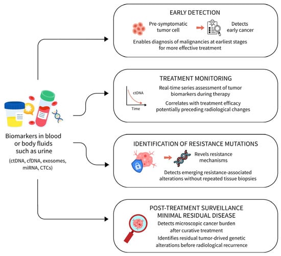
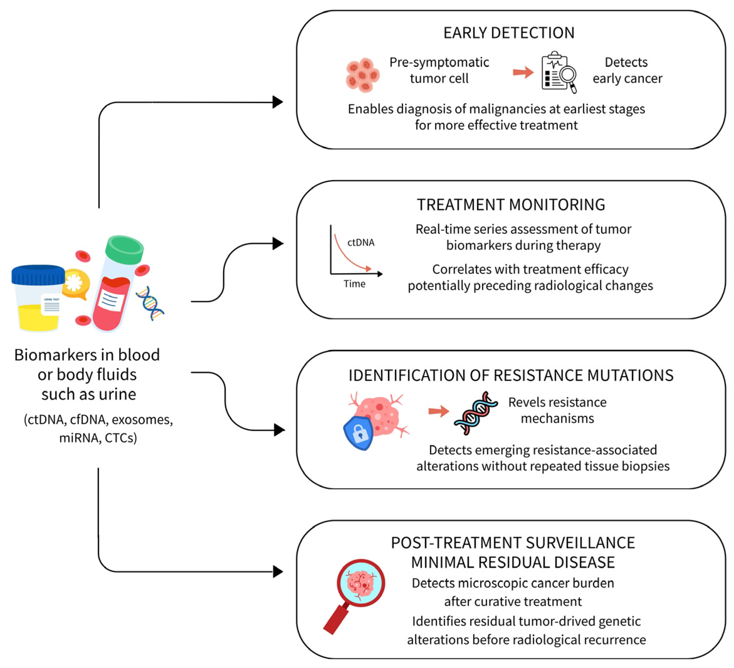

Disclaimer/Publisher’s Note: The statements, opinions and data contained in all publications are solely those of the individual author(s) and contributor(s) and not of MDPI and/or the editor(s). MDPI and/or the editor(s) disclaim responsibility for any injury to people or property resulting from any ideas, methods, instructions or products referred to in the content. |
© 2026 by the authors. Licensee MDPI, Basel, Switzerland. This article is an open access article distributed under the terms and conditions of the Creative Commons Attribution (CC BY) license.