


Highlights
What are the main findings?
- CD57 marks advanced differentiation across T and NK cells, but CD57+ CD4+ and CD57+ CD8+ T cells show distinct developmental trajectories and functional programs rather than a uniform senescent state.
- CMV-driven chronic stimulation is the dominant force shaping CD57+ lymphocyte expansion, linking viral persistence with cytotoxic specialization and immune remodelling.
What are the implications of the main findings?
- CD57 should be interpreted as a context-dependent differentiation and immune-history marker—not a direct therapeutic target—and CD57+ CD4+ and CD57+ CD8+ subsets must be analyzed separately.
- Proper interpretation of CD57+ subsets, with CMV stratification, improves immune profiling in aging, chronic infection, cardiovascular and autoimmune disease, cancer, and vaccine-response studies.
Abstract
CD57-expressing lymphocytes constitute a distinct subset of immune cells with enhanced cytotoxic and pro-inflammatory functions. Initially described in the context of chronic viral infections, most notably cytomegalovirus (CMV), these cells are now recognized as central contributors to immunosenescence and age-related immune dysregulation. Their progressive accumulation reflects prolonged antigenic exposure and sustained immune activation, thereby linking persistent viral infections with long-term disruptions of immune homeostasis. Emerging evidence indicates that CD57 expression denotes a state of terminal differentiation in both T and natural killer (NK) cell compartments, and is associated with cytotoxicity, altered cytokine secretion, and a pro-inflammatory phenotype. This review summarizes the phenotypic and functional characteristics of CD57+ lymphocytes, examines their association with CMV and other chronic viral infections, and explores their potential role in ageing and age-related diseases. Elucidating the biology of CD57+ lymphocytes in the context of chronic viral infections may provide novel insights into immune ageing and help identify potential targets for therapeutic strategies aimed at restoring immune balance in older adults.
1. Introduction
Age-related changes in the immune system reduce its ability to respond to new challenges. This condition, known as immunosenescence, is associated with multiple functional alterations of the immune system, including (i) increased susceptibility to infections, (ii) impaired immunosurveillance with a consequent higher risk of cancer development, (iii) reduced responsiveness to vaccination, (iv) compromised memory T-cell responses, (v) greater propensity to autoimmune disorders, and (vi) decreased telomerase activity in T cells [1,2]
In a longitudinal study of nonagenarian and octogenarian Swedish individuals, it was established that an Immune Risk Profile (IRP) is determined by a variety of immunological parameters associated with lifespan, predicting mortality and morbidity [3,4,5]. The primary feature of this IRP was a reduction in CD8+ naïve T cells and an accumulation of memory CD8+ T cells. Another feature was the increase in the expression of CD57 in these cells, together with the loss of CD28 and CD27 markers. These changes in the T-cell repertoire are associated with thymic involution beginning at puberty, which leads to a progressive decline in the output and replacement of naïve T cells. In addition, chronic immune stimulation by persistent pathogens, particularly cytomegalovirus (CMV) seropositivity, promotes oligoclonal expansions, narrowing the T-cell repertoire and reducing the pool of cells capable of responding to novel antigens [4,6]. CMV is therefore considered one of the main drivers accelerating immunosenescence [7,8].
CMV is a common beta-herpes virus that, after primoinfection, remains latent evading the immune system surveillance [9]. The global average seroprevalence ranges from 45 to 100%, which increases with age and is related to geographic location, ethnicity, socioeconomic status, and education [9,10,11]. In this sense, South America, Asia, and Africa show higher CMV prevalence than the USA and northern European countries such as Germany and England [12]. In CMV-seropositive young individuals, a high frequency of polyfunctional CD57+ T cells has been observed, suggesting that this subpopulation may enhance immune response and infection control [13]. However, in older individuals, these CMV-associated polyfunctional cells may also promote chronic inflammation, thereby impairing optimal immune responses [14].
Both CD57+ CD8+ and CD57+ CD4+ T cells have been linked to several age-associated pathologies. For example, elevated frequencies of CD57+ CD4+ T cells have been detected in patients with acute heart failure and correlate with adverse clinical outcomes [15,16]. Likewise, the expansion of CD57+ CD8+ T cells has been associated with increased arterial stiffness and heightened cardiovascular risk [17]. Beyond the T cell compartment, CD57+ natural killer (NK) cells, often expanded following CMV infection, have also been implicated in chronic inflammation, autoimmune disorders, and cancer [18,19,20]. Thus, the accumulation of these pro-inflammatory lymphocytes during ageing, particularly in CMV-positive individuals, suggests a dual role: while potentially protective during acute infection, they may drive “inflammaging” and contribute to age-related pathologies. Finally, recent evidence reinforces this concept, showing that CMV-seropositive individuals who have experienced SARS-CoV-2 infection display accelerated T-cell immunosenescence, characterized by an expansion of highly differentiated CD57+ T cells and a reduced capacity for immune restoration [11]. Collectively, these findings underscore CD57 expression as a key functional marker linking viral persistence, immune senescence, and the development of chronic age-related diseases.
This review provides an integrated overview of CD57-expressing lymphocytes, emphasizing their central position at the intersection of viral persistence, immune ageing, and chronic inflammation. Understanding how these cells shift from protective immunity to pathogenic contributors offers key insights into the mechanisms linking CMV infection, immunosenescence, and the emergence of age-related inflammatory and degenerative diseases.
Literature Search Strategy
This article is a narrative review and was not designed as a formal systematic review or meta-analysis. The literature discussed was identified primarily through structured searches in PubMed/MEDLINE, complemented by reference-list screening of relevant articles. Search terms included combinations of “CD57”, “HNK-1”, “Leu-7”, “T cells”, “CD4”, “CD8”, “NK cells”, “immunosenescence”, “cytomegalovirus”, “CMV”, “chronic infection”, and “terminal differentiation”. Priority was given to peer-reviewed original studies and high-quality reviews published in English, with particular emphasis on mechanistic, phenotypic, and clinically oriented immunology studies. Study selection was based on relevance to CD57 biology, differentiation status, and clinical associations. No formal systematic quality scoring or meta-analytic methods were applied, consistent with the narrative scope of this review.
2. Biology of CD57 Marker
The CD57 antigen (also known as HNK-1 or Leu-7) is a carbohydrate epitope first identified in 1981 by Abo et al. on human natural killer (NK) cells [21], and later found on subsets of T lymphocytes and neural cells. Structurally, CD57 corresponds to a unique sulphated trisaccharide 3-O-sulfated glucuronic acid (GlcAβ1-3Galβ1-4GlcNAc-R) located at the non-reducing terminus of glycan chains [22]. This glycoepitope is synthesized through the sequential action of β1,3-glucuronyltransferases (B3GAT1/GlcAT-P and B3GAT2/GlcAT-S), which add glucuronic acid to galactose residues, followed by the HNK-1 sulfotransferase (CHST10), which catalyzes sulfation at the C-3 position of GlcA [23]. The sulphate group is critical for the adhesive properties of the molecule [24]. This terminal sulfation step is essential for the biological activity of the HNK-1 epitope, which decorates multiple glycoproteins and glycolipids in both neural and immune tissues [25]. The expression of CD57, therefore, depends not on a single protein, but on the coordinated regulation of glycosyltransferases and sulfotransferases within the Golgi apparatus [26]. A recent study has shown that small extracellular vesicles (sEVs) can mediate the direct transfer of HNK-1 and its major biosynthetic enzyme, GlcAT-P, between cells, highlighting a non-genetic pathway of glycan remodelling through intercellular communication [27].
In the nervous system, the HNK-1 epitope is abundantly expressed on glycoproteins such as myelin-associated glycoprotein (MAG) [28], neural cell adhesion molecule (NCAM) [29], the mammalian glycoproteins PM/Po [30] and tenascin-R [31], where it participates in cell–cell and cell–matrix adhesion, neurite outgrowth, and synaptic plasticity [32,33]. Expression of the HNK-1 epitope in non-neural tissues indicates a broader role in modulating cell–matrix interactions and tissue architecture. In the immune system, CD57 expression constitutes a complex and multifaceted marker that reflects an individual’s cumulative immune history, particularly in the context of persistent infections such as CMV [8]. It is generally accepted that CD57 expression identifies terminally differentiated or senescent lymphocytes, particularly T and NK cells, which exhibit enhanced cytotoxic capacity but limited proliferative potential [18,34,35]. However, in a recent report, [36] analyzed CD57 expression on CD4+ and CD8+ T cells in two cohorts of healthy adults, considering age, CMV serostatus, and cellular differentiation. They combined phenotypic analysis with functional assays for proliferation and senescence-associated β-galactosidase activity. Their findings demonstrated that CD57 expression was strongly associated with CMV seropositivity and T cell differentiation, but not with chronological age. Consequently, they concluded that CD57 should not be considered a strict marker of T cell senescence, but rather of immunological age, reflecting cumulative immune stimulation and viral exposure history.
In summary, CD57 is not only a surface marker but also a conserved molecular motif involved in cell recognition and adhesion across biological systems. It links neural interaction mechanisms with immune maturation and ageing and is strongly associated with chronic viral infection, inflammation, and age-related immune remodelling. Its expression reliably identifies terminally differentiated, highly cytotoxic T-cell subsets, highlighting its value as a functional marker in the study of immune regulation and immunosenescence [8,35,37]. In the following section, we will analyze the relevance of CD57 expression across different lymphocyte populations, exploring its functional implications in immune regulation and ageing.
3. CD57 in αβ T Cells
CD57 expression in T lymphocytes is generally associated with advanced differentiation and is frequently accompanied by the loss of the co-stimulatory receptor CD28. In both CD4+ and CD8+ compartments, CD57+ T cells tend to display a CD28− (CD28null) phenotype, reflecting cumulative antigenic stimulation and progressive differentiation along the effector pathway. The CD28nullCD57+ T-cell profile has been consistently linked to enhanced cytotoxic potential and distinctive transcriptional programmes, although its precise biological meaning remains debated [35,38,39].
CD28 is a costimulatory receptor expressed on naïve CD4+ and CD8+ T cells that binds B7-1 (CD80) and B7-2 (CD86) on antigen-presenting cells. This interaction triggers signalling pathways that promote cytokine production, T-cell proliferation, survival, and differentiation through mediators such as Akt, NF-κB, and mTOR [40]. The decline in CD28 expression on both CD4+ and CD8+ T lymphocytes is a well-recognized feature of immunosenescence [41]. Nonetheless, it is important to distinguish transient downregulation of CD28 from the permanent loss of this molecule, as only the latter reflects long-term changes associated with ageing [42]. Early models of CD8+ T cell differentiation suggested a linear trajectory in which activated CD8+ T cells progressively downregulate CD28 and subsequently acquire CD57 expression. However, accumulating evidence indicates that this process is more complex, with CD28 and CD57 defining multiple CD8+ T-cell subsets characterized by distinct phenotypic, functional and transcriptional profiles. These observations challenge the simplistic view of CD57 as a mere marker of senescence and underscore its role in shaping CD8+ T-cell heterogeneity [43]. Experimental observations further support this non-linear model of CD8+ T cells differentiation, as both CD28nullCD57− and CD28+CD57+ CD8+ T cells can be detected in peripheral blood [44,45,46] questioning the traditional view of mutually exclusive expression of these markers.
The co-stimulatory receptors CD28 and CD27 are widely used to define the differentiation status of CD8+ T cells. In general, early-differentiated CD8+ T cells retain both markers (CD28+CD27+), whereas more differentiated populations progressively lose one or both molecules, resulting in multiple phenotypic combinations that reflect functional and maturational heterogeneity [47,48]. Unlike CD4+ T cells, in which the loss of CD27 and CD28 follows a more sequential pattern, CD8+ T cells display all possible CD27/CD28 expression profiles, indicating that their differentiation does not follow a strictly linear trajectory [47]. Within this framework, CD8+ T cells can be broadly categorized into early (CD28+CD27+), intermediate (CD28−CD27+ or CD28+CD27−), and late (CD28−CD27−) differentiation stages [47,48].
3.1. CD8+ T Cells
CD57 expression is commonly associated with advanced differentiation and replicative senescence in CD8+ T cells. Although CD57+ CD8+ T cells are predominantly enriched in the CD28−CD27− phenotype, this subset is phenotypically heterogeneous, and a proportion of CD57+ cells retain CD27 and/or CD28 expression [45]. Consistent with their late differentiation status, CD28−CD27−CD57+ CD8+ T cells exhibit markedly reduced telomerase activity and features of replicative senescence, supporting their classification as terminally differentiated effector populations [44,49]. In this way, it has been reported that downregulation of costimulatory molecules CD28 and CD27 in terminally differentiated effector memory T cells (TEMRA, CCR7-CD45RA+) can be mediated via epigenetic regulation [50].
CD57+ CD8+ T cells frequently express CD94/NKG2A, inhibitory receptors typically associated with NK cells (iNKRs), which may modulate their activation threshold and survival [51,52].
Within this population, CD27−CD57+ CD8+ T cells have been proposed as the most senescent subset, characterized by resistance to apoptosis [43,44,45,46,47,48,49,50,51,52,53,54,55,56]. This impaired apoptotic response could explain their progressive accumulation in older individuals, contributing to the phenomenon of immunosenescence. Nevertheless, it has been shown that CD57+ CD8+ T cells can still proliferate in response to persistent antigenic stimulation and, in some cases, reactivate telomerase to overcome cell-cycle arrest, indicating that not all are terminally senescent [54]. This dynamic interplay between CD28 and CD57 underscores the plasticity of CD8+ T cell responses and their role in immune surveillance.
Heterogeneous functional capacities have been reported within the CD57+ CD8+ T cell population. Upon activation, these cells exhibit enhanced cytotoxic activity [55], characterized by the expression of perforin, granzyme B, and granulysin [56].
In addition, CD57+ CD8+ T cells produce high levels of pro-inflammatory cytokines such as IFN-γ and TNF-α [13,57], and, interestingly, IL-5 [54]. These cytokines may also contribute to the maintenance and expansion of highly differentiated CD57+ subsets, as inflammatory signals, particularly IFN-γ induce IL-15 expression and trans-presentation by myeloid and tissue cells, and IL-15 is a well-established driver of survival and proliferation in terminally differentiated and CD28null/CD57+ CD8+ T cells [57,58,59]. Moreover, TNF-α has been shown to promote CD28 downregulation, further biassing the pool toward highly differentiated CD28null phenotypes that frequently co-express CD57 [60]. Consequently, their cytotoxic functions enable the recognition and elimination of aberrant cells, while they may also contribute to the exacerbation of inflammatory processes.
Transient FOXP3 expression during CD8+ T-cell activation may function to restrain excessive immune activation and tissue damage at inflammatory sites [61,62]. Within the CD8+ compartment, regulatory T-cell populations (CD8+ Tregs) are phenotypically heterogeneous and can be partly distinguished by CD57 expression, which reflects differentiation status and functional capacity. CD8+ regulatory activity has been most consistently described in CD28null subsets that exert MHC class I–restricted suppression of CD4+ T cells and antigen-presenting cells; these populations are commonly CD57− or phenotypically mixed and retain measurable proliferative potential [61,63]. By contrast, CD57+ CD8+ T cells are predominantly highly differentiated effectors with strong cytotoxic and pro-inflammatory profiles and limited proliferative capacity. Although they can produce regulatory cytokines such as IL-10 under specific conditions, including chronic viral infection, they do not represent a stable FOXP3+ regulatory lineage [43]. Moreover, FOXP3 expression in CD8+ T cells can be transiently induced by persistent antigenic stimulation without conferring durable regulatory identity and is not consistently linked to CD57 expression [63].
Further evidence of the functional heterogeneity of CD8+ T-cell subsets defined by CD28 and CD57 expression comes from transcriptomic and phenotypic analyses in young and elderly individuals. Onyema et al. showed that ageing is associated not only with an expansion of CD28nullCD57+ CD8+ T cells but also with an increased frequency of CD28+CD57+ CD8+ T cells. These subsets displayed distinct patterns of senescence- and apoptosis-related markers, with the highest expression of p16 and p21, and elevated levels of Bcl-2 and CD95, in the CD28+CD57+ subset. Moreover, differentiation and homing markers were unevenly distributed among subsets: CD62L and CCR7 were predominantly expressed in CD28+CD57− CD8+ T cells, whereas CD28nullCD57+ and CD28+CD57+ cells exhibited low and intermediate levels, respectively. In addition, PD-1 and CD45RO were most highly expressed in CD28+CD57+ cells, while CXCR2 expression was preferentially associated with CD28nullCD57+ subset. Altogether, these findings indicate that CD8+ T-cell subsets defined by CD28 and CD57 expression are characterized by distinct molecular and functional profiles, supporting the concept that CD57+ CD8+ T cells constitute a heterogeneous population rather than a uniform senescent compartment [45,46]. CD57+ CD8+ subsets may also display variable expression of checkpoint and inhibitory receptors, including PD-1, whose levels are determined by antigenic and inflammatory conditions rather than representing an intrinsic feature uniformly associated with CD57 expression [64,65,66].
In human disease contexts, such as chronic infections and cancer, CD57+ CD8+ T cells exhibit hallmarks of advanced differentiation, including diminished proliferative capacity and altered expression of survival factors such as Bcl-2, consistent with extensive replicative history and exhaustion-like phenotypes [50]. In the context of chronic inflammation, FOXP3 expression can be transiently induced in CD8+ T cells [63,67] and in CD57+ CD8+ T cells [68]. However, these populations are functionally distinct. Specifically, in a study of patients with stage IV gastric carcinoma undergoing treatment with activated autologous lymphocytes, Akagi et al. [68] demonstrated that FOXP3 expression in CD57+ CD8+ T cells was transient and occurred in the absence of associated regulatory function during the natural course of CD8+ T-cell differentiation. In contrast, CD57−FOXP3+ CD8+ T cells were appropriately classified as CD8+ regulatory T cells. These findings highlight that CD57 expression defines functionally heterogeneous populations within the CD8+ T-cell compartment, encompassing both pro-inflammatory and regulatory subsets.
Within the conventional CD8+ T-cell compartment, NKT-like T cells, expressing the CD56 marker, represent a differentiated subset. Phenotypically, these cells typically display an effector-memory/terminal-effector profile, enriched cytotoxic machinery, and rapid effector responsiveness, and they are known to expand with ageing and chronic viral infection, particularly CMV [69,70,71]. CD57+ NKT-like CD8+ T cells are preferentially expanded in CMV-seropositive individuals and show features of late differentiation, commonly associated with reduced CD28 expression and increased cytotoxic potential. Functionally, these cells exhibit greater polyfunctionality than their CD57− counterparts, with higher combined degranulation and cytokine production, particularly IFN-γ and TNF-α, following polyclonal stimulation. Importantly, CD57+ NKT-like cells display higher functional activity than their CD56−CD57− counterparts, supporting the concept that, in this lineage, CD57 marks terminally differentiated yet functionally potent effectors rather than merely senescent cells [71].
3.2. CD4+ T Cells
In the CD4+ T-cell compartment, CD57 expression is likewise associated with distinct phenotypic and functional features, which differ from those observed in CD8+ T cells. In contrast, CD4+ T lymphocytes tend to lose CD27 and subsequently CD28 expression during differentiation [72,73]. Although, CD27lowCD57+ CD4+ T cells have also been identified [48]. CD57+ CD4+ T cells are functionally distinct from conventional helper T cells, as they display limited cooperation with B cells [74]. Nevertheless, upon stimulation, a subset of these CD57+ CD4+ T cells can upregulate CD40L (CD154), suggesting that they retain partial effector capacity despite their apparent senescent-like phenotype [55]. Activated CD57+ CD4+ T cells are cytotoxic and produce multiple cytokines, with higher functional responses than their CD57− counterparts, supporting the concept that terminal differentiation in this subset is associated with a shift from proliferative capacity toward increased cytotoxic and secretory functions [35,55,75]. Specifically, CD57+ CD4+ T cells can produce a broad range of mediators, including IL-2 (low levels), IFN-γ, TNF-α, perforin, granzyme B and express multiple surface molecules associated with activation, migration, and innate-like responses. These include co-stimulatory molecules (OX40, 4-1 BB), chemokine receptors (CX3CR1, CCR5), Toll-like receptors (TLR2, TLR4), adhesion proteins (VLA-4, ICAM-1), and natural killer cell receptors such as NKG2C, NKG2D, CD11b and CD161 [55]. Together, these findings support the concept that CD57+ CD4+ T cells undergo a functional reprogramming characterized by reduced proliferative capacity but enhanced cytotoxic and secretory functions, positioning this subset as a key contributor to antiviral immunity and chronic inflammatory responses [76].
Thus, CD57 emerges as a robust marker of cytotoxic differentiation in T cells, more closely associated with cytotoxic effector programmes than alternative markers such as CD300a [35]. Consistently, CD57 expression is linked to functional specialization rather than uniform functional decline, as CD57+ T cells in both CD4+ and CD8+ lineages exhibit greater polyfunctional activity than CD57− cells [76].
4. Are CD57+ T Cells Truly Senescent?
CD57 expression has long been employed as a phenotypic marker of senescence in human lymphocytes, particularly within the CD8+ T-cell compartment. Early seminal studies demonstrated that CD57+ CD8+ T cells exhibit hallmarks consistent with replicative senescence, including shortened telomeres, low telomerase activity, and severely impaired proliferative responses following T-cell receptor stimulation, while retaining the ability to produce effector cytokines [44]. Together with the frequent loss of costimulatory molecules such as CD27 and CD28, these features positioned CD57 as a core component of the senescent T-cell phenotype associated with ageing and chronic infection [77]. Thus, these cells have historically been interpreted as a characteristic feature of immunosenescence; however, these phenotypic changes more accurately reflect chronic antigen-driven differentiation [13]. Indeed, within the CD8+ TEMRA subset, CD57 expression distinguishes a terminally differentiated CD57+ population from a previously functionally uncharacterized CD57− “younger” TEMRA population that exhibits high proliferative capacity and greater differentiation plasticity [78].
However, when CD57+ T cells are evaluated against the strict biological definition of cellular senescence, they do not consistently fulfil the required criteria. Cellular senescence is characterized by a combination of parameters, and no single marker is sufficient to establish it; rather, a cell can be reliably classified as senescent only when several of these features are detected simultaneously, including SA-β-galactosidase activity, stable proliferative arrest mediated by the activation of the tumour suppressors TP53 and CDKN2A/p16, and their downstream effectors CDKN1A/p21 and retinoblastoma-1 (RB1) family proteins, telomere attrition, accumulation of DNA damage, and signs of mitochondrial dysfunction [79,80]. Recent functional analyses further reinforce this reinterpretation by showing that CD27/28−CD57+ T cells do not meet core biological criteria of cellular senescence. These cells are not enriched for canonical senescence markers such as p16INK4A or senescence-associated β-galactosidase in either peripheral blood or tumour samples, and they retain measurable proliferative capacity in vitro, including the ability to divide in response to IL-2 or IL-15 stimulation. In parallel, they display preserved or enhanced effector activity, with increased expression of perforin, granzymes and CD107a, together with robust IFN-γ production. These data support the view that CD27/28−CD57+ T cells are better defined as highly differentiated effector-memory cells than as truly senescent lymphocytes [81]. Furthermore, in vivo labelling and longitudinal studies have demonstrated that CD57+ T cells can persist long-term and undergo intracompartmental proliferation, indicating that at least a fraction of these cells retains replicative potential in physiological settings [82]. Additionally, CD57+ T cells can display potent effector functions, including high cytotoxic capacity and robust production of IFN-γ and TNF-α, features that are not typical of classically senescent cells, which are generally characterized by functional decline [83,84,85,86].
Collectively, current evidence indicates that CD57+ T cells are not universally “truly senescent” in the strict biological sense. Rather, they represent a heterogeneous population strongly enriched for advanced differentiation, altered proliferative capacity, and features of immunological ageing, particularly within CD8+ compartments and CMV-experienced individuals (Table 1). While CD57 identifies lymphocytes with a high likelihood of replication incompetence and effector skewing, its expression alone is insufficient to define irreversible cellular senescence. Accurate interpretation, therefore, requires integrating additional phenotypic and molecular markers, including CD28, KLRG-1, PD-1, p16/p21 expression, and SA-β-gal activity, as well as consideration of clinical context and antigenic history [36]. This distinction is clinically relevant, as CD57+ cells may still actively contribute to immune responses and disease processes despite exhibiting senescence-associated traits. Different key markers of CD57+ T-Cell Subsets are described in Table 1. For conceptual clarity, the distinctions between cellular senescence, T-cell exhaustion, terminal differentiation, and immunological age are summarized in Table 2.
Table 1.
Characteristics of CD57+ T-Cell Subsets.
This table summarizes phenotypic markers, senescence-associated molecules, homing receptors, NK-like receptors, immune checkpoints, cytokines, and cytotoxic mediators reported in highly differentiated CD28null/CD57+ CD4+ and CD28null/CD57+ CD8+ T-cell subsets. Data are compiled from multiple published studies in chronic viral infection, immunosenescence, and inflammatory disease contexts. Primary supporting references: [45,48,55,75]. Arrows indicate: ↑, high expression; ↓ low/null expression.
Table 2.
Conceptual distinction between senescence, exhaustion, terminal differentiation, and immunological age in lymphocytes.
5. CD57 in CD4+CD8+ T Cells
Double-positive (DP) T cells account for 1–2% of circulating human T cells and are a heterogeneous population. They may display effector memory-like features, including the CD57 expression [87], suggesting antigen-experienced differentiation [88]. As demonstrated by Clénet et al., in healthy individual DP T cells express CD57 and show enhanced effector functions (INF-γ, CD107a, perforin production) [87]. These cells are distinguished by the lack of CCR7, CD27 and CD2 [89].
Consistent with the findings of Nascimbeni et al., DP T cells increase during viral infections (such as CMV, EBV and HIV). In these contexts, they demonstrate a more advanced differentiation profile compared to single-positive cells. Notably, the frequency of CD57 expression is higher in the DP subset than in CD4 single positive subset (28% vs. 7%), accompanied by the downregulation of CD28 and CD27 [90]. These data support the hypothesis that persistent antigenic stimulation drives the expansion toward a terminally differentiated, highly cytotoxic state.
6. CD57 in γδ T Cells
γδ T cells do not fully conform to “classical” immunosenescence trajectories as they retain robust effector competence with ageing [91,92]. Within this lineage, Vδ1+ cells exhibit a peripheral phenotype that more closely mirrors that of αβ CD8+ T cells during the combined influence of CMV and chronological ageing. In this context, CD57 expression aligns more with advanced differentiation and reduced proliferative potential, while cytotoxic capacity can remain preserved in defined subsets [91,93]. These observations indicate that some senescence-associated markers retain interpretations in Vδ1+ cells that resemble those in αβ T cells, albeit with functional nuances [94]. In contrast, Vδ2+ cells (typically Vγ9/Vδ2) deviate from these canonical patterns, as they are either less prone to cellular ageing or governed by mechanisms distinct from those of αβ T cells [91]. Subsequent studies converge on the view that Vδ2+ cells do not map neatly onto αβ-style differentiation markers (e.g., CD27, CD28) in terms of cytokine production and functional readouts [95,96]. Moreover, ligating KLRG1 on Vδ2+ cells does not reproduce the inhibitory effects observed in CD4+, CD8+, or NK cells, underscoring lineage-specific meanings for so-called senescence markers [97]. Taken together, these data support a model in which Vδ1+ cells exhibit age- and CMV-linked terminal differentiation reminiscent of αβ CD8+ T cells, whereas Vδ2+ cells display relative resistance to classical immunosenescence and maintain strong effector functionality despite the expression of markers like CD57 [92,98].
7. CD57 in NK Cells
The CD57 marker was originally identified on NK cells and was associated with natural killer activity. CD57 is variably expressed on different populations of NK cells. Both CD16+CD56dim cytotoxic NK cells and CD16+CD56bright inflammatory NK cells show a high expression of CD57. In contrast, NK cells with a regulatory profile CD16loCD56bright do not express CD57 [18]. Moreover, CD56− NK cells expressing low levels of CD57, but not displaying canonical features of cellular senescence, have been reported in chronic viral infections, including HIV, hepatitis C virus, EBV and CMV [99].
CD57 is widely regarded as a marker of terminal differentiation in NK cells, with expression increasing along the maturation trajectory from CD56bright to CD56dim subsets and being associated with highly cytotoxic, functionally mature NK cells, whereas CD56bright NK cells typically show little or no CD57 expression [18]. The transition of NK cells from the CD56bright to the CD56dimCD16+ phenotype involves a marked decrease in NKp46, NKG2D, NKp30, and NKG2A expression, concomitant with the acquisition of CD16, and the expression of LIR-1 and KIR [100]. Specifically, the expression of CD57 in mature NK cells is closely related to the increased expression of markers NKG2C and CD8 [101]. Furthermore, CD57+NKG2Chigh NK cells have been proposed as a population of human CMV-specific “memory-like” NK cells [102,103,104], while polyfunctional CD8+ NK cells have been associated with slower disease progression in chronic HIV-1 infection [101]. Similarly to T cells, mature NK cells express CXCR1 and CX3CR1, which are chemokine receptors involved in the homing and migration of NK cells toward peripheral inflamed tissues, including the vascular endothelium. These receptors facilitate lymphocytic recruitment to sites of inflammation by responding to chemokine gradients, thereby enhancing their cytotoxic and immunoregulatory functions within inflamed or infected microenvironments. The expression of these homing receptors, together with the cell–cell adhesion function attributed to the CD57 marker, suggests that CD57+ NK cells, and by extension CD57+ T cells, play a local role in inflammatory processes. This combination of migratory and adhesive properties could facilitate the accumulation and effector activity of these cells within inflamed tissues [105].
Although CD57 expression in NK cells has traditionally been interpreted as a marker of terminal maturation rather than definitive cellular senescence, more recent frameworks propose that CD57 can contribute to the identification of a senescence-like NK phenotype when combined with additional features such as reduced proliferative capacity, impaired cytotoxicity, and senescence-associated signalling pathways. Importantly, CD57 alone shows limited specificity and is insufficient to define NK cell senescence in the absence of complementary molecular and functional criteria [106]. The gain of CD57 by CD56dim NK cells has been associated with a reduction in their proliferative capacity in response to pro-inflammatory cytokines, which could be associated with less expression of IL-2Rβ, IL-12Rβ and IL-18Rα receptors [18,37]. However, CD57+CD56dimCD16+ NK cells exhibit the same proliferative capacity as their CD57-counterparts, showing similar levels of the Ki-67 marker [37]. Additionally, CD57+CD56dimCD16+ NK cells have a marked lytic capacity related to their antibody-dependent cell-mediated cytotoxicity (ADCC) function with high levels of granzyme B, perforin, and degranulation markers such as CD107a [34].
Several studies have highlighted the role of CD57 expression in shaping the functional profile and adaptive-like features of NK cells under different physiological and pathological contexts. CD57 expression, often in combination with NKG2C, is particularly enriched in NK cells from CMV-seropositive individuals [107]. In healthy young CMV+ donors, an expansion of CD57+NKG2C+ NK cells has been described, which is associated with a more mature phenotype but also with a reduced ability to produce interferon-γ in response to heterologous antigens such as Bordetella pertussis and H1N1 influenza virus [108].
Similarly, in CMV+ kidney transplant recipients, dynamic changes in memory-like NK cell subsets have been observed. Pre-existing memory-like NK cells (NKG2C+CD57+FcεRγ−) tend to decrease over time, while pre-memory-like populations (NKG2C+CD57+FcεRγlow/dim) expand during periods of viremia and exhibit a stronger cytotoxic profile compared to non-viremic patients [109]. These findings suggest that viral reactivation and chronic antigenic stimulation drive phenotypic adaptations within the CD57+ NK compartment, promoting the development of memory-like subsets with enhanced effector capacity.
Recent data from COVID-19 convalescent individuals further support the link between CD57 expression and NK cell activation status. Higher frequencies of CD57+ NK cells have been reported in patients who experienced severe disease compared with mild or asymptomatic cases, indicating an association between CD57 expression and sustained NK activation [110]. Moreover, IFN-γ production in response to SARS-CoV-2 peptides has been correlated with the presence of CD57+NKG2C+ NK cells, although some individuals display robust responses even in their absence, suggesting heterogeneity in NK cell memory responses to viral infection. Given that CMV seropositivity is the major driver of CD57+NKG2C+ NK-cell expansion (37), the absence of CMV assessment in this study represents an important limitation. However, in other studies, increased proportions of NKG2ChiCD57+ NK cells have been observed predominantly in CMV-seropositive subjects, but not in CMV-seronegative ones, across several viral settings, including acute chikungunya [111], hantavirus infections [112], as well as chronic infections such as HIV-1 [113] and hepatitis B and C [114].
Altogether, these findings indicate that prior CMV exposure is a key requirement for the emergence of CD57+ NK-cell population, and CD57 definitively identifies a virus-driven, highly cytotoxic, terminally differentiated NK subset resulting from adaptive immune remodelling [115,116].
8. CD57+ Lymphocyte Expansion and Chronic Stimulation
CD57 expression across lymphocyte lineages is broadly associated with prolonged or repeated antigenic and cytokine-driven stimulation. In both T and NK compartments, the accumulation of CD57+ subsets is a recurrent feature of chronic immune activation and sustained environmental pressure, reflecting progressive differentiation and long-term immune adaptation. Rather than representing a lineage-specific phenomenon, CD57 upregulation emerges as a convergent immunophenotypic outcome of persistent stimulation, linking chronic exposure signals with stable shifts in lymphocyte subset composition and functional state.
It has been established that the central driver of CD57+ T-cell accumulation is chronic antigenic stimulation, particularly that imposed by persistent viral infections. Among these, CMV exerts a dominant and sustained impact on the T-cell compartment, promoting the expansion of highly differentiated CD8+ and CD4+ T-cell subsets characterized by CD57 expression, oligoclonal repertoire restriction, and reduced proliferative capacity. CMV-driven immune imprinting has been consistently associated with accelerated immune ageing, altered T-cell diversity, and systemic immune activation, positioning CMV as a key architect of the CD57+ T-cell landscape across the lifespan [8,13,35,55,75].
Importantly, the contribution of CMV to CD57+ T-cell expansion is frequently overlooked in studies examining CD57 expression in chronic diseases. This omission complicates the interpretation of “CD57-high” phenotypes, as CMV serostatus can profoundly shape the size, differentiation state, and functional profile of CD57+ T-cell populations independently of the primary disease under investigation. Recent high-dimensional immunophenotyping studies have reinforced the notion that CMV infection is a major confounder in analyses of late-differentiated T-cell subsets and must be systematically accounted for when attributing CD57+ T-cell expansion to disease-specific mechanisms [117,118].
Against this background, CD57+ T cells should be viewed not merely as markers of terminal differentiation, but as integrative indicators of cumulative immune history shaped by chronic antigenic exposure, viral coinfections, inflammatory signals, and host genetic factors. In the following sections, we review representative groups of representative pathological conditions, including major chronic viral infections, cardiovascular diseases, autoimmune diseases and cancer [119,120,121,122,123,124,125,126,127,128,129,130,131,132,133,134,135,136,137,138,139,140,141,142,143,144,145,146,147,148,149,150,151,152,153,154,155,156,157,158,159,160,161,162,163,164,165,166,167,168,169,170,171,172,173,174,175,176,177,178,179,180,181,182,183,184,185,186,187,188,189,190,191,192,193,194,195,196,197,198,199,200,201,202,203,204,205,206,207,208,209,210,211], in which CD57+ T cells have been implicated. We highlight their contribution to immune dysregulation and disease development, and emphasizing the need to interpret CD57 expression within the broader context of chronic stimulation and CMV-driven immune remodelling.
8.1. Major Chronic Viral Infections
Chronic CMV infection is widely supported as a major—and in many cohorts quantitatively predominant—driver of CD57 acquisition and late T-cell differentiation. However, this process is multifactorial and also influenced by chronological age, inflammatory burden, metabolic status, and co-infections. Comparative cohort studies indicate that CMV serostatus often explains a larger proportion of the variance in CD28null and CD57+ T-cell expansion than age alone, particularly within CD4+ subsets [140], whereas CD8+ compartments show a more mixed contribution of CMV- and age-related effects. Expansions of CD57+ lymphocytes—predominantly within the CD8+ compartment—have also been documented in association with other persistent viral infections, including human immunodeficiency virus (HIV), Epstein–Barr virus (EBV), hepatitis C virus (HCV), varicella–zoster virus (VZV), and herpes simplex virus (HSV) (Table 3, Figure 1) [119,120,121,122,123,124,125,126,127,128,129,130,131,132].
Table 3.
Summary of CD57 findings in Major Chronic Viral Infections and CD57 Expression and CMV evaluation.
Figure 1.
Chronic viral infections shape CD57 expression as a marker of cytotoxic differentiation along gradients of antigenic burden and immune compartment specialization. (A) Viruses are positioned along a continuum of viral load and systemic antigen exposure. Infections with limited or predominantly tissue-restricted persistence (e.g., HPV and HSV) show low or context-dependent CD57 imprinting, whereas viruses characterized by chronic stimulation or frequent reactivation (HBV/HCV, VZV, EBV, HIV, and especially CMV) are associated with progressive accumulation of CD57+ and CD57++ cytotoxic lymphocytes. This gradient reflects a shift from immune resilience toward terminal cytotoxic differentiation across T- and NK-cell compartments. (B) CD57 imprinting is lineage-dependent. CD8+ T cells show the strongest enrichment in CMV (CD57+++), with intermediate accumulation in EBV, VZV, and HCV, and more heterogeneous patterns in HIV and HSV. CD57+ cytotoxic CD4+ T-cell expansion is strongly CMV-associated; reports in other chronic infections lack CMV stratification and remain inconclusive. γδ T cells can acquire CD57 under chronic viral stimulation—most clearly described in CMV—where it marks clonally expanded, cytotoxic, innate-like populations rather than uniform exhaustion. In NK cells, CMV characteristically drives the emergence of adaptive CD57++ NK subsets, whereas other viral infections produce milder, heterogeneous, or tissue-restricted CD57 remodelling. Note: These CD57 scores are literature-based and come from heterogeneous cohorts; many studies did not control for CMV serostatus. Because CMV strongly drives CD57+ differentiation, part of the signal in non-CMV infections may be CMV-related, so interpretation should be cautious. Scoring shown in the table: +++ high, ++ moderate, + low, +/− variable, ? insufficient evidence.
Furthermore, recent cohort-level analyses indicate substantial interindividual heterogeneity in the degree of CMV-associated memory skewing, with marked global compartment distortion occurring only in a subset of CMV-seropositive individuals. This variability suggests that host factors and additional inflammatory or environmental influences modulate the magnitude of CMV-driven immune remodelling [133].
In older adults, these CD28null/CD57+ CD8+ T-cell populations are often enriched for CMV- and EBV-specific responses, indicating that lifelong persistent viral exposure—more than chronological ageing alone—drives oligoclonal accumulation of highly differentiated CD8+ T cells [45,48]. Functionally, CD28nullCD57+ CD8+ T cells show high granzyme and perforin expression supporting antiviral cytotoxicity and immune surveillance, as it has been shown in the context of CMV infection and HIV–CMV co-infection, where CD28nullCD57+CX3CR1+ memory subsets are linked to stable viral control [134,135,136]. However, persistent oligoclonal expansion of these late-differentiated cells may restrict immune repertoire diversity, reduce space for naïve and central memory T cells, and impair responses to new antigens and vaccination. In this sense, CD28null CD8+ subsets, especially CD57+ or senescent/regulatory populations, have also been associated with suppressive activity and immunosenescent profiles linked to poorer outcomes in ageing and malignancy [49,137,138]. Overall, chronic viral infections act as major contributors to CD8+ T-cell remodelling, with ageing often amplifying infection-driven effects [13,55,76,77].
In contrast, CD57-associated differentiation is less consistently observed in the CD4+ compartment. Although virus-specific CD4+ T cells are frequently detectable across chronic viral infections, they are not uniformly linked to CD57 expression nor to the same degree of terminal differentiation seen in CD8+ cells. Current evidence indicates that the emergence and accumulation of CD28nullCD57+ CD4+ T cells is predominantly driven by CMV infection. Quantitative cohort analyses show that age-related increases in these subsets are largely restricted to CMV-seropositive individuals, whereas little or no expansion is observed with ageing alone in CMV-seronegative subjects [11,139,140]. When present, CD57+ CD4+ T cells represent late-differentiated effector populations with reduced proliferative capacity but preserved cytokine and cytotoxic potential.
Similarly, in NK cells, CD57 expression is a hallmark of terminal differentiation which is also strongly influenced by chronic viral exposure. Once more, CMV is the best-established and often dominant determinant of CD57+ NK-cell expansion and adaptive-like NK phenotypes. Although infections such as HIV, EBV, and chronic hepatitis are associated with increased frequencies of differentiated NK cells, studies incorporating CMV serology and CMV-associated phenotypic signatures show that these subsets largely correspond to canonical CMV-driven adaptive NK populations [141,142,143]. In co-infection settings, non-CMV viruses tend to modify activation state, tissue distribution, or clinical associations of CD57+ NK cells rather than generate these populations de novo [144]. Without CMV stratification, attribution of CD57+ NK expansion to other viruses remains unreliable.
8.2. Cardiovascular Diseases
Several studies have observed an expansion of CD57+ T lymphocytes in patients with cardiovascular disease. Coronary artery disease (CAD) was associated with an increase in CD57+ CD8+ T and CD28nullCD8+ T cells in 29 of 43 (67%) CMV-seropositive patients younger than 60 years. Multiple regression analysis showed that CAD per se is a determinant of alterations in T lymphocyte subpopulations. Authors suggest that the expansion of these subpopulations may be stimulated by persistent antigens associated with CMV infection found in cardiovascular tissue, thus exacerbating the inflammatory response [145].
A high frequency of CD57+ CD8+ T cells has also been found in patients with myocardial infarction between 60 and 80 years of age [15,146,147]. These subpopulations were associated with cardiovascular mortality 6 months after the cardiovascular event. Furthermore, CD57+ CD8+ T cells showed a higher cytotoxic profile, releasing perforin and granzymes A and B, compared to their CD57−CD8+ counterparts [145]. The CD28nullCD57+ CD4+ T subpopulations found in cardiovascular diseases are more homogeneous, showing mainly pro-inflammatory characteristics. In patients with unstable angina pectoris, an increase in CD28nullINFγ+ CD4+ T lymphocytes was observed compared to patients with stable angina pectoris and healthy patients [148]. Furthermore, HIV+ patients and CMV coinfections showed a high expansion of memory CD57+ CD4+ T cells in the atherosclerosis plaque. These cells exhibited overexpression of the vascular endothelial receptor CXBCR1 and CD2. Furthermore, the presence of IL-15 stimulated the migration of these cells, associated with a release of TNFα, granzyme B and perforin. Consequently, it can be thought that CMV infection enhances the expansion of CD57+ CD4+ T cells, this being a key factor for the development of vascular damage in immunocompromised individuals [143]. The expansion of CD28nullCD4+ T cells has been observed in patients with a risk factor for developing atherosclerosis and patients with acute coronary syndrome aged between 40 and 80 years. Upon anti-CD3 stimulation these cells produced high levels of perforin, granzyme A and B, and TNFα [149]. Moreover, it has been demonstrated that CD28nullCD4+ T cells are associated with post-operative atrial fibrillation and higher levels of CRP [150].
In CAD, the occurrence of increased frequencies of CD28nullCD57+ CD8+ T cells has also been shown. Importantly, both CMV seropositivity and coronary artery disease were independently linked to this expansion, supporting the idea that chronic viral antigen exposure and CAD-associated inflammation act together to promote immunosenescence [145]. A recent study from our group further confirmed the involvement of CD57+ cells in isolated CAD (iCAD) and CAD associated with aortic stenosis (ASCAD), highlighting their potential utility as biomarkers for identifying individuals at increased risk and for monitoring disease progression [151]. Distinct immunological profiles were identified between iCAD and ASCAD. iCAD was characterized by enhanced immune activation, evidenced by increased inflammatory CD14+CD16+ monocytes, elevated Treg frequencies, and greater differentiation of CD4+ T cells toward effector memory (TEM) and terminally differentiated (TEMRA) phenotypes. Conversely, ASCAD was associated with marked immunosenescence, reflected by increased neutrophil counts, lymphopenia, and heightened cytotoxic activity of NK and T cells. The predictive model accurately discriminated between iCAD and ASCAD, highlighting CD4+ T cell memory subsets and CD57 expression as key distinguishing markers. Overall, the findings indicate that iCAD is primarily driven by immune activation, whereas ASCAD is dominated by immunosenescence and cytotoxicity.
In chronic heart failure (CHF), the increase in the frequency of CD28null T lymphocytes was studied in young and elderly individuals. The frequency of CD45+/− CD28null CD4+ T cells in both age groups was related to CMV seropositivity and to pathology. In contrast, the frequency of CD45+/−CD28null CD8+ T cells was only associated with CMV seropositivity. Furthermore, the CD4+ subpopulation showed higher expression of the CD69 activation receptor than the CD8+ subset. These data suggested that the differentiation of these populations contributed to the exacerbation of the disease [152].
The presence of these expansions in the context of cardiovascular disease is a relevant indicator for predicting and estimating vascular damage in individuals with risk factors. However, few studies account for CMV seropositivity in the target population, even though it may alter T lymphocyte subpopulations regardless of age. Therefore, further research is needed to clarify the causal relationship between these cardiovascular events and underlying immunopathological mechanisms. Such evidence would support the development of effective combination therapies aimed at preventing or slowing cardiovascular damage.
8.3. Autoimmune Diseases
Several studies have evaluated the expression of CD57 on T lymphocytes across a wide range of autoimmune diseases, supporting the concept that CD57+ T cells represent a recurrent immunological feature in chronic autoimmune inflammation. Table 4 summarizes some autoimmune diseases with reported increased frequencies of CD57+ T cells, predominantly within the CD8+ compartment but also affecting CD4+ subsets. In many of these conditions, CD57+ T cells display phenotypic and functional characteristics consistent with terminal differentiation, clonal expansion, and enhanced cytotoxic or pro-inflammatory capacity, suggesting a potential pathogenic role in tissue damage and disease progression.
Table 4.
Main findings on CD57+ T cells in representative autoimmune diseases and CMV evaluation.
In rheumatoid arthritis, the disease with the most extensive evidence, CD57+ T cells have been described both in peripheral blood and inflamed tissues, particularly synovial fluid and bone marrow [153,154,155,156,157]. These cells often exhibit restricted TCR repertoires, high interferon-γ production, and associations with disease activity or duration, indicating antigen-driven expansion and functional relevance in local inflammation. Similar observations of tissue-enriched CD57+ cytotoxic T cells have been reported in multiple sclerosis meningeal infiltrates [158], systemic sclerosis–associated lung fibrosis [159], Crohn’s disease intestinal lesions [153,154,155,156,157,158,159,160], and alopecia areata [161,162]; among others, reinforcing the notion that CD57 expression marks chronically stimulated effector T cells involved in organ-specific autoimmunity
Despite the strong biological overlap between CD57 expression, T-cell senescence, and chronic viral exposure, very few studies summarized in Table 4 explicitly evaluated CMV serostatus. Only early work in rheumatoid arthritis [153], and selected analyses in type 1 diabetes [163] and celiac disease [164] incorporated CMV-related assessments, while the vast majority of studies interpreted CD57+ T-cell expansions solely within an autoimmune framework. This represents a critical limitation, as CMV infection is a major driver of CD57+ CD8+ T-cell accumulation and clonal expansion in the general population. Failure to account for CMV seropositivity may therefore confound the attribution of CD57+ T-cell expansions to autoimmune mechanisms alone. Consequently, incorporating systematic CMV evaluation in future studies will be essential to disentangle virus-driven immune remodelling from disease-specific autoimmune processes and to better define the true pathogenic significance of CD57+ T cells in autoimmunity.
8.4. Cancer
The type of cancer and its stage are characteristics that can affect lymphocyte expansion. T lymphocyte subpopulations could be immunomodulatory or cytotoxic, favouring or combating tumour cell growth. In individuals suffering from gastric cancer, CD57+ CD8+ T lymphocytes have been found to expand only in patients with advanced-stage disease. In addition, a negative correlation was established between the production of IFNγ and that of CD57+ T cells. Specifically, it was observed that in patients with advanced-stage disease, the frequency of CD57+perforin+ T cells was lower. This suggests that this cell population could be ineffective in antitumor defence, favouring a tolerogenic state that allows uncontrolled growth of tumour cells [165].
In another type of cancer, such as melanoma, patients with less than 23% CD57+CD8high T cells before treatment survived longer than patients with more than 23% of this population, suggesting an immunosuppressive activity in these CD8+ T lymphocytes [166]. Similar results were obtained in patients with small cell lung cancer, where patients with a better response to chemotherapy showed less than 20% CD57+CD8high T cells, less than 20% and less than 3% FOXP3+ cells [167].
Other authors have also shown that patients with stage IV carcinoma with high percentages of CD57+ CD8+ T cells had a prolonged, progression-free survival (Progression-Free Survival, PFS) [68], greater than patients with poorly differentiated CD8+ T cells. In patients with clear cell renal carcinoma, CD57+ T cell density was negatively related to some parameters such as grade, disease state (pT), and metastasis. This relation raises a possible antitumor role of CD57+ T lymphocytes [168].
The controversy between antitumor and immunomodulatory activity in various types of cancer is due to the versatility of CD57+ CD8+ T lymphocytes. Some subpopulations could be highly cytotoxic and destroy tumour cells, while others would promote peripheral tolerance, thus allowing continued tumour growth. We could say that leukocyte markers such as CD57, FOXP3 and other NK cell receptors NKG2D, NKG2A, KIRs (killer-cell immunoglobulin-like receptors), CD16 (FcγRIII), and DNAM-1 (CD226) provide significant data on the immune status of the patient, to predict the prognosis of the disease.
In bone marrow and peripheral blood of patients with plasma cell dyscrasia (PCD), CD57+ CD8+ T cells also have high expression of antigen 1 associated with lymphocyte function (LFA-1) and CD57. This phenotype was associated with an inhibition of the antigen-specific T cell response, compared to healthy donors [169].
Beyond the CD8+ compartment, multiple studies indicate that CD57+ CD4+ T cells also expand in several cancer settings, although their frequency, localization, and functional significance appear to be context-dependent. In solid tumours such as breast cancer, accumulation of highly differentiated KLRG1+CD57+ CD4+ T cells has been described in peripheral blood, tumour tissue, and tumour-draining lymph nodes, where these cells display a functionally specialized phenotype within the tumour immune microenvironment. In this study these cells were associated with improved overall survival, highlighting the potential value of monitoring these subsets as prognostic biomarkers during patient follow-up [73]. Similar enrichment of differentiated CD57+ CD4+ subsets has also been reported in hematological malignancies, where CD57 expression marks late-stage differentiated CD4+ T cells with reduced proliferative capacity and altered helper function, contributing to impaired antigen-specific responses in disorders such as plasma cell dyscrasias and related B-cell malignancies. In a cohort of 82 patients with follicular lymphoma, four immune subgroups were identified with distinct T-cell profiles. Higher levels of early-differentiated T cells were linked to better survival, while late-differentiated T cells, especially CD57+ TFH cells, were associated with early disease progression and poorer survival. Single-cell analysis showed CD57+ TFH cells have inflammatory, exhausted, and apoptosis-prone gene signatures [170]. Another study reports that patients with glioblastoma multiforme, particularly those with HCMV-positive tumours, exhibit systemic immunosuppression together with an expansion of highly differentiated CD4+ T-cell subsets, including CD28nullCD4+ and CD57+ CD4+ populations. Increased frequencies of these senescence-associated CD4+ T cells were linked to reduced overall survival, supporting an association between virus-driven immune ageing profiles and poor clinical outcome [171].
9. Clinical Implications and Therapeutic Perspectives
From a clinical standpoint, the implications of CD57 expression in chronic infection have been addressed elsewhere and can be summarized briefly: although CMV is the main and quantitatively dominant driver of CD57-associated differentiation, other chronic infections can also modulate CD57 expression by altering inflammatory tone, tissue trafficking, and immune homeostasis. Nevertheless, the key interpretive challenge is that CMV often defines the baseline differentiation landscape on which other infections act, making CMV stratification essential for accurate clinical inference [141,142].
Importantly, CD57 itself should not be interpreted as a direct therapeutic target. Rather than representing a druggable molecule or a cell-depletion marker, CD57 primarily functions as a phenotypic indicator of cumulative antigenic stimulation and late-stage immune differentiation. Accordingly, current therapeutic strategies are more realistically directed at upstream drivers of CD57+ cell expansion—such as chronic viral activity, inflammatory signalling, and immune dysregulation—rather than at CD57-expressing cells per se. NK and CD8+ T cells expressing CD57 are often expanded in chronic infection and inflammation, reflecting cumulative antigenic pressure rather than irreversible dysfunction. While these cells retain strong cytotoxic potential, their terminal differentiation state is associated with reduced proliferative capacity and diminished adaptability to new immune challenges. Therapeutic strategies that broadly modulate immune activation, such as antiviral treatment of chronic infections, anti-inflammatory interventions, or immune checkpoint modulation, may therefore influence CD57-associated phenotypes indirectly by altering CMV reactivation dynamics rather than by directly targeting CD57+ cells themselves. This is particularly relevant in settings where CMV is not clinically apparent but immunologically active [172]. Regarding vaccine responses, it represents one of the most important translational contexts in which these concepts converge. CMV-associated immune remodelling, characterized by expansion of CD57+ late-differentiated T cells and contraction of naïve T-cell pools, has been repeatedly linked to impaired responses to vaccination, especially in older adults and chronically infected populations. Seminal studies have shown that CMV seropositivity is associated with altered cellular and humoral responses to influenza vaccination, independent of chronological age [173,174,175]. In this framework, reduced vaccine efficacy observed in chronic hepatitis, HIV infection, or other long-standing inflammatory conditions may partially reflect CMV-driven immune differentiation rather than pathogen-specific immune defects.
Other chronic infections can exacerbate CMV-associated limitations on vaccine responsiveness by increasing inflammation or altering lymphocyte trafficking. Without CMV stratification, reduced vaccine-induced immunity may be misattributed to disease-specific effects rather than reflecting differences in the magnitude of an underlying CMV-imprinted immune state, with important consequences for vaccine trial interpretation and consistency. From a therapeutic perspective, these findings suggest that CMV-targeted interventions, through antivirals, immunomodulation, or future CMV vaccines, may enhance immune repertoire diversity and improve responses to heterologous vaccines and immunotherapies, supporting the integration of CMV biology into therapeutic and vaccinology frameworks.
In summary, although multiple chronic infections can influence CD57 expression, CMV remains the most influential and best-characterized driver. Therapeutic and vaccine-related outcomes are therefore best interpreted when CD57 is viewed as a composite marker reflecting CMV history modified by additional infectious and inflammatory pressures, rather than as a pathogen-specific indicator in isolation [141,173].
10. Conclusions and Future Directions
CD57 is a valuable but context-dependent marker of immune differentiation in chronic viral infection, reflecting cumulative antigenic exposure rather than uniform dysfunction. Across infections, CMV is the main and best-characterized driver of CD57-associated phenotypes in NK and T cells, while other chronic viruses mainly influence their magnitude and clinical impact. Clinically, this hierarchy is critical for biomarker interpretation and supports CMV stratification to avoid misattributing immune ageing, exhaustion, or disease severity to the infection under study. Therapeutically, CD57+ populations are best viewed as functionally competent but terminally differentiated—cytotoxic yet proliferation-limited—favouring strategies that restore immune balance rather than broadly depleting differentiated cells. Integrating CMV biology into therapeutic and vaccine frameworks may improve immune resilience and immunotherapy responses, especially in ageing and high-risk groups. Longitudinal and interventional studies will be needed to define when CD57 reflects adaptive specialization versus maladaptive constraint and to guide precise immune stratification and modulation
Author Contributions
Conceptualization, I.M.V.-B., M.R.M.-E., A.B.-D. and A.P.; methodology, I.M.V.-B. and M.R.M.-E.; software, I.M.V.-B. and M.R.M.-E.; validation, A.B.-D. and A.P.; formal analysis, All.; investigation, All.; data curation, I.M.V.-B., M.R.M.-E., S.F.-Á., I.R.-A. and L.B.-P.; writing—original draft preparation, I.M.V.-B., M.R.M.-E., A.B.-D. and A.P.; writing—review and editing, I.M.V.-B., M.R.M.-E., A.B.-D. and A.P.; visualization, A.P. supervision, A.B.-D. and A.P.; project administration, A.P.; funding acquisition, A.B.-D. and A.P. All authors have read and agreed to the published version of the manuscript.
Funding
AB-D is working with a Miguel Servet contract (CP22/00111) from Instituto Salud Carlos III, Spain Government and European Union. L.B.-P. has a contract funded under grant reference FPU24/04370, financed by the Ministry of Science, Innovation and Universities (MCIU)—FPU 2024 Predoctoral Fellowships.
Data Availability Statement
No new data were created or analyzed in this study.
Conflicts of Interest
The authors declare no conflicts of interest.
References
- Pawelec, G. Age and immunity: What is “immunosenescence”? Exp. Gerontol. 2018, 105, 4–9. [Google Scholar] [CrossRef] [Scilit]
- Liu, Z.; Liang, Q.; Ren, Y.; Guo, C.; Ge, X.; Wang, L.; Cheng, Q.; Luo, P.; Zhang, Y.; Han, X. Immunosenescence: Molecular mechanisms and diseases. Signal. Transduct. Target. Ther. 2023, 8, 200. [Google Scholar] [CrossRef] [Scilit]
- Olsson, J.; Wikby, A.; Johansson, B.; Löfgren, S.; Nilsson, B.O.; Ferguson, F. Age-related change in peripheral blood T-lymphocyte subpopulations and cytomegalovirus infection in the very old: The Swedish longitudinal OCTO immune study. Mech. Ageing Dev. 2000, 121, 187–201. [Google Scholar] [CrossRef] [Scilit]
- Wikby, A.; Johansson, B.; Olsson, J.; Löfgren, S.; Nilsson, B.O.; Ferguson, F. Expansions of peripheral blood CD8 T-lymphocyte sub-populations and an association with cytomegalovirus seropositivity in the elderly: The Swedish NONA immune study. Exp. Gerontol. 2002, 37, 445–453. [Google Scholar] [CrossRef] [Scilit]
- Wikby, A.; Ferguson, F.; Forsey, R.; Thompson, J.; Strindhall, J.; Löfgren, S.; Nilsson, B.O.; Ernerudh, J.; Pawelec, G.; Johansson, B. An immune risk phenotype, cognitive impairment, and survival in very late life: Impact of allostatic load in Swedish octogenarian and nonagenarian humans. J. Gerontol. A Biol. Sci. Med. Sci. 2005, 60, 556–565. [Google Scholar] [CrossRef] [Scilit] [PubMed]
- Thomas, R.; Wang, W.; Su, D.M. Contributions of age-related thymic involution to immunosenescence and inflammaging. Immun. Ageing 2020, 17, 2. [Google Scholar] [CrossRef] [Scilit] [PubMed]
- Looney, R.J.; Falsey, A.; Campbell, D.; Torres, A.; Kolassa, J.; Brower, C.; McCann, R.; Menegus, M.; McCormick, K.; Frampton, M.; et al. Role of cytomegalovirus in the T cell changes seen in elderly individuals. Clin. Immunol. 1999, 90, 213–219. [Google Scholar] [CrossRef] [Scilit]
- Pawelec, G.; Akbar, A.; Beverley, P.; Caruso, C.; Derhovanessian, E.; Fülöp, T.; Griffiths, P.; Grubeck-Loebenstein, B.; Hamprecht, K.; Jahn, G.; et al. Immunosenescence and cytomegalovirus: Where do we stand after a decade? Immun. Ageing 2010, 7, 13. [Google Scholar] [CrossRef] [Scilit]
- Cannon, M.J.; Schmid, D.S.; Hyde, T.B. Review of cytomegalovirus seroprevalence and demographic characteristics associated with infection. Rev. Med. Virol. 2010, 20, 202–213. [Google Scholar] [CrossRef] [Scilit]
- Fowler, K.; Mucha, J.; Neumann, M.; Lewandowski, W.; Kaczanowska, M.; Grys, M.; Schmidt, E.; Natenshon, A.; Talarico, C.; Buck, P.O.; et al. A systematic literature review of the global seroprevalence of cytomegalovirus: Possible implications for treatment, screening, and vaccine development. BMC Public Health 2022, 22, 1659. [Google Scholar] [CrossRef] [Scilit] [PubMed]
- Álvarez-Heredia, P.; Reina-Alfonso, I.; Domínguez-Del-Castillo, J.J.; Gutiérrez-González, C.; Hassouneh, F.; Batista-Duharte, A.; Pérez, A.B.; Tarazona, R.; Solana, R.; Pera, A. Accelerated T-cell immunosenescence in cytomegalovirus-seropositive individuals after severe acute respiratory syndrome coronavirus 2 infection. J. Infect. Dis. 2023, 228, 576–585. [Google Scholar]
- Al Mana, H.; Yassine, H.M.; Younes, N.N.; Al-Mohannadi, A.; Al-Sadeq, D.W.; Alhababi, D.; Nasser, E.A.; Nasrallah, G.K. The current status of cytomegalovirus (CMV) prevalence in the MENA region: A systematic review. Pathogens 2019, 8, 213. [Google Scholar] [CrossRef] [Scilit]
- Pera, A.; Campos, C.; Corona, A.; Sanchez-Correa, B.; Tarazona, R.; Larbi, A.; Solana, R. CMV latent infection improves CD8+ T response to SEB due to expansion of polyfunctional CD57+ cells in young individuals. PLoS ONE 2014, 9, e88538. [Google Scholar]
- Schmaltz, H.N.; Fried, L.P.; Xue, Q.L.; Walston, J.; Leng, S.X.; Semba, R.D. Chronic cytomegalovirus infection and inflammation are associated with prevalent frailty in community-dwelling older women. J. Am. Geriatr. Soc. 2005, 53, 747–754. [Google Scholar]
- Youn, J.C.; Jung, M.K.; Yu, H.T.; Kwon, J.S.; Kwak, J.E.; Park, S.H.; Kim, I.C.; Park, M.S.; Lee, S.K.; Choi, S.W.; et al. Increased frequency of CD4+CD57+ senescent T cells in patients with newly diagnosed acute heart failure: Exploring new pathogenic mechanisms with clinical relevance. Sci. Rep. 2019, 9, 12887. [Google Scholar]
- Sornkayasit, K.; Leelayuwat, C.; Jumnainsong, A.; Tippayawat, P.; Wongfieng, W.; Praja, R.K.; Phanabamrung, S.; Raknarong, L.T.; Salao, K.; Nithichanon, A.; et al. Predominant expression of the immunosenescent biomarker CD57 on CD4+ T cells and their subsets in older people associating with cardiovascular disease risk factors. J. Leukoc. Biol. 2025, 117, qiaf124. [Google Scholar] [PubMed]
- Yu, H.T.; Youn, J.C.; Kim, J.H.; Seong, Y.J.; Park, S.H.; Kim, H.C.; Lee, W.W.; Park, S.; Shin, E.C. Arterial stiffness is associated with cytomegalovirus-specific senescent CD8+ T cells. J. Am. Heart Assoc. 2017, 6, e006535. [Google Scholar]
- Nielsen, C.M.; White, M.J.; Goodier, M.R.; Riley, E.M. Functional significance of CD57 expression on human NK cells and relevance to disease. Front. Immunol. 2013, 4, 422. [Google Scholar] [CrossRef] [Scilit] [PubMed]
- Alsulami, K.; Dupuy, F.P.; Gilbert, L.; Messier-Peet, M.; Durand, M.; Tremblay, C.; Routy, J.P.; Bruneau, J.; Baril, J.G.; Trottier, B.; et al. The frequency and function of NKG2C+CD57+ adaptive NK cells in cytomegalovirus co-infected people living with HIV decline with duration of antiretroviral therapy. Viruses 2023, 15, 323. [Google Scholar]
- Vanheusden, M.; Broux, B.; Welten, S.P.M.; Peeters, L.M.; Panagioti, E.; Van Wijmeersch, B.; Somers, V.; Stinissen, P.; Arens, R.; Hellings, N. Cytomegalovirus infection exacerbates autoimmune mediated neuroinflammation. Sci. Rep. 2017, 7, 663. [Google Scholar] [CrossRef] [Scilit]
- Abo, T.; Balch, C.M. A differentiation antigen of human NK and K cells identified by a monoclonal antibody (HNK-1). J. Immunol. 1981, 127, 1024–1029. [Google Scholar]
- Yabuno, K.; Morise, J.; Kizuka, Y.; Hashii, N.; Kawasaki, N.; Takahashi, S.; Miyata, S.; Izumikawa, T.; Kitagawa, H.; Takematsu, H.; et al. A sulfated glycosaminoglycan linkage region is a novel type of human natural killer-1 (HNK-1) epitope expressed on aggrecan in perineuronal nets. PLoS ONE 2015, 10, e0144560. [Google Scholar] [CrossRef] [Scilit]
- Mitsumoto, Y.; Oka, S.; Sakuma, H.; Inazawa, J.; Kawasaki, T. Cloning and chromosomal mapping of human glucuronyltransferase involved in biosynthesis of the HNK-1 carbohydrate epitope. Genomics 2000, 65, 166–173. [Google Scholar] [CrossRef] [Scilit]
- Huang, Y.F.; Aoki, K.; Akase, S.; Ishihara, M.; Liu, Y.S.; Yang, G.; Kizuka, Y.; Mizumoto, S.; Tiemeyer, M.; Gao, X.D.; et al. Global mapping of glycosylation pathways in human-derived cells. Dev. Cell 2021, 56, 1195–1209.e7. [Google Scholar] [CrossRef] [Scilit]
- Morise, J.; Takematsu, H.; Oka, S. The role of human natural killer-1 (HNK-1) carbohydrate in neuronal plasticity and disease. Biochim. Biophys. Acta Gen. Subj. 2017, 1861, 2455–2461. [Google Scholar]
- Kizuka, Y.; Oka, S. Regulated expression and neural functions of human natural killer-1 (HNK-1) carbohydrate. Cell. Mol. Life Sci. 2012, 69, 4135–4147. [Google Scholar] [CrossRef] [Scilit]
- Tokoro, Y.; Kizuka, Y. A neural glycan HNK-1 is transferred to recipient cells via small extracellular vesicles. J. Biol. Chem. 2026, 302, 111144. [Google Scholar] [CrossRef] [Scilit] [PubMed]
- McGarry, R.C.; Helfand, S.L.; Quarles, R.H.; Roder, J.C. Recognition of myelin-associated glycoprotein by the monoclonal antibody HNK-1. Nature 1983, 306, 376–378. [Google Scholar] [CrossRef] [Scilit] [PubMed]
- Chou, D.K.; Ilyas, A.A.; Evans, J.E.; Costello, C.; Quarles, R.H.; Jungalwala, F.B. Structure of sulfated glucuronyl glycolipids in the nervous system reacting with HNK-1 antibody and some IgM paraproteins in neuropathy. J. Biol. Chem. 1986, 261, 11717–11725. [Google Scholar] [CrossRef] [Scilit]
- Hammer, J.A.; O’Shannessy, D.J.; De Leon, M.; Gould, R.; Zand, D.; Daune, G.; Quarles, R.H. Immunoreactivity of PMP-22, P0, and other 19 to 28 kDa glycoproteins in peripheral nerve myelin of mammals and fish with HNK1 and related antibodies. J. Neurosci. Res. 1993, 35, 546–558. [Google Scholar] [PubMed]
- Martini, R. Antibodies to the mammalian adhesion molecule J1/Tenascin and its carbohydrate epitope L2/HNK-1 label the receptor lymph cavities of insect sensilla. Tissue Cell 1989, 21, 639–645. [Google Scholar] [CrossRef] [Scilit]
- Künemund, V.; Jungalwala, F.B.; Fischer, G.; Chou, D.K.; Keilhauer, G.; Schachner, M. The L2/HNK-1 carbohydrate of neural cell adhesion molecules is involved in cell interactions. J. Cell Biol. 1988, 106, 213–223. [Google Scholar] [CrossRef] [Scilit] [PubMed]
- Hall, H.; Liu, L.; Schachner, M.; Schmitz, B. The L2/HNK-1 carbohydrate mediates adhesion of neural cells to laminin. Eur. J. Neurosci. 1993, 5, 34–42. [Google Scholar]
- Kared, H.; Martelli, S.; Ng, T.P.; Pender, S.L.; Larbi, A. CD57 in human natural killer cells and T-lymphocytes. Cancer Immunol. Immunother. 2016, 65, 441–452. [Google Scholar] [CrossRef] [Scilit] [PubMed]
- Formentini, M.; Navas, A.; Hassouneh, F.; Lopez-Sejas, N.; Alonso, C.; Tarazona, R.; Solana, R.; Pera, A. Impact of cytomegalovirus and age on T-cell subsets defined by CD161, CD300a, and/or CD57 expression in healthy Andalusians. J. Gerontol. A Biol. Sci. Med. Sci. 2021, 76, 1946–1953. [Google Scholar]
- Autaa, G.; Korenkov, D.; van Beek, J.; Pellegrin, I.; Parfait, B.; van Baarle, D.; Launay, O.; Tartour, E.; Appay, V. Re-evaluating CD57 as a marker of T cell senescence: Implications for immune ageing and differentiation. Immun. Ageing 2025, 22, 47. [Google Scholar] [CrossRef] [Scilit] [PubMed]
- Lopez-Vergès, S.; Milush, J.M.; Pandey, S.; York, V.A.; Arakawa-Hoyt, J.; Pircher, H.; Norris, P.J.; Nixon, D.F.; Lanier, L.L. CD57 defines a functionally distinct population of mature NK cells in the human CD56dimCD16+ NK-cell subset. Blood 2010, 116, 3865–3874. [Google Scholar]
- Guan, Y.; Cao, M.; Wu, X.; Yan, J.; Hao, Y.; Zhang, C. CD28null T cells in aging and diseases: From biology to assessment and intervention. Int. Immunopharmacol. 2024, 131, 111807. [Google Scholar] [CrossRef] [Scilit]
- Türk, L.; Filippov, I.; Arnold, C.; Zaugg, J.; Tserel, L.; Kisand, K.; Peterson, P. Cytotoxic CD8+ Temra cells show loss of chromatin accessibility at genes associated with T cell activation. Front. Immunol. 2024, 15, 1285798. [Google Scholar]
- Porciello, N.; Tuosto, L. CD28 costimulatory signals in T lymphocyte activation: Emerging functions beyond a qualitative and quantitative support to TCR signalling. Cytokine Growth Factor Rev. 2016, 28, 11–19. [Google Scholar] [CrossRef] [Scilit]
- Effros, R.B.; Boucher, N.; Porter, V.; Zhu, X.; Spaulding, C.; Walford, R.L.; Kronenberg, M.; Cohen, D.; Schächter, F. Decline in CD28+ T cells in centenarians and in long-term T cell cultures: A possible cause for both in vivo and in vitro immunosenescence. Exp. Gerontol. 1994, 29, 601–609. [Google Scholar]
- Weng, N.P.; Akbar, A.N.; Goronzy, J. CD28(-) T cells: Their role in the age-associated decline of immune function. Trends Immunol. 2009, 30, 306–312. [Google Scholar] [CrossRef] [Scilit] [PubMed]
- Pangrazzi, L.; Reidla, J.; Carmona Arana, J.A.; Naismith, E.; Miggitsch, C.; Meryk, A.; Keller, M.; Krause, A.A.N.; Melzer, F.L.; Trieb, K.; et al. CD28 and CD57 define four populations with distinct phenotypic properties within human CD8+ T cells. Eur. J. Immunol. 2020, 50, 363–379. [Google Scholar]
- Brenchley, J.M.; Karandikar, N.J.; Betts, M.R.; Ambrozak, D.R.; Hill, B.J.; Crotty, L.E.; Casazza, J.P.; Kuruppu, J.; Migueles, S.A.; Connors, M.; et al. Expression of CD57 defines replicative senescence and antigen-induced apoptotic death of CD8+ T cells. Blood 2003, 101, 2711–2720. [Google Scholar] [CrossRef] [Scilit] [PubMed]
- Strioga, M.; Pasukoniene, V.; Characiejus, D. CD8+ CD28− and CD8+ CD57+ T cells and their role in health and disease. Immunology 2011, 134, 17–32. [Google Scholar] [CrossRef] [Scilit]
- Onyema, O.O.; Njemini, R.; Forti, L.N.; Bautmans, I.; Aerts, J.L.; De Waele, M.; Mets, T. Aging-associated subpopulations of human CD8+ T-lymphocytes identified by their CD28 and CD57 phenotypes. Arch. Gerontol. Geriatr. 2015, 61, 494–502. [Google Scholar] [PubMed]
- Romero, P.; Zippelius, A.; Kurth, I.; Pittet, M.J.; Touvrey, C.; Iancu, E.M.; Corthesy, P.; Devevre, E.; Speiser, D.E.; Rufer, N. Four functionally distinct populations of human effector-memory CD8+ T lymphocytes. J. Immunol. 2007, 178, 4112–4119. [Google Scholar]
- Focosi, D.; Bestagno, M.; Burrone, O.; Petrini, M. CD57+ T lymphocytes and functional immune deficiency. J. Leukoc. Biol. 2010, 87, 107–116. [Google Scholar] [CrossRef] [Scilit]
- Huff, W.X.; Kwon, J.H.; Henriquez, M.; Fetcko, K.; Dey, M. The evolving role of CD8+CD28− immunosenescent T cells in cancer immunology. Int. J. Mol. Sci. 2019, 20, 2810. [Google Scholar]
- Morris, S.R.; Chen, B.; Mudd, J.C.; Panigrahi, S.; Shive, C.L.; Sieg, S.F.; Cameron, C.M.; Zidar, D.A.; Funderburg, N.T.; Younes, S.A.; et al. Inflammescent CX3CR1+CD57+ CD8+ T cells are generated and expanded by IL-15. JCI Insight 2020, 5, e132963. [Google Scholar]
- Gunturi, A.; Berg, R.E.; Forman, J. Preferential survival of CD8 T and NK cells expressing high levels of CD94. J. Immunol. 2003, 170, 1737–1745. [Google Scholar] [CrossRef] [Scilit]
- Rapaport, A.S.; Schriewer, J.; Gilfillan, S.; Hembrador, E.; Crump, R.; Plougastel, B.F.; Wang, Y.; Le Friec, G.; Gao, J.; Cella, M.; et al. The inhibitory receptor NKG2A sustains virus-specific CD8+ T cells in response to a lethal poxvirus infection. Immunity 2015, 43, 1112–1124. [Google Scholar] [PubMed]
- Koh, J.Y.; Kim, D.U.; Moon, B.H.; Shin, E.C. Human CD8+ T-cell populations that express natural killer receptors. Immune Netw. 2023, 23, e8. [Google Scholar] [PubMed]
- Chong, L.K.; Aicheler, R.J.; Llewellyn-Lacey, S.; Tomasec, P.; Brennan, P.; Wang, E.C. Proliferation and interleukin 5 production by CD8hi CD57+ T cells. Eur. J. Immunol. 2008, 38, 995–1000. [Google Scholar]
- Pera, A.; Vasudev, A.; Tan, C.; Kared, H.; Solana, R.; Larbi, A. CMV induces expansion of highly polyfunctional CD4+ T cell subset coexpressing CD57 and CD154. J. Leukoc. Biol. 2017, 101, 555–566. [Google Scholar]
- Pandya, J.M.; Venalis, P.; Al-Khalili, L.; Hossain, M.S.; Stache, V.; Lundberg, I.E.; Malmström, V.; Fasth, A.E. CD4+ and CD8+ CD28(null) T cells are cytotoxic to autologous muscle cells in patients with polymyositis. Arthritis Rheumatol. 2016, 68, 2016–2026. [Google Scholar] [PubMed]
- Huang, B.; Liu, R.; Wang, P.; Yuan, Z.; Yang, J.; Xiong, H.; Zhang, N.; Huang, Q.; Fu, X.; Sun, W.; et al. CD8+CD57+ T cells exhibit distinct features in human non-small cell lung cancer. J. Immunother. Cancer 2020, 8, e000639. [Google Scholar]
- Setoguchi, R. IL-15 boosts the function and migration of human terminally differentiated CD8+ T cells by inducing a unique gene signature. Int. Immunol. 2016, 28, 293–305. [Google Scholar]
- Bullenkamp, J.; Mengoni, V.; Kaur, S.; Chhetri, I.; Dimou, P.; Astroulakis, Z.M.J.; Kaski, J.C.; Dumitriu, I.E. Interleukin-7 and interleukin-15 drive CD4+CD28null T lymphocyte expansion and function in patients with acute coronary syndrome. Cardiovasc. Res. 2021, 117, 1935–1948. [Google Scholar]
- Bryl, E.; Vallejo, A.N.; Weyand, C.M.; Goronzy, J.J. Down-regulation of CD28 expression by TNF-alpha. J. Immunol. 2001, 167, 3231–3238. [Google Scholar]
- Vuddamalay, Y.; van Meerwijk, J.P. CD28- and CD28lowCD8+ regulatory T cells: Of mice and men. Front. Immunol. 2017, 8, 31. [Google Scholar]
- Liston, A.; Aloulou, M. A fresh look at a neglected regulatory lineage: CD8+Foxp3+ regulatory T cells. Immunol. Lett. 2022, 247, 22–26. [Google Scholar]
- Fenoglio, D.; Ferrera, F.; Fravega, M.; Balestra, P.; Battaglia, F.; Proietti, M.; Andrei, C.; Olive, D.; Antonio, L.C.; Indiveri, F.; et al. Advancements on phenotypic and functional characterization of non-antigen-specific CD8+CD28- regulatory T cells. Hum. Immunol. 2008, 69, 745–750. [Google Scholar]
- Shin, M.S.; Yim, K.; Moon, K.; Park, H.J.; Mohanty, S.; Kim, J.W.; Montgomery, R.R.; Shaw, A.C.; Krishnaswamy, S.; Kang, I. Dissecting alterations in human CD8+ T cells with aging by high-dimensional single cell mass cytometry. Clin. Immunol. 2019, 200, 24–30. [Google Scholar] [PubMed]
- Mogilenko, D.A.; Shpynov, O.; Andhey, P.S.; Arthur, L.; Swain, A.; Esaulova, E.; Brioschi, S.; Shchukina, I.; Kerndl, M.; Bambouskova, M.; et al. Comprehensive profiling of an aging immune system reveals clonal GZMK+CD8+ T cells as conserved hallmark of inflammaging. Immunity 2021, 54, 99–115.e12. [Google Scholar]
- Soto-Heredero, G.; Gómez de Las Heras, M.M.; Escrig-Larena, J.I.; Mittelbrunn, M. Extremely differentiated T cell subsets contribute to tissue deterioration during aging. Annu. Rev. Immunol. 2023, 41, 181–205. [Google Scholar] [CrossRef] [Scilit] [PubMed]
- Fan, J.N.; Ho, H.; Chiang, B.L. Characterization of novel CD8+ regulatory T cells and their modulatory effects in murine model of inflammatory bowel disease. Cell. Mol. Life Sci. 2024, 81, 327. [Google Scholar] [CrossRef] [Scilit]
- Akagi, J.; Baba, H.; Sekine, T.; Ogawa, K. Terminally differentiated CD8+ T cells and CD57-FOXP3+CD8+ T cells are highly associated with the efficacy of immunotherapy using activated autologous lymphocytes. Oncol. Lett. 2018, 15, 9529–9536. [Google Scholar] [PubMed]
- Tarazona, R.; DelaRosa, O.; Alonso, C.; Ostos, B.; Espejo, J.; Peña, J.; Solana, R. Increased expression of NK cell markers on T lymphocytes in aging and chronic activation of the immune system reflects the accumulation of effector/senescent T cells. Mech. Ageing Dev. 2000, 121, 77–88. [Google Scholar] [PubMed]
- Peralbo, E.; Alonso, C.; Solana, R. Invariant NKT and NKT-like lymphocytes: Two different T cell subsets that are differentially affected by ageing. Exp. Gerontol. 2007, 42, 703–708. [Google Scholar] [CrossRef] [Scilit]
- Hassouneh, F.; Campos, C.; López-Sejas, N.; Alonso, C.; Tarazona, R.; Solana, R.; Pera, A. Effect of age and latent CMV infection on CD8+CD56+ T cells (NKT-like) frequency and functionality. Mech. Ageing Dev. 2016, 158, 38–45. [Google Scholar]
- Legac, E.; Autran, B.; Merle-Beral, H.; Katlama, C.; Debre, P. CD4+CD7−CD57+ T cells: A new T-lymphocyte subset expanded during human immunodeficiency virus infection. Blood 1992, 79, 1746–1753. [Google Scholar]
- Ramello, M.C.; Núñez, N.G.; Tosello Boari, J.; Bossio, S.N.; Canale, F.P.; Abrate, C.; Ponce, N.; Del Castillo, A.; Ledesma, M.; Viel, S.; et al. Polyfunctional KLRG-1+CD57+ senescent CD4+ T cells infiltrate tumors and are expanded in peripheral blood from breast cancer patients. Front. Immunol. 2021, 12, 713132. [Google Scholar]
- Fasth, A.E.; Björkström, N.K.; Anthoni, M.; Malmberg, K.J.; Malmström, V. Activating NK-cell receptors co-stimulate CD4(+)CD28(−) T cells in patients with rheumatoid arthritis. Eur. J. Immunol. 2010, 40, 378–387. [Google Scholar] [CrossRef] [Scilit]
- Hassouneh, F.; Goldeck, D.; Pera, A.; van Heemst, D.; Slagboom, P.E.; Pawelec, G.; Solana, R. Functional changes of T-cell subsets with age and CMV infection. Int. J. Mol. Sci. 2021, 22, 9973. [Google Scholar] [CrossRef] [Scilit]
- Hassouneh, F.; Lopez-Sejas, N.; Campos, C.; Sanchez-Correa, B.; Tarazona, R.; Solana, R.; Pera, A. Differential effect of cytomegalovirus infection with age on the expression of CD57, CD300a, and CD161 on T-cell subpopulations. Front. Immunol. 2017, 8, 649. [Google Scholar] [CrossRef] [Scilit] [PubMed]
- Akbar, A.N.; Henson, S.M. Are senescence and exhaustion intertwined or unrelated processes that compromise immunity? Nat. Rev. Immunol. 2011, 11, 289–295. [Google Scholar] [CrossRef] [Scilit] [PubMed]
- Verma, K.; Ogonek, J.; Varanasi, P.R.; Luther, S.; Bünting, I.; Thomay, K.; Behrens, Y.L.; Mischak-Weissinger, E.; Hambach, L. Human CD8+ CD57− TEMRA cells: Too young to be called “old”. PLoS ONE 2017, 12, e0177405. [Google Scholar]
- López-Otín, C.; Blasco, M.A.; Partridge, L.; Serrano, M.; Kroemer, G. Hallmarks of aging: An expanding universe. Cell 2023, 186, 243–278. [Google Scholar] [CrossRef] [Scilit] [PubMed]
- Wiley, C.D.; Campisi, J. The metabolic roots of senescence: Mechanisms and opportunities for intervention. Nat. Metab. 2021, 3, 1290–1301. [Google Scholar] [CrossRef] [Scilit]
- Kinney, B.L.C.; Brammer, B.; Kansal, V.; Parrish, C.J.; Kissick, H.T.; Liu, Y.; Saba, N.F.; Buchwald, Z.S.; El-Deiry, M.W.; Patel, M.R.; et al. CD28−CD57+ T cells from head and neck cancer patients produce high levels of cytotoxic granules and type II interferon but are not senescent. Oncoimmunology 2024, 13, 2367777. [Google Scholar] [CrossRef] [Scilit]
- Ahmed, R.; Miners, K.L.; Lahoz-Beneytez, J.; Jones, R.E.; Roger, L.; Baboonian, C.; Zhang, Y.; Wang, E.C.Y.; Hellerstein, M.K.; McCune, J.M.; et al. CD57+ memory T cells proliferate in vivo. Cell Rep. 2020, 33, 108501. [Google Scholar] [CrossRef] [Scilit]
- Song, K.; Coleman, R.A.; Alber, C.; Ballas, Z.K.; Waldschmidt, T.J.; Mortari, F.; LaBrecque, D.R.; Cook, R.T. TH1 cytokine response of CD57+ T-cell subsets in healthy controls and patients with alcoholic liver disease. Alcohol 2001, 24, 155–167. [Google Scholar] [CrossRef] [Scilit] [PubMed]
- García-Muñoz, R.; Rodríguez-Otero, P.; Galar, A.; Merino, J.; Beunza, J.J.; Páramo, J.A.; Lecumberri, R. Expansion of CD8+CD57+ T cells in an immunocompetent patient with acute toxoplasmosis. Adv. Hematol. 2009, 2009, 173439. [Google Scholar] [PubMed]
- Tong, Z.; Yan, X.; Chen, T.; Song, W.; Su, D.; Wang, N.; Zhang, Y.; Kong, Y.; Wang, D.; Wang, P.; et al. Single-Cell Multi-Omics Identifies Specialized Cytotoxic and Migratory CD8+ Effector T Cells in Acute Myocarditis. Circulation 2025, 152, 1003–1022. [Google Scholar]
- Gloger, E.M.; Judge, S.T.; Reed, R.G.; Presnell, S.R.; Al-Attar, A.; Lutz, C.T.; Segerstrom, S.C. The role of late-differentiated T cells, a proxy for IFN-γ-production, in older adults’ social networks. Brain Behav. Immun. Health 2022, 25, 100512. [Google Scholar]
- Clénet, M.; Gagnon, F.; Moratalla, A.C.; Viel, E.C. Peripheral human CD4+CD8+ T lymphocytes exhibit a memory phenotype and enhanced responses. Sci. Rep. 2017, 7, 11926. [Google Scholar]
- Hagen, M.; Pangrazzi, L.; Rocamora-Reverte, L.; Weinberger, B. Legend or truth: Mature CD4+CD8+ double-positive T cells in the periphery in health and disease. Biomedicines 2023, 11, 2702. [Google Scholar] [CrossRef] [Scilit] [PubMed]
- Ortolani, C.; Forti, E.; Radin, E.; Cibin, R.; Cossarizza, A. Cytofluorimetric identification of two populations of double positive (CD4+,CD8+) T lymphocytes in human peripheral blood. Biochem. Biophys. Res. Commun. 1993, 191, 601–609. [Google Scholar]
- Nascimbeni, M.; Shin, E.C.; Chiriboga, L.; Kleiner, D.E.; Rehermann, B. Peripheral CD4(+)CD8(+) T cells are differentiated effector memory cells with antiviral functions. Blood 2004, 104, 478–486. [Google Scholar] [CrossRef] [Scilit]
- Vasudev, A.; Ying, C.T.; Ayyadhury, S.; Puan, K.J.; Andiappan, A.K.; Nyunt, M.S.; Shadan, N.B.; Mustafa, S.; Low, I.; Rotzschke, O.; et al. γ/δ T cell subsets in human aging using the classical α/β T cell model. J. Leukoc. Biol. 2014, 96, 647–655. [Google Scholar]
- Xu, W.; Monaco, G.; Wong, E.H.; Tan, W.L.W.; Kared, H.; Simoni, Y.; Tan, S.W.; How, W.Z.Y.; Tan, C.T.Y.; Lee, B.T.K.; et al. Mapping of γ/δ T cells reveals Vδ2+ T cells resistance to senescence. EBioMedicine 2019, 39, 44–58. [Google Scholar] [PubMed]
- Alejenef, A.; Pachnio, A.; Halawi, M.; Christmas, S.E.; Moss, P.A.; Khan, N. Cytomegalovirus drives Vδ2neg γδ T cell inflation in many healthy virus carriers with increasing age. Clin. Exp. Immunol. 2014, 176, 418–428. [Google Scholar] [CrossRef] [Scilit] [PubMed]
- Wistuba-Hamprecht, K.; Frasca, D.; Blomberg, B.; Pawelec, G.; Derhovanessian, E. Age-associated alterations in gammadelta T-cells are present predominantly in individuals infected with cytomegalovirus. Immun. Ageing 2013, 10, 26. [Google Scholar] [CrossRef] [Scilit]
- Tan, C.; Wistuba-Hamprecht, K.; Xu, W.; Nyunt, M.S.Z.; Vasudev, A.; Lee, B.T.K.; Pawelec, G.; Puan, K.J.; Rotzschke, O.; Ng, T.P.; et al. Vδ2+ and α/β T cells show divergent trajectories during human aging. Oncotarget 2016, 7, 44906–44918. [Google Scholar]
- Ryan, P.L.; Sumaria, N.; Holland, C.J.; Bradford, C.M.; Izotova, N.; Grandjean, C.L.; Jawad, A.S.; Bergmeier, L.A.; Pennington, D.J. Heterogeneous yet stable Vδ2(+) T-cell profiles define distinct cytotoxic effector potentials in healthy human individuals. Proc. Natl. Acad. Sci. USA 2016, 113, 14378–14383. [Google Scholar] [CrossRef] [Scilit]
- Eberl, M.; Engel, R.; Aberle, S.; Fisch, P.; Jomaa, H. Human Vγ9/Vδ2 effector memory T cells express the killer cell lectin-like receptor G1 (KLRG1). J. Leukoc. Biol. 2005, 77, 16–19. [Google Scholar] [CrossRef] [Scilit]
- Xu, W.; Larbi, A. Markers of T cell senescence in humans. Int. J. Mol. Sci. 2017, 18, 1742. [Google Scholar] [CrossRef] [Scilit]
- Müller-Durovic, B.; Grählert, J.; Devine, O.P.; Akbar, A.N.; Hess, C. CD56-negative NK cells with impaired effector function expand in CMV and EBV co-infected healthy donors with age. Aging 2019, 11, 724–740. [Google Scholar]
- Strauss-Albee, D.M.; Horowitz, A.; Parham, P.; Blish, C.A. Coordinated regulation of NK receptor expression in the maturing human immune system. J. Immunol. 2014, 193, 4871–4879. [Google Scholar] [CrossRef] [Scilit] [PubMed]
- Ahmad, F.; Hong, H.S.; Jäckel, M.; Jablonka, A.; Lu, I.N.; Bhatnagar, N.; Eberhard, J.M.; Bollmann, B.A.; Ballmaier, M.; Zielinska-Skowronek, M.; et al. High frequencies of polyfunctional CD8+ NK cells in chronic HIV-1 infection are associated with slower disease progression. J. Virol. 2014, 88, 12397–12408. [Google Scholar] [CrossRef] [Scilit]
- Lopez-Vergès, S.; Milush, J.M.; Schwartz, B.S.; Pando, M.J.; Jarjoura, J.; York, V.A.; Houchins, J.P.; Miller, S.; Kang, S.M.; Norris, P.J.; et al. Expansion of a unique CD57+NKG2Chi natural killer cell subset during acute human cytomegalovirus infection. Proc. Natl. Acad. Sci. USA 2011, 108, 14725–14732. [Google Scholar]
- Foley, B.; Cooley, S.; Verneris, M.R.; Curtsinger, J.; Luo, X.; Waller, E.K.; Anasetti, C.; Weisdorf, D.; Miller, J.S. Human cytomegalovirus (CMV)-induced memory-like NKG2C(+) NK cells are transplantable and expand in vivo in response to recipient CMV antigen. J. Immunol. 2012, 189, 5082–5088. [Google Scholar] [CrossRef] [Scilit] [PubMed]
- Goodier, M.R.; White, M.J.; Darboe, A.; Nielsen, C.M.; Goncalves, A.; Bottomley, C.; Moore, S.E.; Riley, E.M. Rapid NK cell differentiation in a population with near-universal human cytomegalovirus infection is attenuated by NKG2C deletions. Blood 2014, 124, 2213–2222. [Google Scholar] [CrossRef] [Scilit] [PubMed]
- Carrega, P.; Ferlazzo, G. Natural killer cell distribution and trafficking in human tissues. Front. Immunol. 2012, 3, 347. [Google Scholar] [CrossRef] [Scilit] [PubMed]
- Qiu, Z.; Li, Z.; Zhang, C.; Zhao, Q.; Liu, Z.; Cheng, Q.; Zhang, J.; Lin, A.; Luo, P. NK cell senescence in cancer: From molecular mechanisms to therapeutic opportunities. Aging Dis. 2025, 17, 1002–1033. [Google Scholar] [CrossRef] [Scilit]
- Campos, C.; Pera, A.; Sanchez-Correa, B.; Alonso, C.; Lopez-Fernandez, I.; Morgado, S.; Tarazona, R.; Solana, R. Effect of age and CMV on NK cell subpopulations. Exp. Gerontol. 2014, 54, 130–137. [Google Scholar] [CrossRef] [Scilit]
- Nielsen, C.M.; White, M.J.; Bottomley, C.; Lusa, C.; Rodríguez-Galán, A.; Turner, S.E.; Goodier, M.R.; Riley, E.M. Impaired NK cell responses to pertussis and H1N1 influenza vaccine antigens in human cytomegalovirus-infected individuals. J. Immunol. 2015, 194, 4657–4667. [Google Scholar]
- Ishiyama, K.; Arakawa-Hoyt, J.; Aguilar, O.A.; Damm, I.; Towfighi, P.; Sigdel, T.; Tamaki, S.; Babdor, J.; Spitzer, M.H.; Reed, E.F.; et al. Mass cytometry reveals single-cell kinetics of cytotoxic lymphocyte evolution in CMV-infected renal transplant patients. Proc. Natl. Acad. Sci. USA 2022, 119, e2116588119. [Google Scholar] [CrossRef] [Scilit]
- Herrera, L.; Martin-Inaraja, M.; Santos, S.; Inglés-Ferrándiz, M.; Azkarate, A.; Perez-Vaquero, M.A.; Vesga, M.A.; Vicario, J.L.; Soria, B.; Solano, C.; et al. Identifying SARS-CoV-2 “memory” NK cells from COVID-19 convalescent donors for adoptive cell therapy. Immunology 2022, 165, 234–249. [Google Scholar]
- Petitdemange, C.; Becquart, P.; Wauquier, N.; Béziat, V.; Debré, P.; Leroy, E.M.; Vieillard, V. Unconventional repertoire profile is imprinted during acute chikungunya infection for natural killer cells polarization toward cytotoxicity. PLoS Pathog. 2011, 7, e1002268. [Google Scholar]
- Björkström, N.K.; Lindgren, T.; Stoltz, M.; Fauriat, C.; Braun, M.; Evander, M.; Michaëlsson, J.; Malmberg, K.J.; Klingström, J.; Ahlm, C.; et al. Rapid expansion and long-term persistence of elevated NK cell numbers in humans infected with hantavirus. J. Exp. Med. 2011, 208, 13–21. [Google Scholar] [PubMed]
- Gumá, M.; Cabrera, C.; Erkizia, I.; Bofill, M.; Clotet, B.; Ruiz, L.; López-Botet, M. Human cytomegalovirus infection is associated with increased proportions of NK cells that express the CD94/NKG2C receptor in aviremic HIV-1–positive patients. J. Infect. Dis. 2006, 194, 38–41. [Google Scholar] [CrossRef] [Scilit] [PubMed]
- Béziat, V.; Dalgard, O.; Asselah, T.; Halfon, P.; Bedossa, P.; Boudifa, A.; Hervier, B.; Theodorou, I.; Martinot, M.; Debré, P.; et al. CMV drives clonal expansion of NKG2C+ NK cells expressing self-specific KIRs in chronic hepatitis patients. Eur. J. Immunol. 2012, 42, 447–457. [Google Scholar] [CrossRef] [Scilit]
- Solana, R.; Campos, C.; Pera, A.; Tarazona, R. Shaping of NK cell subsets by aging. Curr. Opin. Immunol. 2014, 29, 56–61. [Google Scholar] [CrossRef] [Scilit]
- Lopez-Sejas, N.; Campos, C.; Hassouneh, F.; Sanchez-Correa, B.; Tarazona, R.; Pera, A.; Solana, R. Effect of CMV and aging on the differential expression of CD300a, CD161, T-bet, and Eomes on NK cell subsets. Front. Immunol. 2016, 7, 476. [Google Scholar]
- Meijers, R.W.; Litjens, N.H.; Hesselink, D.A.; Langerak, A.W.; Baan, C.C.; Betjes, M.G. Primary cytomegalovirus infection significantly impacts circulating T cells in kidney transplant recipients. Am. J. Transplant. 2015, 15, 3143–3156. [Google Scholar] [CrossRef] [Scilit] [PubMed]
- Roesner, L.M.; Bošnjak, B.; Traidl, S.; Huehn, J.; Förster, R.; Werfel, T. Different CMV-specific effector T cell subtypes are associated with age, CMV serostatus, and increased systolic blood pressure. Immun. Ageing 2025, 22, 27. [Google Scholar] [CrossRef] [Scilit]
- Le Priol, Y.; Puthier, D.; Lécureuil, C.; Combadière, C.; Debré, P.; Nguyen, C.; Combadière, B. High cytotoxic and specific migratory potencies of senescent CD8+CD57+ cells in HIV-infected and uninfected individuals. J. Immunol. 2006, 177, 5145–5154. [Google Scholar]
- Vivar, N.; Thang, P.H.; Atlas, A.; Chiodi, F.; Rethi, B. Potential role of CD8+ CD28− T lymphocytes in immune activation during HIV-1 infection. AIDS 2008, 22, 1083–1086. [Google Scholar] [CrossRef] [Scilit] [PubMed]
- Palmer, C.D.; Romero-Tejeda, M.; Scully, E.P.; Lockhart, A.; Seaman, M.S.; Goldenthal, A.; Piechocka-Trocha, A.; Walker, B.D.; Chibnik, L.B.; Jost, S.; et al. Increased frequencies of CD8+CD57+ T cells are associated with antibody neutralization breadth against HIV in viraemic controllers. J. Int. AIDS Soc. 2016, 19, 21136. [Google Scholar]
- Griffith, G.L.; Machmach, K.; Jian, N.; Kim, D.; Costanzo, M.C.; Creegan, M.; Swafford, I.; Kundu, G.; Yum, L.; Bolton, J.S.; et al. CD16 and CD57 expressing gamma delta T cells in acute HIV-1 infection are associated with the development of neutralization breadth. PLoS Pathog. 2025, 21, e1012916. [Google Scholar] [CrossRef] [Scilit]
- Labalette, M.; Salez, F.; Pruvot, F.R.; Noel, C.; Dessaint, J.P. CD8 lymphocytosis in primary cytomegalovirus (CMV) infection of allograft recipients: Expansion of an uncommon CD8+CD57− subset and its progressive replacement by CD8+CD57+ T cells. Clin. Exp. Immunol. 1994, 95, 465–471. [Google Scholar] [CrossRef] [Scilit] [PubMed]
- Petrova, M.; Muhtarova, M.; Nikolova, M.; Magaev, S.; Taskov, H.; Nikolovska, D.; Krastev, Z. Chronic Epstein–Barr virus-related hepatitis in immunocompetent patients. World J. Gastroenterol. 2006, 12, 5711–5716. [Google Scholar] [CrossRef] [Scilit]
- Hu, L.; Zhu, T.; Long, J.; Luo, Q.; Lyu, X. From infection to immune exhaustion: The Epstein–Barr virus and its contribution to immunosenescence. Biochim. Biophys. Acta Rev. Cancer 2025, 1880, 189421. [Google Scholar] [CrossRef] [Scilit] [PubMed]
- Manfras, B.J.; Weidenbach, H.; Beckh, K.H.; Kern, P.; Möller, P.; Adler, G.; Mertens, T.; Boehm, B.O. Oligoclonal CD8+ T-cell expansion in patients with chronic hepatitis C is associated with liver pathology and poor response to interferon-alpha therapy. J. Clin. Immunol. 2004, 24, 258–271. [Google Scholar] [CrossRef] [Scilit] [PubMed]
- Boisvert, J.; Kunkel, E.J.; Campbell, J.J.; Keeffe, E.B.; Butcher, E.C.; Greenberg, H.B. Liver-infiltrating lymphocytes in end-stage hepatitis C virus: Subsets, activation status, and chemokine receptor phenotypes. J. Hepatol. 2003, 38, 67–75. [Google Scholar] [CrossRef] [Scilit] [PubMed]
- Papagno, L.; Spina, C.A.; Marchant, A.; Salio, M.; Rufer, N.; Little, S.; Dong, T.; Chesney, G.; Waters, A.; Easterbrook, P.; et al. Immune activation and CD8+ T-cell differentiation towards senescence in HIV-1 infection. PLoS Biol. 2004, 2, e20. [Google Scholar] [CrossRef] [Scilit]
- Mangmee, S.; Kardkarnklai, S.; Phuphanitcharoenkun, S.; Suthisawat, S.; Li-Khit, O.; Kamchompoo, N.; Coad, R.A.; Wongprompitak, P.; Soongsathitanon, J.; Dharakul, T.; et al. Characterization of neutralizing versus binding antibody and T cell responses to varicella-zoster virus in the elderly. Sci. Rep. 2025, 15, 13776. [Google Scholar] [CrossRef] [Scilit]
- Unemori, P.; Hunt, P.; Leslie, K.; Sinclair, E.; Deeks, S.; Martin, J.; Maurer, T. CD57+, a global marker of immunosenescence, is elevated in an atypical cohort of patients with Kaposi sarcoma and well-controlled HIV. Infect. Agent. Cancer 2009, 4, P43. [Google Scholar]
- Isa, A.; Kasprowicz, V.; Norbeck, O.; Loughry, A.; Jeffery, K.; Broliden, K.; Klenerman, P.; Tolfvenstam, T.; Bowness, P. Prolonged activation of virus-specific CD8+ T cells after acute B19 infection. PLoS Med. 2005, 2, e343. [Google Scholar]
- Lynne, J.E.; Schmid, I.; Matud, J.L.; Hirji, K.; Buessow, S.; Shlian, D.M.; Giorgi, J.V. Major expansions of select CD8+ subsets in acute Epstein–Barr virus infection: Comparison with chronic human immunodeficiency virus disease. J. Infect. Dis. 1998, 177, 1083–1087. [Google Scholar] [PubMed]
- Fuhrmann, S.; Reus, B.; Frey, O.; Pera, A.; Picker, L.J.; Kern, F. Marked skewing of entire T-cell memory compartment occurs only in a minority of CMV-infected individuals and is unrelated to the degree of memory subset skewing among CMV-specific T-cells. Front. Immunol. 2023, 14, 1258339. [Google Scholar]
- Dolstra, H.; Preijers, F.; Van de Wiel-van Kemenade, E.; Schattenberg, A.; Galama, J.; de Witte, T. Expansion of CD8+CD57+ T cells after allogeneic BMT is related with a low incidence of relapse and with cytomegalovirus infection. Br. J. Haematol. 1995, 90, 300–307. [Google Scholar] [CrossRef] [Scilit]
- Eylar, E.H.; Lefranc, C.E.; Yamamura, Y.; Báez, I.; Colón-Martinez, S.L.; Rodriguez, N.; Breithaupt, T.B. HIV infection and aging: Enhanced interferon-γ and tumor necrosis factor-alpha production by the CD8+CD28− T subset. BMC Immunol. 2001, 2, 10. [Google Scholar] [CrossRef] [Scilit]
- Mendes, A.V.; Kallas, E.G.; Benard, G.; Pannuti, C.S.; Menezes, R.; Dulley, F.L.; Evans, T.G.; Salomão, R.; Machado, C.M. Impact of cytomegalovirus and grafts versus host disease on the dynamics of CD57+CD28−CD8+ T cells after bone marrow transplant. Clinics 2008, 63, 667–676. [Google Scholar]
- Reiser, J.; Banerjee, A. Effector, memory, and dysfunctional CD8(+) T cell fates in the antitumor immune response. J. Immunol. Res. 2016, 2016, 8941260. [Google Scholar] [PubMed]
- Naigeon, M.; Roulleaux Dugage, M.; Danlos, F.X.; Boselli, L.; Jouniaux, J.M.; de Oliveira, C.; Ferrara, R.; Duchemann, B.; Berthot, C.; Girard, L.; et al. Human virome profiling identified CMV as the major viral driver of a high accumulation of senescent CD8+ T cells in patients with advanced NSCLC. Sci. Adv. 2023, 9, eadh0708. [Google Scholar] [CrossRef] [Scilit] [PubMed]
- Broadley, I.; Pera, A.; Morrow, G.; Davies, K.A.; Kern, F. Expansions of cytotoxic CD4+CD28− T cells drive excess cardiovascular mortality in rheumatoid arthritis and other chronic inflammatory conditions and are triggered by CMV infection. Front. Immunol. 2017, 8, 195. [Google Scholar] [CrossRef] [Scilit]
- Pera, A.; Caserta, S.; Albanese, F.; Blowers, P.; Morrow, G.; Terrazzini, N.; Smith, H.E.; Rajkumar, C.; Reus, B.; Msonda, J.R.; et al. CD28null pro-atherogenic CD4 T-cells explain the link between CMV infection and an increased risk of cardiovascular death. Theranostics 2018, 8, 4509–4519. [Google Scholar] [CrossRef] [Scilit]
- Booiman, T.; Wit, F.W.; Girigorie, A.F.; Maurer, I.; De Francesco, D.; Sabin, C.A.; Harskamp, A.M.; Prins, M.; Franceschi, C.; Deeks, S.G.; et al. Terminal differentiation of T cells is strongly associated with CMV infection and increased in HIV-positive individuals on ART and lifestyle matched controls. PLoS ONE 2017, 12, e0183357. [Google Scholar] [CrossRef] [Scilit]
- Pollmann, J.; Rölle, A.; Hofmann, M.; Cerwenka, A. Hepatitis C virus and human cytomegalovirus—Natural killer cell subsets in persistent viral infections. Front. Immunol. 2017, 8, 566. [Google Scholar] [CrossRef] [Scilit]
- Woods, E.; Zaiatz-Bittencourt, V.; Bannan, C.; Bergin, C.; Finlay, D.K.; Hoffmann, M.; Brown, A.; Turner, B.; Makvandi-Nejad, S.; Vassilev, V.; et al. Specific human cytomegalovirus signature detected in NK cell metabolic changes post vaccination. NPJ Vaccines 2021, 6, 117. [Google Scholar] [PubMed]
- Lam, J.K.P.; Azzi, T.; Hui, K.F.; Wong, A.M.G.; McHugh, D.; Caduff, N.; Chan, K.H.; Münz, C.; Chiang, A.K.S. Co-infection of cytomegalovirus and Epstein–Barr virus diminishes the frequency of CD56dimNKG2A+KIR− NK cells and contributes to suboptimal control of EBV in immunosuppressed children with post-transplant lymphoproliferative disorder. Front. Immunol. 2020, 11, 1231. [Google Scholar] [CrossRef] [Scilit] [PubMed]
- Jonasson, L.; Tompa, A.; Wikby, A. Expansion of peripheral CD8+ T cells in patients with coronary artery disease: Relation to cytomegalovirus infection. J. Intern. Med. 2003, 254, 472–478. [Google Scholar]
- Tae Yu, H.; Youn, J.C.; Lee, J.; Park, S.; Chi, H.S.; Lee, J.; Choi, C.; Park, S.; Choi, D.; Ha, J.W.; et al. Characterization of CD8(+)CD57(+) T cells in patients with acute myocardial infarction. Cell. Mol. Immunol. 2015, 12, 466–473. [Google Scholar]
- Lu, S.; Yu, Y.; Zhu, Z.; Wang, M.; Liu, R.; Liu, J. Causal relationship between immune cells and heart failure: A Mendelian randomization study. Medicine 2025, 104, e41247. [Google Scholar] [CrossRef] [Scilit] [PubMed]
- Liuzzo, G.; Kopecky, S.L.; Frye, R.L.; O’Fallon, W.M.; Maseri, A.; Goronzy, J.J.; Weyand, C.M. Perturbation of the T-cell repertoire in patients with unstable angina. Circulation 1999, 100, 2135–2139. [Google Scholar] [CrossRef] [Scilit]
- Téo, F.H.; de Oliveira, R.T.; Mamoni, R.L.; Ferreira, M.C.; Nadruz, W., Jr.; Coelho, O.R.; Fernandes, J.L.; Blotta, M.H. Characterization of CD4+CD28null T cells in patients with coronary artery disease and individuals with risk factors for atherosclerosis. Cell. Immunol. 2013, 281, 11–19. [Google Scholar]
- Sulzgruber, P.; Thaler, B.; Koller, L.; Baumgartner, J.; Pilz, A.; Steininger, M.; Schnaubelt, S.; Fleck, T.; Laufer, G.; Steinlechner, B.; et al. CD4+CD28null T lymphocytes are associated with the development of atrial fibrillation after elective cardiac surgery. Sci. Rep. 2018, 8, 9624. [Google Scholar] [CrossRef] [Scilit]
- Domínguez-Del-Castillo, J.J.; Álvarez-Heredia, P.; Reina-Alfonso, I.; Vallejo-Bermúdez, M.I.; López-Romero, R.; Moreno-Moreno, J.A.; Bilbao-Carrasco, L.; Moya-Gonzalez, J.; Muñoz-Calero, M.; Tarazona, R.; et al. Divergent immune pathways in coronary artery disease and aortic stenosis: The role of chronic inflammation and senescence. Int. J. Mol. Sci. 2025, 26, 5248. [Google Scholar] [CrossRef] [Scilit] [PubMed]
- Moro-García, M.A.; López-Iglesias, F.; Marcos-Fernández, R.; Bueno-García, E.; Díaz-Molina, B.; Lambert, J.L.; Suárez-García, F.M.; Morís de la Tassa, C.; Alonso-Arias, R. More intensive CMV-infection in chronic heart failure patients contributes to higher T-lymphocyte differentiation degree. Clin. Immunol. 2018, 192, 20–29. [Google Scholar] [CrossRef] [Scilit]
- Wang, E.C.; Lawson, T.M.; Vedhara, K.; Moss, P.A.; Lehner, P.J.; Borysiewicz, L.K. CD8high+ (CD57+) T cells in patients with rheumatoid arthritis. Arthritis Rheum. 1997, 40, 237–248. [Google Scholar] [CrossRef] [Scilit] [PubMed]
- Dupuy d’Angeac, A.; Monier, S.; Jorgensen, C.; Gao, Q.; Travaglio-Encinoza, A.; Bologna, C.; Combe, B.; Sany, J.; Rème, T. Increased percentage of CD3+, CD57+ lymphocytes in patients with rheumatoid arthritis. Correlation with duration of disease. Arthritis Rheum. 1993, 36, 608–612. [Google Scholar] [PubMed]
- Arai, K.; Yamamura, S.; Seki, S.; Hanyu, T.; Takahashi, H.E.; Abo, T. Increase of CD57+ T cells in knee joints and adjacent bone marrow of rheumatoid arthritis (RA) patients: Implication for an anti-inflammatory role. Clin. Exp. Immunol. 1998, 111, 345–352. [Google Scholar]
- Maeda, T.; Yamada, H.; Nagamine, R.; Shuto, T.; Nakashima, Y.; Hirata, G.; Iwamoto, Y. Involvement of CD4+,CD57+ T cells in the disease activity of rheumatoid arthritis. Arthritis Rheum. 2002, 46, 379–384. [Google Scholar] [CrossRef] [Scilit]
- Yamada, H.; Kaibara, N.; Okano, S.; Maeda, T.; Shuto, T.; Nakashima, Y.; Okazaki, K.; Iwamoto, Y. Interleukin-15 selectively expands CD57+CD28−CD4+ T cells, which are increased in active rheumatoid arthritis. Clin. Immunol. 2007, 124, 328–335. [Google Scholar] [CrossRef] [Scilit]
- Cencioni, M.T.; Magliozzi, R.; Nicholas, R.; Ali, R.; Malik, O.; Reynolds, R.; Borsellino, G.; Battistini, L.; Muraro, P.A. Programmed death 1 is highly expressed on CD8+CD57+ T cells in patients with stable multiple sclerosis and inhibits their cytotoxic response to Epstein–Barr virus. Immunology 2017, 152, 660–676. [Google Scholar] [CrossRef] [Scilit]
- Sasaki, T.; Cao, Y.; Sowerby, J.M.; Higashioka, K.; Marks, K.E.; Elahee, M.; Kamiya, M.; Dellaripa, P.F.; Ainsworth, R.I.; Taylor, K.E.; et al. A CD57+ cytotoxic CD8 T cell subset associated with fibrotic lung disease in systemic sclerosis. J. Clin. Investig. 2026, 17, e194288. [Google Scholar] [CrossRef] [Scilit]
- Chiarolla, C.M.; Schulz, A.R.; Meir, M.; Ferrara, S.; Xiao, Y.; Reu-Hofer, S.; Romero-Olmedo, A.J.; Falcone, V.; Hoffmann, K.; Büttner-Herold, M.; et al. Pro-inflammatory NK-like T cells are expanded in the blood and inflamed intestine in Crohn’s disease. Mucosal Immunol. 2025, 18, 162–175. [Google Scholar]
- Cetin, E.D.; Savk, E.; Uslu, M.; Eskin, M.; Karul, A. Investigation of the inflammatory mechanisms in alopecia areata. Am. J. Dermatopathol. 2009, 31, 53–60. [Google Scholar] [CrossRef] [Scilit] [PubMed]
- Gay-Mimbrera, J.; Gómez-Arias, P.J.; Álvarez-Heredia, P.; Batista-Duharte, A.; Rivera-Ruiz, I.; Aguilar-Luque, M.; Juan-Cencerrado, M.; Mochón-Jiménez, C.; Cebrián-García, Á.; Andújar-Pulido, E.; et al. Integrated single-cell chromatin and transcriptomic analyses of peripheral immune cells in patients with alopecia areata. Front. Immunol. 2025, 16, 1565241. [Google Scholar] [CrossRef] [Scilit]
- Yeo, L.; Woodwyk, A.; Sood, S.; Lorenc, A.; Eichmann, M.; Pujol-Autonell, I.; Melchiotti, R.; Skowera, A.; Fidanis, E.; Dolton, G.M.; et al. Autoreactive T effector memory differentiation mirrors β cell function in type 1 diabetes. J. Clin. Investig. 2018, 128, 3460–3474. [Google Scholar] [CrossRef] [Scilit]
- Jansen, M.A.E.; van den Heuvel, D.; Jaddoe, V.W.V.; van Zelm, M.C.; Moll, H.A. Abnormalities in CD57+ cytotoxic T cells and Vδ1+ γδT cells in subclinical celiac disease in childhood are affected by cytomegalovirus. Clin. Immunol. 2017, 183, 233–239. [Google Scholar] [CrossRef] [Scilit] [PubMed]
- Chochi, K.; Ichikura, T.; Majima, T.; Kawabata, T.; Matsumoto, A.; Sugasawa, H.; Kawarabayashi, N.; Takayama, E.; Hiraide, H.; Seki, S.; et al. The increase of CD57+ T cells in the peripheral blood and their impaired immune functions in patients with advanced gastric cancer. Oncol. Rep. 2003, 10, 1443–1448. [Google Scholar] [CrossRef] [Scilit]
- Characiejus, D.; Pasukoniene, V.; Jonusauskaite, R.; Azlauskaite, N.; Aleknavicius, E.; Mauricas, M.; Otter, W.D. Peripheral blood CD8highCD57+ lymphocyte levels may predict outcome in melanoma patients treated with adjuvant interferon-alpha. Anticancer. Res. 2008, 28, 1139–1142. [Google Scholar] [PubMed]
- Dobrovolskienė, N.T.; Cicėnas, S.; Kazlauskaitė, N.; Mišeikytė-Kaubrienė, E.; Krasko, J.A.; Ostapenko, V.; Pašukonienė, V.; Strioga, M.M. CD8(high)CD57(+) T-cell population as an independent predictor of response to chemoradiation therapy in extensive-stage small cell lung cancer. Lung Cancer 2015, 90, 326–333. [Google Scholar] [CrossRef] [Scilit]
- Nakanishi, H.; Miyata, Y.; Mochizuki, Y.; Yasuda, T.; Nakamura, Y.; Araki, K.; Sagara, Y.; Matsuo, T.; Ohba, K.; Sakai, H. Pathological significance and prognostic roles of densities of CD57+ cells, CD68+ cells, and mast cells, and their ratios in clear cell renal cell carcinoma. Hum. Pathol. 2018, 79, 102–108. [Google Scholar] [CrossRef] [Scilit]
- Plaumann, J.; Engelhardt, M.; Awwad, M.H.S.; Echchannaoui, H.; Amman, E.; Raab, M.S.; Hillengass, J.; Halama, N.; Neuber, B.; Müller-Tidow, C.; et al. IL-10 inducible CD8+ regulatory T-cells are enriched in patients with multiple myeloma and impact the generation of antigen-specific T-cells. Cancer Immunol. Immunother. 2018, 67, 1695–1707. [Google Scholar] [CrossRef] [Scilit]
- Yang, Z.Z.; Kim, H.J.; Wu, H.; Tang, X.; Yu, Y.; Krull, J.; Larson, D.P.; Moore, R.M.; Maurer, M.J.; Pavelko, K.D.; et al. T-cell phenotype including CD57+ T follicular helper cells in the tumor microenvironment correlate with a poor outcome in follicular lymphoma. Blood Cancer J. 2023, 13, 124. [Google Scholar] [CrossRef] [Scilit]
- Fornara, O.; Odeberg, J.; Wolmer Solberg, N.; Tammik, C.; Skarman, P.; Peredo, I.; Stragliotto, G.; Rahbar, A.; Söderberg-Nauclér, C. Poor survival in glioblastoma patients is associated with early signs of immunosenescence in the CD4 T-cell compartment after surgery. Oncoimmunology 2015, 4, e1036211. [Google Scholar] [CrossRef] [Scilit]
- El Baba, R.; Herbein, G. Immune landscape of CMV infection in cancer patients: From “canonical” diseases toward virus-elicited oncomodulation. Front. Immunol. 2021, 12, 730765. [Google Scholar] [CrossRef] [Scilit] [PubMed]
- Derhovanessian, E.; Maier, A.B.; Hähnel, K.; Beck, R.; de Craen, A.J.M.; Slagboom, E.P.; Westendorp, R.G.J.; Pawelec, G. Infection with cytomegalovirus but not herpes simplex virus induces the accumulation of late-differentiated CD4+ and CD8+ T-cells in humans. J. Gen. Virol. 2011, 92, 2746–2756. [Google Scholar] [CrossRef] [Scilit] [PubMed]
- Borghans, J.A.M.; van Beek, J.; van Baarle, D.; de Wit, J. Latent CMV infection is associated with lower influenza virus-specific memory T-cell frequencies, but not with an impaired T-cell response to acute influenza virus infection. Front. Immunol. 2021, 12, 663664. [Google Scholar]
- Espinar-García, M.; Vallejo-Bermúdez, I.M.; Onieva-García, M.Á.; Reina-Alfonso, I.; Llapa-Chino, L.; Álvarez-Heredia, P.; Salcedo, I.; Solana, R.; Pera, A.; Batista-Duharte, A. Epitope variation in hemagglutinin and antibody responses to successive A/Victoria A(H1N1) strains in young and older adults following seasonal influenza vaccination: A pilot study. Vaccines 2025, 13, 774. [Google Scholar]
- Collins, D.R.; Urbach, J.M.; Racenet, Z.J.; Arshad, U.; Power, K.A.; Newman, R.M.; Mylvaganam, G.H.; Ly, N.L.; Lian, X.; Rull, A.; et al. Functional impairment of HIV-specific CD8+ T cells precedes aborted spontaneous control of viremia. Immunity 2021, 54, 2372–2384.e7. [Google Scholar] [CrossRef] [Scilit]
- Mueller, M.C.; Kern, W.V.; Usadel, S.; Pauly, M.C.; Cathomen, T.; Salzer, U. Assessing the differential impact of chronic CMV and treated HIV infection on CD8+ T-cell differentiation in a matched cohort study: Is CMV the key? AIDS Res. Ther. 2021, 18, 37. [Google Scholar] [CrossRef] [Scilit]
- Kaplan, R.C.; Sinclair, E.; Landay, A.L.; Lurain, N.; Sharrett, A.R.; Gange, S.J.; Xue, X.; Hunt, P.; Karim, R.; Kern, D.M.; et al. T cell activation and senescence predict subclinical carotid artery disease in HIV-infected women. J. Infect. Dis. 2011, 203, 452–463. [Google Scholar] [CrossRef] [Scilit]
- Palmer, B.E.; Blyveis, N.; Fontenot, A.P.; Wilson, C.C. Functional and phenotypic characterization of CD57+ CD4+ T cells and their association with HIV-1-induced T cell dysfunction. J. Immunol. 2005, 175, 8415–8423. [Google Scholar]
- Fernandez, S.; French, M.A.; Price, P. Immunosenescent CD57+ CD4+ T-cells accumulate and contribute to interferon-γ responses in HIV patients responding stably to ART. Dis. Markers 2011, 31, 337–342. [Google Scholar]
- Phetsouphanh, C.; Aldridge, D.; Marchi, E.; Munier, C.M.L.; Meyerowitz, J.; Murray, L.; Van Vuuren, C.; Goedhals, D.; Fidler, S.; Kelleher, A.; et al. Maintenance of functional CD57+ cytolytic CD4+ T cells in HIV+ elite controllers. Front. Immunol. 2019, 10, 1844. [Google Scholar]
- Chen, B.; Morris, S.R.; Panigrahi, S.; Michaelson, G.M.; Wyrick, J.M.; Komissarov, A.A.; Potashnikova, D.; Lebedeva, A.; Younes, S.A.; Harth, K.; et al. Cytomegalovirus coinfection is associated with increased vascular-homing CD57+ CD4 T cells in HIV infection. J. Immunol. 2020, 204, 2722–2733. [Google Scholar]
- Flórez-Álvarez, L.; Blanquiceth, Y.; Ramírez, K.; Ossa-Giraldo, A.C.; Velilla, P.A.; Hernandez, J.C.; Zapata, W. NK cell activity and CD57+/NKG2Chigh phenotype are increased in men who have sex with men at high risk for HIV. Front. Immunol. 2020, 11, 537044. [Google Scholar]
- Peppa, D.; Pedroza-Pacheco, I.; Pellegrino, P.; Williams, I.; Maini, M.K.; Borrow, P. Adaptive reconfiguration of natural killer cells in HIV-1 infection. Front. Immunol. 2018, 9, 474. [Google Scholar] [CrossRef] [Scilit]
- Desimio, M.G.; Covino, D.A.; Rivalta, B.; Cancrini, C.; Doria, M. The role of NK cells in EBV infection and related diseases: Current understanding and hints for novel therapies. Cancers 2023, 15, 1914. [Google Scholar] [CrossRef] [Scilit]
- Hendricks, D.W.; Balfour, H.H., Jr.; Dunmire, S.K.; Schmeling, D.O.; Hogquist, K.A.; Lanier, L.L. Cutting edge: NKG2C(hi)CD57+ NK cells respond specifically to acute infection with cytomegalovirus and not Epstein–Barr virus. J. Immunol. 2014, 192, 4492–4496. [Google Scholar] [PubMed]
- Golden-Mason, L.; Rosen, H.R. Natural killer cells: Multifaceted players with key roles in hepatitis C immunity. Immunol. Rev. 2013, 255, 68–81. [Google Scholar] [CrossRef] [Scilit] [PubMed]
- Cachem, F.C.O.F.; Dias, A.S.; Monteiro, C.; Fernandes, G.; Delphim, L.; Tavares, F.; Maciel, A.M.A.; Amendola-Pires, M.M.; Brandão-Mello, C.E.; Andrade, R.M.; et al. Different core-specific T cell subsets are expanded in chronic hepatitis C with advanced liver disease. Cytokine 2019, 124, 154456. [Google Scholar] [CrossRef] [Scilit]
- Lee, J.; Suh, W.I.; Shin, E.C. T-cell dysfunction and inhibitory receptors in hepatitis C virus infection. Immune Netw. 2010, 10, 120–125. [Google Scholar] [CrossRef] [Scilit] [PubMed]
- Barathan, M.; Mohamed, R.; Vadivelu, J.; Chang, L.Y.; Saeidi, A.; Yong, Y.K.; Ram, M.R.; Gopal, K.; Velu, V.; Larsson, M.; et al. Peripheral loss of CD8(+)CD161(++)TCRVα7.2(+) mucosal-associated invariant T cells in chronic hepatitis C virus-infected patients. Eur. J. Clin. Investig. 2016, 46, 170–180. [Google Scholar]
- Strunz, B.; Hengst, J.; Deterding, K.; Manns, M.P.; Cornberg, M.; Ljunggren, H.G.; Wedemeyer, H.; Björkström, N.K. Chronic hepatitis C virus infection irreversibly impacts human natural killer cell repertoire diversity. Nat. Commun. 2018, 9, 2275. [Google Scholar] [CrossRef] [Scilit]
- Serti, E.; Chepa-Lotrea, X.; Kim, Y.J.; Keane, M.; Fryzek, N.; Liang, T.J.; Ghany, M.; Rehermann, B. Successful interferon-free therapy of chronic hepatitis C virus infection normalizes natural killer cell function. Gastroenterology 2015, 149, 190–200.e2. [Google Scholar] [CrossRef] [Scilit]
- Mele, D.; Oliviero, B.; Mantovani, S.; Ludovisi, S.; Lombardi, A.; Genco, F.; Gulminetti, R.; Novati, S.; Mondelli, M.U.; Varchetta, S. Adaptive natural killer cell functional recovery in hepatitis C virus cured patients. Hepatology 2021, 73, 79–90. [Google Scholar] [CrossRef] [Scilit]
- Varchetta, S.; Mele, D.; Mantovani, S.; Oliviero, B.; Cremonesi, E.; Ludovisi, S.; Michelone, G.; Alessiani, M.; Rosati, R.; Montorsi, M.; et al. Impaired intrahepatic natural killer cell cytotoxic function in chronic hepatitis C virus infection. Hepatology 2012, 56, 841–849. [Google Scholar] [CrossRef] [Scilit]
- Shiraki, T.; Takayama, E.; Magari, H.; Nakata, T.; Maekita, T.; Enomoto, S.; Mori, Y.; Shingaki, N.; Moribata, K.; Deguchi, H.; et al. Altered cytokine levels and increased CD4+CD57+ T cells in the peripheral blood of hepatitis C virus-related hepatocellular carcinoma patients. Oncol. Rep. 2011, 26, 201–208. [Google Scholar] [PubMed]
- Verjans, G.M.; Hintzen, R.Q.; van Dun, J.M.; Poot, A.; Milikan, J.C.; Laman, J.D.; Langerak, A.W.; Kinchington, P.R.; Osterhaus, A.D. Selective retention of herpes simplex virus-specific T cells in latently infected human trigeminal ganglia. Proc. Natl. Acad. Sci. USA 2007, 104, 3496–3501. [Google Scholar] [PubMed]
- Liu, T.; Khanna, K.M.; Chen, X.; Fink, D.J.; Hendricks, R.L. CD8(+) T cells can block herpes simplex virus type 1 reactivation from latency in sensory neurons. J. Exp. Med. 2000, 191, 1459–1466. [Google Scholar]
- Chentoufi, A.A.; Kritzer, E.; Tran, M.V.; Dasgupta, G.; Lim, C.H.; Yu, D.C.; Afifi, R.E.; Jiang, X.; Carpenter, D.; Osorio, N.; et al. The herpes simplex virus 1 latency-associated transcript promotes functional exhaustion of virus-specific CD8+ T cells in latently infected trigeminal ganglia. J. Virol. 2011, 85, 9127–9138. [Google Scholar] [CrossRef] [Scilit] [PubMed]
- Chentoufi, A.A.; Khan, A.A.; Srivastava, R.; Karan, S.; Lekbach, Y.; Vahed, H.; BenMohamed, L. Dysfunctional senescent herpes simplex virus-specific CD57+ CD8+ T cells are associated with symptomatic recurrent ocular herpes in humans. Viruses 2025, 17, 606. [Google Scholar]
- Dai, H.S.; Caligiuri, M.A. Molecular basis for the recognition of herpes simplex virus type 1 infection by human natural killer cells. Front. Immunol. 2018, 9, 183. [Google Scholar] [CrossRef] [Scilit]
- Della Chiesa, M.; De Maria, A.; Muccio, L.; Bozzano, F.; Sivori, S.; Moretta, L. Human NK cells and herpesviruses: Mechanisms of recognition, response and adaptation. Front. Microbiol. 2019, 10, 2297. [Google Scholar] [CrossRef] [Scilit] [PubMed]
- Moraru, M.; Black, L.E.; Muntasell, A.; Portero, F.; López-Botet, M.; Reyburn, H.T.; Pandey, J.P.; Vilches, C. NK cell and Ig interplay in defense against herpes simplex virus type 1. J. Immunol. 2015, 195, 1676–1684. [Google Scholar] [CrossRef] [Scilit] [PubMed]
- Björkström, N.K.; Svensson, A.; Malmberg, K.J.; Eriksson, K.; Ljunggren, H.G. Characterization of natural killer cell phenotype and function during recurrent human HSV-2 infection. PLoS ONE 2011, 6, e27664. [Google Scholar]
- Freer, G.; Pistello, M. Varicella-zoster virus infection: Natural history, clinical manifestations, immunity and current and future vaccination strategies. New Microbiol. 2018, 41, 95–105. [Google Scholar]
- Weinberg, A.; Canniff, J.; Rouphael, N.; Mehta, A.; Mulligan, M.; Whitaker, J.A.; Levin, M.J. Varicella-zoster virus-specific cellular immune responses to the live attenuated zoster vaccine in young and older adults. J. Immunol. 2017, 199, 604–612. [Google Scholar]
- Vukmanovic-Stejic, M.; Sandhu, D.; Seidel, J.A.; Patel, N.; Sobande, T.O.; Agius, E.; Jackson, S.E.; Fuentes-Duculan, J.; Suárez-Fariñas, M.; Mabbott, N.A.; et al. The characterization of varicella zoster virus-specific T cells in skin and blood during aging. J. Investig. Dermatol. 2015, 135, 1752–1762. [Google Scholar]
- Laing, K.J.; Ouwendijk, W.J.D.; Campbell, V.L.; McClurkan, C.L.; Mortazavi, S.; Elder Waters, M.; Krist, M.P.; Tu, R.; Nguyen, N.; Basu, K.; et al. Selective retention of virus-specific tissue-resident T cells in healed skin after recovery from herpes zoster. Nat. Commun. 2022, 13, 6957. [Google Scholar]
- Campbell, T.M.; McSharry, B.P.; Steain, M.; Ashhurst, T.M.; Slobedman, B.; Abendroth, A. Varicella zoster virus productively infects human natural killer cells and manipulates phenotype. PLoS Pathog. 2018, 14, e1006999. [Google Scholar]
- van Gent, M.; Ouwendijk, W.J.D.; Campbell, V.L.; Laing, K.J.; Verjans, G.M.G.M.; Koelle, D.M. Varicella-zoster virus proteome-wide T-cell screening demonstrates low prevalence of virus-specific CD8 T-cells in latently infected human trigeminal ganglia. J. Neuroinflamm. 2023, 20, 141. [Google Scholar]
- Jiang, J.; Yang, M.; Zhu, H.; Long, D.; He, Z.; Liu, J.; He, L.; Tan, Y.; Akbar, A.N.; Reddy, V.; et al. CD4+CD57+ senescent T cells as promoters of systemic lupus erythematosus pathogenesis and the therapeutic potential of senolytic BCL-2 inhibitor. Eur. J. Immunol. 2024, 54, e2350603. [Google Scholar] [PubMed]
- Batista, M.D.; Tincati, C.; Milush, J.M.; Ho, E.L.; Ndhlovu, L.C.; York, V.A.; Kallas, E.G.; Kalil, J.; Keating, S.M.; Norris, P.J.; et al. CD57 expression and cytokine production by T cells in lesional and unaffected skin from patients with psoriasis. PLoS ONE 2013, 8, e52144. [Google Scholar] [CrossRef] [Scilit] [PubMed]
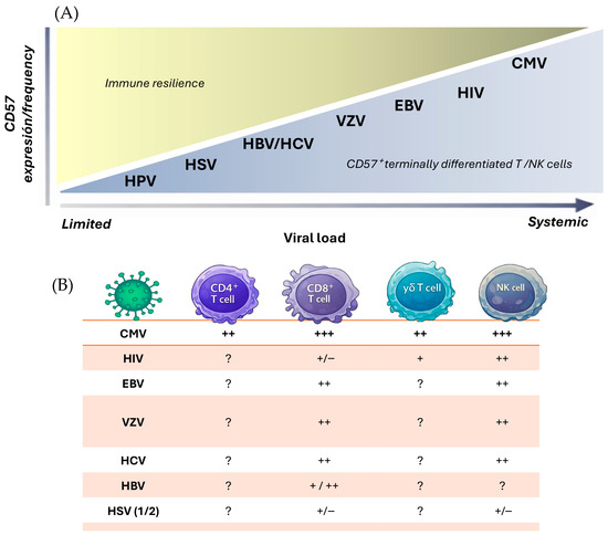
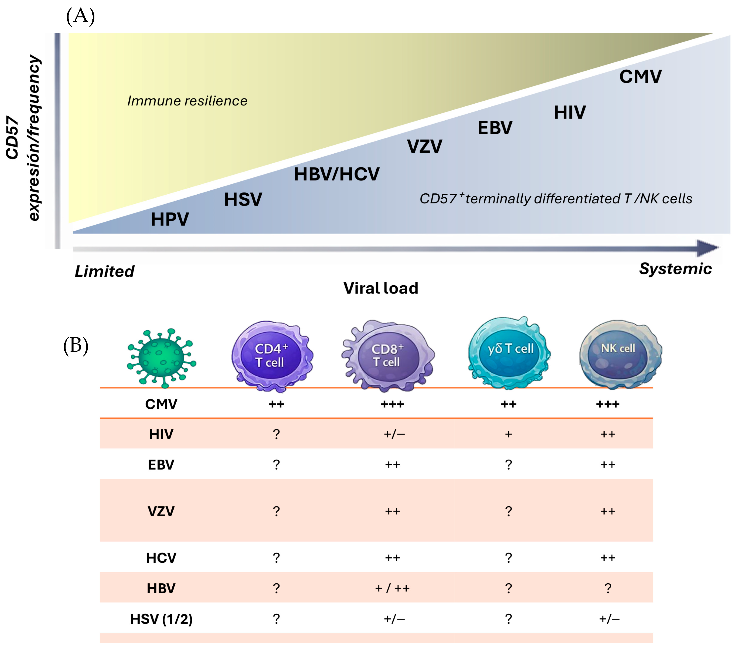

Disclaimer/Publisher’s Note: The statements, opinions and data contained in all publications are solely those of the individual author(s) and contributor(s) and not of MDPI and/or the editor(s). MDPI and/or the editor(s) disclaim responsibility for any injury to people or property resulting from any ideas, methods, instructions or products referred to in the content. |
© 2026 by the authors. Licensee MDPI, Basel, Switzerland. This article is an open access article distributed under the terms and conditions of the Creative Commons Attribution (CC BY) license.