


Highlights
What are the main findings?
- Ectopic fat comprises metabolically active, organ-specific fat depots whose biological effects depend on their anatomical location, cellular composition, and local interactions with adjacent tissues.
- Ectopic fat contributes to metabolic dysfunction through multiple interconnected mechanisms, including excess free fatty acid flux, chronic inflammation, oxidative stress, mitochondrial dysfunction, endoplasmic reticulum stress, fibrosis, and impaired insulin signaling.
What are the implications of the main findings?
- Individual ectopic fat depots exert distinct pathogenic effects, contributing to cardiovascular disease, metabolic dysfunction-associated steatotic liver disease, β-cell dysfunction, chronic kidney disease, skeletal muscle insulin resistance, and other obesity-related complications.
- Quantification of ectopic fat using contemporary imaging techniques, combined with advanced molecular phenotyping, may provide clinically relevant information beyond conventional measures of adiposity and contribute to a better understanding and assessment of obesity-related organ dysfunction.
Abstract
Ectopic fat is increasingly recognized as a heterogeneous, metabolically active contributor to obesity-related cardiometabolic disorders, with clinical implications extending beyond conventional measures of adiposity. This narrative review synthesizes experimental, translational, and clinical evidence on the mechanisms of ectopic fat accumulation, depot-specific biological characteristics, clinical significance, and therapeutic implications. Impaired subcutaneous adipose tissue expandability and lipid buffering capacity promote ectopic lipid deposition, contributing to lipotoxicity, inflammation, oxidative stress, mitochondrial dysfunction, fibrosis, and impaired insulin signaling. Despite shared pathogenic mechanisms, individual depots exhibit distinct organ-specific associations. Epicardial, pericardial, and perivascular fat are implicated in cardiovascular dysfunction; hepatic fat is closely associated with metabolic dysfunction-associated steatotic liver disease and insulin resistance; pancreatic fat may contribute to β-cell dysfunction; skeletal muscle fat is linked to metabolic and functional impairment; and perirenal and renal sinus fat are implicated in renal dysfunction and alterations in cardiovascular structure and function. Advanced imaging enables depot-specific quantification and characterization of ectopic fat, providing information beyond conventional measures of adiposity. Lifestyle modifications and pharmacological therapies may reduce ectopic fat accumulation in selected depots, although their effects vary across organs and interventions. Collectively, these findings highlight ectopic adiposity as a potentially modifiable component of obesity-related cardiometabolic complications. Integrating imaging-based assessment with molecular phenotyping may improve understanding and assessment of obesity-related organ dysfunction.
1. Introduction
Obesity is a chronic, multifactorial disease characterized by an abnormal or excessive accumulation of adipose tissue that substantially impairs health and represents a major global public health burden [1]. Although body mass index (BMI) remains the most widely used clinical metric for defining and stratifying obesity, it inadequately captures the biological complexity and heterogeneity of this condition. By failing to distinguish between fat mass and lean body mass or to account for regional fat distribution, BMI offers an imprecise estimate of obesity-related health risk. Individuals with comparable BMI values frequently display striking differences in metabolic health, susceptibility to cardiometabolic complications, and long-term clinical outcomes. Such variability reflects differences in adipose tissue distribution, its functional properties, and the propensity to accumulate lipids within non-adipose organs, rather than differences in total body weight alone [2,3]. Accordingly, current approaches to obesity assessment consider not only body weight but also fat distribution, adipose tissue function, and their biological and clinical consequences [4].
Adipose tissue is a dynamic endocrine organ with distinct regional depots that differ in morphology, metabolic activity, and systemic effects [5]. Subcutaneous adipose tissue (SAT) is the largest adipose depot and serves as the principal physiological site for excess lipid storage, thereby preserving lipid homeostasis and preventing lipid accumulation in non-adipose organs [6]. In contrast, visceral adipose tissue (VAT) exhibits higher lipolytic activity and contributes to increased flux of free fatty acids (FFAs) into the portal and systemic circulation [7]. However, during sustained energy surplus, the capacity of SAT to accommodate increasing lipid loads through adaptive expansion may become progressively impaired. This disruption of adipose tissue homeostasis promotes lipid spillover and ectopic lipid deposition within visceral compartments and organs not specialized for lipid storage. This process gives rise to ectopic fat deposition, defined as the accumulation of triglycerides in organs not specialized for fat storage, including the liver, skeletal muscle, pancreas, heart, and kidneys [6]. However, ectopic fat encompasses distinct forms of lipid accumulation, ranging from intracellular lipid deposition to anatomically defined adipose tissue depots, each differing in structural organization, cellular composition, and biological functions [6,8].
Ectopic fat is now recognized as a central feature of metabolic dysfunction, exerting both local and systemic effects. Lipid accumulation within non-adipose tissues disrupts cellular homeostasis through multiple lipotoxic mechanisms, including the accumulation of toxic lipid intermediates, mitochondrial dysfunction, alterations in cellular energy metabolism, and impaired insulin signaling. These processes ultimately compromise organ-specific function and contribute to systemic metabolic dysregulation [8]. In parallel, ectopic and visceral fat depots function as metabolically active endocrine compartments, releasing FFAs, adipokines, pro-inflammatory cytokines, and other bioactive mediators that exacerbate insulin resistance and sustain chronic low-grade inflammation. Notably, ectopic fat depots are biologically heterogeneous; their anatomical location, cellular composition, and proximity to target organs determine their relative contributions to disease pathogenesis. Consequently, distinct depots differentially influence the development and progression of key metabolic disorders, including insulin resistance, type 2 diabetes mellitus (T2DM), cardiovascular diseases, and metabolic dysfunction-associated steatotic liver disease (MASLD) [9,10,11]. An overview of the anatomical characteristics, imaging modalities, measurement limitations, and clinical significance of organ-specific ectopic fat depots is provided in Table 1.
In this narrative review, we synthesize current evidence on the mechanisms linking impaired adipose tissue function, ectopic fat accumulation, and metabolic dysfunction. We discuss the molecular basis of ectopic fat-induced cellular injury, the heterogeneity of ectopic fat depots, and their implications for disease progression. This perspective highlights the importance of ectopic fat biology in refining risk assessment and identifying potential therapeutic targets.
2. Methods
This study was conducted as a non-systematic, expert-informed narrative review aimed at synthesizing current knowledge on the biological characteristics, pathophysiological mechanisms, and clinical significance of organ-specific ectopic fat accumulation. The review focused on major ectopic fat depots and lipid compartments, including epicardial, pericardial, perivascular, perirenal, hepatic, pancreatic, and skeletal muscle fat.
A literature search was performed using PubMed, Scopus, and Web of Science as the primary databases. Google Scholar was additionally used to identify relevant publications not indexed in major biomedical databases. The search covered studies published up to 2026, with a primary focus on literature published between 2010 and 2026 to capture recent advances in imaging, molecular characterization, and metabolic profiling of ectopic fat, while earlier studies were considered when relevant to fundamental concepts and mechanisms. Search terms were applied iteratively and included combinations of keywords related to ectopic fat depots and metabolic disease, including: “ectopic adipose tissue”, “ectopic fat”, “visceral adiposity”, “epicardial adipose tissue”, “pericardial adipose tissue”, “perivascular adipose tissue”, “perirenal fat”, “pancreatic steatosis”, “fatty pancreas”, “hepatic steatosis”, “skeletal muscle fat”, “insulin resistance”, “lipotoxicity”, “metabolic dysfunction”, “inflammation”, “cardiovascular disease”, and “obesity”. Manual screening of the reference lists of selected publications was also performed to identify relevant foundational and recently published studies.
Publications were selected based on their relevance to the review objectives, with particular consideration given to studies addressing biological mechanisms, organ-specific characteristics, clinical associations, and therapeutic implications. Original experimental and clinical studies, as well as relevant systematic reviews and meta-analyses, were considered to provide complementary mechanistic and clinical perspectives. Where available, greater emphasis was placed on original human studies, longitudinal investigations, interventional trials, and systematic reviews, while experimental studies were used primarily to contextualize proposed biological mechanisms. Given the narrative nature of this review, no formal systematic screening, predefined quality assessment, or quantitative evidence synthesis was performed. Accordingly, the review may be subject to selection bias and does not aim to provide an exhaustive or systematically graded assessment of the available evidence. Artificial intelligence tools were used in preparing the figures, as described in the Acknowledgements Section. Language-related support is also described in the Acknowledgements Section [12].
3. Pathophysiology of Ectopic Fat Deposition
The development of ectopic fat is closely linked to chronic positive energy balance and the limited capacity of SAT to expand and safely store excess energy. Under physiological conditions, surplus energy is preferentially stored in SAT through the expansion of existing adipocytes (hypertrophy) and the recruitment and differentiation of new adipocytes (hyperplasia), thereby facilitating the safe storage of excess lipids [13]. However, the capacity of SAT to accommodate excess energy varies substantially between individuals and is influenced by genetic, sex-specific, and environmental factors [14]. When this adaptive capacity is exceeded—particularly in the setting of sustained overnutrition and hyperinsulinemia—SAT progressively loses its ability to function as a metabolically protective “sink,” resulting in lipid spillover into non-adipose tissues such as the liver, skeletal muscle, pancreas, and myocardium [14,15]. In addition to impaired lipid storage, adipose tissue dysfunction is associated with dysregulation of lipolysis. Under physiological conditions, insulin suppresses adipocyte lipolysis, whereas during energy deprivation or increased energy demand, catecholamine-mediated signaling promotes the breakdown of stored triglycerides into free fatty acids and glycerol. Impaired insulin signaling in adipocytes can therefore result in excessive basal lipolysis and increased release of free fatty acids and glycerol into the circulation, further contributing to ectopic lipid accumulation in non-adipose tissues [16,17]. This concept is encapsulated in the “adipose tissue expandability hypothesis”, which posits that impaired adipogenesis and a predominance of hypertrophic expansion promote ectopic lipid accumulation and subsequent lipotoxicity [14,18]. Notably, ectopic fat deposition can also occur in individuals without overt obesity, underscoring that adipose tissue dysfunction, rather than total fat mass per se, is a key determinant of metabolic risk [14].
Once ectopic lipid accumulation exceeds the metabolic capacity of non-adipose tissues, triglycerides and bioactive lipid intermediates, including diacylglycerols (DAG), ceramides, and long-chain acyl-CoA, accumulate within cells, initiating lipotoxicity [8,18]. Rather than serving as inert energy stores, these lipid intermediates interfere with intracellular signaling pathways, particularly insulin signaling, through activation of serine/threonine kinases such as protein kinase C, c-Jun N-terminal kinase, and IκB kinase. This results in inhibitory phosphorylation of insulin receptor substrates, impaired PI3K–Akt signaling, and progressive insulin resistance [8,19]. Persistent lipid overload further disrupts cellular homeostasis by promoting mitochondrial dysfunction, excessive reactive oxygen species (ROS) production, endoplasmic reticulum stress, and activation of the unfolded protein response, ultimately impairing cellular function and viability. These stress responses trigger chronic low-grade inflammation, recruit immune cells, activate tissue-specific fibroblasts or stellate cells, and promote extracellular matrix remodeling and fibrosis [9,19]. Simultaneously, dysfunctional adipose tissue exhibits enhanced lipolysis and an altered secretory profile characterized by increased production of pro-inflammatory cytokines and adipokines together with reduced secretion of protective adipokines, thereby perpetuating both local tissue injury and systemic metabolic dysfunction [9,13,14]. Although these fundamental mechanisms are shared across ectopic fat depots, their biological and clinical consequences depend on the anatomical location of lipid accumulation and the physiological characteristics of the affected organ, as summarized in Figure 1.
Figure 1.
Anatomical distribution of ectopic fat depots.
4. Thoracic Fat: Epicardial, Pericardial, and Perivascular Depots
4.1. Thoracic Fat Accumulation and Its Role in Metabolic Dysfunction
Epicardial adipose tissue (EAT) and pericardial adipose tissue (PAT) represent anatomically and functionally distinct ectopic fat depots surrounding the heart. EAT is located between the myocardium and the visceral pericardium, in direct contact with the coronary arteries and myocardial surface, without a separating fascial layer, whereas PAT is situated outside the parietal pericardium within the thoracic cavity (Figure 2) [20]. In contrast, perivascular adipose tissue (PVAT) surrounds blood vessels, including the coronary arteries and larger thoracic vessels such as the aorta. Coronary PVAT may anatomically overlap with EAT [21]. Importantly, EAT shares a common embryological origin and microcirculation with the myocardium through the coronary arteries, enabling intense paracrine and vasocrine crosstalk between adipocytes, vascular structures, and cardiomyocytes. In contrast, PAT originates from thoracic mesenchymal cells and is supplied by non-coronary vessels [9,20]. Under physiological conditions, EAT exerts several cardioprotective functions, including mechanical protection of the heart and coronary arteries; thermoregulation; regulation of local FFA homeostasis by supplying energy substrates to the myocardium during periods of increased metabolic demand; and protection of cardiomyocytes from lipotoxicity by buffering excess FFAs [22]. Unlike most white adipose depots, EAT retains partial brown adipose tissue-like characteristics, including expression of uncoupling protein-1 (UCP-1) and increased metabolic activity, although these features progressively decline with ageing, obesity, DM, and coronary artery disease (CAD) [23].
Figure 2.
Anatomical distribution of epicardial, pericardial and perivascular adipose tissue.
EAT is increasingly recognized as an active cardiometabolic organ rather than a passive lipid storage compartment. Due to the absence of a fascial barrier, bioactive molecules released from EAT can directly diffuse into the adjacent myocardium and coronary arteries through paracrine and vasocrine signaling pathways [24]. The physiological phenotype of EAT is characterized by the expression of cardioprotective, anti-inflammatory, and anti-atherogenic mediators, including adiponectin and adrenomedullin. In obesity, T2DM, and CAD, this protective profile is progressively replaced by a pro-inflammatory and pro-atherogenic phenotype driven by extensive transcriptomic and functional remodeling [23]. This transition is characterized by increased expression of inflammatory cytokines, oxidative stress-related pathways, and profibrotic mediators, accompanied by infiltration of pro-inflammatory immune cells, including M1 macrophages, mast cells, and CD8+ T lymphocytes. Activation of NF-κB-, JNK-, and toll-like receptor-dependent signaling further amplifies local inflammatory responses, promoting endothelial dysfunction, vascular inflammation, and progression of atherosclerosis [25]. Simultaneously, reduced adiponectin expression weakens local anti-inflammatory and vasoprotective mechanisms, thereby enhancing susceptibility to CAD and adverse myocardial remodeling [26].
Excessive FFA release from dysfunctional EAT contributes to myocardial lipotoxicity. When myocardial fatty acid uptake exceeds oxidative capacity, intracellular accumulation of lipotoxic intermediates such as ceramides and DAG occurs, leading to mitochondrial dysfunction, oxidative stress, endoplasmic reticulum stress, and impaired calcium homeostasis [9,22]. Oxidative stress induced by EAT-derived inflammatory mediators further disrupts calcium handling and myocardial contractility, thereby promoting structural and functional cardiac remodeling [9]. Increased secretion of phospholipase A2 and fatty-acid-binding proteins from pericoronary EAT may further enhance lipid accumulation within atherosclerotic plaques and accelerate coronary artery calcification [27,28]. Importantly, EAT in patients with T2DM demonstrates altered expression of genes involved in lipid transport, fatty acid handling, and innate immune activation, including lipase G (LIPG), solute carrier family 7 member 5 (SLC7A5), and Pentraxin3 (PTX3), indicating that T2DM is associated with profound depot-specific metabolic and inflammatory remodeling of EAT, which may further amplify local coronary inflammation and myocardial dysfunction [23,29,30].
Excessive expansion of EAT may contribute to adverse cardiac remodeling not only through inflammatory and metabolic pathways but also by mechanically affecting cardiac function. Increased EAT has been associated with impaired ventricular filling and ventricular diastolic dysfunction, as well as elevated cardiac workload, atrial enlargement, and left ventricular hypertrophy, collectively contributing to myocardial dysfunction and arrhythmogenesis [20,31]. These structural alterations, together with the close anatomical relationship between EAT and the atrial myocardium, appear to play a central role in the pathogenesis of atrial fibrillation (AF). EAT-derived inflammatory and profibrotic mediators promote atrial fibrosis, structural remodeling of the atrial myocardium, and conduction abnormalities [32]. Direct extension of EAT into the adjacent atrial myocardium has been demonstrated histologically and may contribute to disruption of myocardial architecture, conduction slowing, and the formation of re-entry circuits [33]. Increased EAT volume and thickness are consistently associated with AF prevalence, persistence, and atrial conduction abnormalities independent of overall adiposity. Importantly, meta-analytic data indicate a strong, graded relationship between epicardial fat burden and AF risk, with higher EAT volume associated with markedly increased odds of both paroxysmal and persistent AF. Notably, these associations appear stronger than those observed for conventional measures of adiposity, including BMI and abdominal fat distribution, highlighting the potential role of EAT as a depot-specific marker of arrhythmogenic risk [34]. Similarly, excessive EAT accumulation has been linked to impaired diastolic function, elevated cardiac filling pressures, pulmonary hypertension, and heart failure with preserved ejection fraction (HFpEF) [35,36]. Increased EAT burden has been consistently observed in patients with HFpEF compared with BMI-matched controls, supporting a potential role for epicardial fat in myocardial stiffness, impaired ventricular compliance, and adverse cardiac remodeling [35]. In addition to structural remodeling, EAT-derived cytokines, including tumor necrosis factor-alpha (TNF-α) and interleukin-6 (IL-6), together with locally released FFAs, directly modulate atrial electrophysiology, promoting conduction slowing, electrical heterogeneity, and the maintenance of arrhythmogenic substrates [32].
Compared with EAT, PAT exhibits lower lipolytic activity and a different fatty acid composition, indicating distinct metabolic regulation of these neighboring depots [37]. PAT is a metabolically active depot characterized by relatively small, poorly differentiated adipocytes, abundant vascularization, and substantial immune-cell content [21]. Its inflammatory and lipolytic properties are closely linked to systemic metabolic status, particularly insulin resistance, suggesting that PAT may participate in the metabolic disturbances accompanying visceral adiposity [21]. Unlike EAT, whose biological activity is strongly influenced by local myocardial signals, PAT appears to respond predominantly to systemic metabolic stimuli, potentially contributing to cardiovascular risk through endocrine and inflammatory pathways rather than direct myocardial or coronary paracrine signaling [38]. Clinically, increased pericardial fat volume has been associated with coronary atherosclerosis and calcification. Nafakhi et al. [39] reported an association between CT-measured pericardial fat volume and coronary atherosclerotic burden in postmenopausal women, while Liu et al. [40] found that greater EAT and PAT volumes were associated with more extensive coronary artery calcification in patients with suspected CAD. In the Framingham Heart Study, Mahabadi et al. [41] demonstrated that pericardial fat was associated with prevalent cardiovascular disease independently of conventional measures of adiposity, although this relationship was attenuated after adjustment for cardiovascular risk factors. Importantly, these findings require cautious interpretation because the term pericardial fat has been applied to different anatomical compartments, including intrapericardial fat, which does not correspond to strictly defined extracardiac PAT [37,42].
PVAT is a metabolically active adipose tissue compartment surrounding blood vessels, including the coronary arteries and thoracic aorta, and plays an important role in the regulation of vascular homeostasis [21]. Unlike PAT, PVAT is positioned in close proximity to the vascular adventitia, enabling bidirectional communication between adipocytes, vascular cells, and infiltrating immune cells. Under physiological conditions, PVAT contributes to vascular homeostasis through the release of adiponectin, nitric oxide-related mediators, and other vasoactive factors that modulate vascular tone and endothelial function. However, its cellular composition and metabolic characteristics vary according to vascular location, with coronary PVAT displaying features distinct from those of PVAT surrounding larger systemic arteries [21]. In obesity, T2DM, and atherosclerosis, PVAT undergoes metabolic and inflammatory remodeling that shifts its local activity from vascular protection toward the promotion of inflammation [43,44]. Adipocyte hypertrophy and reduced adiponectin availability weaken the vasodilatory and anti-inflammatory properties of PVAT, while increased oxidative stress, partly driven by NADPH oxidase activity, impairs nitric oxide (NO) signaling and endothelial function [43,44]. At the same time, increased monocyte chemoattractant protein-1 (MCP-1) production promotes monocyte and macrophage recruitment, establishing a local inflammatory environment in which TNF-α and IL-6 enhance endothelial expression of adhesion molecules, including VCAM-1 and ICAM-1 [43]. This facilitates leukocyte adhesion and migration into the vascular wall, linking PVAT inflammation to early atherogenic changes [43,45]. Inflammatory mediators released from dysfunctional PVAT may also influence vascular smooth muscle cell migration and proliferation, contributing to vascular remodeling, although these processes can have different consequences depending on the stage of atherosclerotic disease [43].
Importantly, communication between PVAT and the vascular wall is bidirectional. Inflammatory signals originating from diseased arteries can induce changes in adjacent adipocytes, including inhibition of adipogenesis and lipid accumulation, thereby altering local adipose tissue composition [21,46]. Antonopoulos et al. [46] demonstrated that coronary inflammation is associated with CT-detectable changes in PVAT, providing the basis for the fat attenuation index (FAI) as a non-invasive imaging marker of coronary inflammation. In the CRISP-CT study, Oikonomou et al. [47] further demonstrated that higher perivascular FAI was associated with increased cardiac mortality independently of conventional cardiovascular risk factors and CT-derived measures of coronary atherosclerosis. Similar associations have been reported beyond the coronary circulation. Zhang et al. [48] found that increased CT-derived PVAT attenuation surrounding the descending thoracic aorta was independently associated with atherosclerotic plaque burden and complexity. Beyond conventional measurements of adipose tissue volume, CT-based characterization of PVAT attenuation has therefore emerged as a promising approach to cardiovascular risk assessment [21,49]. As highlighted by Foran et al. [49], advances in PVAT imaging and quantification, including radiotranscriptomic approaches, may provide additional information about the biological state of the vascular wall. Nevertheless, PVAT attenuation is an indirect imaging marker influenced by vascular inflammation and technical factors, rather than a direct measurement of inflammatory cell activity. Although these approaches have demonstrated prognostic potential, further standardization and prospective validation are needed before they can be considered universally applicable clinical tools.
4.2. Effects of Dietary Interventions and Physical Activity on EAT, PAT, PVAT
Lifestyle interventions can reduce EAT, although the magnitude of this response depends on the intervention and the population studied. In a 12-week randomized trial involving physically inactive adults with abdominal obesity, Christensen et al. [50] reported that supervised aerobic and resistance training reduced MRI-assessed EAT mass by 32% and 24%, respectively, relative to baseline, whereas EAT mass increased in the non-exercising control group. In a 16-week pilot randomized trial involving 73 patients with T2DM, Leroux-Stewart et al. [51] found that combining caloric restriction with physical activity reduced echocardiographically measured EAT thickness by 1.4 ± 1.4 mm, compared with 0.4 ± 1.6 mm with caloric restriction alone, despite comparable weight loss between intervention groups. However, additional exercise does not consistently provide benefits beyond dietary restriction. In a six-month trial involving 61 premenopausal women with overweight or obesity, Hens et al. [52] observed a reduction in cardiac epicardial fat volume across the study population, but adding unsupervised exercise to a hypocaloric diet did not significantly improve outcomes compared with diet alone. More recently, Xue et al. [53] demonstrated that a 12-month structured exercise program in community-dwelling older adults produced a greater reduction in EAT volume than usual care (between-group difference: −15.15 mL), accompanied by a modest decrease in circulating macrophage colony-stimulating factor (M-CSF). Greater exercise frequency was associated with larger reductions in EAT volume, although the study did not establish that changes in M-CSF mediated the adipose tissue response. Overall, meta-analyses support the effectiveness of lifestyle interventions in reducing EAT, while indicating heterogeneity in treatment response [54,55]. The response of PAT to lifestyle modification appears to differ from that of EAT. In the same 12-week randomized trial, Christensen et al. [50] found that resistance training reduced MRI-assessed PAT mass by 31% relative to baseline, whereas the 11% reduction observed after aerobic training was not statistically significant compared with the control group. Notably, resistance training produced a significantly greater PAT reduction than no exercise, despite relatively modest changes in total body fat mass, suggesting that changes in this depot may not simply parallel generalized fat loss.
Evidence regarding PVAT comes predominantly from animal studies, in which exercise-related improvements have been demonstrated through changes in vascular reactivity and local inflammatory signaling rather than reductions in PVAT volume. In obese mice, Sousa et al. [56] showed that aerobic exercise training prevented PVAT-associated endothelial dysfunction in the thoracic aorta, supporting preservation of vascular responsiveness despite exposure to an obesogenic diet. Araujo et al. [57] similarly investigated rats fed a high-fat diet and demonstrated that aerobic training modified the anticontractile influence of thoracic aortic PVAT, indicating that exercise can alter the functional relationship between perivascular adipocytes and vascular smooth muscle. More recently, Li et al. [58] used a high-fat diet-induced mouse model followed by eight weeks of treadmill training and observed improved acetylcholine-mediated relaxation in aortic rings with intact PVAT, from approximately 40% in sedentary obese mice to 59% in exercised obese mice. This functional improvement was accompanied by reduced PVAT adipocyte hypertrophy, lower expression of inflammatory mediators, increased adiponectin, and enhanced Akt/AMPK/eNOS phosphorylation, consistent with partial restoration of vasoprotective signaling. Although these findings provide mechanistic support for exercise-induced improvements in PVAT function, corresponding human intervention studies remain insufficient to determine whether similar vascular effects occur in clinical practice.
4.3. Effects of Pharmacological Interventions on EAT, PAT, PVAT
Pharmacological reduction of EAT has been demonstrated primarily with GLP-1RAs and sodium–glucose cotransporter-2 inhibitors (SGLT2is). In the randomized, placebo-controlled LIRAFLAME trial involving 102 patients with T2DM, Rasmussen et al. [59] reported that 26 weeks of liraglutide treatment reduced CT-assessed cardiac adipose tissue volume by 11.5 mL, compared with virtually no change in the placebo group; however, the between-group difference was no longer significant after adjustment for changes in body mass index, suggesting that the effect was largely associated with weight loss. In turn, in a 12-week controlled study involving patients with T2DM and obesity, treatment with semaglutide or dulaglutide was associated with an approximately 20% reduction in echocardiographically measured EAT thickness, whereas no comparable reduction was observed in the metformin-treated control group [60]. More specifically, a prespecified analysis of the randomized STOP trial demonstrated that semaglutide reduced CT-measured EAT volume by approximately 9% compared with placebo and increased EAT attenuation by approximately 5%, suggesting changes in both depot size and imaging characteristics [61]. SGLT2 inhibition has also shown beneficial effects: in the EMPA-TROPISM trial, six months of empagliflozin treatment in nondiabetic patients with heart failure with reduced ejection fraction reduced MRI-assessed EAT volume by 5.14 mL, compared with 0.75 mL in the placebo group, alongside improvements in myocardial fibrosis and aortic stiffness [62]. Nevertheless, these effects are not uniform across populations. In the 12-week randomized ASSET study involving 44 patients with early-stage T2DM, empagliflozin did not significantly outperform sitagliptin in reducing EAT accumulation [63]. The efficacy of pharmacological interventions in reducing EAT has also been evaluated in meta-analyses. Myasoedova et al. [64] demonstrated significant reductions in EAT with GLP-1RAs and SGLT2 inhibitors, with the greatest effect observed for GLP-1RAs. In contrast, a more recent network meta-analysis by Bao et al. [65] found that SGLT2 inhibitors were more effective than GLP-1 receptor agonists in reducing EAT thickness in patients with T2DM and/or obesity.
Evidence regarding pharmacological modulation of PAT remains substantially more limited, and interpretation is complicated by differences in how pericardial fat is anatomically defined. In a 24-week randomized, double-blind trial involving 78 men with T2DM, Jonker et al. [66] found that pioglitazone increased MRI-measured pericardial fat volume from 30.5 to 33.1 mL, whereas metformin produced no meaningful change (29.2 to 29.6 mL). Importantly, the increase in pericardial fat was not accompanied by deterioration in left ventricular diastolic function, indicating that expansion of this imaging-defined depot does not necessarily translate into measurable cardiac dysfunction over the treatment period. Conversely, in the ASSET trial, Hiruma et al. [63] separately evaluated epicardial and paracardial fat using cardiac MRI and found no significant differences in either compartment between empagliflozin and sitagliptin after 12 weeks.
Pharmacological effects on PVAT have increasingly been investigated using CT-derived pericoronary adipose tissue (PCAT) attenuation, an imaging marker associated with coronary vascular inflammation rather than a direct measure of PVAT volume. In a multicenter observational study of 96 patients with stable coronary artery disease undergoing serial coronary CT angiography, Cheng et al. [67] reported that lesion-level PCAT attenuation decreased from −79.4 to −86.5 Hounsfield units (HU) among patients who initiated statin therapy and from −83.5 to −90.6 HU among those receiving continuous statin treatment, whereas the change was not statistically significant in statin-naïve patients over a median follow-up of 3.8 years. These findings are consistent with an association between statin exposure and reduced CT-derived markers of pericoronary inflammation, although the observational design precludes causal inference. Similarly, in a retrospective study of 497 patients with T2DM, Li et al. [68] found that semaglutide-treated patients had lower PCAT fat attenuation index values than untreated patients; following propensity score matching, this association was observed around all three major coronary arteries and remained significant in multivariable analyses.
5. Intrahepatic Lipid Accumulation
5.1. Intrahepatic Fat Accumulation and Its Role in Metabolic Dysfunction
Intrahepatic fat (IHF) represents one of the most clinically relevant ectopic fat depots and is the pathological hallmark of metabolic dysfunction-associated steatotic liver disease and metabolic dysfunction–associated steatohepatitis (MASH) [69,70]. Under physiological conditions, hepatic lipid homeostasis is maintained through a dynamic balance between fatty acid uptake, de novo lipogenesis, mitochondrial β-oxidation, and triglyceride export as very low-density lipoproteins (VLDL) [69,71]. Chronic positive energy balance disrupts this equilibrium by increasing hepatic lipid influx through enhanced adipose tissue lipolysis, increased dietary fat delivery, and accelerated hepatic de novo lipogenesis. Notably, adipose tissue lipolysis, which is exacerbated by insulin resistance, accounts for approximately 60% of hepatic FFA delivery [70,72]. Initially, excess FFAs are esterified into triglycerides and either stored within hepatocytes or exported as VLDL, representing an adaptive mechanism that limits fatty acid-induced cellular toxicity [71,73]. However, when lipid influx exceeds the capacity for mitochondrial β-oxidation and VLDL secretion, hepatocellular triglyceride accumulation is accompanied by the generation of lipotoxic lipid intermediates, particularly DAG, ceramides, and other bioactive lipid species that actively disrupt intracellular metabolic homeostasis (Figure 3) [73]. Consequently, hepatic steatosis should be regarded not merely as passive triglyceride accumulation but as a metabolically active process that may contribute to cellular dysfunction and the development and progression of MASLD/MASH [70].
Figure 3.
Pathophysiological Mechanisms of Hepatic Fat Accumulation and Insulin Resistance; Abbreviations: Cer—ceramides; DAG—diacylglycerols; DNL—de novo lipogenesis; IRS-1—insulin receptor substrate 1; IRS-2—insulin receptor substrate 2; PI3K—phosphatidylinositol 3-kinase; PKC-ε—protein kinase C epsilon; TG—triglycerides; VLDL—very-low-density lipoproteins.
Following lipid accumulation, hepatocellular DAG content increases and activates protein kinase C-ε (PKC-ε), which inhibits insulin receptor kinase activity and impairs IRS-1/IRS-2-mediated phosphatidylinositol-3 kinase (PI3K) signaling, thereby promoting hepatic insulin resistance. Hepatic insulin resistance is characterized by impaired insulin-mediated suppression of gluconeogenesis, reduced glycogen synthesis, and increased VLDL production, contributing to hyperglycemia, hypertriglyceridemia, and the redistribution of lipids to extrahepatic tissues [72,73]. Clinical studies have also demonstrated associations between intrahepatic lipid accumulation and impaired insulin sensitivity. In a study of 42 individuals with obesity but without T2DM, Korenblat et al. [74] observed an inverse correlation between hepatic triglyceride content, measured using proton magnetic resonance spectroscopy (1H-MRS), and hepatic insulin sensitivity assessed by the hyperinsulinemic-euglycemic clamp. Hepatic triglyceride content was also associated with insulin sensitivity in skeletal muscle and adipose tissue, independently of BMI and total body fat percentage. Similarly, in a European cohort of 1307 individuals with normal glucose tolerance, a higher fatty liver index was associated with reduced insulin-stimulated glucose disposal and higher circulating FFA concentrations during the clamp procedure, suggesting concomitant impairment of peripheral and adipose tissue insulin sensitivity [75]. Further evidence comes from a cross-sectional and longitudinal study by Tricò et al. [76] involving 632 children and adolescents with obesity. Higher IHF content, assessed by MRI, was associated with increased hepatic insulin resistance and reduced endogenous insulin clearance, independently of ethnicity and several potential confounders, including adiposity and pubertal status. In a subgroup of 89 participants reassessed after approximately two years, increases in IHF content were associated with reduced insulin clearance and worsening hepatic insulin resistance. Although these findings support a relationship between intrahepatic lipid accumulation and impaired hepatic insulin metabolism, the relatively small longitudinal sample and observational study design limit causal interpretation. Experimental studies further suggest that hepatic insulin resistance may develop before detectable peripheral insulin resistance, supporting the concept that hepatic steatosis represents an early pathogenic event in the development of metabolic syndrome and T2DM [14,77]. Moreover, compensatory hyperinsulinemia paradoxically maintains hepatic de novo lipogenesis through activation of lipogenic transcription factors, including sterol regulatory element-binding protein-1c (SREBP-1c) and carbohydrate-responsive element-binding protein (ChREBP), thereby creating a self-perpetuating cycle of hepatic lipid accumulation [70,78]. Concomitantly, excessive FFAs and other lipotoxic lipid species induce mitochondrial dysfunction, endoplasmic reticulum stress, and excessive ROS generation, leading to hepatocyte injury and apoptosis [73,79]. These alterations trigger the activation of Kupffer cells and hepatic stellate cells through the release of pro-inflammatory and profibrogenic mediators, particularly transforming growth factor-β (TGF-β), thereby perpetuating chronic hepatic inflammation, extracellular matrix deposition, and progressive liver fibrosis [70].
Beyond its role in MASLD/MASH, IHF is increasingly recognized as a central determinant of systemic metabolic dysfunction. Hepatic triglyceride content correlates more strongly with hepatic and whole-body insulin resistance than overall adiposity or VAT, indicating that IHF represents a more robust marker of metabolic impairment than conventional anthropometric measures [74,80]. Increased hepatic lipid accumulation promotes excessive VLDL secretion and ectopic lipid redistribution to peripheral tissues, including skeletal muscle and the pancreas, thereby contributing to peripheral insulin resistance, β-cell dysfunction, atherogenic dyslipidemia, chronic low-grade inflammation, and an increased risk of cardiovascular disease [14]. Importantly, IHF is a highly dynamic and metabolically responsive ectopic fat depot. Clinical studies consistently demonstrate that moderate weight loss markedly reduces hepatic triglyceride content, frequently preceding substantial changes in visceral adiposity and resulting in significant improvements in hepatic insulin sensitivity and glycemic control [14,81]. Moreover, sustained lifestyle-induced weight reduction is associated with partial histological resolution of hepatic steatosis and, in selected patients, improvement of steatohepatitis and liver fibrosis, highlighting IHF as a reversible therapeutic target in obesity, MASLD/MASH, and T2DM [8,82].
5.2. Effects of Dietary Interventions and Physical Activity on IHF
Dietary interventions and physical activity are effective strategies for reducing IHF content, although their effects depend on the characteristics of the intervention and the metabolic profile of the study population. Randomized controlled trials have demonstrated that caloric restriction effectively reduces IHF content, while the extent to which dietary macronutrient composition contributes to this reduction, independent of energy intake, remains unclear. In a six-month randomized trial, Haufe et al. [83] reported comparable relative reductions in intrahepatic lipid (IHL) content following energy-restricted diets with reduced carbohydrate (−47%) or fat (−42%) intake, with no significant between-group differences. Participants with elevated baseline IHL (>5.56%) exhibited approximately sevenfold greater absolute reductions in hepatic lipid content than those with lower baseline values, despite similar changes in visceral adiposity and insulin sensitivity. In patients with non-alcoholic fatty liver disease (NAFLD), Holmer et al. [84] demonstrated that both a low-carbohydrate, high-fat (LCHF) diet and intermittent calorie restriction (5:2 diet) produced greater absolute reductions in hepatic steatosis than standard lifestyle advice (−7.2, −6.1, and −3.6 percentage points, respectively), without significant differences between the two dietary interventions. The effects of macronutrient composition independent of prescribed energy restriction were investigated by London et al. [85], who demonstrated that four days of an eucaloric low-carbohydrate, high-fat diet reduced hepatic triglyceride content by 35.3%, whereas a high-carbohydrate, low-fat diet did not significantly alter hepatic triglyceride content. The reduction was accompanied by increased fasting fat oxidation and ketogenesis, reduced markers of de novo lipogenesis, and improved hepatic insulin sensitivity. Dietary quality was further examined in the 18-month DIRECT-PLUS trial, in which a polyphenol-rich green Mediterranean diet resulted in a greater relative reduction in IHF than a conventional Mediterranean diet or healthy dietary guidelines [86]. Together, these studies demonstrate that both energy restriction and dietary composition influence IHF content, although their relative contributions vary according to intervention duration and dietary characteristics.
Exercise training has also been shown to reduce IHF content independently of changes in body weight and abdominal adiposity. Hallsworth et al. [87] demonstrated that eight weeks of resistance training reduced hepatic lipid content by 13% (from 14.0% to 12.2%) in patients with NAFLD, without significant changes in body weight, visceral adipose tissue, or total body fat. The intervention was also associated with improved substrate oxidation and glycemic regulation. Similarly, Sullivan et al. [88] reported a 10.3% relative reduction in intrahepatic triglyceride content following 16 weeks of moderate-intensity aerobic training, despite unchanged body weight and body fat percentage. Exercise did not significantly alter hepatic very-low-density lipoprotein triglyceride or apolipoprotein B-100 secretion rates, indicating that the reduction in hepatic triglyceride content was not accompanied by measurable changes in these components of hepatic lipoprotein export. The influence of exercise intensity was evaluated by Winn et al. [89], who compared four weeks of high-intensity interval training (HIIT) and moderate-intensity continuous training (MICT), matched for exercise energy expenditure (approximately 400 kcal per session). Both interventions significantly reduced IHL content (HIIT: −37.0%; MICT: −20.1%), without significant changes in body weight or visceral adiposity. However, the between-group difference in IHL reduction was not statistically significant. These findings demonstrate that aerobic, resistance, and interval training may reduce hepatic lipid accumulation without substantial weight loss, while the relative effectiveness of different exercise intensities remains uncertain when energy expenditure is controlled.
The beneficial effects of exercise training on IHF content are further supported by evidence from systematic reviews and meta-analyses. In a systematic review and meta-analysis of 21 randomized controlled trials involving 1530 participants, Smart et al. [90] reported a significant reduction in IHF content following exercise interventions with a cumulative energy expenditure exceeding 10,000 kcal (mean difference [MD]: −3.46 percentage points; 95% CI: −5.20 to −1.73). Exercise-only interventions were additionally associated with reductions in fasting FFA and insulin concentrations, although no significant pooled effects were observed for liver enzyme activities. In a meta-analysis of 11 randomized controlled trials involving 577 patients with NAFLD, Nam et al. [91] reported a significant reduction in IHL following exercise interventions (MD: −2.03; 95% CI: −3.26 to −0.79), with a larger reduction in studies lasting more than three months (MD: −3.62; 95% CI: −5.76 to −1.48). Interventions lasting less than three months did not produce a statistically significant pooled effect, although this finding does not exclude benefits from shorter exercise programs. A more comprehensive systematic review and network meta-analysis by Khalafi et al. [92], including 38 studies and 1880 participants, reported a significant reduction in IHL content following exercise interventions compared with non-exercise controls (standardized mean difference [SMD]: −0.33). Based on network ranking probabilities, HIIT ranked highest (p-score = 0.95), followed by aerobic training (p-score = 0.77), combined aerobic and resistance training (p-score = 0.52), and resistance training (p-score = 0.10). These rankings indicate the relative ordering of interventions within the network model but do not, by themselves, establish statistically significant differences between exercise modalities. In contrast, a meta-analysis by Chai et al. [93], comprising 34 randomized controlled trials and 2652 participants with MAFLD, reported a greater reduction in IHF content with resistance training than with aerobic training. The same analysis also favored low-carbohydrate over low-fat dietary interventions (SMD: −0.25; 95% CI: −0.45 to −0.06). These discrepancies may reflect differences in study populations, exercise protocols, and comparator groups, particularly given that the meta-analysis by Chai et al. [93] exclusively included participants with obesity.
The reduction in IHF content following dietary and exercise interventions may result from alterations in hepatic lipid synthesis, oxidation, and substrate availability. Carbohydrate restriction may decrease hepatic de novo lipogenesis while increasing fatty acid oxidation and ketogenesis, thereby reducing triglyceride accumulation in hepatocytes [85]. Exercise training may further influence hepatic lipid metabolism by increasing fatty acid utilization and modifying circulating FFA availability, even in the absence of substantial changes in body weight or visceral adiposity [87,88,90]. Additionally, dietary components such as polyphenols may contribute to reductions in hepatic fat through changes in metabolic regulation and gut microbiota composition, although the underlying mechanisms in humans remain incompletely understood [86].
5.3. Effects of Pharmacological Interventions on IHF
Pharmacological interventions targeting incretin and glucagon receptor signaling have demonstrated substantial effects on IHF content in individuals with obesity, T2DM, and MASLD. Glucagon-like peptide-1 receptor agonists (GLP-1RAs) have been investigated extensively, with several randomized controlled trials demonstrating reductions in hepatic steatosis assessed by MRI. In the D-LIFT trial, 24 weeks of dulaglutide treatment resulted in a significantly greater reduction in liver fat content than standard treatment in patients with T2DM and NAFLD, with a between-group difference of −3.5 percentage points (95% CI: −6.6 to −0.4) and a relative treatment difference of −26.4% [94]. Semaglutide has also demonstrated beneficial effects on hepatic steatosis. In a randomized placebo-controlled trial, Flint et al. [95] reported a significant reduction in MRI-assessed liver fat content following semaglutide treatment, although the primary endpoint of change in liver stiffness assessed by magnetic resonance elastography was not met. More recently, the 52-week SAMARA trial demonstrated that a greater proportion of participants receiving semaglutide 2.4 mg achieved a relative reduction in MRI-derived proton density fat fraction (MRI-PDFF) of at least 30% compared with placebo (60% vs. 17%). Semaglutide also produced greater improvements in the FibroScan-AST (FAST) score and liver enzyme activities [96]. Collectively, these findings support the effectiveness of GLP-1RAs in reducing hepatic fat accumulation, although improvements in steatosis do not necessarily correspond to measurable reductions in liver stiffness over the same treatment period.
Liraglutide has also been evaluated in populations with different metabolic disorders and in direct comparison with lifestyle modification. In a placebo-controlled trial involving women with polycystic ovary syndrome (PCOS), 26 weeks of liraglutide treatment reduced liver fat content by 44%, accompanied by reductions in body weight and visceral adipose tissue [97]. Importantly, a randomized trial directly comparing liraglutide with a structured program of dietary energy restriction and exercise demonstrated similar reductions in liver fat fraction after 26 weeks (−7.0 and −8.1 percentage points, respectively). However, during the subsequent 26-week follow-up, discontinuation of liraglutide was associated with partial regain of body weight and liver fat, whereas the improvements achieved through lifestyle modification were maintained [98]. These findings indicate that pharmacologically induced reductions in hepatic steatosis may not persist after treatment withdrawal and emphasize the importance of long-term weight management and sustained lifestyle modification.
The development of dual- and triple-hormone receptor agonists has expanded the range of pharmacological interventions investigated to reduce hepatic fat accumulation. Tirzepatide, a dual glucose-dependent insulinotropic polypeptide (GIP) and GLP-1 receptor agonist, was evaluated in the SURPASS-3 MRI substudy involving individuals with T2DM. After 52 weeks, the pooled tirzepatide 10 mg and 15 mg groups achieved a significantly greater absolute reduction in liver fat content (measured by MRI-PDFF) than insulin degludec (−8.09 vs. −3.38 percentage points; estimated treatment difference: −4.71 percentage points; 95% CI: −6.72 to −2.70). Reductions in liver fat were also correlated with decreases in body weight, visceral adipose tissue, and abdominal subcutaneous adipose tissue [99]. Greater reductions in hepatic steatosis were reported with retatrutide, a triple GIP, GLP-1, and glucagon receptor agonist. In a randomized phase 2a trial, 24 weeks of treatment produced relative reductions in liver fat content of 81.4% and 82.4% with the 8 mg and 12 mg doses, respectively, compared with a 0.3% increase in the placebo group. Liver fat content below 5% was achieved in 79% and 86% of participants receiving these doses, respectively [100]. Although these results demonstrate substantial effects on hepatic triglyceride accumulation, differences in study populations, baseline steatosis, treatment duration, and comparator groups preclude direct conclusions regarding the comparative efficacy of tirzepatide and retatrutide.
Dual GLP-1/glucagon receptor agonists have also demonstrated substantial reductions in liver fat content. In a randomized placebo-controlled trial, 12 weeks of pemvidutide treatment produced relative reductions in MRI-PDFF of 46.6%, 68.5%, and 57.1% at doses of 1.2, 1.8, and 2.4 mg, respectively, compared with 4.4% in the placebo group [101]. In a subsequent 12-week extension involving participants who completed the original trial, relative reductions reached 56.3%, 75.2%, and 76.4% at the corresponding doses after 24 weeks of treatment, compared with 14.0% with placebo [102]. The efficacy of survodutide, another dual GLP-1/glucagon receptor agonist, was evaluated in the phase 3 SYNCHRONIZE-MASLD trial. After 48 weeks, 84.2% of participants receiving survodutide achieved a relative reduction in MRI-PDFF of at least 30%, compared with 24.3% receiving placebo. Mean relative reductions in liver fat content were 58.7% and 9.5%, respectively [103]. Importantly, a post hoc mediation analysis of a separate phase 2 survodutide trial in patients with MASH estimated that 58.2% of the treatment effect on MRI-PDFF was mediated by weight reduction. In contrast, a smaller proportion of the effects on several markers of hepatic inflammation and fibrosis was attributable to weight loss [104]. These findings suggest that weight reduction is a major contributor to the improvement in hepatic steatosis during survodutide treatment, whereas additional weight-independent pathways may contribute to changes in other liver-related endpoints.
6. Skeletal Muscle Fat
6.1. Skeletal Muscle Fat and Its Role in Metabolic Dysfunction
Intermuscular adipose tissue (IMAT), intramuscular adipose tissue (intraMAT), and intramyocellular lipids (IMCLs) represent distinct compartments of skeletal muscle ectopic fat that differ in anatomical localization, cellular composition, and metabolic significance. IMAT consists of adipose tissue located between muscle groups, beneath the deep fascia, whereas IMCLs comprise intracellular lipid droplets stored within myofibers as an endogenous energy substrate [9]. In contrast, intraMAT refers to extracellular adipose tissue located within individual skeletal muscles, between muscle fibers or fascicles (Figure 4) [105]. Modern imaging techniques enable reliable, non-invasive quantification of skeletal muscle ectopic fat, with CT and MRI primarily assessing IMAT and 1H-MRS measuring IMCL content. MRI, particularly using Dixon-based techniques, also enables assessment of intramuscular fatty infiltration and quantification of muscle fat fraction. However, these measurements do not selectively quantify intraMAT, as the detected fat signal may reflect contributions from multiple lipid compartments [106]. IMAT and intramuscular fat infiltration generally increase with aging and are associated with obesity and T2DM, but they contribute to metabolic dysfunction through distinct biological mechanisms [107]. Numerous imaging studies have demonstrated strong associations between IMAT accumulation and insulin resistance, impaired physical performance, and sarcopenia, whereas IMCL accumulation is primarily linked to altered skeletal muscle lipid metabolism [108]. Despite its relatively small contribution to total adipose tissue mass, IMAT is independently associated with insulin resistance across several measures of overall adiposity. Experimental studies further suggest that factors secreted by IMAT may directly impair skeletal muscle insulin sensitivity, highlighting its potential metabolic significance beyond its absolute volume [107,109].
Figure 4.
Skeletal muscle lipid deposition: intermuscular, intramuscular and intramyocellular compartments.
The close anatomical proximity of IMAT to skeletal muscle enables direct paracrine communication between adipocytes and myofibers. Human explant studies have shown that IMAT-conditioned medium impairs insulin signaling in primary myotubes similarly to VAT, supporting a direct role for IMAT in skeletal muscle insulin resistance [107]. This effect may be mediated, at least in part, by the local release of pro-inflammatory cytokines, adipokines, and other bioactive factors from IMAT, which can alter the skeletal muscle microenvironment and impair insulin signaling [9,109]. In addition, IMAT contains abundant macrophages and other immune cells with predominantly pro-inflammatory M1 polarization, reinforcing chronic low-grade inflammation and further compromising skeletal muscle metabolic homeostasis [110]. Increased lipolysis within IMAT may also enhance local FFA availability and the accumulation of lipotoxic intermediates, further disrupting insulin signaling [111]. Experimental models further support a structural contribution of IMAT to skeletal muscle dysfunction. Adipocyte infiltration between myofibers may impair lateral force transmission, interfere with muscle regeneration and post-injury hypertrophy, and potentially disrupt neuromuscular connectivity and local perfusion [111]. Although these mechanisms have been demonstrated primarily in experimental models, they provide a plausible biological explanation for the consistent association between IMAT accumulation, reduced muscle strength, impaired mobility, and sarcopenia that is observed independently of muscle mass loss in aging and metabolic diseases [9]. The relationship between IMAT and metabolic health also appears to be influenced by age. In a study of healthy young and older adults without obesity, diabetes, or frailty, lower-leg IMAT was substantially higher in older compared with young adults and was negatively associated with insulin sensitivity, particularly among older participants. Twelve weeks of exercise training reduced IMAT in young but not older adults, whereas insulin sensitivity improved in both groups, indicating that the metabolic benefits of exercise were not directly dependent on reductions in IMAT. These findings suggest that although age-related IMAT accumulation may be associated with impaired metabolic function, exercise-induced improvements in insulin sensitivity can occur independently of changes in IMAT [112].
Unlike IMCL, which serve as intracellular energy reserves, intraMAT consists of adipocytes located within the skeletal muscle interstitium and may contribute to muscle dysfunction through structural and paracrine mechanisms. The development of intraMAT is primarily associated with the adipogenic differentiation of muscle-resident fibro-adipogenic progenitors (FAPs), which normally support tissue homeostasis and regeneration but may differentiate into adipocytes or profibrotic cells in response to aging, injury, or pathological changes in the muscle microenvironment [111]. Excessive intraMAT accumulation may disrupt muscle architecture and contribute to local metabolic dysfunction through altered adipokine secretion, inflammatory signaling, and interactions with neighboring myofibers, although the specific contribution of intraMAT to insulin resistance in humans remains incompletely established [111]. Experimental evidence further suggests that intraMAT may actively impair muscle regeneration rather than merely reflect tissue degeneration. In a mouse model of adipogenic muscle injury, selective inhibition of adipocyte differentiation in FAPs improved muscle regeneration and functional recovery, supporting a potential causal role of intraMAT in impaired muscle repair [113]. Collectively, these findings indicate that intraMAT may represent a biologically active component of skeletal muscle dysfunction, although further human studies are needed to distinguish its effects from those of other muscle lipid compartments.
Unlike IMAT, the metabolic consequences of IMCL accumulation are highly context dependent. Increased IMCL content is consistently associated with obesity, insulin resistance, and T2DM; however, similarly elevated IMCL stores are also observed in endurance-trained athletes who retain excellent insulin sensitivity, a phenomenon known as the “athlete’s paradox” [8,114]. This paradox highlights that the metabolic implications of IMCL accumulation depend not merely on total lipid content but also on lipid droplet localization and characteristics, lipid turnover, and mitochondrial oxidative capacity, which may be more informative determinants of metabolic dysfunction than IMCL quantity alone. In insulin-resistant individuals, chronic excess FFA delivery together with impaired mitochondrial fatty acid oxidation promotes the accumulation of bioactive lipid intermediates, particularly DAG and ceramides, which interfere with insulin signaling pathways. Conversely, endurance training increases mitochondrial content and oxidative capacity, allowing rapid turnover and oxidation of IMCLs and preventing lipotoxicity despite high intracellular lipid stores [8,115]. Importantly, exercise-induced improvements in skeletal muscle metabolism do not necessarily require a reduction in IMCL content. In men with T2DM, 12 weeks of combined aerobic and resistance training increased mitochondrial function by 48% and insulin-stimulated glucose disposal by 63%, while restoring metabolic flexibility to levels comparable to those of healthy controls. These improvements occurred despite a non-significant 27% increase in IMCL content, suggesting that greater IMCL storage capacity, together with enhanced mitochondrial oxidative capacity, may allow skeletal muscle to accommodate higher lipid stores without impairing insulin action [116]. Whether mitochondrial dysfunction is a primary cause or a consequence of lipid accumulation remains unresolved, and current evidence suggests a bidirectional relationship between impaired oxidative metabolism and skeletal muscle lipotoxicity [8,115].
The biological heterogeneity of skeletal muscle-associated adipose tissue is further reflected in the diverse cellular origins and metabolic characteristics of its adipocytes. Experimental studies suggest that adipocytes accumulating within and between skeletal muscles may originate from several progenitor populations, including muscle-resident fibro-adipogenic progenitors (FAPs), mesenchymal stromal cells, and adipose-derived stromal cells [108,110]. Although these adipocytes predominantly exhibit white adipose tissue-like characteristics, UCP1-positive brown-like adipocytes have also been identified in skeletal muscle-associated fat depots, particularly in obesity-resistant mouse strains [110]. Experimental findings suggest that brown adipogenesis within skeletal muscle may contribute to energy expenditure and glucose homeostasis; however, the anatomical specificity of these effects and their relevance to human metabolic health remain uncertain [110].
Collectively, IMAT, intraMAT, and IMCLs represent biologically distinct components of skeletal muscle lipid accumulation. While IMAT may contribute to metabolic dysfunction primarily through local adipose tissue-derived signaling, intraMAT may additionally affect muscle architecture and regeneration. In contrast, the metabolic significance of IMCLs depends largely on intracellular lipid handling and oxidative capacity rather than lipid quantity alone. These differences highlight the importance of distinguishing individual skeletal muscle lipid compartments when interpreting their associations with insulin resistance and impaired muscle function.
6.2. Effects of Dietary Interventions and Physical Activity on Skeletal Muscle Fat
Dietary interventions and physical exercise can modify skeletal muscle lipid accumulation, although their effects vary by the anatomical compartment assessed, the degree of energy deficit, and the characteristics of the intervention. In particular, changes in IMAT, intraMAT, and IMCLs should not be interpreted as equivalent responses. This distinction is supported by a systematic review of 19 RCTs, in which Prokopidis et al. [117] reported heterogeneous effects of aerobic and resistance exercise on intermuscular and intramuscular fat. Reductions in thigh IMAT were observed in some studies combining exercise with hypocaloric diets, whereas interventions performed without a prescribed energy deficit generally did not demonstrate significant between-group reductions. Evidence concerning intramuscular fat was particularly limited, precluding firm conclusions regarding the effectiveness of specific exercise modalities for this compartment.
Several randomized trials suggest that structured exercise may enhance the reduction of IMAT during weight-loss interventions, although the magnitude of this effect varies across populations and protocols. In a 12-month trial involving sedentary adults aged 50–60 years, Murphy et al. [118] compared exercise-induced weight loss with dietary caloric restriction. Both interventions reduced thigh IMAT, but after adjustment for differences in total fat mass loss, the reduction was approximately twice as large in the exercise group (−45 versus −25 mL). Moreover, reductions in IMAT were correlated with improvements in an oral glucose tolerance test-derived insulin sensitivity index among participants assigned to exercise, although this association does not establish that IMAT loss mediated the metabolic response. In a six-month RCT involving older adults with obesity, Brennan et al. [119] reported a 15% reduction in IMAT following combined dietary weight loss and exercise, compared with changes of +2% in the diet-only group and +9% in the health education control group. The combined intervention also improved peripheral insulin sensitivity relative to control and attenuated the reductions in lean mass and muscle strength observed with dietary weight loss alone. Similarly, Waters et al. [120] compared aerobic training, resistance training, and their combination during approximately matched diet-induced weight loss in older adults with obesity. Combined training produced a greater reduction in IMAT than aerobic or resistance training alone (41%, 28%, and 23%, respectively), together with larger improvements in insulin sensitivity and several measures of physical function.
However, exercise-induced improvements in insulin sensitivity do not necessarily require a measurable reduction in IMAT. In a 12-week randomized trial involving adults with obesity, Zemski Berry et al. [121] compared dietary weight loss with endurance exercise training undertaken without weight loss. Dietary weight loss reduced IMAT volume by 12.7% and improved insulin sensitivity by 42%, whereas exercise increased insulin sensitivity by 23% without significantly altering IMAT volume. These findings suggest that reduction of IMAT and improvement of insulin action may represent partly dissociable responses to lifestyle interventions. Dietary composition may also influence changes in IMAT, although its independent contribution remains difficult to distinguish from differences in energy balance. In an eight-week RCT involving older adults with obesity, Goss et al. [122] found that a very-low-carbohydrate diet reduced thigh IMAT by 24.4%, compared with approximately 1% following a low-fat diet, and produced greater improvements in insulin sensitivity. Nevertheless, the very-low-carbohydrate intervention also resulted in substantially greater total fat loss, preventing attribution of the observed differences specifically to carbohydrate restriction.
Resistance training may provide additional benefits by preserving or improving muscle function during weight management, even when its effects on IMAT are not enhanced by concurrent caloric restriction. In a five-month RCT involving older adults with overweight or obesity, Nicklas et al. [123] demonstrated that resistance training, both with and without caloric restriction, reduced thigh IMAT and improved muscle strength and physical performance. Although the addition of caloric restriction produced greater weight loss and improved selected mobility outcomes, it did not result in a significantly greater reduction in IMAT and was accompanied by a decrease in lean mass. The response of skeletal muscle adiposity to exercise has also been examined in clinical and non-obese populations. In an exploratory secondary analysis of an RCT involving individuals with knee osteoarthritis, Holm et al. [124] reported reductions in MRI-measured intermuscular fat of 21% following neuromuscular exercise and 25% following neuromuscular exercise supplemented with quadriceps strengthening. However, the between-group difference was not statistically significant, and changes in MRI-derived intramuscular adiposity were small.
Nutritional interventions may also influence skeletal muscle composition, including fat accumulation within and around skeletal muscles. In a 12-week randomized trial involving middle-aged and older adults, Peng et al. reported that a protein-enriched diet providing 25% of daily energy from protein improved muscle endurance and was associated with a marginal reduction in MRI-assessed intramuscular adiposity compared with a diet providing 15% of energy from protein [125]. Further evidence for the potential benefits of nutritional support during exercise comes from the VIVE2 trial. In a prespecified secondary analysis of a six-month randomized, double-blind, placebo-controlled trial involving 149 mobility-limited older adults with vitamin D insufficiency, Englund et al. [126] investigated the effects of a daily nutritional supplement containing 20 g of whey protein and 800 IU of vitamin D in addition to a structured physical activity program. Both intervention groups exhibited improvements in muscle strength and body composition, whereas supplementation resulted in a significantly greater reduction in CT-measured thigh intermuscular fat cross-sectional area (−0.63 versus −0.26 cm2) and a greater increase in normal-density muscle cross-sectional area (+4.28 versus +1.00 cm2) compared with placebo. Notably, these findings were derived from a secondary analysis, and the intervention combined protein and vitamin D supplementation, precluding attribution of the observed effects to either nutrient individually. Moreover, the primary VIVE2 analysis did not demonstrate a significant additional improvement in walking performance with supplementation. Together, these findings suggest that nutritional support may enhance selected aspects of skeletal muscle composition during exercise in older adults, although improvements in muscle composition do not necessarily translate into additional functional benefits.
In contrast to extracellular fat compartments, the metabolic response of IMCLs to dietary restriction and exercise cannot be adequately characterized by changes in total lipid content alone. In a small randomized study involving 16 older adults with overweight or obesity and impaired glucose regulation, Dubé et al. [127] compared diet-induced weight loss with aerobic exercise. Both interventions improved insulin sensitivity by approximately 20%, despite opposing effects on intramyocellular triacylglycerol content, which decreased following dietary weight loss but increased after exercise training. Both interventions reduced skeletal muscle diacylglycerol concentrations, whereas total ceramide content decreased significantly only in the exercise group. Importantly, changes in these lipid intermediates were not significantly correlated with improvements in glucose disposal, limiting conclusions about their individual contributions to insulin sensitization. Acute IMCL utilization may also remain altered despite improvements in metabolic fitness. Ipavec-Levasseur et al. [128] found that one hour of moderate-intensity cycling reduced IMCL content in normal-weight controls but not in men with obesity. Following a 16-week lifestyle intervention combining caloric restriction and exercise, participants with obesity lost weight and showed improvements in aerobic capacity and insulin resistance, yet no significant reduction in IMCL content was observed during a subsequent exercise bout. These findings indicate that improvements in systemic metabolic health do not necessarily restore a measurable acute decline in IMCL stores under the exercise conditions examined.
Beyond changes in total skeletal muscle lipid content, lifestyle interventions may also affect intracellular lipid distribution and the cellular composition of muscle-associated fat. In a three-month intervention involving 21 adults with obesity, Zemski Berry et al. [129] observed a 68% improvement in insulin sensitivity following combined dietary weight loss and exercise training, despite unchanged whole-muscle triacylglycerol concentrations. Subcellular analyses demonstrated reductions in sarcolemmal and nuclear triacylglycerols, accompanied by increases in whole-muscle diacylglycerols and sphingolipids, reflecting changes in their intracellular distribution. Single-nucleus RNA sequencing also revealed changes in the cellular composition of the adipose tissue sampled in association with skeletal muscle, including increased proportions of fibro-adipogenic progenitors, vascular cells, and macrophages and a reduced proportion of preadipocytes. Although these observations suggest that metabolic improvements may accompany lipid redistribution and adipose tissue remodeling, the study design does not establish the causal contribution of these changes or distinguish the independent effects of dietary restriction and exercise.
6.3. Effects of Pharmacological Interventions on Skeletal Muscle Fat
Pharmacological interventions targeting obesity and glucose metabolism may influence skeletal muscle fat accumulation, although changes in lipid content, muscle volume, and insulin sensitivity do not necessarily occur in parallel. Among incretin-based therapies, liraglutide has demonstrated favorable effects on thigh muscle composition. In a secondary analysis of an RCT involving adults with overweight or obesity, Pandey et al. [130] reported a reduction in MRI-derived thigh muscle fat following liraglutide 3.0 mg treatment, with an adjusted between-group difference of −0.24 percentage points compared with placebo. Similarly, a secondary analysis of the STEP UP trial found that semaglutide treatment was associated with a greater reduction in thigh muscle fat infiltration than placebo over 72 weeks (estimated treatment difference: −0.92 percentage points). Although thigh muscle volume declined numerically, the between-group difference was not statistically significant, while performance in the 30-s sit-to-stand test was maintained [131]. In contrast, a retrospective CT study by Nelson et al. documented changes in muscle and fat compartments during semaglutide treatment that varied with weight trajectories, but its observational design precluded direct attribution of these changes to the drug [132].
The effects of tirzepatide on skeletal muscle composition were evaluated in a post hoc analysis of the SURPASS-3 MRI substudy involving adults with T2DM. After 52 weeks, pooled tirzepatide treatment reduced thigh muscle fat infiltration by 0.36 percentage points and thigh muscle volume by 0.64 L, with both changes differing significantly from those observed with insulin degludec [133]. Comparisons with population-based reference data suggested that the reduction in muscle fat infiltration exceeded that expected from weight change, whereas the reduction in muscle volume was broadly consistent with weight-related changes. However, these comparisons do not establish a weight-loss-independent drug effect, and muscle strength and physical performance were not directly assessed [133]. Overall, findings from incretin-based therapies suggest improvements in imaging-derived muscle fat infiltration during weight reduction, but these measurements do not selectively quantify intraMAT or IMCLs, and their functional implications remain uncertain. As discussed by Kołakowski and Karczewska-Kupczewska, the extent to which GLP-1 receptor agonists directly modify skeletal muscle lipid metabolism independently of weight loss remains unresolved [134].
Other glucose-lowering agents may influence skeletal muscle lipid metabolism through different pathways. In a randomized placebo-controlled trial involving adults with T2DM, Bajpeyi et al. [108] found that 12 weeks of pioglitazone improved insulin sensitivity and metabolic flexibility, reduced intramyocellular lipid content, and increased extramyocellular lipid content without a detectable improvement in maximal mitochondrial ATP synthetic capacity. These findings suggest that improved glucose metabolism may accompany redistribution of muscle-associated lipids rather than uniform lipid depletion. Importantly, extramyocellular lipid measurements obtained using proton magnetic resonance spectroscopy cannot be directly equated with anatomically defined IMAT or intraMAT. The effects of metformin and sodium-glucose cotransporter-2 inhibitors on skeletal muscle fat are less clearly established. In a 12-week randomized trial involving individuals with prediabetes, Yu et al. [135] reported that aerobic exercise reduced an MRI-derived intermuscular fat measure, whereas metformin alone did not produce a significant change. Combining metformin with exercise did not demonstrate a statistically significant additional reduction compared with exercise alone. In another randomized trial involving patients with early-stage T2DM and nonalcoholic fatty liver disease, Hiruma et al. [136] found that empagliflozin reduced intrahepatic lipid content more effectively than sitagliptin, but did not produce a significant between-group improvement in skeletal muscle insulin sensitivity. These findings do not establish a corresponding benefit for skeletal muscle lipid accumulation.
A distinct pharmacological approach involves targeting skeletal muscle growth rather than glucose metabolism. In a randomized placebo-controlled study involving healthy men with immobilization-induced muscle atrophy, Rooks et al. [137] investigated bimagrumab, a monoclonal antibody targeting activin type II receptors. Twelve weeks after cast removal, thigh muscle volume increased by 5.1% relative to pre-immobilization values in the bimagrumab group, compared with −0.1% in the placebo group. IMAT volume decreased to 6.6% below baseline with bimagrumab, whereas it remained slightly above baseline with placebo. However, knee extension strength recovered in both groups without a demonstrated additional functional benefit, and the small experimental study does not establish efficacy in obesity-related myosteatosis.
7. Kidney Adiposity
7.1. Kidney Adiposity and Its Role in Metabolic Dysfunction
Perirenal adipose tissue (PRAT) is a specialized ectopic fat depot located within the retroperitoneal space between the renal capsule and Gerota’s fascia, surrounding both the kidneys and adrenal glands (Figure 5). Structurally and functionally, PRAT differs from conventional VAT due to its dense vascularization, lymphatic drainage and extensive sympathetic innervation [9]. Histologically, PRAT displays a heterogeneous white-brown adipose tissue phenotype. During fetal and early postnatal development, it is predominantly composed of brown adipocytes, whereas aging is accompanied by progressive whitening, leaving only scattered clusters of dormant UCP1-positive adipocytes, particularly in regions enriched with sympathetic nerve endings. These brown adipocyte remnants retain thermogenic potential and suggest that PRAT remains a metabolically active tissue throughout adult life despite age-related phenotypic remodeling [138,139]. In addition to mature adipocytes, PRAT contains preadipocytes, immune cells, abundant capillaries and nerve fibers, as well as mesenchymal stem cells capable of adipogenic, osteogenic, chondrogenic, and epithelial differentiation, highlighting its potential role in tissue remodeling and regenerative processes [140].
Figure 5.
Anatomical Distribution of Perirenal and Renal Sinus Fat.
Renal sinus fat (RSF) represents a distinct kidney-associated fat depot located within the renal sinus, surrounding the renal pelvis, calyces, and intrarenal vessels and lymphatic structures. Unlike PRAT, which lies outside the renal capsule, RSF occupies an anatomically confined space within the kidney and should also be distinguished from intracellular lipid accumulation in renal parenchymal cells [141]. Despite their proximity, these fat depots exhibit different metabolic and inflammatory characteristics. In a comparative analysis of adipose tissue obtained from living kidney donors, Pereira et al. [142] demonstrated that PRAT contained larger adipocytes, exhibited greater insulin-stimulated glucose uptake, and showed higher expression of thermogenic and mitochondrial markers, including UCP1, CIDEA, and CKMT1B, than RSAT. In contrast, RSAT was enriched in inflammatory and immune-related gene signatures, including NFKBIA, BIRC3, and IRF1, and contained more CD68- and IL-1β-positive cells, indicating a more pronounced inflammatory phenotype.
Beyond serving as a structural fat depot, PRAT functions as a highly active endocrine, paracrine and immunometabolic organ. Compared with other visceral fat depots, it demonstrates increased metabolic activity and secretes a broad spectrum of adipokines and inflammatory mediators, including adiponectin, leptin, resistin, visfatin, TNF-α, IL-6 and IL-1β, which directly influence renal metabolism, vascular homeostasis and systemic cardiometabolic regulation [138,143]. PRAT-derived mesenchymal stem cells exhibit pronounced immunomodulatory properties that are enhanced under inflammatory conditions. Exposure to TNF-α, IL-1β or IFN-γ stimulates the secretion of IL-6 and IL-8, promotes angiogenesis and augments their immunosuppressive capacity, indicating that PRAT actively participates in local inflammatory responses rather than acting as a passive energy reservoir [140].
The anatomical proximity of PRAT to the renal capsule and adjacent vascular structures enables potential mechanical and paracrine interactions with the kidney, whereas RSF lies in direct proximity to the renal collecting system, intrarenal vessels, and lymphatic structures. As PRAT expands, the limited compliance of the renal fascia results in progressive compression of the renal parenchyma and intrarenal vessels, increasing interstitial hydrostatic pressure, impairing renal perfusion, and enhancing tubular sodium reabsorption. These hemodynamic alterations activate the renin–angiotensin–aldosterone system and sympathetic nervous system, promoting hypertension, glomerular hyperfiltration, and progressive nephron injury [144,145]. Mechanical compression is accompanied by profound changes in the secretory profile of PRAT. Adipocyte hypertrophy and immune cell infiltration promote the release of FFAs and pro-inflammatory adipokines into the adjacent renal microenvironment [146]. Excessive exposure to leptin, TNF-α, IL-6 and IL-1β, together with accumulation of lipotoxic metabolites such as ceramides, induces oxidative stress, endothelial dysfunction, macrophage recruitment, and progressive tubulointerstitial fibrosis [138]. In parallel, leptin enhances renal sympathetic activity and glomerular endothelial remodeling, whereas TNF-α further impairs renal vascular function, amplifying the progression of chronic kidney disease (CKD) and diabetic kidney disease (DKD) [9,138,145]. These pathophysiological mechanisms are supported by clinical studies demonstrating that increased PRAT thickness is independently associated with impaired renal function, reflected by lower estimated glomerular filtration rate (eGFR) and higher serum creatinine concentrations, as well as with markers of kidney injury, including albuminuria. Increased PRAT has also been linked to a higher prevalence of CKD, DKD, and obesity-related glomerulopathy, independent of overall adiposity [147,148,149]. Beyond the kidney, PRAT expansion is also linked to arterial stiffness, hypertension, atherosclerosis, and increased cardiovascular risk, supporting its role as a pathophysiological interface between obesity, renal dysfunction, and cardiovascular disease [150,151,152].
The anatomical location of RSF may also have important hemodynamic implications. Its expansion within the confined renal sinus has been proposed to compress low-pressure venous and lymphatic structures, potentially impairing renal drainage, increasing intrarenal interstitial pressure, and altering sodium handling [141,152,153]. However, these mechanisms remain incompletely established in humans. Clinical observations support an association between RSF accumulation and adverse renal and cardiovascular outcomes. In a study of 205 middle-aged and older individuals at increased cardiovascular risk, Chughtai et al. [153] found that greater MRI-measured RSF volume was associated with stage II hypertension and a higher number of prescribed antihypertensive medications, independently of BMI and intraperitoneal fat. Similarly, the Framingham Heart Study, involving 2923 participants, demonstrated that increased CT-measured RSF was associated with hypertension and CKD even after adjustment for BMI or visceral adipose tissue. Notably, the association with CKD was evident when kidney function was assessed using cystatin C but was not significant for creatinine-based CKD after multivariable adjustment [152]. More recent investigations have extended these observations to patients with T2DM. Peng et al. [154] reported progressive enlargement of RSF area and changes in its CT attenuation across individuals without T2DM, patients with T2DM, and those with DKD, with RSF measurements also associated with indices of renal function. Importantly, in a longitudinal Japanese cohort of 190 individuals with T2DM initially free of DKD, Tanabe et al. [155] found that a higher RSF volume index predicted incident DKD over a median follow-up of 8.5 years, providing additional evidence for its potential prognostic relevance beyond conventional measures of adiposity. Nevertheless, differences in imaging protocols, renal fat quantification, and adjustment for confounding factors limit direct comparisons between studies. Furthermore, although RSF may contribute to local mechanical and inflammatory disturbances, its independent causal role in kidney disease progression remains uncertain.
7.2. Effects of Dietary Interventions and Physical Activity on Kidney Adiposity
Dietary interventions and physical activity may modify kidney-associated fat accumulation, although their effects appear to differ between renal fat compartments. In the 18-month RCT, Zelicha et al. [156] demonstrated that weight loss significantly reduced RSF, whereas changes in renal parenchymal fat were not statistically significant. RSF reduction was comparable across dietary and exercise intervention groups, suggesting that overall weight loss may be more important than the specific intervention strategy. Similarly, Spurny et al. [157] observed sustained reductions in RSF following dietary weight loss, while renal cortical fat remained largely unchanged. Importantly, reductions in RSF were associated with improvements in several cardiometabolic parameters, including insulin and lipid-related indices, but did not consistently translate into improvements in renal function or blood pressure [156,157]. These findings suggest that RSF is responsive to negative energy balance, whereas lipid accumulation within the renal parenchyma may be less readily modified.
The independent contribution of physical activity to kidney fat reduction remains less clearly established. In the CENTRAL trial, Gepner et al. [158] demonstrated that dietary composition and exercise differentially influenced the mobilization of visceral and ectopic fat depots, although these effects were not uniform across anatomical compartments. Specifically, the renal fat analysis by Zelicha et al. did not demonstrate a significant additional reduction in RSF attributable to exercise [156]. Experimental findings nevertheless suggest that physical activity may influence the biological characteristics of PRAT. Ruegsegger et al. [159] reported that cessation of voluntary running in rats rapidly altered PRAT gene expression, particularly pathways involved in lipid metabolism, extracellular matrix organization, and inflammatory signaling. Although these findings indicate that PRAT is responsive to changes in physical activity, their relevance to human kidney adiposity and renal function remains uncertain. A systematic review and meta-analysis by Kazeminasab et al. [160] further demonstrated that combined dietary and exercise interventions can reduce ectopic fat accumulation in individuals with overweight or obesity, although kidney-specific effects could not be established from the pooled findings.
7.3. Effects of Pharmacological Interventions on Kidney Adiposity
Pharmacological modulation of kidney-associated fat has attracted increasing attention, particularly regarding GLP-1RAs and SGLT2is. In a study involving 860 individuals with obesity, Cuatrecasas et al. [161] evaluated changes in abdominal adipose tissue distribution following six months of liraglutide treatment using ultrasonography. Significant reductions were observed across several abdominal fat compartments, with perirenal fat thickness decreasing by approximately 22%. Notably, reductions in perirenal fat were associated with improvements in lipid parameters and blood pressure, suggesting a potential relationship between local adipose tissue remodeling and cardiometabolic health. However, the absence of an untreated control group limits conclusions regarding the independent effect of liraglutide on this depot. More recently, the randomized, placebo-controlled trial by Tuttle et al. [162] demonstrated that 52 weeks of semaglutide treatment in patients with T2DM and CKD reduced MRI-measured perirenal fat and RSF volumes by approximately 25% and 13%, respectively, compared with placebo. These changes were accompanied by lower renal vascular resistance, stabilization of MRI-derived markers of cortical fibrosis, and reduced albuminuria, although the trial did not establish whether reductions in kidney-associated fat directly mediated these renal benefits.
SGLT2 inhibitors have also been associated with favorable changes in PRAT among patients with CKD. In the observational ADIPO-CKD cohort, Checa-Ros et al. [163] assessed ultrasound-derived adipose tissue thickness over eight months in 189 patients with CKD stages 1–4. SGLT2 inhibitor treatment was associated with a significant reduction in PRAT thickness, whereas no comparable change was observed among patients not receiving these medications. This association persisted after adjustment for changes in BMI and visceral adiposity, suggesting that PRAT remodeling may not simply parallel overall weight reduction. In a subsequent analysis of the ADIPO-CKD cohort, Checa-Ros et al. [164] compared patients receiving GLP-1RA-based therapy, SGLT2 inhibitors, or neither treatment. Both drug classes were associated with reductions in PRAT thickness, with a larger observed decrease in the GLP-1RA-based group. Importantly, changes in PRAT were not correlated with changes in body weight or measured body composition, raising the possibility of depot-specific responses to pharmacological treatment. Nevertheless, these findings are observational and do not establish a direct adipose tissue effect independent of other treatment-related metabolic changes.
8. Fatty Pancreas
8.1. Pancreatic Fat Deposition and Its Role in Metabolic Dysfunction
Pancreatic fat deposition, also referred to as intrapancreatic fat deposition (IPFD), involves lipid accumulation in distinct anatomical compartments within the pancreas and should be differentiated from peripancreatic adipose tissue. Unlike hepatic steatosis, where triglycerides are predominantly stored within hepatocytes, IPFD frequently reflects the presence of mature adipocytes within interlobular septa and intralobular pancreatic tissue, although intracellular lipid droplets may also occur in acinar and endocrine cells [9,165,166,167]. These compartments represent distinct biological entities and should not be considered interchangeable. Pancreatic fat deposition may involve two distinct processes: intracellular lipid droplet accumulation within pancreatic parenchymal cells and fatty replacement of damaged or atrophic pancreatic tissue by adipocytes (Figure 6) [168,169]. Lipid accumulation affects both interlobular and intralobular compartments, although infiltrating interlobular adipocytes constitute the predominant pancreatic lipid reservoir [9,170]. Importantly, IPFD is now considered a pancreatic manifestation of metabolic dysfunction, rather than merely an age-related or incidental imaging finding, linking obesity and metabolic syndrome with structural remodeling of the pancreas [170,171]. Its assessment is further complicated by differences in imaging techniques and sampling protocols. Although MRI, particularly MRI-PDFF techniques, enables quantitative estimation of pancreatic fat content, imaging-derived measurements cannot reliably distinguish adipocyte infiltration from intracellular lipid accumulation. Furthermore, despite proposed diagnostic criteria and severity classifications, no universally accepted and clinically validated threshold for IPFD has been established across populations and imaging methods [172,173].
Figure 6.
Pancreatic Fat Deposition: Intracellular Lipid Accumulation and Fatty Replacement.
The pathogenesis of IPFD is driven by chronic energy surplus and impaired lipid handling, which promote excessive FFA delivery to the pancreas and progressive intracellular triglyceride accumulation [170]. Under insulin-resistant conditions, re-esterification of circulating FFAs becomes the predominant pathway of triglyceride synthesis, while nutrient-sensitive lipogenic pathways involving carbohydrate response element-binding protein (ChREBP), sterol regulatory element-binding protein-1c (SREBP-1c), and mechanistic target of rapamycin complex 1 (mTORC1) further enhance ectopic lipid deposition [9]. Progressive lipid overload induces lipotoxicity, leading to oxidative stress, endoplasmic reticulum stress, mitochondrial dysfunction, impaired protein folding, and disruption of cellular homeostasis within pancreatic tissue [174,175]. These stress responses promote local inflammation, pancreatic stellate cell activation, extracellular matrix remodeling, and fibrosis, thereby perpetuating pancreatic injury [170,176]. Experimental studies additionally implicate alterations in the gut–pancreas axis, gut microbiota-derived metabolites, and adipose-derived hormonal signaling as modulators of pancreatic lipid metabolism and disease progression [177,178]. However, these mechanisms are supported predominantly by experimental and translational evidence and do not establish that adipocyte infiltration directly causes pancreatic inflammation or fibrosis in humans. Indeed, quantitative histological analysis by Dyson et al. identified distinct fatty and fibrotic pancreatic phenotypes in T2DM, suggesting that adipocyte accumulation and fibrotic remodeling may represent partially independent processes [179].
These structural and molecular alterations may have important functional consequences for both endocrine and exocrine pancreatic physiology. Experimental studies suggest that prolonged exposure to excess FFAs and bioactive lipid intermediates may impair β-cell glucose sensing and glucose-stimulated insulin secretion, potentially contributing to β-cell dysfunction [171,180,181]. However, intracellular triglyceride accumulation does not necessarily indicate lipotoxic injury, and these experimental findings cannot be directly extrapolated to imaging-derived IPFD. Clinical studies have also reported associations between pancreatic fat content, obesity, insulin resistance, and impaired β-cell function, although findings are inconsistent [182]. In human pancreatic tissue, Zhang et al. [183] identified associations between intrapancreatic adipocytes and markers of β-cell dedifferentiation in T2DM, while Divoux et al. [184] reported an association between interlobular pancreatic adipose tissue and reduced glucose-induced insulin secretion. In contrast, Begovatz et al. [167] found no significant association between pancreatic adipose tissue infiltration or parenchymal steatosis and β-cell function in individuals with different degrees of glucose tolerance. Furthermore, Dyson et al. [179] reported that pancreatic fibrosis, rather than adipocyte abundance, was associated with a reduced β-cell-to-α-cell ratio.
Moreover, pancreatic steatosis frequently coexists with MASLD, and the degree of pancreatic fat accumulation parallels hepatic steatosis severity, supporting the concept of common lipotoxic and metabolic pathways driving ectopic fat deposition in both organs [185,186]. Beyond metabolic dysfunction, IPFD has also been associated with diseases of the exocrine pancreas, including acute pancreatitis, chronic pancreatitis, pancreatic fibrosis, and pancreatic cancer [9,170,187]. In a prospective UK Biobank study involving 42,599 participants, Dong et al. [188] reported associations between higher MRI-derived IPFD and incident acute pancreatitis, T2DM, and pancreatic cancer after adjustment for several demographic and metabolic factors, including measures of adiposity and hepatic fat content. Nevertheless, these findings do not establish that IPFD directly causes pancreatic injury, particularly because pancreatic inflammation, acinar atrophy, and tissue remodeling may themselves be accompanied by fatty replacement [169,173]. Furthermore, the histological distinction between fatty and fibrotic pancreatic phenotypes argues against assuming a uniform progression from adipocyte accumulation to pancreatic fibrosis [179]. Associations between pancreatic fat accumulation and pancreatic cancer require particularly cautious interpretation. A systematic review by Sreedhar et al. [189] identified substantial heterogeneity in study populations, pancreatic fat assessment methods, and adjustment for confounding factors, limiting causal inference. More recently, Yamazaki et al. [190] reported an association between higher IPFD and pancreatic cancer risk, supporting a possible causal relationship. Although these findings strengthen the hypothesis that pancreatic fat may contribute to pancreatic carcinogenesis, they do not establish a definitive biological pathway or demonstrate that reducing pancreatic fat prevents malignancy. Overall, IPFD should be regarded as a heterogeneous pancreatic characteristic with potentially distinct endocrine and exocrine associations rather than an established independent cause of β-cell dysfunction, fibrosis, pancreatitis, or pancreatic cancer.
8.2. Effects of Dietary Interventions and Physical Activity on Pancreatic Fat Deposition
Lifestyle interventions provide evidence that pancreatic fat deposition is modifiable, although the metabolic consequences of its reduction remain incompletely understood. Early studies by Lim et al. [191] demonstrated that an 8-week very-low-calorie diet (approximately 600 kcal/day) in individuals with T2DM reduced pancreatic triglyceride content from 8.0% to 6.2%, accompanied by recovery of first-phase insulin secretion and normalization of fasting plasma glucose. Extending these observations, Steven et al. [192] investigated 30 individuals with T2DM undergoing 8 weeks of severe energy restriction followed by a structured weight-maintenance program. Weight loss was largely maintained over the subsequent 6 months, while restoration of glycemic control occurred predominantly in participants with shorter diabetes duration and preserved capacity for insulin secretion. Further evidence from the Diabetes Remission Clinical Trial (DiRECT) demonstrated that dietary weight loss reduced pancreatic fat in both participants who achieved diabetes remission and those who remained diabetic. However, sustained recovery of first-phase insulin secretion was observed primarily in responders, indicating that pancreatic fat reduction alone is insufficient to explain the restoration of β-cell function [193]. Together, these findings suggest that dietary energy restriction can reduce pancreatic fat accumulation, whereas its endocrine benefits depend on additional factors, particularly the residual functional capacity of pancreatic β-cells.
Beyond severe caloric restriction, the composition of the diet and its combination with physical activity may influence pancreatic fat dynamics. In an 18-month RCT involving 277 participants, Tene et al. [194] compared Mediterranean/low-carbohydrate and low-fat dietary strategies, with or without a structured exercise intervention. Pancreatic fat decreased modestly across the study population (mean change: −0.26 percentage points), with a greater reduction following the Mediterranean/low-carbohydrate diet than the low-fat diet. The largest decrease was observed when the Mediterranean/low-carbohydrate diet was combined with exercise (−0.69 percentage points), compared with a slight increase in the low-fat diet group without exercise (+0.12 percentage points). Notably, changes in pancreatic fat were associated with improvements in lipid-related metabolic parameters rather than measures of glycemic control, highlighting the possibility that reductions in IPFD do not necessarily translate into improved β-cell function. Pancreatic fat accumulation may also be influenced by dietary macronutrient composition, particularly carbohydrate restriction and increased protein intake, as well as by the pattern of energy restriction. Reductions in pancreatic fat have been observed even in the absence of substantial weight loss, suggesting that changes in pancreatic lipid content may not depend exclusively on overall weight reduction [195,196].
Physical activity may also reduce pancreatic fat accumulation, including over relatively short intervention periods. In an RCT, Heiskanen et al. [197] investigated the effects of 2 weeks of exercise training in participants with normal glucose tolerance and those with prediabetes or T2DM. Pancreatic fat content decreased in both metabolically healthy men (from 4.4% to 3.6%) and men with impaired glucose regulation (from 8.7% to 6.7%), while exercise training improved indices of β-cell function in participants with prediabetes or T2DM. Both high-intensity interval training and moderate-intensity continuous training produced favorable metabolic responses, suggesting that the benefits were not restricted to a single exercise modality. Importantly, the reduction in pancreatic fat occurred without detectable changes in pancreatic glucose or fatty acid uptake, indicating that changes in pancreatic lipid content cannot be explained solely by alterations in the measured substrate uptake. These findings support exercise training as a potential strategy for reducing IPFD, although the short intervention period and relatively small study population limit conclusions regarding long-term effects and the mechanisms linking pancreatic fat reduction to endocrine function. The broader evidence has recently been evaluated in a systematic review and meta-analysis by Ahmad et al. [198], which included 23 interventional studies involving 1036 participants. Lifestyle modification was associated with a significant reduction in MRI-measured IPFD (SMD: −0.26; 95% CI: −0.36 to −0.16), corresponding to a pooled absolute decrease of approximately 1.02 percentage points. Combined dietary and exercise interventions showed the largest numerical effect (SMD: −0.41), followed by exercise alone (SMD: −0.26) and dietary interventions alone (SMD: −0.22), although differences between intervention categories were not statistically significant. Greater percentage weight loss was associated with larger reductions in IPFD in univariable meta-regression, supporting the importance of overall energy balance, but this association did not remain independently significant in the multivariable model.
8.3. Effects of Pharmacological Interventions on Pancreatic Fat Deposition
Pharmacological interventions have also emerged as a potential approach to reducing IPFD, particularly in individuals with T2DM. Among glucose-lowering medications, GLP-1RAs and SGLT2is have attracted attention because of their effects on body weight, energy metabolism, and ectopic fat accumulation. Kuriyama et al. [199] investigated the effects of liraglutide on pancreatic fat in patients with T2DM using computed tomography-derived indices of fat deposition. Compared with glimepiride, liraglutide was associated with a reduction in pancreatic fat among patients with fatty pancreas at baseline, whereas no corresponding improvement was observed with glimepiride. However, the overall change in pancreatic fat in the liraglutide group did not reach statistical significance, and the retrospective study design limits causal interpretation. SGLT2 inhibitors have also demonstrated potential to reduce pancreatic fat alongside improvements in systemic metabolic parameters. In an RCT involving patients with T2DM and non-alcoholic fatty liver disease, Shi et al. [200] reported that 24 weeks of dapagliflozin treatment significantly reduced MRI-derived pancreatic and hepatic fat content compared with the control group. Consistent with these findings, Ghosh et al. [201] observed reductions in pancreatic fat fraction, hepatic fat, body weight, and indices of insulin resistance following dapagliflozin treatment in patients with T2DM. However, the absence of a control group makes it difficult to determine whether the observed reduction in pancreatic fat was attributable specifically to dapagliflozin treatment.
The available randomized evidence was recently synthesized by Agon et al. [202] in a systematic review and meta-analysis of eight RCTs, including five placebo-controlled studies. Glucose-lowering medications were associated with a significant reduction in IPFD (mean difference: −1.5 percentage points; 95% CI: −1.8 to −1.1), with a larger pooled effect in the placebo-controlled subgroup (−1.8 percentage points; 95% CI: −3.1 to −0.5). Subgroup analyses demonstrated significant reductions with both GLP-1RAs (−1.6 percentage points; 95% CI: −2.5 to −0.7) and SGLT2 inhibitors (−1.4 percentage points; 95% CI: −1.6 to −1.1). These findings support the pharmacological modifiability of imaging-derived pancreatic fat, although the limited number of trials and differences in study populations, treatment duration, and imaging methods warrant cautious interpretation. Furthermore, a reduction in pancreatic fat fraction does not necessarily indicate reversal of adipocyte infiltration or demonstrate a direct improvement in pancreatic endocrine or exocrine function.
Table 1.
Organ-Specific Ectopic Fat: Anatomical Characteristics, Imaging Modalities, Measurement Limitations, and Clinical Significance.; Abbreviations: AF, atrial fibrillation; CAD, coronary artery disease; CAP, controlled attenuation parameter; CKD, chronic kidney disease; CT, computed tomography; EAT, epicardial adipose tissue; EMCL, extramyocellular lipids; EUS, endoscopic ultrasonography; HFpEF, heart failure with preserved ejection fraction; HU, Hounsfield units; IHF, intrahepatic fat; IMAT (interMAT), intermuscular adipose tissue; IMCL, intramyocellular lipids; intraMAT, intramuscular adipose tissue; IPFD, intrapancreatic fat deposition; IR, insulin resistance; MASH, metabolic dysfunction-associated steatohepatitis; MASLD, metabolic dysfunction-associated steatotic liver disease; MRI, magnetic resonance imaging; MRI-PDFF, magnetic resonance imaging-derived proton density fat fraction; MRS, magnetic resonance spectroscopy; PAT, pericardial adipose tissue; PRAT, perirenal adipose tissue; RSF, renal sinus fat; T2DM, type 2 diabetes mellitus.
9. Limitations, Controversies, and Future Research Directions
Despite the reported associations between ectopic fat accumulation and systemic and organ-specific diseases, several methodological and interpretative limitations remain. Inconsistent terminology and anatomical definitions, together with the lack of standardized imaging protocols and validated thresholds, hinder comparisons across studies. Much of the available evidence derives from cross-sectional or observational investigations, limiting causal inference and raising concerns about reverse causation. Furthermore, incomplete adjustment for total and visceral adiposity complicates the assessment of whether individual ectopic fat depots contribute independently to disease risk. Differences in study populations, imaging techniques, and clinical characteristics further contribute to heterogeneous findings. Importantly, many studies rely on surrogate markers rather than hard clinical outcomes, and the ability to quantify ectopic fat does not necessarily establish its incremental prognostic or clinical utility.
Future research should prioritize large, well-characterized longitudinal cohorts with standardized anatomical definitions, harmonized imaging acquisition and analysis protocols, and consistent adjustment for overall and visceral adiposity. Studies should evaluate whether depot-specific measurements improve risk prediction beyond established clinical factors and identify clinically meaningful thresholds. Further research and adequately designed interventional trials are also needed to determine whether changes in specific ectopic fat compartments mediate improvements in clinical outcomes, rather than merely reflecting broader changes in adiposity or metabolic health. Such efforts will be essential to establish the causal relevance and potential clinical applications of depot-specific fat assessment.
10. Conclusions
Ectopic fat represents a biologically heterogeneous group of metabolically active fat depots that extend far beyond passive lipid storage. Although all ectopic fat depots arise from adipose tissue dysfunction and impaired lipid buffering capacity, their anatomical location, cellular composition, developmental origin, and secretory profiles determine distinct pathophysiological effects on adjacent organs and on systemic metabolic homeostasis. Rather than acting as isolated entities, these depots participate in interconnected networks of lipotoxicity, chronic low-grade inflammation, oxidative stress, fibrosis, and insulin resistance that collectively may drive the development and progression of obesity-related cardiometabolic disease.
Assessment of organ-specific ectopic fat depots may provide complementary insights into the distribution and organ-specific consequences of excess adiposity that are not adequately reflected by conventional anthropometric measures such as BMI. Although the clinical relevance and strength of evidence vary across depots, their characterization may contribute to a more comprehensive assessment of obesity-related cardiometabolic complications and help identify patterns of fat accumulation associated with organ dysfunction. However, the extent to which ectopic fat measurements improve risk prediction beyond established clinical and metabolic parameters remains to be determined. Further advances in multimodal imaging, molecular phenotyping, and metabolomic profiling may improve our understanding of depot-specific biological mechanisms and facilitate the identification of clinically relevant biomarkers, provided that their diagnostic and prognostic value is confirmed in well-designed prospective studies.
Importantly, ectopic fat is a dynamic and potentially reversible component of metabolic dysfunction. Lifestyle modification, sustained weight reduction and emerging pharmacological therapies have demonstrated the capacity to reduce ectopic fat accumulation, although the magnitude of response varies among individual depots. Future research should focus on elucidating the molecular determinants of depot-specific remodeling, standardizing imaging-based quantification, and integrating imaging, metabolic, and omics-derived biomarkers to support precision medicine approaches for obesity and cardiometabolic disease.
Author Contributions
Conceptualization, D.G., J.M., B.P. and N.S.-G.; methodology, D.G.; formal analysis, D.G.; investigation, D.G.; resources, D.G.; data curation, D.G.; writing—original draft preparation, D.G.; writing—review and editing, B.P., J.M. and N.S.-G.; visualization, D.G.; supervision, M.R. and N.S.-G. All authors have read and agreed to the published version of the manuscript.
Funding
This research received no external funding.
Institutional Review Board Statement
Not applicable.
Informed Consent Statement
Not applicable.
Data Availability Statement
No new data were created or analyzed in this study. Data sharing is not applicable to this article.
Acknowledgments
Language-related support for this manuscript was provided using Grammarly (version 6.8.263) and ChatGPT, an artificial intelligence-based conversational tool developed by OpenAI and powered by the GPT-5.2 language model. These tools were used solely for linguistic refinement, including grammar and stylistic consistency. The authors have reviewed and edited the output and take full responsibility for the content of this publication. FigureLabs, an artificial intelligence-based tool, was also used to prepare and generate the figures presented in this manuscript. Its use was limited to figure generation and did not involve the generation, modification, or interpretation of the underlying scientific content or data. The authors reviewed the generated figures and take full responsibility for their accuracy and scientific content.
Conflicts of Interest
The authors declare no conflicts of interest.
Abbreviations
The following abbreviations are used in this manuscript:
| AF | Atrial fibrillation |
| AMPK | AMP-activated protein kinase |
| BMI | Body mass index |
| CAD | Coronary artery disease |
| CAP | Controlled attenuation parameter |
| ChREBP | Carbohydrate-responsive element-binding protein |
| CKD | Chronic kidney disease |
| CT | Computed tomography |
| DAG | Diacylglycerol |
| DKD | Diabetic kidney disease |
| EAT | Epicardial adipose tissue |
| EMCL | Extramyocellular lipids |
| eNOS | Endothelial nitric oxide synthase |
| eGFR | Estimated glomerular filtration rate |
| EUS | Endoscopic ultrasonography |
| FAI | Fat attenuation index |
| FAPs | Fibro/adipogenic progenitors |
| FFAs | Free fatty acids |
| GLP-1RA | Glucagon-like peptide-1 receptor agonist |
| HFpEF | Heart failure with preserved ejection fraction |
| HU | Hounsfield unit; |
| ICAM-1 | Intercellular adhesion molecule-1 |
| IHF | Intrahepatic fat |
| IL-1 β | Interleukin-1 beta |
| IL-6 | Interleukin-6 |
| IL-8 | Interleukin-8 |
| IMAT | Intermuscular adipose tissue |
| IMCLs | Intramyocellular lipids |
| IFN -γ | Interferon gamma |
| IRS-1/IRS-2 | Insulin receptor substrate 1/2 |
| IR | Insulin resistance |
| IRS | Insulin receptor substrate |
| JNK-c | c-Jun N-terminal kinase |
| LIPG | Endothelial lipase |
| MASH | Metabolic dysfunction-associated steatohepatitis |
| MASLD | Metabolic dysfunction-associated steatotic liver disease |
| MCP-1 | Monocyte chemoattractant protein-1 |
| M-CSF | Macrophage colony-stimulating factor |
| MRI | Magnetic resonance imaging |
| MRI-PDFF | Magnetic resonance imaging–proton density fat fraction |
| MRS | Magnetic resonance spectroscopy |
| mTORC1 | Mechanistic target of rapamycin complex 1 |
| NF-κB | Nuclear factor kappa B |
| NO | Nitric oxide |
| PAT | Pericardial adipose tissue |
| PCAT | Pericoronary adipose tissue |
| PI3K | Phosphatidylinositol 3-kinase |
| PKC-ε | Protein kinase C epsilon |
| PRAT | Perirenal adipose tissue |
| PTX3 | Pentraxin 3 |
| PVAT | Perivascular adipose tissue |
| ROS | Reactive oxygen species |
| RSF | Renal sinus fat |
| SAT | Subcutaneous adipose tissue |
| SGLT2i | Sodium–glucose cotransporter-2 inhibitor |
| SLC7A5 | Solute carrier family 7-member 5 |
| SREBP-1c | Sterol regulatory element-binding protein-1c |
| T2DM | Type 2 diabetes mellitus |
| TGF-β | Transforming growth factor beta |
| TNF-α | Tumor necrosis factor alpha |
| UCP-1 | Uncoupling protein 1 |
| VAT | Visceral adipose tissue |
| VCAM | Vascular cell adhesion molecule-1; |
| VLDL | Very low-density lipoproteins |
| 1H-MRS | Proton magnetic resonance spectroscopy |
References
- Sawicka-Gutaj, N.; Gruszczyński, D.; Nijakowski, K.; Derwich-Rudowicz, A.; Krenz, M.; Kumar, A.; Lu, P.-Y.; Ruchała, M. GLP-1 Agonists and Changes in Body Mass and Composition in Adults with Overweight or Obesity with or without Type 2 Diabetes Mellitus: A Systematic Review and Meta-Analysis. Int. J. Obes. 2026, 50, 1603–1621. [Google Scholar] [CrossRef] [Scilit] [PubMed]
- Açmaz, B.; Bahçebaşı, S.; Biçer, N.S.; Biçer, İ.İ.; Aydın, E.; Türkmen, M.Y.; Açmaz, G. Patients with Obesity: Baseline Body Composition, Follow-Up Retention, and Longitudinal Changes—A Single-Center Real-World Study. Life 2026, 16, 617. [Google Scholar] [CrossRef] [Scilit] [PubMed]
- Tanasescu, M.-D.; Rosu, A.-M.; Minca, A.; Rosu, A.-L.; Grigorie, M.-M.; Timofte, D.; Ionescu, D. Beyond BMI: Rethinking Obesity Metrics and Cardiovascular Risk in the Era of Precision Medicine. Diagnostics 2025, 15, 3025. [Google Scholar] [CrossRef] [Scilit] [PubMed]
- Rubino, F.; Cummings, D.E.; Eckel, R.H.; Cohen, R.V.; Wilding, J.P.H.; Brown, W.A.; Stanford, F.C.; Batterham, R.L.; Farooqi, I.S.; Farpour-Lambert, N.J.; et al. Definition and Diagnostic Criteria of Clinical Obesity. Lancet Diabetes Endocrinol. 2025, 13, 221–262, Correction in Lancet Diabetes Endocrinol. 2025, 13, E6. [Google Scholar] [CrossRef] [Scilit] [PubMed]
- Coelho, M.; Oliveira, T.; Fernandes, R. Biochemistry of Adipose Tissue: An Endocrine Organ. Arch. Med. Sci. 2013, 9, 191–200. [Google Scholar] [CrossRef] [Scilit] [PubMed]
- Zhang, P.; Konja, D.; Wang, Y. Adipose Tissue Secretory Profile and Cardiometabolic Risk in Obesity. Endocr. Metab. Sci. 2020, 1, 100061. [Google Scholar] [CrossRef] [Scilit]
- Chait, A.; den Hartigh, L.J. Adipose Tissue Distribution, Inflammation and Its Metabolic Consequences, Including Diabetes and Cardiovascular Disease. Front. Cardiovasc. Med. 2020, 7, 22. [Google Scholar] [CrossRef] [Scilit] [PubMed]
- Snel, M.; Jonker, J.T.; Schoones, J.; Lamb, H.; de Roos, A.; Pijl, H.; Smit, J.W.A.; Meinders, A.E.; Jazet, I.M. Ectopic Fat and Insulin Resistance: Pathophysiology and Effect of Diet and Lifestyle Interventions. Int. J. Endocrinol. 2012, 2012, 983814. [Google Scholar] [CrossRef] [Scilit] [PubMed]
- Luo, J.; Wang, Y.; Mao, J.; Yuan, Y.; Luo, P.; Wang, G.; Zhou, S. Features, Functions, and Associated Diseases of Visceral and Ectopic Fat: A Comprehensive Review. Obesity 2025, 33, 825–838. [Google Scholar] [CrossRef] [Scilit] [PubMed]
- Lim, S.; Meigs, J.B. Links Between Ectopic Fat and Vascular Disease in Humans. Arterioscler. Thromb. Vasc. Biol. 2014, 34, 1820–1826. [Google Scholar] [CrossRef] [Scilit] [PubMed]
- Britton, K.A.; Fox, C.S. Ectopic Fat Depots and Cardiovascular Disease. Circulation 2011, 124, e837–e841. [Google Scholar] [CrossRef] [Scilit] [PubMed]
- Gruszczyński, D.; Nijakowski, K.; Halupczok-Żyła, J.; Ruchała, M.; Walkowiak, J.; Sawicka-Gutaj, N. Generative Artificial Intelligence in Scientific Publishing: Ethical Governance, Challenges, and Responsibilities. J. Med. Sci. 2026, 95, e1571. [Google Scholar] [CrossRef] [Scilit]
- An, S.-M.; Cho, S.-H.; Yoon, J.C. Adipose Tissue and Metabolic Health. Diabetes Metab. J. 2023, 47, 595–611. [Google Scholar] [CrossRef] [Scilit] [PubMed]
- Janssen, J.A.M.J.L. The Causal Role of Ectopic Fat Deposition in the Pathogenesis of Metabolic Syndrome. Int. J. Mol. Sci. 2024, 25, 13238. [Google Scholar] [CrossRef] [Scilit] [PubMed]
- Neeland, I.J.; Ross, R.; Després, J.-P.; Matsuzawa, Y.; Yamashita, S.; Shai, I.; Seidell, J.; Magni, P.; Santos, R.D.; Arsenault, B.; et al. Visceral and Ectopic Fat, Atherosclerosis, and Cardiometabolic Disease: A Position Statement. Lancet Diabetes Endocrinol. 2019, 7, 715–725. [Google Scholar] [CrossRef] [Scilit] [PubMed]
- Nielsen, T.S.; Jessen, N.; Jørgensen, J.O.L.; Møller, N.; Lund, S. Dissecting Adipose Tissue Lipolysis: Molecular Regulation and Implications for Metabolic Disease. J. Mol. Endocrinol. 2014, 52, R199–R222. [Google Scholar] [CrossRef] [Scilit] [PubMed]
- Morigny, P.; Houssier, M.; Mouisel, E.; Langin, D. Adipocyte Lipolysis and Insulin Resistance. Biochimie 2016, 125, 259–266. [Google Scholar] [CrossRef] [Scilit] [PubMed]
- White, U. Adipose Tissue Expansion in Obesity, Health, and Disease. Front. Cell Dev. Biol. 2023, 11, 1188844. [Google Scholar] [CrossRef] [Scilit] [PubMed]
- Morino, K.; Petersen, K.F.; Shulman, G.I. Molecular Mechanisms of Insulin Resistance in Humans and Their Potential Links with Mitochondrial Dysfunction. Diabetes 2006, 55, S9–S15. [Google Scholar] [CrossRef] [Scilit] [PubMed]
- Ren, J.; Wu, N.N.; Wang, S.; Sowers, J.R.; Zhang, Y. Obesity Cardiomyopathy: Evidence, Mechanisms, and Therapeutic Implications. Physiol. Rev. 2021, 101, 1745–1807. [Google Scholar] [CrossRef] [Scilit] [PubMed]
- Antoniades, C.; Tousoulis, D.; Vavlukis, M.; Fleming, I.; Duncker, D.J.; Eringa, E.; Manfrini, O.; Antonopoulos, A.S.; Oikonomou, E.; Padró, T.; et al. Perivascular Adipose Tissue as a Source of Therapeutic Targets and Clinical Biomarkers. Eur. Heart J. 2023, 44, 3827–3844. [Google Scholar] [CrossRef] [Scilit] [PubMed]
- Antonopoulos, A.S.; Antoniades, C. The Role of Epicardial Adipose Tissue in Cardiac Biology: Classic Concepts and Emerging Roles. J. Physiol. 2017, 595, 3907–3917. [Google Scholar] [CrossRef] [Scilit] [PubMed]
- Bodenstab, M.L.; Varghese, R.T.; Iacobellis, G. Cardio-Lipotoxicity of Epicardial Adipose Tissue. Biomolecules 2024, 14, 1465. [Google Scholar] [CrossRef] [Scilit] [PubMed]
- Iacobellis, G.; Barbaro, G. Epicardial Adipose Tissue Feeding and Overfeeding the Heart. Nutrition 2019, 59, 1–6. [Google Scholar] [CrossRef] [Scilit] [PubMed]
- Iacobellis, G. Epicardial Adipose Tissue in Contemporary Cardiology. Nat. Rev. Cardiol. 2022, 19, 593–606. [Google Scholar] [CrossRef] [Scilit] [PubMed]
- Iacobellis, G.; Pistilli, D.; Gucciardo, M.; Leonetti, F.; Miraldi, F.; Brancaccio, G.; Gallo, P.; di Gioia, C.R.T. Adiponectin Expression in Human Epicardial Adipose Tissue In Vivo Is Lower in Patients with Coronary Artery Disease. Cytokine 2005, 29, 251–255. [Google Scholar] [CrossRef] [Scilit] [PubMed]
- Dutour, A.; Achard, V.; Sell, H.; Naour, N.; Collart, F.; Gaborit, B.; Silaghi, A.; Eckel, J.; Alessi, M.-C.; Henegar, C.; et al. Secretory Type II Phospholipase A2 Is Produced and Secreted by Epicardial Adipose Tissue and Overexpressed in Patients with Coronary Artery Disease. J. Clin. Endocrinol. Metab. 2010, 95, 963–967. [Google Scholar] [CrossRef] [Scilit] [PubMed]
- Furuhashi, M.; Fuseya, T.; Murata, M.; Hoshina, K.; Ishimura, S.; Mita, T.; Watanabe, Y.; Omori, A.; Matsumoto, M.; Sugaya, T.; et al. Local Production of Fatty Acid-Binding Protein 4 in Epicardial/Perivascular Fat and Macrophages Is Linked to Coronary Atherosclerosis. Arterioscler. Thromb. Vasc. Biol. 2016, 36, 825–834. [Google Scholar] [CrossRef] [Scilit] [PubMed]
- Hatem, S.N.; Redheuil, A.; Gandjbakhch, E. Cardiac Adipose Tissue and Atrial Fibrillation: The Perils of Adiposity. Cardiovasc. Res. 2016, 109, 502–509. [Google Scholar] [CrossRef] [Scilit] [PubMed]
- Camarena, V.; Sant, D.; Mohseni, M.; Salerno, T.; Zaleski, M.L.; Wang, G.; Iacobellis, G. Novel Atherogenic Pathways from the Differential Transcriptome Analysis of Diabetic Epicardial Adipose Tissue. Nutr. Metab. Cardiovasc. Dis. 2017, 27, 739–750. [Google Scholar] [CrossRef] [Scilit] [PubMed]
- Iacobellis, G. Local and Systemic Effects of the Multifaceted Epicardial Adipose Tissue Depot. Nat. Rev. Endocrinol. 2015, 11, 363–371. [Google Scholar] [CrossRef] [Scilit] [PubMed]
- Wong, C.X.; Ganesan, A.N.; Selvanayagam, J.B. Epicardial Fat and Atrial Fibrillation: Current Evidence, Potential Mechanisms, Clinical Implications, and Future Directions. Eur. Heart J. 2017, 38, 1294–1302. [Google Scholar] [CrossRef] [Scilit] [PubMed]
- Hatem, S.N.; Sanders, P. Epicardial Adipose Tissue and Atrial Fibrillation. Cardiovasc. Res. 2014, 102, 205–213. [Google Scholar] [CrossRef] [Scilit] [PubMed]
- Wong, C.X.; Sun, M.T.; Odutayo, A.; Emdin, C.A.; Mahajan, R.; Lau, D.H.; Pathak, R.K.; Wong, D.T.; Selvanayagam, J.B.; Sanders, P.; et al. Associations of Epicardial, Abdominal, and Overall Adiposity with Atrial Fibrillation. Circ. Arrhythmia Electrophysiol. 2016, 9, e004378. [Google Scholar] [CrossRef] [Scilit] [PubMed]
- van Woerden, G.; Gorter, T.M.; Westenbrink, B.D.; Willems, T.P.; van Veldhuisen, D.J.; Rienstra, M. Epicardial Fat in Heart Failure Patients with Mid-range and Preserved Ejection Fraction. Eur. J. Heart Fail. 2018, 20, 1559–1566. [Google Scholar] [CrossRef] [Scilit] [PubMed]
- van Woerden, G.; van Veldhuisen, D.J.; Westenbrink, B.D.; de Boer, R.A.; Rienstra, M.; Gorter, T.M. Connecting Epicardial Adipose Tissue and Heart Failure with Preserved Ejection Fraction: Mechanisms, Management and Modern Perspectives. Eur. J. Heart Fail. 2022, 24, 2238–2250. [Google Scholar] [CrossRef] [Scilit] [PubMed]
- Lobeek, M.; van Vugt, J.L.A.; Gorter, T.M. Epicardial and Pericardial Adipose Tissue: A Plea for Standardized Definitions and Assessment. Eur. Heart J. 2025, ehaf583. [Google Scholar] [CrossRef] [Scilit] [PubMed]
- Demmert, T.T.; Klambauer, K.; Moser, L.J.; Mergen, V.; Eberhard, M.; Alkadhi, H. Epicardial and Pericardial Adipose Tissue: Anatomy, Physiology, Imaging, Segmentation, and Treatment Effects. Br. J. Radiol. 2026, 99, 1962–1972. [Google Scholar] [CrossRef] [Scilit] [PubMed]
- Nafakhi, H.; Al-Mosawi, A.A.; Alkhalil, Q.Q.M. Cardiovascular Risk Factors and Pericardial Fat Volume as Predictors of Coronary Atherosclerosis Burden in Postmenopausal Women: A Retrospective, Single Center Study. Health Sci. Rep. 2023, 6, e1155. [Google Scholar] [CrossRef] [Scilit] [PubMed]
- Liu, J.; Fan, W.; Liu, Y.; Bu, H.; Song, J.; Sun, L. Association of Epicardial and Pericardial Adipose Tissue Volumes with Coronary Artery Calcification. Int. Heart J. 2022, 63, 1019–1025. [Google Scholar] [CrossRef] [Scilit] [PubMed]
- Mahabadi, A.A.; Massaro, J.M.; Rosito, G.A.; Levy, D.; Murabito, J.M.; Wolf, P.A.; O’Donnell, C.J.; Fox, C.S.; Hoffmann, U. Association of Pericardial Fat, Intrathoracic Fat, and Visceral Abdominal Fat with Cardiovascular Disease Burden: The Framingham Heart Study. Eur. Heart J. 2009, 30, 850–856. [Google Scholar] [CrossRef] [Scilit] [PubMed]
- Chhabra, L.; Gurukripa Kowlgi, N. Cardiac Adipose Tissue: Distinction Between Epicardial and Pericardial Fat Remains Important! Int. J. Cardiol. 2015, 201, 274–275. [Google Scholar] [CrossRef] [Scilit] [PubMed]
- Qi, X.-Y.; Qu, S.-L.; Xiong, W.-H.; Rom, O.; Chang, L.; Jiang, Z.-S. Perivascular Adipose Tissue (PVAT) in Atherosclerosis: A Double-Edged Sword. Cardiovasc. Diabetol. 2018, 17, 134. [Google Scholar] [CrossRef] [Scilit] [PubMed]
- Fitzgibbons, T.P.; Czech, M.P. Epicardial and Perivascular Adipose Tissues and Their Influence on Cardiovascular Disease: Basic Mechanisms and Clinical Associations. J. Am. Heart Assoc. 2014, 3, e000582. [Google Scholar] [CrossRef] [Scilit] [PubMed]
- Fantuzzi, G.; Mazzone, T. Adipose Tissue and Atherosclerosis: Exploring the Connection. Arterioscler. Thromb. Vasc. Biol. 2007, 27, 996–1003. [Google Scholar] [CrossRef] [Scilit] [PubMed]
- Antonopoulos, A.S.; Sanna, F.; Sabharwal, N.; Thomas, S.; Oikonomou, E.K.; Herdman, L.; Margaritis, M.; Shirodaria, C.; Kampoli, A.-M.; Akoumianakis, I.; et al. Detecting Human Coronary Inflammation by Imaging Perivascular Fat. Sci. Transl. Med. 2017, 9, eaal2658. [Google Scholar] [CrossRef] [Scilit] [PubMed]
- Oikonomou, E.K.; Marwan, M.; Desai, M.Y.; Mancio, J.; Alashi, A.; Hutt Centeno, E.; Thomas, S.; Herdman, L.; Kotanidis, C.P.; Thomas, K.E.; et al. Non-Invasive Detection of Coronary Inflammation Using Computed Tomography and Prediction of Residual Cardiovascular Risk (the CRISP CT Study): A Post-Hoc Analysis of Prospective Outcome Data. Lancet 2018, 392, 929–939. [Google Scholar] [CrossRef] [Scilit] [PubMed]
- Zhang, Q.-H.; Li, Y.; Xie, L.-H.; Ren, X.; Zhao, Y.; Wang, N.; Chen, L.-H.; Ju, Y.; Liu, Y.-J.; Xie, L.-Z.; et al. Association Between Perivascular Adipose Tissue Density and Atherosclerosis in the Descending Thoracic Aorta. Angiology 2023, 74, 216–226. [Google Scholar] [CrossRef] [Scilit] [PubMed]
- Foran, D.; Chan, K.; Antoniades, C. Imaging and Quantification of Perivascular Adipose Tissue. Arterioscler. Thromb. Vasc. Biol. 2026, 46, e321704. [Google Scholar] [CrossRef] [Scilit] [PubMed]
- Christensen, R.H.; Wedell-Neergaard, A.-S.; Lehrskov, L.L.; Legaard, G.E.; Dorph, E.; Larsen, M.K.; Launbo, N.; Fagerlind, S.R.; Seide, S.K.; Nymand, S.; et al. Effect of Aerobic and Resistance Exercise on Cardiac Adipose Tissues: Secondary Analyses from a Randomized Clinical Trial. JAMA Cardiol. 2019, 4, 778–787, Correction in JAMA Cardiol. 2019, 4, 833. [Google Scholar] [CrossRef] [Scilit] [PubMed]
- Leroux-Stewart, J.; Elisha, B.; Tagougui, S.; Suppère, C.; Bernard, S.; Mircescu, H.; Desjardin, K.; Messier, V.; Iacobellis, G.; Rabasa-Lhoret, R. Effect of Caloric Restriction with or without Physical Activity on Body Composition and Epicardial Fat in Type 2 Diabetic Patients: A Pilot Randomized Controlled Trial. Nutr. Metab. Cardiovasc. Dis. 2021, 31, 921–929. [Google Scholar] [CrossRef] [Scilit] [PubMed]
- Hens, W.; Vissers, D.; Verhaeghe, N.; Gielen, J.; Van Gaal, L.; Taeymans, J. Unsupervised Exercise Training Was Not Found to Improve the Metabolic Health or Phenotype over a 6-Month Dietary Intervention: A Randomised Controlled Trial with an Embedded Economic Analysis. Int. J. Environ. Res. Public Health 2021, 18, 8004. [Google Scholar] [CrossRef] [Scilit] [PubMed]
- Xue, X.; Chen, S.; Cheng, Y.; Li, Q.; Ye, Z.; Li, H.; Huang, F.; Wang, F. A 12-Month Structured Exercise Reduces Epicardial Adipose Volume in a Frequency-Dependent Manner and Lowers Serum M-CSF in Community-Dwelling Older Adults: A Randomized Controlled Trial. Front. Public Health 2026, 14, 1938784. [Google Scholar] [CrossRef] [Scilit] [PubMed]
- Mączyńska, A.; Olbryś, P.; Smoliński, M.; Rosa, M.; Kacperska, M.; Froń, M.; Leśniewski, P.; Łuniewska, O.; Sander, A.; Mączewski, M.; et al. Association between Body Mass Index Reduction and Epicardial Adipose Tissue Changes across Pharmacological and Lifestyle Interventions: A Multilevel Meta-Regression Analysis. Cardiovasc. Diabetol. 2026, 25, 304. [Google Scholar] [CrossRef] [Scilit] [PubMed]
- Launbo, N.; Zobel, E.H.; von Scholten, B.J.; Faerch, K.; Jørgensen, P.G.; Christensen, R.H. Targeting Epicardial Adipose Tissue with Exercise, Diet, Bariatric Surgery or Pharmaceutical Interventions: A Systematic Review and Meta-Analysis. Obes. Rev. 2021, 22, e13136. [Google Scholar] [CrossRef] [Scilit] [PubMed]
- Sousa, A.S.; Sponton, A.C.S.; Trifone, C.B.; Delbin, M.A. Aerobic Exercise Training Prevents Perivascular Adipose Tissue-Induced Endothelial Dysfunction in Thoracic Aorta of Obese Mice. Front. Physiol. 2019, 10, 1009. [Google Scholar] [CrossRef] [Scilit] [PubMed]
- Araujo, H.N.; Victório, J.A.; Valgas da Silva, C.P.; Sponton, A.C.S.; Vettorazzi, J.F.; de Moraes, C.; Davel, A.P.; Zanesco, A.; Delbin, M.A. Anti-Contractile Effects of Perivascular Adipose Tissue in Thoracic Aorta from Rats Fed a High-Fat Diet: Role of Aerobic Exercise Training. Clin. Exp. Pharmacol. Physiol. 2018, 45, 293–302. [Google Scholar] [CrossRef] [Scilit] [PubMed]
- Li, M.; Li, W.; Zheng, Y.; Jo, W.; Lee, S.; Jang, S.; Kim, M.; Kim, H.M.; Jeong, G.-S.; Kim, K.K.; et al. Aerobic Exercise Attenuates Obesity-Associated Vascular Dysfunction via Restoration of Perivascular Adipose Tissue Homeostasis in Mice. Int. Neurourol. J. 2025, 29, S35–S43. [Google Scholar] [CrossRef] [Scilit] [PubMed]
- Rasmussen, I.K.B.; Zobel, E.H.; Ripa, R.S.; von Scholten, B.J.; Curovic, V.R.; Jensen, J.K.; Kjaer, A.; Hansen, T.W.; Rossing, P. Liraglutide Reduces Cardiac Adipose Tissue in Type 2 Diabetes: A Secondary Analysis of the LIRAFLAME Randomized Placebo-Controlled Trial. Diabetes Obes. Metab. 2021, 23, 2651–2659. [Google Scholar] [CrossRef] [Scilit] [PubMed]
- Iacobellis, G.; Villasante Fricke, A.C. Effects of Semaglutide Versus Dulaglutide on Epicardial Fat Thickness in Subjects with Type 2 Diabetes and Obesity. J. Endocr. Soc. 2020, 4, bvz042. [Google Scholar] [CrossRef] [Scilit] [PubMed]
- Manubolu, V.S.; Lakshmanan, S.; Kinninger, A.; Ahmad, K.; Susarla, S.; Seok, H.J.; Hamal, S.; Dahal, S.; Roy, S.K.; Budoff, M.J. Effect of Semaglutide on Epicardial Adipose Tissue in Type 2 Diabetes: Insights From the STOP (Semaglutide Treatment Effect On Coronary Atherosclerosis Progression) Randomized Trial. J. Am. Coll. Cardiol. 2024, 84, 865–867. [Google Scholar] [CrossRef] [Scilit] [PubMed]
- Requena-Ibáñez, J.A.; Santos-Gallego, C.G.; Rodriguez-Cordero, A.; Vargas-Delgado, A.P.; Mancini, D.; Sartori, S.; Atallah-Lajam, F.; Giannarelli, C.; Macaluso, F.; Lala, A.; et al. Mechanistic Insights of Empagliflozin in Nondiabetic Patients with HFrEF: From the EMPA-TROPISM Study. JACC Heart Fail. 2021, 9, 578–589. [Google Scholar] [CrossRef] [Scilit] [PubMed]
- Hiruma, S.; Shigiyama, F.; Hisatake, S.; Mizumura, S.; Shiraga, N.; Hori, M.; Ikeda, T.; Hirose, T.; Kumashiro, N. A Prospective Randomized Study Comparing Effects of Empagliflozin to Sitagliptin on Cardiac Fat Accumulation, Cardiac Function, and Cardiac Metabolism in Patients with Early-Stage Type 2 Diabetes: The ASSET Study. Cardiovasc. Diabetol. 2021, 20, 32. [Google Scholar] [CrossRef] [Scilit] [PubMed]
- Myasoedova, V.A.; Parisi, V.; Moschetta, D.; Valerio, V.; Conte, M.; Massaiu, I.; Bozzi, M.; Celeste, F.; Leosco, D.; Iaccarino, G.; et al. Efficacy of Cardiometabolic Drugs in Reduction of Epicardial Adipose Tissue: A Systematic Review and Meta-Analysis. Cardiovasc. Diabetol. 2023, 22, 23. [Google Scholar] [CrossRef] [Scilit] [PubMed]
- Bao, Y.; Hu, Y.; Shi, M.; Zhao, Z. SGLT2 Inhibitors Reduce Epicardial Adipose Tissue More than GLP-1 Agonists or Exercise Interventions in Patients with Type 2 Diabetes Mellitus and/or Obesity: A Systematic Review and Network Meta-Analysis. Diabetes Obes. Metab. 2025, 27, 1096–1112. [Google Scholar] [CrossRef] [Scilit] [PubMed]
- Jonker, J.T.; Lamb, H.J.; van der Meer, R.W.; Rijzewijk, L.J.; Menting, L.J.; Diamant, M.; Bax, J.J.; de Roos, A.; Romijn, J.A.; Smit, J.W.A. Pioglitazone Compared with Metformin Increases Pericardial Fat Volume in Patients with Type 2 Diabetes Mellitus. J. Clin. Endocrinol. Metab. 2010, 95, 456–460. [Google Scholar] [CrossRef] [Scilit] [PubMed]
- Cheng, K.; Hii, R.; Lim, E.; Yuvaraj, J.; Nicholls, S.J.; Dey, D.; Lin, A.; Wong, D.T.L. Effect of Statin Therapy on Coronary Inflammation Assessed by Pericoronary Adipose Tissue Computed Tomography Attenuation. Eur. Heart J. Cardiovasc. Imaging 2025, 26, 784–793. [Google Scholar] [CrossRef] [Scilit] [PubMed]
- Li, Y.; Yao, W.; Wang, T.; Yang, Q.; Song, K.; Zhang, F.; Wang, F.; Dang, Y. Association of Semaglutide Treatment with Coronary Artery Inflammation in Type 2 Diabetes Mellitus Patients: A Retrospective Study Based on Pericoronary Adipose Tissue Attenuation. Cardiovasc. Diabetol. 2024, 23, 348. [Google Scholar] [CrossRef] [Scilit] [PubMed]
- Fabbrini, E.; Sullivan, S.; Klein, S. Obesity and Nonalcoholic Fatty Liver Disease: Biochemical, Metabolic, and Clinical Implications. Hepatology 2010, 51, 679–689. [Google Scholar] [CrossRef] [Scilit] [PubMed]
- Lucaciu, R.L.; Coste, S.C.; Hangan, A.C.; Iancu, M.; Orășan, O.H.; Cozma, A.; Gog Bogdan, S.; Procopciuc, L.M. Pathogenesis and Clinical Management of Metabolic Dysfunction-Associated Steatotic Liver Disease. Int. J. Mol. Sci. 2025, 26, 5717. [Google Scholar] [CrossRef] [Scilit] [PubMed]
- Gou, C.; Zhang, W.; Xu, H.; Zhang, H.; Ding, R.; Zhang, X. Pathogenesis of Metabolic Dysfunction-Associated Steatotic Liver Disease and Donor Liver Damage. iLIVER 2025, 4, 100195. [Google Scholar] [CrossRef] [Scilit] [PubMed]
- Samuel, V.T.; Shulman, G.I. The Pathogenesis of Insulin Resistance: Integrating Signaling Pathways and Substrate Flux. J. Clin. Investig. 2016, 126, 12–22. [Google Scholar] [CrossRef] [Scilit] [PubMed]
- Perry, R.J.; Samuel, V.T.; Petersen, K.F.; Shulman, G.I. The Role of Hepatic Lipids in Hepatic Insulin Resistance and Type 2 Diabetes. Nature 2014, 510, 84–91. [Google Scholar] [CrossRef] [Scilit] [PubMed]
- Korenblat, K.M.; Fabbrini, E.; Mohammed, B.S.; Klein, S. Liver, Muscle and Adipose Tissue Insulin Action Is Directly Related to Intrahepatic Triglyceride Content in Obese Subjects. Gastroenterology 2008, 134, 1369–1375. [Google Scholar] [CrossRef] [Scilit] [PubMed]
- Gastaldelli, A.; Kozakova, M.; Højlund, K.; Flyvbjerg, A.; Favuzzi, A.; Mitrakou, A.; Balkau, B.; RISC Investigators. Fatty Liver Is Associated with Insulin Resistance, Risk of Coronary Heart Disease, and Early Atherosclerosis in a Large European Population. Hepatology 2009, 49, 1537–1544. [Google Scholar] [CrossRef] [Scilit] [PubMed]
- Tricò, D.; Galderisi, A.; Mari, A.; Polidori, D.; Galuppo, B.; Pierpont, B.; Samuels, S.; Santoro, N.; Caprio, S. Intrahepatic Fat, Irrespective of Ethnicity, Is Associated with Reduced Endogenous Insulin Clearance and Hepatic Insulin Resistance in Obese Youths: A Cross-Sectional and Longitudinal Study from the Yale Pediatric NAFLD Cohort. Diabetes Obes. Metab. 2020, 22, 1628–1638. [Google Scholar] [CrossRef] [Scilit] [PubMed]
- Samuel, V.T.; Liu, Z.-X.; Qu, X.; Elder, B.D.; Bilz, S.; Befroy, D.; Romanelli, A.J.; Shulman, G.I. Mechanism of Hepatic Insulin Resistance in Non-Alcoholic Fatty Liver Disease. J. Biol. Chem. 2004, 279, 32345–32353. [Google Scholar] [CrossRef] [Scilit] [PubMed]
- Beaven, S.W.; Matveyenko, A.; Wroblewski, K.; Chao, L.; Wilpitz, D.; Hsu, T.W.; Lentz, J.; Drew, B.; Hevener, A.L.; Tontonoz, P. Reciprocal Regulation of Hepatic and Adipose Lipogenesis by Liver X Receptors in Obesity and Insulin Resistance. Cell Metab. 2013, 18, 106–117. [Google Scholar] [CrossRef] [Scilit] [PubMed]
- Fu, S.; Watkins, S.M.; Hotamisligil, G.S. The Role of Endoplasmic Reticulum in Hepatic Lipid Homeostasis and Stress Signaling. Cell Metab. 2012, 15, 623–634. [Google Scholar] [CrossRef] [Scilit] [PubMed]
- Fabbrini, E.; Magkos, F.; Mohammed, B.S.; Pietka, T.; Abumrad, N.A.; Patterson, B.W.; Okunade, A.; Klein, S. Intrahepatic Fat, Not Visceral Fat, Is Linked with Metabolic Complications of Obesity. Proc. Natl. Acad. Sci. USA 2009, 106, 15430–15435. [Google Scholar] [CrossRef] [Scilit] [PubMed]
- Sato, F.; Tamura, Y.; Watada, H.; Kumashiro, N.; Igarashi, Y.; Uchino, H.; Maehara, T.; Kyogoku, S.; Sunayama, S.; Sato, H.; et al. Effects of Diet-Induced Moderate Weight Reduction on Intrahepatic and Intramyocellular Triglycerides and Glucose Metabolism in Obese Subjects. J. Clin. Endocrinol. Metab. 2007, 92, 3326–3329. [Google Scholar] [CrossRef] [Scilit] [PubMed]
- Huang, M.A.; Greenson, J.K.; Chao, C.; Anderson, L.; Peterman, D.; Jacobson, J.; Emick, D.; Lok, A.S.; Conjeevaram, H.S. One-Year Intense Nutritional Counseling Results in Histological Improvement in Patients with Non-Alcoholic Steatohepatitis: A Pilot Study. Am. J. Gastroenterol. 2005, 100, 1072–1081. [Google Scholar] [CrossRef] [Scilit] [PubMed]
- Haufe, S.; Engeli, S.; Kast, P.; Böhnke, J.; Utz, W.; Haas, V.; Hermsdorf, M.; Mähler, A.; Wiesner, S.; Birkenfeld, A.L.; et al. Randomized Comparison of Reduced Fat and Reduced Carbohydrate Hypocaloric Diets on Intrahepatic Fat in Overweight and Obese Human Subjects. Hepatology 2011, 53, 1504–1514. [Google Scholar] [CrossRef] [Scilit] [PubMed]
- Holmer, M.; Lindqvist, C.; Petersson, S.; Moshtaghi-Svensson, J.; Tillander, V.; Brismar, T.B.; Hagström, H.; Stål, P. Treatment of NAFLD with Intermittent Calorie Restriction or Low-Carb High-Fat Diet—A Randomised Controlled Trial. JHEP Rep. 2021, 3, 100256. [Google Scholar] [CrossRef] [Scilit] [PubMed]
- London, A.; Richter, M.M.; Sjøberg, K.A.; Wewer Albrechtsen, N.J.; Považan, M.; Drici, L.; Schaufuss, A.; Madsen, L.; Øyen, J.; Madsbad, S.; et al. The Impact of Short-Term Eucaloric Low- and High-Carbohydrate Diets on Liver Triacylglycerol Content in Males with Overweight and Obesity: A Randomized Crossover Study. Am. J. Clin. Nutr. 2024, 120, 283–293. [Google Scholar] [CrossRef] [Scilit] [PubMed]
- Yaskolka Meir, A.; Rinott, E.; Tsaban, G.; Zelicha, H.; Kaplan, A.; Rosen, P.; Shelef, I.; Youngster, I.; Shalev, A.; Blüher, M.; et al. Effect of Green-Mediterranean Diet on Intrahepatic Fat: The DIRECT PLUS Randomised Controlled Trial. Gut 2021, 70, 2085–2095. [Google Scholar] [CrossRef] [Scilit] [PubMed]
- Hallsworth, K.; Fattakhova, G.; Hollingsworth, K.G.; Thoma, C.; Moore, S.; Taylor, R.; Day, C.P.; Trenell, M.I. Resistance Exercise Reduces Liver Fat and Its Mediators in Non-Alcoholic Fatty Liver Disease Independent of Weight Loss. Gut 2011, 60, 1278–1283. [Google Scholar] [CrossRef] [Scilit] [PubMed]
- Sullivan, S.; Kirk, E.P.; Mittendorfer, B.; Patterson, B.W.; Klein, S. Randomized Trial of Exercise Effect on Intrahepatic Triglyceride Content and Lipid Kinetics in Nonalcoholic Fatty Liver Disease. Hepatology 2012, 55, 1738–1745. [Google Scholar] [CrossRef] [Scilit] [PubMed]
- Winn, N.C.; Liu, Y.; Rector, R.S.; Parks, E.J.; Ibdah, J.A.; Kanaley, J.A. Energy-Matched Moderate and High Intensity Exercise Training Improves Nonalcoholic Fatty Liver Disease Risk Independent of Changes in Body Mass or Abdominal Adiposity—A Randomized Trial. Metabolism 2018, 78, 128–140. [Google Scholar] [CrossRef] [Scilit] [PubMed]
- Smart, N.A.; King, N.; McFarlane, J.R.; Graham, P.L.; Dieberg, G. Effect of Exercise Training on Liver Function in Adults Who Are Overweight or Exhibit Fatty Liver Disease: A Systematic Review and Meta-Analysis. Br. J. Sports Med. 2018, 52, 834–843. [Google Scholar] [CrossRef] [Scilit] [PubMed]
- Nam, H.; Yoo, J.-J.; Cho, Y.; Kang, S.H.; Ahn, S.B.; Lee, H.-W.; Jun, D.W.; Song, D.S.; Choi, M. Effect of Exercise-Based Interventions in Nonalcoholic Fatty Liver Disease: A Systematic Review with Meta-Analysis. Dig. Liver Dis. 2023, 55, 1178–1186. [Google Scholar] [CrossRef] [Scilit] [PubMed]
- Khalafi, M.; Fatolahi, S.; Symonds, M.E.; Rosenkranz, S.K.; Dinizadeh, F.; Batrakoulis, A. Comparative Efficacy of Different Exercise Interventions on Intrahepatic Lipid Content, Glucose Homeostasis, and Liver Function in Adults with and Without Nonalcoholic Fatty Liver Disease: A Systematic Review with Pairwise and Network Meta-Analyses. Obes. Rev. 2026, 27, e70052. [Google Scholar] [CrossRef] [Scilit] [PubMed]
- Chai, X.-N.; Zhou, B.-Q.; Ning, N.; Pan, T.; Xu, F.; He, S.-H.; Chen, N.-N.; Sun, M. Effects of Lifestyle Intervention on Adults with Metabolic Associated Fatty Liver Disease: A Systematic Review and Meta-Analysis. Front. Endocrinol. 2023, 14, 1081096. [Google Scholar] [CrossRef] [Scilit] [PubMed]
- Kuchay, M.S.; Krishan, S.; Mishra, S.K.; Choudhary, N.S.; Singh, M.K.; Wasir, J.S.; Kaur, P.; Gill, H.K.; Bano, T.; Farooqui, K.J.; et al. Effect of Dulaglutide on Liver Fat in Patients with Type 2 Diabetes and NAFLD: Randomised Controlled Trial (D-LIFT Trial). Diabetologia 2020, 63, 2434–2445. [Google Scholar] [CrossRef] [Scilit] [PubMed]
- Flint, A.; Andersen, G.; Hockings, P.; Johansson, L.; Morsing, A.; Sundby Palle, M.; Vogl, T.; Loomba, R.; Plum-Mörschel, L. Randomised Clinical Trial: Semaglutide Versus Placebo Reduced Liver Steatosis but Not Liver Stiffness in Subjects with Non-Alcoholic Fatty Liver Disease Assessed by Magnetic Resonance Imaging. Aliment. Pharmacol. Ther. 2021, 54, 1150–1161. [Google Scholar] [CrossRef] [Scilit] [PubMed]
- Ajmera, V.; Vuppalanchi, R.; Khalili, M.; Sheikh, M.Y.; Risser, J.; Klein, S.; Tincopa, M.; Madamba, E.; Singh, S.; Siddiqi, H.; et al. Clinical Trial: Semaglutide Versus Placebo in NIT-Assessed MASH-A Multicenter Randomised Placebo-Controlled Trial (SAMARA). Aliment. Pharmacol. Ther. 2026, 63, 1080–1088. [Google Scholar] [CrossRef] [Scilit] [PubMed]
- Frøssing, S.; Nylander, M.; Chabanova, E.; Frystyk, J.; Holst, J.J.; Kistorp, C.; Skouby, S.O.; Faber, J. Effect of Liraglutide on Ectopic Fat in Polycystic Ovary Syndrome: A Randomized Clinical Trial. Diabetes Obes. Metab. 2018, 20, 215–218. [Google Scholar] [CrossRef] [Scilit] [PubMed]
- Khoo, J.; Hsiang, J.C.; Taneja, R.; Koo, S.-H.; Soon, G.-H.; Kam, C.J.; Law, N.-M.; Ang, T.-L. Randomized Trial Comparing Effects of Weight Loss by Liraglutide with Lifestyle Modification in Non-Alcoholic Fatty Liver Disease. Liver Int. 2019, 39, 941–949. [Google Scholar] [CrossRef] [Scilit] [PubMed]
- Gastaldelli, A.; Cusi, K.; Fernández Landó, L.; Bray, R.; Brouwers, B.; Rodríguez, Á. Effect of Tirzepatide Versus Insulin Degludec on Liver Fat Content and Abdominal Adipose Tissue in People with Type 2 Diabetes (SURPASS-3 MRI): A Substudy of the Randomised, Open-Label, Parallel-Group, Phase 3 SURPASS-3 Trial. Lancet Diabetes Endocrinol. 2022, 10, 393–406. [Google Scholar] [CrossRef] [Scilit] [PubMed]
- Sanyal, A.J.; Kaplan, L.M.; Frias, J.P.; Brouwers, B.; Wu, Q.; Thomas, M.K.; Harris, C.; Schloot, N.C.; Du, Y.; Mather, K.J.; et al. Triple Hormone Receptor Agonist Retatrutide for Metabolic Dysfunction-Associated Steatotic Liver Disease: A Randomized Phase 2a Trial. Nat. Med. 2024, 30, 2037–2048. [Google Scholar] [CrossRef] [Scilit] [PubMed]
- Harrison, S.A.; Browne, S.K.; Suschak, J.J.; Tomah, S.; Gutierrez, J.A.; Yang, J.; Roberts, M.S.; Harris, M.S. Effect of Pemvidutide, a GLP-1/Glucagon Dual Receptor Agonist, on MASLD: A Randomized, Double-Blind, Placebo-Controlled Study. J. Hepatol. 2025, 82, 7–17, Correction in J. Hepatol. 2025, 85, 431. [Google Scholar] [CrossRef] [Scilit] [PubMed]
- Browne, S.K.; Suschak, J.J.; Tomah, S.; Gutierrez, J.A.; Yang, J.; Georges, B.; Roberts, M.S.; Harris, M.S. Safety and Efficacy of 24 Weeks of Pemvidutide in Metabolic Dysfunction-Associated Steatotic Liver Disease: A Randomized, Controlled Clinical Trial. JHEP Rep. 2025, 7, 101483. [Google Scholar] [CrossRef] [Scilit] [PubMed]
- Kaplan, L.M.; Startseva, E.; le Roux, C.W.; Wharton, S.; Bozkurt, B.; Mazo, D.F.; von Schlippenbach, J.; González Maldonado, S.; Ajaz Hussain, S.; Neff, G.W.; et al. Survodutide in Adults with Obesity and Metabolic Dysfunction-Associated Steatotic Liver Disease: SYNCHRONIZE-MASLD, a Randomized, Double-Blind, Placebo-Controlled Phase 3 Trial. Nat. Med. 2026, 32, 2948–2958, Correction in Nat. Med. 2026, 32, 3913. [Google Scholar] [CrossRef] [Scilit] [PubMed]
- Noureddin, M.; Sanyal, A.J.; Bedossa, P.; Fraessdorf, M.; Schoelch, C.; Startseva, E.; Marshall, S.R.; Alhussein, A.; Neff, G.W.; Lawitz, E.J.; et al. Weight Reduction-Dependent/-Independent Effects of Survodutide on Liver Endpoints: Mediation Analysis of a Phase 2 Trial in MASH. Hepatology 2026. [Google Scholar] [CrossRef] [Scilit] [PubMed]
- Correa-de-Araujo, R.; Addison, O.; Miljkovic, I.; Goodpaster, B.H.; Bergman, B.C.; Clark, R.V.; Elena, J.W.; Esser, K.A.; Ferrucci, L.; Harris-Love, M.O.; et al. Myosteatosis in the Context of Skeletal Muscle Function Deficit: An Interdisciplinary Workshop at the National Institute on Aging. Front. Physiol. 2020, 11, 963. [Google Scholar] [CrossRef] [Scilit] [PubMed]
- Spence, C.; Belzunce, M.A.; Di Laura, A.; Henckel, J.; Hart, A.J. Intramuscular Fat as a Biomarker of Muscle Health: Strengths, Limitations and Open Challenges. J. Cachexia Sarcopenia Muscle 2026, 17, e70389. [Google Scholar] [CrossRef] [Scilit] [PubMed]
- Goodpaster, B.H.; Bergman, B.C.; Brennan, A.M.; Sparks, L.M. Intermuscular Adipose Tissue in Metabolic Disease. Nat. Rev. Endocrinol. 2023, 19, 285–298. [Google Scholar] [CrossRef] [Scilit] [PubMed]
- Hausman, G.J.; Basu, U.; Du, M.; Fernyhough-Culver, M.; Dodson, M.V. Intermuscular and Intramuscular Adipose Tissues: Bad vs. Good Adipose Tissues. Adipocyte 2014, 3, 242–255. [Google Scholar] [CrossRef] [Scilit] [PubMed]
- Sachs, S.; Zarini, S.; Kahn, D.E.; Harrison, K.A.; Perreault, L.; Phang, T.; Newsom, S.A.; Strauss, A.; Kerege, A.; Schoen, J.A.; et al. Intermuscular Adipose Tissue Directly Modulates Skeletal Muscle Insulin Sensitivity in Humans. Am. J. Physiol. Endocrinol. Metab. 2019, 316, E866–E879. [Google Scholar] [CrossRef] [Scilit] [PubMed]
- Zhang, T.; Li, J.; Li, X.; Liu, Y. Intermuscular Adipose Tissue in Obesity and Related Disorders: Cellular Origins, Biological Characteristics and Regulatory Mechanisms. Front. Endocrinol. 2023, 14, 1280853. [Google Scholar] [CrossRef] [Scilit] [PubMed]
- Jones, H.G.; Kopinke, D.; Meyer, G.A. Intramuscular Adipose Tissue: From Progenitor to Pathology. Am. J. Physiol. Cell Physiol. 2025, 329, C1268–C1282. [Google Scholar] [CrossRef] [Scilit] [PubMed]
- Alexander, A.M.; Fitzgibbons, L.M.; Kunz, H.E.; Ortiz Correa, Z.K.; Nair, K.S.; Port, J.; Lanza, I.R. Intermuscular Adipose Tissue in Healthy Human Aging-Effects of Exercise Training and Implications to Metabolic Health. J. Appl. Physiol. 2025, 139, 1300–1308. [Google Scholar] [CrossRef] [Scilit] [PubMed]
- Norris, A.M.; Palzkill, V.R.; Appu, A.B.; Fierman, K.E.; Noble, C.D.; Ryan, T.E.; Kopinke, D. Intramuscular Adipose Tissue Restricts Functional Muscle Recovery. Cell Rep. 2025, 44, 116021. [Google Scholar] [CrossRef] [Scilit] [PubMed]
- Zacharewicz, E.; Hesselink, M.K.C.; Schrauwen, P. Exercise Counteracts Lipotoxicity by Improving Lipid Turnover and Lipid Droplet Quality. J. Intern. Med. 2018, 284, 505–518. [Google Scholar] [CrossRef] [Scilit] [PubMed]
- Jang, S.Y.; Choi, K.M. Impact of Adipose Tissue and Lipids on Skeletal Muscle in Sarcopenia. J. Cachexia Sarcopenia Muscle 2025, 16, e70000. [Google Scholar] [CrossRef] [Scilit] [PubMed]
- Meex, R.C.R.; Schrauwen-Hinderling, V.B.; Moonen-Kornips, E.; Schaart, G.; Mensink, M.; Phielix, E.; van de Weijer, T.; Sels, J.-P.; Schrauwen, P.; Hesselink, M.K.C. Restoration of Muscle Mitochondrial Function and Metabolic Flexibility in Type 2 Diabetes by Exercise Training Is Paralleled by Increased Myocellular Fat Storage and Improved Insulin Sensitivity. Diabetes 2010, 59, 572–579. [Google Scholar] [CrossRef] [Scilit] [PubMed]
- Prokopidis, K.; Varanoske, A.N.; Veronese, N.; Kirk, B.; Triantafyllidis, K.K.; Giannaki, C.D.; Stavrinou, P.S.; Church, D.D.; Duque, G. Effects of Exercise with or without a Hypocaloric Diet on Intermuscular and Intramuscular Fat: A Systematic Review. Aging Clin. Exp. Res. 2025, 37, 183. [Google Scholar] [CrossRef] [Scilit] [PubMed]
- Murphy, J.C.; McDaniel, J.L.; Mora, K.; Villareal, D.T.; Fontana, L.; Weiss, E.P. Preferential Reductions in Intermuscular and Visceral Adipose Tissue with Exercise-Induced Weight Loss Compared with Calorie Restriction. J. Appl. Physiol. 2012, 112, 79–85. [Google Scholar] [CrossRef] [Scilit] [PubMed]
- Brennan, A.M.; Standley, R.A.; Anthony, S.J.; Grench, K.E.; Helbling, N.L.; DeLany, J.P.; Cornnell, H.H.; Yi, F.; Stefanovic-Racic, M.; Toledo, F.G.S.; et al. Weight Loss and Exercise Differentially Affect Insulin Sensitivity, Body Composition, Cardiorespiratory Fitness, and Muscle Strength in Older Adults with Obesity: A Randomized Controlled Trial. J. Gerontol. Ser. A Biol. Sci. Med. Sci. 2022, 77, 1088–1097. [Google Scholar] [CrossRef] [Scilit] [PubMed]
- Waters, D.L.; Aguirre, L.; Gurney, B.; Sinacore, D.R.; Fowler, K.; Gregori, G.; Armamento-Villareal, R.; Qualls, C.; Villareal, D.T. Effect of Aerobic or Resistance Exercise, or Both, on Intermuscular and Visceral Fat and Physical and Metabolic Function in Older Adults with Obesity While Dieting. J. Gerontol. Ser. A Biol. Sci. Med. Sci. 2022, 77, 131–139. [Google Scholar] [CrossRef] [Scilit] [PubMed]
- Zemski Berry, K.; Garfield, A.; Macias, E.; Zarini, S.; Bowen, S.; Cree, M.G.; Snell-Bergeon, J.K.; Bodkin, S.; Bergman, B.C. Weight Loss Reduces Intermuscular Adipose Tissue and Liver Fat Compared to Exercise: Implications for Muscle Quality and Metabolic Health. J. Clin. Endocrinol. Metab. 2026, 111, 1994–2007. [Google Scholar] [CrossRef] [Scilit] [PubMed]
- Goss, A.M.; Gower, B.; Soleymani, T.; Stewart, M.; Pendergrass, M.; Lockhart, M.; Krantz, O.; Dowla, S.; Bush, N.; Garr Barry, V.; et al. Effects of Weight Loss during a Very Low Carbohydrate Diet on Specific Adipose Tissue Depots and Insulin Sensitivity in Older Adults with Obesity: A Randomized Clinical Trial. Nutr. Metab. 2020, 17, 64. [Google Scholar] [CrossRef] [Scilit] [PubMed]
- Nicklas, B.J.; Chmelo, E.; Delbono, O.; Carr, J.J.; Lyles, M.F.; Marsh, A.P. Effects of Resistance Training with and Without Caloric Restriction on Physical Function and Mobility in Overweight and Obese Older Adults: A Randomized Controlled Trial. Am. J. Clin. Nutr. 2015, 101, 991–999. [Google Scholar] [CrossRef] [Scilit] [PubMed]
- Holm, P.M.; Blankholm, A.D.; Nielsen, J.L.; Bandholm, T.; Wirth, W.; Wisser, A.; Kemnitz, J.; Eckstein, F.; Schrøder, H.M.; Wernbom, M.; et al. Effects of Neuromuscular Control and Strengthening Exercises on MRI-Measured Thigh Tissue Composition and Muscle Properties in People with Knee Osteoarthritis—An Exploratory Secondary Analysis from a Randomized Controlled Trial. Semin. Arthritis Rheum. 2024, 65, 152390. [Google Scholar] [CrossRef] [Scilit] [PubMed]
- Peng, L.-N.; Yu, P.-C.; Lee, H.-F.; Lin, M.-H.; Chen, L.-K. Protein-Enriched Diet Improved Muscle Endurance and Marginally Reduced Intramuscular Adiposity: Results from a Randomized Controlled Trial among Middle-Aged and Older Adults. Arch. Gerontol. Geriatr. 2021, 96, 104436. [Google Scholar] [CrossRef] [Scilit] [PubMed]
- Englund, D.A.; Kirn, D.R.; Koochek, A.; Zhu, H.; Travison, T.G.; Reid, K.F.; von Berens, Å.; Melin, M.; Cederholm, T.; Gustafsson, T.; et al. Nutritional Supplementation with Physical Activity Improves Muscle Composition in Mobility-Limited Older Adults, The VIVE2 Study: A Randomized, Double-Blind, Placebo-Controlled Trial. J. Gerontol. Ser. A Biol. Sci. Med. Sci. 2017, 73, 95–101, Correction in J. Gerontol. Ser. A Biol. Sci. Med. Sci. 2019, 74, 1993. [Google Scholar] [CrossRef] [Scilit] [PubMed]
- Dubé, J.J.; Amati, F.; Toledo, F.G.S.; Stefanovic-Racic, M.; Rossi, A.; Coen, P.; Goodpaster, B.H. Effects of Weight Loss and Exercise on Insulin Resistance, and Intramyocellular Triacylglycerol, Diacylglycerol and Ceramide. Diabetologia 2011, 54, 1147–1156. [Google Scholar] [CrossRef] [Scilit] [PubMed]
- Ipavec-Levasseur, S.; Croci, I.; Choquette, S.; Byrne, N.M.; Cowin, G.; O’Moore-Sullivan, T.M.; Prins, J.B.; Hickman, I.J. Effect of 1-h Moderate-Intensity Aerobic Exercise on Intramyocellular Lipids in Obese Men Before and After a Lifestyle Intervention. Appl. Physiol. Nutr. Metab. 2015, 40, 1262–1268. [Google Scholar] [CrossRef] [Scilit] [PubMed]
- Zemski Berry, K.; Garfield, A.; Whytock, K.L.; Macias, E.; Zarini, S.; Jambal, P.; Stepaniak, T.; Bowen, S.; Perreault, L.; Johnson, C.; et al. Combined Weight Loss and Exercise Training Alters Skeletal Muscle Subcellular Lipid Localization and Intermuscular Adipose Tissue Cellular Composition. Diabetes 2025, 74, 2249–2263. [Google Scholar] [CrossRef] [Scilit] [PubMed]
- Pandey, A.; Patel, K.V.; Segar, M.W.; Ayers, C.; Linge, J.; Leinhard, O.D.; Anker, S.D.; Butler, J.; Verma, S.; Joshi, P.H.; et al. Effect of Liraglutide on Thigh Muscle Fat and Muscle Composition in Adults with Overweight or Obesity: Results from a Randomized Clinical Trial. J. Cachexia Sarcopenia Muscle 2024, 15, 1072–1083. [Google Scholar] [CrossRef] [Scilit] [PubMed]
- Hjelmesæth, J.; Bhat, S.; Garvey, W.T.; Kolnes, K.J.; Linge, J.; Lingvay, I.; Rasmussen, N.K.; Rosenstock, J.; Wharton, S. Effect of Semaglutide on Thigh Skeletal Muscle Volume, Fat Infiltration and Physical Function in People with Obesity: Secondary Analysis of STEP UP. Diabetes Obes. Metab. 2026, 28, 11653–11663. [Google Scholar] [CrossRef] [Scilit] [PubMed]
- Nelson, L.W.; Lee, M.H.; Garrett, J.W.; Pickhardt, S.G.; Warner, J.D.; Summers, R.M.; Pickhardt, P.J. Intrapatient Changes in CT-Based Body Composition After Initiation of Semaglutide (Glucagon-like Peptide-1 Receptor Agonist) Therapy. AJR Am. J. Roentgenol. 2024, 223, e2431805. [Google Scholar] [CrossRef] [Scilit] [PubMed]
- Sattar, N.; Neeland, I.J.; Dahlqvist Leinhard, O.; Fernández Landó, L.; Bray, R.; Linge, J.; Rodriguez, A. Tirzepatide and Muscle Composition Changes in People with Type 2 Diabetes (SURPASS-3 MRI): A Post-Hoc Analysis of a Randomised, Open-Label, Parallel-Group, Phase 3 Trial. Lancet Diabetes Endocrinol. 2025, 13, 482–493. [Google Scholar] [CrossRef] [Scilit] [PubMed]
- Kołakowski, A.; Karczewska-Kupczewska, M. Do GLP-1 Receptor Agonists Improve Insulin Sensitivity and Reduce Lipid Accumulation in Skeletal Muscles and the Liver Independent of Weight Loss? Endokrynol. Pol. 2026, 77, 134–146. [Google Scholar] [CrossRef] [Scilit] [PubMed]
- Yu, F.; Dong, Y.; Ye, N.; Xing, C.; Lyu, X.; Chen, Y.; Yang, X.; Pan, S. Multiparameter MRI Assessment of Metformin and Exercise Effects on Skeletal Muscle in Prediabetes: A Randomized Controlled Trial. Eur. Radiol. Exp. 2025, 9, 123. [Google Scholar] [CrossRef] [Scilit] [PubMed]
- Hiruma, S.; Shigiyama, F.; Kumashiro, N. Empagliflozin versus Sitagliptin for Ameliorating Intrahepatic Lipid Content and Tissue-Specific Insulin Sensitivity in Patients with Early-Stage Type 2 Diabetes with Non-Alcoholic Fatty Liver Disease: A Prospective Randomized Study. Diabetes Obes. Metab. 2023, 25, 1576–1588. [Google Scholar] [CrossRef] [Scilit] [PubMed]
- Rooks, D.S.; Laurent, D.; Praestgaard, J.; Rasmussen, S.; Bartlett, M.; Tankó, L.B. Effect of Bimagrumab on Thigh Muscle Volume and Composition in Men with Casting-Induced Atrophy. J. Cachexia Sarcopenia Muscle 2017, 8, 727–734. [Google Scholar] [CrossRef] [Scilit] [PubMed]
- Grigoraș, A.; Balan, R.A.; Căruntu, I.-D.; Giușcă, S.E.; Lozneanu, L.; Avadanei, R.E.; Rusu, A.; Riscanu, L.A.; Amalinei, C. Perirenal Adipose Tissue-Current Knowledge and Future Opportunities. J. Clin. Med. 2021, 10, 1291. [Google Scholar] [CrossRef] [Scilit] [PubMed]
- Jespersen, N.Z.; Feizi, A.; Andersen, E.S.; Heywood, S.; Hattel, H.B.; Daugaard, S.; Peijs, L.; Bagi, P.; Feldt-Rasmussen, B.; Schultz, H.S.; et al. Heterogeneity in the Perirenal Region of Humans Suggests Presence of Dormant Brown Adipose Tissue That Contains Brown Fat Precursor Cells. Mol. Metab. 2019, 24, 30–43. [Google Scholar] [CrossRef] [Scilit] [PubMed]
- Baer, P.C.; Koch, B.; Hickmann, E.; Schubert, R.; Cinatl, J.; Hauser, I.A.; Geiger, H. Isolation, Characterization, Differentiation and Immunomodulatory Capacity of Mesenchymal Stromal/Stem Cells from Human Perirenal Adipose Tissue. Cells 2019, 8, 1346. [Google Scholar] [CrossRef] [Scilit] [PubMed]
- Zhang, Y. Kidney Adiposity in Obesity and Chronic Kidney Disease: Mechanisms, Measurement, and Clinical Implications. Front. Med. 2026, 13, 1967640. [Google Scholar] [CrossRef] [Scilit] [PubMed]
- Pereira, M.J.; Mathioudaki, A.; Hetty, S.; Sedigh, A.; Eriksson, J.W.; Svensson, M.K. Depot-Specific Metabolic and Inflammatory Profiles in Perirenal and Renal Sinus Adipose Tissue. Mol. Med. 2025, 31, 262. [Google Scholar] [CrossRef] [Scilit] [PubMed]
- Liu, B.-X.; Sun, W.; Kong, X.-Q. Perirenal Fat: A Unique Fat Pad and Potential Target for Cardiovascular Disease. Angiology 2019, 70, 584–593. [Google Scholar] [CrossRef] [Scilit] [PubMed]
- Huang, N.; Mao, E.-W.; Hou, N.-N.; Liu, Y.-P.; Han, F.; Sun, X.-D. Novel Insight into Perirenal Adipose Tissue: A Neglected Adipose Depot Linking Cardiovascular and Chronic Kidney Disease. World J. Diabetes 2020, 11, 115–125. [Google Scholar] [CrossRef] [Scilit] [PubMed]
- Hall, J.E.; Mouton, A.J.; da Silva, A.A.; Omoto, A.C.M.; Wang, Z.; Li, X.; do Carmo, J.M. Obesity, Kidney Dysfunction, and Inflammation: Interactions in Hypertension. Cardiovasc. Res. 2020, 117, 1859–1876. [Google Scholar] [CrossRef] [Scilit] [PubMed]
- Fazeli, S.A.; Nourollahi, S.; Alirezaei, A.; Mirhashemi, S.; Davarian, A.; Hosseini, I. Perirenal Adipose Tissue: Clinical Implication and Therapeutic Interventions. Indian J. Nephrol. 2024, 34, 573–582. [Google Scholar] [CrossRef] [Scilit] [PubMed]
- Lamacchia, O.; Nicastro, V.; Camarchio, D.; Valente, U.; Grisorio, R.; Gesualdo, L.; Cignarelli, M. Para- and Perirenal Fat Thickness Is an Independent Predictor of Chronic Kidney Disease, Increased Renal Resistance Index and Hyperuricaemia in Type-2 Diabetic Patients. Nephrol. Dial. Transplant. 2011, 26, 892–898. [Google Scholar] [CrossRef] [Scilit] [PubMed]
- Fang, Y.; Xu, Y.; Yang, Y.; Liu, C.; Zhao, D.; Ke, J. The Relationship between Perirenal Fat Thickness and Reduced Glomerular Filtration Rate in Patients with Type 2 Diabetes. J. Diabetes Res. 2020, 2020, 6076145. [Google Scholar] [CrossRef] [Scilit] [PubMed]
- Sun, X.; Han, F.; Miao, W.; Hou, N.; Cao, Z.; Zhang, G. Sonographic Evaluation of Para- and Perirenal Fat Thickness Is an Independent Predictor of Early Kidney Damage in Obese Patients. Int. Urol. Nephrol. 2013, 45, 1589–1595. [Google Scholar] [CrossRef] [Scilit] [PubMed]
- De Pergola, G.; Campobasso, N.; Nardecchia, A.; Triggiani, V.; Caccavo, D.; Gesualdo, L.; Silvestris, F.; Manno, C. Para- and Perirenal Ultrasonographic Fat Thickness Is Associated with 24-Hours Mean Diastolic Blood Pressure Levels in Overweight and Obese Subjects. BMC Cardiovasc. Disord. 2015, 15, 108. [Google Scholar] [CrossRef] [Scilit] [PubMed]
- Ricci, M.A.; Scavizzi, M.; Ministrini, S.; De Vuono, S.; Pucci, G.; Lupattelli, G. Morbid Obesity and Hypertension: The Role of Perirenal Fat. J. Clin. Hypertens. 2018, 20, 1430–1437. [Google Scholar] [CrossRef] [Scilit] [PubMed]
- Foster, M.C.; Hwang, S.-J.; Porter, S.A.; Massaro, J.M.; Hoffmann, U.; Fox, C.S. Fatty Kidney, Hypertension, and Chronic Kidney Disease: The Framingham Heart Study. Hypertension 2011, 58, 784–790. [Google Scholar] [CrossRef] [Scilit] [PubMed]
- Chughtai, H.L.; Morgan, T.M.; Rocco, M.; Stacey, B.; Brinkley, T.E.; Ding, J.; Nicklas, B.; Hamilton, C.; Hundley, W.G. Renal Sinus Fat and Poor Blood Pressure Control in Middle-Aged and Elderly Individuals at Risk for Cardiovascular Events. Hypertension 2010, 56, 901–906. [Google Scholar] [CrossRef] [Scilit] [PubMed]
- Peng, X.-G.; Cui, Y.; Sun, J.; Chen, X.; Chen, Z.; Tian, W.; Jiang, Y.; Zhang, J.; Na, D.; Dai, J.; et al. Stratified CT Analysis of Renal, Perirenal and Renal Sinus Fat in Type 2 Diabetes Mellitus and Diabetic Kidney Disease. Nutr. Metab. Cardiovasc. Dis. 2026, 36, 104367. [Google Scholar] [CrossRef] [Scilit] [PubMed]
- Tanabe, H.; Kono, T.; Saito, H.; Maimaituxun, G.; Tanaka, K.; Kazama, J.J.; Shimabukuro, M. Peri-Renal and Renal Sinus Fat Accumulation and Diabetic Kidney Disease in Type 2 Diabetes: A Japanese Cohort Study. Diabetes Res. Clin. Pract. 2026, 232, 113074. [Google Scholar] [CrossRef] [Scilit] [PubMed]
- Zelicha, H.; Schwarzfuchs, D.; Shelef, I.; Gepner, Y.; Tsaban, G.; Tene, L.; Yaskolka Meir, A.; Bilitzky, A.; Komy, O.; Cohen, N.; et al. Changes of Renal Sinus Fat and Renal Parenchymal Fat during an 18-Month Randomized Weight Loss Trial. Clin. Nutr. 2018, 37, 1145–1153. [Google Scholar] [CrossRef] [Scilit] [PubMed]
- Spurny, M.; Jiang, Y.; Sowah, S.A.; Nonnenmacher, T.; Schübel, R.; Kirsten, R.; Johnson, T.; von Stackelberg, O.; Ulrich, C.M.; Kaaks, R.; et al. Changes in Kidney Fat upon Dietary-Induced Weight Loss. Nutrients 2022, 14, 1437. [Google Scholar] [CrossRef] [Scilit] [PubMed]
- Gepner, Y.; Shelef, I.; Schwarzfuchs, D.; Zelicha, H.; Tene, L.; Yaskolka Meir, A.; Tsaban, G.; Cohen, N.; Bril, N.; Rein, M.; et al. Effect of Distinct Lifestyle Interventions on Mobilization of Fat Storage Pools: CENTRAL Magnetic Resonance Imaging Randomized Controlled Trial. Circulation 2018, 137, 1143–1157. [Google Scholar] [CrossRef] [Scilit] [PubMed]
- Ruegsegger, G.N.; Company, J.M.; Toedebusch, R.G.; Roberts, C.K.; Roberts, M.D.; Booth, F.W. Rapid Alterations in Perirenal Adipose Tissue Transcriptomic Networks with Cessation of Voluntary Running. PLoS ONE 2015, 10, e0145229. [Google Scholar] [CrossRef] [Scilit] [PubMed]
- Kazeminasab, F.; Mahboobi, M.H.; Mohebinejad, M.; Nojoumi, M.; Belyani, S.; Camera, D.M.; Moradi, S.; Bagheri, R. The Impact of Exercise Training Plus Dietary Interventions on Ectopic Fat in Population with Overweight/Obesity with and without Chronic Disease: A Systematic Review, Meta-Analysis, and Metaregression of Randomized Clinical Trials. Curr. Dev. Nutr. 2025, 9, 104574. [Google Scholar] [CrossRef] [Scilit] [PubMed]
- Cuatrecasas, G.; Calbo, M.; Rossell, O.; Dachs, L.; Aguilar-Soler, G.; Coves, M.-J.; Patrascioiu, I.; Benito, C.E.; March, S.; Balfegó, M.; et al. Effect of Liraglutide in Different Abdominal Fat Layers Measured by Ultrasound: The Importance of Perirenal Fat Reduction. Obes. Facts 2024, 17, 347–354. [Google Scholar] [CrossRef] [Scilit]
- Tuttle, K.R.; Bjornstad, P.; Smith, C.; Pruijm, M.; McCown, P.J.; Hodgin, J.B.; Cherney, D.Z.I.; Belmar, N.; Chacko, M.M.; Das, V.; et al. Effects of Semaglutide on Kidney Disease in Type 2 Diabetes: A Randomized Placebo-Controlled Trial. Nat. Med. 2026, 32, 4307–4318. [Google Scholar] [CrossRef] [Scilit] [PubMed]
- Checa-Ros, A.; Arias, Ó.; Okojie, O.-J.; Salvador, P.; D’Marco, L. Depot-Specific Cardiorenal Adipose Remodeling with SGLT2i in Chronic Kidney Disease. J. Clin. Med. 2026, 15, 3641. [Google Scholar] [CrossRef] [Scilit] [PubMed]
- Checa-Ros, A.; Arias-Mutis, O.; Okojie, O.-J.; Salvador, P.; D’Marco, L. The Effects of GLP-1RA-Based and SGLT2 Inhibitor Therapy on Perirenal Adipose Tissue in CKD Patients. Diabetes Obes. Metab. 2026. [Google Scholar] [CrossRef] [Scilit] [PubMed]
- Petrov, M.S.; Taylor, R. Intra-Pancreatic Fat Deposition: Bringing Hidden Fat to the Fore. Nat. Rev. Gastroenterol. Hepatol. 2022, 19, 153–168. [Google Scholar] [CrossRef] [Scilit] [PubMed]
- Pinnick, K.E.; Collins, S.C.; Londos, C.; Gauguier, D.; Clark, A.; Fielding, B.A. Pancreatic Ectopic Fat Is Characterized by Adipocyte Infiltration and Altered Lipid Composition. Obesity 2008, 16, 522–530. [Google Scholar] [CrossRef] [Scilit] [PubMed]
- Begovatz, P.; Koliaki, C.; Weber, K.; Strassburger, K.; Nowotny, B.; Nowotny, P.; Müssig, K.; Bunke, J.; Pacini, G.; Szendrödi, J.; et al. Pancreatic Adipose Tissue Infiltration, Parenchymal Steatosis and Beta Cell Function in Humans. Diabetologia 2015, 58, 1646–1655. [Google Scholar] [CrossRef] [Scilit] [PubMed]
- Rebours, V.; Gaujoux, S.; d’Assignies, G.; Sauvanet, A.; Ruszniewski, P.; Lévy, P.; Paradis, V.; Bedossa, P.; Couvelard, A. Obesity and Fatty Pancreatic Infiltration Are Risk Factors for Pancreatic Precancerous Lesions (PanIN). Clin. Cancer Res. 2015, 21, 3522–3528. [Google Scholar] [CrossRef] [Scilit] [PubMed]
- Gerst, F.; Wagner, R.; Oquendo, M.B.; Siegel-Axel, D.; Fritsche, A.; Heni, M.; Staiger, H.; Häring, H.-U.; Ullrich, S. What Role Do Fat Cells Play in Pancreatic Tissue? Mol. Metab. 2019, 25, 1–10. [Google Scholar] [CrossRef] [Scilit] [PubMed]
- Ye, J.; Wang, J.-G.; Liu, R.-Q.; Shi, Q.; Wang, W.-X. Association between Intra-Pancreatic Fat Deposition and Diseases of the Exocrine Pancreas: A Narrative Review. World J. Gastroenterol. 2025, 31, 101180. [Google Scholar] [CrossRef] [Scilit] [PubMed]
- Wagner, R.; Eckstein, S.S.; Yamazaki, H.; Gerst, F.; Machann, J.; Jaghutriz, B.A.; Schürmann, A.; Solimena, M.; Singer, S.; Königsrainer, A.; et al. Metabolic Implications of Pancreatic Fat Accumulation. Nat. Rev. Endocrinol. 2022, 18, 43–54. [Google Scholar] [CrossRef] [Scilit] [PubMed]
- Petrov, M.S.; Yamazaki, H.; Lu, G.; Al-Mrabeh, A.H.; Luchini, C.; Dong, X.; Hamada, T.; Liu, Y.; Oyama, H.; Rospleszcz, S.; et al. Conceptual Framework and Expert Guidance on Intrapancreatic Fat Deposition: The Melbourne Consensus. Nat. Rev. Gastroenterol. Hepatol. 2026. [Google Scholar] [CrossRef] [Scilit] [PubMed]
- Vujasinovic, M.; Demir, I.E.; Marchegiani, G.; Hegyi, P.; Archibugi, L.; Valente, R.; Capurso, G.; Witt, H.; Bonovas, S.; Piovani, D.; et al. International Multidisciplinary Consensus Report on Definitions, Diagnostic Criteria, and Management of Fatty Pancreas: A Joint Statement Endorsed by EPC, APA, EASD, EASL, ESGAR, ESGE, ESP, ESPCG, ESPEN, ESPGHAN, IAP, JPS, KPBA, LAPSG, and UEG. United Eur. Gastroenterol. J. 2026, 14, e70185. [Google Scholar] [CrossRef] [Scilit] [PubMed]
- Fujii, J.; Homma, T.; Kobayashi, S.; Seo, H.G. Mutual Interaction between Oxidative Stress and Endoplasmic Reticulum Stress in the Pathogenesis of Diseases Specifically Focusing on Non-Alcoholic Fatty Liver Disease. World J. Biol. Chem. 2018, 9, 1–15. [Google Scholar] [CrossRef] [Scilit] [PubMed]
- Kim, G.; Lee, J.; Ha, J.; Kang, I.; Choe, W. Endoplasmic Reticulum Stress and Its Impact on Adipogenesis: Molecular Mechanisms Implicated. Nutrients 2023, 15, 5082. [Google Scholar] [CrossRef] [Scilit] [PubMed]
- Kong, F.; Pan, Y.; Wu, D. Activation and Regulation of Pancreatic Stellate Cells in Chronic Pancreatic Fibrosis: A Potential Therapeutic Approach for Chronic Pancreatitis. Biomedicines 2024, 12, 108. [Google Scholar] [CrossRef] [Scilit] [PubMed]
- Thomas, R.M.; Jobin, C. Microbiota in Pancreatic Health and Disease: The next Frontier in Microbiome Research. Nat. Rev. Gastroenterol. Hepatol. 2020, 17, 53–64. [Google Scholar] [CrossRef] [Scilit] [PubMed]
- Kirsoy, F.; Yalniz, M.; Bahçecioğlu, İ.H.; Artaş, H.; Türkoğlu, S.; Solmaz, O.; Tawheed, A. The Gut-Pancreas Axis: Investigating the Relationship Between Microbiota Metabolites and Pancreatic Steatosis. Intern. Emerg. Med. 2024, 19, 1887–1896. [Google Scholar] [CrossRef] [Scilit] [PubMed]
- Dyson, N.J.; Kattner, N.; Al-Selwi, Y.; Honkanen-Scott, M.; Shaw, M.F.; Brack, C.A.; Coulthard, R.; Flaxman, C.S.; Richardson, S.J.; Shaw, J.A.M. Quantitative Analysis of Human Adult Pancreatic Histology Reveals Separate Fatty and Fibrotic Phenotypes in Type 2 Diabetes. Diabetologia 2025, 68, 2840–2853. [Google Scholar] [CrossRef] [Scilit] [PubMed]
- Imai, Y.; Cousins, R.S.; Liu, S.; Phelps, B.M.; Promes, J.A. Connecting Pancreatic Islet Lipid Metabolism with Insulin Secretion and the Development of Type 2 Diabetes. Ann. N. Y. Acad. Sci. 2020, 1461, 53–72. [Google Scholar] [CrossRef] [Scilit] [PubMed]
- Oberhauser, L.; Maechler, P. Lipid-Induced Adaptations of the Pancreatic Beta-Cell to Glucotoxic Conditions Sustain Insulin Secretion. Int. J. Mol. Sci. 2021, 23, 324. [Google Scholar] [CrossRef] [Scilit] [PubMed]
- Lu, T.; Wang, Y.; Dou, T.; Xue, B.; Tan, Y.; Yang, J. Pancreatic Fat Content Is Associated with β-Cell Function and Insulin Resistance in Chinese Type 2 Diabetes Subjects. Endocr. J. 2019, 66, 265–270. [Google Scholar] [CrossRef] [Scilit] [PubMed]
- Zhang, N.; Sun, Q.; Zhang, J.; Zhang, R.; Liu, S.; Zhao, X.; Ma, J.; Li, X. Intrapancreatic Adipocytes and Beta Cell Dedifferentiation in Human Type 2 Diabetes. Diabetologia 2025, 68, 1242–1260. [Google Scholar] [CrossRef] [Scilit] [PubMed]
- Divoux, A.; Nielsen, H.; Rowe, C.; Pratley, R.E.; Casu, A. Association between Interlobular Pancreatic Adipose Tissue and Low Glucose-Induced Insulin Secretion by β-Cells. Am. J. Physiol. Gastrointest. Liver Physiol. 2026, 331, G102–G106. [Google Scholar] [CrossRef] [Scilit] [PubMed]
- Siddiqui, M.S.; Cheang, K.L.; Luketic, V.A.; Boyett, S.; Idowu, M.O.; Patidar, K.; Puri, P.; Matherly, S.; Stravitz, R.T.; Sterling, R.K.; et al. Nonalcoholic Steatohepatitis (NASH) Is Associated with a Decline in Pancreatic Beta Cell (β-Cell) Function. Dig. Dis. Sci. 2015, 60, 2529–2537. [Google Scholar] [CrossRef] [Scilit] [PubMed]
- Zhang, C.-L.; Wang, J.-J.; Li, J.-N.; Yang, Y. Nonalcoholic Fatty Pancreas Disease: An Emerging Clinical Challenge. World J. Clin. Cases 2021, 9, 6624–6638. [Google Scholar] [CrossRef] [Scilit] [PubMed]
- Otsuka, N.; Shimizu, K.; Taniai, M.; Tokushige, K. Risk Factors for Fatty Pancreas and Effects of Fatty Infiltration on Pancreatic Cancer. Front. Physiol. 2023, 14, 1243983. [Google Scholar] [CrossRef] [Scilit] [PubMed]
- Dong, X.; Zhu, Q.; Yuan, C.; Wang, Y.; Ma, X.; Shi, X.; Chen, W.; Dong, Z.; Chen, L.; Shen, Q.; et al. Associations of Intrapancreatic Fat Deposition with Incident Diseases of the Exocrine and Endocrine Pancreas: A UK Biobank Prospective Cohort Study. Am. J. Gastroenterol. 2024, 119, 1158–1166. [Google Scholar] [CrossRef] [Scilit] [PubMed]
- Sreedhar, U.L.; DeSouza, S.V.; Park, B.; Petrov, M.S. A Systematic Review of Intra-Pancreatic Fat Deposition and Pancreatic Carcinogenesis. J. Gastrointest. Surg. 2020, 24, 2560–2569. [Google Scholar] [CrossRef] [Scilit] [PubMed]
- Yamazaki, H.; Streicher, S.A.; Wu, L.; Fukuhara, S.; Wagner, R.; Heni, M.; Grossman, S.R.; Lenz, H.-J.; Setiawan, V.W.; Le Marchand, L.; et al. Evidence for a Causal Link Between Intra-Pancreatic Fat Deposition and Pancreatic Cancer: A Prospective Cohort and Mendelian Randomization Study. Cell Rep. Med. 2024, 5, 101391, Correction in Cell Rep. Med. 2024, 5, 101842. [Google Scholar] [CrossRef] [Scilit] [PubMed]
- Lim, E.L.; Hollingsworth, K.G.; Aribisala, B.S.; Chen, M.J.; Mathers, J.C.; Taylor, R. Reversal of Type 2 Diabetes: Normalisation of Beta Cell Function in Association with Decreased Pancreas and Liver Triacylglycerol. Diabetologia 2011, 54, 2506–2514. [Google Scholar] [CrossRef] [Scilit] [PubMed]
- Steven, S.; Hollingsworth, K.G.; Al-Mrabeh, A.; Avery, L.; Aribisala, B.; Caslake, M.; Taylor, R. Very Low-Calorie Diet and 6 Months of Weight Stability in Type 2 Diabetes: Pathophysiological Changes in Responders and Nonresponders. Diabetes Care 2016, 39, 808–815, Correction in Diabetes Care 2018, 41, 1321. [Google Scholar] [CrossRef] [Scilit] [PubMed]
- Taylor, R.; Al-Mrabeh, A.; Zhyzhneuskaya, S.; Peters, C.; Barnes, A.C.; Aribisala, B.S.; Hollingsworth, K.G.; Mathers, J.C.; Sattar, N.; Lean, M.E.J. Remission of Human Type 2 Diabetes Requires Decrease in Liver and Pancreas Fat Content but Is Dependent upon Capacity for β Cell Recovery. Cell Metab. 2018, 28, 547–556.e3, Correction in Cell Metab. 2018, 28, P667. [Google Scholar] [CrossRef] [Scilit] [PubMed]
- Tene, L.; Shelef, I.; Schwarzfuchs, D.; Gepner, Y.; Yaskolka Meir, A.; Tsaban, G.; Zelicha, H.; Bilitzky, A.; Komy, O.; Cohen, N.; et al. The Effect of Long-Term Weight-Loss Intervention Strategies on the Dynamics of Pancreatic-Fat and Morphology: An MRI RCT Study. Clin. Nutr. ESPEN 2018, 24, 82–89, Correction in Clin. Nutr. ESPEN 2021, 42, 417. [Google Scholar] [CrossRef] [Scilit] [PubMed]
- Della Pepa, G.; Salamone, D.; Testa, R.; Bozzetto, L.; Costabile, G. Intrapancreatic Fat Deposition and Nutritional Treatment: The Role of Various Dietary Approaches. Nutr. Rev. 2024, 82, 1820–1834. [Google Scholar] [CrossRef] [Scilit] [PubMed]
- Leiu, K.H.; Poppitt, S.D.; Miles-Chan, J.L.; Sequeira, I.R. Fatty Pancreas and Cardiometabolic Risk: Response of Ectopic Fat to Lifestyle and Surgical Interventions. Nutrients 2022, 14, 4873. [Google Scholar] [CrossRef] [Scilit] [PubMed]
- Heiskanen, M.A.; Motiani, K.K.; Mari, A.; Saunavaara, V.; Eskelinen, J.-J.; Virtanen, K.A.; Koivumäki, M.; Löyttyniemi, E.; Nuutila, P.; Kalliokoski, K.K.; et al. Exercise Training Decreases Pancreatic Fat Content and Improves Beta Cell Function Regardless of Baseline Glucose Tolerance: A Randomised Controlled Trial. Diabetologia 2018, 61, 1817–1828. [Google Scholar] [CrossRef] [Scilit] [PubMed]
- Ahmad, S.; Sul, H.H.; Danpanichkul, P.; Souza, M.; Petrov, M.S. Global Intrapancreatic FaT (GIFT) Alliance. Effects of Lifestyle Modification on Intrapancreatic Fat Deposition: A Systematic Review and Meta-Analysis. Curr. Obes. Rep. 2026, 15, 60. [Google Scholar] [CrossRef] [Scilit] [PubMed]
- Kuriyama, T.; Ishibashi, C.; Kozawa, J.; Baden, M.Y.; Horii, T.; Niki, A.; Ozawa, H.; Hosokawa, Y.; Fujita, Y.; Sadahiro, K.; et al. Effects of Liraglutide on Intrapancreatic Fat Deposition in Patients with Type 2 Diabetes. Clin. Nutr. ESPEN 2024, 59, 208–213. [Google Scholar] [CrossRef] [Scilit] [PubMed]
- Shi, M.; Zhang, H.; Wang, W.; Zhang, X.; Liu, J.; Wang, Q.; Wang, Y.; Zhang, C.; Guo, X.; Qiao, Q.; et al. Effect of Dapagliflozin on Liver and Pancreatic Fat in Patients with Type 2 Diabetes and Non-Alcoholic Fatty Liver Disease. J. Diabetes Complicat. 2023, 37, 108610. [Google Scholar] [CrossRef] [Scilit] [PubMed]
- Ghosh, A.; Dutta, K.; Bhatt, S.P.; Gupta, R.; Tyagi, K.; Ansari, I.A.; Venugopal, V.K.; Mahajan, H.; Pandey, R.M.; Pandey, S.; et al. Dapagliflozin Improves Body Fat Patterning, and Hepatic and Pancreatic Fat in Patients with Type 2 Diabetes in North India. J. Clin. Endocrinol. Metab. 2022, 107, e2267–e2275. [Google Scholar] [CrossRef] [Scilit] [PubMed]
- Agon, H.C.; Shen, Y.; Petrov, M.S. Effects of Glucose-Lowering Medications on Intrapancreatic Fat Deposition: A Systematic Review and Meta-Analysis of Randomized Controlled Trials. Obes. Rev. 2026, 27, e70087. [Google Scholar] [CrossRef] [Scilit] [PubMed]
- Trimarchi, G.; Carerj, M.L.; Zito, C.; Bella, G.D.; Taverna, G.; Cusmà Piccione, M.; Crea, P.; Lo Giudice, S.; Buonpane, A.; Bonanni, M.; et al. Epicardial Adipose Tissue: A Multimodal Imaging Diagnostic Perspective. Medicina 2025, 61, 961. [Google Scholar] [CrossRef] [Scilit] [PubMed]
- Nerlekar, N.; Muthalaly, R.G.; Wong, N.; Thakur, U.; Wong, D.T.L.; Brown, A.J.; Marwick, T.H. Association of Volumetric Epicardial Adipose Tissue Quantification and Cardiac Structure and Function. J. Am. Heart Assoc. 2018, 7, e009975. [Google Scholar] [CrossRef] [Scilit] [PubMed]
- Pala, B.; Piscione, M.; Cribari, F.; De Mitri, S.; Gualtieri, P.; De Lorenzo, A.; Barrea, L.; Perrone, M.A.; Iacobellis, G.; Di Renzo, L. The Role of Echocardiography in the Assessment of Epicardial Adipose Tissue: A Systematic Review and Meta-Analysis. Curr. Obes. Rep. 2026, 15, 63, Correction in Curr. Obes. Rep. 2026, 15, 90. [Google Scholar] [CrossRef] [Scilit] [PubMed]
- Gronwald, J.; Gersch, S.S.; Böttiger, Z.; Hagedorn, C.; Lange, T.; Backhaus, S.J.; Kelle, S.; Schmidt, C.; Toischer, K.; Schuster, A.; et al. Standardized MRI-Based Quantification of Epicardial Adipose Tissue Volume: Reproducibility and Agreement with Cardiac Computed Tomography. J. Magn. Reson. Imaging 2026, 64, 1052–1063. [Google Scholar] [CrossRef] [Scilit] [PubMed]
- Nichols, J.H.; Samy, B.; Nasir, K.; Fox, C.S.; Schulze, P.C.; Bamberg, F.; Hoffmann, U. Volumetric Measurement of Pericardial Adipose Tissue from Contrast-Enhanced Coronary Computed Tomography Angiography: A Reproducibility Study. J. Cardiovasc. Comput. Tomogr. 2008, 2, 288–295. [Google Scholar] [CrossRef] [Scilit] [PubMed]
- Sicari, R.; Sironi, A.M.; Petz, R.; Frassi, F.; Chubuchny, V.; De Marchi, D.; Positano, V.; Lombardi, M.; Picano, E.; Gastaldelli, A. Pericardial Rather than Epicardial Fat Is a Cardiometabolic Risk Marker: An MRI vs Echo Study. J. Am. Soc. Echocardiogr. 2011, 24, 1156–1162. [Google Scholar] [CrossRef] [Scilit] [PubMed]
- Wilson, M.P.; Tang, A.; Low, G.; Zhang, L.X.; Costa, A.F.; Pang, E.; Chang, S.; Medellin, A.; Shaheen, A.-A.; Dana, J.; et al. Part 1: CAR Metabolic Dysfunction-Associated Steatotic Liver Disease Working Group Guidance Statements for Detecting and Grading Hepatic Steatosis Using Ultrasound, CT, or MRI. Can. Assoc. Radiol. J. 2026, 77, 58–72. [Google Scholar] [CrossRef] [Scilit] [PubMed]
- Starekova, J.; Hernando, D.; Pickhardt, P.J.; Reeder, S.B. Quantification of Liver Fat Content with CT and MRI: State of the Art. Radiology 2021, 301, 250–262. [Google Scholar] [CrossRef] [Scilit] [PubMed]
- Wu, S.; Pan, J.; Song, M.; Zhao, Y.-C.; Chen, W.; Huang, H.; Zhu, Y.; Chen, F. Performance of Magnetic Resonance Imaging and Ultrasound for Identifying the Different Degrees of Hepatic Steatosis: A Systematic Review and Meta-Analysis. Acad. Radiol. 2025, 32, 6528–6540. [Google Scholar] [CrossRef] [Scilit] [PubMed]
- Syed, K.A.; Manoj, M.A.; Forsmark, C.E.; Hughes, S.J.; Grajo, J.R.; Feely, M.M.; Novikov, A.A. The Fatty Pancreas: Current Evidence, Treatment Implications, and Future Directions. Gastro Hep Adv. 2026, 5, 100933. [Google Scholar] [CrossRef] [Scilit] [PubMed]
- Engelke, K.; Chaudry, O.; Gast, L.; Eldib, M.A.B.; Wang, L.; Laredo, J.-D.; Schett, G.; Nagel, A.M. Magnetic Resonance Imaging Techniques for the Quantitative Analysis of Skeletal Muscle: State of the Art. J. Orthop. Transl. 2023, 42, 57–72. [Google Scholar] [CrossRef] [Scilit] [PubMed]
- Ding, F.; Nakamoto, A.; Zheng, X.; Honda, T.; Wang, G.; Liu, Y.; Fukui, H.; Ota, T.; Yanagawa, M.; Tomiyama, N. Intermuscular Adipose Tissue as a Multimodality Quantitative Imaging Biomarker: Evidence Across Metabolic, Cardiovascular and Oncologic Diseases. J. Cachexia Sarcopenia Muscle 2026, 17, e70366. [Google Scholar] [CrossRef] [Scilit] [PubMed]
- Garcia-Diez, A.I.; Porta-Vilaro, M.; Isern-Kebschull, J.; Naude, N.; Guggenberger, R.; Brugnara, L.; Milinkovic, A.; Bartolome-Solanas, A.; Soler-Perromat, J.C.; Del Amo, M.; et al. Myosteatosis: Diagnostic Significance and Assessment by Imaging Approaches. Quant. Imaging Med. Surg. 2024, 14, 7937–7957. [Google Scholar] [CrossRef] [Scilit] [PubMed]
- Krššák, M.; Lindeboom, L.; Schrauwen-Hinderling, V.; Szczepaniak, L.S.; Derave, W.; Lundbom, J.; Befroy, D.; Schick, F.; Machann, J.; Kreis, R.; et al. Proton Magnetic Resonance Spectroscopy in Skeletal Muscle: Experts’ Consensus Recommendations. NMR Biomed. 2021, 34, e4266. [Google Scholar] [CrossRef] [Scilit] [PubMed]
- Prado, C.M.; Gonzalez, M.C.; Norman, K.; Orsso, C.E.; Barazzoni, R.; Cederholm, T.; Compher, C.; Jensen, G.L.; Abe, T.; Barbosa-Silva, T.G.; et al. Methodological Standards for Body Composition Assessment—An Expert-Endorsed Guide for Research and Clinical Applications: Bioimpedance, Dual-Energy X-Ray Absorptiometry, Computerized Tomography, and Ultrasound Methods. Am. J. Clin. Nutr. 2026, 123, 101283. [Google Scholar] [CrossRef] [Scilit] [PubMed]
- Baumann, V.J.A.; Banati, R.; Clarke, J.L. Ultrasound Measurement of Perirenal Adipose Tissue Indicates Cardiovascular Disease, but Standardisation Is Needed: A Systematic Review. Australas. J. Ultrasound Med. 2025, 28, e12407. [Google Scholar] [CrossRef] [Scilit] [PubMed]
- Raphael, H.; Klang, E.; Konen, E.; Leibowitz, A.; Frenkel-Nir, Y.; Apter, S.; Grossman, E. Renal Fat Accumulation Assessed by MRI or CT and Its Association with Clinical and Metabolic Disorders: A Systematic Imaging Review. J. Clin. Med. 2025, 14, 4305. [Google Scholar] [CrossRef] [Scilit] [PubMed]
- Kang, P.; Sun, B.; Hao, J.; Wang, C.; Chen, X. Perirenal Fat and Chronic Kidney Disease: A Systematic Review and Meta-Analysis. Kidney Blood Press. Res. 2025, 50, 240–248. [Google Scholar] [CrossRef] [Scilit] [PubMed]
- Foster, M.C.; Hwang, S.-J.; Porter, S.A.; Massaro, J.M.; Hoffmann, U.; Fox, C.S. Development and Reproducibility of a Computed Tomography-Based Measurement of Renal Sinus Fat. BMC Nephrol. 2011, 12, 52. [Google Scholar] [CrossRef] [Scilit] [PubMed]
- Zhang, Q.-H.; Chen, L.-H.; An, Q.; Pi, P.; Dong, Y.-F.; Zhao, Y.; Wang, N.; Fang, X.; Pu, R.-W.; Song, Q.-W.; et al. Quantification of the Renal Sinus Fat and Exploration of Its Relationship with Ectopic Fat Deposition in Normal Subjects Using MRI Fat Fraction Mapping. Front. Endocrinol. 2023, 14, 1187781. [Google Scholar] [CrossRef] [Scilit] [PubMed]
- Kaneko, K.; Mitsuno, R.; Kojima, D.; Azegami, T.; Kosugi, S.; Nakamura, T.; Hashiguchi, A.; Yamada, Y.; Jinzaki, M.; Yamaguchi, S.; et al. Renal Sinus Fat Is Associated with Intrarenal Hemodynamic Abnormalities Independent of Visceral Fat in Patients with Chronic Kidney Disease. Obes. Res. Clin. Pract. 2024, 18, 118–123. [Google Scholar] [CrossRef] [Scilit] [PubMed]
- Michelotti, F.C.; Koshiba, R.; Möser, C.; Massold, K.S.; Mori, T.; Kupriyanova, Y.; Roden, M.; Wagner, R.; Schrauwen-Hinderling, V.B. A Comparison Study to Assess U-Net Driven Volumetric versus Single-Slice Analysis and MRI Sequences with Different Volume Coverage to Detect Renal Sinus Fat in People with and without Diabetes. Sci. Rep. 2026, 16, 4826. [Google Scholar] [CrossRef] [Scilit] [PubMed]
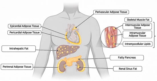
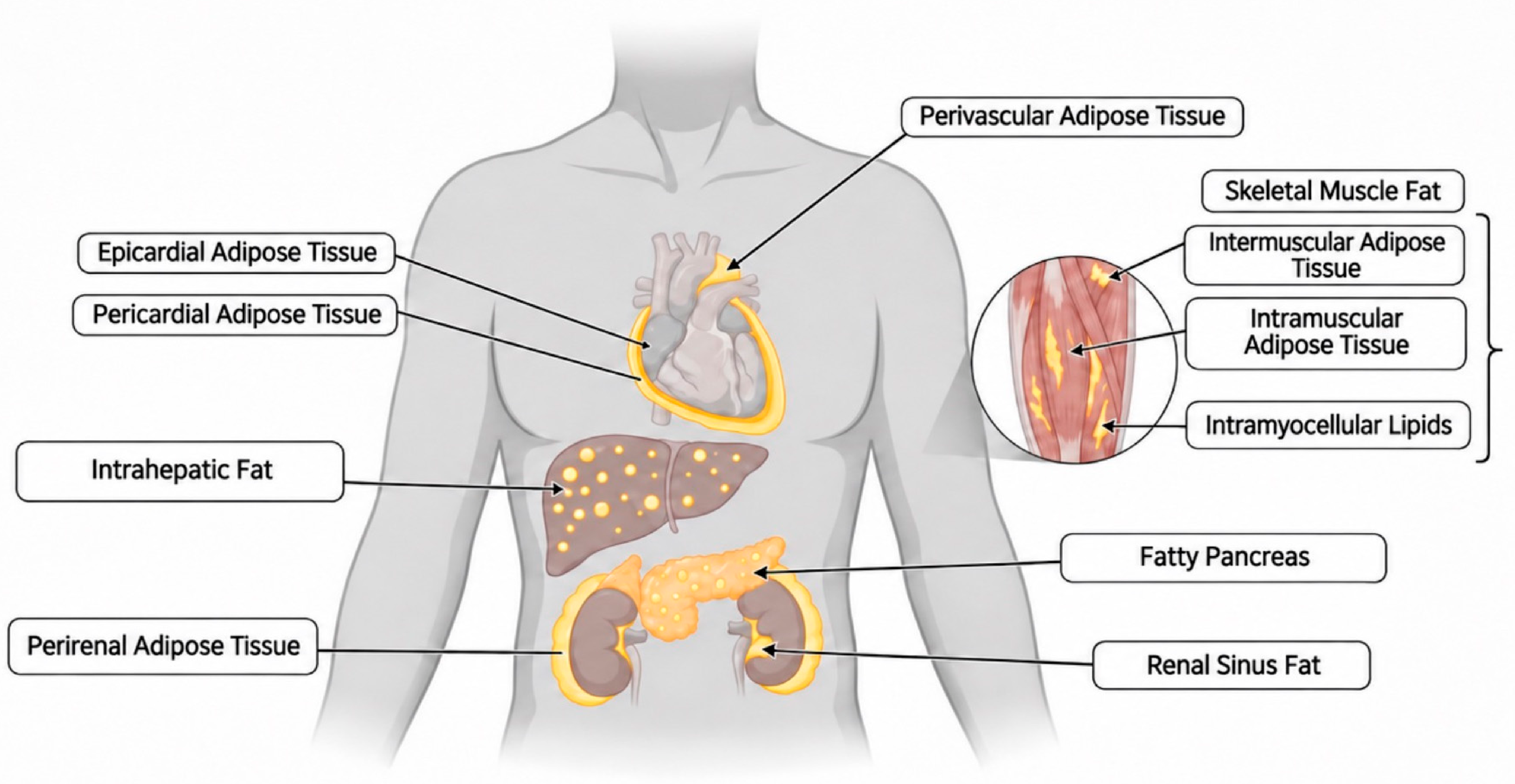
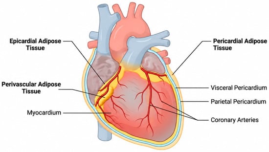
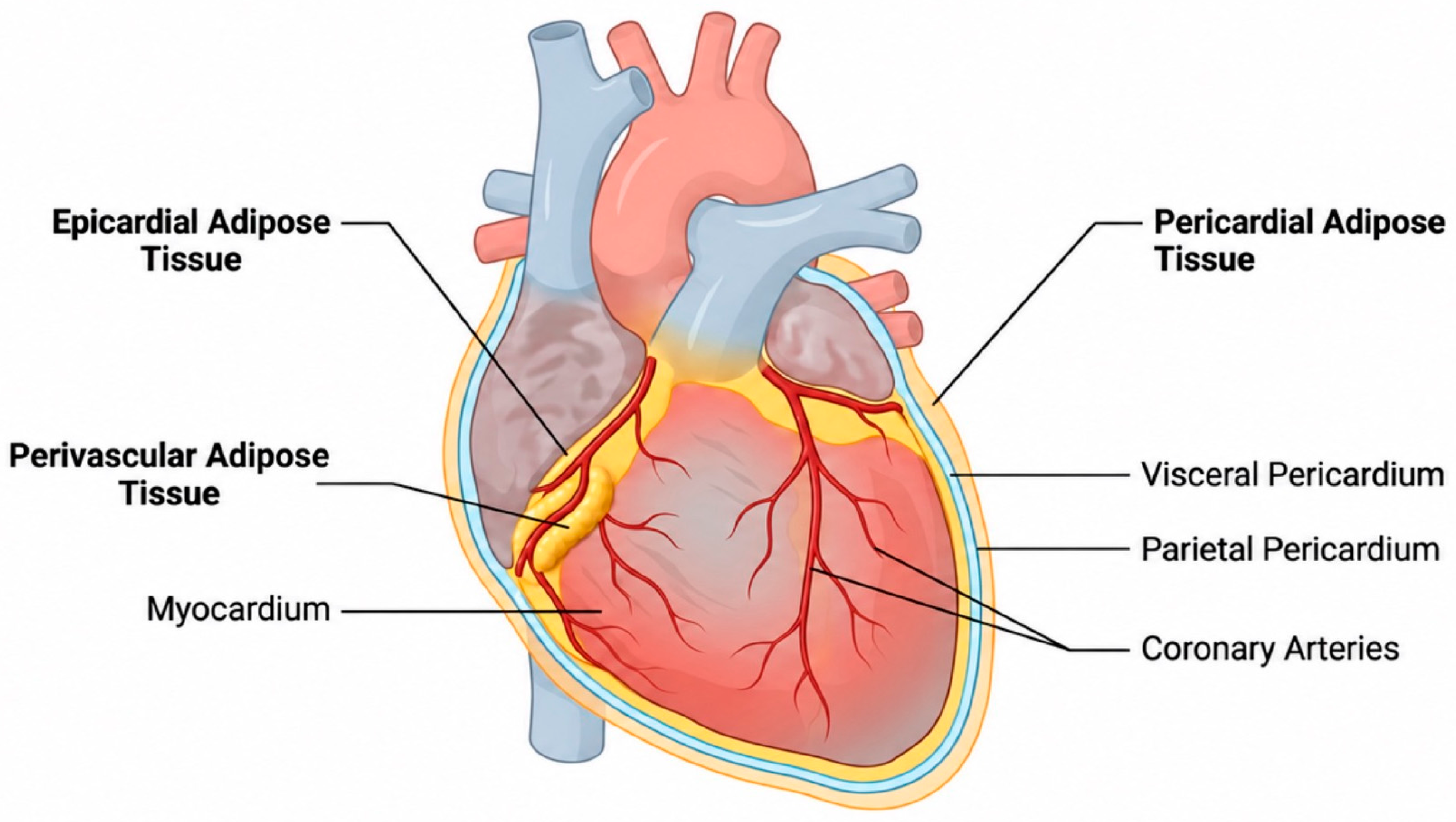
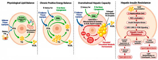
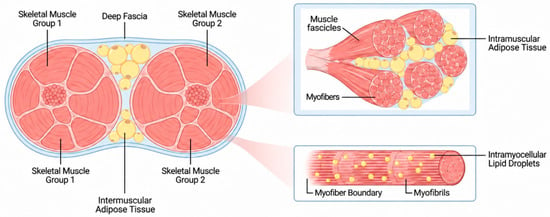
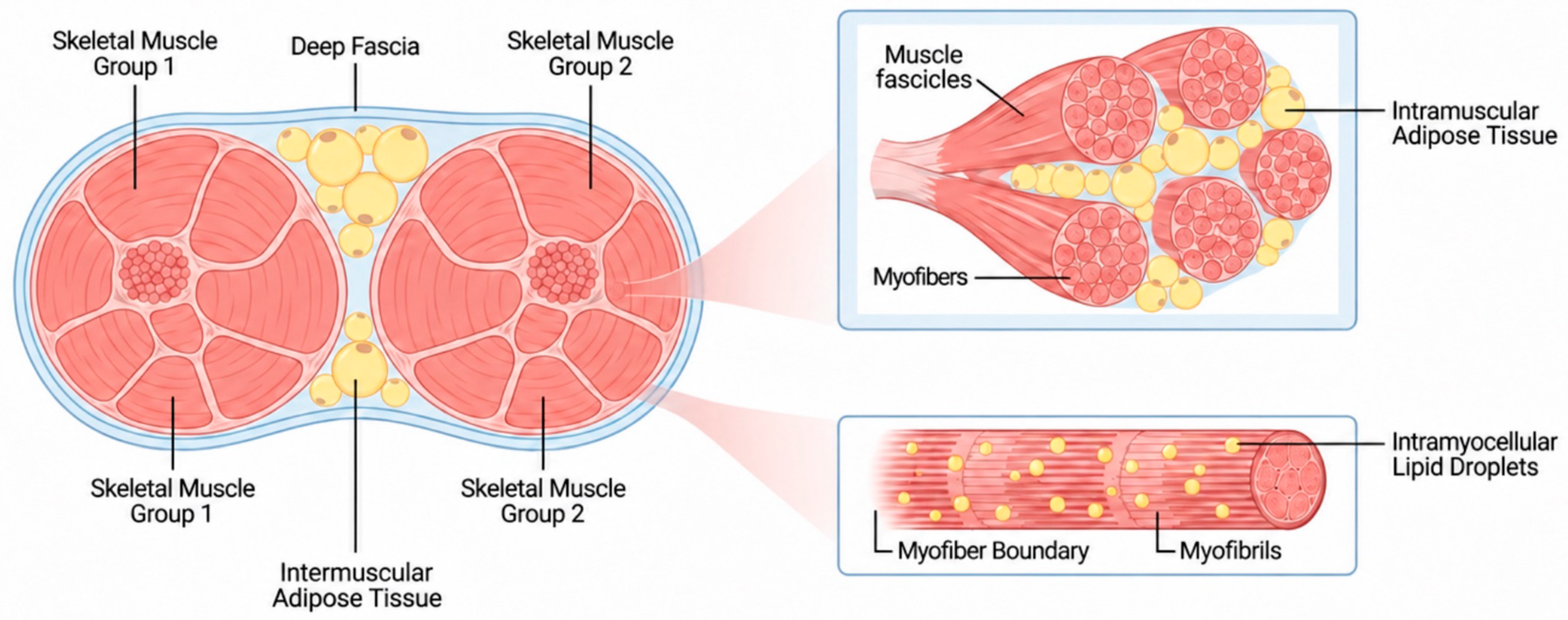
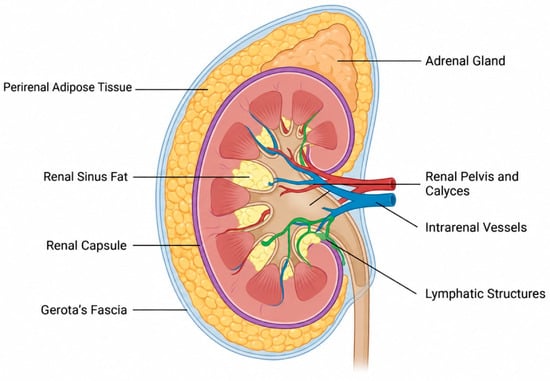
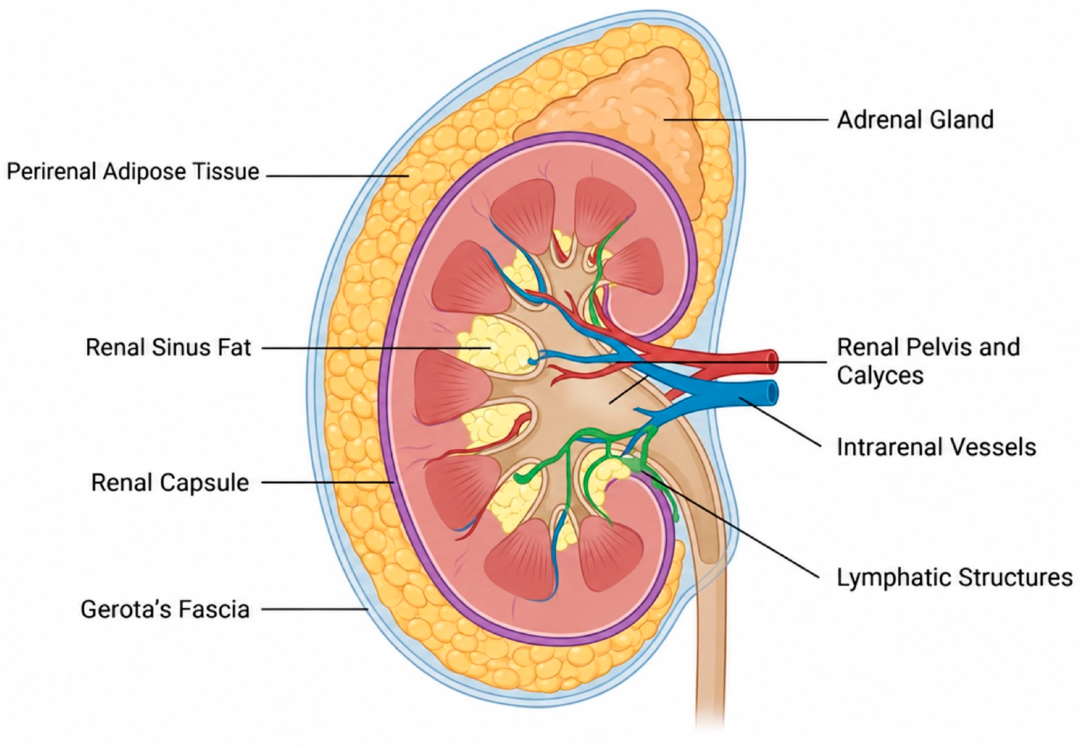
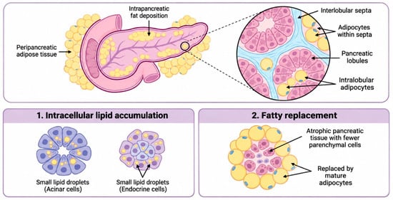
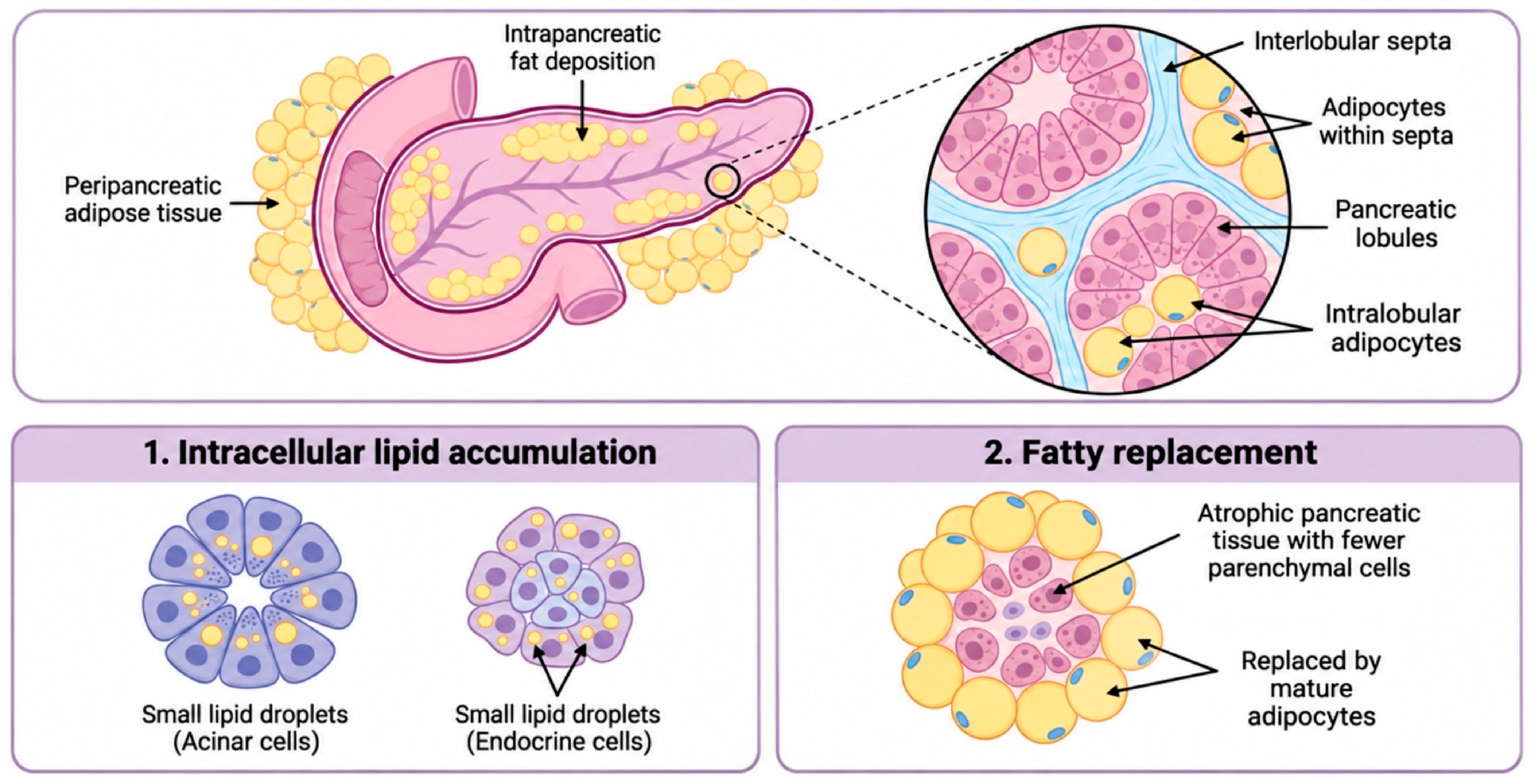
Disclaimer/Publisher’s Note: The statements, opinions and data contained in all publications are solely those of the individual author(s) and contributor(s) and not of MDPI and/or the editor(s). MDPI and/or the editor(s) disclaim responsibility for any injury to people or property resulting from any ideas, methods, instructions or products referred to in the content. |
© 2026 by the authors. Licensee MDPI, Basel, Switzerland. This article is an open access article distributed under the terms and conditions of the Creative Commons Attribution (CC BY) license.