


Highlights
What are the main findings?
- •
- DNA methyltransferase (DNMT) 3A is upregulated in fibrotic corneas.
- •
- DNMT3A inhibition using the FDA-approved agent decitabine suppresses corneal fibrosis.
What are the implications of the main findings?
- •
- DNMT3A-dependent DNA methylation is implicated in corneal myofibroblast differentiation and corneal scar formation.
- •
- Repurposing decitabine as a pharmacological, donor-independent strategy to reduce corneal fibrosis and preserve corneal transparency.
Abstract
Corneal fibrosis and opacity are major causes of visual impairment and blindness, while the conventional treatment by corneal transplantation remains limited due to the global shortage of donor materials and the risk of immune-mediated graft rejection. In this study, we investigated DNA methyltransferase (DNMT) inhibition using the FDA-approved agent decitabine (DAC; 5-aza-2′-deoxycytidine) to suppress corneal fibrosis. DNMT3A is a predicted target of the anti-fibrotic microRNA miR-29a, which was previously linked to the scar-reducing effects of cornealstromal stem cell-derived extracellular vesicles (doi:10.1016/j.jare.2022.05.008). DNMT3A expression was upregulated in scarred mouse corneas. In donor-derived corneal stromal fibroblasts, DAC dose-dependently suppressed TGFβ1-induced αSMA expression and attenuated myofibroblast-associated morphological and functional changes, including collagen gel contraction, at concentrations up to 50 μM. RNA sequencing further showed that DAC reversed TGFβ1-driven extracellular matrix remodeling, including changes in SPOCK1 and CTHRC1 expression. These effects were supported in injured mouse corneas, where topical DAC treatment reduced SPOCK1 and CTHRC1 expression, decreased scar formation, and restored corneal thickness toward normal levels. Fibrosis- and inflammation-associated gene expression was also downregulated, with no apparent treatment-related toxicity. In conclusion, these findings demonstrate that DNMT inhibition suppresses corneal fibrosis and myofibroblast differentiation, supporting the repurposing of DAC as a pharmacological, donor-independent strategy for reducing corneal fibrosis and preserving corneal transparency.
1. Introduction
Corneal transparency is essential for normal vision, as a clear cornea allows light to pass through and be effectively refracted onto the retina. However, trauma, infection, and chemical or thermal burns can disrupt the highly organized corneal structure, resulting in haze, opacity, and scarring. Following injury, inflammatory and fibrotic responses activate normally quiescent corneal stromal keratocytes (CSKs), which transition into repair-type stromal fibroblasts (SFs) and may further differentiate into myofibroblasts (myoFs) under stimulation by pro-fibrotic cytokines and signaling pathways, including transforming growth factor β (TGFβ)/Smad, MAPK/p38/ERK/JNK, PI3K/AKT, interleukins (ILs), and tumor necrosis factor α (TNFα) [1,2,3,4]. Activated SFs and myoFs produce and deposit excessive and disorganized extracellular matrix (ECM), disrupting the highly ordered stromal structure. MyoFs characteristically express α-smooth muscle actin (αSMA) and exhibit a highly contractile phenotype that contributes to wound contraction and closure but can also promote tissue distortion and fibrotic remodeling. Together with abnormal ECM deposition and organization, these fibrotic tissues increase stromal light scattering and lead to corneal haze and opacity, blocking light passage and leading to visual impairment [5].
Corneal opacity and scarring are major causes of blindness worldwide, affecting more than 12 million individuals (Eye Bank Association of America; https://restoresight.org/cornea-donation/; accessed 20 August 2026) and accounting for about 3.2% of global blindness [6]. Each year, an estimated two million new cases occur [7]. Donor corneal transplantation remains the conventional treatment for restoring vision in patients with severe corneal opacity. However, its widespread use and long-term success are limited by multiple challenges, including immune-related graft rejection, the need for prolonged immunosuppression and its associated adverse effects, postoperative complications, and, most importantly, the global shortage of suitable donor tissue [8,9]. Although the cornea is the most transplanted tissue worldwide, with about 185,000 procedures performed annually, only 1 in 70 patients in need of corneal transplantation has access to suitable donor tissue [10]. Therefore, restoring corneal transparency in these patients can be transformative for their vision, quality of life, and independence. This situation underscores an urgent need for effective, accessible, and donor-independent therapies that prevent or reduce corneal fibrosis and preserve or restore corneal transparency.
Treatment with human corneal stromal stem cells (hCSSCs) has emerged as a potential therapeutic strategy to suppress injury-induced corneal inflammation and fibrosis while promoting regenerative healing with reduced opacities. In mouse models, topical administration of hCSSCs exerted potent anti-inflammatory effects through secretion of TNFα-stimulated gene 6 (TSG6), which suppressed neutrophil infiltration into injured corneas [11]. Human CSSCs also produce the anti-fibrotic and regenerative cytokine TGFβ3 and deliver TGFB3 mRNA through extracellular vesicles (EVs), thereby promoting regenerative wound healing [12,13]. Consistent with these effects, hCSSC-treated corneas showed reduced deposition of fibrosis-associated matrix components, including collagen type III (Col3), fibronectin (FN), tenascin C (TNC), and αSMA, together with reduced light-scattering opacity, demonstrating their potent scar-inhibitory activity [14]. Recently, we showed that hCSSC-derived EVs contain microRNAs with anti-fibrotic activity [15]. In a mouse model of corneal stromal injury, treatment with hCSSCs producing miR-29a-enriched EVs resulted in greater suppression of inflammation- and fibrosis-associated gene expression than treatment with cells producing EVs with lower miR-29a abundance [16]. Together, these findings indicate that the therapeutic effects of hCSSCs are mediated through multiple complementary paracrine mechanisms rather than a single pathway. Soluble anti-inflammatory factors, regenerative cytokines, and EV-mediated delivery of anti-fibrotic miRNAs appear to act in concert to limit fibrosis and promote regenerative, scar-reducing tissue healing.
DNA methylation is increasingly recognized as an important epigenetic mechanism regulating fibrotic responses [17,18]. Aberrant DNA hypermethylation within the gene regulatory regions can silence anti-fibrotic genes, favoring fibroblast activation and myoF differentiation [19]. Such changes may reinforce pro-fibrotic signaling, including TGFβ-mediated pathways that promote myoF persistence and excessive, disorganized ECM deposition. DNA methylation is mediated by a family of DNA methyltransferases (DNMTs), primarily DNMT1, DNMT3A, and DNMT3B, with DNMT3L functioning as a regulatory cofactor [20]. Conversely, DNA demethylation mediated by ten-eleven translocation (TET) enzymes counterbalances DNMT activity and modulates pathways involved in tissue fibrosis and angiogenesis [21]. Evidence from ocular and non-ocular fibrotic tissues supports the therapeutic potential of targeting aberrant DNA methylation to attenuate fibrosis. DNMT inhibition may suppress fibrotic responses, at least in part, by restoring the expression of anti-fibrotic genes silenced through DNA hypermethylation. For example, reactivation of RASAL1 and PTEN following DNMT inhibition has been associated with reduced fibrotic responses in models of liver fibrosis [22]. In human conjunctival fibroblasts, treatment with the DNMT inhibitor 5-aza-2′-deoxycytidine (5-Aza-dC; decitabine, DAC) reduced TGFβ2-induced expression of αSMA and type I collagen [23]. Given that DAC is already clinically used for the treatment of hematologic malignancies [24], these findings provide a strong rationale for investigating whether pharmacological inhibition of DNMT activity with DAC can suppress corneal fibrosis and reduce scar formation.
Of particular interest, DNMT3A is a predicted target of miR-29a and ranks highly in both miRTARGET and miRDB analyses (see Results). In our previous study, miR-29a was enriched in EVs derived from hCSSCs with potent anti-fibrotic and scar-inhibitory activity [16]. This finding raises the possibility that EV-delivered miR-29a may suppress DNMT3A expression and thereby modulate DNA methylation programs associated with fibroblast activation and myoF differentiation. Reduced DNMT3A activity could, in turn, suppress fibrotic responses, including αSMA expression and excessive or aberrant ECM deposition. Validating this hypothesis could suggest a potential hCSSC-EV-associated miR-29a–DNMT3A axis through which epigenetic regulation may contribute to the suppression of corneal fibrosis. Based on this premise, the present study investigated the involvement of DNMT3A in corneal myoF and scar formation and evaluated whether pharmacological DNMT inhibition with DAC could provide a donor-independent therapeutic strategy to reduce corneal fibrosis and promote regenerative, scar-reducing corneal tissue healing.
2. Materials and Methods
2.1. Donor Corneas
Research-grade corneas from de-identified donors were obtained from the Center for Organ Recovery and Education (CORE; protocol #161, approved 12 September 2024), Pittsburgh, PA, and Eversight Eye Bank (Cleveland, OH, USA). The study was approved by the University of Pittsburgh Human Stem Cell Research Oversight Committee (protocol #202300049; approved 17 February 2025). Donors had no history of ocular disease or cancer and tested negative for human immunodeficiency virus (HIV), hepatitis B and C viruses, and syphilis. Corneas had no prior injury and an endothelial cell count >2500 cells/mm2. Tissues were recovered under sterile conditions, preserved in Optisol-GS (Bausch & Lomb, Rochester, NY, USA) within 12 h of donor death, and used within 12 days. Seven donor corneas (age of 63 ± 14 years and male/female ratio of 1:2) were used (Supplementary Table S1).
2.2. Primary Cultures of Human Corneal Stromal Keratocytes (hCSKs) and Human Stromal Fibroblasts (hSFs)
After removal of the corneal epithelium and endothelium, the central stroma (8-mm diameter) was isolated and digested with 0.1% (w/v) collagenase A for 6–8 h at 37 °C. The digest was passed through a 40 mm cell strainer (Corning, NY, USA), and a single-cell suspension was collected, washed, and seeded on collagen-I-coated tissue culture polystyrene surfaces using the ERI protocol supplemented with 0.5% heat-inactivated fetal bovine serum (FBS, Gibco, Thermo Fisher Sci, Waltham, MA, USA), referred to as 0.5SERI medium, to expand hCSKs through P4 [25,26]. To induce quiescent hCSKs, expanded cells were cultured in serum-free ERI medium for a week and characterized by the expression of keratocyte markers, keratocan (KERA) and aldehyde dehydrogenase 3A1 (ALDH3A1). hSFs were generated by culturing hCSKs in DMEM/F12 containing 5% FBS (DF5) for two passages, and the fibroblastic phenotype was confirmed by collagen type III a1 (COL3A1) and fibronectin (FN1) expression.
2.3. In Vitro Fibrosis Assay
Human SFs seeded at <2000 cells/cm2 on collagen-I-coated coverslips were cultured in DF5. Myofibroblast (myoF) differentiation was induced by treatment with 5 to 10 ng/mL TGFβ1 (Sigma-Aldrich, St Louis, MO, USA) under 0.5% FBS conditions. To assess the effect of decitabine (DAC; Sigma-Aldrich) on fibroblast-to-myofibroblast differentiation, hSFs were co-treated with TGFβ1 and varying concentrations of DAC (5–250 μM). MyoF differentiation was evaluated by αSMA expression.
2.4. Collagen Gel Contraction Assay
A 24-well culture plate was coated with sterile PBS containing 1% Pluronic F127 (Sigma-Aldrich), washed, and air-dried. Human SFs were suspended in rat type I collagen (0.8 mg/mL, Corning) at 5 × 10⁴ cells/mL, and 0.5 mL of the cell–collagen mixture was added to each F127-coated well. After 15–30 min at 37 °C, the polymerized gel was overlaid with culture medium containing 0.5% FBS and TGFβ1 (5 ng/mL) with or without DAC (10–50 µM) and released from the wells using a sterile 21-G needle to initiate contraction. After 36 h, the gel was imaged and its area was quantified using ImageJ (NIH). Gel area was expressed as a percentage of the well area. Experiments were performed in quadruplicate, and data are presented as mean ± SD with statistical significance assessed using the Mann–Whitney U test for two-group comparisons or the Kruskal–Wallis test with Dunn–Bonferroni correction for multiple comparisons. Gels were fixed for immunofluorescence or lysed for total RNA extraction to analyze αSMA expression.
2.5. Immunostaining
Cells on glass coverslips were fixed with ice-cold absolute ethanol for 20 min on ice and blocked/permeabilized in PBS containing 0.15% saponin (Sigma-Aldrich), 1% bovine serum albumin (BSA, Sigma-Aldrich), and 1.5% normal goat serum (NGS, Invitrogen) for 1 h at room temperature (RT). Samples were incubated with an AlexaFluor488-conjugated mouse-anti-human αSMA antibody (DAKO, Agilent, Santa Clara, CA, USA) for 2 h at RT in the dark, followed by nuclear counterstaining with 4,6-diamidino-2-phenylindole (DAPI, Sigma-Aldrich) for 15 min. After PBS washes, samples were mounted using Immuno-Mount (Thermo Fisher Sci) and imaged using a confocal microscope (FV1000, Olympus, Tokyo, Japan) under identical acquisition settings. αSMA-positive cells were quantified from at least 50 randomly selected cells imaged with a 10× objective, and the percentage of positive cells was calculated. For quantitative fluorescence analysis, RGB images were split into individual channels using ImageJ version 1.54u8, and the αSMA channel was converted to 8-bit grayscale and thresholded consistently to generate binary images defining regions of interest (ROIs). Mean gray values within the ROIs were measured after background subtraction using identical processing parameters across all samples.
2.6. Flow Cytometry
Human SFs were incubated with LIVE/DEAD Fixable Aqua Dead Cell Stain (Invitrogen, Thermo Fisher Scientific, Waltham, MA, USA) for 5–7 min at RT and washed with PBS. For intracellular staining, cells were fixed with freshly prepared neutral-buffered 3% paraformaldehyde for 15 min, permeabilized with Perm Buffer (Invitrogen) for 7 min on ice, and washed with PBS. After filtration through a 70 µm cell strainer, cells were resuspended in staining buffer (PBS containing 1% FBS) with Fc-Block and incubated with an AlexaFluor488-conjugated mouse-anti-human αSMA antibody (Invitrogen) for 30 min on ice in the dark. Unstained and isotype controls were included for gating and assessment of non-specific fluorescence. Samples were analyzed using a CytoFLEX LX flow cytometer (Beckman Coulter, Brea, CA, USA), and αSMA expression was quantified in the viable cell population.
2.7. Cell Viability and Growth Assays
Cell viability counting was performed by 0.4% trypan blue dye exclusion using a Countess Automatic Cell Counter (Invitrogen). Each sample was counted three times, and the mean ± SD was calculated. The cell growth assay was performed after seeding at 4 × 10³ cells/well in E-plate 96 plates. After 24 h, the medium was replaced with fresh culture medium containing TGFβ1 (10 ng/mL) with or without DAC (10–25 μM), and cell growth was monitored using an xCELLigence Real-Time Cell Analyzer RTCA SP (Agilent/ACEA). Experiments were performed in triplicate, and impedance was recorded every 15 min for 120 h and expressed as cell index (CI). Significance between groups was assessed using the Mann–Whitney U test.
2.8. Total RNA Extraction and Gene Expression
RNA was extracted from cells using the RNeasy Mini Plus Kit (QIAGEN #24136) following the manufacturer’s instructions with an additional homogenization step using the QIAshedder (QIAGEN, Germantown, MD, USA). For RNA extracted from mouse corneal tissues, the homogenization step was replaced by mechanical disruption using MagNA Lyser beads in a MagNA Lyser Instrument (Roche, Carlsbad, CA, USA). RNA was resuspended in nuclease-free water and quantified on a NanoDrop spectrophotometer (Thermo Fisher). Total RNA (500 ng) was reverse transcribed with the SuperScript III Reverse Transcriptase kit (Thermo Fisher) and random primer hexanucleotides (10 ng/mL). cDNA was assayed for target gene expression using SYBR Green Real-Time Master Mix (Life Technologies, Carlsbad, CA, USA) and specific primers (Supplementary Table S2) in a QuantStudio3 Real-Time PCR System (Applied Biosyst, Thermo Fisher). Experiments were performed in triplicate. Melting curves were constructed to validate the specificity of primer sets, and negative controls with no-template (no cDNA) reactions and minus RT reactions were included. Relative RNA abundance was determined using the ΔCt method after normalization to housekeeping 18S. The expression fold change was calculated using the 2−ΔΔCt method and expressed as mean ± SD. Statistical significance was determined using a non-parametric Mann–Whitney U test.
2.9. RNA-Seq, Data Processing, and Pathway Analysis
Total RNA was purified from HC579, HC616, and HC618 under three experimental conditions (naïve, TGFβ1-treated, and TGFβ1+DAC-treated; nine samples in total). RNA quality and quantity were assessed using a 5300 Bioanalyzer (Agilent Technologies) and a NanoDrop ND2000 spectrophotometer (Thermo Fisher). High-quality RNA samples (concentration ≥20 ng/mL, total RNA >100 ng, and RQN >1) were used for bulk RNA-seq. Library preparation and sequencing were performed by Novogene Co., Ltd. using the NovaSeq X Plus platform with 150-bp paired-end (PE150) sequencing to generate ~40 million reads per sample. The quality of FASTQ files was assessed using FastQC and summarized across all samples using MultiQC. Adaptor sequences and low-quality bases (quality score <20) were removed before alignment. Processed reads were aligned to the human GRCh38 reference genome (Ensembl release 102) using STAR (version 2.7.11b). Gene-level read counts were generated during alignment using a reverse-stranded protocol, and counts from all nine samples were combined into a single expression matrix. Differential gene expression analysis was performed in R (version 4.6.0) using DESeq2 (version 1.52.0), with donor included as a covariate to account for the paired experimental design. Log2 fold-change estimates were shrunk using the apeglm method. p values were adjusted for multiple testing using the Benjamini–Hochberg false discovery rate (FDR) method, and differentially expressed genes (DEGs) were defined as those with an adjusted p value (FDR) < 0.05 and an absolute log2(fold change) > 1. Principal component analysis (PCA), volcano plots, and heatmaps were generated in R.
DEGs were further assessed using CLC Genomics Workbench (version 26), with donor included as a blocking factor. For the functional enrichment analysis, protein-coding DEGs were selected in CLC and submitted to the Database for Annotation, Visualization, and Integrated Discovery (DAVID; https://david.ncifcrf.gov/, accessed 30 May 2026) for functional annotation and enrichment analysis. Enriched terms were then filtered using DAVID’s Bonferroni-corrected p < 0.05. Enriched Gene Ontology (GO) terms encompassing molecular function (MF), biological process (BP), and cellular component (CC) were identified using an enrichment score ≥ 3. Selected enriched pathways were visualized as enrichment bubble plots generated with SRplot (http://www.bioinformatics.com.cn/SRplot, accessed 30 May 2026) [27]. Heatmaps illustrating expression changes in genes associated with selected pathways were generated using log2 fold-change values.
2.10. Mouse Models of Acute Anterior Stromal Injury and Treatment
The research strictly adhered to the guidelines outlined in the Care and Use of Laboratory Animals of the National Institutes of Health (NIH) and the Association for Research in Vision and Ophthalmology (ARVO) Statement for the Use of Animals in Ophthalmic and Vision Research. The protocol was approved by the Institutional Animal Care and Use Committee (IACUC), University of Pittsburgh, under protocol 24115664 (approved 21 November 2024). A total of 38 BALB/c mice (breeders from Charles River Lab., Wayne, PA) aged 10 to 12 weeks were used. They were housed in an AAALAC-accredited animal facility with 12 h light–12 h dark cycles and provided with unrestricted food and water. After anesthetization by intraperitoneal injection of ketamine (50 mg/kg) and xylazine (5 mg/kg) (Dechra, Fort Worth, TX, USA), the right eyes of all mice received topical proparacaine hydrochloride (0.5%, Alcon, Fort Worth, TX, USA) for local analgesia [28]. The central corneal epithelium (2 mm diameter) was removed by a high-speed Algerbrush II (Accutome Inc, Malvern, PA, USA). The basement membrane and anterior stroma of 0.5 mm diameter and 10 to 20 μm depth were ablated by a second Algerbrush burr [28]. After rinses with normal saline and brief drying with a sterile cotton spear, the injured corneas were randomly assigned to treatment or control groups. Eye drops (5 µL) containing either saline or DAC (25 or 50 µM) were administered thrice daily for three consecutive days. In addition, all treated eyes received topical tobramycin (0.3%, USP, Somerset Therapeutics, Hollywood, FL, USA) thrice daily for 3 days to prevent infection. Each group had a minimum of six corneas according to the sample size calculation in the Section 2.12.
2.11. Ophthalmic Examination and Scar Measurement
Before and after injury, and at day 10 after treatment, mouse corneas were examined for cross-sectional changes using spectral-domain optical coherence tomography (SD-OCT, Envisu R2210, Leica [Bioptigen], Morrisville, NC, USA) with a linear volume scan (4 × 4 mm area, 1024 × 1000 × 100 volumes) [29]. The scanning was performed in a masked fashion. Images were processed using NIS Elements software v6.10.01 (Nikon, Melville, NY, USA). Central corneal thickness (CCT) was measured as the average of five measurements taken at the center (0 mm) and 0.5 mm on both vertical and horizontal meridians. Mice were euthanized by an overdose of intraperitoneal sodium pentobarbital followed by cervical dislocation. Corneas were isolated and imaged, and the entire corneal and scar areas were measured using ImageJ software (version 1.54u8, NIH, Bethesda, MD, USA) and MetaMorph 7.7.3 (Molecular Devices Inc., San Jose, CA, USA) in a blinded manner. Significance between groups was assessed using the Mann–Whitney U test.
2.12. Statistics
All experiments were done in triplicate. For in vivo studies, a minimum of six corneas per group were analyzed. Sample size was determined using G*Power 3.1, n = 1 + 2C(s/d)2 [30], where s is the standard deviation (SD), d is the difference to be detected, and C = 10.51 at p = 0.05 (α) and power (1 − β) = 0.8. Using an expected difference of 30% and an SD of 20%, the calculated sample size was six corneas per group. Data normality was assessed using the Shapiro–Wilk test. Comparisons between two independent groups were performed using an unpaired two-tailed Student’s t-test for normally distributed data, or the Mann–Whitney U test for non-normally distributed (non-parametric) data. Statistical analyses were conducted using GraphPad Prism 11.0.2, with p < 0.05 considered statistically significant.
3. Results
3.1. Elevated DNMT3A Expression in Corneal Scarring
Cell-based therapy using hCSSCs has been shown to suppress inflammation and fibrosis and promote regeneration of native-like corneal stroma in multiple mouse models of corneal injury [11,12,14,15,16,28,31,32,33,34,35]. Treated corneas showed reduced deposition of fibrotic matrix and decreased formation of light-scattering opacities, demonstrating the potent scar-inhibitory activity of hCSSCs. In our recent study, hCSSCs producing EVs enriched in hsa-miR-29a exhibited enhanced suppression of fibrosis and improved stromal regeneration and restoration of transparency in injured mouse corneas (Supplementary Figure S1) [16]. To identify potential targets of hsa-miR-29a, we performed target prediction analysis using miRTARGET and miRDB. DNMT3A was identified as a high-ranking predicted target of hsa-miR-29a, ranking sixth in miRTARGET (https://mirtarget.com/; accessed 2 June 2026) (Figure 1A) and 11th in miRDB (https://mirdb.org; accessed 2 June 2026) (Supplementary Figure S2) [36,37,38]. Conversely, when candidate miRNAs predicted to target DNMT3A were ranked, hsa-miR-29a ranked second in miRTARGET (Figure 1A) and fourth in miRDB (Supplementary Figure S2). These reciprocal in silico analyses consistently identified a potential regulatory association between hsa-miR-29a and DNMT3A. Therefore, we hypothesized that suppression of DNMT3A-mediated DNA methylation may contribute to the anti-fibrotic and regenerative effects associated with hCSSC treatment.
Figure 1.
Elevated DNMT3A expression in corneal scarring. (A) miRTARGET analysis showed the top 10 predicted miR-29a-target genes and the top 10 predicted miRNAs targeting DNMT3A, ranked by calculated prediction scores. (B) qPCR analysis showing that DNMT3A was consistently upregulated in myoFs relative to their matched SFs (donors YD67, 384, 579, and 652), whereas DNMT1 and 3B showed no significant changes. (C) Representative mouse corneas with anterior stromal injury followed by treatment with different batches of hCSSCs, showing varying degrees of scar inhibition. Injury-only corneas developed severe stromal scarring, whereas naïve controls remained clear. (D) DNMT3A expression was significantly lower in corneas exhibiting scar inhibition, whereas injury-only controls and corneas that developed scars showed significantly higher DNMT3A expression (Mann–Whitney U test; n > 8 per group). Naïve clear corneas had minimal DNMT3A expression [39,40,41,42].
To investigate the association of DNMT3A with the fibrotic reaction of corneal stromal cells, we examined the expression of different DNMT family members in four pairs of donor-matched SFs and their TGFβ1-induced myoFs. qPCR analysis showed that DNMT3A was consistently upregulated in myoFs relative to their matched SFs, with increases up to >6000-fold (YD/67 cells showed a 194-fold increase in DNMT3A in myoFs compared with SFs, MyoF579 had 6518-fold higher expression than SF579, MyoF384 had 8-fold higher expression than SF384, and MyoF652 had around 1.6-fold higher expression than SF652) (Figure 1B). However, DNMT3B and DNMT1 showed no consistent changes in myoFs.
To further determine whether DNMT3A expression was also associated with corneal fibrosis in vivo, we analyzed injured mouse corneas exhibiting different degrees of scar inhibition following treatment with different donor CSSC batches, as previously reported (Figure 1C) [16,28]. Corneas with scar inhibition (n = 22 corneas) showed significantly attenuated DNMT3A expression when compared with injured controls (n = 20) and treated corneas that developed scars (n = 20) (p < 0.001 compared with injured controls; p = 0.003 compared with hCSSC-treated corneas with scar formation) (Figure 1D). Together, these findings indicate a consistent association between elevated DNMT3A expression and the fibrotic phenotype, supporting the potential involvement of DNMT3A in corneal myoF differentiation and scar formation.
3.2. DNMT Inhibition Suppressed Myofibroblast Phenotype
To determine whether DNMT3A inhibition suppresses myoF differentiation, primary hSFs (SF579, SF584, and YD/67) were treated with recombinant human TGFβ1 (10 ng/mL) with or without the DNMT inhibitor decitabine (DAC) for 72 h. TGFβ1 treatment induced robust expression of αSMA, a characteristic marker of myoFs (Figure 2A). Co-treatment with DAC markedly suppressed TGFβ1-induced αSMA expression. Quantitative fluorescence analysis using ImageJ showed that mean αSMA intensity per unit area was significantly reduced by DAC at both 5 and 10 μM (n > 50 cells per group; p < 0.001 for both concentrations; Mann–Whitney U test) (Figure 2B).
Figure 2.
Decitabine suppressed myofibroblast phenotype. (A) Compared with naïve cells, TGFβ1-treated human corneal stromal fibroblasts (SFs) exhibited strong αSMA expression, indicative of myofibroblast differentiation. Co-treatment with DAC (5 and 10 μM) substantially suppressed TGFβ1-induced αSMA expression. (B) Quantification of αSMA staining intensity showed that DAC co-treatment significantly reduced αSMA staining compared with TGFβ1 treatment only, whereas naïve SFs exhibited minimal αSMA expression (p < 0.05). A minimum of 50 cells were analyzed per group. (C) ACTA2 expression was significantly upregulated in hSFs following TGFβ1 treatment. Co-treatment with DAC (10 μM) for 1, 5, and 10 days significantly reduced TGFβ1-induced ACTA2 expression (p < 0.05). (D) Flow cytometry analysis showed that DAC (5 to 50 μM) dose-dependently reduced the percentage of αSMA-positive cells following co-treatment with TGFβ1 compared with TGFβ1 alone. Naïve and isotype-control cells exhibited minimal αSMA staining. (E) Across three independent SF batches (SF579, SF584, and YD/67), DAC treatments reduced the mean percentage of αSMA-positive cells, with a significant reduction observed at 25 μM, reaching levels close to those of naïve control cells. Data are presented as mean ± SD. p values were calculated using the non-parametric Mann–Whitney U test.
Increased cell size is another characteristic morphological feature associated with myoF differentiation [43]. Consistent with αSMA suppression, a 3-day treatment with DAC (5 and 10 μM) significantly reduced the TGFβ1-induced increase in cell area (p < 0.05 for both concentrations, paired Student’s t-test) (Supplementary Figure S3). Consistent with the changes in αSMA protein expression, qPCR analysis showed that co-treatment with DAC (10 μM) significantly reduced TGFβ1-induced ACTA2 mRNA expression at days 1, 5, and 10 (p = 0.01 for day 1, p = 0.02 for day 5, and p = 0.01 for day 10, Mann–Whitney U test) (Figure 2C). These findings indicate that DAC suppresses TGFβ1-induced αSMA expression and associated morphological changes and that its inhibitory effect on ACTA2 expression is sustained during prolonged treatment without potential reversal of the suppressive effect.
Changes in αSMA expression were further assessed by flow cytometry. In SF579 cells, treatment with TGFβ1 (10 ng/mL) for 3 days increased the percentage of αSMA-positive cells from 3% to 43% (Figure 2D). Co-treatment with DAC reduced the proportion of αSMA-positive cells in a dose-dependent manner to 29%, 19%, and 12% at 5, 10, and 50 μM, respectively. Consistent with these findings, analysis of three independent donor-derived hSF batches (SF579, SF618, and YD/67) showed an overall reduction in the percentages of αSMA-positive cells following DAC co-treatment compared with TGFβ1 alone (Figure 2E). In response to DAC treatment, the proportion of αSMA-positive cells decreased to levels close to those in naïve control cells. These results thus indicate that DAC suppresses TGFβ1-induced myoF differentiation in donor-derived hSFs.
3.3. DAC Reduced Myofibroblast-Mediated Collagen Gel Contraction
The functional contractile activity of TGFβ1-treated myoFs was assessed using a collagen gel contraction assay (Figure 3A). As a control, collagen gel containing donor-derived hSFs (SF579, SF616, and SF618) cultured in 0.5% FBS showed minimal contraction, with the gel area remaining at 91 ± 2.9% of the culture well size after 36 h (Figure 3B). Treatment with TGFβ1 (10 ng/mL) markedly increased gel contraction, reducing the mean gel area to 33.6 ± 7.6% (p = 0.017, Mann–Whitney U test). In contrast, DAC substantially inhibited TGFβ1-induced gel contraction, with the resulting gel area maintained at 83.5 ± 6.8% for DAC at 5 μM, 90 ± 6.8% at 25 μM, and 79.3 ± 9.4% at 50 μM, respectively. A similar inhibitory effect was observed with mitomycin C (MMC, 20 μg/mL), which was included as a reference control for suppression of myoF contractile activity. Multiple group comparisons using the Kruskal–Wallis test with Dunn–Bonferroni correction showed no significant differences among the DAC- and MMC-treated groups.
Figure 3.
Decitabine functionally reduced myofibroblast-mediated contractility while maintaining cell viability at low doses. (A) Collagen gel contraction assay. Human SFs were seeded at 5 × 10⁴ cells/mL in collagen gel (0.8 mg/mL) under the indicated treatment conditions. After 36 h, gel area was quantified using ImageJ software, and the percentage of gel area relative to the original well area is presented as mean ± SD in (B). Representative immunofluorescence images showed αSMA expression in collagen gels under the respective treatment conditions. Nuclei were stained with DAPI. Scale bars: 25 μm. (B) Compared with gel containing SFs only, TGFβ1 (10 ng/mL) treatment induced significant collagen gel contraction (p = 0.017; n > 6 samples), whereas co-treatment with DAC (5 to 50 μM) or MMC (20 μg/mL) significantly inhibited TGFβ1-induced contraction (p = 0.022 for 5 μM DAC, p = 0.017 for 25 μM DAC, p = 0.024 for 50 μM DAC, and p = 0.013 for 20 μg/mL MMC). No significant differences were observed among the DAC- and MMC-treated groups (Kruskal–Wallis test and Dunn–Bonferroni correction). (C) qPCR analysis of cells in collagen gels revealed significant downregulation of TGFβ1-induced ACTA2 expression following treatment with DAC (25 and 50 μM) or MMC. p = 0.011 for 25 μM DAC, p = 0.02 for 50 μM DAC, and p = 0.007 for 20 μg/mL MMC, when compared with TGFβ1 treatment only. (D) Trypan blue dye exclusion assay showed that co-treatment with TGFβ1 and DAC at concentrations below 100 μM maintained >85% hSF viability. (E,F) Real-time cell impedance analysis using xCELLigence showed significantly increased cell index in TGFβ1-treated cells compared with untreated control and DAC co-treated cells (p = 0.036 for both 10 μM DAC and 25 μM DAC added with TGFβ1), consistent with morphological changes associated with TGFβ1-induced myoF differentiation and their attenuation by DAC. Data are presented as mean ± SD. p values were calculated using the non-parametric Mann–Whitney U test.
Consistent with these functional contractile responses, αSMA-positive cells were detectable in TGFβ1-treated collagen gels and those treated with 5 μM DAC, whereas expression was minimal following treatments with 25 or 50 μM DAC and MMC (Figure 3A). Similarly, qPCR analysis showed significant downregulation of ACTA2 expression in collagen gel samples treated with 25 or 50 μM DAC or MMC compared with TGFβ1 treatment alone (p < 0.05; Mann–Whitney U test) (Figure 3C). These results indicate that DAC suppresses the contractile phenotype of TGFβ1-induced myoFs, which aligns well with the inhibitory effects on αSMA expression and myoF differentiation.
3.4. Effect of Decitabine on Human Corneal Stromal Cell Viability and Growth
Although DAC is an FDA-approved agent for the treatment of hematologic malignancies in patients, we evaluated its potential cytotoxicity in human corneal cells. The viability of hSFs treated with TGFβ1 and/or DAC (10 to 100 μM) was assessed using a trypan blue dye exclusion assay. Treatment with TGFβ1 (10 ng/mL) or DAC at concentrations up to 50 μM maintained >85% cell viability, with no significant differences compared with untreated controls (Figure 3D). In contrast, treatment with 100 μM DAC or MMC alone (20 μg/mL) significantly reduced cell viability (p = 0.02 for 100 μM DAC and p = 0.017 for MMC; Mann–Whitney U test).
To further assess potential cellular stress responses, expression of apoptosis- and autophagy-related genes was examined. Treatment with 100 μM DAC modestly elevated BAX expression, leading to significant upregulation of the BAX/BCL-2 expression ratio (Supplementary Figure S4). In contrast, expression of the autophagy-associated genes ATG5 and ATG7 remained unchanged. These findings indicate that DAC at concentrations up to 50 μM was well tolerated by hSFs. Treatment with higher DAC concentrations reduced cell viability, accompanied by potential activation of apoptotic pathways.
We next examined changes in SF adhesion and morphology using real-time impedance monitoring with the xCELLigence system during treatment with TGFβ1 and/or DAC (10 to 25 μM) for up to 108 h. The cell index profile showed that treatment with TGFβ1 alone significantly increased hSF coverage, indicating cell spreading and enlargement, which are morphological changes associated with myoF differentiation (Figure 3E,F) (full cell index profile with SD in Supplementary Figure S5). In contrast, co-treatment with DAC suppressed the TGFβ1-induced increase in cell index, maintaining levels close to those of the untreated control cells. These observed changes in real-time impedance support the ability of DAC to attenuate cellular and morphological changes associated with myoF differentiation.
3.5. Transcriptomic Profiling Reveals ECM Modulation by DAC Treatment
To assess the molecular response to DAC, we performed global RNA sequencing (RNA-seq) of hSFs (SF579, SF616, and SF618) following a 3-day treatment with TGFβ1 (10 ng/mL), with or without DAC (25 μM), together with untreated controls. These hSF batches were confirmed to exhibit DAC-mediated suppression of TGFβ1-induced ACTA2 expression (Supplementary Figure S6). Nine libraries (three donor SFs × three conditions) yielded 39.7–47.4 million read pairs each and passed quality control. Alignment to the human genome (GRCh38, Ensembl release 102) was efficient and uniform across donors, with 92.8–94.9% of reads uniquely mapped, confirming high data quality (Supplementary Table S4). Principal component analysis (PCA) after donor correction revealed clear separation among the three control and treatment conditions, confirming a reproducible, condition-specific transcriptional response (Figure 4A). Pairwise differential expression analysis showed that TGFβ1 treatment resulted in 1691 differentially expressed genes (DEGs), 722 upregulated and 969 downregulated, compared with untreated controls (absolute log2 fold change >1 and <−1; adjusted p (FDR) <0.05) (Figure 4B). Compared with TGFβ1 alone, co-treatment with DAC induced substantial transcriptomic reprogramming, with 1032 DEGs, including 908 upregulated and 124 downregulated genes (volcano plot in Figure 4C). Supplementary Figure S7 shows volcano plots for TGFβ1 versus control and TGFβ1+DAC versus control. The RNA-seq datasets generated in this study have been deposited in the Gene Expression Omnibus (GEO) under accession number GSE349101.
Figure 4.
Differentially expressed gene analysis of bulk RNA-seq datasets. (A) Principal component analysis (PCA) of hSFs under naïve control, TGFβ1-treated, and combined TGFβ1- and DAC-treated conditions. Samples from three independent hSF batches (SF579, SF616, and SF618) showed condition-dependent clustering, indicating distinct and reproducible transcriptional profiles across treatment groups. (B) Venn diagram showing the overlap of differentially expressed genes (DEGs) identified in TGFβ1-treated versus TGFβ1+DAC-treated hSFs, and the respective comparisons with naïve controls. Data were obtained from n = 3 biological repeats. The top 50 genes are labeled. (C) Volcano plot showing DEGs in TGFβ1+DAC-treated hSFs compared with TGFβ1 treatment alone, with 908 upregulated genes (red) and 124 downregulated genes (blue). (D) Gene Ontology (GO) enrichment analysis showing the top-ranked enriched GO terms (enrichment score >3) for DEGs identified in TGFβ1-treated versus naïve hSFs and TGFβ1+DAC-treated versus TGFβ1-treated hSFs. p values were adjusted using the Benjamini–Hochberg procedure. Bubble size represents the percentage of genes associated with each GO term, whereas statistical significance is represented by −log10(adjusted p value). DEGs from the DESeq2 analysis (B,C) were defined as genes with an adjusted p (FDR) < 0.05 and an absolute log2 fold change > 1.
To identify the biological processes involved in TGFβ1-induced SF-to-myoF transition and modulated by DAC treatment, we performed functional enrichment analysis of DEGs using DAVID Bioinformatics (https://davidbioinformatics.nih.gov/; accessed 30 April 2026). Gene Ontology (GO) analysis of TGFβ1-treated cells revealed prominent enrichment of “GO:0005576~extracellular region” and “GO:0005201~ECM structural constituent” with enrichment scores of 25.81 and 24.19, respectively (Figure 4D), indicating substantial changes in the expression of ECM remodeling genes during TGFβ1-induced myoF differentiation. In cells co-treated with DAC, the score for “GO:0005201~ECM structural constituent” was substantially reduced from 24.19 to 3.6, suggesting an alleviation of ECM remodeling. In addition, functional categories associated with fibrotic and growth factor signaling, including “GO:0045236~CXCR chemokine receptor binding” (enrichment score: 3.22) and “GO:0005520~insulin-like growth factor binding” (enrichment score: 3.55), were enriched in TGFβ1-treated cells but were absent after DAC treatment. In contrast, KEGG pathways related to integrin signaling, focal adhesion, and ECM–receptor interaction remained frequently associated with hSFs under TGFβ1 with or without DAC (Supplementary Figure S8). Collectively, these findings indicate that DAC induces substantial transcriptomic reprogramming of TGFβ1-activated hSFs and selectively reduces gene expression associated with ECM remodeling and pro-fibrotic signaling. A potential limitation of this experiment is that enrichment results reflect changes in the expression of ECM-associated genes at the transcript level. They do not, on their own, confirm altered ECM protein deposition or direct DNMT3A-dependent regulation, which would require further protein-level studies.
3.6. Topical Treatment with Decitabine Eye Drops Suppresses Corneal Scarring
Injured mouse corneas (n = 32) with anterior stromal wounds were randomly assigned to receive DAC (25 or 50 μM) or saline eye drops. Eye drops (5 μL per application) were administered thrice daily for 3 days until corneal re-epithelialization was complete. All injured corneas in DAC-treated (n = 20) and saline-treated (n = 12) groups, as well as naïve corneas (n = 32), showed similar epithelial wound closure at 48 to 54 h, as revealed by fluorescein staining. At day 14, saline-treated injured corneas developed prominent stromal scarring, whereas DAC-treated corneas generally displayed reduced scar formation and severity (Figure 5A). Treatment with 50 μM DAC (n = 9) consistently inhibited scar formation, with a significantly smaller opaque area, as quantified using ImageJ, compared with saline-treated injured controls (p = 0.002; Mann–Whitney U test) (Figure 5B). Corneas treated with 25 μM DAC (n = 11) also showed an overall reduction in opaque area, although the response was variable. Nine of the 11 corneas exhibited substantial scar inhibition, whereas two developed prominent scarring with opaque areas comparable to the mean level observed in the saline-treated injured group. This variability could have resulted from reduced drug retention on the corneal surface due to rapid tear drainage or blinking.
Figure 5.
Topical DAC treatment inhibits corneal scarring following anterior stromal injury in mice. (A) Representative corneal images showing scar outcomes at day 14 following topical treatment with DAC (25 or 50 μM) or saline (n > 6 eyes per group). Eye drops (5 μL per application) were administered three times daily for 3 days until complete corneal epithelial wound closure. DAC treatment markedly reduced corneal scar formation, whereas saline-treated injured corneas developed prominent scarring. Representative anterior-segment OCT images show the cross-sectional changes in corneal structure at day 14 post-injury. (B) Quantification of scar area showed significant scar inhibition in nine of 11 eyes treated with 25 μM DAC (p = 0.01) and all nine eyes treated with 50 μM DAC (p = 0.002) compared with saline-treated injured corneas (n = 12). Two corneas treated with 25 μM DAC showed no apparent reduction in scar formation. (C) Central corneal thickness (CCT), measured from ASOCT images, was significantly reduced in DAC-treated corneas compared with saline-treated corneal samples, consistent with reduced injury-related stromal thickening (p = 0.017 for 25 μM DAC and p = 0.011 for 50 μM DAC). (D) qPCR analysis showed reduced expression of fibrosis-associated (ACTA2 and TNC) and inflammation-associated (NOS2 and MCP1) genes in DAC-treated corneas compared with saline-treated injured corneas. Data are presented as mean ± SD. p values were calculated using the non-parametric Mann–Whitney U test.
Analysis of serial OCT images further showed that central corneal thickness (CCT) was significantly reduced (Figure 5A) in DAC-treated corneas compared with saline-treated injured corneas (p = 0.017 for 25 μM DAC and p = 0.011 for 50 μM DAC treatments). At day 14, CCT in both DAC-treated groups remained within the normal range and was comparable to that of naïve controls (n = 6), whereas saline-treated injured corneas exhibited significantly increased CCT compared with naïve controls (p = 0.03; Mann–Whitney U test) (Figure 5C). Collectively, these findings demonstrate that short-term topical DAC treatment reduces corneal scar formation and maintains CCT within the normal range following acute stromal injury.
No appreciable systemic or behavioral abnormalities were observed in DAC-treated mice, including changes in body weight, mobility, light or pain sensitivity, food or water intake, or routine daily behavior.
We next examined fibrosis- and inflammation-associated gene expression to further evaluate the effects of DAC treatment on corneal wound healing and scar formation. qPCR analysis showed that the fibrosis-associated genes ACTA2 and TNC were markedly upregulated in saline-treated injured corneas, whereas their induction was suppressed following treatment with either 25 or 50 μM DAC eye drops (Figure 5D). Similarly, the inflammation-associated genes NOS2 (nitric oxide synthase 2, inducible) and MCP1 (monocyte chemoattractant protein-1) were prominently upregulated in saline-treated injured corneas but substantially reduced following DAC treatment. These molecular findings are consistent with the reduced stromal opacity observed in DAC-treated corneas and support the scar-inhibitory effects of topical DAC on both fibrotic and inflammatory responses following acute stromal injury.
3.7. Reversal of ECM-Associated Genes by DAC Treatment in Mouse Corneas
DESeq2 analysis of bulk RNA-seq datasets identified 44 ECM-associated genes shown in the heatmap analysis of GO:0005201~extracellular matrix structural constituent, of which 14 genes showed significant reversal of expression following co-treatment with TGFβ1 and DAC compared with TGFβ1 alone (Supplementary Figure S9 and Table S5). Among these genes, LAMB3, encoding the β3 subunit of laminin-322, exhibited a complete reversal, changing from downregulation by TGFβ1 (log2 fold change [log2FC] = −1.39) to upregulation following TGFβ1+DAC treatment (log2FC = +1.36). The remaining 13 genes showed partial reversal by DAC, with changes in log2FC of <1. Notably, SPOCK1 (SPARC-osteonectin, CWCV, and Kazal-like domains proteoglycan 1; testican-1) was upregulated by TGFβ1 (log2FC = +1.25), which was reversed following DAC co-treatment (log2FC = −0.36). Similarly, CTHRC1 (collagen triple helix repeat containing 1) was upregulated by TGFβ1 (log2FC = +1.32) and reversed by TGFβ1+DAC treatment (log2FC = −0.29). These findings indicate that DAC counteracts TGFβ1-induced transcriptional changes in a subset of ECM-associated genes (Figure 6A).
Figure 6.
DAC treatment reverses injury-induced expression of ECM-associated genes. (A) Selected ECM-associated genes differentially expressed in hSFs (SF579, SF616, and SF618) following treatment with TGFβ1 with or without DAC, as determined by bulk RNA-seq. DESeq2-based heatmap analysis identified LAMB3, SPOCK1, and CTHRC1 in the GO:0005201 extracellular matrix structural constituent pathway. LAMB3 exhibited complete reversal, whereas SPOCK1 and CTHRC1 showed partial reversal following DAC co-treatment when compared with TGFβ1 treatment only (Supplementary Table S5). (B) qPCR analysis of LAMB3, SPOCK1, and CTHRC1 expression in mouse corneas with anterior stromal wounds treated with DAC (25 or 50 μM) or saline eye drops. Injury-associated upregulation of these genes in saline-treated corneas was attenuated by DAC treatment. Each group contained >6 mouse corneas, with two corneas pooled as one sample for RNA isolation. Data are presented as mean ± SD. p values were calculated using the non-parametric Mann–Whitney U test.
We validated these gene expression changes using mouse corneal samples with anterior stromal wounds treated with DAC (25 or 50 μM) or saline eye drops. qPCR analysis showed that the injury-associated upregulation of mouse LAMB3 expression (11.4 ± 4.2-fold compared with naïve corneas) was reversed by DAC treatment (1.5 ± 0.1-fold for 25 μM DAC and 2.4 ± 0.3-fold for 50 μM DAC) (Figure 6B). Interestingly, this in vivo expression pattern differed from that observed in cultured hSFs: TGFβ1 suppressed LAMB3 expression, whereas DAC induced its downregulation. This discrepancy may reflect differences between the two experimental systems. The LAMB3-encoded β3 subunit of laminin-322 is a major epithelial basement membrane component that participates in epithelial cell–basement membrane adhesion and tissue remodeling and has been implicated in morphogenic epithelial-to-mesenchymal transition (EMT) and fibrotic responses, such as in junctional epidermolysis bullosa [44,45]. Therefore, LAMB3 upregulation in injured mouse corneas may predominantly reflect epithelial and basement membrane remodeling during wound healing rather than the SF-specific response detected in vitro. DAC-mediated suppression of LAMB3 may therefore indicate normalization of injury-associated epithelial–stromal remodeling.
The expression of SPOCK1 and CTHRC1 in injured mouse corneas was similarly reversed by DAC treatment, consistent with that observed in cultured hSFs (Figure 6A,B). Compared with naïve corneas, saline-treated injured corneas showed a 10.6 ± 4.5-fold increase in SPOCK1 expression, which was markedly reduced to 2.4 ± 0.4-fold and 5.1 ± 0.7-fold following treatment with 25 and 50 μM DAC, respectively. Similarly, CTHRC1 expression was increased by 3.7 ± 0.8-fold in saline-treated injured corneas compared with naïve controls and was reduced to 1.5 ± 0.2-fold and 2.1 ± 0.4-fold following treatment with 25 and 50 μM DAC, respectively. These findings demonstrate that DAC counteracts injury-induced changes in the expression of different ECM-associated genes in vivo, consistent with its ECM-modulating effects observed in cultured SFs.
4. Discussion
In this study, we have demonstrated that DNMT3A is upregulated in association with the corneal fibrotic phenotype and that the pharmacological inhibition of DNMT3A using decitabine (DAC; 5-aza-2′-deoxycytidine) suppresses both the molecular and functional features of TGFβ1-induced myoF differentiation and inhibits corneal scar formation in a mouse model of acute corneal injury. These findings extend our previous hCSSC-based therapeutic approach for corneal fibrosis and scarring by identifying epigenetic modulation as an alternative pharmacological strategy. Importantly, DAC is an FDA-approved nucleoside metabolic inhibitor used for the treatment of myelodysplastic syndromes, raising the possibility of repurposing this drug for the topical treatment of corneal fibrosis.
Building on our previous findings that hCSSC-derived extracellular vesicles (EVs) enriched in hsa-miR-29a promote corneal healing and suppress fibrosis [16], we identified DNMT3A as a high-ranking target of hsa-miR-29a using a reciprocal prediction approach with miRTARGET and miRDB. In fibrotic disease models, miR-29a is generally associated with an anti-fibrotic phenotype. Increased miR-29a expression has been associated with reduced fibroblast activation, ECM production, and fibrotic responses, partly through downregulation of DNMT3A. This regulatory interaction has been investigated in multiple cell types, including small-cell lung cancer cells [41], hepatic fibroblasts [46], neonatal fibroblasts [47], hematopoietic stem cells [48], and mesenchymal stem cells [49]. More recently, miR-29a-mediated suppression of DNMT3A and DNMT3B has been implicated in attenuating fibrosis in experimental models of cardiac fibrosis and diabetic kidney disease [50,51]. The miR-29a–DNMT3A axis has also been associated with ECM remodeling and pulmonary fibrosis [52], further supporting its broader involvement in tissue fibrosis. Importantly, the direct interaction between miR-29a and DNMT3A has been experimentally validated. Fabbri et al. demonstrated that miR-29a directly targets the 3′-UTR of DNMT3A mRNA, as confirmed by a dual-luciferase reporter assay, resulting in suppression of DNMT3A expression [41]. Collectively, these findings provide a strong rationale for investigating DNMT inhibition as a potential anti-fibrotic strategy associated with hCSSCs or their derived EVs for addressing corneal fibrosis.
In donor-derived primary hSFs, in vitro DAC treatment markedly reduced TGFβ1-induced αSMA expression, indicating suppression of myoF differentiation. Functionally, DAC substantially attenuated TGFβ1-treated SF-mediated collagen gel contraction across the tested concentrations, maintaining gel size similar to that of non-TGFβ1-treated controls. Importantly, DAC at concentrations up to 50 μM maintained high cell viability, indicating that its anti-fibrotic effects were not attributable to cytotoxicity, in contrast to the cytotoxic mechanisms associated with MMC-mediated inhibition of myoFs. In vivo, topical administration of DAC (25–50 μM) reduced scar formation following anterior stromal injury, supporting its therapeutic potential for promoting scar-reducing corneal wound healing. DAC-treated corneas also exhibited reduced expression of key fibrotic and inflammatory genes and showed no observable ocular toxicity or systemic or behavioral abnormalities. Together, these findings provide evidence that pharmacological DNMT inhibition by DAC can attenuate corneal fibrosis at both cellular and tissue levels, while preserving stromal cell and tissue viability.
Among the three major DNMTs, DNMT1 maintains existing DNA methylation, whereas DNMT3A and DNMT3B mediate de novo methylation. DNMT3A is of particular interest in fibrosis due to its potential role in the epigenetic regulation of fibroblast activation and ECM deposition. Studies have linked DNMT3A-mediated RASSF1A regulation to ERK1/2 signaling and cardiac fibrosis [53,54]. Our study shows that DNMT inhibition represents a potential therapeutic strategy for corneal fibrosis. Several nucleoside and non-nucleoside DNMT inhibitors are under investigation for their ability to modulate DNA methylation and fibrosis-related gene expression. Among these, azacitidine and DAC are FDA-approved DNMT inhibitors, whereas agents such as zebularine, guadecitabine, and other next-generation inhibitors remain investigational [55,56]. An earlier study by Yonemura et al. reported DAC-mediated downregulation of TGFβ2-induced αSMA and type I collagen in human conjunctival fibroblasts [23]. Hence, we selected DAC because it is a clinically established DNMT inhibitor that incorporates into DNA, leading to DNMT trapping and depletion and subsequent DNA hypomethylation [57]. Importantly, DAC at low concentrations has limited cytotoxicity and can exert epigenetic effects, whereas higher concentrations may induce DNA damage and cell death [58]. Its established clinical use and well-characterized safety and pharmacological profiles therefore provide a rationale for evaluating DAC as a repurposed anti-fibrotic therapy for the cornea. Consistent with this rationale, our in vitro findings showed that DAC effectively attenuated fibrotic responses at concentrations up to 50 μM while maintaining high cell viability, without evidence of increased apoptosis or altered mitophagy in primary corneal epithelial cells and SFs. In vivo, topical DAC did not impair corneal epithelial wound closure or produce observable adverse effects, including ocular inflammation or corneal edema. Together, these findings support the safety and local tolerability of DAC at the tested doses and provide proof of concept for repurposing DAC for DNMT inhibition as a therapeutic approach to prevent or reduce corneal fibrosis and scarring.
By screening DEGs in hSFs treated with TGFβ1 with or without DAC, we identified ECM structural constituents as a major pathway modulated by DAC, with the enrichment score dropping from 22.88 following TGFβ1 treatment to 3.17 after TGFβ1+DAC treatment, suggesting substantial attenuation of TGFβ1-induced ECM remodeling. In particular, TGFβ1-induced expression of SPOCK1 and CTHRC1 was counteracted by DAC. SPOCK1 encodes testican-1, a highly conserved matricellular proteoglycan that regulates matrix metalloproteinase (MMP)-2 activity through upstream interactions with membrane type (MT)-MMPs, including MT1-MMP and MT3-MMP, hence modulating ECM degradation and turnover during matrix remodeling [59]. SPOCK1 has also been associated with fibroblast activation through the integrin α5β1/PI3K/AKT signaling pathway and with pathological fibrotic ECM organization [60]. Thus, elevated SPOCK1 expression following TGFβ1 stimulation or corneal injury may contribute to an imbalance in ECM turnover and promote the transition to pathological fibrotic tissue remodeling. Similarly, CTHRC1 encodes a secreted collagen-binding ECM-associated protein increasingly recognized as a marker of activated fibroblast populations. CTHRC1-positive fibroblasts are associated with active collagen production, excessive ECM deposition, and matrix remodeling [61,62], and CTHRC1 expression has been linked to TGFβ signaling, fibroblast activation, and fibrosis development in multiple organs, including idiopathic pulmonary fibrosis and systemic sclerosis [63]. Importantly, our in vivo data showed that topical DAC treatment counteracted the injury-induced changes in both SPOCK1 and CTHRC1 expression in mouse corneas, recapitulating the trend of changes observed in TGFβ1-stimulated hSFs. Together, these findings suggest that DAC not only suppresses fibrotic markers but may also attenuate a broader TGFβ-associated ECM remodeling program involving both matrix turnover (SPOCK1) and activated-fibroblast-associated collagen remodeling (CTHRC1). Suppression of these pathways could restrain the accumulation and disorganization of fibrotic stromal ECM that compromises corneal transparency. De novo DNA methyltransferases DNMT3A and DNMT3B may promote fibrotic remodeling by methylating and suppressing anti-fibrotic regulators such as SMAD7 and PTEN, which restrain TGFβ and PI3K/AKT signaling, respectively [64,65]. Dysregulation of these pathways may contribute to the expression of pro-fibrotic genes, including CTHRC1 and SPOCK1; however, direct epigenetic regulation of these genes by DNMTs requires further investigation. This feature could provide a potential molecular basis for the scar-inhibitory effect of topical DAC observed in injured corneas. Although our data showed that DAC treatment reduced injury-induced SPOCK1 and CTHRC1 expression, their levels were not fully restored to those observed in naïve corneas, and the reduction did not achieve statistical significance. This may be partly attributed to the limited sample size, as two corneas were pooled to generate one RNA sample, despite an average of 10 corneas being collected from each of the DAC-treated and saline control groups. Also, RNA was extracted from whole corneal tissues rather than isolated corneal stroma, potentially diluting stromal-specific changes in ECM-related gene expression with signals from the corneal epithelium and endothelium. Future studies with a larger number of biological replicates and stromal-specific tissue sampling would provide a more sensitive and accurate assessment of DAC-mediated regulation of ECM expression during corneal fibrosis.
Although we have demonstrated a proof-of-concept anti-scarring effect of DAC in corneal application, a limitation of this study is that the acute wound model used does not fully recapitulate clinical scenarios that involve chronic wounds and established scarring. Therefore, the therapeutic efficacy of DAC observed here may not be directly translatable to chronic wound environments, necessitating further investigation. In addition, the in vivo efficacy of DAC was tested using a single mechanical injury model; whether its anti-fibrotic effects extend to other injury etiologies remains to be determined. Our ongoing studies evaluating DAC treatment in models of corneal fibrosis induced by alkali burns and Pseudomonas-infected keratitis will provide further insight into its applicability and therapeutic efficacy across different types of corneal injury. Further histological analyses of DAC-treated and placebo-treated injured corneas will provide additional information to assess epithelial and stromal structures, basement membrane integrity, and potential alterations in corneal tissue organization following DAC treatment. These analyses could help clarify the overall therapeutic effects and safety issues of DAC application to corneas. Moreover, the epigenetic consequences of DAC treatment in hSFs and during myoF generation were not directly investigated. A key limitation is the lack of direct assessment of DNA methylation profiles and associated epigenetic changes before and after DAC treatment. Whole-genome bisulfite sequencing or targeted promoter methylation analysis, particularly focusing on the promoters and regulatory regions of pro- and anti-fibrotic genes, could clarify how DAC-mediated DNMT inhibition alters DNA methylation patterns during fibrotic remodeling and mediates anti-fibrotic effects. Nevertheless, studies in other fibrotic disease models have provided evidence that DNMT inhibition can reverse aberrant DNA methylation associated with persistent fibroblast activation. In pulmonary fibrosis, hypermethylation of the promoter of the anti-fibrotic gene PTGER2 results in reduced expression and impaired anti-fibrotic signaling. Treatment with DAC or DNMT-specific siRNAs reduced PTGER2 promoter methylation, restored its expression, and recovered prostaglandin E2 (PGE2) responsiveness in fibrotic fibroblasts [66]. Similarly, THY1 promoter hypermethylation is associated with the loss of the anti-fibrotic THY1 phenotype in pulmonary fibrosis, whereas pharmacological DNMT inhibition epigenetically restores THY1 expression [67]. In liver fibrogenesis, DNMT-mediated hypermethylation of the anti-fibrotic PTEN promoter silences PTEN and activates PI3K/AKT and ERK signaling, whereas DAC-mediated DNMT inhibition restores PTEN expression and suppresses hepatic stellate cell (HSC) activation [68]. Likewise, HSC activation is associated with DNMT1/DNMT3B-mediated hypermethylation of the promoter of prostacyclin synthase (PTGIS), an anti-fibrotic gene, leading to reduced PTGIS expression, and pharmacological DNMT inhibition with DAC restores PTGIS expression and attenuates HSC activation and fibrosis [69]. Collectively, these studies suggest that DNMT inhibition reduces fibrosis by relieving methylation-mediated silencing of anti-fibrotic pathways and/or altering methylation of pro-fibrotic regulatory networks. The present study primarily focused on establishing, for the first time, the anti-fibrotic efficacy of DAC in corneal fibrosis inferred from downstream gene expression and functional data, demonstrating its ability to attenuate fibrotic responses both in vitro and in vivo. Regarding the in silico RNA-seq and enriched pathway analyses, the small number of independent donor cell preparations (three per condition) could limit the generalizability of the results, although we used a paired design with donor included as a covariate and the transcriptional responses were consistent across the three donor-derived cell preparations. Further validation using a larger number of independent samples would be appropriate to confirm the broad application of the findings. Single-cell transcriptomic analyses may provide insight into the cellular pathways affected by DAC. Additionally, the cell-type-specific responses, such as those of corneal epithelial and endothelial cells, warrant further investigation to establish a more comprehensive pharmacological profile of DAC, including its efficacy, cytotoxicity, and potential off-target effects, in the treatment of corneal fibrosis. Given that DNMT1, 3A, and 3L are detectable in human corneas [70], our ongoing studies are examining DAC-induced DNA methylation changes, including promoter methylation of key pro- and anti-fibrotic genes, to define the epigenetic mechanisms underlying its anti-fibrotic effects in the cornea. Furthermore, we did not directly assess collagen fibril organization, which is a key structural determinant of corneal transparency. Further ultrastructural and advanced imaging analyses of stromal collagen organization will provide a more comprehensive assessment of corneal tissue reorganization.
5. Conclusions
Our findings demonstrate for the first time a close association between DNMT3A upregulation and the corneal fibrotic phenotype and show that the pharmacological inhibition of DNA methylation with DAC suppresses both the molecular and functional features of TGFβ1-induced myoF differentiation. The marked reduction in αSMA and myoF phenotypes supports a functional role for DNMT-dependent epigenetic regulation in corneal fibrosis and provides a rationale for targeting this pathway pharmacologically to mitigate corneal scarring.
Supplementary Materials
The following supporting information can be downloaded at: https://www.mdpi.com/article/10.3390/cells15222071/s1, Supplementary Figure S1: Human corneal stromal stem cells (hCSSCs) producing extracellular vesicles (EVs) enriched in miR-29a exhibited strong anti-scarring efficacy. Supplementary Figure S2: Target gene prediction analysis. Supplementary Figure S3: Decitabine (DAC) treatment suppressed myofibroblast phenotype. Supplementary Figure S4: Decitabine (DAC) at doses below 100 μM did not induce apoptosis or autophagy. Supplementary Figure S5: Decitabine (DAC) suppressed TGFβ1-induced proliferation of human stromal fibroblasts. Supplementary Figure S6: Three donor stromal fibroblast batches selected for RNA-seq analysis. Supplementary Figure S7: Volcano plots for treatment of human stromal fibroblasts with TGFβ1 alone versus control and TGFβ1 + decitabine (DAC) versus control. Supplementary Figure S8: KEGG pathways of differentially expressed genes identified in TGFβ1 versus control and TGFβ1 + DAC treatment versus control. Supplementary Figure S9: Heatmap illustrating significant DEGs related to the enriched Gene Ontology pathways. Supplementary Table S1: Donor cornea information. Supplementary Table S2: Expression primers used in this study. Supplementary Table S3: Antibodies used in this study. Supplementary Table S4: Sequencing depth and STAR alignment metrics. Supplementary Table S5: Significant ECM reversal gene list derived from bulk RNA-seq analysis.
Author Contributions
M.S. contributed to conception and design, data acquisition, analysis and validation, and manuscript preparation; S.R.A., M.L.G., J.T.C. and E.J.D. contributed to data acquisition, analysis and validation, and manuscript preparation; C.-Y.C. and K.-C.C. contributed to data analysis and interpretation, and manuscript preparation; D.K.D. and V.J. contributed to resources, administrative support, and manuscript preparation; G.H.F.Y. contributed to funding support, conception and design, data interpretation, and manuscript preparation. All authors have read and agreed to the published version of the manuscript.
Funding
We gratefully acknowledge funding support from the Hillman Foundation (GY), the Eye and Ear Foundation of Pittsburgh (GY), NIH RMIP U01 EY035252 (GY), and unrestricted funds from Research to Prevent Blindness and the National Eye Institute [P30, EY008098]. The funders had no role in study design, data collection and analysis, decision to publish, or preparation of the manuscript.
Institutional Review Board Statement
Research-grade corneas from de-identified donors were procured from the Center for Organ Recovery and Education (CORE, protocol #161; approved September 2024), Pittsburgh, PA, and Eversight Eye Bank. The study was approved by the Human Stem Cell Research Oversight (hSCRO protocol #202300049; approved February 2025) at the University of Pittsburgh, PA. The mouse research strictly adhered to the guidelines outlined in the Care and Use of Laboratory Animals of the National Institutes of Health (NIH) and the Association for Research in Vision and Ophthalmology Statement for the Use of Animals in Ophthalmic and Vision Research. The detailed protocol was approved by the Institutional Animal Care and Use Committee, University of Pittsburgh, under protocol 24115664 (approved November 2024).
Informed Consent Statement
Patient consent was waived in this study due to the use of research-grade de-identified donor corneas procured from certified eye banks.
Data Availability Statement
The original contributions presented in this study are included in the article/Supplementary Material. Further inquiries can be directed to the corresponding author.
Acknowledgments
We thank the Center for Organ Recovery and Education (CORE), Pittsburgh, PA, and Eversight Eye Bank for their assistance in procuring donor corneas; the Ophthalmology core facilities: Image Acquisition & Analysis (K.L. Lathrop), Systems Biology (A.J. St. Leger), and Virus Production and Manipulation of Protein/Gene Expression modules (P.R. Kinchington). We especially thank the Department of Health Research, New Delhi, India, for the NRI Fellowship (M. Santra) and J.A. Sahel for overall support of this study. The authors have reviewed and edited the output and take full responsibility for the content of this publication. During the preparation of this manuscript, the authors used Grammarly Version v.1.2.274.1916 to improve readability and language. After using AI, the authors reviewed and edited the content as needed and take full responsibility for the content of the published article.
Conflicts of Interest
The authors declare no conflicts of interest.
Abbreviations
The following abbreviations are used in this manuscript:
| αSMA | α-smooth muscle actin |
| CCT | Central corneal thickness |
| Col3 | Collagen type III |
| CSK | Corneal stromal keratocyte |
| CSSC | Corneal stromal stem cell |
| DAC | Decitabine |
| DEG | Differentially expressed gene |
| DNMT | DNA methyltransferase |
| ECM | Extracellular matrix |
| EV | Extracellular vesicle |
| FBS | Fetal bovine serum |
| FN | Fibronectin |
| GO | Gene Ontology |
| IL | Interleukin |
| miR | MicroRNA |
| MMC | Mitomycin C |
| MyoF | Myofibroblast |
| PCA | Principal component analysis |
| SF | Stromal fibroblast |
| TET | Ten-eleven translocation |
| TGFβ | Transforming growth factor β |
| TNC | Tenascin C |
| TNF | Tumor necrosis factor |
References
- Frangogiannis, N. Transforming growth factor-beta in tissue fibrosis. J. Exp. Med. 2020, 217, e20190103. [Google Scholar] [CrossRef] [Scilit] [PubMed]
- Yam, G.H.F.; Riau, A.K.; Funderburgh, M.L.; Mehta, J.S.; Jhanji, V. Keratocyte biology. Exp. Eye Res. 2020, 196, 108062. [Google Scholar] [CrossRef] [Scilit] [PubMed]
- Mohan, R.R.; Kempuraj, D.; D’Souza, S.; Ghosh, A. Corneal stromal repair and regeneration. Prog. Retin. Eye Res. 2022, 91, 101090. [Google Scholar] [CrossRef] [Scilit] [PubMed]
- Zhang, L.; Zhang, X.; Deng, X.; Wang, P.; Mo, Y.; Zhang, Y.; Tong, X. Cytokines as drivers: Unraveling the mechanisms of epithelial-mesenchymal transition in COVID-19 lung fibrosis. Biochem. Biophys. Res. Commun. 2023, 686, 149118. [Google Scholar] [CrossRef] [Scilit] [PubMed]
- Wilson, S.E. Corneal myofibroblasts and fibrosis. Exp. Eye Res. 2020, 201, 108272. [Google Scholar] [CrossRef] [Scilit] [PubMed]
- Flaxman, S.R.; Bourne, R.R.A.; Resnikoff, S.; Ackland, P.; Braithwaite, T.; Cicinelli, M.; Das, A.; Jonas, J.B.; Keeffe, J.; Kempen, J.H.; et al. Global causes of blindness and distance vision impairment 1990-2020: A systematic review and meta-analysis. Lancet Glob. Health 2017, 5, e1221–e1234. [Google Scholar] [CrossRef] [Scilit] [PubMed]
- Wang, E.Y.; Kong, X.; Wolle, M.; Gasquet, N.; Ssekasanvu, J.; Mariotti, S.P.; Bourne, M.R.; Taylor, M.H.; Resnikoff, M.S.; West, P.S. Global Trends in Blindness and Vision Impairment Resulting from Corneal Opacity 1984–2020: A Meta-analysis. Ophthalmology 2023, 130, 863–871. [Google Scholar] [CrossRef] [Scilit] [PubMed]
- Tan, D.T.; Dart, J.K.; Holland, E.J.; Kinoshita, S. Corneal transplantation. Lancet 2012, 379, 1749–1761. [Google Scholar] [CrossRef] [Scilit] [PubMed]
- Armitage, W.J.; Goodchild, C.; Griffin, M.D.; Gunn, D.J.; Hjortdal, J.; Lohan, P.; Murphy, C.C.; Pleyer, U.; Ritter, T.; Tole, D.M.; et al. High-risk Corneal Transplantation: Recent Developments and Future Possibilities. Transplantation 2019, 103, 2468–2478. [Google Scholar] [CrossRef] [Scilit] [PubMed]
- Gain, P.; Jullienne, R.; He, Z.; Aldossary, M.; Acquart, S.; Cognasse, F.; Thuret, G. Global Survey of Corneal Transplantation and Eye Banking. JAMA Ophthalmol. 2016, 134, 167–173. [Google Scholar] [CrossRef] [Scilit] [PubMed]
- Hertsenberg, A.J.; Shojaati, G.; Funderburgh, M.L.; Mann, M.M.; Du, Y.; Funderburgh, J.L. Corneal stromal stem cells reduce corneal scarring by mediating neutrophil infiltration after wounding. PLoS ONE 2017, 12, e0171712. [Google Scholar] [CrossRef] [Scilit] [PubMed]
- Weng, L.; Funderburgh, J.; Khandaker, I.; Geary, M.L.; Yang, T.; Basu, R.; Funderburgh, M.L.; Du, Y.; Yam, G.H.-F. The anti-scarring effect of corneal stromal stem cell therapy is mediated by transforming growth factor b3. Eye Vis. 2020, 7, 52. [Google Scholar] [CrossRef] [Scilit] [PubMed]
- Santra, M.; Rubin, E.; Ali, S.R.; Geary, M.L.; Chandran, C.; Coelho, J.T.; Dewald, E.J.; Cheng, X.; Funderburgh, M.L.; Dhaliwal, D.K.; et al. M1-primed human corneal stromal stem cell-derived exosomes delivering protein-coding transforming growth factor-β3 RNA promote corneal regeneration. Stem Cells Trans. Med. 2026, 15, szag079. [Google Scholar] [CrossRef] [Scilit] [PubMed]
- Basu, S.; Hertsenberg, A.J.; Funderburgh, M.L.; Burrow, M.K.; Mann, M.M.; Du, Y.; Lathrop, K.L.; Syed-Picard, F.N.; Adams, S.M.; Birk, D.E.; et al. Human limbal biopsy-derived stromal stem cells prevent corneal scarring. Sci. Transl. Med. 2014, 6, 266ra172. [Google Scholar] [CrossRef] [Scilit] [PubMed]
- Shojaati, G.; Khandaker, I.; Funderburgh, M.L.; Mann, M.M.; Basu, R.; Stolz, D.B.; Geary, M.L.; Dos Santos, A.; Deng, S.X.; Funderburgh, J.L. Mesenchymal Stem Cells Reduce Corneal Fibrosis and Inflammation via Extracellular Vesicle-Mediated Delivery of miRNA. Stem Cells Transl. Med. 2019, 8, 1192–1201. [Google Scholar] [CrossRef] [Scilit] [PubMed]
- Yam, G.H.; Yang, T.; Geary, M.L.; Santra, M.; Funderburgh, M.; Rubin, E.; Du, Y.; A Sahel, J.; Jhanji, V.; Funderburgh, J.L. Human corneal stromal stem cells express anti-fibrotic microRNA-29a and 381-5p—A robust cell selection tool for stem cell therapy of corneal scarring. J. Adv. Res. 2023, 45, 141–155. [Google Scholar] [CrossRef] [Scilit] [PubMed]
- Xue, T.; Qiu, X.; Liu, H.; Gan, C.; Tan, Z.; Xie, Y.; Wang, Y.; Ye, T. Epigenetic regulation in fibrosis progress. Pharmacol. Res. 2021, 173, 105910. [Google Scholar] [CrossRef] [Scilit] [PubMed]
- Huang, J.; Qin, J.; Zhu, Y.; Shen, A. The role of epigenetics in pulmonary fibrosis: Recent advances in mechanistic insights and therapeutic implications. Front. Mol. Biosci. 2025, 12, 1647300. [Google Scholar] [CrossRef] [Scilit] [PubMed]
- Bechtel, W.; McGoohan, S.; Zeisberg, E.M.; Muller, G.A.; Kalbacher, H.; Salant, D.J.; Müller, C.A.; Kalluri, R.; Zeisberg, M. Methylation determines fibroblast activation and fibrogenesis in the kidney. Nat. Med. 2010, 16, 544–550. [Google Scholar] [CrossRef] [Scilit] [PubMed]
- Klose, R.J.; Bird, A.P. Genomic DNA methylation: The mark and its mediators. Trends Biochem. Sci. 2006, 31, 89–97. [Google Scholar] [CrossRef] [Scilit] [PubMed]
- Li, J.; Du, S.; Shi, Y.; Han, J.; Niu, Z.; Wei, L.; Yang, P.; Chen, L.; Tian, H.; Gao, L. Rapamycin ameliorates corneal injury after alkali burn through methylation modification in mouse TSC1 and mTOR genes. Exp. Eye Res. 2021, 203, 108399. [Google Scholar] [CrossRef] [Scilit] [PubMed]
- Asada, K.; Kaji, K.; Sato, S.; Seki, K.; Shimozato, N.; Kawaratani, H.; Takaya, H.; Sawada, Y.; Nakanishi, K.; Furukawa, M.; et al. Hydralazine Sensitizes to the Antifibrotic Effect of 5-Aza-2′-deoxycytidine in Hepatic Stellate Cells. Biology 2020, 9, 117. [Google Scholar] [CrossRef] [Scilit] [PubMed]
- Yonemura, H.; Futakuchi, A.; Inoue-Mochita, M.; Fujimoto, T.; Takahashi, E.; Tanihara, H.; Inoue, T. DNA methyltransferase inhibitor suppresses fibrogenetic changes in human conjunctival fibroblasts. Mol. Vis. 2019, 25, 382–390. [Google Scholar] [PubMed]
- Jabbour, E.; Giralt, S.; Kantarjian, H.; Garcia-Manero, G.; Jagasia, M.; Kebriaei, P.; de Padua, L.; Shpall, E.J.; Champlin, R.; de Lima, M. Low-dose azacitidine after allogeneic stem cell transplantation for acute leukemia. Cancer 2009, 115, 1899–1905. [Google Scholar] [CrossRef] [Scilit] [PubMed]
- Yam, G.H.; Yusoff, N.Z.; Kadaba, A.; Tian, D.; Myint, H.H.; Beuerman, R.W.; Zhou, L.; Mehta, J.S. Ex vivo propagation of human corneal stromal “activated keratocytes” for tissue engineering. Cell Transplant. 2015, 24, 1845–1861. [Google Scholar] [CrossRef] [Scilit] [PubMed]
- Riau, A.K.; Look, Z.; Yam, G.H.; Boote, C.; Ma, Q.; Han, E.J.; Yusoff, N.Z.B.M.; Ong, H.S.; Goh, T.-W.; Halim, N.S.H.B.; et al. Impact of keratocyte differentiation on corneal opacity resolution and visual function recovery in male rats. Nat. Comm. 2024, 15, 4959. [Google Scholar] [CrossRef] [Scilit] [PubMed]
- Tang, D.; Chen, M.; Huang, X.; Zhang, G.; Zeng, L.; Zhang, G.; Wu, S.; Wang, Y. SRplot: A free online platform for data visualization and graphing. PLoS ONE 2023, 18, e0294236. [Google Scholar] [CrossRef] [Scilit] [PubMed]
- Santra, M.; Geary, M.L.; Rubin, E.; Hsu, M.Y.; Funderburgh, M.L.; Chandran, C.; Du, Y.; Dhaliwal, D.K.; Jhanji, V.; Yam, G.H.-F. Good manufacturing practice production of human corneal limbus-derived stromal stem cells and in vitro quality screening for therapeutic inhibition of corneal scarring. Stem Cell Res. Ther. 2024, 15, 11. [Google Scholar] [CrossRef] [Scilit] [PubMed]
- Lathrop, K.L.; Coelho, J.T.; Chandran, C.; Ali, S.R.; Geary, M.L.; Dhaliwal, D.K.; Jhanji, V.; Santra, M.; Yam, G.H.F. Human Corneal Stromal Stem Cell Treatment Reduces Established Opacities in Chronic Corneal Scarring. Cells 2026, 15, 615. [Google Scholar] [CrossRef] [Scilit] [PubMed]
- Faul, F.; Erdfelder, E.; Buchner, A.; Lang, A.G. Statistical power analyses using G*Power 3.1: Tests for correlation and regression analyses. Behav. Res. Methods 2009, 41, 1149–1160. [Google Scholar] [CrossRef] [Scilit] [PubMed]
- Morgan, S.R.; Dooley, E.P.; Kamma-Lorger, C.; Funderburgh, J.L.; Funderburgh, M.L.; Meek, K.M. Early wound healing of laser in situ keratomileusis-like flaps after treatment with human corneal stromal stem cells. J. Cataract Refract. Surg. 2016, 42, 302–309. [Google Scholar] [CrossRef] [Scilit] [PubMed]
- Shojaati, G.; Khandaker, I.; Sylakowski, K.; Funderburgh, M.L.; Du, Y.; Funderburgh, J.L. Compressed Collagen Enhances Stem Cell Therapy for Corneal Scarring. Stem Cells Trans. Med. 2018, 7, 487–494. [Google Scholar] [CrossRef] [Scilit] [PubMed]
- Ghoubay, D.; Borderie, M.; Grieve, K.; Martos, R.; Bocheux, R.; Nguyen, T.M.; Callard, P.; Chédotal, A.; Borderie, V.M. Corneal stromal stem cells restore transparency after N2 injury in mice. Stem Cells Trans. Med. 2020, 9, 917–935. [Google Scholar] [CrossRef] [Scilit] [PubMed]
- Khandaker, I.; Funderburgh, J.L.; Geary, M.L.; Funderburgh, M.L.; Jhanji, V.; Du, Y.; Yam, G.H.-F. A novel transgenic mouse model for corneal scar visualization. Exp. Eye Res. 2020, 200, 108270. [Google Scholar] [CrossRef] [Scilit] [PubMed]
- Jhanji, V.; Santra, M.; Riau, A.K.; Geary, M.L.; Yang, T.; Rubin, E.; Yusoff, N.Z.B.M.; Dhaliwal, D.K.; Mehta, J.S.; Yam, G.H.-F. Combined Therapy Using Human Corneal Stromal Stem Cells and Quiescent Keratocytes to Prevent Corneal Scarring after Injury. Int. J. Mol. Sci. 2022, 23, 6980. [Google Scholar] [CrossRef] [Scilit]
- Liu, W.; Wang, X. Prediction of functional microRNA targets by integrative modeling of microRNA binding and target expression data. Genome Biol. 2019, 20, 18. [Google Scholar] [CrossRef] [Scilit] [PubMed]
- Chen, Y.; Wang, X. miRDB: An online database for prediction of functional microRNA targets. Nucl. Acids Res. 2020, 48, D127–D131. [Google Scholar] [CrossRef] [Scilit] [PubMed]
- Rokavec, M.; Hermeking, H. miRTARGET: An integrated web tool for the identification of microRNA targets with potential therapeutic or prognostic value in cancer. Neoplasia 2025, 67, 101202. [Google Scholar] [CrossRef] [Scilit] [PubMed]
- Morita, S.; Horii, T.; Kimura, M.; Ochiya, T.; Tajima, S.; Hatada, I. miR-29 represses the activities of DNA methyltransferases and DNA demethylases. Int. J. Mol. Sci. 2013, 14, 14647–14658. [Google Scholar] [CrossRef] [Scilit] [PubMed]
- Du, B.; Ma, L.; Huang, M.; Zhou, H.; Huang, H.; Shao, P.; Chen, Y.; Qu, L.-H. High glucose downregulates miR-29a to increase collagen IV production in HK-2 cells. FEBS Lett. 2010, 584, 811–816. [Google Scholar] [CrossRef] [Scilit] [PubMed]
- Fabbri, M.; Garzon, R.; Cimmino, A.; Liu, Z.; Zanesi, N.; Callegari, E.; Liu, S.; Alder, H.; Costinean, S.; Fernandez-Cymering, C.; et al. MicroRNA-29 family reverts aberrant methylation in lung cancer by targeting DNA methyltransferases 3A and 3B. Proc. Nat. Acad. Sci. USA 2007, 104, 15805–15810. [Google Scholar] [CrossRef] [Scilit] [PubMed]
- Plaisier, C.L.; Pan, M.; Baliga, N.S. A miRNA-regulatory network explains how dysregulated miRNAs perturb oncogenic processes across diverse cancers. Genome Res. 2012, 22, 2302–2314. [Google Scholar] [CrossRef] [Scilit] [PubMed]
- Hillsley, A.; Santoso, M.S.; Engels, S.M.; Halwachs, K.N.; Contreras, L.M.; Rosales, A.M. A strategy to quantify myofibroblast activation on a continuous spectrum. Sci. Rep. 2022, 12, 12239. [Google Scholar] [CrossRef] [Scilit] [PubMed]
- Alzaben, K.A.; Albakri, A.; Alshabeeb, R.S.; Almontashiri, N.; Abu-Amero, K.K.; Alzendi, N.A. Ophthalmic phenotype associated with a novel mutation in LAMB3 gene linked to uncommon Intermediate Junctional Epidermolysis Bullosa. Am. J. Ophthalmol. Case Rep. 2026, 41, 102498. [Google Scholar] [CrossRef] [Scilit] [PubMed]
- Kumar, R.; Sinha, N.R.; Jeffrey, M.T.; Hofmann, A.C.; Mohan, R.R. Single-Cell Transcriptomics Shows Cellular Heterogeneity, Intercellular Communication, and Extracellular Matrix Remodeling in Corneal Fibrosis In Vivo. Investig. Ophthalmol. Vis. Sci. 2025, 66, 48. [Google Scholar] [CrossRef] [Scilit]
- Roderburg, C.; Urban, G.W.; Bettermann, K.; Vucur, M.; Zimmermann, H.; Schmidt, S.; Janssen, J.; Koppe, C.; Knolle, P.; Castoldi, M.; et al. Micro-RNA profiling reveals a role for miR-29 in human and murine liver fibrosis. Hepatology 2011, 53, 209–218. [Google Scholar] [CrossRef] [Scilit] [PubMed]
- Suh, E.J.; Remillard, M.Y.; Legesse-Miller, A.; Johnson, E.L.; Lemons, J.M.; Chapman, T.R.; Forman, J.J.; Kojima, M.; Silberman, E.S.; A Coller, H. A microRNA network regulates proliferative timing and extracellular matrix synthesis during cellular quiescence in fibroblasts. Genome Biol. 2012, 13, R121. [Google Scholar] [CrossRef] [Scilit]
- Hu, W.; Dooley, J.; Chung, S.S.; Chandramohan, D.; Cimmino, L.; Mukherjee, S.; Mason, C.E.; de Strooper, B.; Liston, A.; Park, C.Y. miR-29a maintains mouse hematopoietic stem cell self-renewal by regulating Dnmt3a. Blood 2015, 125, 2206–2216. [Google Scholar] [CrossRef] [Scilit] [PubMed]
- Jung, Y.D.; Park, S.K.; Kang, D.; Hwang, S.; Kang, M.H.; Hong, S.W.; Moon, J.-H.; Shin, J.-S.; Jin, D.-H.; You, D.; et al. Epigenetic regulation of miR-29a/miR-30c/DNMT3A axis controls SOD2 and mitochondrial oxidative stress in human mesenchymal stem cells. Redox Biol. 2020, 37, 101716. [Google Scholar] [CrossRef] [Scilit] [PubMed]
- Qin, R.H.; Tao, H.; Ni, S.H.; Shi, P.; Dai, C.; Shi, K.H. MicroRNA-29a inhibits cardiac fibrosis in Sprague-Dawley rats by downregulating the expression of DNMT3A. Anatol. J. Cardiol. 2018, 20, 198–205. [Google Scholar] [CrossRef] [Scilit] [PubMed]
- Yang, Y.; Chen, Y.; Tang, J.Y.; Chen, J.; Li, G.Q.; Feng, B.; Mu, J. MiR-29a-3p inhibits fibrosis of diabetic kidney disease in diabetic mice via downregulation of DNA methyl transferase 3A and 3B. World J. Diabetes 2025, 16, 93630. [Google Scholar] [CrossRef] [Scilit]
- Cheng, M.H.; Kuo, H.F.; Chang, C.Y.; Chang, J.C.; Liu, I.F.; Hsieh, C.C.; Hsu, C.-H.; Li, C.-Y.; Wang, S.-C.; Chen, Y.-H.; et al. Curcumin regulates pulmonary extracellular matrix remodeling and mitochondrial function to attenuate pulmonary fibrosis by regulating the miR-29a-3p/DNMT3A axis. Biomed. Pharmacother. 2024, 174, 116572. [Google Scholar] [CrossRef] [Scilit]
- Tao, H.; Yang, J.J.; Chen, Z.W.; Xu, S.S.; Zhou, X.; Zhan, H.Y.; Shi, K.-H. DNMT3A silencing RASSF1A promotes cardiac fibrosis through upregulation of ERK1/2. Toxicology 2014, 323, 42–50. [Google Scholar] [CrossRef] [Scilit] [PubMed]
- Zhang, X.; Hu, M.; Lyu, X.; Li, C.; Thannickal, V.J.; Sanders, Y.Y. DNA methylation regulated gene expression in organ fibrosis. Biochim. Biophys. Acta Mol. Basis Dis. 2017, 1863, 2389–2397. [Google Scholar] [CrossRef] [Scilit] [PubMed]
- Michael, D.C.; Mehdipour, P. DNA methyltransferase inhibitors in oncology: Clinical progress, limitations and future directions. Epigenomics 2026, 18, 463–491. [Google Scholar] [CrossRef] [Scilit] [PubMed]
- Zhou, Z.; Li, H.Q.; Liu, F. DNA Methyltransferase Inhibitors and their Therapeutic Potential. Curr. Top. Med. Chem. 2018, 18, 2448–2457. [Google Scholar] [CrossRef] [Scilit]
- Hu, C.; Liu, X.; Zeng, Y.; Liu, J.; Wu, F. DNA methyltransferase inhibitors combination therapy for the treatment of solid tumor: Mechanism and clinical application. Clin. Epigenet. 2021, 13, 166. [Google Scholar] [CrossRef] [Scilit]
- Tsai, H.C.; Li, H.; Van Neste, L.; Cai, Y.; Robert, C.; Rassool, F.V.; Shin, J.J.; Harbom, K.M.; Beaty, R.; Pappou, E.; et al. Transient low doses of DNA-demethylating agents exert durable antitumor effects on hematological and epithelial tumor cells. Cancer Cell 2012, 21, 430–446. [Google Scholar] [CrossRef] [Scilit] [PubMed]
- Berger, E.A.; McClellan, S.A.; Barrett, R.P.; Hazlett, L.D. Testican-1 promotes resistance against Pseudomonas aeruginosa-induced keratitis through regulation of MMP-2 expression and activation. Investig. Ophthalmol. Vis. Sci. 2011, 52, 5339–5346. [Google Scholar] [CrossRef] [Scilit] [PubMed]
- Du, Z.; Lin, Z.; Wang, Z.; Liu, D.; Tian, D.; Xia, L. SPOCK1 overexpression induced by platelet-derived growth factor-BB promotes hepatic stellate cell activation and liver fibrosis through the integrin α5β1/PI3K/Akt signaling pathway. Lab. Investig. 2020, 100, 1042–1056. [Google Scholar] [CrossRef] [Scilit]
- Cao, M.; Ke, D.; Zhou, H. The role and molecular mechanism of CTHRC1 in fibrosis. Life Sci. 2024, 350, 122745. [Google Scholar] [CrossRef] [Scilit] [PubMed]
- Mukhatayev, Z.; Adilbayeva, A.; Kunz, J. CTHRC1: An Emerging Hallmark of Pathogenic Fibroblasts in Lung Fibrosis. Cells 2024, 13, 946. [Google Scholar] [CrossRef] [Scilit]
- Durmus, T.; LeClair, R.J.; Park, K.S.; Terzic, A.; Yoon, J.K.; Lindner, V. Expression analysis of the novel gene collagen triple helix repeat containing-1 (Cthrc1). Gene Expr. Patterns 2006, 6, 935–940. [Google Scholar] [CrossRef] [Scilit] [PubMed]
- Zhao, Z.; Wu, Q.; Cheng, J.; Qiu, X.; Zhang, J.; Fan, H. Depletion of DNMT3A suppressed cell proliferation and restored PTEN in hepatocellular carcinoma cell. J. Biomed. Biotechnol. 2010, 2010, 737535. [Google Scholar]
- Tian, H.; Liu, C.; Yu, J.; Han, J.; Du, J.; Liang, S.; Wang, W.; Liu, Q.; Lian, R.; Zhu, T.; et al. PHF14 enhances DNA methylation of SMAD7 gene to promote TGF-beta-driven lung adenocarcinoma metastasis. Cell Discov. 2023, 9, 41. [Google Scholar] [CrossRef] [Scilit] [PubMed]
- Huang, S.K.; Fisher, A.S.; Scruggs, A.M.; White, E.S.; Hogaboam, C.M.; Richardson, B.C.; Peters-Golden, M. Hypermethylation of PTGER2 confers prostaglandin E2 resistance in fibrotic fibroblasts from humans and mice. Am. J. Pathol. 2010, 177, 2245–2255. [Google Scholar] [CrossRef] [Scilit] [PubMed]
- Sanders, Y.Y.; Pardo, A.; Selman, M.; Nuovo, G.J.; Tollefsbol, T.O.; Siegal, G.P.; Hagood, J.S. Thy-1 promoter hypermethylation: A novel epigenetic pathogenic mechanism in pulmonary fibrosis. Am. J. Respir. Cell Mol. Biol. 2008, 39, 610–618. [Google Scholar] [PubMed]
- Bian, E.B.; Huang, C.; Ma, T.T.; Tao, H.; Zhang, H.; Cheng, C.; Lv, X.-W.; Li, J. DNMT1-mediated PTEN hypermethylation confers hepatic stellate cell activation and liver fibrogenesis in rats. Toxicol. Appl. Pharmacol. 2012, 264, 13–22. [Google Scholar] [CrossRef] [Scilit] [PubMed]
- Pan, X.Y.; Yang, Y.; Meng, H.W.; Li, H.D.; Chen, X.; Huang, H.M.; Bu, F.-T.; Yu, H.-X.; Wang, Q.; Huang, C.; et al. DNA Methylation of PTGIS Enhances Hepatic Stellate Cells Activation and Liver Fibrogenesis. Front. Pharmacol. 2018, 9, 553. [Google Scholar] [CrossRef] [Scilit] [PubMed]
- Bonnin, N.; Belville, C.; Chiambaretta, F.; Sapin, V.; Blanchon, L. DNA methyl transferases are differentially expressed in the human anterior eye segment. Acta Ophthalmol. 2014, 92, e366–e371. [Google Scholar] [CrossRef] [Scilit] [PubMed]
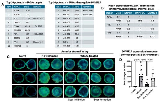
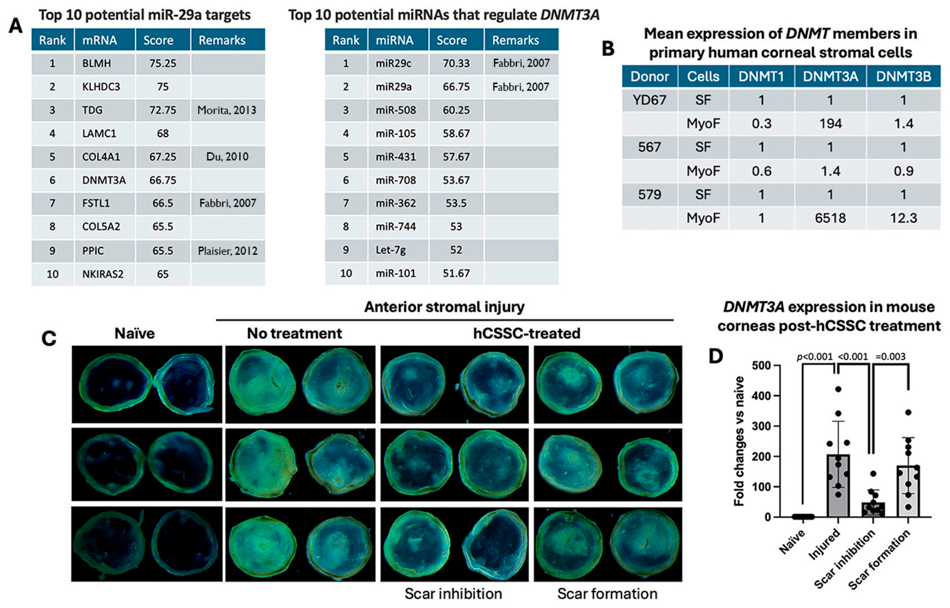
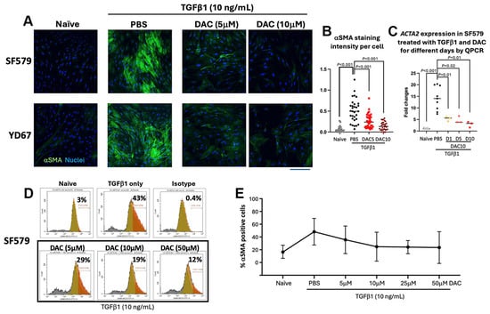
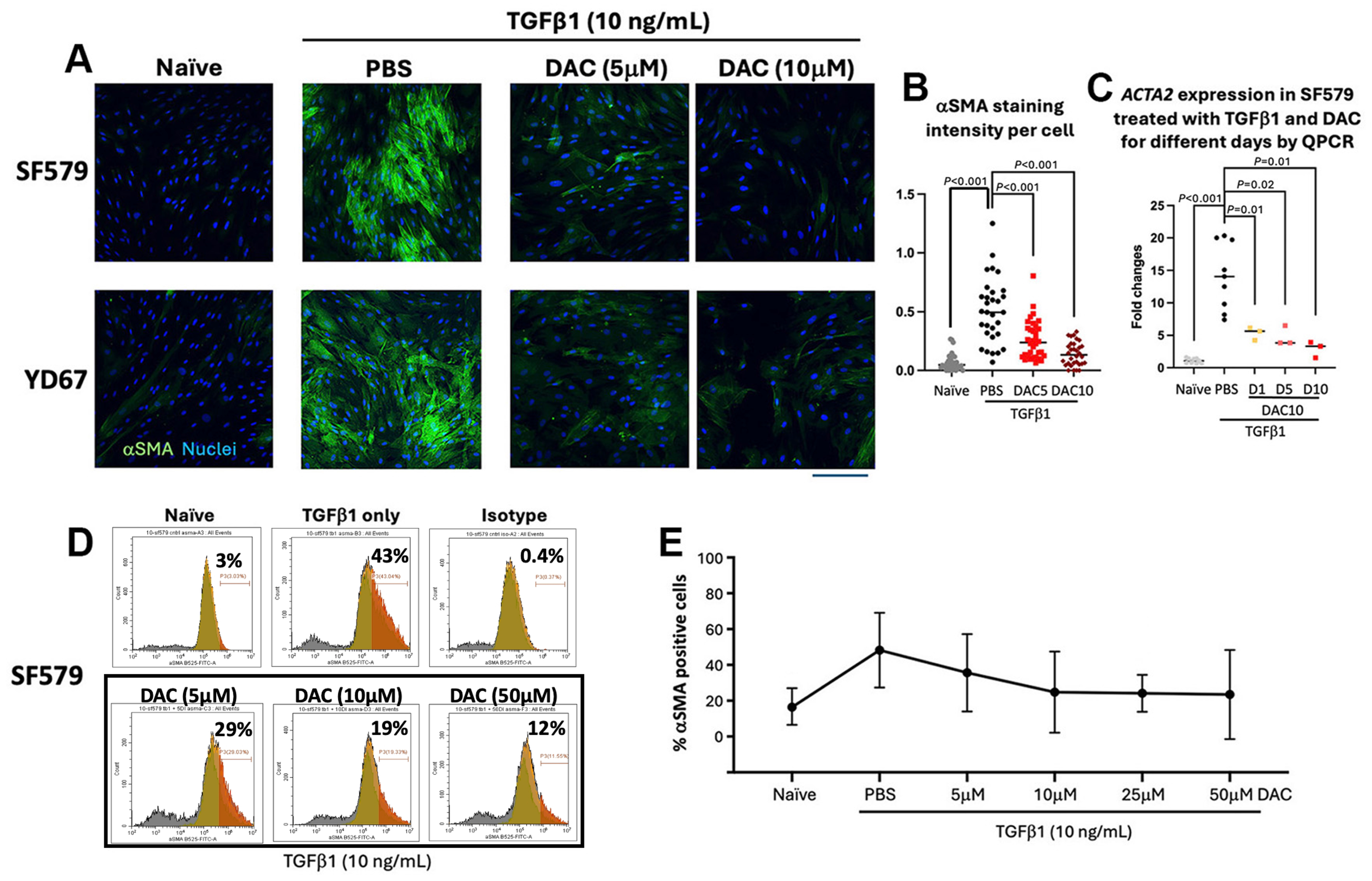
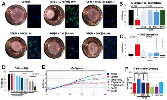
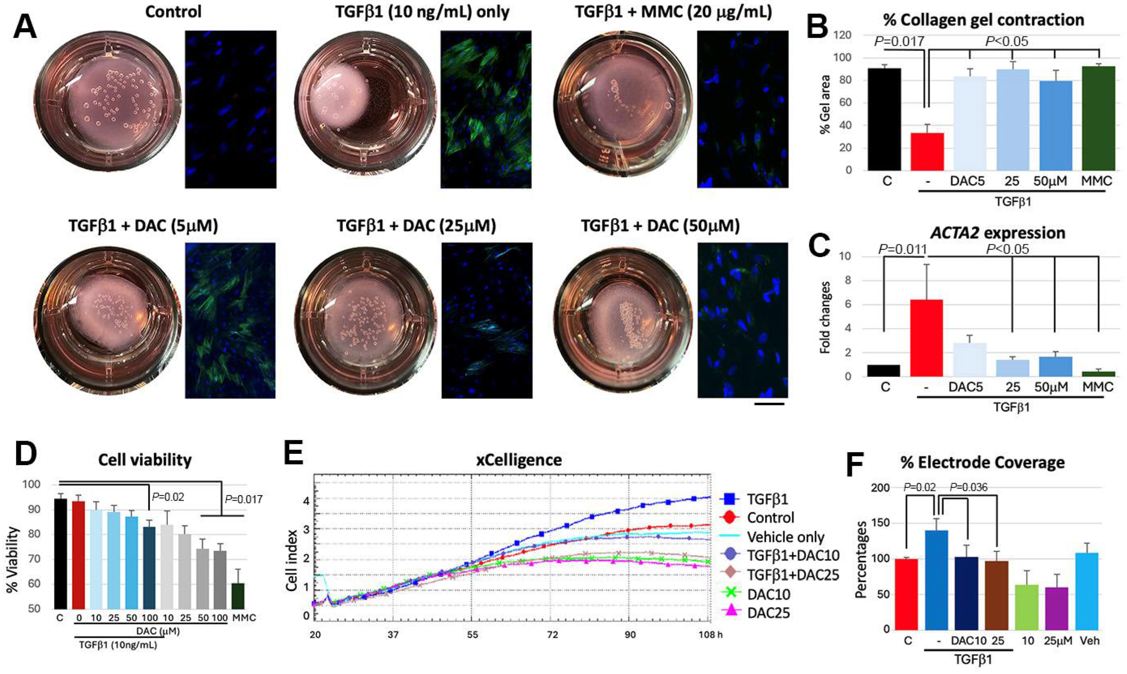
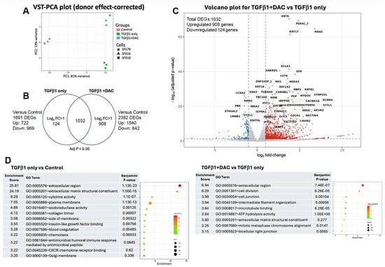
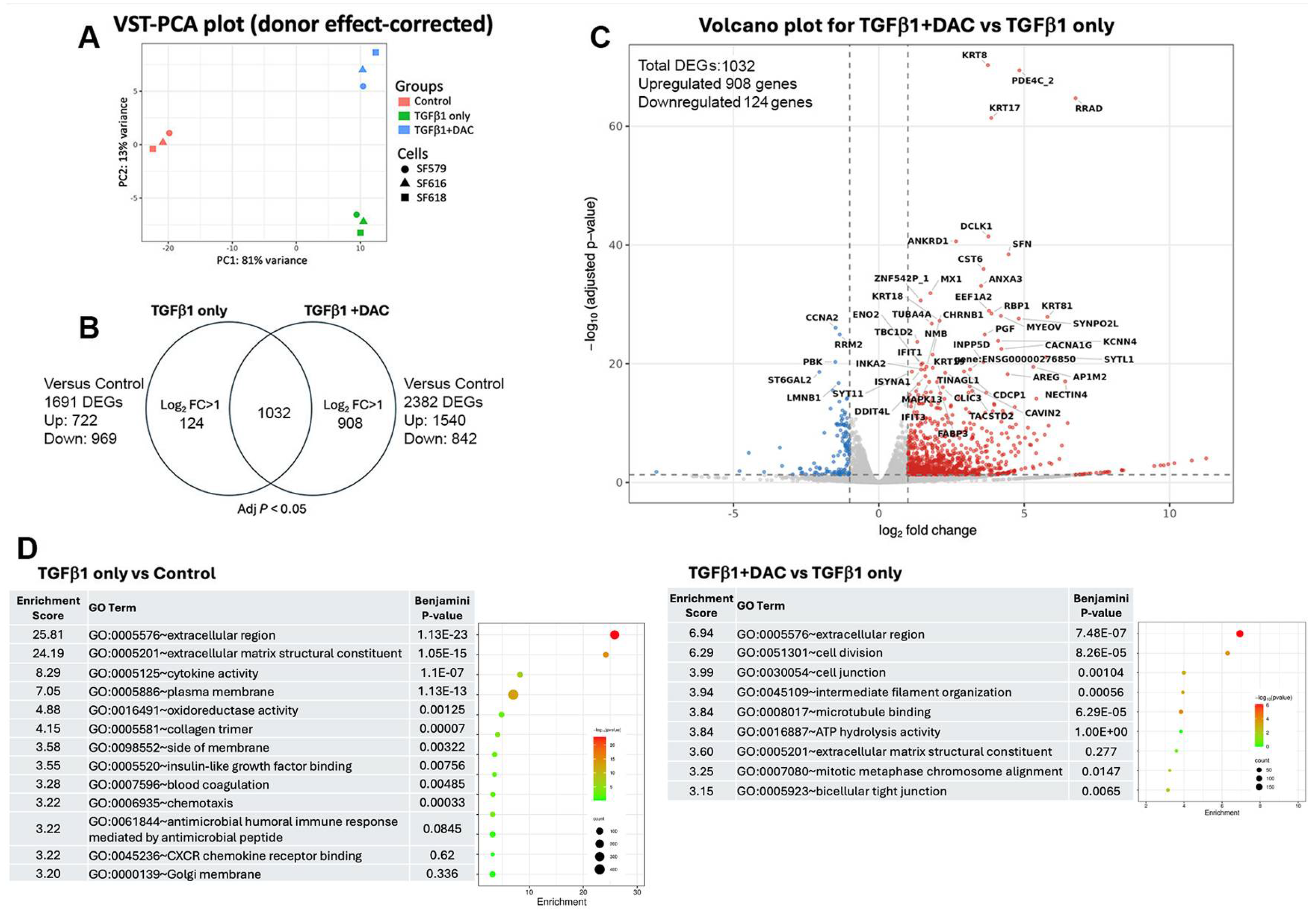
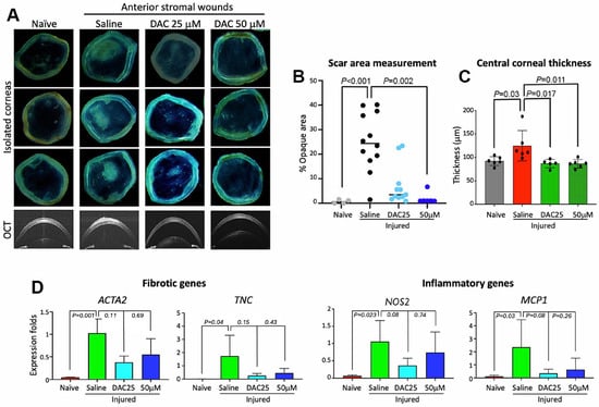
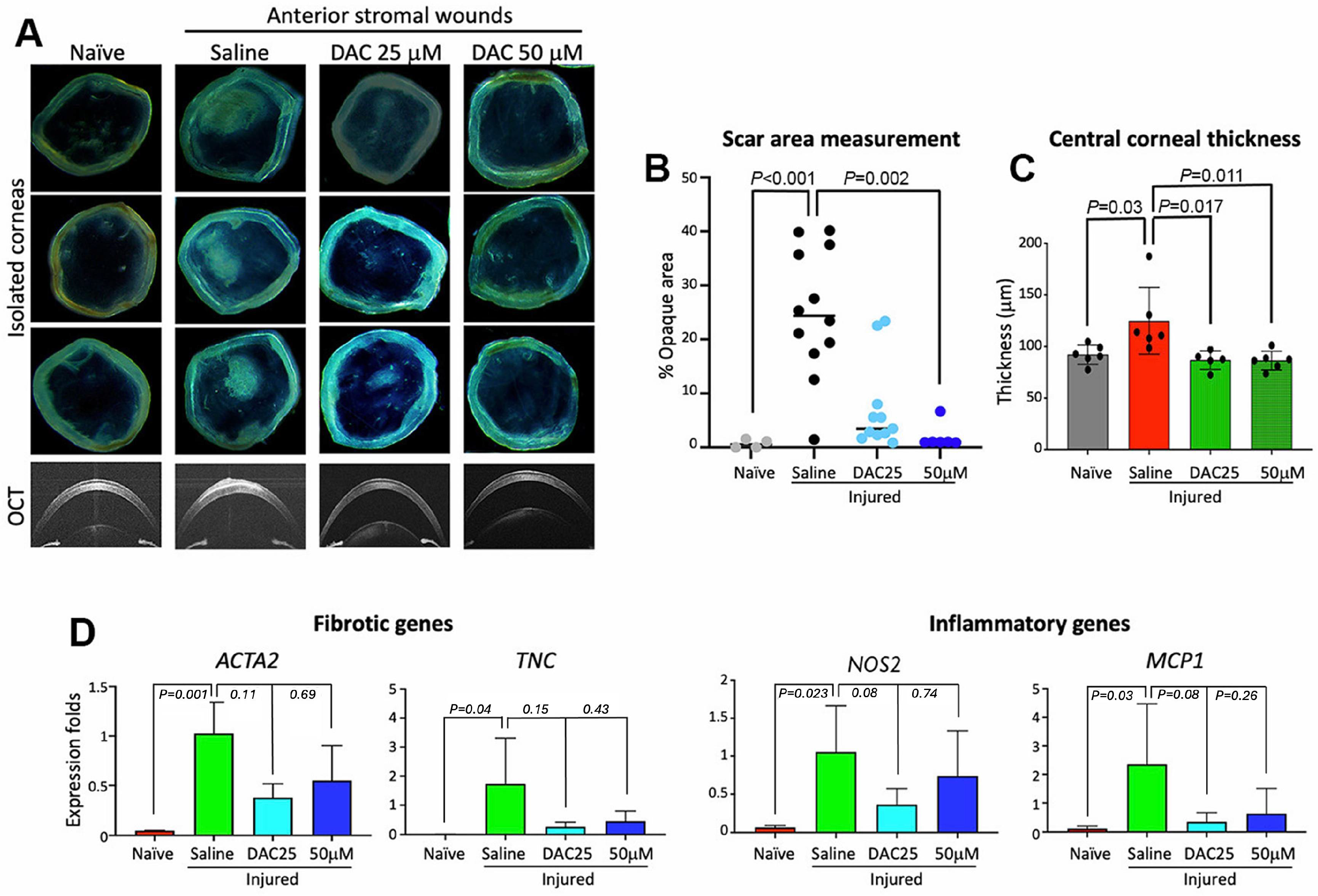
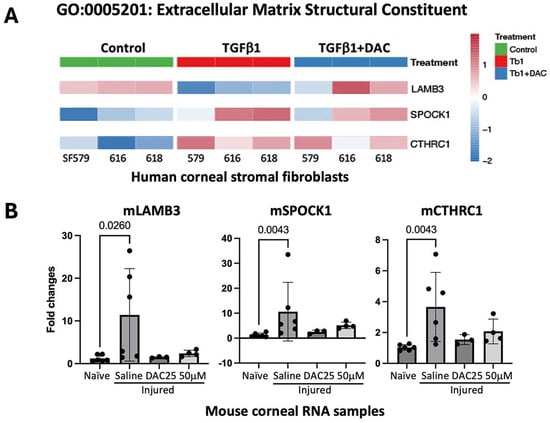
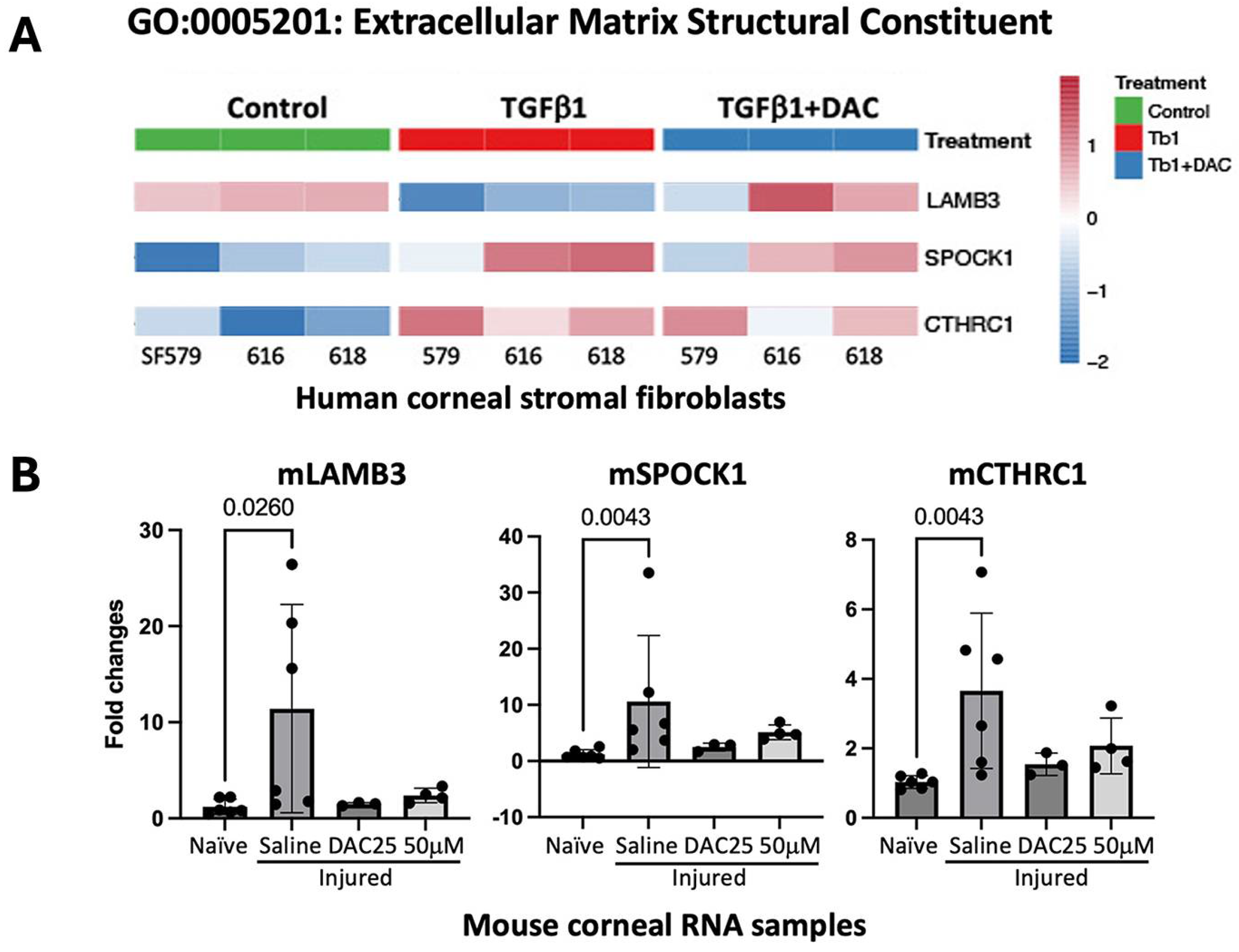
Disclaimer/Publisher’s Note: The statements, opinions and data contained in all publications are solely those of the individual author(s) and contributor(s) and not of MDPI and/or the editor(s). MDPI and/or the editor(s) disclaim responsibility for any injury to people or property resulting from any ideas, methods, instructions or products referred to in the content. |
© 2026 by the authors. Licensee MDPI, Basel, Switzerland. This article is an open access article distributed under the terms and conditions of the Creative Commons Attribution (CC BY) license.