


Highlights
What are the main findings?
- TSST-1 impairs endothelial repair by disrupting cell polarity, lamellipodia formation, rear-edge retraction, and directional migration.
- TSST-1 inhibits angiogenic sprouting and produces dysmorphic vascular networks through perturbations in cytoskeletal regulation, adhesion dynamics, extracellular matrix remodeling, and trafficking pathways.
What are the implications of the main findings?
- These findings identify suppression of vascular regeneration as a previously underrecognized, nonclassical pathogenic activity of a staphylococcal superantigen that extends beyond immune activation.
- By preventing reendothelialization and reparative angiogenesis, TSST-1 may promote non-healing vascular niches that favor tissue damage and S. aureus persistence during invasive infection.
Abstract
Staphylococcus aureus causes invasive infections characterized by extensive tissue injury and impaired healing. Although staphylococcal superantigens are best known for immune activation, emerging evidence suggests that they also directly disrupt vascular repair. This study investigated how toxic shock syndrome toxin-1 (TSST-1) affects endothelial migration and angiogenic remodeling. Immortalized human aortic endothelial cells were evaluated using viability and proliferation assays, cell-exclusion assays and live-cell imaging, actin staining, and proteomic profiling. Angiogenic effects were examined using a rabbit aortic ring explant culture model combined with tissue and secreted proteomics. TSST-1 suppressed endothelial migration by reducing forward displacement and directional persistence. Treated cells failed to form broad lamellipodia and instead developed multipolar protrusions, persistent rear tethers, and prominent actin bundles, suggesting TSST-1 disrupts protrusion and retraction dynamics. TSST-1 also inhibited angiogenic sprouting and produced dysmorphic vascular networks. Proteomic analyses identified perturbations in endothelial cell pathways associated with polarity, adhesion, extracellular matrix organization, cytoskeletal regulation, trafficking, and stress responses. In rabbit aortic rings, TSST-1 exposure induced proteomic changes associated with adhesion stabilization, reduced proteolytic invasion, and altered matrix composition. These findings expand the pathogenic activities of staphylococcal superantigens beyond immune activation and support direct suppression of vascular regeneration as a potential contributor to persistent tissue injury during invasive S. aureus infection.
1. Introduction
Staphylococcus aureus causes aggressive and invasive illnesses associated with exceedingly high mortality. These infections include pneumonia, infective endocarditis (IE), and sepsis [1,2]. Pneumonia and IE are prime examples of infections that present with tissue injury that distinctly lacks signs of healing [3,4]. In IE, the bacterium rapidly assembles valve-bound vegetations composed of bacterial microcolonies interwoven with fibrin and platelets, admixed with inflammatory and necrotic debris [5]. These friable structures fragment, seed emboli, and precipitate end-organ injury, driving morbidity and mortality [6]. S. aureus pneumonia and skin/soft-tissue infections share a signature of endothelial damage and toxin-driven necrosis that can persist despite appropriate antimicrobial therapy [7,8,9]. Much still needs to be learned about the underlying mechanisms driving wound healing defects. Staphylococcal superantigens (SAgs) are notoriously present in strains causing severe disease [10]. We showed that staphylococcal enterotoxin C (SEC), toxic shock syndrome toxin-1 (TSST-1), and select enterotoxin gene cluster SAgs play a novel and essential role in S. aureus IE and its systemic pathologies [11,12,13,14]. SAgs get their name due to their potent over-activation of the immune system, leading to toxic shock syndrome. Hence, the pathogenesis of SAgs is largely understood through this context. Our studies demonstrated that SAgs promote disease and deficits in reendothelialization and revascularization via mechanisms not dependent on their classical SAg activity. These results highlight a critical contribution of an alternative function of SAgs in S. aureus invasive diseases.
Angiogenic repair programs that restore endothelial integrity and revascularize injured tissue have emerged as convergent targets of S. aureus virulence factors critical for initiation and expansion of vegetations. In IE, mechanical or inflammatory injury to the valve endothelium creates a nidus for bacterial adhesion and rapid vegetation growth [15]. Efficient reendothelialization would ordinarily limit thrombus formation and microbial colonization, yet S. aureus appears to actively subvert this repair process by targeting cell migration programs via various exotoxin-driven mechanisms [11,16,17]. Studies in the rabbit model of native-valve IE with MRSA strain MW2 demonstrate that SEC is required for vegetation growth and organ injury, as SEC-deficient strains are attenuated in vivo [11,17]. Strikingly, SEC retains much of its pathogenic activity when the TCR and MHC II binding sites are inactivated, revealing superantigenicity-independent functions essential for disease [17]. Consistent with these observations, SEC has been shown to suppress microvessel formation in the ex vivo rabbit aortic ring model of angiogenesis and to reduce expression of endothelial VEGF-A, a master regulator of vascular repair [17]. Additionally, SEC inhibits pentraxin-3 (PTX3), amphiregulin (AREG), and TIMP-4 expression from aortic endothelial cells, converging on core motility programs and positioning anti-angiogenesis as a direct driver of failed vascular repair in IE.
TSST-1, a highly prevalent SAg among mucosa-associated S. aureus strains, is likewise associated with invasive disease and promotes vegetation formation in experimental IE [12,18,19]. TSST-1 directly targets aortic endothelial cells independently of canonical MHC II engagement, activating endothelium by upregulating adhesion molecules, increasing monolayer permeability, and impairing reendothelialization [13]. These effects occur in the absence of T cell–driven inflammation, demonstrating direct modification of endothelial function. Notably, TSST-1 suppresses endothelial interleukin-8 and interleukin-6 production, chemokines essential for neutrophil recruitment, suggesting that SAgs attenuate local inflammatory responses while promoting vascular dysfunction [13]. Collectively, the non-hematopoietic actions of SAgs provide a mechanism for sustained tissue disruption at infection sites, highlighting a role in vascular repair interference that is distinct from their immune-activating properties.
Despite these emerging insights, the cellular mechanisms through which SAgs, such as TSST-1 and SEC, directly impair vascular repair remain poorly defined. Building on our observation that TSST-1 inhibits reendothelialization [13], we sought to delineate mechanisms responsible for this impairment and consequences for angiogenic sprouting. Here we show that TSST-1 specifically disrupts directional endothelial migration by suppressing a broad network of motility-associated factors and by inducing profound reorganization of the actin cytoskeleton. Furthermore, TSST-1 inhibits microvessel sprouting or yields dysmorphic, non-productive vascular networks. Proteomic analyses identify perturbations in matrix remodeling, adhesion-complex dynamics, and angiogenic signaling that may converge to impair vessel formation. Our findings place TSST-1 alongside SEC and β-toxin [20] as part of a coordinated strategy that targets distinct but convergent nodes in the angiogenic program.
2. Materials and Methods
2.1. Media and Reagents
Phenol red–free Medium 200 (M200PRF500), low serum growth supplement (LSGS; S00310), 0.025% trypsin-EDTA (R001100), VEGF (PHC9344), Pierce Protease Inhibitor Mini Tablets (A32953), Halt™ Protease Inhibitor Cocktail, EDTA-Free (78439), Dynabeads™ His-Tag (10103D), Detoxi-Gel™ (20339), penicillin–streptomycin (15140122), amphotericin B (15290018), Qubit™ Protein Broad Range kit (A50668) and human umbilical vein endothelial cells (HUVECs; C0035C; RRID: CVCL K312) were purchased from Thermofisher Scientific/Fisher Scientific, New York, NY, USA. HiTrap Heparin HP columns (17040701) were obtained from Cytiva, Marlborough, MA, USA. ToxinSensor™ LAL Endotoxin Assay Kit (L00350) was purchased from Genscript, Nanjing, China. CellTiter 96® AQueous One Solution (G3582) was purchased from Promega, Madison, WI, USA. Mitomycin C (M4287) was purchased from Sigma-Aldrich, St. Louis, MA, USA. Proteome Profiler™ Human Angiogenesis Antibody Array (ARY007) was purchased from R&D Systems, Minneapolis, MN, USA. IRDye 800CW Streptavidin (926-32230) was purchased from LI-COR, Lincoln, NE, USA. Growth factor-reduced Matrigel (GFR-Matrigel; 356231) and 12-well (3513) and 48-well (3548) Costar tissue culture plates were purchased from Corning, New York, NY, USA. 96-well plates (P96-1.5P) were purchased from Cellvis, Mountain View, CA, USA. Axitinib (HY-10065) was purchased from MedChemExpress, Monmouth Junction, NJ, USA. Glass beads (0.1 mm; P000929LYSK0A.0) and soft tissue homogenizing kits (P000933-LYSK0-A) were purchased from Bertin Corp, Rockville, MD, USA. 4-well culture inserts (80466) and angiogenesis slides (81506) were purchased from ibidi, Gräfelfing, Germany. Mycostrip™ Plus (LT07-701) and MycoStrip® (REP-MYS-10) Mycoplasma Detection kits were purchased from Lonza, Basel, Switzerland and InvivoGen, San Diego, CA, USA respectively.
2.2. Endothelial Cell Culture Conditions
Immortalized human aortic endothelial cells (iHAEC) are a recently established cell line shown to retain phenotypic and functional characteristics of primary cells, serving as a large-vessel model system in which to address questions relevant to vascular biology [13]. Cells were grown at 37 °C, 5% CO2 in Medium 200 supplemented with LSGS, at final concentrations of: FBS 2%, hydrocortisone 1 μg mL−1, human epidermal growth factor 10 ng mL−1, basic fibroblast growth factor 3 ng mL−1, and heparin 10 μg mL−1. Cells were maintained on 1% gelatin-coated plates unless otherwise stated. Cells were passaged at least twice before use in experiments. iHAECs were used at passages between 4 and 12 from a single clone. Mycoplasma-testing was conducted every 6 months using MycoAlert™ Plus (Lonza) or MycoStrip® (InvivoGen) Mycoplasma Detection Kit.
2.3. TSST-1 Expression Vector Construction
The construct was generated in the pET25bHSVdelTEV plasmid backbone and cloned into Escherichia coli DH5α before being transformed into E. coli BL21 (DE3). Unless otherwise noted, reagents were used as supplied by the manufacturer, and incubations were performed at recommended temperatures and times. tstH was PCR-amplified from S. aureus strain MN8 with Phusion high-fidelity DNA polymerase (New England Biolabs, Ipswich, MA, USA) using primers tsthNdeIF (ttacCATATGTCTACAAACGATAATATAAAGG) and tsthpET25bXhoIR (ttacCTCGAGATTAATTTCTGCTTCTATAGTTT). The tstH PCR fragment and pET25bHSVdelTEV backbone were digested with NdeI and XhoI and ligated with T4 DNA ligase (New England Biolabs), creating the plasmid pET25bHSVdelTEV_tstH. Coding sequence and vector–insert junctions were confirmed by Sanger sequencing using the T7_prom primer (TAATACGACTCACTATAGGG). Primers were purchased from Integrated DNA Technologies.
2.4. TSST-1 Purification and Concentrations Used
TSST-1 was expressed in E. coli BL21 (DE3) cells harboring a plasmid encoding N-terminal His6-tagged constructs. Cultures were initiated in 1 L LB broth (5 g yeast extract, 10 g tryptone, 10 g NaCl) containing 100 μg mL−1 carbenicillin and grown at 37 °C to an OD600 of 0.4–0.8. Expression was induced with 1 mM IPTG, and cultures were maintained in the incubator shaker for 4 h at 30 °C, 250 rpm. Cultures were pelleted via centrifugation (6000× g, 10 min, 4 °C) and resuspended in 30 mL binding buffer (20 mM NH2C(CH2OH)3 · HCl, 500 mM NaCl, 20 mM imidazole, pH 7.9) along with three Pierce Protease Inhibitor Mini Tablets. Ten-millimeter aliquots were homogenized with 7 g of 0.1 mm glass beads using a Precellys Cryolys Evolution homogenizer (9900 rpm, six 30 s cycles, 60 s rests, 4 °C) (Bertin Corp., Rockville, MD, USA). Lysates were centrifuged at 50,000× g, 40 min, and 4 °C. The resulting supernatant was manually syringe-filtered through a 0.45 μm membrane. All proteins were purified by Ni2+-affinity chromatography on a HiTrap Heparin HP column (Cytiva) via gradient elution (20–250 mM imidazole, 50 mM NH2C(CH2OH)3. HCl, 500 mM NaCl, pH 7.9) and subsequently dialyzed overnight against 4 L PBS (pH 7.4) at 4 °C. Protein capture and purity were confirmed by sodium dodecyl sulfate-polyacrylamide gel electrophoresis (SDS-PAGE) followed by Coomassie blue staining to reveal a single band of 22 kDa. Protein concentration was determined using the Qubit™ Protein Broad Range Assay Kit (Thermofisher Scientific). Endotoxin contamination was reduced using Detoxi-Gel™ endotoxin removal resin, and final endotoxin levels were measured with the ToxinSensor™ Chromogenic Limulus Amebocyte Lysate (LAL) Endotoxin Assay Kit, ensuring levels ≤ 0.05 ng mL−1. TSST-1 was used at the subtoxic concentrations of 6.125, 12.25, 25.5, and 50 μg mL−1, depending on assay sensitivity. S. aureus produces TSST-1 as high as μg mL−1 in liquid culture and 16,000 μg mL−1 in thin-film biofilms [21]. Septic lesions, such as vegetations, support bacterial growth as high as 5 × 10⁹ cfu [12]. We expect infected tissues to be exposed to these toxins at high levels.
2.5. EdU Incorporation Assay
Passage 11 iHAEC cultured in Medium 200 supplemented with LSGS were seeded into a 96-well plate at 7000 cells per well. Following 4 h of incubation at 37 °C with 5% CO2 for cell attachment, media were gently aspirated and replaced with toxin-conditioned media (50 μg mL−1) as measured by the Qubit™ assay, allowing for biological/technical replicates per toxin condition along with media-only controls in triplicate. Cells were then incubated overnight to reach ~70% confluency. After experimental treatments, cells were pulsed with EdU (5-ethynyl-2′-deoxyuridine; 10 µM), a nucleoside analog of thymidine, for 10 min at 37 °C in complete growth medium to label cells actively replicating DNA (S-phase). Following EdU incorporation, cells were washed twice with phosphate-buffered saline (PBS) and fixed in 1% formaldehyde/PBS for 10 min at room temperature and washed three times in PBS. The cells were then permeabilized with 0.1% Triton X-100/PBS for 15 min. Samples were incubated for the Click reaction in a solution containing working concentrations of 2.5 μg mL−1 488 Azide, 2 μg mL−1 Sodium Ascorbate, and 2 nM Copper Sulfate in PBS. After washing, nuclear counterstaining was performed using DAPI. The plate was imaged using the ImageXpress Nano (Molecular Devices, San Jose, CA, USA) imaging system. The system allows single-cell, high-content automated acquisition (3000 cells per condition). Images were analyzed for average integrated nuclear intensity using the MetaXpress high-content image analysis software.
2.6. Cell-Exclusion Assay
Cell-exclusion assays were performed using 4-chamber silicone inserts (ibidi) to assess the effects of TSST-1 on migration and proliferation of iHAECs. All experiments were conducted using cells between passages 7 and 13 grown in Medium 200 supplemented with LSGS. Inserts were placed into uncoated 12-well, tissue culture-treated plates using sterile tweezers. Each chamber was subsequently seeded with 27,500 cells and incubated at 37 °C with 5% CO2 for 4 h for cell attachment. The medium was then replaced with fresh toxin-conditioned medium at 50 μg mL−1, and cells were incubated overnight. Following overnight treatment, the inserts were removed. The conditioned media was collected, returned to the wells, and supplemented with additional toxin-conditioned media to achieve a final volume of 1.5 mL per well. Toxin conditions included TSST-1 (50 μg mL−1). Mitomycin C (2 μg mL−1) was also paired with each condition to isolate contributions of proliferation throughout the time course and was added immediately following insert removal. Plates were set in a Leica DMi8 microscope equipped with a Tokai Hit stage-top incubator set to 37 °C and 5% CO2 for the duration of image capture. Phase-contrast images were captured every 30 min over a 24 h period using a HC PL FLUOTAR 10x/0.13 objective lens to monitor wound closure. Three independent experiments were conducted for each treatment condition, and images were analyzed automatically using ImageJ. Wound area was quantified using the Wound Healing Size Tool plugin in ImageJ software [22]. Images were processed by applying the following initial settings: Variance window radius = 50, Threshold value = 100, Percentage of saturated pixels = 0.001%, with additional adjustments for individual runs to ensure accuracy of gap borders.
2.7. Quantification of Single-Cell Migration Using CellTraxx
Single-cell migration trajectories were obtained using CellTraxx, an automated cell-tracking platform designed for high-throughput and time-resolved analysis of motile cells. Time-lapse phase–contrast images from cell-exclusion assays were acquired every 30 min for 24 h using a Leica DMi8 equipped with a Tokai Hit stage-top incubator for live cell imaging at 37 °C and 5% CO2. Raw image sequences were imported into CellTraxx, and tracking was performed using the software’s integrated centroid-detection and object-linking algorithms [23]. Briefly, CellTraxx identifies individual cells in each frame by adaptive thresholding followed by morphological segmentation, then links these detections across frames using a predictive nearest-neighbor model to reconstruct full cell trajectories. Default tracking parameters were used. To ensure high-quality trajectories, tracks with fewer than 5 continuous frames, excessive frame gaps, or algorithm-flagged ambiguities were excluded prior to analysis. Each validated track was visually inspected within the CellTraxx interface to confirm accurate linking, especially in regions of high cell density. For each trajectory, CellTraxx automatically computed frame-to-frame displacements (μm), instantaneous velocity (μm min−1), cumulative path length (μm), Euclidean displacement from origin (μm), and angular changes between successive steps. Average velocity (μm min−1) was calculated from the frame-to-frame instantaneous velocities of cells in the track. Forward distance was calculated as net displacement along the wound axis (x2 − x1). Directional persistence was quantified using the Euclidean distance, calculated as , divided by the cumulative path length.
2.8. Fluorescence Microscopy
For actin immunofluorescence, iHAECs were cultured to confluence, or semi-confluence, on fibronectin-coated glass slides in the presence or absence of TSST-1 (25 μg mL−1) for 4 h. Cells were fixed in cold methanol, washed, permeabilized with 0.1% Tween-20, and blocked in 1% BSA. Actin filaments were stained using Acti-Stain 555 phalloidin (Cytoskeleton, Inc., Denver, CO, USA). Slides were mounted with ProLong™ Gold Antifade containing 4′,6-diamidino-2-phenylindole (DAPI; Molecular Probes, Life Technologies, Carlsbad, CA, USA). Fluorescence images were acquired using a Leica DMi8 upright epifluorescence microscope equipped with appropriate filter sets. Multichannel images were pseudocolored and merged using ImageJ (NIH, Bethesda, MD, USA).
2.9. Proteome Profiler™ Human Angiogenesis Array
For cells in monolayer, iHAECs were seeded onto gelatin-coated 96-well plates at 7000 cells per well and cultured to near confluence. Fresh media containing 50 μg mL−1 TSST-1 was added, and plates were incubated for 24 h at 37 °C with 5% CO2. For migrating cells, the cell-exclusion assay was performed as previously described. Conditioned media was collected and stored at −80 °C until analysis. Relative protein expression was assessed using the Proteome Profiler™ Human Angiogenesis Antibody Array kit (R&D Systems/Bio-Techne), a membrane-based sandwich immunoassay containing duplicate capture-antibody spots for 55 angiogenesis-related proteins (Table S1). Samples were processed according to the manufacturer’s instructions and signals were detected by fluorescence. Each membrane contained assay-embedded reference spots that verified incubation with streptavidin–HRP and facilitated alignment of the array template, as well as negative-control spots used to estimate background signal. Analyte signals were quantified as the mean pixel density of duplicate spots after subtraction of the averaged background signal determined from the negative-control spots. Conditioned media (120 μL) was incubated with biotinylated detection antibodies for 1 h at room temperature. During this time, capture antibody membranes were blocked with kit-provided blocking buffer. The sample-antibody mixture was then applied to membranes and incubated overnight at 4 °C. Following three sequential 10 min washes, membranes were incubated with IRDye 800CW Streptavidin (1:2000) for 30 min while protected from light exposure. Membranes were washed three times and imaged using an Azure c600 (120 μm resolution, auto-intensity). Mean pixel intensities from duplicate spots were averaged using Image Studio Software (LI-COR; RRID: SCR_013715), and fold-changes relative to untreated controls were calculated. Proteins unlikely to be produced by endothelial cells (angiopoietin-1, angiostatin/plasminogen, EG-VEGF, FGF-4, leptin, platelet factor 4, and serpin B5) were identified through a comprehensive review and were excluded based on GTExPortal and Expression Atlas data. These analytes were not detected in iHAECs. Data was collected from three independent experiments.
2.10. Ex-Vivo Aortic Ring Model of Sprouting Angiogenesis
Juvenile mixed-sex New Zealand white rabbits (2–3 kg) were obtained from Charles River Laboratories (Wilmington, MA, USA) and Western Oregon Rabbit Co. (Philomath, OR, USA) and housed at the Research Vivarium at the School of Veterinary Medicine, University of Wisconsin–Madison, according to established guidelines and the protocol approved by the Institutional Animal Care and Use Committee (protocol V006634-R02). All animals were individually housed with ad libitum access to food and water. Rabbits were allowed at least four days for acclimatization and were clinically determined to be healthy by a veterinarian prior to euthanasia. For humane euthanasia, rabbits were sedated with a combination of ketamine and xylazine, followed by 1 mL injection of Euthasol (Virbac) per protocol. Following euthanasia, the thoracic and abdominal aorta segments were immediately excised and placed in phosphate-buffered saline (PBS) for dissection. Excess fascia and connective tissue were removed, and 1–1.5 mm2 aortic cross-sections were prepared using a scalpel. Aortic ring explants were cultured by modifying the thin-layer method [24]. The sections were embedded in 200 μL of phenol-red-free, growth-factor-reduced Matrigel (GFR-Matrigel, Corning) in 24-well plates. After 10 min of polymerization at 37 °C in 5% CO2, 200 μL of supplemented Medium 200 was added to each well, and the plates were incubated at 37 °C in 5% CO2 for up to 14 days. Medium 200 was supplemented with LSGS, 100 U mL−1 penicillin-streptomycin (Thermofisher Scientific), 2.5 μg mL−1 amphotericin B (Thermofisher Scientific), and relevant treatment conditions. Media (with or without treatments) were changed every 3–5 days. Images were captured after 14 days using a Leica DMi8 microscope equipped with a Tokai Hit stage-top incubator set to 37 °C, 5% CO2. A 4x/0.13 HC PL FLUOTAR objective lens was used for image acquisition, and sprouting of aortic rings was assessed for surface area in millimeters squared on day 14 using ImageJ [25]. Three rabbits were used for each experimental condition: a total of 9−12 thoracic sections and 9−12 abdominal sections per condition.
2.11. Protein Extraction and Processing for Mass Spectrometry Analysis
For protein extraction from tissue samples, aortic rings were prepared and cultured as described under the ex vivo explant model methodology section. Following a 14-day culture period, aortic tissues were homogenized in RIPA buffer (10 mM Tris-HCl pH 7.5, 150 mM NaCl, 0.5 mM EDTA, 0.1% SDS, 1.0% Triton X-100, 1.0% sodium deoxycholate) supplemented with Halt™ Protease Inhibitor Cocktail, EDTA-Free. For every milligram of wet tissue, 15 µL of RIPA buffer was added to 2 mL O-ring tubes containing 2.8 mm beads (1.2 g beads per 30 mg tissue). For each treatment condition, approximately 20 rings from the same rabbit constituted one biological replicate. Tissue was homogenized using a Bead Mill 4 instrument (Fisher Scientific) at 5000 rpm for 45 s, followed by cooling on ice for 5 min, repeated 10 times. Lysates were centrifuged at 12,000 rpm (13,680× g) for 15 min at 4 °C and the supernatants were collected. Protein concentrations were measured using a Qubit™ Protein Assay Kit (Invitrogen, Hong Kong, China). To remove superantigens from aortic ring supernatant, magnetic bead separation was completed using Dynabeads™ His-Tag. One milligram of Dynabeads was washed three times with 900 µL PBS (pH 7.5), incubated in a rotator at room temperature for 2 min between washes and magnetized for 2 min at room temperature to prepare beads for sample loading. Conditioned medium from ring cultures was collected on day 14, centrifuged (12,000× g, 15 min, 4 °C), supplemented with 9 µL Halt™ Protease Inhibitor Cocktail per 900 µL, and then incubated with the prepared Dynabeads™ for 1 h at 4 °C. The sample was then magnetized and supernatant was removed and saved. Samples were stored at −80 °C until DIA LC-MS/MS analysis. Protein samples, including Dynabeads™ eluates, were analyzed by SDS-PAGE to confirm purity. Selected bands were excised, digested with trypsin, and desalted. Peptides were dried and prepared for DIA LC–MS/MS analysis to identify and quantify protein abundance.
2.12. Data-Independent Acquisition Liquid Chromatography-Tandem Mass Spectrometry
Proteins from fresh media, tissues, and supernatants from untreated controls or TSST-1-treated conditions were submitted to the University of Wisconsin Biotechnology Center Mass Spectrometry Core Facility. Proteomic profiling was performed using data-independent acquisition liquid chromatography–tandem mass spectrometry (DIA LC–MS/MS), followed by CHIMERYS-based peptide identification and label-free protein quantification. To obtain sufficient protein yield for downstream mass spectrometry analysis, material from 20 parallel technical extractions was pooled per rabbit. Equal protein amounts from three rabbits (aortas or cell-free conditioned media) from control and treated conditions were submitted independently for analysis. DIA LC–MS/MS data were searched against Oryctolagus cuniculus (rabbit) and Bos taurus (cattle) UniProt databases using standard search parameters. Raw data from DIA LC–MS/MS were analyzed by the UW–Madison Mass Spectrometry Facility utilizing the Proteome Discoverer DIA workflow. Peptide-spectrum matches (peptides, protein groups, and proteins) were filtered at 1% false discovery rate confidence assignments generated by the processing workflow. Only proteins classified as Master proteins and meeting the selected protein-group and protein FDR-confidence criteria were retained for downstream analysis. Protein identifications were further characterized by sequence coverage, total peptide count, number of peptide-spectrum matches, number of unique peptides, CHIMERYS score, molecular mass, calculated isoelectric point, and the presence of the protein in individual samples. Label-free protein abundance was determined from DIA precursor signals. In Proteome Discoverer DIA analysis, the quantitative value for a peptide group is obtained by summing the peak areas of its associated DIA precursors. Protein-level abundance values were normalized within the processing workflow and reported for each individual sample. Grouped abundance values were calculated separately for each condition. These values were used for downstream fold-change calculations and log2 transformations. Untransformed abundance ratios, log2-transformed ratios, ratio variability, nominal p-values, and adjusted p-values were exported from Proteome Discoverer. Adjustment for multiple comparisons was performed within the Proteome Discoverer workflow. Proteome Discoverer calculates adjusted p-values using the Benjamini–Hochberg procedure. Proteins were considered differentially abundant when they met the prespecified thresholds of adjusted p-value < 0.05 (−log10 ≥ 1.3) and absolute fold change ≥ 2 (log2 fold change ≥ 1). Abundant proteins matching to fresh media (Table S2), Matrigel components, and common contaminants were eliminated. Only proteins with ≥2 unique peptide counts were retained. The remaining proteins in the list were bona fide aortic tissue or secreted proteins (Tables S3 and S4).
2.13. Pathway and Process Enrichment Analysis
Pathway and process enrichment analysis on protein lists from significantly increased or decreased proteins in tissues and conditioned media was performed with the Metascape platform [26]. For each given gene list, the Metascape platform performs pathway and process enrichment analysis with the following ontology sources: KEGG Pathway, GO Biological Processes, Reactome Gene Sets, Canonical Pathways, CORUM, WikiPathways, and PANTHER Pathway. All genes in the genome were used as the enrichment background. Terms with a p-value < 0.01, a minimum count of 3, and an enrichment factor > 1.5 (the enrichment factor is the ratio between the observed counts and the counts expected by chance) were collected and grouped into clusters based on their membership similarities. p-values were calculated based on the cumulative hypergeometric distribution, and q-values were calculated using the Benjamini–Hochberg procedure to account for multiple testing. Kappa scores were used as the similarity metric when performing hierarchical clustering on the enriched terms, and sub-trees with a similarity of >0.3 were considered a cluster. The most statistically significant term within a cluster was chosen to represent the cluster (Tables S5 and S6). To further capture the relationships between the terms, Metascape carried out protein–protein interaction enrichment analysis for each given gene list with the following databases: STRING, BioGrid, OmniPath, and InWeb_IM. The resultant network contained the subset of proteins that form physical interactions with at least one other member in the list. The Molecular Complex Detection (MCODE) algorithm was applied to identify densely connected network components.
2.14. Statistical Analysis
Statistical analyses were conducted using GraphPad Prism software (RRID: SCR_002798). For each experiment, the precision measures as well as the number of technical and biological replicates are detailed in the figure legends and methods. Cell-exclusion assay and aortic ring sprouting data were analyzed using two-way ANOVA with Holm–Šídák’s multiple comparisons test. EdU incorporation assay was evaluated via non-parametric ANOVA. For single-cell tracking, non-parametric one-way ANOVA and the Mann–Whitney test were used. Statistical significance with α = 0.05 is indicated as follows: * p < 0.05, ** p < 0.01, *** p < 0.001, and **** p < 0.0001 (GraphPad Prism 11.0.1).
3. Results
3.1. TSST-1 Targets Directed Endothelial Cell Migration
Previous work showed that TSST-1 impairs reendothelialization in an in vitro scratch assay [13]. However, whether this defect resulted from altered endothelial cell proliferation or migration remained unclear. We first evaluated cell viability following toxin exposure to confirm that TSST-1 was not cytotoxic under the conditions tested. Treatment of immortalized human aortic endothelial cells (iHAECs) with TSST-1 at concentrations up to 112 μg mL−1 did not reduce metabolic activity, as measured by the MTS assay, confirming a subtoxic dose range (Figure 1A). We next evaluated reendothelialization using a cell-exclusion assay and live-cell imaging, where phase-contrast images were captured every 30 min over a 24 h period. As previously demonstrated, exposure to TSST-1 significantly reduced gap closure. This defect was more pronounced when cell division was inhibited with mitomycin C (Figure 1B). These findings suggest that TSST-1 primarily impairs endothelial cell migration rather than proliferation. To directly examine the effect of TSST-1 on proliferation, we measured EdU incorporation into newly synthesized DNA as a marker of active cellular replication. TSST-1 treatment did not significantly alter EdU incorporation relative to untreated cells (Figure 1C), further indicating that the reendothelialization defect was not attributable to impaired cell proliferation.
Figure 1.
TSST-1 targets directional displacement required for effective gap closure. (A) Cell survival based on MTS metabolic activity. iHAECs grown to near confluency on 1% gelatin-coated plates. (B) Time course analysis of iHAECs grown to confluency in silicone inserts that create uniform gaps upon removal and treated with either TSST-1 (50 µg mL−1) or untreated ± mitomycin C (MMC; 2 µg mL−1). Images were acquired every 30 min for 24 h. Data represent mean ± SEM; **** p < 0.0001; two-way ANOVA with Holm–Šídák’s multiple comparisons test. (C) EdU incorporation of iHAEC seeded at 7000 cell/well in a 96-well plate and treated with TSST-1 (50 μg mL−1) overnight to ~70% confluency. Edu incorporation measured for 2 h in control versus TSST-1-treated cells. Data captured from 3000 cells per condition. (D–G) Single-cell tracking of iHAECs of the leading edge in the cell-exclusion assay ± TSST-1 (50 μg mL−1). (D) Representative images of tracked migration paths from individual cells (color lines represent individual tracks). Trajectories were median-filtered and drift-corrected prior to analysis. (E) Average velocity per track (μm min−1). (F) Forward distance; directional efficiency along the axis of wound closure calculated as x2−x1. (G) Directional persistence index; directed movement toward closing the gap, calculated as Euclidean distance/total distance traveled. Euclidean distance; net displacement from the point of origin calculated as . Each symbol represents an individual cell. Data are pooled from 3 independent experiments, shown as violin plots; *** p < 0.001. Non-parametric one-way ANOVA, Mann–Whitney test.
Having established that TSST-1 inhibits endothelial cell migration without compromising cell viability or proliferation, we next investigated how TSST-1 alters endothelial migratory behavior. In untreated control cultures, iHAECs migrated progressively into the cell-free gap, with the leading edge advancing from both wound edges toward the center over the 24 h imaging period (Video S1A). In contrast, TSST-1-treated cells migrated into the open gap with less efficiency, showing reduced leading-edge spreading and persistent cell clustering near the wound margin (Video S1B). To quantify the effects on cell motility, we performed single-cell tracking using the time-lapse images collected during the cell-exclusion assays. For quantitative analysis, individual endothelial cells of the leading edge were tracked using CellTraxx, an automated platform that identifies cell centroids, links positions across sequential frames, and reconstructs complete migration trajectories [23]. Representative trajectory plots for control and TSST-1-treated conditions are shown (Figure 1D). Cell-migration behavior was evaluated using three parameters: (i) average velocity, representing instantaneous migration speed; (ii) forward distance, a measure of directional efficiency along the axis of wound closure; and (iii) directional persistence, a measure of forward, directional movement across the entire trajectory. These are standard metrics for motility analysis that distinguish speed from directionality in an unbiased fashion [27]. Control monolayers displayed rapid and consistent directional migration. Average velocities were high (0.58 ± 0.02 μm min−1; mean ± SEM), and cells achieved substantial forward distance (12.6 ± 0.5 μm) and directional persistence (0.8 ± 0.02), indicating effective translocation across the substrate (Figure 1E–G). Cells exposed to TSST-1 demonstrated significantly reduced velocities (0.33 ± 0.01 μm min−1; Figure 1E), decreased forward displacement (3.7 ± 0.2 μm; Figure 1F), and decreased directional persistence (0.5 ± 0.02; Figure 1G). Together, these findings indicate that TSST-1 does not simply suppress cellular movement but preferentially disrupts the ability of endothelial cells to sustain directional migration into the gap.
3.2. TSST-1 Disrupts Lamellipodia Architecture and Actin Cytoskeletal Dynamics During Endothelial Cell Migration
Live-cell imaging analyses indicated that TSST-1 disrupts cytoskeletal or polarization mechanisms required for persistent, directed movement. To characterize these altered migration patterns, we examined individual cell morphologies in the time-lapse images. During migration, control iHAECs at the leading edge displayed broad lamellipodial extensions, cell elongation toward the gap, and coordinated retraction of the trailing edge (Figure 2A and Video S2A). In stark contrast, TSST-1–treated cells showed abnormal migratory and trailing-edge morphology characterized by filamented cytoskeletons, reduced lamellipodia, and defective rear-edge retraction (Figure 2B, Video S2). Individual cells frequently exhibited multipolar extensions (Figure 2C), reduced or narrow lamellipodial protrusions (Figure 2D), persistent tether-like structures or long aberrant protrusions (Figure 2E), and incomplete rear-edge retraction (Figure 2F). These single-cell features indicate that TSST-1 disrupts leading-edge adhesions and release of trailing-edge contacts required for productive migration.
Figure 2.
TSST-1 disrupts lamellipodia formation and retraction of the trailing edge. (A,B) Analysis of cell morphology during 24 h migration in the cell-exclusion assay. Representative phase-contrast images of immortalized human aortic endothelial cells (iHAECs) migrating into the gap ± TSST-1 (20 μg mL−1). Cell shape was followed over 12 consecutive frames to assess morphology changes during migration. Images show shape over time, not trajectory. Colors identify individual cells. (A) Control cells showing formation of broad lamellipodia, elongation, and rear retraction. (B) TSST-1-treated cells showing narrow and small lamellipodia or abnormal protrusive behavior, filamented cytoskeletons, and/or tethers that remain upon rear retraction. (C–F) Distinct morphological phenotypes observed with TSST-1 treatment. (C) Multipolar morphology with cells exhibiting multiple radiating protrusions. (D) Reduced lamellipodial sheets. (E) Filamented cytoskeletons with long, persistent tethers or aberrant multipolar protrusions. (F) Long, stretched retraction fibers tethered to a point of contact with another cell. Small lamellipodia with short, stretched extensions anchored by adhesion-rich regions.
To directly assess cytoskeletal organization, we next examined F-actin architecture in endothelial cells seeded onto fibronectin-coated slides and treated at confluency (monolayers) or at sub-confluency (cell migration). In confluent control monolayers, VE- cadherin is localized continuously along cell–cell borders, and F-actin is organized primarily as cortical actin associated with intact junctional contacts to preserve monolayer cohesion (Figure 3A). TSST-1 treatment of intact monolayers led to disruption of junctional organization, including reduced and discontinuous VE-cadherin staining, cell retraction, and formation of intercellular gaps (Figure 3B). These changes were accompanied by increased F-actin staining, with actin redistributed along cell circumferences and gap borders, indicating extensive remodeling of the junction-associated cortical cytoskeleton. At sub-confluency, control cells displayed diffuse cortical and filamentous actin organization (Figure 3C). Sub-confluent cells treated with TSST-1 exhibited prominent filamentous F-actin structures, elongated protrusions, and abnormal cell extensions, similar to the aberrant protrusive and trailing-edge morphologies observed by live-cell imaging (Figure 3D). Together, these findings indicate that TSST-1 impairs directed collective endothelial cell migration by disrupting lamellipodia architecture, trailing-edge retraction, and actin cytoskeletal dynamics.
Figure 3.
TSST-1 disrupts the actin cytoskeleton. (A–D) Human endothelial cells seeded onto fibronectin-coated slides ± TSST-1 (25 μg mL−1) for 4 h. (A,B) 40X images of confluent monolayers stained with VE-cadherin-AF488 (green) antibody, Acti-Stain 555 phalloidin (magenta), and nuclei-DAPI (blue). (A) Control monolayer. Bottom panel, zoomed inset showing junctional integrity and cortical actin. (B) TSST-1-treated monolayer. Bottom panel, zoomed inset showing loss of VE-cadherin, gap formation, and increased actin staining. (C,D) Cells seeded at 70% sub-confluency and stained with Acti-Stain 555 phalloidin (magenta) and DAPI (white). (C) Control cells showing spreading and migrating morphologies. (D) TSST-1-treated cells showing abnormal actin morphologies. Scale bar, 100 μm.
3.3. TSST-1 Alters the Pro-Migratory Secretome in Human Aortic Endothelial Cells
This loss of migratory efficiency suggests that TSST-1 interferes with cellular processes governing polarity and directional sensing during reendothelialization. To address this, we used a human angiogenesis proteome array to profile 48 secreted proteins in either monolayers or actively migrating cells in the presence or absence of TSST-1. This approach distinguished signals present under basal junction-maintaining conditions from those associated with active protrusion and cell migration. The secretion profiles of iHAECs were expressed as fold change from untreated controls, with a cut-off of ≥1.5-fold or ≤0.5-fold as thresholds for 50% increases or decreases in protein levels, respectively [28]. Relative to monolayers, migrating cells showed increased levels of CSF2, PGF, DPP4, and ENG (Figure 4A), demonstrating their activated, reparative, and pro-angiogenic state. In TSST-1-treated monolayers, levels of TIMP-1 and EDN1 were reduced (Figure 4B). These reductions can alter MMP-dependent pericellular matrix remodeling, contributing to the defective adhesion dynamics observed in our study [29,30,31]. TSST-1-treated cells in the migration assay also exhibited a broadly reduced secretory profile across multiple categories of motility-associated factors (Figure 4B). Treatment induced a >2-fold decrease in secreted HGF, PTX3, TIMP-1, and DPP4 (Figure 4B). Reduced HGF can lead to attenuation of pro-migratory HGF–c-MET signaling, whereas changes in PTX3, TIMP-1, and DPP4 suggest broader perturbation of extracellular matrix and cell migration regulatory networks [32,33,34]. In contrast to these widespread reductions, FGF basic (FGF2) was increased with TSST-1 treatment. As a classic stress-induced survival factor, elevated FGF2 likely reflects a compensatory response to TSST-1-induced disruption of endothelial homeostasis [35]. Collectively, these changes provide insight into the mechanisms by which TSST-1 impairs endothelial cell migration.
Figure 4.
TSST-1 suppresses polarity and adhesion programs. (A,B) Proteome Profiler Human Angiogenesis Array of iHAECs in monolayer or in the wound healing (cell-exclusion) assay. Monolayer: iHAECs grown to near-confluency on gelatin-coated plates ± TSST-1 (50 μg mL−1) for 24 h. Wound healing (cell-exclusion assay): iHAECs cultured to confluency in silicone inserts to generate uniform gaps ± TSST-1 (50 μg mL−1) for 24 h. (A) Untreated controls, mean absolute values. (•) Angiogenesis-related factors showing >1.5-fold increase or decrease in wound-healing versus monolayer. (B) TSST-1-treated cells, fold change relative to controls. (°) Angiogenesis-related factors showing >1.5-fold increase or decrease in TSST-1-treated monolayers to monolayer controls, and TSST-1-treated wounds to wound controls. Data from three independent experiments.
3.4. TSST-1 Induces a Suppressive and Dysmorphic Sprouting Phenotype in Rabbit Aortic Ring Explants
Angiogenesis remodels the vascular network by growing new capillaries from pre-existing vessels. Pathogens that modulate angiogenesis typically do so by targeting multiple steps of this process [36]. To evaluate the impact of TSST-1 in a physiologically relevant context, we employed the ex vivo rabbit aortic ring assay, a model that preserves the native vessel microenvironment. Thoracic and abdominal aortic segments (~1 mm width) from New Zealand White rabbits were embedded in a thin layer of growth factor-reduced Matrigel to stimulate microvascular outgrowth from the cut edge. In untreated controls, both ring types generated robust radial sprouts that increased in density and complexity over 14 days. In contrast, TSST-1 at 12.5–25 µg mL−1 reduced microvascular outgrowth by approximately 25%, whereas 50 µg mL−1 nearly abolished angiogenic activity (Figure 5A). Representative images of rings exposed to the submaximal 20 µg mL−1 dose demonstrated that despite the rings producing measurable microvascular outgrowth, their architecture was distinctly different from controls (Figure 5B). Without treatment, aortic rings exhibited dense multicellular sprouts that extended radially from the explant and formed a highly interconnected microvascular plexus. Sprouts appeared continuous, aligned, and organized into cohesive cord-like structures typical of robust endothelial outgrowth (Figure 5C). Submaximal TSST-1 exposure generated aberrant sprouting phenotypes with markedly reduced branch density. Sprouts frequently appeared fragmented, more tortuous, and forming dense clusters or knots rather than smooth radial extensions. Many sprouts displayed a jagged, spiky appearance forming filopodia-like extensions extending from central cellular masses (Figure 5D). Thus, depending on exposure levels, TSST-1 suppresses angiogenic sprouting or causes the formation of dysmorphic vascular networks.
Figure 5.
TSST-1 reduces angiogenic branching and disrupts structural organization in the rabbit aortic ring model of angiogenesis. Thoracic and abdominal aortic ring explants from New Zealand White rabbits cultured on GFR-Matrigel for 14 days ± TSST-1. Data comes from three rabbits per condition, with 9−12 sections per condition and per aorta type. (A) Quantification of sprouting area in aortic rings treated with increasing concentrations of TSST-1 (6.25, 12.5, 25, 50 μg mL−1). Data represent mean ± SEM. Statistics, two-way ANOVA. (B) Phase-contrast microscopy of abdominal and thoracic aortic rings ± TSST-1 (25 μg mL−1) imaged o day 14. (C,D) Phase-contrast images illustrating the effect of TSST-1 (25 μg mL−1) on vascular network morphology on day 14. (C) Untreated abdominal and thoracic ring sprouts characterized by a dense and highly interconnected network of elongated structures. (D) Rings treated with TSST-1 showing dysmorphic networks. Abdominal: arrows highlight terminal sprout ends with extended thin, tapered protrusions. Thoracic: arrows point to dense radial clusters or star-like formations.
3.5. TSST-1 Induces Proteomic Shifts That Promote Matrix Rigidity, Adhesion Stabilization, and Stress Responses
To define the TSST-1-induced molecular pathways underlying its anti-angiogenic activity, we performed DIA LC–MS/MS-based proteomic profiling on aortic ring explants cultured for 14 days in the presence or absence of TSST-1. Because the proteome was measured from ring tissue only and not isolated sprouts, the data reflects the tissue state leading to deficient sprouting. Changes could arise from endothelial cells, smooth-muscle cells, fibroblasts, pericyte-like cells, or resident immune cells. DIA LC–MS/MS identified 2647 proteins in sprouted aortas and 2655 proteins in TSST-1-treated aortas. Most proteins (2598) were shared between conditions. Analysis of protein abundance ratios identified 216 proteins with significantly lower abundance in TSST-1-treated tissue and 42 proteins with significantly higher abundance relative to control (Figure 6A). Pathway enrichment analysis [26] of the differentially abundant datasets revealed perturbations in biological programs associated with vascular remodeling (Figure 6B). Proteins increased in TSST-1-treated rings were enriched in the PID integrin-1 pathway and pathways related to fibrin formation and hemostasis, indicating increased representation of cell-matrix adhesion and matrix-stabilizing processes. Conversely, proteins decreased in TSST-1-treated rings were enriched in lysosome biogenesis, proteolysis and protein catabolism, vesicle-mediated transport, and metabolic and stress-response pathways.
Figure 6.
TSST-1 suppresses protein networks supporting vascular remodeling. Rabbit aortic-ring explants cultured for 14 days ± TSST-1 (50 μg mL−1). Changes delineate the TSST-1-induced aortic tissue proteome associated with suppression of angiogenic sprouting. Data represent n = 3 rabbits per condition, with 20 aortic sections per animal per condition. Aortic tissue proteome analyzed by data-independent acquisition liquid chromatography-tandem mass spectrometry (DIA LC–MS/MS). (A) Volcano plot of protein abundance changes in TSST-1-treated aortic rings relative to control rings represented as log2 fold change (FC) against −log10 adjusted p-value. Dashed lines indicate significant thresholds: absolute log2 fold change ≥1 and −log10 adjusted p-value > 1.3. Proteins meeting both thresholds are colored blue; proteins not meeting these criteria are shown in gray. (B) Pathway enrichment analysis among proteins with decreased (Decr) or increased (Incr) abundance analyzed in the Metascape portal, which uses the following ontology sources: KEGG Pathway, GO Biological Processes, Reactome Gene Sets, Canonical Pathways, CORUM, WikiPathways, and PANTHER Pathway. The heatmap shows significantly enriched Gene Ontology terms clustered into biological categories. Enriched ontology terms are hierarchically clustered based on gene-membership similarity. Closely clustered terms represent overlapping or related biological processes. Color intensity reflects −log10 (p-value). (C,D) Protein–protein interaction modules identified among proteins decreased (C) or increased (D) in TSST-1-treated rings. Metascape performs these analyses with the following databases: STRING, BioGrid, OmniPath, InWeb_IM and the Molecular Complex Detection (MCODE) algorithm to identify densely connected network components. Node color identifies unique MCODEs.
Protein–protein interaction enrichment analysis further identified physically connected protein modules. Proteins decreased in TSST-1-treated rings formed interaction networks associated with microtubule cytoskeleton organization, cargo recognition during clathrin-mediated endocytosis, mRNA splicing, and ribosome-associated quality control (Figure 6C). Reductions in these interacting modules could lead to disruptions in cytoskeletal organization, intracellular cargo trafficking, RNA processing, and cellular proteostasis and translation surveillance. The only interaction network among proteins increased in TSST-1-treated rings was among a small number of proteins involved in fibrin formation (Figure 6D). Collectively, TSST-1 treatment induces a matrix-stabilizing proteomic signature while perturbing interconnected pathways that support cellular remodeling and homeostasis.
3.6. The TSST-1 Induced Secretome Favors Tissue Anchorage and Matrix Stabilization
To fully capture the aortic response to TSST-1, we profiled the cell-free conditioned media from the aortic rings cultured in the presence or absence of TSST-1. DIA LC–MS/MS identified 541 proteins in sprouted aortas and 519 proteins in TSST-1-treated aortas. Of these, 506 proteins were shared between conditions. Analysis of protein abundance ratios identified 81 proteins with significantly lower abundance in TSST-1-treated conditioned media and 11 proteins with significantly higher abundance relative to control (Figure 7A). Pathway analysis of significantly decreased proteins revealed enrichment of extracellular matrix-related processes, including the core matrisome, matrix organization, and integrin-mediated adhesion, together with complement signaling, cellular responses to TGF-β, and regulation of IGF transport (Figure 7B). Hence, the conditioned media exhibited a broad loss of matrix scaffolding, adhesion molecules, extracellular processing enzymes, complement-derived cues, and growth-factor-regulatory proteins. Protein–protein interaction analysis further resolved connected modules within the extracellular matrix organization pathway. The most densely connected networks were associated with the collagen biosynthesis and modification pathway and the complement activation pathway, with the latter more specifically associated with extracellular signaling (Figure 7C). In contrast, the smaller set of increased proteins showed more restricted enrichment in the regulation of the cell adhesion pathway, influencing extracellular adhesion signaling or matrix-cell interactions (Figure 7B). Protein interaction clusters were not identified in this group. Altogether, the TSST-1-induced secretome was characterized by broad perturbations of programs that support matrix organization, cell adhesion, and paracrine signaling, suggesting that TSST-1 promotes an adhesion environment that favors tissue anchorage and extracellular matrix stabilization.
Figure 7.
The TSST-1-secreted proteome favors extracellular matrix stabilization. Rabbit aortic-ring explants cultured for 14 days ± TSST-1 (50 μg mL−1). Cell-free conditioned media were analyzed by data-independent acquisition liquid chromatography–tandem mass spectrometry (DIA LC–MS/MS). Changes delineate the TSST-1-induced secretome associated with inhibition of angiogenic sprouting. Data represent n = 3 rabbits per condition, with 20 samples per animal per condition. Cell-free conditioned media proteome analyzed by data-independent acquisition liquid chromatography-tandem mass spectrometry (DIA LC–MS/MS). (A) Volcano plot of protein abundance changes with TSST-1 treatment relative to control, represented as log2 fold change (FC) and −log10 adjusted p-value. Dashed lines indicate significant thresholds: absolute log2 fold change ≥1 and −log10 adjusted p-value > 1.3. Proteins meeting both thresholds are colored blue; proteins not meeting these criteria are shown in gray. (B) Pathway enrichment analysis among proteins with decreased (Decr) or increased (Incr) abundance was performed using Metascape, which uses the following ontology sources: KEGG Pathway, GO Biological Processes, Reactome Gene Sets, Canonical Pathways, CORUM, WikiPathways, and PANTHER Pathway. The heatmap shows significantly enriched Gene Ontology terms clustered into biological categories. Enriched ontology terms are hierarchically clustered based on gene-membership similarity. Closely clustered terms represent overlapping or related biological processes. Color intensity reflects −log10 (p-value). (C) Protein–protein interaction modules identified among proteins decreased with TSST-1 treatment. Metascape performs these analyses with the following databases: STRING, BioGrid, OmniPath, InWeb_IM and the Molecular Complex Detection (MCODE) algorithm to identify densely connected network components. Interacting proteins fall within the extracellular matrix organization pathway. Complement activation, and collagen biosynthesis, and modifying enzymes are molecular complexes within the pathway. Node color identifies unique MCODEs.
4. Discussion
S. aureus deploys a broad repertoire of virulence factors that injure the vasculature, promoting tissue damage and impairing reparative angiogenesis [8,37]. Although staphylococcal SAgs are classically defined by immune activation, accumulating evidence indicates that they also act directly on non-hematopoietic tissues through novel mechanisms of vascular disruption. This work provides evidence for a mechanism by which TSST-1 disrupts endothelial repair and angiogenic remodeling. TSST-1 selectively impairs directed endothelial migration by limiting forward displacement and directional persistence during wound closure. This migration defect was accompanied by disrupted lamellipodia architecture, impaired rear-edge retraction, and altered actin organization, indicating that TSST-1 interferes with the cytoskeletal and polarity programs required for productive endothelial movement. Proteomic analyses further linked these cellular defects to broad changes in adhesion, cytoskeletal, matrix-associated, and stress-response pathways. In aortic ring explants, these changes provide mechanistic insights into TSST-1-induced inhibition of angiogenic sprouting. These findings support a model in which TSST-1 subverts vascular regeneration by uncoupling endothelial migration, adhesion dynamics, and matrix remodeling, thereby generating a microenvironment that is unfavorable for regenerative processes.
Productive reendothelialization requires coordinated cycles of leading-edge protrusion, nascent adhesion formation, actomyosin-dependent cell-body translocation, and rear-edge detachment. These processes depend on the spatial and temporal regulation of lamellipodial actin dynamics and adhesion remodeling [38]. TSST-1 induced marked changes in endothelial morphology, including defective protrusion, impaired rear-edge release, and cytoskeletal imbalance, suggesting that the toxin disrupts both junction-associated cortical actin and the cytoskeletal organization required for directed migration [39]. The proteomic profile of migrating TSST-1-treated iHAECs provides molecular support for some of these mechanisms. Specifically, TSST-1 reduced the abundance of multiple secreted factors associated with lamellipodial activity, cell polarity, adhesion remodeling, extracellular matrix regulation, and vascular signaling, including IL-8, HGF, AREG, ANG, IGFBP2, TIMP-1, THBS2, DPP4, and ENG. TIMP-1 exerts both metalloproteinase-inhibitory and MMP-independent signaling functions and has been linked to cell migration through CD63/PI3K/AKT signaling and, in some cellular contexts, FAK-associated PI3K/AKT pathways [40]. ENG is also an established endothelial regulator of vascular homeostasis and remodeling, with reported roles in inflammatory signaling and adhesive responses [41]. Collectively, these findings support a model in which TSST-1 suppresses the extracellular signals and intracellular cytoskeletal dynamics required to maintain a polarized, migratory endothelial phenotype, thereby promoting a constricted and poorly coordinated state that is less permissive to endothelial migration and reendothelialization.
The ex vivo aortic ring findings extend our understanding of TSST-1-mediated vascular dysfunction to a three-dimensional, multicellular tissue context. In this model, vascular outgrowth requires the coordinated integration of endothelial sprouting, mural-cell support, extracellular matrix engagement, growth-factor signaling, inflammatory and paracrine cues, and proteolytic matrix remodeling. TSST-1 suppressed sprouting at higher concentrations, whereas submaximal concentrations altered microvessel architecture, producing fragmented, tortuous, and poorly branched sprouts that failed to form the cohesive radial networks characteristic of productive angiogenesis. These phenotypes may reflect disruption of the coordinated processes required for angiogenic invasion, including directional migration, cytoskeletal remodeling, integrin-dependent matrix interactions, and proteolytic remodeling of extracellular barriers [42]. The effects of TSST-1 on endothelial migratory polarity and actin organization, together with the cytoskeletal and signaling changes identified in migrating endothelial cells, are consistent with this interpretation and provide a mechanistic framework for the dysmorphic sprouting phenotype. Under conditions in which sprouting was strongly suppressed, the aortic ring proteome and secretome represent the tissue state associated with deficient angiogenesis rather than the molecular composition of developing sprouts. Thus, changes reflect tissue responses that, upon TSST-1 exposure, prevent aortas from mounting the coordinated tissue program required for sprout initiation and extension.
Proteins increased in TSST-1-treated rings were enriched in the PID integrin-1 pathway, as well as in pathways related to fibrin formation and hemostasis, indicating increased representation of cell-matrix adhesion and matrix-stabilizing processes. Conversely, proteins decreased in TSST-1-treated rings are part of the lysosome biogenesis, vesicle-mediated transport, and cytoskeletal remodeling pathways. The strongest decreased pathway is lysosome biogenesis. These decreases are especially important because endothelial cells leaving the ring must degrade and traverse the surrounding matrix. Reduced lysosomal function is likely to disrupt intracellular turnover of matrix-derived cargo, protease maturation, membrane trafficking, and recycling of receptors and adhesion complexes. In parallel, decreased abundance of proteins involved in vesicle-mediated transport may further compromise receptor localization and adhesion turnover, while reductions in proteins that regulate actin-microtubule dynamics could impair the cytoskeletal remodeling required for directed migration. Collectively, these alterations are predicted to limit the cellular plasticity necessary for invasive sprouting, resulting in endothelial cells that remain largely confined to and anchored within the aortic ring rather than adopting a migratory, sprouting phenotype.
Control rings release a broad extracellular matrix program, complement components, and growth-factor modulators that collectively establish a permissive environment for endothelial migration and sprout extension. TSST-1-treated rings fail to release much of this extracellular program. The strongest pathway signals among proteins decreased in TSST-1-treated supernatants are in the NABA core matrisome and NABA matrisome- associated pathways. This is mechanistically important because sprouting cells require more than a pre-existing collagen gel. Cells within the ring must continuously secrete and organize a local pericellular matrix that provides adhesion sites, mechanical guidance, and a substrate for migration. The reduction of collagens, laminin, fibulin-family proteins, and matricellular organizers indicates that TSST-1-treated rings do not generate the extracellular tracks normally used by endothelial and supporting stromal cells during outgrowth. Therefore, deficient sprouting likely reflects both a compromised capacity of cells within the explant to migrate, as suggested by the aortic ring-tissue proteome, and aortic rings that are prevented from constructing a migration-permissive microenvironment, as indicated by the secretome. A limitation of the study is that it identifies pathways associated with impaired sprouting but does not establish their causal contribution or the direct molecular target of TSST-1. Current studies are defining these signaling mechanisms to determine their role in impaired vascular repair.
5. Conclusions
TSST-1 compromises vascular repair by disrupting endothelial cell migration and perturbing cytoskeletal, adhesive, and extracellular matrix-remodeling pathways required for productive angiogenesis. These findings establish vascular regeneration as an important nonimmune target of TSST-1 and broaden our understanding of how staphylococcal SAgs contribute to tissue injury and defective healing. This direct interference with vascular repair may convert sites of tissue injury into persistently non-healing niches that favor S. aureus persistence.
Supplementary Materials
The following supporting information can be downloaded at: https://www.mdpi.com/article/10.3390/cells15222050/s1, Video S1: Cell migration. Video S2. Morphology during migration. Table S1: iHAEC Proteome. Table S2: Fresh media components. Table S3: Tissue DIA mass spec. Table S4: Secreted DIA mass spec. Table S5: Tissue pathway analysis. Table S6: Secreted pathway analysis.
Author Contributions
S.S.T. and W.S.-P. conceptualized the study. D.S., K.P.M.M., and W.S.-P. contributed to the methodology. S.S.T., O.F.B., P.M.T., X.-J.W., and A.G.C. performed the investigation. S.S.T., O.F.B., P.M.T., D.S., K.P.M.M., and W.S.-P. conducted the formal analysis. S.S.T., P.M.T., A.G.C., and W.S.-P. prepared the visualizations. S.S.T. and W.S.-P. wrote the manuscript. W.S.-P. was responsible for project administration. All authors have read and agreed to the published version of the manuscript.
Funding
This research was funded by NIH grant R01AI34692-01 to W.S.-P., AHA grant 23PRE1026138 to S.S.T., and NIEHS grant 1R00ES034058-05 to K.P.M.M.
Institutional Review Board Statement
The animal study protocol was approved by the University of Wisconsin–Madison, Institutional Animal Care and Use Committee (protocol V006634-R02, approved 19 August 2019).
Informed Consent Statement
Not applicable.
Data Availability Statement
The original contributions presented in this study are included in the article/Supplementary Materials. Further inquiries can be directed to the corresponding author.
Acknowledgments
We are grateful to Grzegorz Sabat, M.S., Proteomics Specialist, at the University of Wisconsin Biotechnology Center Mass Spectrometry Core Facility for mass spectrometry support.
Conflicts of Interest
The authors declare no conflicts of interest.
Abbreviations
The following abbreviations are used in this manuscript:
| Abbreviation | Definition |
| AHA | American Heart Association |
| ANOVA | analysis of variance |
| AREG | amphiregulin |
| BSA | bovine serum albumin |
| cfu | colony-forming units |
| DAPI | 4′,6-diamidino-2-phenylindole |
| DIA LC–MS/MS | data-independent acquisition liquid chromatography–tandem mass spectrometry |
| DPP4 | dipeptidyl peptidase IV |
| ECM | extracellular matrix |
| EDTA | ethylenediaminetetraacetic acid |
| EdU | 5-ethynyl-2′-deoxyuridine |
| ENG | endoglin |
| F-actin | filamentous actin |
| FAK | focal adhesion kinase |
| FBS | fetal bovine serum |
| FDR | false discovery rate |
| FGF2 | fibroblast growth factor 2 |
| GFR-Matrigel | growth factor-reduced Matrigel |
| GM-CSF | granulocyte–macrophage colony-stimulating factor |
| GO | Gene Ontology |
| HGF | hepatocyte growth factor |
| HUVECs | human umbilical vein endothelial cells |
| iHAECs | immortalized human aortic endothelial cells |
| IE | infective endocarditis |
| IGF | insulin-like growth factor |
| IL | interleukin |
| IPTG | isopropyl β-D-1-thiogalactopyranoside |
| KEGG | Kyoto Encyclopedia of Genes and Genomes |
| LAL | limulus amebocyte lysate |
| LB | Luria–Bertani |
| LSGS | low-serum growth supplement |
| MCODE | Molecular Complex Detection |
| MHC II | major histocompatibility complex class II |
| MMP | matrix metalloproteinase |
| MRSA | methicillin-resistant Staphylococcus aureus |
| MTS | 3-(4,5-dimethylthiazol-2-yl)-5-(3-carboxymethoxyphenyl)-2-(4-sulfophenyl)-2H-tetrazolium |
| NIEHS | National Institute of Environmental Health Sciences |
| NIH | National Institutes of Health |
| OD600 | optical density at 600 nm |
| PBS | phosphate-buffered saline |
| PCR | polymerase chain reaction |
| PI3K | phosphoinositide 3-kinase |
| PGF | placental growth factor |
| PPI | protein–protein interaction |
| PTX3 | pentraxin 3 |
| RIPA | radioimmunoprecipitation assay |
| RRID | Research Resource Identifier |
| SAg | superantigen |
| SDS | sodium dodecyl sulfate |
| SDS-PAGE | sodium dodecyl sulfate–polyacrylamide gel electrophoresis |
| SEC | staphylococcal enterotoxin C |
| SEM | standard error of the mean |
| TCR | T-cell receptor |
| TGF-β | transforming growth factor β |
| TIMP-1 | tissue inhibitor of metalloproteinases-1 |
| TSST-1 | toxic shock syndrome toxin-1 |
| VEGF-A | vascular endothelial growth factor-A |
References
- Salgado-Pabón, W.; Schlievert, P.M. Models matter: The search for an effective Staphylococcus aureus vaccine. Nat. Rev. Microbiol. 2014, 12, 585–591. [Google Scholar] [CrossRef] [Scilit] [PubMed]
- Powers, M.E.; Bubeck Wardenburg, J. Igniting the fire: Staphylococcus aureus virulence factors in the pathogenesis of sepsis. PLoS Pathog. 2014, 10, e1003871. [Google Scholar] [CrossRef] [Scilit] [PubMed] [PubMed Central]
- Liesman, R.M.; Pritt, B.S.; Maleszewski, J.J.; Patel, R. Laboratory Diagnosis of Infective Endocarditis. J. Clin. Microbiol. 2017, 55, 2599–2608. [Google Scholar] [CrossRef] [Scilit] [PubMed] [PubMed Central]
- Hayashida, A.; Bartlett, A.H.; Foster, T.J.; Park, P.W. Staphylococcus aureus beta-toxin induces lung injury through syndecan-1. Am. J. Pathol. 2009, 174, 509–518. [Google Scholar] [CrossRef] [Scilit] [PubMed] [PubMed Central]
- Baudoin, J.P.; Camoin-Jau, L.; Prasanth, A.; Habib, G.; Lepidi, H.; Hannachi, N. Ultrastructure of a late-stage bacterial endocarditis valve vegetation. J. Thromb. Thrombol. 2021, 51, 821–826. [Google Scholar] [CrossRef] [Scilit] [PubMed]
- Serban, A.M.; Pepine, D.; Inceu, A.; Dadarlat, A.; Achim, A. Embolic risk management in infective endocarditis: Predicting the ‘embolic roulette’. Open Heart 2025, 12, e003060. [Google Scholar] [CrossRef] [Scilit] [PubMed] [PubMed Central]
- Kitur, K.; Parker, D.; Nieto, P.; Ahn, D.S.; Cohen, T.S.; Chung, S.; Wachtel, S.; Bueno, S.; Prince, A. Toxin-Induced Necroptosis Is a Major Mechanism of Staphylococcus aureus Lung Damage. PLoS Pathog. 2015, 11, e1004820. [Google Scholar] [CrossRef] [Scilit] [PubMed]
- Yang, C.; Robledo-Avila, F.H.; Partida-Sanchez, S.; Montgomery, C.P. α-Hemolysin-mediated endothelial injury contributes to the development of Staphylococcus aureus-induced dermonecrosis. Infect. Immun. 2024, 92, e0013324. [Google Scholar] [CrossRef] [Scilit] [PubMed] [PubMed Central]
- Bai, S.; Wen, X.; Li, B.; Hu, R.; Yang, J.; Yu, Q.; Zeng, X.; Feng, H.; Zhu, F.; Cai, Z.; et al. Extracellular vesicles from alveolar macrophages harboring phagocytosed methicillin-resistant Staphylococcus aureus induce necroptosis. Cell Rep. 2024, 43, 114453. [Google Scholar] [CrossRef] [Scilit] [PubMed]
- King, J.M.; Kulhankova, K.; Stach, C.S.; Vu, B.G.; Salgado-Pabón, W. Phenotypes and Virulence among Staphylococcus aureus USA100, USA200, USA300, USA400, and USA600 Clonal Lineages. mSphere 2016, 1, e00071-16. [Google Scholar] [CrossRef] [Scilit] [PubMed] [PubMed Central]
- Salgado-Pabón, W.; Breshears, L.; Spaulding, A.R.; Merriman, J.A.; Stach, C.S.; Horswill, A.R.; Peterson, M.L.; Schlievert, P.M. Superantigens are critical for Staphylococcus aureus Infective endocarditis, sepsis, and acute kidney injury. mBio 2013, 4, e00494-13. [Google Scholar] [CrossRef] [Scilit] [PubMed] [PubMed Central]
- Stach, C.S.; Vu, B.G.; Merriman, J.A.; Herrera, A.; Cahill, M.P.; Schlievert, P.M.; Salgado-Pabón, W. Novel Tissue Level Effects of the Staphylococcus aureus Enterotoxin Gene Cluster Are Essential for Infective Endocarditis. PLoS ONE 2016, 11, e0154762. [Google Scholar] [CrossRef] [Scilit] [PubMed] [PubMed Central]
- Kulhankova, K.; Kinney, K.J.; Stach, J.M.; Gourronc, F.A.; Grumbach, I.M.; Klingelhutz, A.J.; Salgado-Pabón, W. The Superantigen Toxic Shock Syndrome Toxin 1 Alters Human Aortic Endothelial Cell Function. Infect. Immun. 2018, 86, e00848-17. [Google Scholar] [CrossRef] [Scilit] [PubMed] [PubMed Central]
- Nienaber, J.J.; Sharma Kuinkel, B.K.; Clarke-Pearson, M.; Lamlertthon, S.; Park, L.; Rude, T.H.; Barriere, S.; Woods, C.W.; Chu, V.H.; Marin, M.; et al. Methicillin-susceptible Staphylococcus aureus endocarditis isolates are associated with clonal complex 30 genotype and a distinct repertoire of enterotoxins and adhesins. J. Infect. Dis. 2011, 204, 704–713. [Google Scholar] [CrossRef] [Scilit] [PubMed] [PubMed Central]
- Werdan, K.; Dietz, S.; Loffler, B.; Niemann, S.; Bushnaq, H.; Silber, R.E.; Peters, G.; Muller-Werdan, U. Mechanisms of infective endocarditis: Pathogen-host interaction and risk states. Nat. Rev. Cardiol. 2014, 11, 35–50. [Google Scholar] [CrossRef] [Scilit] [PubMed]
- Cai, D.P.; Chen, A.C.; Zhou, R.M.; Murashita, T.; Fay, W.P.; Chen, S.Y. Enhanced Reendothelialization and Thrombosis Prevention with a New Drug-Eluting Stent. Cardiovasc. Drugs Ther. 2025, 39, 765–774. [Google Scholar] [CrossRef] [Scilit] [PubMed]
- Kinney, K.J.; Tang, S.S.; Wu, X.J.; Tran, P.M.; Bharadwaj, N.S.; Gibson-Corley, K.N.; Forsythe, A.N.; Kulhankova, K.; Gumperz, J.E.; Salgado-Pabón, W. SEC is an antiangiogenic virulence factor that promotes Staphylococcus aureus endocarditis independent of superantigen activity. Sci. Adv. 2022, 8, eabo1072. [Google Scholar] [CrossRef] [Scilit] [PubMed]
- Spaulding, A.R.; Salgado-Pabón, W.; Kohler, P.L.; Horswill, A.R.; Leung, D.Y.; Schlievert, P.M. Staphylococcal and streptococcal superantigen exotoxins. Clin. Microbiol. Rev. 2013, 26, 422–447. [Google Scholar] [CrossRef] [Scilit] [PubMed] [PubMed Central]
- Tuffs, S.W.; Goncheva, M.I.; Xu, S.X.; Craig, H.C.; Kasper, K.J.; Choi, J.; Flannagan, R.S.; Kerfoot, S.M.; Heinrichs, D.E.; McCormick, J.K. Superantigens promote Staphylococcus aureus bloodstream infection by eliciting pathogenic interferon-gamma production. Proc. Natl. Acad. Sci. USA 2022, 119, e2115987119. [Google Scholar] [CrossRef] [Scilit] [PubMed] [PubMed Central]
- Tran, P.M.; Tang, S.S.; Salgado-Pabón, W. Staphylococcus aureus β-toxin exerts anti-angiogenic effects by Inhibiting re-endothelialization and neovessel formation. Front. Microbiol. 2022, 13, 840236. [Google Scholar] [CrossRef] [Scilit] [PubMed] [PubMed Central]
- Yarwood, J.M.; Schlievert, P.M. Oxygen and carbon dioxide regulation of toxic shock syndrome toxin 1 production by Staphylococcus aureus MN8. J. Clin. Microbiol. 2000, 38, 1797–1803. [Google Scholar] [CrossRef] [Scilit] [PubMed] [PubMed Central]
- Suarez-Arnedo, A.; Torres Figueroa, F.; Clavijo, C.; Arbelaez, P.; Cruz, J.C.; Munoz-Camargo, C. An image J plugin for the high throughput image analysis of in vitro scratch wound healing assays. PLoS ONE 2020, 15, e0232565. [Google Scholar] [CrossRef] [Scilit] [PubMed] [PubMed Central]
- Holme, B.; Bjornerud, B.; Pedersen, N.M.; de la Ballina, L.R.; Wesche, J.; Haugsten, E.M. Automated tracking of cell migration in phase contrast images with CellTraxx. Sci. Rep. 2023, 13, 22982. [Google Scholar] [CrossRef] [Scilit] [PubMed] [PubMed Central]
- Zhu, W.H.; Nicosia, R.F. The thin prep rat aortic ring assay: A modified method for the characterization of angiogenesis in whole mounts. Angiogenesis 2002, 5, 81–86. [Google Scholar] [CrossRef] [Scilit] [PubMed]
- Kapoor, A.; Chen, C.G.; Iozzo, R.V. A simplified aortic ring assay: A useful ex vivo method to assess biochemical and functional parameters of angiogenesis. Matrix Biol. Plus 2020, 6–7, 100025. [Google Scholar] [CrossRef] [Scilit] [PubMed] [PubMed Central]
- Zhou, Y.Y.; Zhou, B.; Pache, L.; Chang, M.; Khodabakhshi, A.H.; Tanaseichuk, O.; Benner, C.; Chanda, S.K. Metascape provides a biologist-oriented resource for the analysis of systems-level datasets. Nat. Commun. 2019, 10, 1523. [Google Scholar] [CrossRef] [Scilit] [PubMed]
- Gorelik, R.; Gautreau, A. Quantitative and unbiased analysis of directional persistence in cell migration. Nat. Protoc. 2014, 9, 1931–1943. [Google Scholar] [CrossRef] [Scilit] [PubMed]
- Barron, G.A.; Goua, M.; Wahle, K.W.J.; Bermano, G. Circulating levels of angiogenesis-related growth factors in breast cancer: A study to profile proteins responsible for tubule formation. Oncol. Rep. 2017, 38, 1886–1894. [Google Scholar] [CrossRef] [Scilit] [PubMed]
- Wilkins-Port, C.E.; Higgins, S.P.; Higgins, C.E.; Kobori-Hotchkiss, I.; Higgins, P.J. Complex Regulation of the Pericellular Proteolytic Microenvironment during Tumor Progression and Wound Repair: Functional Interactions between the Serine Protease and Matrix Metalloproteinase Cascades. Biochem. Res. Int. 2012, 2012, 454368. [Google Scholar] [CrossRef] [Scilit] [PubMed] [PubMed Central]
- Sugimoto, K.; Yokokawa, T.; Misaka, T.; Kaneshiro, T.; Yamada, S.; Yoshihisa, A.; Nakazato, K.; Takeishi, Y. Endothelin-1 Upregulates Activin Receptor-Like Kinase-1 Expression via Gi/RhoA/Sp-1/Rho Kinase Pathways in Human Pulmonary Arterial Endothelial Cells. Front. Cardiovasc. Med. 2021, 8, 648981. [Google Scholar] [CrossRef] [Scilit] [PubMed] [PubMed Central]
- Planas-Rigol, E.; Terrades-Garcia, N.; Corbera-Bellalta, M.; Lozano, E.; Alba, M.A.; Segarra, M.; Espigol-Frigole, G.; Prieto-Gonzalez, S.; Hernandez-Rodriguez, J.; Preciado, S.; et al. Endothelin-1 promotes vascular smooth muscle cell migration across the artery wall: A mechanism contributing to vascular remodelling and intimal hyperplasia in giant-cell arteritis. Ann. Rheum. Dis. 2017, 76, 1624–1634. [Google Scholar] [CrossRef] [Scilit] [PubMed]
- Noriega-Guerra, H.; Freitas, V.M. Extracellular Matrix Influencing HGF/c-MET Signaling Pathway: Impact on Cancer Progression. Int. J. Mol. Sci. 2018, 19, 3300. [Google Scholar] [CrossRef] [Scilit] [PubMed] [PubMed Central]
- Doyle, A.D.; Nazari, S.S.; Yamada, K.M. Cell-extracellular matrix dynamics. Phys. Biol. 2022, 19, 021002. [Google Scholar] [CrossRef] [Scilit] [PubMed] [PubMed Central]
- Ghersi, G.; Zhao, Q.; Salamone, M.; Yeh, Y.; Zucker, S.; Chen, W.T. The protease complex consisting of dipeptidyl peptidase IV and seprase plays a role in the migration and invasion of human endothelial cells in collagenous matrices. Cancer Res. 2006, 66, 4652–4661. [Google Scholar] [CrossRef] [Scilit] [PubMed] [PubMed Central]
- Lee, H.T.; Kay, E.P. FGF-2 induced reorganization and disruption of actin cytoskeleton through PI 3-kinase, Rho, and Cdc42 in corneal endothelial cells. Mol. Vis. 2003, 9, 624–634. [Google Scholar] [PubMed]
- Vasil, M.L.; Stonehouse, M.J.; Vasil, A.I.; Wadsworth, S.J.; Goldfine, H.; Bolcome, R.E.; Chan, J. A complex extracellular sphingomyelinase of Pseudomonas aeruginosa inhibits angiogenesis by selective cytotoxicity to endothelial cells. PLoS Pathog. 2009, 5, e1000420. [Google Scholar] [CrossRef] [Scilit] [PubMed] [PubMed Central]
- Speziale, P.; Foster, T.J.; Arciola, C.R. The endothelium at the interface between tissues and Staphylococcus aureus in the bloodstream. Clin. Microbiol. Rev. 2025, 38, e00098-24. [Google Scholar] [CrossRef] [Scilit] [PubMed]
- Michaelis, U.R. Mechanisms of endothelial cell migration. Cell Mol. Life Sci. 2014, 71, 4131–4148. [Google Scholar] [CrossRef] [Scilit] [PubMed] [PubMed Central]
- Patel, S.; McKeon, D.; Sao, K.; Yang, C.; Naranjo, N.M.; Svitkina, T.M.; Petrie, R.J. Myosin II and Arp2/3 cross-talk governs intracellular hydraulic pressure and lamellipodia formation. Mol. Biol. Cell 2021, 32, 579–589. [Google Scholar] [CrossRef] [Scilit] [PubMed] [PubMed Central]
- Grunwald, B.; Schoeps, B.; Kruger, A. Recognizing the Molecular Multifunctionality and Interactome of TIMP-1. Trends Cell Biol. 2019, 29, 6–19. [Google Scholar] [CrossRef] [Scilit] [PubMed]
- Banecki, K.; Dora, K.A. Endothelin-1 in Health and Disease. Int. J. Mol. Sci. 2023, 24, 11295. [Google Scholar] [CrossRef] [Scilit] [PubMed] [PubMed Central]
- Lamalice, L.; Le Boeuf, F.; Huot, J. Endothelial cell migration during angiogenesis. Circ. Res. 2007, 100, 782–794. [Google Scholar] [CrossRef] [Scilit] [PubMed]
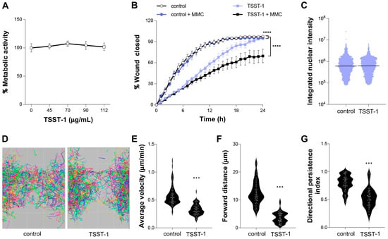
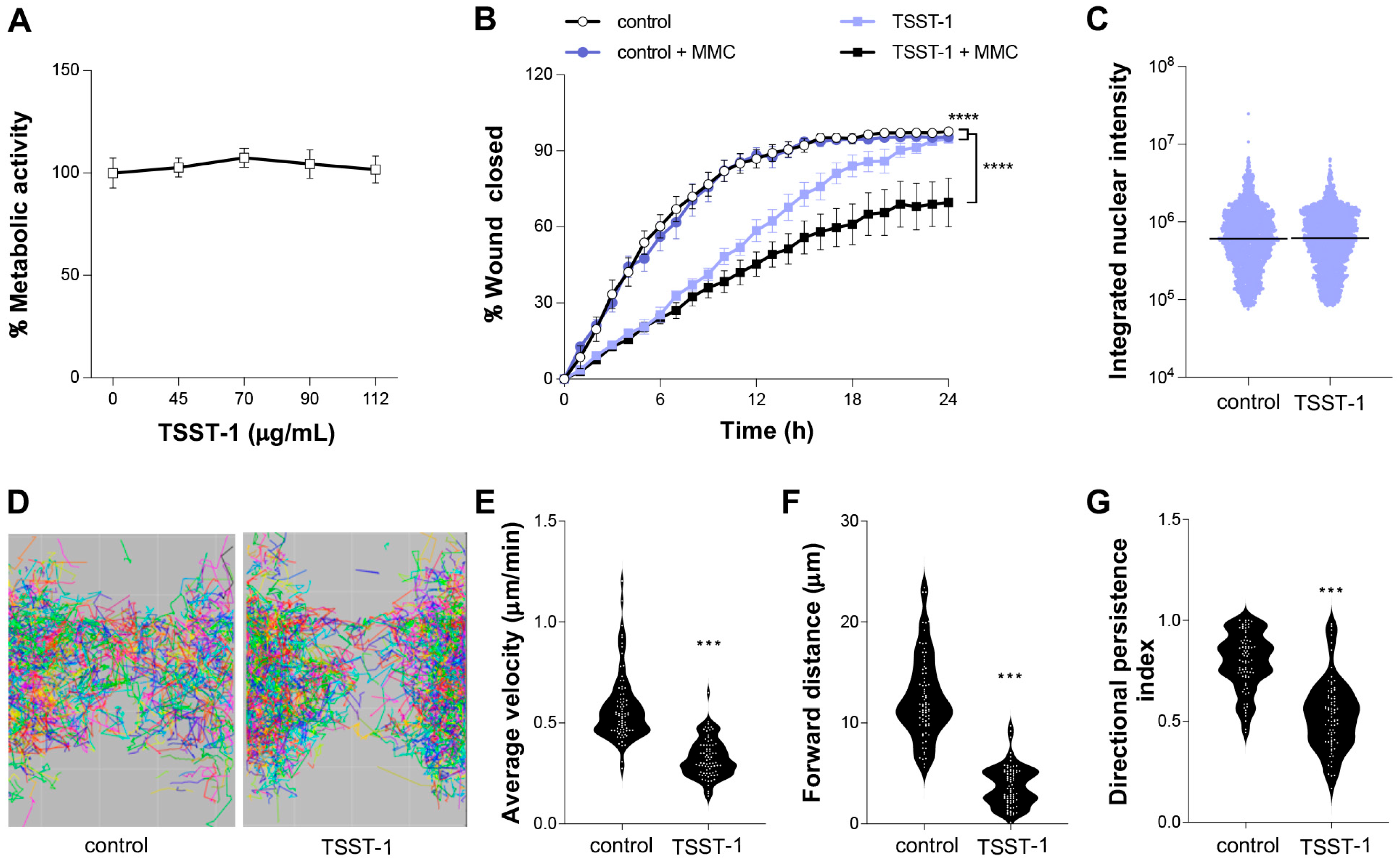
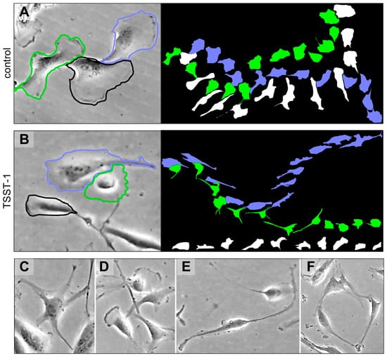
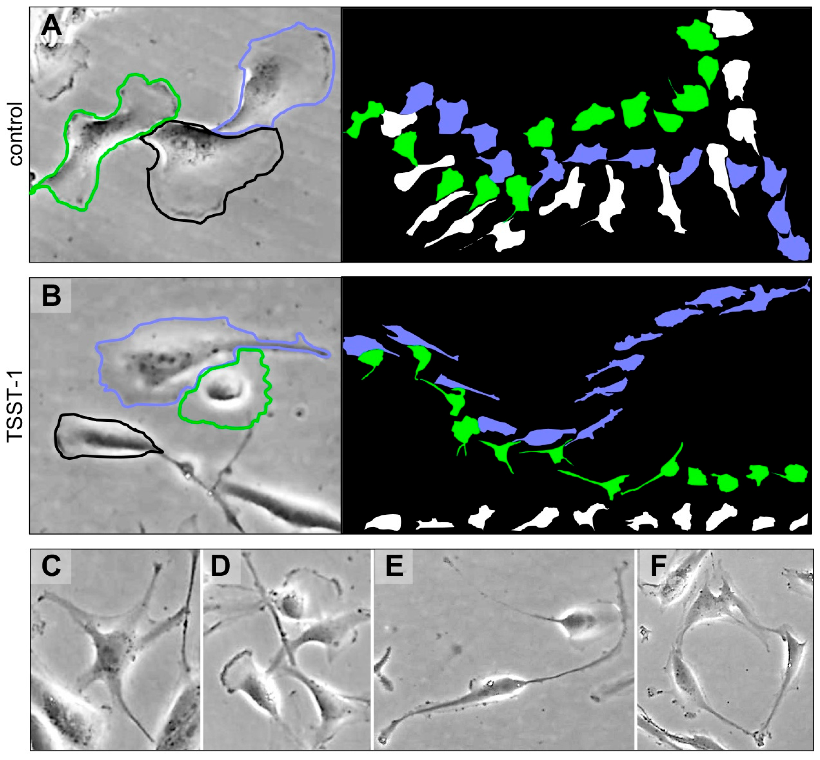
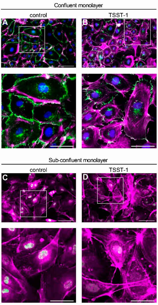
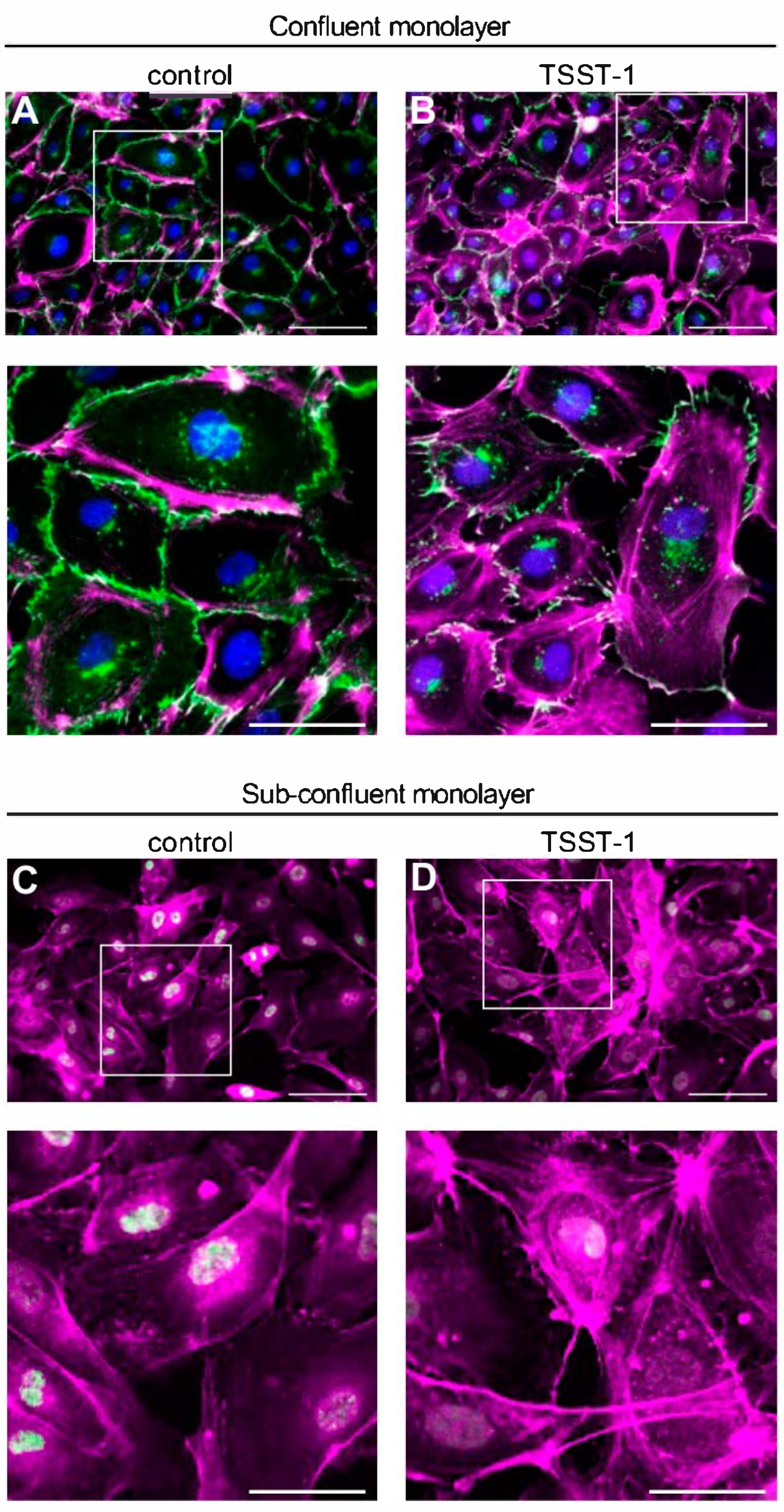
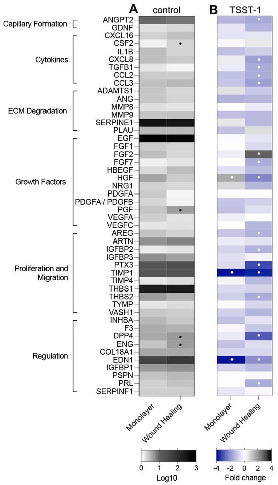
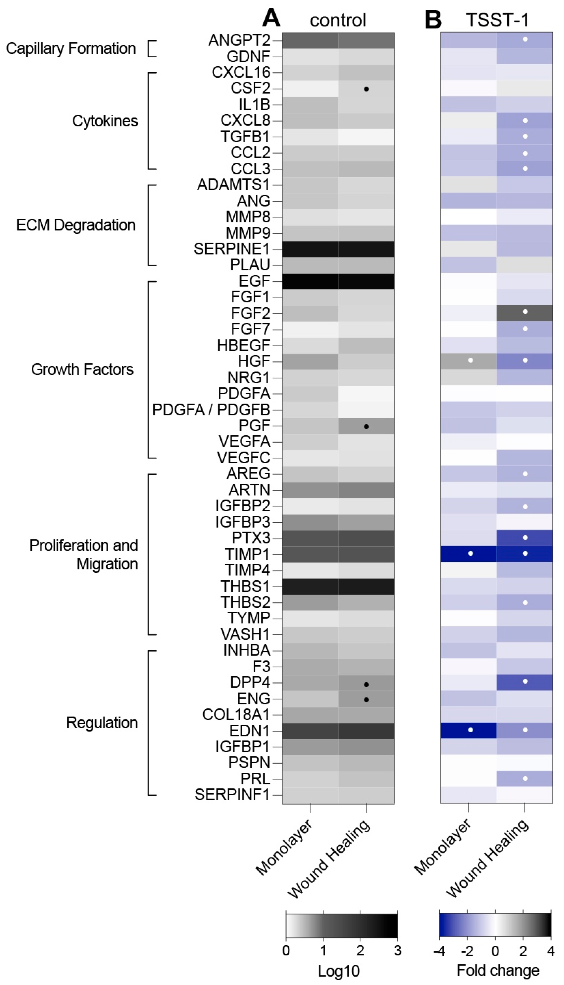
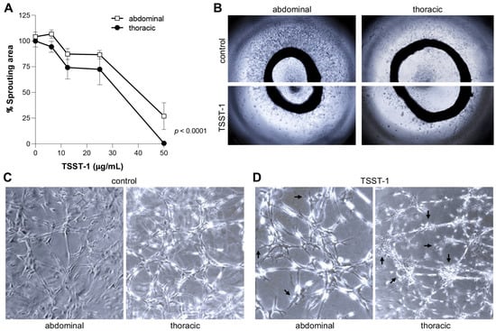
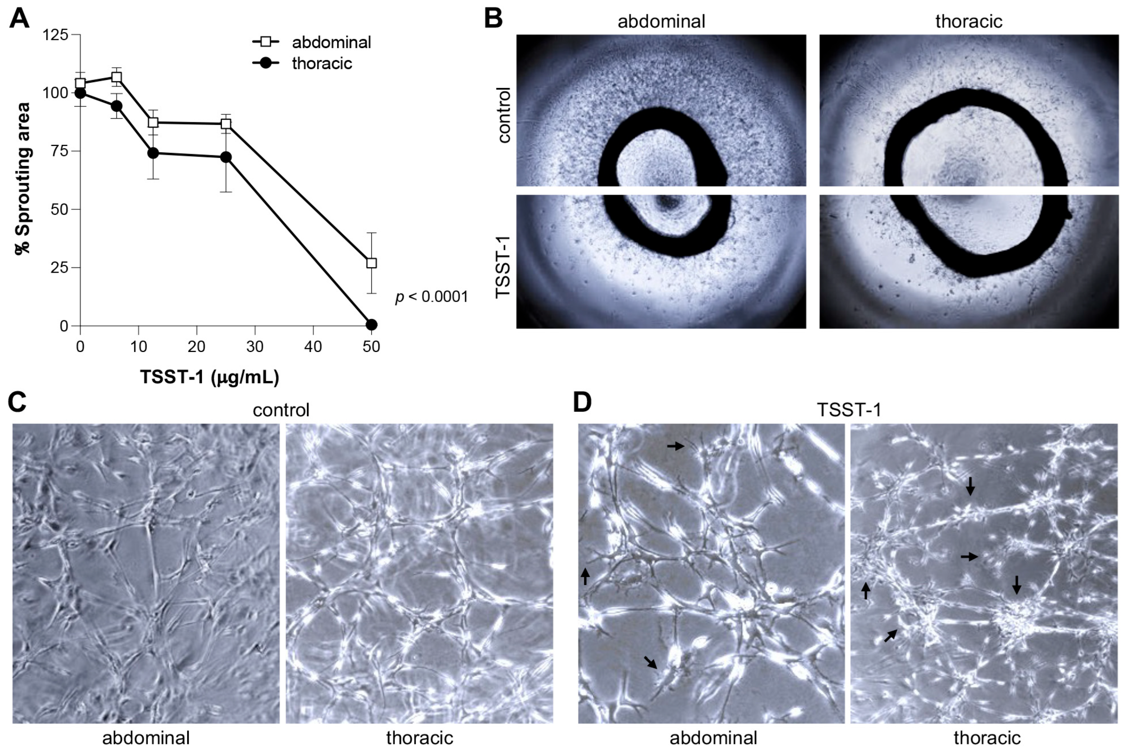
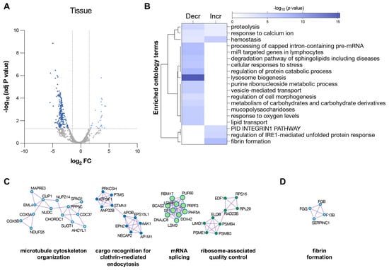
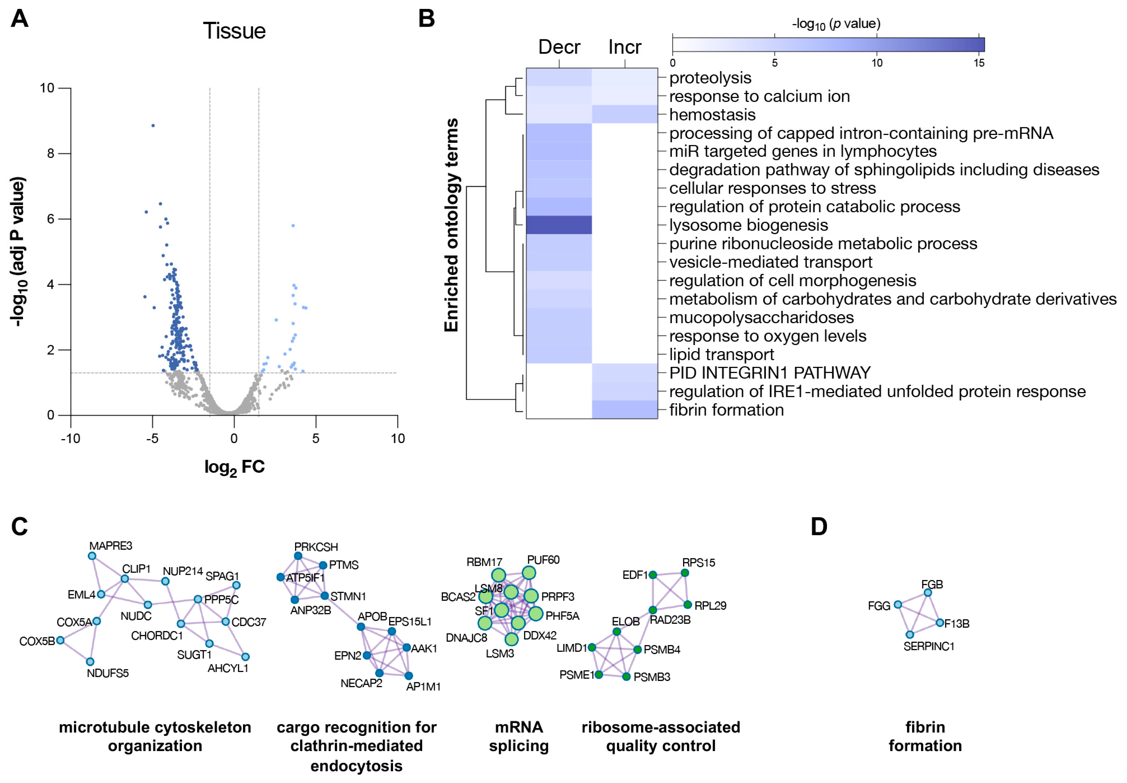
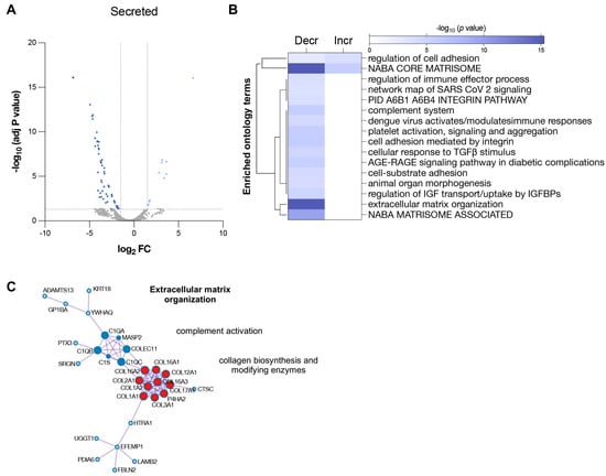
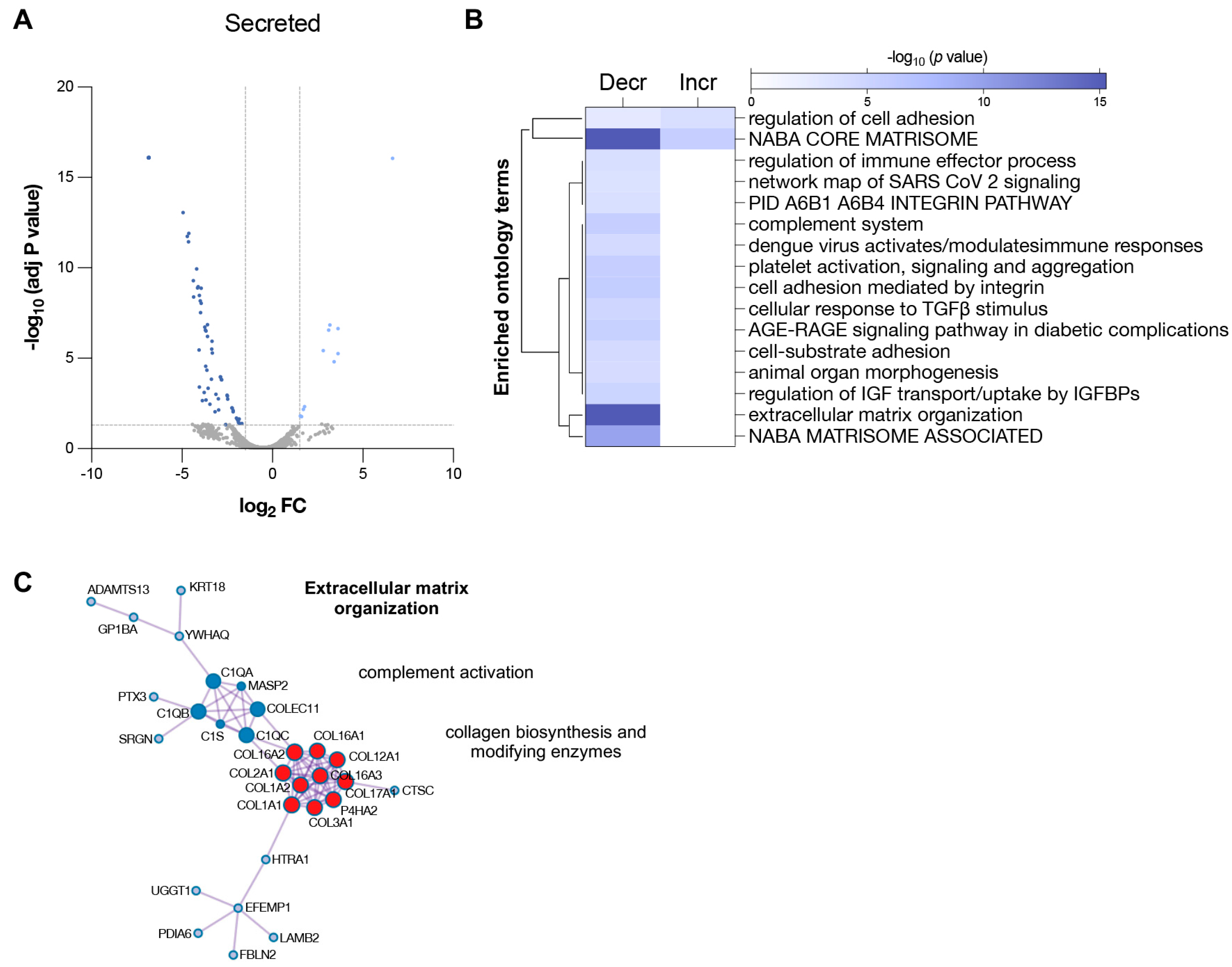
Disclaimer/Publisher’s Note: The statements, opinions and data contained in all publications are solely those of the individual author(s) and contributor(s) and not of MDPI and/or the editor(s). MDPI and/or the editor(s) disclaim responsibility for any injury to people or property resulting from any ideas, methods, instructions or products referred to in the content. |
© 2026 by the authors. Licensee MDPI, Basel, Switzerland. This article is an open access article distributed under the terms and conditions of the Creative Commons Attribution (CC BY) license.