


Highlights
What are the main findings?
- •
- Clinical improvement and reverse remodeling in heart failure do not necessarily imply complete biological recovery. Persistent alterations across immune, stromal, vascular, metabolic, mitochondrial, and hematopoietic compartments may constitute residual biological heart failure (RBHF), particularly when they remain biologically active, impair physiological reserve, or increase vulnerability to relapse.
- •
- We propose heart failure resolution competence (HF-RC) as a complementary framework describing the capacity of the cardiac and systemic environment to terminate injury-associated responses, complete adaptive repair, and establish durable post-injury homeostasis. HF-RC is conceived as a dynamic property of recovery rather than a surrogate for ejection fraction, inflammatory burden, or conventional disease severity.
What are the implications of the main findings?
- •
- Heart failure recovery may be more meaningfully characterized by the depth, kinetics, coordination, and durability of biological normalization than by isolated measurements obtained at a single time point. Longitudinal trajectories and responses to physiological perturbation may therefore provide information that cannot be captured by conventional resting phenotypes alone.
- •
- RBHF and HF-RC provide testable research frameworks rather than new diagnostic categories. Their translational value will depend on demonstrating that residual biological abnormalities predict impaired reserve or relapse beyond established clinical measures and that modifying relevant mechanisms improves the durability of recovery.
Abstract
Heart failure (HF) remains a major cause of morbidity and mortality despite substantial advances in disease-modifying therapy, and clinical improvement does not necessarily indicate complete restoration of myocardial homeostasis. Increasing evidence suggests that inflammatory, stromal, vascular, metabolic, and mitochondrial programs initiated during cardiac injury may persist beyond apparent phenotypic recovery. This narrative review examines HF through the complementary framework of biological resolution and evaluates whether incomplete recovery may reflect not only persistence of the original disease substrate but also incomplete termination of injury-associated biological programs. Evidence was synthesized from contemporary experimental, translational, and clinical studies addressing inflammatory cell dynamics, efferocytosis, immune and fibroblast state transitions, extracellular matrix remodeling, endothelial and microvascular dysfunction, mitochondrial and metabolic adaptation, cellular senescence, hematopoietic conditioning, and tissue-resident immunity. Collectively, these data indicate that reverse remodeling may coexist with persistent biological abnormalities whose significance depends on their activity, functional consequences, and effects on physiological reserve. We therefore propose residual biological heart failure (RBHF) to describe biologically relevant abnormalities persisting despite substantial phenotypic improvement and heart failure resolution competence (HF-RC) to describe the capacity of the cardiac and systemic environment to terminate injury-associated responses, complete adaptive repair, and restore durable homeostasis. These constructs extend rather than replace established concepts of reverse remodeling, remission, and myocardial recovery. RBHF and HF-RC remain hypothesis-generating frameworks requiring longitudinal human validation, but they provide a testable basis for distinguishing phenotypic improvement from the biological depth and durability of HF recovery.
1. Introduction
Heart failure (HF) represents a major and growing global health burden, affecting more than 60 million people worldwide, with prevalence expected to increase further as populations age and survival from cardiovascular disease improves [1]. Rather than constituting a single disease entity, HF encompasses heterogeneous clinical syndromes arising from distinct structural, functional, and systemic substrates [2]. Contemporary phenotyping distinguishes, among others, HF with reduced ejection fraction (HFrEF) and HF with preserved ejection fraction (HFpEF), which differ substantially in ventricular phenotype, comorbidity burden, therapeutic responsiveness, and underlying biology. Ischemic heart disease and cardiomyopathies remain major substrates of HFrEF, whereas hypertension, obesity, diabetes, chronic kidney disease, aging, and systemic metabolic dysfunction contribute prominently to HFpEF; valvular heart disease and other loading conditions may contribute across the ejection-fraction spectrum [3]. This clinical and etiological heterogeneity is particularly relevant to biological recovery because the mechanisms that initiate myocardial injury, sustain remodeling, and limit subsequent restoration of homeostasis are unlikely to be uniform across HF phenotypes [4].
Contemporary classifications appropriately recognize heterogeneity in left ventricular ejection fraction, etiology, hemodynamic phenotype, and clinical trajectory [5]. These frameworks predominantly describe how HF presents and progresses; they provide less insight into why biological recovery remains incomplete in some patients even after the dominant disease mechanism has been effectively treated. This distinction has become increasingly relevant as modern therapy can produce substantial reverse remodeling, improvements, normalization of biomarkers, and, in selected patients, recovery of ventricular function [6]. The biological meaning of such improvement, however, remains incompletely defined [7].
Inflammation has emerged as an important component of HF pathophysiology. Activation of innate and adaptive immune pathways has been demonstrated across ischemic and non-ischemic myocardial injury, pressure and volume overload, cardiometabolic disease, and advanced HF. Damage-associated molecular patterns, inflammasome activation, complement signaling, cytokine release, oxidative stress, leukocyte recruitment, and macrophage activation can contribute to myocardial injury and remodeling [8,9]. Attempts to translate this knowledge into broadly effective anti-inflammatory treatments have yielded inconsistent results [10]. One explanation is biological heterogeneity: inflammation may be causal in some settings, secondary in others, and inseparable from ongoing tissue injury in still others. A complementary possibility is that the relevant abnormality is not simply excessive inflammatory activation, but a failure of the injured cardiovascular system to complete the transition from inflammation through repair to stable homeostasis.
Resolution is not synonymous with suppression of inflammation. It is an active and coordinated biological process through which injury-associated responses are appropriately terminated, damaged cells and extracellular material are cleared, reparative programs evolve, and tissue function is re-established [11,12]. This process involves efferocytosis, temporal transitions in neutrophil and macrophage states, restoration of endothelial integrity, deactivation or reprogramming of activated fibroblasts, extracellular-matrix reorganization, mitochondrial quality control, and recovery of metabolic and vascular homeostasis [13]. Successful resolution requires coordinated change across immune, stromal, vascular, metabolic, and parenchymal compartments rather than normalization of an isolated inflammatory mediator.
In heart failure, mechanisms initially required for survival and tissue repair can become maladaptive when they persist beyond their appropriate biological window. Leukocyte recruitment is essential for the removal of damaged material, but becomes harmful when sustained. Fibroblast activation preserves structural integrity after injury but contributes to stiffness and impaired function when deactivation is incomplete. Extracellular matrix deposition stabilizes damaged myocardium, yet may subsequently alter mechanotransduction and cellular behavior [14]. Mitochondrial stress responses preserve cellular viability but can perpetuate oxidative and danger signaling when damaged organelles are not effectively removed. Similarly, cellular senescence, hematopoietic adaptation, and tissue-resident immune responses may initially serve adaptive functions while subsequently contributing to persistent biological vulnerability. HF may reflect not only the consequences of injury, but in selected settings, an inability to terminate processes that were initially appropriate [15].
Such persistence provides a potential link between transient injury and long-term biological memory. Previous HF may leave durable changes in myocardial cellular composition, extracellular matrix, microvascular function, mitochondrial quality, immune-cell states, or hematopoietic output [16,17]. Importantly, persistence alone does not establish pathology. Some post-injury changes may represent appropriate adaptation, others may be biologically neutral, and still others may reflect continuing exposure to an unresolved causal stimulus.
Improvement in left ventricular ejection fraction, chamber geometry, natriuretic peptides, filling pressures, and symptoms represents genuine and clinically meaningful recovery. It does not, however, establish that the biological processes underlying HF have normalized to an equivalent degree. Different compartments may recover at different rates, and some may remain altered after conventional indices have substantially improved [18]. Clinical recovery and biological recovery should be regarded as related but not necessarily synonymous processes.
To describe this discrepancy, we propose the concept of residual biological heart failure (RBHF). RBHF refers to persistent disease-associated biological abnormalities that remain after substantial improvement of the clinically measurable HF phenotype [19]. It is not intended as a new diagnostic category, an extension of LVEF-based classification, or evidence that HF can never genuinely resolve. Rather, it provides a framework for examining the biological distance between phenotypic improvement and restoration of stable tissue homeostasis.
We further propose heart failure resolution competence (HF-RC) as a complementary framework describing the capacity of the cardiac and systemic biological environment to terminate injury-associated responses, complete repair, and establish a stable post-injury state. Resolution competence is unlikely to depend on a single pathway. It may instead emerge from interactions among immune cells, fibroblasts, endothelial cells, cardiomyocytes, mitochondria, extracellular matrix, and hematopoietic compartments [20].
The proposed framework is conceptually distinct from established descriptions of reverse remodeling, myocardial recovery, remission, and heart failure with improved ejection fraction. These constructs primarily characterize changes in the clinically observable phenotype, ventricular structure and function, or the longitudinal course of disease. RBHF addresses a different level of the recovery process: the biological abnormalities that persist despite substantial phenotypic improvement and may remain relevant to physiological reserve or susceptibility to relapse. HF-RC, in turn, describes the capacity of the cardiac and systemic biological environment to terminate injury-associated programs, complete adaptive repair, and establish a stable post-injury state. RBHF and HF-RC should therefore not be viewed as alternative classifications of heart failure, but as complementary mechanistic constructs intended to bridge the gap between phenotypic improvement and the depth, completeness, and durability of biological recovery. The proposed constructs address a level of recovery not directly captured by existing clinical terminology. Reverse remodeling describes a favorable change in cardiac structure and function; HFimpEF identifies improvement in LVEF within a defined clinical phenotype; and myocardial recovery or remission describes varying degrees of functional and clinical improvement. RBHF instead asks whether disease-associated biological abnormalities remain functionally relevant after such improvement, whereas HF-RC asks whether the recovering cardiovascular system retains the capacity to terminate injury-associated programs and re-establish homeostasis after perturbation. The distinction is therefore not primarily semantic or classificatory: it separates the observable phenotype of recovery from its underlying biological depth and resilience. Whether this additional dimension provides prognostic information beyond established clinical measures remains to be demonstrated.
These concepts remain hypotheses rather than validated clinical entities. Much of the mechanistic evidence supporting resolution biology derives from experimental cardiovascular injury, and persistent abnormalities in human HF cannot automatically be interpreted as evidence of failed resolution. Ongoing injury must be distinguished from defective termination, and association from causal persistence. Resolution is also intrinsically temporal: a single measurement cannot reliably discriminate active disease, delayed recovery, stable adaptation, recurrent stimulation, and genuine non-resolution. Testing this framework will require longitudinal studies that follow biological trajectories through active HF, effective treatment, reverse remodeling, and longer-term recovery.
We integrate evidence from inflammatory-cell dynamics, efferocytosis, immune and fibroblast state transitions, extracellular matrix remodeling, endothelial and microvascular dysfunction, mitochondrial and metabolic adaptation, cellular senescence, hematopoietic conditioning, and tissue-resident immunity. On this basis, we introduce residual biological heart failure (RBHF) and heart failure resolution competence (HF-RC) as complementary, testable constructs intended to extend—not replace—established concepts of reverse remodeling, myocardial recovery, remission, and HF with improved ejection fraction. The specific contribution of this framework is to shift attention from the magnitude of phenotypic improvement alone toward the biological depth, completeness, resilience, and durability of recovery.
1.1. Physiological Versus Pathological Subcellular Remodeling in Heart Failure
The myocardium continuously adapts its cellular and subcellular organization to changes in workload, substrate availability, neurohumoral signaling, and mechanical stress. Under physiological conditions, these responses preserve contractile performance through coordinated adjustments in excitation–contraction coupling, sarcoplasmic-reticulum Ca2+ handling, myofilament function, mitochondrial energetics, membrane signaling, and cytoskeletal organization [21]. Such adaptation is generally proportionate to demand and retains the capacity to regress when the initiating stimulus is removed. In heart failure, however, sustained hemodynamic, neurohumoral, inflammatory, or metabolic stress can convert initially compensatory responses into maladaptive subcellular remodeling [22]. Altered Ca2+ cycling, impaired β-adrenergic signaling, myofilament dysfunction, mitochondrial energetic failure, oxidative stress, and changes in membrane and extracellular-matrix interactions progressively uncouple energetic supply from mechanical demand and reduce myocardial reserve [23]. Importantly, the distinction between physiological and pathological remodeling is determined not simply by the presence of structural or molecular changes, but by their magnitude, persistence, reversibility, and functional consequences.
This distinction is particularly relevant to the concept of biological resolution. Subcellular abnormalities may persist after improvement in chamber geometry, ejection fraction, filling pressures, or symptoms, indicating that reverse remodeling at the organ level does not necessarily imply equivalent recovery of the cellular machinery responsible for contraction, energetics, and stress adaptation [24]. Conversely, not every residual molecular alteration should be interpreted as pathological, because some changes may represent stable compensation rather than active disease. Biological recovery should therefore be considered a coordinated process in which adaptive remodeling is retained where necessary, whereas maladaptive signaling, energetic inefficiency, disturbed Ca2+ homeostasis, oxidative injury, and inappropriate reparative activity are progressively terminated or normalized [25]. This distinction between adaptive and maladaptive remodeling provides an important mechanistic basis for considering why apparently similar degrees of clinical recovery may coexist with different levels of physiological reserve and vulnerability to subsequent stress [26].
1.2. Literature Search Strategy
This article was conceived as a narrative, hypothesis-generating review rather than a systematic review or meta-analysis. The literature was identified through structured searches of PubMed/MEDLINE, supplemented by examining reference lists of relevant original studies, reviews, and consensus documents. The search covered publications available from database inception through 30 October 2026, which was the date of the final literature search.
Search terms were used individually and in relevant combinations and included “heart failure”, “myocardial recovery”, “reverse remodeling”, “HFimpEF”, “inflammation resolution”, “efferocytosis”, “macrophage”, “neutrophil”, “regulatory T cell”, “fibroblast”, “extracellular matrix”, “endothelial dysfunction”, “microvascular dysfunction”, “mitochondrial dysfunction”, “mitophagy”, “cellular senescence”, “trained immunity”, “hematopoiesis”, “tissue-resident immunity”, “immune memory”, “specialized pro-resolving mediators”, and related terms.
Priority was given to original mechanistic studies, human translational investigations, longitudinal clinical studies, and studies directly addressing myocardial injury, repair, reverse remodeling, or recovery. Experimental cardiovascular studies were included when they provided mechanistic information not directly obtainable in humans, whereas clinical studies were preferentially used when human HF-specific evidence was available. Selected high-quality reviews and consensus documents were used to provide broader biological or clinical context and to identify the additional primary literature. Studies were evaluated for relevance to the central question of whether injury-associated biological programs terminate, persist, or become reactivated during HF progression and recovery.
Because the objective was conceptual synthesis rather than exhaustive quantification of evidence, no formal risk-of-bias assessment or systematic evidence-grading procedure was performed. Accordingly, the literature summarized in this review should be interpreted as a structured narrative synthesis, and distinctions between experimental evidence, observations in human HF, and mechanisms that remain predominantly hypothetical are maintained throughout the manuscript.
1.3. Five Interconnected Domains of HF Pathophysiology
Contemporary HF pathophysiology can be viewed through five interconnected biological domains rather than a single dominant mechanism. First, cardiomyocyte dysfunction encompasses abnormalities in excitation–contraction coupling, calcium handling, sarcomeric performance, energetic failure, and cell death signaling, ultimately reducing contractile reserve [27]. Second, sustained neurohormonal and autonomic activation, including sympathetic and renin–angiotensin–aldosterone signaling, initially supports perfusion but chronically promotes hypertrophy, oxidative stress, sodium retention, vascular dysfunction, and adverse remodeling [28]. Third, innate and adaptive immune activation modifies myocardial injury and repair through leukocyte recruitment, cytokine signaling, inflammasome activity, complement pathways, and macrophage-state transitions [29]. Fourth, fibroblasts and the extracellular matrix translate mechanical, inflammatory, and neurohormonal signals into structural remodeling; although fibroblast activation is essential for tissue stabilization after injury, persistent myofibroblast activity and matrix deposition promote fibrosis, stiffness, and impaired reversibility [30]. Fifth, endothelial, microvascular, mitochondrial, and metabolic dysfunction constrain perfusion and energetic flexibility while reinforcing oxidative and inflammatory stress [31]. These domains are not independent: cardiomyocytes, fibroblasts, endothelial cells, resident and recruited immune cells, and the extracellular matrix form an interacting myocardial ecosystem in which adaptive responses may become maladaptive when their intensity, duration, or termination is inappropriate.
1.4. Contemporary Therapeutic Pillars of Heart Failure: Mechanistic Foundations
Contemporary management of heart failure (HF) is founded on interventions targeting the neurohormonal, hemodynamic, and metabolic mechanisms responsible for disease progression [32,33]. In heart failure with reduced ejection fraction (HFrEF), four established pharmacological pillars constitute the foundation of disease-modifying therapy: angiotensin receptor–neprilysin inhibition (ARNI) or alternative renin–angiotensin system inhibition, evidence-based β-blockade, mineralocorticoid receptor antagonism (MRA), and sodium–glucose cotransporter-2 (SGLT2) inhibition [34]. Their complementary mechanisms extend beyond conventional hemodynamic improvement. ARNI combines attenuation of angiotensin II-mediated signaling with enhancement of natriuretic peptide activity, thereby modifying vascular loading, sodium balance, and adverse myocardial remodeling. β-Blockers counter sustained sympathetic stimulation, reduce adrenergic myocardial stress, and facilitate functional and structural recovery. MRAs limit aldosterone-dependent sodium retention and mineralocorticoid receptor-mediated profibrotic and inflammatory signaling, whereas SGLT2 inhibitors confer cardiovascular and renal protection through mechanisms involving renal sodium handling, altered loading conditions, and cardiometabolic regulation [35]. A fifth, complementary therapeutic dimension comprises individualized etiological, device-based, and advanced interventions, including cardiac resynchronization, coronary revascularization, correction of valvular disease, mechanical circulatory support, and heart transplantation, according to the underlying substrate and disease severity [36]. This fifth dimension represents an organizing framework rather than a universally established pharmacological pillar. Importantly, therapeutic priorities differ across the ejection-fraction spectrum, with SGLT2 inhibition, management of congestion, and treatment of associated comorbidities assuming particular importance in HF with preserved ejection fraction [37]. Collectively, these interventions modify the biological conditions sustaining HF and may facilitate reverse remodeling. However, whether their clinical benefits are consistently accompanied by the complete termination of injury-associated cellular and reparative programs remains an important and incompletely resolved question.
2. From Inflammatory Activation to Resolution: A Missing Dimension in Heart Failure Biology
The inflammatory response to cardiac injury is intrinsically dynamic. Its biological significance cannot be inferred from the magnitude of leukocyte recruitment, cytokine production, or activation of individual innate immune pathways at a single time point. An appropriate response must be initiated early enough to contain the injury and facilitate the removal of damaged tissue, but it must also transition toward repair and restoration of homeostasis. Inflammation and resolution are not opposing states; they are temporally overlapping components of a coordinated tissue response whose outcome depends on the nature of the injury, cellular context, and capacity to terminate programs that were initially adaptive [38,39].
Current models have largely emphasized mechanisms of inflammatory activation in heart failure pathology, including damage-associated molecular patterns (DAMPs), pattern-recognition receptors, inflammasome and complement signaling, oxidative stress, cytokine release, and myeloid cell recruitment [40]. These pathways explain how inflammation is initiated and amplified, but less adequately why inflammatory and reparative programs persist after the dominant stimulus has diminished, or why successful correction of hemodynamic stress does not invariably restore tissue homeostasis [41]. The complementary question is not simply what activates inflammation in HF, but what determines whether the response can successfully resolve.
2.1. Resolution Is an Active Biological Program
Resolution was historically regarded as the passive consequence of the decline of inflammatory stimuli. This view has been replaced by recognition of an actively regulated process involving coordinated changes in leukocyte trafficking, cell death and clearance, phagocyte function, lipid and cytokine signaling, stromal cell behavior, vascular integrity, and extracellular matrix organization. Its purpose is not merely to reduce inflammation, but to terminate injury-associated responses while preserving host defense, remove damaged cells and debris, complete appropriate repair, and establish functional homeostasis [42,43].
Several events are central to this transition. Further neutrophil recruitment must decline; apoptotic and damaged cells must be efficiently cleared; macrophage programs must shift from inflammatory amplification toward clearance and repair; and endothelial, stromal, and parenchymal compartments must progressively disengage from injury-associated signaling. These processes are accompanied by the reorganization of cytokine and lipid-mediator networks and the eventual contraction or redistribution of inflammatory cell populations [44].
Suppression reduces selected components of an inflammatory response; resolution coordinates its termination with clearance and tissue repair [45]. Inhibition of a single mediator may reduce measurable inflammation without restoring the biological processes required for recovery. Conversely, successful resolution does not require elimination of immune activity, but rather its appropriate temporal, spatial, and functional reorganization [46].
Following acute myocardial injury, neutrophils and inflammatory monocytes participate in the removal of damaged material, after which macrophage-mediated clearance, fibroblast activation, matrix deposition, angiogenesis, and scar maturation become increasingly important. Disruption of this sequence can impair healing even when the targeted pathway is conventionally considered inflammatory. The relevant biological objective is not minimal inflammation, but an appropriately initiated and successfully terminated response [47,48].
Chronic HF is more complex than acute injury because cardiomyocyte stress, altered loading, neurohormonal and metabolic dysfunction, congestion, endothelial abnormalities, and recurrent episodes of decompensation can provide continuing or recurrent biological stimuli. Activation and attempted repair may coexist, generating persistent cellular states that cannot be adequately classified as simply inflammatory or reparative [49,50].
2.2. The Transition from Inflammation to Repair Is a Cellular State Change
Traditional descriptions of cardiac inflammation frequently assign immune-cell populations fixed pro- or anti-inflammatory identities. The M1/M2 macrophage paradigm has been a useful concept, but it does not capture the diversity of myocardial macrophage states demonstrated by contemporary transcriptomic and single-cell studies. Macrophage behavior is shaped by developmental origin, tissue localization, recruitment history, metabolic environment, phagocytic activity, and local cellular interactions [51,52].
This plasticity is central to resolution. Recognition and engulfment of apoptotic cells not only remove material that can sustain inflammation but also reprogram phagocytes toward states conducive to clearance and repair [53]. Changes in cytokine production, lipid metabolism, chemokine signaling, and interactions with fibroblasts and endothelial cells accompany this transition [54].
Cellular origin adds another level of complexity. The healthy myocardium contains tissue-resident macrophage populations that differ from recruited monocyte-derived cells. Severe or recurrent injury can alter their relative abundance and spatial organization [55]. Consequently, normalization of total macrophage numbers does not necessarily imply restoration of the pre-injury immune ecosystem. Resolution should be understood not simply as the disappearance of inflammatory cells, but as the recovery of an appropriately organized and functional cellular environment.
2.3. Efferocytosis as a Checkpoint Between Inflammation and Resolution
Efferocytosis occupies a pivotal position at the interface between injury and resolution. Efficient clearance of apoptotic cells prevents secondary necrosis and limits extracellular release of nucleic acids, mitochondrial constituents, and other potentially immunogenic material. At the same time, engulfment activates intracellular programs within phagocytes that favor inflammatory termination and tissue repair [56,57].
Apoptotic-cell recognition depends on coordinated “find-me” and “eat-me” signals, including phosphatidylserine exposure and receptor systems involving MerTK and associated bridging molecules. Successful clearance also requires sufficient metabolic competence to process engulfed material. Efferocytosis is not merely a receptor-mediated event, but a functional test of the capacity of the phagocyte to complete clearance [58,59].
Experimental myocardial infarction and ischemia–reperfusion studies demonstrate that disruption of efferocytic pathways can impair clearance and worsen remodeling [60]. Their relevance to established human HF remains less certain. Nevertheless, chronic oxidative stress, mitochondrial dysfunction, aging, metabolic disturbance, and repeated cellular injury could plausibly create an imbalance between the amount of damaged material generated and the tissue’s capacity to remove it [61].
Such an imbalance may be particularly important in chronic HF, where modest but recurrent injury could repeatedly exceed local clearance capacity. Delayed removal permits apoptotic cells to progress toward secondary necrosis, increasing DAMP exposure and potentially sustaining innate immune activation. The biological consequence of cell death may depend not only on its magnitude but also on the efficiency with which its products are cleared [62].
Defective efferocytosis represents a mechanistically well-supported determinant of adverse repair in experimental cardiovascular injury, whereas direct evidence that impaired myocardial efferocytosis constitutes a quantitatively important driver of non-resolution in chronic human HF remains limited [63,64]. In the present framework, it should therefore be regarded as a biologically plausible candidate mechanism rather than an established human HF phenotype.
2.4. Pro-Resolving Signaling and the Concept of a Mediator-Class Transition
Resolution also involves qualitative changes in the mediators exchanged among immune and structural cells. Particular interest has focused on specialized pro-resolving mediators (SPMs), including lipoxins, resolvins, protectins, and maresins [65]. These lipid-derived mediators have been proposed to limit further neutrophil recruitment, enhance macrophage efferocytosis, modify cytokine production, and promote tissue repair through receptor-dependent mechanisms [66].
Their conceptual relevance lies in the possibility that successful resolution requires not simply declining concentrations of inflammatory mediators but a transition toward signaling networks that actively facilitate termination and repair. Such a mechanism is attractive in cardiovascular disease because lipid mediators can simultaneously influence leukocytes, platelets, endothelial function, vascular tone, and tissue remodeling.
However, the evidence requires careful interpretation. Several candidate SPMs occur at very low endogenous concentrations, and their measurement is complicated by analytical issues involving LC–MS/MS methodology, stability, stereochemical identification, biosynthetic plausibility, and receptor specificity [67,68]. Experimental benefit following administration of an exogenous mediator also does not establish that endogenous deficiency of that mediator causes human HF. SPM biology should be regarded as an evolving component of the resolution framework rather than an established explanation for failed resolution in patients. Defining a clinically meaningful cardiovascular “resolution lipidome” will require rigorously validated quantitative lipidomics integrated with longitudinal cellular and tissue phenotyping.
2.5. Resolution Failure Is Not Necessarily Persistent Inflammation
A crucial distinction follows from this framework, which is based on the hypothesis that failed resolution is not synonymous with chronic inflammation [69]. Persistent inflammatory activity can arise through several mechanisms. The initiating insult may remain active; recurrent injury may repeatedly reactivate an otherwise competent response; resolution machinery itself may be impaired; or structural remodeling may generate secondary stimuli that maintain inflammation after the original insult has diminished [70]. These mechanisms may produce similar biomarker profiles while representing fundamentally different biological states.
This distinction is especially relevant across HF phenotypes. Ischemic HFrEF may retain scar-associated or recurrent ischemic stimuli, whereas genetic, toxic, metabolic, infectious, or immune-mediated cardiomyopathies create different patterns of injury and repair [71]. In HFpEF, obesity, diabetes, chronic kidney disease, hypertension, and aging may continuously expose the cardiovascular system to systemic metabolic and inflammatory signals [72]. Apparent cardiac non-resolution in such settings may reflect repeated extracardiac re-initiation rather than an intrinsic defect in myocardial resolution.
The persistence of the initiating stimulus must consequently be considered before non-resolution is inferred. A tissue cannot be expected to achieve complete resolution while injury is continuously renewed. More informative—and more difficult to establish—is persistence of inflammatory or reparative activity after effective control of the dominant insult, suggesting that components of the injury response have become partly autonomous from the process that originally initiated them [73].
2.6. The Resolution–Repair Paradox: When Successful Healing Becomes Maladaptive Remodeling
Resolution and repair are interdependent but not synonymous. Processes required to restore short-term structural integrity can become sources of long-term dysfunction if they fail to terminate appropriately [74]. Fibroblast activation and extracellular-matrix deposition illustrate this tension particularly clearly [75].
Following substantial cardiomyocyte injury, fibroblast activation, myofibroblast differentiation, matrix deposition, angiogenesis, and macrophage-derived reparative signaling are essential for tissue stabilization [76]. Persistence of these programs, however, can promote fibrosis, myocardial stiffness, impaired relaxation, electrical heterogeneity, and altered ventricular mechanics [77]. The same pathway may be protective during one phase of injury and maladaptive during another.
This creates a resolution–repair paradox, as termination of leukocyte-dominated inflammation does not necessarily signify restoration of myocardial homeostasis [78]. Inflammation may resolve while fibrotic or other reparative programs remain active. Conversely, premature inhibition of repair may compromise tissue integrity [79]. Successful recovery consequently requires not maximal suppression of repair, but its appropriate initiation, spatial restriction, maturation, and termination.
Endothelial activation facilitates immune recruitment but becomes maladaptive when sustained; macrophage-derived growth factors promote repair but may perpetuate stromal activation; and extracellular matrix stabilizes injured tissue while subsequently influencing mechanotransduction and cellular behavior [80,81]. Resolution must occur across interconnected compartments whose kinetics need not be synchronized.
2.7. From Inflammatory Burden to Resolution Capacity
These observations expose a limitation of conventional inflammatory measurements in HF. Circulating cytokines, CRP, leukocyte subsets, or tissue inflammatory transcripts may identify inflammatory activity but cannot establish why that activity persists [82]. An apparently similar inflammatory phenotype could reflect severe, ongoing injury with intact resolution; modest injury combined with impaired resolution; repeated cycles of activation and incomplete recovery; or combinations of these states [83].
A more informative framework considers the relationship between injury burden and resolution capacity. Tissue homeostasis is favored when cellular clearance, inflammatory termination, vascular recovery, and reparative completion are sufficient for the magnitude and duration of injury [84]. Persistent disease becomes more likely when injury repeatedly exceeds this capacity or when resolution mechanisms themselves become compromised [85].
Aging, metabolic disease, renal dysfunction, recurrent ischemia, infection, neurohormonal activation, and previous inflammatory exposures may simultaneously increase injury burden and reduce the capacity for macrophage clearance, mitochondrial recovery, endothelial restoration, and stromal plasticity [86]. HF progression may reflect not only accumulation of injury, but progressive loss of the ability to resolve an equivalent degree of injury [87].
This perspective also provides a potential explanation for biological heterogeneity among patients with apparently similar clinical HF. Comparable ventricular dysfunction or systemic inflammatory activity may conceal fundamentally different capacities for recovery [88]. In one patient, control of the dominant insult may permit coordinated restoration of tissue homeostasis; in another, persistent immune, stromal, vascular, or metabolic states may continue despite equivalent clinical treatment [89].
Resolution capacity is best understood as an emergent property of a multicellular system rather than the function of a single inflammatory pathway. Immune cells, fibroblasts, endothelial cells, cardiomyocytes, extracellular matrix, mitochondria, and systemic hematopoietic compartments participate in interdependent processes whose recovery occurs on different temporal scales [90]. The critical question is consequently no longer only which pathways initiate or amplify inflammation, but whether the post-injury cardiovascular system retains the capacity to terminate inflammation, complete repair, and establish a stable biological state [91]. This transition from inflammatory burden to resolution capacity provides the conceptual basis for examining the cellular and tissue mechanisms that may determine the depth and durability of recovery in HF. The biological processes that distinguish successful from incomplete resolution in heart failure are presented in Table 1.
Table 1.
Biological processes distinguishing successful resolution from incomplete resolution in heart failure.
These observations support a dynamic model in which the biological consequences of cardiac injury depend not only on the magnitude of the initial response but also on whether inflammatory and reparative programs are appropriately terminated and on whether the myocardium successfully transitions toward stable homeostasis (Figure 1).
Figure 1.
Divergent Biological Trajectories After Cardiac Injury. Legend: Cardiac injury initiates inflammatory, clearance, and reparative responses whose long-term consequences depend not only on the magnitude and persistence of the initiating insult, but also on the capacity of the cardiovascular system to terminate injury-associated programs and complete tissue repair. Successful resolution involves coordinated restriction of further leukocyte recruitment, efficient clearance of damaged material, appropriate evolution of immune-cell states, fibroblast deactivation and matrix maturation, restoration of vascular homeostasis, and recovery of mitochondrial and metabolic function. These processes need not restore a pristine pre-injury myocardium but may establish a stable post-injury state with sufficient physiological reserve. Incomplete resolution may instead permit persistent or recurrent immune activation, inadequate clearance, continued stromal and extracellular-matrix remodeling, microvascular dysfunction, metabolic stress, and reinforcing multicellular interactions. The resulting state may comprise residual substrate, residual biological activity, and residual vulnerability, collectively providing the conceptual basis for residual biological heart failure (RBHF). A subsequent physiological or pathological stressor may expose differences between apparently similar resting phenotypes by revealing preserved versus impaired capacity to return toward homeostasis. Persistent abnormalities should not, however, be automatically equated with pathological non-resolution, as some may represent stable adaptation or irreversible but biologically inactive consequences of prior injury. Created by Urbanowicz T., on 31 October 2026 with www.figurelabs.ai (ID: FL-PUB-20261031-1ZFV0P).
3. The Cellular Architecture of Cardiac Resolution
Resolution of cardiac inflammation is not executed by a single cell population or molecular pathway. It emerges from coordinated transitions among recruited leukocytes, tissue-resident immune cells, vascular cells, and stromal populations as the injured myocardium progresses from recognition and containment toward clearance and repair [108,109]. The biological consequences of inflammation depend not only on which cells are present but also on their origin, functional state, spatial organization, and position within the temporal trajectory of injury [110]. This distinction is particularly relevant in heart failure (HF), where recurrent or persistent stress can blur the boundaries between inflammatory and reparative phases and allow cellular programs that were adaptive during acute injury to persist in a maladaptive context.
3.1. Neutrophils: From Acute Injury to Resolution Signaling
Neutrophils are among the earliest cellular responders to substantial myocardial injury [111]. Their rapid recruitment facilitates removal of damaged material and host defense, but also exposes surrounding tissue to proteases, reactive oxygen species, inflammatory mediators, and neutrophil extracellular traps [112]. Their biological effect is strongly time-dependent: appropriate early recruitment contributes to tissue clearance, whereas excessive accumulation, prolonged survival, or dysregulated activation can amplify collateral injury and delay subsequent repair.
Neutrophils should not, however, be regarded solely as terminal pro-inflammatory effectors. Their timely apoptosis constitutes an important component of the transition toward resolution. Recognition and engulfment of apoptotic neutrophils by macrophages simultaneously remove potentially damaging cellular material and modify phagocyte behavior toward clearance and repair [113,114]. Neutrophils can participate in both initiation and termination of inflammation, depending on their functional state and temporal context [115]. Most mechanistic evidence for temporally ordered neutrophil states derives from experimental acute-injury models, particularly myocardial infarction. Chronic HF is unlikely to reproduce this sharply demarcated response. Recurrent decompensation, congestion, hypoperfusion, infection, and systemic comorbidity may instead produce intermittent episodes of neutrophil activation superimposed on an already remodeled immune environment. Whether these episodes actively impede resolution or predominantly reflect continuing systemic injury remains uncertain.
The more informative question is not whether neutrophils are increased in HF, but whether their recruitment, activation, apoptosis, and clearance remain appropriately synchronized [116]. Disruption of this sequence provides a plausible mechanism through which a normally transient defensive response could become a recurrent source of tissue injury [117].
3.2. Monocyte–Macrophage State Transitions: Orchestrators of Resolution
Macrophages occupy a central position at the interface between inflammation, clearance, and tissue repair. The myocardial macrophage compartment includes tissue-resident populations and macrophages derived from recruited circulating monocytes, with important differences in developmental origin, self-renewal, localization, and response to injury [118]. This heterogeneity makes binary classification into inflammatory and reparative phenotypes inadequate for contemporary HF biology [119].
In the healthy myocardium, resident macrophages contribute to surveillance and maintain functional relationships with cardiomyocytes, endothelial cells, fibroblasts, and the microvasculature [120]. Injury disrupts this ecosystem. Resident populations may be depleted or redistributed, while recruitment of circulating monocytes generates macrophage states shaped by the inflammatory and metabolic environment [121]. During successful repair, macrophage function progressively shifts from inflammatory amplification and debris clearance toward efferocytosis, matrix regulation, angiogenic support, and communication with reparative stromal populations [122].
Disappearance of overt inflammation does not necessarily restore the pre-injury macrophage landscape. Monocyte-derived populations may persist, and phenotypically similar cells can retain distinct transcriptional, metabolic, and functional programs determined by origin and exposure history [123]. Normalization of macrophage abundance consequently provides limited information about restoration of the myocardial immune niche.
This issue becomes particularly relevant in chronic HF, where macrophages remain exposed to cardiomyocyte stress, altered perfusion, extracellular-matrix remodeling, mitochondrial dysfunction, neurohormonal activation, and systemic comorbidities. Rather than occupying stable phenotypes, they may move along overlapping inflammatory, phagocytic, angiogenic, reparative, and profibrotic trajectories [124]. Such plasticity provides an important mechanistic interface between inflammation and structural remodeling: macrophages may contribute to clearance while simultaneously generating signals that sustain fibroblast activation and matrix turnover [125].
Successful resolution should not be conceptualized as an M1-to-M2 switch [126]. It requires restoration of an appropriately organized macrophage ecosystem in which inflammatory amplification terminates, cellular debris is efficiently removed, reparative signaling remains proportionate to tissue requirements, and stromal activation does not become self-sustaining [127].
These state transitions are supported by extensive experimental and increasingly sophisticated single-cell and spatial datasets [128]; their temporal behavior during clinical HF recovery remains incompletely characterized. In particular, persistence of a disease-associated macrophage state in human myocardium should not, in isolation, be interpreted as evidence of failed resolution unless it can be linked to persistent biological activity, impaired recovery, or altered responses to subsequent stress [129].
3.3. Regulatory and Adaptive Immune Control
Although innate immune cells dominate the early response to myocardial injury, adaptive immunity contributes to its subsequent regulation and may influence persistence or recurrence [130]. T lymphocytes are particularly relevant because they can provide both inflammatory amplification and counter-regulatory control while retaining antigen-experienced states that persist beyond the initiating event [131].
Regulatory T cells (Tregs) constitute an important component of this regulatory architecture. Through IL-10, TGF-β, CTLA-4-dependent modulation of antigen-presenting cells, and direct interactions with innate and stromal populations, Tregs can constrain excessive inflammatory activity and facilitate repair [132]. Experimental cardiovascular studies suggest that their function extends beyond generalized immunosuppression to modulation of macrophage and fibroblast behavior, placing them within a broader multicellular resolution network [133].
Conversely, adaptive responses may impede resolution when antigenic stimulation persists or recurrent tissue injury maintains activated and memory populations [134]. Th1- and Th17-associated programs can reinforce macrophage activation and endothelial dysfunction, while cytotoxic lymphocytes may contribute directly to tissue injury in selected etiologies [135]. B-cell and antibody-mediated mechanisms add complexity, but their importance varies substantially with disease substrate and should not be generalized across HF phenotypes [136].
As with innate immunity, cellular abundance alone is an inadequate measure of biological function. Circulating Treg numbers do not establish suppressive competence within the myocardium, and peripheral lymphocyte phenotypes may incompletely reflect tissue-localized states [137]. Spatial organization is likely to be important because regulatory and effector lymphocytes exert their effects within specific endothelial, macrophage, and stromal niches [138].
The adaptive compartment introduces a dual dimension into resolution biology: regulatory capacity and immunological persistence. This distinction becomes increasingly relevant as HF evolves from an acute injury response toward a chronic tissue state and provides a mechanistic bridge to tissue-resident immune memory discussed later in this review.
3.4. Efferocytosis as the Cellular Checkpoint of Resolution
Efferocytosis provides one of the principal cellular links between the termination of inflammation and tissue repair. Its importance extends beyond disposal of apoptotic cells: efficient clearance prevents secondary release of intracellular danger signals while simultaneously inducing transcriptional and metabolic changes in the engulfing phagocyte that favor resolution [46].
This coupling is particularly relevant in the myocardium, where inadequately cleared cellular material contains mitochondrial constituents, nucleic acids, and other DAMPs that can sustain innate immune signaling [139]. Failure of clearance can transform cell death from a finite event into a continuing inflammatory stimulus. Conversely, successful efferocytosis removes the source of danger signaling while facilitating macrophage transition toward reparative functions.
The MerTK axis, together with phosphatidylserine-recognition systems and bridging molecules such as Gas6, Protein S, and MFG-E8, represents a well-characterized component of this machinery [140]. However, receptor-mediated recognition alone does not determine clearance competence. Following engulfment, phagocytes must process substantial quantities of lipids, cholesterol, proteins, and mitochondrial material, thereby linking efferocytosis to metabolic and mitochondrial fitness [141]. Consequently, clearance may become inadequate even when apoptotic-cell recognition remains intact. For chronic HF, the more relevant concept may be a mismatch between clearance demand and phagocytic capacity, rather than complete failure of efferocytosis. Recurrent low-grade injury could thereby exceed local clearance capacity even in the absence of a discrete defect in the efferocytic machinery. These observations support a view of cardiac resolution as a coordinated ecosystem transition. Neutrophil apoptosis must be matched by macrophage clearance; macrophage programs must evolve from inflammatory amplification toward proportionate repair; adaptive immunity must provide regulatory control without sustaining pathological activation; and damaged cellular material must be removed before it becomes a persistent source of danger signaling [142]. Failure within one compartment can alter the behavior of others and prevent restoration of tissue homeostasis [143].
This interdependence may become increasingly important as HF progresses. Recurrent injury does not occur within a biologically naïve myocardium, but within tissue already modified by previous inflammation, cellular loss, metabolic stress, vascular dysfunction, and structural remodeling. Each subsequent insult is interpreted by an altered cellular ecosystem [144]. Among the mechanisms that stabilize this altered state, fibroblasts and the extracellular matrix are particularly important, as they can convert transient inflammatory and reparative signals into persistent structural information [145]. The transition from cellular non-resolution to stromal and matrix remodeling represents the next level of biological persistence in HF.
4. When Repair Fails to Terminate: Fibroblasts and Persistent Stromal Activation
Once injury has engaged the stromal compartment, the reparative program required to preserve tissue integrity must itself eventually terminate. This transition from active repair to structural quiescence is particularly important in the heart, where limited regenerative capacity makes fibroblast activation and extracellular-matrix (ECM) deposition essential after substantial cardiomyocyte injury. The same processes become maladaptive when maintained beyond the requirements of tissue stabilization [146]. Chronic heart failure (HF) may reflect not only persistent injury or inflammation, but also failure to disengage a reparative program that has outlived its original purpose.
Cardiac fibroblasts are central to this transition. Rather than functioning simply as collagen-producing cells, they integrate mechanical stress, inflammatory and neurohormonal signals, metabolic conditions, and cues from cardiomyocytes, endothelial cells, immune populations, and the surrounding matrix [147]. Fibrosis should be regarded not merely as the cumulative consequence of injury, but as an active tissue state capable of modifying the environment in which subsequent injury and recovery occur.
4.1. Fibroblast State Transitions After Cardiac Injury
In the healthy myocardium, fibroblasts maintain ECM homeostasis and tissue architecture. Injury disrupts this equilibrium through mechanical stretch, cellular damage, inflammatory mediators, neurohormonal activation, and changes in matrix composition. Fibroblasts consequently enter proliferative, secretory, and matrix-producing states; after substantial injury, acquisition of a contractile myofibroblast phenotype is essential for wound stabilization [148].
Single-cell and spatial studies have substantially refined this conventional model. Fibroblasts do not follow a uniform linear trajectory from quiescence to activation and then back to rest. Injured myocardium contains heterogeneous populations with inflammatory, proliferative, matrix-producing, contractile, and remodeling-associated programs that vary with anatomical location, disease substrate, stage of injury, and local cellular environment [149]. “Activated fibroblast” describes a spectrum of context-dependent states rather than a single pathogenic phenotype.
A state required for early tissue stabilization may become harmful when chronically maintained, whereas premature suppression of fibroblast activity may compromise structural integrity [150]. Successful recovery requires appropriate progression through reparative states followed by contraction, deactivation, or transition toward a phenotype compatible with long-term homeostasis [151]. The mechanisms governing fibroblast deactivation remain less well understood than those initiating fibrosis. This asymmetry is relevant to a resolution-centered model of HF: the removal of the initiating stimulus cannot be assumed to automatically restore stromal quiescence [152]. Termination of fibroblast activation should instead be considered an active component of tissue recovery.
4.2. Failure of Myofibroblast Deactivation
Myofibroblast persistence illustrates the boundary between adaptive repair and pathological remodeling. Increased contractility and fibrillar collagen synthesis initially stabilize vulnerable myocardium. Once sufficient structural integrity has been achieved, however, continued matrix production and cross-linking increase myocardial stiffness, impair relaxation, alter electrical and mechanical coupling, and contribute to adverse ventricular remodeling [153].
Persistent TGF-β and neurohormonal signaling, mechanical stretch, altered integrin signaling, oxidative stress, and inflammatory mediators can maintain profibrotic programs [154]. Importantly, these stimuli need not remain dependent on the original cardiac insult. As matrix architecture and stiffness change, the ECM itself modifies mechanotransduction and cellular signaling, generating feedback that sustains fibroblast activation [155]. Repair may thereby acquire partial autonomy from the event that initiated it.
This possibility changes the interpretation of persistent fibrosis. Continued matrix remodeling need not imply ongoing cardiomyocyte injury of equivalent magnitude. Instead, the stromal compartment may enter a self-maintaining state in which altered mechanical and molecular feedback sustains fibroblast activity despite attenuation of the original stimulus. This provides a plausible explanation for the incomplete regression of fibrosis despite substantial hemodynamic or functional improvement [156].
Experimental evidence indicates that activated fibroblasts retain plasticity and that fibrosis can regress after removal of pathological loading or signaling [157]. Yet this capacity is neither universal nor unlimited. Disease duration, matrix organization, collagen cross-linking, cellular aging, metabolic dysfunction, and recurrent injury may progressively constrain stromal plasticity. The transition from reversible activation to a more stable profibrotic state may represent an important threshold between adaptive repair and chronic tissue remodeling.
4.3. Immune–Fibroblast Crosstalk in Persistent Remodeling
Fibroblast behavior is inseparable from the immune environment. Macrophages, lymphocytes, mast cells, and other immune populations provide cytokines, growth factors, and contact-dependent signals that regulate fibroblast proliferation, differentiation, and matrix production [158]. Fibroblasts reciprocally influence leukocyte recruitment and function through chemokines, cytokines, matricellular proteins, and changes in ECM composition [159].
During successful repair, these interactions must evolve as tissue requirements change. Signals promoting fibroblast recruitment and activation should diminish as structural stabilization is achieved. Persistent inflammatory or profibrotic immune states may instead prevent stromal deactivation, while activated fibroblasts can create an environment that favors immune-cell retention and continued signaling [160]. Such reciprocal reinforcement need not produce marked inflammatory infiltration; relatively subtle alterations in chemokine gradients, matrix composition, adhesion, and local growth-factor availability may be sufficient to sustain remodeling.
Immune–stromal interactions occur within specific myocardial microenvironments—including perivascular regions, replacement scars, interstitial fibrotic zones, and injury border regions—rather than uniformly throughout the myocardium. Single-cell methods identify the states present, but spatial approaches are required to determine which populations are anatomically positioned to interact. The relevant unit of investigation may be the cellular neighborhood, rather than an isolated immune or stromal population. This perspective also avoids categorizing immune–fibroblast signaling as intrinsically pathological. Many profibrotic pathways are indispensable during early repair [161]. Their pathological significance arises from inappropriate magnitude, localization, or persistence after their reparative function has been fulfilled.
4.4. Extracellular Matrix as a Reservoir of Biological Memory
The ECM is not merely the structural product of fibroblast activity. Its composition, architecture, stiffness, cross-linking, and proteolytic remodeling actively regulate cellular behavior through integrins, focal adhesions, mechanosensitive pathways, and local growth-factor availability [162]. The matrix is both an output of previous injury and an input into subsequent cellular responses.
This reciprocity creates the possibility that previous injury becomes structurally encoded within the myocardium. Fibrosis alters the mechanical environment sensed by fibroblasts and cardiomyocytes; increased matrix stiffness can reinforce pathways favoring continued stromal activation; matrix-bound mediators can remain locally available after their initial production; and matrix degradation can generate biologically active fragments [163]. The tissue constructed during repair may influence whether repair subsequently terminates.
In this sense, the ECM can be considered a form of structural biological memory. This does not imply immunological or epigenetic memory. Rather, it describes the capacity of previous injury to leave persistent alterations in tissue architecture that modify responses to future mechanical and molecular signals [164]. Consequently, recurrent stress occurs within a myocardium whose physical and biochemical properties already contain information about its disease history. Residual fibrosis and abnormal matrix organization can preserve myocardial stiffness, impair mechanical efficiency, alter microvascular–myocyte relationships, and change the threshold at which stromal cells respond to renewed stress. Functional improvement need not erase the structural consequences of previous disease [165]. ECM turnover persists, and reverse remodeling demonstrates that pathological structure remains at least partly modifiable [166]. The clinically important questions are how much abnormal matrix can regress, over what time scale, and when remodeling becomes sufficiently stabilized to resist normalization despite effective treatment of the initiating condition.
This framework helps reconcile the frequent dissociation between functional and structural recovery. Hemodynamic improvement may occur relatively rapidly, whereas fibrosis regression and restoration of matrix architecture may follow a slower, potentially incomplete trajectory. Fibroblasts thus translate inflammatory, mechanical, and neurohormonal information into persistent tissue remodeling, while the resulting matrix feeds information back to the cells embedded within it [167]. When this reciprocal system fails to disengage, repair itself becomes a source of continued pathological signaling.
Failed resolution extends beyond persistent inflammation. A heart may no longer exhibit prominent leukocyte activation yet remain constrained by tissue architecture generated during previous injury. The vascular compartment represents a further level of this persistence because endothelial cells and the coronary microcirculation regulate perfusion, leukocyte trafficking, barrier function, and the organization of local immune–stromal niches. Failure to restore vascular homeostasis may sustain biological vulnerability after overt inflammation has subsided.
5. Mitochondrial Damage and Metabolic Persistence: When the Danger Signal Does Not Switch Off
The heart is exceptionally dependent on mitochondrial integrity. Beyond sustaining ATP production, mitochondria regulate calcium handling, redox homeostasis, intermediary metabolism, cell death pathways, and the communication between cellular stress and innate immunity [168]. Mitochondrial dysfunction in heart failure (HF) extends beyond energetic insufficiency. Once mitochondrial quality deteriorates, the organelle itself can become a source of signals that perpetuate cellular stress and impede restoration of homeostasis.
This dual function makes mitochondrial injury particularly relevant to resolution biology. Dysfunctional mitochondria produce energy inefficiently but can also generate excessive reactive oxygen species, release mitochondrial constituents, disturb calcium homeostasis, and constrain the metabolic flexibility required for repair [169]. The critical question is not whether mitochondrial dysfunction accompanies HF, but whether damaged mitochondrial networks are effectively removed, replaced, and functionally reintegrated after the dominant stress has diminished.
5.1. Mitochondrial Damps and Persistent Innate Sensing
Mitochondrial DNA (mtDNA), formylated peptides, cardiolipin, and other constituents are normally compartmentalized and largely concealed from immune surveillance. Loss of mitochondrial integrity exposes these molecules to cytosolic or extracellular pattern-recognition systems, transforming essential intracellular components into damage-associated molecular patterns [170].
Cytosolic mtDNA is particularly relevant. Membrane disruption, defective mitochondrial turnover, and severe cellular stress can expose mtDNA to nucleic-acid sensors, including the cyclic GMP–AMP synthase–stimulator of interferon genes (cGAS–STING) pathway [171]. Mitochondrial material can also contribute to inflammasome and related innate immune signaling [172]. An organelle essential for cardiomyocyte survival can thus become a continuing source of sterile danger signals.
This mechanism provides a potential bridge between primary injury and biological persistence. When mitochondrial damage is not adequately corrected, danger signals may continue to be generated after the initiating insult has diminished. The relationship is also bidirectional: inflammation, oxidative stress, calcium dysregulation, and neurohormonal activation further impair mitochondrial function, potentially creating a feed-forward circuit between organellar dysfunction and cellular stress [173].
Importantly, such persistence does not require catastrophic mitochondrial failure. Dysfunction affecting only part of the mitochondrial network may reduce energetic reserve while maintaining stress signaling in cardiomyocytes exposed to continuous mechanical demand. Experimental cardiovascular evidence supports individual components of this model, including mtDNA-associated signaling, cGAS–STING activation, and inflammasome engagement [174]. Their independent contribution to established human HF, however, remains uncertain and must be distinguished from signaling that is secondary to ongoing tissue injury.
5.2. Defective Mitophagy and Mitochondrial Quality Control
Mitochondrial damage is unavoidable, making quality control essential for cellular homeostasis. Mitochondrial proteins are renewed, networks undergo continuous fusion and fission, and irreversibly damaged organelles can be selectively removed by mitophagy. These processes maintain a functional mitochondrial population while limiting the accumulation of organelles that generate energetic and inflammatory stress [175].
Mitophagy is particularly relevant to resolution because it provides a mechanism for actively removing the intracellular consequences of injury. The PINK1–Parkin pathway is the best-characterized example, although receptor-mediated mechanisms provide additional routes of mitochondrial clearance [176]. The relevant physiological objective is not maximal mitophagy but balanced mitochondrial turnover: elimination of damaged organelles must be coupled to sufficient biogenesis and network restoration.
Reduction in ventricular loading can lower energy requirements relatively rapidly without necessarily restoring mitochondrial architecture, respiratory efficiency, substrate utilization, or quality-control mechanisms. Cardiomyocytes may consequently recover adequate resting function while retaining reduced energetic reserve. Recurrent stress further introduces a temporal dimension. If a subsequent hemodynamic or metabolic challenge occurs before mitochondrial recovery is complete, it acts on a cardiomyocyte already carrying diminished reserve and residual organellar injury. Progressive HF may reflect not only the magnitude of individual insults but also incomplete mitochondrial recovery between them [177].
5.3. Immunometabolic Barriers to Resolution
The relevance of mitochondrial biology extends beyond cardiomyocytes. Glycolysis, oxidative phosphorylation, fatty acid and amino acid metabolism, and mitochondrial redox state influence inflammatory signaling, phagocytosis, proliferation, biosynthesis, and tissue repair [178]. These relationships cannot be reduced to a binary model of glycolytic inflammatory cells and oxidative reparative cells. Cellular metabolism in vivo is substantially more heterogeneous. Cells unable to reorganize energetic and biosynthetic pathways may remain trapped in states no longer appropriate to tissue requirements.
5.4. Mitochondrial Recovery as a Requirement for Durable Cardiac Recovery
Mitochondrial recovery should be distinguished from normalization of individual stress markers or restoration of ATP production alone. Durable recovery requires coordinated restoration of mitochondrial quality, network organization, turnover, redox control, metabolic flexibility, and cessation of inappropriate danger signaling [179]. Resting measurements can demonstrate substantial functional improvement while failing to reveal limited mitochondrial reserve. Exercise, tachycardia, infection, recurrent ischemia, and pressure or volume loading increase energetic demand and may expose a myocardium whose mitochondrial network has recovered only partially [180].
Residual mitochondrial dysfunction could contribute to vulnerability after apparently successful reverse remodeling. This does not imply that mitochondrial abnormalities are the principal determinant of relapse in all patients. Rather, mitochondrial competence represents one dimension of biological recovery whose trajectory may diverge from chamber-level improvement. HF-associated mitochondrial dysfunction is not a single lesion: altered substrate utilization, electron transport, mitochondrial dynamics, mitophagy, calcium handling, oxidative stress, and danger signaling may coexist in different combinations across etiologies and disease stages [181]. Interventions directed at isolated pathways may consequently require biological selection and appropriate timing rather than indiscriminate application.
A more informative translational objective is to determine whether mitochondrial competence marks the depth and durability of recovery. Longitudinal integration of metabolic imaging, circulating mitochondrial signals, myocardial metabolomics, molecular signatures, and functional measures of energetic reserve could establish whether mitochondrial normalization accompanies durable reverse remodeling or whether residual dysfunction identifies biologically incomplete recovery. Mitochondria thus occupy a distinctive position within the resolution framework: they are simultaneously targets of injury, determinants of adaptation, and potential generators of secondary danger signals. Failure of mitochondrial quality control permits previous injury to remain biologically active within the cell after extracellular conditions have improved. This intracellular persistence provides one mechanism by which the consequences of HF may outlast its dominant initiating stimulus.
6. Neurocardiac and Circadian Regulation of Heart Failure Resolution
6.1. The Bidirectional Heart–Brain Axis in Heart Failure
Heart failure is accompanied by reciprocal interactions between the cardiovascular and central nervous systems that extend beyond the consequences of reduced cardiac output alone [182]. Cerebral hypoperfusion, impaired vascular autoregulation, endothelial dysfunction, venous congestion, systemic inflammation, and neurohormonal activation may adversely affect cerebral homeostasis, particularly in advanced HF [183]. Conversely, disruption of central autonomic and neuroendocrine regulation may reinforce cardiovascular stress through sustained sympathetic activation, impaired parasympathetic restraint, altered hypothalamic–pituitary–adrenal signaling, and secondary effects on vascular tone, renal function, metabolism, and immune activity [184]. The heart–brain axis may therefore form a self-reinforcing biological circuit in which cardiac dysfunction modifies central regulation, while maladaptive central responses subsequently increase the hemodynamic and cellular burden imposed on the myocardium [185].
This reciprocity is relevant to a resolution-centered view of HF. Improvement in ventricular function or loading conditions does not necessarily imply normalization of the systemic regulatory adaptations established during preceding disease. Persistent autonomic imbalance or altered neuroendocrine and cerebrovascular regulation could continue to influence myocardial metabolism, vascular function, and inflammatory signaling despite substantial phenotypic recovery [186]. Such abnormalities should not themselves be interpreted as evidence of failed resolution, and their independent contribution to RBHF remains to be established in longitudinal human studies. Nevertheless, they broaden the concept of recovery beyond the myocardium and suggest that durable cardiovascular homeostasis may require recalibration of the regulatory networks governing responses to subsequent stress [187]. The temporal organization of these responses provides a direct link between the heart–brain axis and circadian biology [188].
6.2. Circadian Disruption and Cellular Homeostasis in Heart Failure
Central and peripheral molecular clocks coordinate daily variation in cardiac workload, substrate utilization, mitochondrial function, redox balance, autophagy, vascular activity, and immune-cell trafficking, thereby aligning cellular demand with energy availability and repair. In HF, disruption of this organization may be particularly relevant because metabolic flexibility and energetic reserve are already constrained. Experimental and translational evidence suggests that circadian disturbance can alter myocardial substrate utilization, mitochondrial efficiency, oxidative stress responses, and cellular quality-control pathways, potentially increasing the mismatch between energetic demand and adaptive capacity [189].
From a resolution perspective, the importance of circadian biology lies less in any single clock-regulated pathway than in the temporal coordination of recovery. Effective resolution requires injury, inflammatory, metabolic, and reparative programs to be activated and terminated in an appropriate sequence [190]. Disruption of this temporal architecture could therefore impair recovery even in the absence of persistent elevation of individual biomarkers. Conversely, normalization of ventricular function at rest may not indicate restoration of the biological timing required for an appropriate response to subsequent stress. Circadian integrity may thus represent a previously underappreciated component of HF resolution competence, although its contribution to incomplete recovery and relapse in human HF remains to be established prospectively.
6.3. Neurocircadian Dysfunction as a Modifier of Heart Failure Resolution Competence
The integration of neurocardiac and circadian biology extends HF resolution competence beyond the capacity of individual cells or tissues to terminate injury-associated programs. Recovery also requires restoration of coordinated systemic responses to physiological challenge. In this context, HF-RC may depend not only on whether inflammatory, metabolic, vascular, and reparative pathways normalize, but also on whether they regain appropriate temporal organization and the ability to return efficiently toward homeostasis after perturbation [191,192]. Persistent dysregulation of these higher-order control systems could therefore remain biologically relevant even when conventional indices suggest substantial cardiac recovery.
This perspective emphasizes resilience rather than resting phenotype. Two patients with comparable reverse remodeling may differ in their capacity to accommodate recurrent hemodynamic, metabolic, or inflammatory stress and subsequently re-establish homeostasis. Neurocircadian regulation may contribute to this difference, but its role in determining the depth of recovery or relapse remains hypothetical and requires longitudinal validation. Accordingly, it should presently be viewed as a potential modifier of HF-RC rather than a defining component of RBHF.
7. The Heart–Bone Marrow Axis: Systemic Memory of Cardiac Injury
The biological consequences of heart failure (HF) extend beyond the myocardium. Cardiac injury generates neural, humoral, metabolic, and inflammatory signals that influence distant organs, including the bone marrow and spleen, where circulating immune populations are generated and maintained. This communication is bidirectional: cardiovascular injury modifies hematopoietic activity, while the resulting cells subsequently return to cardiovascular tissues and participate in inflammatory and reparative responses. The hematopoietic system may function not merely as a source of leukocytes but as a dynamic component of HF biology, whose output is conditioned by prior and ongoing cardiovascular stress [193].
This perspective expands the concept of resolution beyond the myocardium. Local inflammation may diminish while hematopoietic compartments remain altered, potentially changing the magnitude or quality of subsequent immune responses. The heart–bone marrow axis thus provides a plausible mechanism through which cardiac injury can acquire a biological time horizon extending beyond the original event.
7.1. Cardiac Injury as a Driver of Hematopoietic Remodeling
Acute cardiovascular injury can rapidly alter hematopoiesis. Experimental myocardial infarction increases proliferation and mobilization of hematopoietic stem and progenitor cells (HSPCs), promotes myelopoiesis, and enhances recruitment of monocytes and neutrophils to injured tissues. Sympathetic activation, circulating cytokines and growth factors, remodeling of the bone-marrow niche, and extramedullary hematopoiesis contribute to this response [194].
Such mobilization is initially adaptive, supplying cells required for debris clearance and tissue repair. The potential transition toward pathology arises when hematopoietic activation persists, repeatedly recurs, or leaves more durable changes in progenitor behavior after the immediate requirement for repair has diminished. Chronic HF provides multiple conditions capable of maintaining this communication, including neurohormonal activation, congestion, renal dysfunction, altered perfusion, metabolic disease, episodic ischemia, and recurrent systemic inflammation. Unlike a discrete experimental injury, HF frequently follows a fluctuating trajectory, with periods of relative stability interrupted by decompensation and extracardiac stress. The marrow may experience repeated stimulation rather than a single, clearly bounded event.
The evidence nevertheless requires careful qualification. Persistent hematopoietic remodeling is better established in experimental cardiovascular injury and atherosclerosis than in longitudinal human HF. Although heart–marrow communication is well supported, its magnitude and persistence across HF phenotypes remain incompletely defined. Inter-organ signaling should be distinguished from the stronger hypothesis that durable hematopoietic reprogramming independently sustains HF.
7.2. Myeloid Bias and Systemic Conditioning of the Cellular Supply Chain
Altered hematopoietic output may influence cardiac inflammation even without permanent stem-cell reprogramming. Increased myelopoiesis, shifts in monocyte populations, and altered circulating cell states alter the cellular substrate available for recruitment into cardiovascular tissues. The phenotype of myocardial inflammation is determined partly before recruited cells reach the heart [195]. Comparable myocardial stimuli may recruit biologically different cells depending on the state of systemic hematopoiesis. Local chemotactic and adhesion signals determine which cells enter the myocardium, but the hematopoietic system determines which cells are available to respond. The relationship can be viewed as systemic conditioning of the cellular supply chain.
Age and cardiometabolic disease add substantial complexity. Aging promotes changes in HSPC function and myeloid bias, while obesity, dysglycemia, chronic kidney disease, and chronic inflammatory exposures can independently alter hematopoiesis [196]. The cellular phenotype observed in HF may reflect the combined effects of cardiac disease, comorbidity, age, prior inflammation, and treatment rather than HF alone. A new cardiac insult does not occur from an immunologically neutral baseline. It acts within an organism whose hematopoietic system has already been shaped by its biological history, potentially contributing to heterogeneity in the response to apparently similar myocardial injuries.
7.3. Trained Immunity as One Component of Failed Resolution
Trained immunity provides one potential mechanism for persistence within this system. Unlike classical adaptive immune memory, trained immunity refers to sustained functional reprogramming of innate immune cells or their progenitors following prior stimulation. Epigenetic remodeling, altered chromatin accessibility, and metabolic reprogramming can modify subsequent responsiveness to stimuli that need not be identical to the original trigger [197].
Atherogenic lipids, metabolic abnormalities, and inflammatory mediators can induce persistent changes in myeloid cells and HSPCs. Reprogramming at the progenitor level is particularly important because it allows altered innate responsiveness to outlast individual circulating monocytes and potentially propagate into subsequent cellular generations [198].
Its role in HF, however, should not be overstated. Direct evidence that durable HSPC-trained immunity independently drives human HF progression remains limited, and chronic HF continuously exposes the immune system to multiple stimuli. Activated circulating monocytes or elevated inflammatory mediators cannot, by themselves, establish trained immunity. A stronger demonstration requires persistence of altered responsiveness after the initiating exposure has been removed. Trained immunity should consequently be regarded as one mechanism within a broader spectrum of hematopoietic persistence rather than a universal explanation for HF-associated inflammation. This distinction also has therapeutic implications: continued stimulation requires control of the underlying driver, whereas stable progenitor reprogramming persisting after that driver has been corrected may require a fundamentally different strategy.
7.4. Recurrent Injury and the Heart–Marrow Feedback Loop
The broader significance of the heart–bone marrow axis may lie in its capacity to generate feedback. Cardiac injury increases hematopoietic demand; altered hematopoiesis modifies circulating immune populations; these cells influence the heart, vasculature, kidney, and other tissues; and renewed tissue stress generates additional systemic signals. A process initiated locally can thereby become distributed across multiple organs [199]. The intensity of this circuit is unlikely to remain constant. Hematopoietic activation may increase during acute decompensation and regress during clinical stability. Repeated episodes, however, could prevent complete resetting or progressively alter the baseline from which subsequent responses begin. Disease progression may involve not only cumulative myocardial injury but also incomplete biological resetting between episodes.
This perspective offers a hypothesis for studying recurrent HF hospitalizations beyond their hemodynamic significance. Repeated decompensation may represent serial systemic stress capable of re-engaging hematopoietic, vascular, metabolic, and immune pathways. Whether such episodes produce cumulative and measurable changes in human hematopoietic compartments remains unknown. Moreover, this network extends beyond the heart and marrow. The spleen contributes through immune-cell storage and extramedullary myelopoiesis, while renal dysfunction, adipose inflammation, intestinal barrier abnormalities, and autonomic signaling influence systemic immune tone. The heart–bone marrow axis is better understood as part of a broader cardio-hematopoietic network rather than an isolated two-organ pathway.
7.5. Does the Hematopoietic System Retain a Biological Record of Heart Failure?
The possibility that HF modifies subsequent hematopoietic responses raises a broader question: Does the systemic immune compartment retain a biologically meaningful record of previous cardiac disease? This should not be conflated with permanent immune memory. Hematopoiesis remains dynamic, and many disease-associated abnormalities may regress after removal of the relevant stimulus. The critical question is whether hematopoietic recovery proceeds in parallel with cardiac recovery.
This distinction may become particularly relevant after reverse remodeling. Ventricular geometry, natriuretic peptides, and symptoms may improve while hematopoietic output remains conditioned by previous disease or continuing comorbidity. A subsequent myocardial stressor would then encounter both a previously remodeled heart and a systemic immune compartment whose response characteristics may remain altered.
Testing this hypothesis requires longitudinal investigation. Peripheral leukocyte counts and isolated cytokines are unlikely to capture the relevant biology. Serial integration of circulating-cell phenotyping with HSPC characterization, chromatin accessibility, transcriptomics, metabolic profiling, clonal architecture, and functional restimulation could determine whether hematopoietic abnormalities regress with cardiac recovery or persist independently.
Clonal hematopoiesis must also be considered. Age-related expansion of hematopoietic clones carrying somatic mutations can modify inflammatory behavior and cardiovascular risk [200,201]. Although clonal hematopoiesis and trained immunity may converge on overlapping pathways, they are mechanistically distinct. Persistent myeloid activation in HF may reflect environmental conditioning, epigenetic reprogramming, clonal selection, continuing stimulation, or combinations thereof.
The heart–bone marrow axis thus extends incomplete resolution from the myocardium to the organism. A clinically improved heart may remain embedded within a host whose hematopoietic environment has not recovered to the same extent. Systemic conditioning, however, is only one form of biological persistence. Immune populations that remain within the myocardium may preserve a more localized record of prior injury, providing a complementary mechanism by which past disease shapes future cardiac responses.
8. From Cellular Memory to Tissue Memory: Why Heart Failure Persists After the Initial Insult
A central challenge in heart failure (HF) biology is explaining why the consequences of cardiac injury may persist after the dominant initiating stimulus has been attenuated or removed. No single mechanism adequately accounts for this phenomenon. Fibrosis, microvascular dysfunction, mitochondrial abnormalities, cellular senescence, hematopoietic conditioning, and persistent immune states each contribute, but their significance ultimately derives from interaction within the same tissue. The post-injury myocardium should be considered not as a collection of residual abnormalities, but as an integrated biological system whose subsequent behavior is conditioned by its previous state [202].
We use the term tissue memory to describe this phenomenon. It does not imply a discrete molecular mechanism analogous to adaptive immune memory, nor does it suggest that the myocardium literally “remembers” injury. Rather, tissue memory refers to the persistence and interaction of structural, cellular, metabolic, and regulatory changes that modify the heart’s response to subsequent physiological or pathological stress. It is an emergent property of the post-injury myocardium, arising when recovery across multiple biological compartments remains incomplete or asynchronous.
8.1. Biological Recovery Is Not a Return to the Starting Point
Recovery after cardiac injury should not be assumed to reconstruct the pre-disease myocardium. Even a successful repair can leave a durable biological imprint. Replacement fibrosis provides the clearest example, but less visible changes may be equally consequential: interstitial matrix architecture may remain altered, microvascular reserve incompletely restored, cardiomyocyte metabolism adapted to prior stress, and immune or stromal composition may differ from that preceding disease. The recovered myocardium may represent a reorganized tissue rather than a restored replica of its original state [203]. Revascularization, valve intervention, rhythm control, mechanical unloading, and guideline-directed HF therapy can substantially improve ventricular geometry and function, yet correcting the dominant driver does not necessarily reverse all consequences generated during its activity [204].
The duration of the disease may influence the depth of this recovery. Early intervention may encounter cellular and tissue states that remain highly plastic, whereas prolonged exposure can stabilize structural and regulatory abnormalities and progressively constrain reversibility [205]. Biological recovery may depend not only on whether the initiating mechanism is corrected, but also on the state of the tissue when correction occurs.
8.2. Multiple Forms of Persistence Converge Within the Same Tissue
The forms of persistence described in the preceding sections do not operate independently. An altered ECM modifies mechanical forces experienced by cardiomyocytes, fibroblasts, and vascular cells; microvascular dysfunction changes oxygen delivery and metabolic conditions; mitochondrial abnormalities reduce energetic flexibility; senescent cells reshape local signaling; and immune populations respond to—and further modify—the remodeled environment. Each abnormality becomes part of the biological context in which the others operate [206]. This interconnectedness suggests that pathology may persist even after the original pathway is no longer dominant. Once sufficiently established, the remodeled myocardium may create conditions that perpetuate its dysfunction. Disease persistence can shift from dependence on the initiating insult toward dependence on the secondary biological architecture created in response to that insult. The severity of HF at a given time point provides only incomplete information about the myocardium’s biological state. Similar clinical phenotypes may conceal markedly different degrees of tissue plasticity and potential for recovery [207].
8.3. Tissue Memory as an Altered Response to a Second Hit
The functional significance of tissue memory may become most apparent when the myocardium encounters a subsequent challenge. A previously injured heart does not receive a second insult under the biological conditions that preceded the first. Its matrix may be stiffer, capillary reserve reduced, energetic flexibility constrained, cellular composition altered, and regulatory thresholds shifted. The response to renewed stress is consequently shaped by previous disease.
This concept provides a mechanistic framework for persistent susceptibility after apparent HF recovery. Previous disease may alter the myocardial response landscape, changing the magnitude or type of additional stress required to produce recurrent dysfunction. A second hit may include recurrent ischemia or abnormal loading, but also infection, arrhythmia, renal deterioration, metabolic stress, pregnancy, cardiotoxic exposure, or withdrawal of effective therapy [208]. In each case, the outcome reflects the interaction between the new challenge and the biological substrate on which it acts.
A previous injury may establish protective adaptations, improve surveillance or clearance, or facilitate more efficient responses to subsequent stress. Other changes may narrow physiological reserve or lower the threshold for decompensation [209]. The relevant question is not simply whether previous injury leaves a trace, but which elements of that trace promote resilience and which preserve vulnerability. This functional definition is important because persistence becomes clinically meaningful when it modifies future behavior. A residual molecular or cellular abnormality that neither compromises reserve nor alters the response to subsequent stress should not automatically be interpreted as pathological non-resolution.
8.4. From Reversible Adaptation to Stabilized Disease
Tissue memory should not be equated with irreversibility. The substantial reverse remodeling observed after reduction in pathological loading, neurohormonal blockade, rhythm restoration, mechanical unloading, and other effective interventions demonstrates considerable myocardial plasticity [210]. The critical question is when adaptive or reparative changes become sufficiently stabilized to resist reversal.
At one extreme, disease-associated states remain dependent on the initiating stimulus and regress after its removal. At the other, irreversible structural replacement, stable cellular reprogramming, or mutually reinforcing tissue interactions constrain recovery. Between these extremes lies a broad spectrum of partially reversible states whose trajectory may remain therapeutically modifiable [211].
Importantly, different cardiac compartments may simultaneously occupy distinct positions along this continuum. Hemodynamic improvement can precede metabolic recovery; vascular adaptation may follow a different trajectory; matrix remodeling may require months or remain incomplete; and selected tissue-resident populations may persist after other abnormalities have regressed.
8.5. Tissue Memory as an Emergent Property of the Post-HF Myocardium
Tissue memory is best regarded as an integrative framework rather than an additional molecular pathway. No single cell population, signaling cascade, or structural abnormality is required to preserve the complete history of disease. Instead, that history is distributed across tissue architecture, cellular composition, metabolic state, vascular function, and the relationships among these compartments.
Tissue memory requires more than showing that a biomarker remains abnormal after treatment. The stronger question is whether relationships among cardiac compartments remain altered and whether those alterations influence subsequent function. Longitudinal integration of imaging, physiological testing, cellular phenotyping, spatial analysis, and molecular and metabolic profiling is likely to be more informative than isolated cross-sectional measurements [212]. Some residual changes may be irreversible yet clinically neutral; others may represent appropriate adaptation. The more meaningful endpoint may be restoration of biological flexibility: the capacity of the post-HF myocardium to accommodate changing physiological demand without re-entering a pathological state.
This proposed framework clarifies an important clinical paradox. Contemporary therapy can produce striking improvements in symptoms, ventricular geometry, natriuretic peptides, and ejection fraction, yet susceptibility to recurrent dysfunction may persist and become apparent after treatment withdrawal or renewed stress. Reverse remodeling and biological recovery are closely related but potentially discordant processes. The next question is consequently not whether reverse remodeling represents genuine recovery—it clearly does—but how completely conventional measures of recovery capture restoration of the underlying myocardial substrate. These mechanisms suggest that biological persistence in HF is unlikely to reside within a single cell population or signaling pathway, but may emerge from reciprocal interactions among immune, stromal, vascular, metabolic, and systemic compartments that collectively reshape the post-injury myocardial environment (Figure 2).
Figure 2.
Multicompartment Persistence and Tissue Memory in Heart Failure.
The biological consequences of previous heart failure may persist across interacting myocardial and systemic compartments even after attenuation of the dominant disease driver and substantial clinical improvement. Persistent immune-cell states, incomplete fibroblast deactivation, extracellular-matrix remodeling, endothelial and microvascular dysfunction, mitochondrial and metabolic abnormalities, cellular senescence, tissue-resident populations, and altered hematopoietic output may recover at different rates and reciprocally influence one another. Tissue memory is proposed here as an emergent property of this post-injury ecosystem rather than as a discrete molecular mechanism or analog of adaptive immune memory. Reciprocal immune–stromal, matrix–cellular, vascular, metabolic, and heart–bone marrow interactions may generate persistent multicellular niches that modify the environment in which subsequent stress is encountered. The resulting biological state may include residual substrate, residual activity, and residual vulnerability, the latter becoming evident only when physiological or pathological perturbation exposes reduced reserve or delayed restoration of homeostasis. Persistent abnormalities should not, however, be automatically interpreted as pathological non-resolution, because they may also reflect irreversible injury, stable adaptation, ongoing disease, or biologically neutral consequences of prior cardiac injury.
Created by Urbanowicz T., on 31 October 2026 with ww.figurelabs.ai (ID: FL-PUB-20261031-3YQN7H).
9. Reverse Remodeling Is Not Necessarily Biological Recovery
Reverse remodeling is among the clearest demonstrations of myocardial plasticity in heart failure (HF). Pharmacological therapy, cardiac resynchronization, correction of valvular disease, restoration of sinus rhythm, revascularization in appropriately selected patients, and mechanical unloading can substantially improve ventricular geometry and function. In some patients, these changes are accompanied by normalization of symptoms, natriuretic peptides, filling pressures, and exercise capacity, challenging the historical view of HF as an invariably progressive disorder [213].
9.1. Clinical Recovery Versus Cellular Recovery
Most definitions of HF improvement rely appropriately on macroscopic measures such as left ventricular ejection fraction (LVEF), chamber dimensions, natriuretic peptides, congestion, functional status, and exercise capacity. These variables quantify clinically meaningful recovery but were not designed to determine whether the biological substrate underlying prior dysfunction has disappeared. LVEF is influenced by loading conditions, geometry, contractility, heart rate, valvular function, and treatment. Its improvement cannot establish normalization of cardiomyocyte biology, just as a reduction in ventricular volume cannot determine whether interstitial composition, microvascular function, energetic reserve, or cellular organization has fully recovered [23]. Hemodynamic recovery concerns pressures and loading; functional recovery concerns pump performance and exercise capacity; structural recovery concerns chamber and tissue remodeling; and biological recovery concerns the cellular and molecular systems required to sustain these improvements. These dimensions may evolve together, but they need not do so synchronously or to the same extent. The clinically relevant question is not whether recovery is absolutely complete or absent, but how deeply it extends across the biological hierarchy of HF.
9.2. HFimpEF as a Model of Incomplete Biological Resolution
Heart failure with improved ejection fraction (HFimpEF) provides a particularly informative human model for examining this distinction. A patient whose LVEF improves after previously documented systolic dysfunction is not necessarily biologically equivalent to an individual who has always had the same LVEF. The numerical phenotype may converge while the biological history remains fundamentally different.
Persistent vulnerability after improvement reinforces this principle. Patients with HFimpEF remain at risk of recurrent dysfunction and HF events, and withdrawal of effective disease-modifying therapy can precipitate relapse in a substantial proportion of selected patients with recovered dilated cardiomyopathy. These observations have appropriately shifted terminology from permanent “recovery” toward improvement or remission. Relapse should not, however, be interpreted as evidence that every disease-associated abnormality remained continuously active. Its more fundamental implication is that normalization of the observable phenotype does not necessarily establish independence from the therapeutic conditions that produced it.
In recovery, the mechanisms responsible for dysfunction have been sufficiently corrected for homeostasis to remain stable. In remission, disease expression is effectively suppressed or reversed, but susceptibility persists. The two states may be indistinguishable under stable conditions and diverge only when treatment is withdrawn or a new physiological challenge is imposed. HFimpEF represents more than a classification based on LVEF trajectory. It provides a clinically accessible model in which the depth and durability of biological recovery can be investigated.
9.3. The Residual Substrate Beneath an Improved Phenotype
The substrate that remains after reverse remodeling is unlikely to be uniform. Its composition depends on the etiology, disease duration, severity of prior remodeling, irreversible myocardial injury, genetic background, age, comorbidities, and treatment [214].
Genetic cardiomyopathy provides an especially clear example: ventricular function can improve despite persistence of the causal substrate [215]. Conversely, correction of tachyarrhythmia, abnormal valvular loading, or ischemia may remove the principal driver while leaving variable secondary remodeling. Similar improvement in LVEF may emerge through biologically distinct trajectories and leave different forms of residual vulnerability [216]. This heterogeneity becomes most difficult to recognize when conventional parameters approach normality [217]. Residual disease is evident when ventricular enlargement, biomarkers, or symptoms persist despite improved LVEF; it is less apparent when resting measurements normalize.
Exercise is particularly informative because it requires coordinated recruitment of contractile, chronotropic, vascular, metabolic, and peripheral reserve. A myocardium that performs normally at rest may reveal impaired reserve only when demand increases [218]. The depth of recovery may be reflected not solely by normalization of resting indices but also by the cardiovascular system’s capacity to respond appropriately to stress.
9.4. Why the Recovered Heart May Remain Biologically Primed
We use biologically primed to describe a clinically stable myocardium whose previous disease has altered its response threshold to renewed stress. Priming does not require persistent high-grade inflammation or continuous cellular injury. It requires only that the previous HF has sufficiently modified the substrate so that a subsequent challenge elicits a response different from that expected in an otherwise unaffected heart. The concept extends the resolution framework from normalization toward resilience. Successful biological resolution should not require restoration of every molecular variable to a theoretical pre-disease value. More importantly, it should restore the capacity to maintain homeostasis across a physiologically meaningful range of stress. Recurrent HF after apparent recovery need not represent entirely new disease; it may reveal vulnerability that remained compensated during clinical stability. Conversely, the absence of relapse alone cannot establish complete biological recovery if the myocardium has not been meaningfully challenged. “Biological priming” should remain a mechanistic hypothesis rather than a clinical label. No validated test currently determines whether an individual patient has achieved complete biological recovery.
9.5. From Reverse Remodeling to Biological Remission
Reverse remodeling should be regarded as compelling evidence of cardiac recovery, but not necessarily as evidence that the biological history of HF has been erased. The clinically improved heart may occupy a spectrum ranging from near-complete biological recovery to treatment-dependent remission with substantial residual susceptibility. Persistent matrix remodeling, vascular dysfunction, mitochondrial abnormalities, senescent-cell states, hematopoietic conditioning, or altered immune niches need not be present in every patient, nor need they remain pathological whenever detectable [219]. Their importance lies in whether they collectively constrain reserve or reduce the durability of recovery.
This creates the conceptual space for residual biological heart failure (RBHF). RBHF does not describe every persistent abnormality after HF improvement, nor does it deny genuine recovery. It refers specifically to residual disease-associated biology that remains functionally relevant after substantial clinical improvement and may help explain why apparently comparable degrees of reverse remodeling are followed by different long-term trajectories. The next section develops this construct and defines the criteria required to distinguish biologically meaningful residual HF from the expected molecular and structural legacy of previous cardiac disease.
Accordingly, comparable improvement in conventional measures of cardiac function may conceal fundamentally different depths of recovery, with some patients achieving stable biological re-equilibration and others retaining a residual substrate, ongoing biological activity, or latent vulnerability that becomes apparent only during subsequent stress (Figure 3).
Figure 3.
Reverse Remodeling Does Not Necessarily Equal Biological Recovery.
Effective treatment may produce substantial, clinically meaningful reverse remodeling, including improvements in ventricular geometry and function, symptoms, hemodynamics, and circulating biomarkers. Comparable phenotypic improvement, however, may coexist with different depths of underlying biological recovery. In one trajectory, coordinated normalization across immune, stromal, vascular, metabolic, mitochondrial, and systemic compartments establishes a stable post-injury state with preserved physiological reserve and an appropriate response to subsequent stress. In another, selected disease-associated abnormalities persist despite improvement of the clinically measurable HF phenotype. We propose residual biological heart failure (RBHF) as a conceptual framework for this latter state and distinguish three potentially overlapping dimensions: residual substrate, representing persistent structural or cellular consequences of previous disease; residual activity, denoting ongoing disease-associated biological processes; and residual vulnerability, reflecting impaired reserve or an abnormal capacity to restore homeostasis after physiological or pathological perturbation. These dimensions are neither mutually exclusive nor necessarily present in all patients after reverse remodeling. Persistent abnormalities alone do not establish pathological non-resolution, and RBHF is proposed as a testable biological framework rather than a diagnostic category or alternative classification of heart failure.
Created by Urbanowicz T., on 31 October 2026 with ww.figurelabs.ai (ID: FL-PUB-20261031-N550AX).
Reverse remodeling should be regarded as an observable manifestation of recovery rather than proof of complete biological normalization [220]. The clinically relevant question is not whether every post-injury abnormality disappears, but whether residual abnormalities remain biologically active, constrain physiological reserve, or alter the response to subsequent stress [221]. This distinction provides the conceptual basis for residual biological heart failure developed in the following section.
10. Residual Biological Heart Failure: A New Conceptual Framework
HF can improve substantially without demonstrating that every component of the disease process has resolved. Ventricular function and geometry may recover, biomarkers normalize, and symptoms disappear, while the previous disease has left persistent changes in tissue architecture, cellular composition, metabolic reserve, vascular function, or responses to subsequent stress [23]. We propose residual biological heart failure (RBHF) to describe disease-associated biological abnormalities that persist after substantial improvement of the clinically measurable HF phenotype. RBHF is not a new diagnosis, an additional LVEF category, or a replacement for established HF classifications. It is a conceptual framework for defining the biological distance between phenotypic improvement and restoration of stable tissue homeostasis. The clinical importance of any residual abnormality depends not simply on its persistence but on whether it remains biologically active, limits physiological reserve, or modifies susceptibility to recurrent dysfunction.
10.1. Defining the Residual Biological State
RBHF must first be distinguished from active HF. Persistent congestion, elevated filling pressures, progressive ventricular dysfunction, or continuing myocardial injury indicate ongoing disease rather than a residual state. RBHF becomes relevant when substantial clinical improvement has occurred but disease-associated biological abnormalities remain below the level of the improved phenotype. Such abnormalities may be structural, including residual fibrosis or altered tissue architecture; vascular, including impaired microvascular reserve; metabolic, including incomplete restoration of cardiomyocyte energetics; or cellular and regulatory, including persistent stromal, immune, senescent, or systemic states. Their relative contribution will vary with etiology, disease duration, irreversible injury, treatment, age, and comorbidity. Equally important, persistence does not establish residual disease. Genetic traits, aging, comorbidities, or abnormalities that preceded HF may remain detectable after improvement without representing consequences of the HF episode. Even disease-associated changes may represent stable adaptation rather than pathology.
10.2. Residual Substrate, Residual Activity, and Residual Vulnerability
Residual abnormalities are unlikely to have equivalent biological significance. We propose distinguishing three dimensions of RBHF: residual substrate, residual activity, and residual vulnerability.
Residual substrate denotes relatively stable consequences of the preceding disease process that persist after the process responsible for generating them has diminished or ceased. Replacement scar provides the clearest example, but persistent changes in tissue architecture or cellular composition may have similar significance. Residual substrate represents the biological legacy of disease rather than necessarily its continued activity.
Residual activity indicates that disease-associated processes remain operational despite clinical improvement. Persistent pathological signaling, low-level cellular stress, maladaptive matrix turnover, or other ongoing biological programs may remain insufficient to produce overt deterioration yet prevent complete restoration of homeostasis [222]. This distinguishes active residual biology from an inert structural consequence of previous injury.
Residual vulnerability refers to reduced capacity to preserve homeostasis when the recovered cardiovascular system is challenged. It may remain invisible at rest and emerge only during infection, arrhythmia, recurrent loading, ischemia, metabolic stress, pregnancy, or withdrawal of effective therapy. Vulnerability is defined primarily by the response to perturbation rather than the baseline phenotype.
These dimensions may coexist but should not be conflated. Persistent fibrosis may constitute residual substrate without evidence of ongoing activity; abnormal signaling may remain active despite limited structural damage; and either may or may not translate into impaired reserve. Conversely, clinically relevant vulnerability could arise from several modest abnormalities whose individual effects are insufficient to identify a dominant residual mechanism.
This distinction is essential if RBHF is to remain a biologically useful, rather than an indiscriminate, construct. The presence of an abnormality after recovery is insufficient; its origin, persistence, activity, and functional consequence must be established. RBHF should be reserved for residual disease-associated biology that plausibly contributes to incomplete homeostasis or reduced durability of recovery. Table 2 presents the proposed framework of residual biological heart failure: distinguishing residual substrate, residual activity, and residual vulnerability.
Table 2.
Proposed framework of residual biological heart failure: distinguishing residual substrate, residual activity, and residual vulnerability.
10.3. Residual Biological HF Is Likely to Be Etiology-Specific
The biological content of RBHF will depend strongly on the mechanism that produced HF; a universal residual phenotype is improbable. In ischemic cardiomyopathy, irreversible myocardial loss and scar may dominate despite successful revascularization and reverse remodeling. In inflammatory cardiomyopathy, persistent or recurrent immune activity may remain relevant in selected patients. Tachycardia-mediated cardiomyopathy may show extensive recovery after rhythm control while retaining variable susceptibility to recurrent arrhythmia. After correction of valvular disease, the loading abnormality may resolve before hypertrophy, fibrosis, microvascular dysfunction, or right ventricular remodeling [228]. Genetic cardiomyopathies provide an especially clear example: ventricular function may improve while the causal genotype remains unchanged.
HFpEF is more complex because cardiac residual biology may be difficult to separate from continuing systemic exposure. Obesity, diabetes, hypertension, renal dysfunction, and aging can maintain cardiovascular stress despite symptomatic or hemodynamic improvement [229]. Persistent abnormalities in this setting may reflect continuing disease drivers rather than remnants of a resolved HF episode.
10.4. Treatment Dependence as a Window into Residual Biology
Continued dependence on disease-modifying therapy provides an important clinical window into residual susceptibility. When ventricular dysfunction recurs after withdrawal of treatment that produced reverse remodeling, phenotypic normalization has clearly not conferred complete independence from the therapeutic environment. Such relapse does not identify a specific persistent molecular mechanism. HF therapies simultaneously influence loading, neurohormonal signaling, renal and vascular physiology, and cellular biology. Treatment withdrawal cannot establish which component remained abnormal. It does, however, demonstrate that the recovered state remained conditional.
Therapy may consequently do more than initiate recovery; in some patients, it may become part of the biological conditions required to sustain it. Withdrawal may then expose a residual substrate whose clinical expression had remained suppressed rather than recreate the original disease de novo. This concept does not justify treatment withdrawal as an experimental test of recovery. Rather, existing withdrawal studies illustrate why an improved phenotype cannot be assumed to signify elimination of disease susceptibility.
10.5. Making Residual Biological HF Testable
RBHF has scientific value only if it generates falsifiable predictions. It should remain a research construct until its biological and prognostic significance can be demonstrated.
Several predictions follow. Among patients achieving comparable clinical reverse remodeling, biological heterogeneity should persist. Selected residual abnormalities should independently predict impaired physiological reserve or relapse, regardless of conventional measures of HF severity. Their trajectories should differ over time, with more complete normalization accompanying durable recovery than unstable remission. Ultimately, modifying a biologically relevant residual mechanism should improve the stability of recovery rather than merely alter its associated biomarker.
Testing these hypotheses requires cohorts defined by trajectory rather than a single LVEF measurement [230]. Ideally, phenotyping should extend from active disease through treatment, reverse remodeling, stabilization, and subsequent physiological or clinical stress. Comparison with persistent HF and appropriately matched individuals without previous HF will be necessary to distinguish residual disease from abnormalities attributable to age, comorbidity, or pre-existing susceptibility.
No single analytical platform is likely to be sufficient [231]. Imaging can characterize tissue architecture and perfusion; exercise testing can interrogate reserve capacity; circulating biomarkers enable serial assessment; and cellular, molecular, and multi-omic approaches can identify candidate mechanisms. In selected settings, myocardial tissue may provide direct validation. The objective should be integration around a defined biological question rather than the accumulation of measurements.
Crucially, prognostic association alone cannot distinguish residual markers from residual mechanisms. Persistent abnormalities may simply record previous disease severity. Longitudinal mediation, experimental validation, and ultimately targeted intervention will be required before causality can be inferred.
11. Heart Failure Resolution Competence: A Framework for the Biology of Recovery
RBHF describes what remains after clinical improvement; it does not explain why the depth and durability of recovery differ among patients. Similar ventricular dysfunction can be followed by near-complete reverse remodeling, persistent disease, or initial improvement followed by relapse. Etiology and treatment explain part of this heterogeneity, but variation may also exist in the cardiovascular system’s capacity to recover.
We propose heart failure resolution competence (HF-RC) to describe this capacity. HF-RC is the ability of the cardiac and systemic biological environment to terminate injury-associated responses appropriately, complete necessary repair, and establish a stable post-injury state without maintaining unnecessary inflammatory, stromal, metabolic, or vascular activity. Its focus is not primarily on the magnitude of injury, but on the efficiency, completeness, and durability of recovery after injury.
HF-RC is distinct from conventional disease severity. Severe injury may be followed by effective resolution once its cause is controlled, whereas a less dramatic insult may produce persistent dysfunction when repair is inefficient, interrupted, or repeatedly reactivated. Resolution competence should be regarded as an additional dimension of HF biology rather than a surrogate for LVEF, fibrosis, biomarkers, or inflammatory burden.
11.1. Defining Resolution Competence
Biological resolution requires coordinated transitions across multiple compartments: clearance of damaged material, termination of unnecessary immune recruitment, restoration of vascular homeostasis, appropriate evolution of reparative states, stabilization of extracellular matrix, recovery of cellular energetics, and re-establishment of tissue-level regulation. Defects in one domain may be compensated; failure across interacting domains may prevent complete recovery. This is deliberately not a mathematical equation or proposed clinical score. It emphasizes that resolution emerges from relationships among biological processes rather than from the concentration of a single mediator.
HF-RC should also not be regarded as immutable. Age, genetics, disease duration, recurrent injury, metabolic and renal health, previous cardiovascular events, environmental exposures, and treatment may all modify the capacity for resolution. The same patient may exhibit different resolution competence at different stages of the disease. Nor is HF-RC synonymous with resilience. Resolution competence describes the ability to complete recovery after injury; resilience describes the ability to preserve or rapidly regain function when challenged. Effective resolution may restore resilience, whereas incomplete resolution may diminish it, but the concepts refer to different phases of the disease trajectory.
11.2. Resolution-Competent Heart Failure
At one end of the proposed continuum lies a resolution-competent trajectory, in which effective treatment of the principal disease driver is followed by coordinated regression of injury-associated biological programs. Reverse remodeling is accompanied by restoration of sufficient cardiovascular reserve and declining dependence on pathological signaling. Resolution competence does not require reconstruction of a pristine pre-disease myocardium. Such a criterion would be unrealistic after irreversible cardiomyocyte loss or scar formation. Rather, residual abnormalities no longer sustain progressive pathology or materially compromise the stability of recovery. A healed myocardial infarction may leave a permanent scar while the surrounding myocardium reaches stable homeostasis. A resolution-competent trajectory would be expected to show broad concordance between clinical improvement, stable reverse remodeling, restored functional reserve, and normalization or stabilization of biologically relevant processes. This remains a hypothetical composite phenotype; no validated criteria currently identify it in individual patients.
11.3. Resolution-Delayed Heart Failure
Between successful resolution and persistent non-resolution lies resolution-delayed HF. Here, biological change remains favorable, but different compartments recover at different rates, allowing clinical improvement to precede deeper tissue restoration. An abnormality detected soon after effective treatment may represent ongoing recovery rather than failure to resolve. Slowly reversible matrix, vascular, metabolic, or cellular changes may lag behind rapid hemodynamic improvement, particularly after prolonged disease or in the presence of aging and systemic comorbidity.
Only longitudinal observation can distinguish delayed recovery from persistent abnormality [232]. Cross-sectional studies may inadvertently combine patients whose biology is still improving with those who have reached a pathological plateau, potentially contributing to inconsistent molecular findings in apparently recovered HF.
This distinction also matters therapeutically. Persistent pathway activity during recovery is not necessarily pathological; it may remain necessary for ongoing repair. Intervention should depend not simply on whether a pathway is active, but on whether its activity is appropriate to the phase and trajectory of recovery.
11.4. Resolution-Incompetent Heart Failure
At the opposite end lies resolution-incompetent HF, in which injury-associated programs fail to terminate appropriately despite adequate control of the dominant clinical driver. This does not imply persistence of every disease mechanism or preclude future recovery; rather, transition toward stable homeostasis remains incomplete.
Several mechanisms could produce such a state: injury may exceed tissue restorative capacity; repeated insults may interrupt repair; structural changes may generate secondary stress; or systemic conditions may continuously interfere with recovery. In other patients, the apparent failure of resolution may simply reflect an inadequately identified or treated primary driver.
Persistent HF due to continuing ischemia, uncontrolled hypertension, recurrent arrhythmia, untreated valvular disease, or inadequate disease-modifying therapy should not be relabeled as defective resolution. HF-RC becomes informative only after clinically relevant initiating and perpetuating factors have been considered.
11.5. Resolution Competence Is Context Dependent
The competent–delayed–incompetent framework is best regarded as a continuum rather than a set of fixed phenotypes. Resolution competence depends on both the nature of the injury and the biological environment in which recovery occurs. The same myocardium may resolve one stress effectively but respond poorly to another, and recurrent injury may repeatedly interrupt an otherwise competent recovery process. Cumulative persistence can develop without an intrinsic defect in a single resolution pathway.
This context dependence is particularly relevant in multimorbid HF. Diabetes, obesity, chronic kidney disease, anemia, pulmonary disease, and aging continuously modify the environment in which cardiac recovery occurs. Distinguishing intrinsic cardiac non-resolution from systemic conditions that repeatedly impose new stress will be essential. HF-RC is consequently best considered a property of the heart within the host, not of the myocardium in isolation [233]. The relevant biological system includes its metabolic, renal, hematopoietic, neurohumoral, and environmental context.
11.6. From a Conceptual Framework to a Testable Phenotype
The value of HF-RC depends on whether it predicts aspects of recovery not already captured by conventional HF classification. HF-RC must be assessed longitudinally because its defining feature is a trajectory toward—or failure to reach—stable homeostasis [234]. Candidate measures should interrogate complementary biological domains rather than repeatedly quantify inflammatory burden. Most importantly, HF-RC must provide information beyond established predictors, including etiology, LVEF, ventricular volumes, natriuretic peptides, renal function, fibrosis, treatment intensity, and comorbidity.
The experimental question differs from conventional biomarker discovery. Rather than asking which molecules distinguish HF from health, studies should determine which biological trajectories distinguish durable recovery from fragile remission. The most informative comparison may be between patients who achieve similar clinical improvement and subsequently remain stable or relapse. Such studies may identify coordinated normalization, delayed but progressive recovery, or persistent abnormalities despite equivalent reverse remodeling. Whether these patterns predict physiological reserve and recurrent HF would provide an initial test of the framework.
Critically, HF-RC must be allowed to fail as a hypothesis. If biological trajectories provide no meaningful information beyond established clinical variables, or if residual abnormalities are unrelated to the durability of recovery, the construct would have limited translational value. Explicit falsifiability is essential if HF-RC is to represent a scientific framework rather than new terminology for established observations.
11.7. Resolution Competence as a Dynamic Therapeutic Endpoint
Mortality, hospitalization, symptoms, quality of life, and cardiac structure and function remain the appropriate endpoints of HF therapy. Some therapies may rapidly improve loading while biological recovery proceeds more slowly; others may modify the tissue environment in ways that facilitate durable repair. Conversely, improvement in an inflammatory or molecular marker does not demonstrate enhanced resolution. A resolution-promoting effect requires evidence that the trajectory and stability of recovery have changed. The objective would not be to force every patient toward an arbitrary molecular norm, but to identify modifiable biological barriers preventing an otherwise treatable heart from establishing stable homeostasis.
HF-RC thus reframes heterogeneity of recovery as a biological problem in its own right. Patients differ not only in the cause and severity of HF, but potentially in how effectively they exit the HF state once the dominant injury has been controlled. The framework remains deliberately provisional. Resolution-competent, resolution-delayed, and resolution-incompetent HF are not validated clinical phenotypes, and no biomarker or threshold can currently assign individual patients to these states. Their purpose is to define a testable continuum centered on the kinetics, completeness, and durability of recovery.
If resolution competence cannot be inferred from conventional measurements of inflammation or cardiac function, it must be studied using approaches that track biological change over time and distinguish ongoing injury, necessary repair, delayed normalization, and persistent non-resolution. HF-RC can be conceptualized as a dynamic property expressed through both the depth of biological recovery and the capacity to maintain or rapidly restore homeostasis when the recovering cardiovascular system is subsequently challenged (Figure 4).
Figure 4.
Heart Failure Resolution Competence: Response and Recovery After Biological Stress.
Heart failure resolution competence (HF-RC) is proposed as the capacity of the cardiac and systemic biological environment to terminate injury-associated responses, complete adaptive repair, establish a stable post-injury state, and restore homeostasis after subsequent perturbation. Rather than reflecting a single molecular pathway, HF-RC may emerge from coordinated recovery across immune, stromal, extracellular-matrix, vascular, mitochondrial and metabolic, cellular, and systemic hematopoietic compartments. The framework is conceptualized as a continuum of resolution-competent, resolution-delayed, and resolution-incompetent trajectories rather than as a categorical classification of heart failure. Its biological expression may become particularly apparent during physiological or pathological challenge: a resolution-competent system would be expected to mount a proportionate response and efficiently return toward baseline, whereas delayed or incomplete resolution may be associated with greater perturbation, slower recovery, or failure to restore the previous stable state. Accordingly, longitudinal assessment of response magnitude, recovery kinetics, completeness, coordination, and durability may be more informative than isolated resting measurements. RBHF and HF-RC describe complementary aspects of recovery: RBHF concerns the biological abnormalities that remain after phenotypic improvement, whereas HF-RC concerns the capacity to resolve injury-associated biology and sustain or restore homeostasis. HF-RC remains a testable research framework; no validated biomarker, imaging measure, cellular signature, or composite score currently defines it.
Created by Urbanowicz T., on 31 October 2026 with ww.figurelabs.ai (ID: FL-PUB-20261031-YW91NM).
12. Measuring Resolution Rather than Inflammation
If heart failure resolution competence (HF-RC) is a dynamic property, its measurement requires a different experimental logic from conventional inflammatory phenotyping. Most studies assess whether a mediator is increased or decreased in HF compared with controls. Such comparisons identify disease-associated pathways but cannot determine whether an activated program is progressing toward termination, persisting, or repeatedly reinitiating. A high inflammatory signal may accompany active injury, necessary repair, or failed resolution; a low signal does not establish restoration of homeostasis. Resolution must consequently be measured through direction, timing, coordination, and durability rather than abundance alone [235].
12.1. From Static Biomarkers to Resolution Trajectories
Resolution biology adds time to conventional biomarker interpretation. The same measurement obtained during acute decompensation, early treatment response, reverse remodeling, or stable recovery may represent fundamentally different biological states. Candidate signals should be interpreted relative to the initiating disease process, therapeutic intervention, and subsequent functional recovery. A marker may normalize in parallel with clinical improvement, lag behind reverse remodeling, persist despite apparent recovery, or re-emerge before relapse. These trajectories are biologically distinct but largely indistinguishable in cross-sectional studies [236]. Inflammatory, angiogenic, proteolytic, and matrix-remodeling programs may be required transiently for successful repair. The relevant unit of analysis becomes the kinetics of biological activity relative to the recovery trajectory, rather than its value at an isolated time point.
12.2. Circulating Biomarkers: Measuring Direction Rather than Burden
Blood provides the most practical compartment for repeated assessment, but circulating signals integrate biology from multiple organs and cannot be assumed to represent the myocardium. Renal dysfunction, hepatic congestion, adipose tissue, skeletal muscle, vascular disease, infection, and aging are particularly important sources of confounding in HF. Accordingly, a single circulating “resolution biomarker” is unlikely to be sufficient. More informative signatures may combine complementary domains—including tissue injury, immune activation and clearance, vascular homeostasis, matrix turnover, cellular stress, and metabolic adaptation—and determine how their relationships change during recovery. Composite indices or ratios may theoretically capture these transitions, but mathematical combination does not establish biological validity. Any proposed index must demonstrate temporal coherence, reproducibility, and incremental association with durable recovery.
Specialized pro-resolving mediators illustrate both the promise and difficulty of this approach. Their biological rationale is compelling, but human plasma measurements remain technically demanding because of low concentrations, rapid metabolism, pre-analytical sensitivity, and analytical identification requirements [68,237]. Their relevance to HF will depend on the use of validated targeted methods embedded within longitudinal designs. Circulating biomarkers should ultimately be regarded as accessible windows into a distributed recovery process rather than direct measures of myocardial resolution.
12.3. Functional Immune Phenotyping: What Cells Do May Matter More than What They Express
Cellular phenotyping provides information unavailable from soluble biomarkers, yet abundance and surface phenotype alone may remain insufficient. Similar circulating monocyte counts, for example, can conceal substantial differences in responsiveness, metabolism, phagocytic capacity, or behavior after secondary stimulation.
Ex vivo stimulation can test whether innate immune cells remain hyper-responsive after clinical improvement; phagocytic and efferocytic assays can interrogate clearance competence; metabolic studies can identify constraints on cellular state transitions. Repeated assessment can then determine whether these abnormalities regress as recovery occurs.
This approach is especially relevant to biological memory. Persistent gene expression alone does not demonstrate altered memory; stronger evidence requires showing that the previous disease has changed the cellular response to a defined subsequent challenge. Similarly, adaptive immune analysis may need to incorporate functional state, clonality, antigen experience, and tissue localization rather than relying solely on circulating lymphocyte abundance. These assays remain sensitive to cell isolation, storage, stimulation protocols, and laboratory processing. They should initially be regarded as mechanistic research tools that require rigorous standardization rather than as components of routine HF classification.
12.4. Single-Cell Approaches: From Cell Catalogs to State Transitions
Single-cell technologies have transformed cardiovascular biology by revealing cellular heterogeneity obscured by bulk analysis. Their contribution to resolution research, however, will depend on moving beyond increasingly detailed catalogs of disease-associated populations. Trajectory-informed studies could determine whether improvement involves the restoration of pre-disease populations, the establishment of alternative homeostatic states, or the persistence of disease-associated cells despite reverse remodeling. Computational trajectory analysis can generate hypotheses, but should not be equated with observed temporal change. Cross-sectional pseudotime cannot demonstrate that cells in human HF actually progress through an inferred sequence. Longitudinal sampling, lineage-resolved experimental models, or complementary functional validation remain necessary.
Single-cell approaches may be especially useful for identifying rare but functionally important populations. Biological priority should reflect regulatory position and tissue context rather than cell frequency alone. Comparisons across persistent HF, reverse remodeling, stable recovery, and relapse are likely to be more informative than conventional failing-versus-non-failing designs [238].
12.5. Spatial Biology: Has the Tissue Reorganized?
Dissociation-based methods remove the anatomical relationships through which cardiac cells function. Yet resolution depends substantially on proximity: immune cells communicate with fibroblasts, endothelial cells regulate leukocyte access, and the extracellular matrix organizes the physical environment in which these interactions occur. Spatial transcriptomics and multiplex imaging can address a question that single-cell profiling cannot, namely, the reorganization of pathological tissue architecture [239].
A small population of abnormal cells dispersed within otherwise recovered myocardium may have different consequences from the same cells concentrated within a persistent perivascular or fibrotic niche. Persistent multicellular neighborhoods could represent anatomical components of tissue memory, particularly if their presence predicts impaired reserve or relapse.
Serial myocardial biopsy is inappropriate solely for research in most stable patients, whereas transplantation specimens predominantly represent advanced disease. Opportunistic sampling during clinically indicated procedures, selected biopsy cohorts, and integration with experimental models will be required.
12.6. Multi-Omic Profiling: From Molecular Layers to Biological Processes
Resolution occurs across regulatory levels that need not recover synchronously. Chromatin state, transcription, protein abundance, lipid mediators, metabolism, and cellular function may provide complementary information about the depth of recovery [240]. Epigenomics may identify regulatory states that persist after transcriptional normalization; proteomics can capture extracellular signaling and matrix turnover; metabolomics can interrogate energetic and redox recovery; and lipidomics may characterize inflammatory and pro-resolving mediator networks. Yet greater molecular coverage does not necessarily provide greater biological understanding.
A signature containing thousands of features may predict outcome while revealing little about why recovery remains incomplete. Multi-omic studies should begin with predefined mechanistic questions: Does regulatory memory persist after transcriptional recovery? Does metabolic normalization lag behind reverse remodeling? Do patients destined to relapse retain a reproducible molecular state during apparent remission?
The objective is not maximal data generation, but temporal and mechanistic coherence across biological layers.
12.7. Measuring Reserve Through Perturbation
Perhaps the most informative assessment of resolution will occur not at rest, but during and after controlled stress. A biological system may appear normal under low demand while revealing limited reserve when challenged. Exercise provides the most immediately applicable perturbation because it simultaneously interrogates cardiac, vascular, metabolic, autonomic, and peripheral responses [241]. Coupling exercise with serial molecular measurements could determine whether apparently recovered patients both mount and terminate stress responses appropriately. Other carefully controlled research paradigms may include metabolic challenges or ex vivo stimulation of circulating cells. Their purpose is not to provoke decompensation, but to determine whether the recovered system responds proportionately and efficiently returns toward baseline.
This distinguishes resting normalization from dynamic normalization. Normal baseline measurements may coexist with an exaggerated response, delayed recovery, or incomplete return to baseline after stress. Such behavior may reveal residual vulnerability more effectively than a static abnormality in an otherwise stable patient. HF-RC may ultimately be reflected in a response-and-recovery curve: the relationship among the applied perturbation, the amplitude of the biological response, and the kinetics of return toward homeostasis.
12.8. Longitudinal Sampling as the Core Experimental Design
Longitudinal sampling is the central methodological requirement for studying resolution. Without repeated assessment, delayed recovery cannot be distinguished reliably from persistent activation, recurrent stimulation, or stable interindividual differences. An informative study should ideally begin during active HF, continue after initiation or intensification of effective treatment, capture structural and clinical improvement, and extend into stable follow-up. Sampling intervals should reflect the expected kinetics of the biological process rather than impose an identical schedule on pathways operating over different timescales. Intercurrent events—including infection, hospitalization, arrhythmia, renal deterioration, and medication changes—must be incorporated into these trajectories because they can substantially perturb inflammatory and metabolic biology.
Endpoints should likewise reflect the proposed construct. If HF-RC describes the establishment of stable homeostasis, the durability of reverse remodeling, the preservation of functional reserve, and recurrent HF, then these are more informative than short-term changes in an isolated biomarker. Repeated measurements also permit within-patient analysis, allowing each individual to serve partly as their own biological reference.
12.9. Toward a Resolution Phenotype
Conventional clinical measures should first be used to determine whether meaningful recovery has occurred. Accessible biological measurements can then determine whether major domains are changing concordantly with that recovery. Advanced cellular, spatial, and multi-omic approaches should be reserved for mechanistically informative cohorts with discordant trajectories, thereby enabling subsequent reduction in discovery findings to smaller signatures suitable for independent validation.
A candidate resolution phenotype should demonstrate coherent longitudinal behavior, provide information beyond conventional HF severity, distinguish durable recovery from fragile remission, and remain reproducible across appropriately selected populations. Ideally, at least part of the phenotype should also be modifiable, linking biological observation to therapeutic opportunity.
The purpose of measuring resolution is not to generate a more elaborate molecular description of HF. It is to identify modifiable barriers that remain after conventional therapy has produced its major clinical effects. Once these can be distinguished from irreversible damage, adaptive remodeling, and biologically inconsequential residue, the therapeutic question changes—from how to suppress persistent inflammation to whether completion of recovery itself can be promoted (Table 3).
Table 3.
Proposed framework for longitudinal assessment of heart failure resolution competence.
A pragmatic first validation study should recruit an etiologically homogeneous HF cohort rather than a broad all-comer population. Patients could be phenotyped during active disease, after optimization of disease-modifying therapy, at documented reverse remodeling, and during stable follow-up using standardized echocardiography or CMR, natriuretic peptides and selected circulating biomarkers, functional or exercise testing, and detailed documentation of treatment exposure and intercurrent clinical events. Comparisons should include patients with persistent HF and appropriately matched individuals without previous HF. Candidate resolution signatures should be defined before subsequent relapse or sustained recovery is known and tested for incremental prognostic information beyond established clinical variables, followed by replication in an independent cohort. Tissue, single-cell, spatial, and multi-omic analyses would be most appropriately reserved for nested mechanistic substudies rather than required for the primary validation framework.
13. Limitations, Knowledge Gaps, and Priorities for Future Research
The framework developed in this review is intentionally integrative, but its limitations should be stated explicitly. The central proposition—that HF progression and incomplete recovery may partly reflect failure to complete biological resolution—is supported by converging observations across cardiovascular biology, but it has not yet been demonstrated as a unified mechanism in human HF [255]. Several components of the model are well established individually; their integration into a coherent resolution phenotype remains largely conceptual.
13.1. Much of the Mechanistic Evidence Does Not Derive from Chronic Human Heart Failure
A major limitation is the origin of the available evidence. Detailed understanding of inflammatory resolution, efferocytosis, macrophage transitions, cellular senescence, mitochondrial quality control, and hematopoietic reprogramming has been derived extensively from experimental systems. Acute myocardial infarction and other controlled injury models have been particularly informative because the initiating event can be defined precisely and the subsequent inflammatory and reparative phases followed over time [256].
Clinical HF is fundamentally different. The initiating insult may be uncertain, prolonged, recurrent, or still present when the patient is studied. HFrEF and HFpEF represent heterogeneous syndromes rather than single biological diseases, and their trajectories are further modified by age, renal dysfunction, obesity, diabetes, vascular disease, arrhythmias, and treatment [257]. Mechanisms demonstrated after acute experimental injury cannot be assumed to operate with equivalent importance in chronic human HF.
This distinction is particularly important for the language of “failed resolution.” Persistent inflammatory or reparative activity does not establish a primary defect in resolution if the tissue remains exposed to ongoing injury. Future studies must distinguish failure to terminate a response from continued activation driven by a stimulus that has never disappeared.
The strength of evidence supporting individual components of the proposed resolution framework varies considerably, particularly when experimental cardiovascular models are distinguished from chronic human HF and from studies performed during reverse remodeling or recovery (Table 4).
Table 4.
Current level of evidence supporting major components of the heart failure resolution framework.
13.2. Resolution Cannot Yet Be Measured Directly in Patients
There is currently no validated biomarker, cellular signature, imaging measure, or composite score that quantifies resolution competence in HF. HF-RC and the proposed resolution-competent, resolution-delayed, and resolution-incompetent states should remain research constructs rather than clinical phenotypes.
Resolution is a process, whereas most available measurements provide snapshots. A single biomarker cannot reliably distinguish persistent activation from delayed recovery or recurrent stimulation, and even single-cell or multi-omic datasets remain cross-sectional unless anchored to a known clinical trajectory.
Studies should follow patients from active disease through effective treatment and reverse remodeling into stable follow-up, with particular emphasis on individuals who achieve comparable clinical improvement but subsequently demonstrate different degrees of stability. Such trajectories provide the most informative setting in which to determine whether resolution can be measured meaningfully.
13.3. Association Must Be Distinguished from Biological Persistence and Causality
Increasingly sensitive molecular technologies will almost inevitably identify differences between previously failing and never-failing hearts. Such differences do not, by themselves, establish residual biological HF.
A persistent abnormality may represent irreversible injury, adaptation, previous disease severity, continuing pathology, aging, or comorbidity. Its translational significance depends not on detectability but on whether it contributes to impaired reserve, recurrent dysfunction, or instability of recovery.
Persistent macrophage populations, altered chromatin states, remodeled extracellular matrix, or resident lymphocytes should not automatically be interpreted as pathological memory. Persistence becomes mechanistically relevant when it demonstrably modifies subsequent tissue behavior. A candidate mechanism of failed resolution becomes compelling when its persistence tracks an abnormal recovery trajectory, experimental manipulation alters that trajectory, and therapeutic modification improves the durability—not merely the molecular appearance—of recovery.
13.4. The Appropriate Reference State Is Uncertain
Biological recovery requires a reference state, but that reference cannot simply be the molecular profile of a young healthy heart. Such a comparator may be inappropriate for an older patient with cardiovascular comorbidities, whereas comparison only with persistent HF cannot establish whether recovery has reached an appropriate physiological state. Nor should successful resolution require restoration of the pre-disease molecular configuration. Some consequences of cardiac injury are irreversible; others may represent adaptive restructuring necessary for long-term stability. Molecular normalization and biological recovery are not synonymous.
A more meaningful endpoint is functional homeostasis: a state in which residual changes no longer sustain progressive pathology and sufficient reserve exists to accommodate physiological stress. Defining such a state will require appropriately matched longitudinal populations rather than conventional case–control comparisons.
13.5. Cardiac and Systemic Resolution Are Difficult to Separate
HF is a multisystem disorder, making the anatomical origin of residual biology difficult to determine. Circulating inflammatory, metabolic, and cellular signals integrate contributions from the heart and extracardiac tissues, particularly in the presence of renal dysfunction, obesity, diabetes, vascular disease, and aging. Conversely, myocardial improvement may occur while systemic conditions continue to impose biological stress. A heart may retain substantial intrinsic capacity for recovery yet remain embedded within an environment that repeatedly prevents its completion. Conversely, some systemic abnormalities may themselves have been initiated or amplified by the preceding HF state.
Circulating biology should consequently not be treated as a direct surrogate for myocardial resolution. Studies will require integrated assessment of cardiac phenotype, systemic physiology, circulating cells and mediators, and, where clinically and ethically feasible, myocardial tissue to determine where biological persistence resides and how cardiac and systemic compartments interact during recovery.
13.6. Recovery Is Etiology- and Context-Dependent
The biological constraints on recovery are unlikely to be uniform across HF. Ischemic scar, genetic cardiomyopathy, previous myocarditis, pressure overload, tachyarrhythmia, cardiotoxic injury, and cardiometabolic HF establish fundamentally different substrates. Accordingly, the contributions of immune persistence, stromal remodeling, microvascular dysfunction, mitochondrial impairment, and systemic conditioning will vary across etiologies. A universal molecular definition of resolution may be neither achievable nor biologically appropriate. A more realistic objective is to identify shared principles of successful recovery while preserving etiological specificity.
Large heterogeneous cohorts remain valuable for discovery, but mechanistic validation will require deeply phenotyped populations in which the initiating disease, therapeutic intervention, and subsequent trajectory are sufficiently defined to support causal interpretation.
13.7. A Research Agenda for Resolution Biology in Heart Failure
These limitations suggest a focused research agenda. The priority is not simply greater molecular resolution, but experimental designs capable of distinguishing durable recovery from persistent or delayed biological disease.
Prospective cohorts should be constructed around interventions capable of producing substantial reverse remodeling and be followed long enough to distinguish stable recovery from relapse. Sampling should reflect the expected kinetics of the biological process under investigation, allowing abnormalities to be classified as regressing, plateauing, or re-emerging.
Physiological perturbation should complement resting phenotyping where appropriate. Exercise and other standardized challenges may expose impaired reserve or abnormal recovery kinetics that remain invisible under stable conditions. Particularly informative are discordant phenotypes: patients achieving comparable clinical improvement but markedly different subsequent trajectories. Such comparisons provide a natural experiment for identifying biological determinants of durable recovery.
Candidate mechanisms must ultimately progress beyond prognostic association to intervention. The decisive question is whether a persistent abnormality represents a modifiable barrier to recovery and whether correcting it improves physiological resilience or reduces relapse. Without this step, resolution biology risks becoming increasingly sophisticated description rather than actionable mechanism.
This principle defines the potential scope of resolution-directed medicine. Such an approach would neither replace guideline-directed HF therapy nor imply generalized suppression of inflammation or indiscriminate enhancement of pro-resolving pathways. It would instead identify specific biological processes that remain inappropriately active or fail to normalize after the dominant disease driver has been adequately treated, and determine whether they can be redirected toward stable homeostasis.
14. Conclusions
Heart failure recovery is conventionally assessed by improvement in symptoms, ventricular function, hemodynamics, and circulating biomarkers, yet these measures may not fully capture the biological completeness of recovery. These remain indispensable measures of therapeutic success, but they may not fully describe the biological state of the recovering myocardium. Evidence considered throughout this review suggests that immune, stromal, vascular, metabolic, mitochondrial, and hematopoietic compartments may recover with different kinetics and to different degrees. The consequence is not necessarily persistent disease, but rather a spectrum of post-injury states ranging from stable adaptation to biologically active residual abnormalities that may limit physiological reserve or increase susceptibility to subsequent stress.
Within this framework, we propose residual biological heart failure (RBHF) and heart failure resolution competence (HF-RC) as complementary research constructs. RBHF describes the potentially relevant biological substrate that persists despite substantial improvement of the clinically measurable HF phenotype, whereas HF-RC refers to the capacity of the cardiac and systemic environment to terminate injury-associated responses, complete adaptive repair, and establish durable homeostasis. Neither concept is intended to replace established definitions of reverse remodeling, remission, myocardial recovery, or HF with improved ejection fraction. Rather, they provide a framework for asking a different question: not only whether the heart has improved, but how completely and durably the underlying biological response to injury has resolved.
This distinction shifts attention from the magnitude of residual inflammation toward the coordination and completion of recovery across interacting biological compartments. Such a perspective also emphasizes time. Resolution is a trajectory rather than a static phenotype, and its adequacy may ultimately be better captured by longitudinal changes and responses to physiological perturbation than by isolated measurements obtained at rest. Whether persistent biological abnormalities constitute causal determinants of relapse, markers of previous disease severity, stable adaptations, or irreversible but biologically inactive consequences of injury remains to be established.
The immediate value of RBHF and HF-RC is therefore conceptual rather than diagnostic. Their relevance will depend on prospective validation demonstrating that specific patterns of incomplete biological recovery predict impaired reserve, recurrent dysfunction, or relapse beyond established clinical measures, and, ultimately, that modifying these processes improves the durability of recovery. A resolution-centered approach may thereby extend the study of HF beyond the mechanisms that initiate and sustain the disease.
Author Contributions
Conceptualization, T.U., M.K., A.K., P.S. and G.M.R.; investigation, T.U., M.K., A.K., P.S. and G.M.R.; writing—original draft preparation, T.U., M.K., A.K., P.S. and G.M.R.; writing—review and editing, T.U., M.K., A.K., P.S. and G.M.R.; supervision, T.U.; project administration, T.U.; All authors have read and agreed to the published version of the manuscript.
Funding
This research received no external funding.
Institutional Review Board Statement
Not applicable.
Informed Consent Statement
Not applicable.
Data Availability Statement
No new data were created or analyzed in this study. Data sharing is not applicable to this article.
Acknowledgments
During preparation of the revised manuscript and response letter, the authors used Grammarly (version 1.186.0.0) and OpenAI Codex (GPT-5) to assist with language editing, organization, and consistency checking. No generative AI tool generated or analyzed study data. The authors reviewed and edited all output and take full responsibility for the content of this publication.
Conflicts of Interest
The authors declare no conflicts of interest.
Abbreviations
The following abbreviations are used in this manuscript:
| ATP | adenosine triphosphate |
| cGAS | cyclic GMP–AMP synthase |
| cGAS–STING | cyclic GMP–AMP synthase–stimulator of interferon genes |
| CMR | cardiovascular magnetic resonance |
| CRP | C-reactive protein |
| CTLA-4 | cytotoxic T-lymphocyte-associated protein 4 |
| DAMPs | damage-associated molecular patterns |
| DNA | deoxyribonucleic acid |
| ECM | extracellular matrix |
| Gas6 | growth arrest-specific protein 6 |
| GLS | global longitudinal strain |
| HF | heart failure |
| HFimpEF | heart failure with improved ejection fraction |
| HFpEF | heart failure with preserved ejection fraction |
| HF-RC | heart failure resolution competence |
| HFrEF | heart failure with reduced ejection fraction |
| HSPC | hematopoietic stem and progenitor cell |
| HSPCs | hematopoietic stem and progenitor cells |
| IL-10 | interleukin-10 |
| LA | left atrial |
| LC–MS/MS | liquid chromatography–tandem mass spectrometry |
| LV | left ventricular |
| LVEF | left ventricular ejection fraction |
| MerTK | MER proto-oncogene tyrosine kinase |
| MFG-E8 | milk fat globule–epidermal growth factor 8 |
| mtDNA | mitochondrial DNA |
| NYHA | New York Heart Association |
| PINK1 | PTEN-induced kinase 1 |
| RBHF | residual biological heart failure |
| RV | right ventricular |
| SASP | senescence-associated secretory phenotype |
| SPMs | specialized pro-resolving mediators |
| STING | stimulator of interferon genes |
| T1 | longitudinal relaxation time |
| T2 | transverse relaxation time |
| TGF-β | transforming growth factor beta |
| Tregs | regulatory T cells |
| T_RM | tissue-resident memory T cells |
References
- GBD 2023 Heart Failure Collaborators; Johnson, C.O.; Abbasi, M.; Abbasifard, M.; Abbastabar, H.; ElHafeez, S.A.; Razeq, N.M.A.; Abdelgalil, A.A.; Adbolizadeh, A.; Abdoun, M.; et al. Global, Regional, and National Burden of Heart Failure, 1990–2023: A Systematic Analysis for the Global Burden of Disease Study 2023. JAMA Cardiol. 2026, Epub ahead of printing. [Google Scholar] [CrossRef] [Scilit] [PubMed]
- Meijs, C.; Handoko, M.L.; Savarese, G.; Vernooij, R.W.M.; Vaartjes, I.; Banerjee, A.; Koudstaal, S.; Brugts, J.J.; Asselbergs, F.W.; Uijl, A. Discovering Distinct Phenotypical Clusters in Heart Failure Across the Ejection Fraction Spectrum: A Systematic Review. Curr. Heart Fail Rep. 2023, 20, 333–349. [Google Scholar] [CrossRef] [Scilit] [PubMed]
- Anker, S.D.; Usman, M.S.; Anker, M.S.; Butler, J.; Böhm, M.; Abraham, W.T.; Adamo, M.; Chopra, V.K.; Cicoira, M.; Cosentino, F.; et al. Patient phenotype profiling in heart failure with preserved ejection fraction to guide therapeutic decision making. A scientific statement of the Heart Failure Association, the European Heart Rhythm Association of the European Society of Cardiology, and the European Society of Hypertension. Eur. J. Heart Fail. 2023, 25, 936–955. [Google Scholar] [CrossRef] [Scilit] [PubMed]
- Roh, J.; Hill, J.A.; Singh, A.; Valero-Muñoz, M.; Sam, F. Heart Failure With Preserved Ejection Fraction: Heterogeneous Syndrome, Diverse Preclinical Models. Circ. Res. 2022, 130, 1906–1925. [Google Scholar] [CrossRef] [Scilit] [PubMed]
- Falcão-Pires, I.; Ferreira, A.F.; Trindade, F.; Bertrand, L.; Ciccarelli, M.; Visco, V.; Dawson, D.; Hamdani, N.; Van Laake, L.W.; Lezoualc’H, F.; et al. Mechanisms of myocardial reverse remodelling and its clinical significance: A scientific statement of the ESC Working Group on Myocardial Function. Eur. J. Heart Fail. 2024, 26, 1454–1479. [Google Scholar] [CrossRef] [Scilit] [PubMed]
- Kodur, N.; Tang, W.H.W. Myocardial Recovery and Relapse in Heart Failure With Improved Ejection Fraction. Curr. Treat. Options Cardiovasc. Med. 2024, 26, 139–160. [Google Scholar] [CrossRef] [Scilit] [PubMed]
- Riccardi, M.; Pabon, M.A.; Bhatt, A.S.; Savarese, G.; Metra, M.; Volterrani, M.; Lombardi, C.M.; Vaduganathan, M.; Solomon, S.D.; Vardeny, O.; et al. Heart Failure With Improved Ejection Fraction: Definitions, Epidemiology, and Management. J. Am. Coll. Cardiol. 2025, 85, 2401–2415. [Google Scholar] [PubMed]
- Boulet, J.; Sridhar, V.S.; Bouabdallaoui, N.; Tardif, J.C.; White, M. Inflammation in heart failure: Pathophysiology and therapeutic strategies. Inflamm. Res. 2024, 73, 709–723. [Google Scholar] [CrossRef] [Scilit] [PubMed]
- Mendieta, G.; Pironti, G.; Tromp, J.; Lund, L.H. Inflammatory cytokines and immune signalling in heart failure: Mechanisms, models, and emerging therapies. Cardiovasc. Res. 2026, 122, 1901–1921. [Google Scholar] [CrossRef] [Scilit] [PubMed]
- Taylor, E.B.; Hall, J.E.; Mouton, A.J. Current anti-inflammatory strategies for treatment of heart failure: From innate to adaptive immunity. Pharmacol. Res. 2025, 216, 107761–107802. [Google Scholar] [CrossRef] [Scilit] [PubMed]
- Fredman, G.; Serhan, C.N. Specialized pro-resolving mediators in vascular inflammation and atherosclerotic cardiovascular disease. Nat. Rev. Cardiol. 2024, 21, 808–823. [Google Scholar] [CrossRef] [Scilit] [PubMed]
- Gonzalez, A.L.; Dungan, M.M.; Smart, C.D.; Madhur, M.S.; Doran, A.C. Inflammation Resolution in the Cardiovascular System: Arterial Hypertension, Atherosclerosis, and Ischemic Heart Disease. Antioxid. Redox Signal. 2024, 40, 292–316. [Google Scholar] [CrossRef] [Scilit] [PubMed]
- Schilperoort, M.; Ngai, D.; Sukka, S.R.; Avrampou, K.; Shi, H.; Tabas, I. The role of efferocytosis-fueled macrophage metabolism in the resolution of inflammation. Immunol. Rev. 2023, 319, 65–80. [Google Scholar] [CrossRef] [Scilit] [PubMed]
- Zhang, X.Z.; Li, Q.L.; Tang, T.T.; Cheng, X. Emerging Role of Macrophage-Fibroblast Interactions in Cardiac Homeostasis and Remodeling. JACC Basic Transl. Sci. 2024, 10, 113–127. [Google Scholar] [CrossRef] [Scilit] [PubMed]
- Halade, G.V.; Bäck, M.; Kain, V. Inflammation-resolution signalling in cardiac repair, remodelling, and heart failure. Eur. Heart J. Open 2025, 5, oeaf157. [Google Scholar] [CrossRef] [Scilit] [PubMed]
- Lunde, I.G.; Rypdal, K.B.; Van Linthout, S.; Diez, J.; González, A. Myocardial fibrosis from the perspective of the extracellular matrix: Mechanisms to clinical impact. Matrix Biol. 2024, 134, 1–22. [Google Scholar] [CrossRef] [Scilit] [PubMed]
- Huang, X.; Hu, L.; Shi, X.; Hou, C.; Zhu, M. From marrow memory to adverse ventricular remodeling: Trained immunity after myocardial infarction. Front Immunol. 2026, 17, 1962412. [Google Scholar] [CrossRef] [Scilit] [PubMed]
- Sreedevi, K.; Shankar, T.S.; Zaitsev, A.V.; Korte, A.; Tseliou, E.; Kyriakoulis, I.; Selzman, C.H.; Drakos, S.G.; Warren, J.S. PERM1 Is Selectively Restored in Left Ventricular Assist Device-Mediated Myocardial Recovery in Heart Failure. J. Am. Heart Assoc. 2026, 15, e044558. [Google Scholar] [CrossRef] [Scilit] [PubMed]
- Kyodo, A.; Nakada, Y.; Nogi, K.; Ishihara, S.; Ueda, T.; Tohyama, T.; Enzan, N.; Matsushima, S.; Tsutsui, H.; Hikoso, S. Advancing Heart Failure Classification Beyond Left Ventricular Ejection Fraction: Unsupervised Machine Learning Insights From the Japanese Nationwide Acute Decompensated Heart Failure Cohort: JROADHF. J. Am. Heart Assoc. 2026, 15, e050615. [Google Scholar] [CrossRef] [Scilit] [PubMed]
- Amrute, J.M.; Luo, X.; Penna, V.; Yang, S.; Yamawaki, T.; Hayat, S.; Bredemeyer, A.; Jung, I.-H.; Kadyrov, F.F.; Heo, G.S.; et al. Targeting immune-fibroblast cell communication in heart failure. Nature 2024, 635, 423–433. [Google Scholar] [CrossRef] [Scilit] [PubMed]
- Babick, A.P.; Dhalla, N.S. Role of subcellular remodeling in cardiac dysfunction due to congestive heart failure. Med. Princ. Pract. 2007, 16, 81–89. [Google Scholar] [CrossRef] [Scilit] [PubMed]
- Balderas, E.; Lee, S.H.J.; Rai, N.K.; Mollinedo, D.M.; Duron, H.E.; Chaudhuri, D. Mitochondrial Calcium Regulation of Cardiac Metabolism in Health and Disease. Physiology 2024, 39, 247–268. [Google Scholar] [CrossRef] [Scilit] [PubMed]
- Altman, N.L.; Gill, E.A.; Kahwash, R.; Meyer, L.K.; Wagner, J.A.; Karimpour-Fard, A.; Berning, A.A.; Minobe, W.A.; Carroll, I.A.; Jonas, E.R.; et al. Heart Rate Reduction Is Associated With Reverse Left Ventricular Remodeling and Mechanism-Specific Molecular Phenotypes in Dilated Cardiomyopathy. Circ. Heart Fail. 2025, 18, e012484. [Google Scholar] [CrossRef] [Scilit] [PubMed]
- Nakano, K.; Yamamoto, M.; Yamada, Y.; Nakatsukasa, T.; Kawamatsu, N.; Sato, K.; Machino-Ohtsuka, T.; Murakoshi, N.; Ishizu, T. Mitochondrial Structural Abnormalities and Cardiac Reverse Remodeling in Patients With Systolic Dysfunction. Circ. J. 2024, 89, 101–108. [Google Scholar] [CrossRef] [Scilit] [PubMed]
- Meekers, E.; Dupont, M. Role of Imaging and Biomarkers in Identifying, Monitoring, and Promoting Myocardial Recovery. Methodist Debakey Cardiovasc. J. 2024, 20, 42–53. [Google Scholar] [CrossRef] [Scilit] [PubMed]
- Park, A.C.; Mann, D.L. The Pathobiology of Myocardial Recovery and Remission: From Animal Models to Clinical Observations in Heart Failure Patients. Methodist Debakey Cardiovasc. J. 2024, 20, 16–30. [Google Scholar] [CrossRef] [Scilit] [PubMed]
- Benitah, J.P.; Pereira, L.; Perrier, R.; Mercadier, J.J.; Sabourin, J.; Gómez, A.M. Local calcium dynamics and signalling in cardiomyocytes. Nat. Rev. Cardiol. 2026, 23, 816–831. [Google Scholar] [CrossRef] [Scilit] [PubMed]
- Manolis, A.A.; Manolis, T.A.; Manolis, A.S. Neurohumoral Activation in Heart Failure. Int. J. Mol. Sci. 2023, 24, 15472–15495. [Google Scholar] [CrossRef] [Scilit] [PubMed]
- Alcaide, P.; Kallikourdis, M.; Emig, R.; Prabhu, S.D. Myocardial Inflammation in Heart Failure With Reduced and Preserved Ejection Fraction. Circ. Res. 2024, 134, 1752–1766. [Google Scholar] [CrossRef] [Scilit] [PubMed]
- Ibrahim, S.Y.; Sadaka, K.; Shepard, M.E.; Bagchi, A.K.; Bagchi, R.A. Cardiac fibroblasts in extracellular matrix homeostasis and remodeling. Am. J. Physiol. Heart Circ. Physiol. 2026, 331, H779–H795. [Google Scholar] [PubMed]
- Wang, H.; Li, Y.; Qin, C.; Chen, X.; Wang, K.; Zhang, M.; Cheng, W. Bidirectional metabolic-vascular feedback loops in heart failure: From myocardial energetics to multisystem cardiometabolic crosstalk. iScience 2026, 29, 117553–117580. [Google Scholar] [CrossRef] [Scilit] [PubMed]
- Kasprzak, J.D.; Gorczyca-Głowacka, I.; Sobczak-Kaleta, M.; Barylski, M.; Drożdż, J.; Filipiak, K.J.; Kapłon-Cieślicka, A.; Lelonek, M.; Mamcarz, A.; Ochijewicz, D.; et al. Pharmacotherapy of heart failure A.D. 2023. Expert opinion of Working Group on Cardiovascular Pharmacotherapy, Polish Cardiac Society. Pol. Heart J. 2023, 81, 537–556. [Google Scholar] [CrossRef] [Scilit] [PubMed]
- Straburzyńska-Migaj, E.; Pawlak, A.; Bielecka-Dąbrowa, A.; Kapłon-Cieślicka, A.; Kasprzak, J.D.; Nessler, J.; Rubiś, P.; Tomaszuk-Kazberuk, A.; Lelonek, M. Heart failure with improved ejection fraction: From diagnosis to management. Are phenotypes such as improvement, remission, and recovery possible? Expert opinion of the Heart Failure Association. Pol. Heart J. 2026, Epub ahead of printing. [Google Scholar] [CrossRef] [Scilit] [PubMed]
- Heidenreich, P.A.; Bozkurt, B.; Aguilar, D.; Allen, L.A.; Byun, J.J.; Colvin, M.M.; Deswal, A.; Drazner, M.H.; Dunlay, S.M.; Evers, L.R.; et al. 2022 AHA/ACC/HFSA Guideline for the Management of Heart Failure: A Report of the American College of Cardiology/American Heart Association Joint Committee on Clinical Practice Guidelines. J. Am. Coll. Cardiol. 2022, 79, e263–e421. [Google Scholar] [PubMed]
- McDonagh, T.A.; Metra, M.; Adamo, M.; Gardner, R.S.; Baumbach, A.; Böhm, M.; Burri, H.; Butler, J.; Čelutkienė, J.; Chioncel, O.; et al. 2021 ESC Guidelines for the diagnosis and treatment of acute and chronic heart failure: Developed by the Task Force for the diagnosis and treatment of acute and chronic heart failure of the European Society of Cardiology (ESC). With the special contribution of the Heart Failure Association (HFA) of the ESC. Eur. J. Heart Fail. 2022, 24, 4–131. [Google Scholar] [CrossRef] [Scilit]
- Chung, M.K.; Patton, K.K.; Lau, C.P.; Dal Forno, A.R.J.; Al-Khatib, S.M.; Arora, V.; Birgersdotter-Green, U.M.; Cha, Y.-M.; Chung, E.H.; Cronin, E.M.; et al. 2023 HRS/APHRS/LAHRS guideline on cardiac physiologic pacing for the avoidance and mitigation of heart failure. Heart Rhythm. 2023, 20, e17–e91. [Google Scholar] [CrossRef] [Scilit] [PubMed]
- McDonagh, T.A.; Metra, M.; Adamo, M.; Gardner, R.S.; Baumbach, A.; Böhm, M.; Burri, H.; Butler, J.; Čelutkienė, J.; Chioncel, O.; et al. 2023 Focused Update of the 2021 ESC Guidelines for the diagnosis and treatment of acute and chronic heart failure: Developed by the task force for the diagnosis and treatment of acute and chronic heart failure of the European Society of Cardiology (ESC) With the special contribution of the Heart Failure Association (HFA) of the ESC. Eur. J. Heart Fail. 2024, 26, 5–17. [Google Scholar] [PubMed]
- Chen, R.; Zhang, H.; Tang, B.; Luo, Y.; Yang, Y.; Zhong, X.; Chen, S.; Xu, X.; Huang, S.; Liu, C. Macrophages in cardiovascular diseases: Molecular mechanisms and therapeutic targets. Signal Transduct. Target Ther. 2024, 9, 130–174. [Google Scholar] [CrossRef] [Scilit] [PubMed]
- Liu, Y.; Ren, Q.; Wang, H.; Jiang, X.; Zhao, M.; Min, Q.; Xie, Y.; Cai, D.; Nie, Y. Cardiac immunity and heart repair: Mechanism, challenge, and future direction. Chin. Med. J. 2026, 139, 2237–2279. [Google Scholar] [CrossRef] [Scilit] [PubMed]
- Vlachakis, P.K.; Theofilis, P.; Kachrimanidis, I.; Giannakopoulos, K.; Drakopoulou, M.; Apostolos, A.; Kordalis, A.; Leontsinis, I.; Tsioufis, K.; Tousoulis, D. The Role of Inflammasomes in Heart Failure. Int. J. Mol. Sci. 2024, 25, 5372–5386. [Google Scholar] [CrossRef] [Scilit] [PubMed]
- Fragasso, G.; Stolfo, D.; Anker, M.S.; Bayes-Genis, A.; Chioncel, O.; Heymans, S.; Jhund, P.S.; Lewis, B.S.; Lopaschuk, G.D.; Lund, L.H.; et al. The crosstalk between immune activation and metabolism in heart failure. A scientific statement of the Heart Failure Association of the ESC. Eur. J. Heart Fail. 2025, 27, 1700–1719. [Google Scholar] [CrossRef] [Scilit] [PubMed]
- Zhang, J.; Liu, S.; Ding, W.; Wan, J.; Qin, J.J.; Wang, M. Resolution of inflammation, an active process to restore the immune microenvironment balance: A novel drug target for treating arterial hypertension. Ageing Res. Rev. 2024, 99, 102352–102364. [Google Scholar] [CrossRef] [Scilit] [PubMed]
- De Bartolo, A.; Romeo, N.; Angelone, T.; Rocca, C. Specialized Pro-Resolving Mediators as Emerging Players in Cardioprotection: From Inflammation Resolution to Therapeutic Potential. Acta Physiol. 2025, 241, e70062. [Google Scholar] [CrossRef] [Scilit] [PubMed]
- Soliman, A.M.; Soliman, M.; Shah, S.S.H.; Baig, H.A.; Gouda, N.S.; Alenezi, B.T.; Alenezy, A.; Hegazy, A.M.S.; Jan, M.; Eltom, E.H. Molecular dynamics of inflammation resolution: Therapeutic implications. Front. Cell Dev. Biol. 2025, 13, 1600149–1600170. [Google Scholar] [CrossRef] [Scilit] [PubMed]
- Costa, V.V.; Resende, F.; Melo, E.M.; Teixeira, M.M. Resolution pharmacology and the treatment of infectious diseases. Br. J. Pharmacol. 2024, 181, 917–937. [Google Scholar] [CrossRef] [Scilit] [PubMed]
- Inui, H.; Yu, X.; Pfrender, E.; Bednarek, J.; Zhang, L.; Thorp, E.B. Macrophage Efferocytosis in Heart. Arterioscler. Thromb. Vasc. Biol. 2026, 46, e323209. [Google Scholar] [CrossRef] [Scilit] [PubMed]
- Lindsey, M.L.; Oliver, A.F.; Gaye, A.; Nde, P.N.; DeLeon-Pennell, K.Y.; González, G.E. Cellular Interactions of Cardiac Repair After Myocardial Infarction. Cells 2025, 14, 1903–1918. [Google Scholar] [CrossRef] [Scilit] [PubMed]
- Toba, H.; Halade, G.V.; DeLeon-Pennell, K.Y.; Chiao, Y.A.; Yabluchanskiy, A.; Holm Nielsen, S.; Chalise, U.; Becirovic-Agic, M.; de Castro Braz, L.E.; Lindsey, M.L. Cellular and molecular signals of cardiac wound healing after myocardial infarction. Am. J. Physiol. Heart Circ. Physiol. 2026, 330, H594–H609. [Google Scholar] [CrossRef] [Scilit] [PubMed]
- Kumar, V.; Bansal, S.S. Immunological Regulation of Fibrosis During Heart Failure: It Takes Two to Tango. Biomolecules 2025, 15, 58–71. [Google Scholar] [CrossRef] [Scilit] [PubMed]
- Cabrera-Fuentes, H.A.; Barreto, G.; Perez-Campos, E.; Nivon-Torres, G.F.; Garcia González, A.A.; Al-Suhaim, E.A.; Liehn, E.A. Targeting Inflammation and Fibrosis in Cardiovascular Disease: Emerging Mechanisms and Therapies. FASEB J. 2025, 39, e71008. [Google Scholar] [CrossRef] [Scilit] [PubMed]
- Kadyrov, F.F.; Koenig, A.L.; Amrute, J.M.; Dun, H.; Li, W.; Weinheimer, C.J.; Nigro, J.M.; Kovacs, A.; Bredemeyer, A.L.; Yang, S.; et al. Hypoxia sensing in resident cardiac macrophages regulates monocyte fate specification following ischemic heart injury. Nat. Cardiovasc. Res. 2024, 3, 1337–1355. [Google Scholar] [CrossRef] [Scilit] [PubMed]
- Liao, Y.; Zhu, L. At the heart of inflammation: Unravelling cardiac resident macrophage biology. J. Cell Mol. Med. 2024, 28, e70050. [Google Scholar] [CrossRef] [Scilit] [PubMed]
- Sukka, S.R.; Ampomah, P.B.; Darville, L.N.F.; Ngai, D.; Wang, X.; Kuriakose, G.; Xiao, Y.; Shi, J.; Koomen, J.M.; McCusker, R.H.; et al. Efferocytosis drives a tryptophan metabolism pathway in macrophages to promote tissue resolution. Nat. Metab. 2024, 6, 1736–1755. [Google Scholar] [CrossRef] [Scilit] [PubMed]
- Gong, S.; Zhai, M.; Shi, J.; Yu, G.; Lei, Z.; Shi, Y.; Zeng, Y.; Ju, P.; Yang, N.; Zhang, Z.; et al. TREM2 macrophage promotes cardiac repair in myocardial infarction by reprogramming metabolism via SLC25A53. Cell Death Differ. 2024, 31, 239–253. [Google Scholar] [CrossRef] [Scilit] [PubMed]
- Weinberger, T.; Denise, M.; Joppich, M.; Fischer, M.; Garcia Rodriguez, C.; Kumaraswami, K.; Wimmler, V.; Ablinger, S.; Räuber, S.; Fang, J.; et al. Resident and recruited macrophages differentially contribute to cardiac healing after myocardial ischemia. eLife 2024, 12, R89377–R89400. [Google Scholar] [CrossRef] [PubMed]
- Doran, A.C.; Yurdagul, A., Jr.; Tabas, I. Efferocytosis in health and disease. Nat. Rev. Immunol. 2020, 20, 254–267. [Google Scholar] [CrossRef] [Scilit] [PubMed]
- Boada-Romero, E.; Martinez, J.; Heckmann, B.L.; Green, D.R. The clearance of dead cells by efferocytosis. Nat. Rev. Mol. Cell Biol. 2020, 21, 398–414. [Google Scholar] [CrossRef] [Scilit] [PubMed]
- Nagata, S.; Segawa, K. Sensing and clearance of apoptotic cells. Curr. Opin. Immunol. 2021, 68, 1–8. [Google Scholar] [CrossRef] [Scilit] [PubMed]
- Yurdagul, A., Jr.; Subramanian, M.; Wang, X.; Crown, S.B.; Ilkayeva, O.R.; Darville, L.; Kolluru, G.K.; Rymond, C.C.; Gerlach, B.D.; Zheng, Z.; et al. Macrophage Metabolism of Apoptotic Cell-Derived Arginine Promotes Continual Efferocytosis and Resolution of Injury. Cell Metab. 2020, 31, 518–533.e10. [Google Scholar] [CrossRef] [Scilit] [PubMed]
- Cai, S.; Zhao, M.; Zhou, B.; Yoshii, A.; Bugg, D.; Villet, O.; Sahu, A.; Olson, G.S.; Davis, J.; Tian, R. Mitochondrial dysfunction in macrophages promotes inflammation and suppresses repair after myocardial infarction. J. Clin. Investig. 2023, 133, e159498. [Google Scholar] [CrossRef] [Scilit] [PubMed]
- Thorp, E.B. Cardiac macrophages and emerging roles for their metabolism after myocardial infarction. J. Clin. Investig. 2023, 133, e171953. [Google Scholar] [CrossRef] [Scilit] [PubMed]
- Zhang, L.; Chen, Y.; Xu, P.; Zheng, T.; Sheng, J.; Wang, Z.; Wang, S.; Ding, Y.; Lu, J.; Liu, X.; et al. CD40-TRAF2/3/5 Signaling Promotes Cardiac Repair by Mediating Macrophage Efferocytosis After Myocardial Infarction. Circulation 2026, 154, 354–374. [Google Scholar] [CrossRef] [Scilit] [PubMed]
- Wang, X.; Du, W.; Li, Y.; Yang, H.H.; Zhang, Y.; Akbar, R.; Morgan, H.; Peng, T.; Chen, J.; Sadayappan, S.; et al. Macrophage-enriched Sectm1a promotes efficient efferocytosis to attenuate ischemia/reperfusion-induced cardiac injury. JCI Insight 2024, 9, e173832. [Google Scholar] [CrossRef] [Scilit] [PubMed]
- Zheng, H.; Liang, Y.; Tian, Y. Ferroptosis-driven immune remodeling in heart failure: The central role of macrophage reprogramming and impaired efferocytosis. J. Nanobiotechnology 2026, 24, 747–771. [Google Scholar] [CrossRef] [Scilit] [PubMed]
- Jiang, Y.; Du, L. Specialized pro-resolving mediators in myocardial infarction: Orchestrators of inflammation resolution and tissue repair. Front Cardiovasc. Med. 2026, 13, 1728794–1728805. [Google Scholar] [CrossRef] [Scilit] [PubMed]
- Sosnowski, D.K.; Hébert, T.E.; Dobrev, D.; Nattel, S. G protein-coupled receptors and inflammation resolution signaling networks in the heart: Pharmacology and potential for innovative therapeutics. Pharmacol. Rev. 2026, 78, 100129–100159. [Google Scholar] [CrossRef] [Scilit] [PubMed]
- O’Donnell, V.B.; Schebb, N.H.; Milne, G.L.; Murphy, M.P.; Thomas, C.P.; Steinhilber, D.; Gelhaus, S.L.; Kühn, H.; Gelb, M.H.; Jakobsson, P.-J.; et al. Failure to apply standard limit-of-detection or limit-of-quantitation criteria to specialized pro-resolving mediator analysis incorrectly characterizes their presence in biological samples. Nat. Commun. 2023, 14, 7172–7177. [Google Scholar] [CrossRef] [Scilit] [PubMed]
- Schebb, N.H.; Kühn, H.; Kampschulte, N.; Flamand, N.; Peters-Golden, M.; Jakobsson, P.-J.; Claesson, H.-E.; Weylandt, K.H.; Rohwer, N.; Saul, M.J.; et al. Biosynthesis and analytics of specialized pro-resolving mediators: Facts and faults. J. Lipid Res. 2026, 67, 101133. [Google Scholar] [CrossRef] [Scilit] [PubMed]
- Panigrahy, D.; Gilligan, M.M.; Serhan, C.N.; Kashfi, K. Resolution of inflammation: An organizing principle in biology and medicine. Pharmacol. Ther. 2021, 227, 107879–107893. [Google Scholar] [CrossRef] [Scilit] [PubMed]
- Doran, A.C. Inflammation Resolution: Implications for Atherosclerosis. Circ. Res. 2022, 130, 130–148. [Google Scholar] [CrossRef] [Scilit] [PubMed]
- Nazaruk, J.; Bilnik, B.; Niewiadomski, M.; Pawlak, W.; Gajewski, P. Immunometabolism in Cardiac Remodeling: Mechanisms and Therapeutic Perspectives. Int. J. Mol. Sci. 2026, 27, 3906–3924. [Google Scholar] [CrossRef] [Scilit] [PubMed]
- Teng, Y.; Bian, W.; Li, Q.; Eyk, J.E.V.; Yan, X.; Zhang, S. Inflammation, metabolism, and aging in heart failure with preserved ejection fraction: Mechanisms and treatment perspectives. J. Transl. Int. Med. 2026, 14, 379–397. [Google Scholar] [CrossRef] [Scilit] [PubMed]
- Hu, H.; Ji, R.; Hu, J. Senescent repair memory in chronic disease: Failed resolution as a driver of persistent organ dysfunction. Ageing Res. Rev. 2026, 122, 103321–103341. [Google Scholar] [CrossRef] [Scilit] [PubMed]
- Frangogiannis, N.G. TGF-β as a therapeutic target in the infarcted and failing heart: Cellular mechanisms, challenges, and opportunities. Expert Opin. Ther. Targets 2024, 28, 45–56. [Google Scholar] [CrossRef] [Scilit] [PubMed]
- Venugopal, H.; Hanna, A.; Humeres, C.; Frangogiannis, N.G. Properties and Functions of Fibroblasts and Myofibroblasts in Myocardial Infarction. Cells 2022, 11, 1386. [Google Scholar] [CrossRef] [Scilit] [PubMed]
- Frangogiannis, N.G. Transforming growth factor-β in myocardial disease. Nat. Rev. Cardiol. 2022, 19, 435–455. [Google Scholar] [CrossRef] [Scilit] [PubMed]
- Frangogiannis, N.G. Cardiac fibrosis. Cardiovasc. Res. 2021, 117, 1450–1488. [Google Scholar] [CrossRef] [Scilit] [PubMed]
- Psarras, S. The Macrophage-Fibroblast Dipole in the Context of Cardiac Repair and Fibrosis. Biomolecules 2024, 14, 1403–1438. [Google Scholar] [CrossRef] [Scilit] [PubMed]
- Neff, L.S.; Biggs, R.M.; Zhang, Y.; Van Laer, A.O.; Baicu, C.F.; Subramanian, S.; Berto, S.; DeLeon-Pennell, K.; Zile, M.R.; Bradshaw, A.D. Role of macrophages in regression of myocardial fibrosis following alleviation of left ventricular pressure overload. Am. J. Physiol. Heart Circ. Physiol. 2024, 326, H1204–H1218. [Google Scholar] [CrossRef] [Scilit] [PubMed]
- Li, R.; Hanna, A.; Huang, S.; Hernandez, S.C.; Tuleta, I.; Kubota, A.; Humeres, C.; Chen, B.; Liu, Y.; Zheng, D.; et al. Macrophages in the infarcted heart acquire a fibrogenic phenotype, expressing matricellular proteins, but do not undergo fibroblast conversion. J. Mol. Cell Cardiol. 2024, 196, 152–167. [Google Scholar] [CrossRef] [Scilit] [PubMed]
- Bowers, S.L.K.; Meng, Q.; Molkentin, J.D. Fibroblasts orchestrate cellular crosstalk in the heart through the ECM. Nat. Cardiovasc. Res. 2022, 1, 312–321. [Google Scholar] [CrossRef] [Scilit] [PubMed]
- Roman-Pepine, D.; Serban, A.M.; Capras, R.D.; Cismaru, C.M.; Filip, A.G. A Comprehensive Review: Unraveling the Role of Inflammation in the Etiology of Heart Failure. Heart Fail Rev. 2025, 30, 931–954. [Google Scholar] [CrossRef] [Scilit] [PubMed]
- Kovenskiy, A.; Mukhatayev, Z.; Sailybayeva, A.; Bekbossynova, M.; Kushugulova, A. Diagnostic and prognostic value of circulating biomarkers in heart failure. Front Cardiovasc. Med. 2025, 12, 1633164–1633174. [Google Scholar] [CrossRef] [Scilit] [PubMed]
- Fu, M.; Jia, S.; Xu, L.; Li, X.; Lv, Y.; Zhong, Y.; Ai, S. Single-cell multiomic analysis identifies macrophage subpopulations in promoting cardiac repair. J. Clin. Investig. 2024, 134, e175297. [Google Scholar] [CrossRef] [Scilit] [PubMed]
- Silva, E.D.; Tomé, I.; Vasques-Nóvoa, F.; Conceição, G.; Silva, A.; Barros, A.S.; Mendes-Ferreira, P.; Santos-Ribeiro, D.; Gonçalves, A.; Angélico-Gonçalves, A.; et al. Pharmacological clearance of senescent cells reduces inflammation, endothelial damage and cardiac fibrosis in HFpEF. Cardiovasc. Res. 2025, 121, 2385–2403. [Google Scholar] [CrossRef] [Scilit] [PubMed]
- Grzeczka, A.; Graczyk, S.; Gröschel, J.; Spethmann, S.; Kordowitzki, P. HFpEF, Aging, Inflammaging, and the Future of Anti-Inflammatory: Therapy in Heart Failure with Preserved Ejection Fraction. Aging Dis. 2026, Epub ahead of printing. [Google Scholar] [CrossRef] [Scilit] [PubMed]
- Lantz, C.; Becker, A.; DeBerge, M.; Filipp, M.; Glinton, K.; Ananthakrishnan, A.; Urbanczyk, J.; Cetlin, M.; Alzamroon, A.; Abdel-Latif, A.; et al. Early-age efferocytosis directs macrophage arachidonic acid metabolism for tissue regeneration. Immunity 2025, 58, 344–361.e7. [Google Scholar] [CrossRef] [Scilit] [PubMed]
- Versnjak, J.; Kuehne, T.; Fahjen, P.; Jovanovic, N.; Löber, U.; Schiattarella, G.G.; Wilck, N.; Gerhardt, H.; Müller, D.N.; Edelmann, F.; et al. Deep phenotyping of heart failure with preserved ejection fraction through multi-omics integration. Eur. J. Heart Fail. 2025, 27, 3243–3259. [Google Scholar] [CrossRef] [Scilit] [PubMed]
- Lanzer, J.D.; Ramirez Flores, R.O.; Liñares Blanco, J.; Steier, M.; Rangrez, A.Y.; Frey, N.; Saez-Rodriguez, J. A cross-study transcriptional patient map of heart failure defines conserved multicellular coordination in cardiac remodeling. Nat. Commun. 2025, 16, 9659–9680. [Google Scholar] [CrossRef] [Scilit] [PubMed]
- Xiao, X.; Wu, W.; Mao, Q.; Li, B.; Wang, J.; Liu, S.; Zhao, H.; Long, E.; Wang, J. Single-cell transcriptomic profiling reveals cell type heterogeneity between HFpEF and HFrEF. Commun. Biol. 2025, 8, 1436–1451. [Google Scholar] [CrossRef] [Scilit] [PubMed]
- Komuro, J.; Hashimoto, H.; Katsuki, T.; Kusumoto, D.; Katoh, M.; Kubota, M.; Yamada, S.; Nakamura, T.; Akiba, Y.; Kouka, T.; et al. Heart failure-specific cardiac fibroblasts contribute to cardiac dysfunction via the MYC-CXCL1-CXCR2 axis. Nat. Cardiovasc. Res. 2025, 4, 1135–1151. [Google Scholar] [CrossRef] [Scilit] [PubMed]
- Meng, C.; Tao, S.; Li, Y.; Li, J.; Huang, X.; Xia, X.; Liu, Y. Efferocytosis in myocardial infarction: The regulatory core from inflammation resolution to cardiac repair. Front Immunol. 2026, 17, 1782933–1782952. [Google Scholar] [CrossRef] [Scilit] [PubMed]
- Chalise, U.; Becirovic-Agic, M.; Lindsey, M.L. Neutrophil crosstalk during cardiac wound healing after myocardial infarction. Curr. Opin. Physiol. 2021, 24, 100485–100496. [Google Scholar] [CrossRef] [Scilit]
- Tan, H.; Li, W.; Pang, Z.; Weng, X.; Gao, J.; Chen, J.; Wang, Q.; Li, Q.; Yang, H.; Dong, Z.; et al. Genetically Engineered Macrophages Co-Loaded with CD47 Inhibitors Synergistically Reconstruct Efferocytosis and Improve Cardiac Remodeling Post Myocardial Ischemia Reperfusion Injury. Adv. Healthc. Mater. 2024, 13, e2303267. [Google Scholar] [CrossRef] [Scilit] [PubMed]
- Kopecky, B.J.; Lavine, K.J. Cardiac macrophage metabolism in health and disease. Trends Endocrinol. Metab. 2024, 35, 249–262. [Google Scholar] [CrossRef] [Scilit] [PubMed]
- Zhuang, R.; Meng, Q.; Ma, X.; Shi, S.; Gong, S.; Liu, J.; Li, M.; Gu, W.; Li, D.; Zhang, X.; et al. CD4+FoxP3+CD73+ regulatory T cell promotes cardiac healing post-myocardial infarction. Theranostics 2022, 12, 2707–2721. [Google Scholar] [CrossRef] [Scilit] [PubMed]
- Harrington, A.; Moore-Morris, T. Cardiac fibroblasts in heart failure and regeneration. Front Cell Dev. Biol. 2024, 12, 1388378–1388385. [Google Scholar] [CrossRef] [Scilit] [PubMed]
- Perestrelo, A.R.; Silva, A.C.; Oliver-De La Cruz, J.; Martino, F.; Horváth, V.; Caluori, G.; Polanský, O.; Vinarský, V.; Azzato, G.; de Marco, G.; et al. Multiscale Analysis of Extracellular Matrix Remodeling in the Failing Heart. Circ. Res. 2021, 128, 24–38. [Google Scholar] [CrossRef] [Scilit] [PubMed]
- D’Amario, D.; Laborante, R.; Bianchini, E.; Ciliberti, G.; Paglianiti, D.A.; Galli, M.; Restivo, A.; Stolfo, D.; Vergallo, R.; Rosano, G.M.; et al. Impact of coronary microvascular dysfunction in heart failure with preserved ejection fraction: A meta-analysis. ESC Heart Fail. 2024, 11, 2063–2075. [Google Scholar] [CrossRef] [Scilit] [PubMed]
- Parker, A.M.; Lees, J.G.; Murray, A.J.; Velagic, A.; Lim, S.Y.; De Blasio, M.J.; Ritchie, R.H. Precision Medicine: Therapeutically Targeting Mitochondrial Alterations in Heart Failure. JACC Basic Transl. Sci. 2025, 10, 101345–101366. [Google Scholar] [PubMed]
- Lopaschuk, G.D.; Karwi, Q.G.; Tian, R.; Wende, A.R.; Abel, E.D. Cardiac Energy Metabolism in Heart Failure. Circ. Res. 2021, 128, 1487–1513. [Google Scholar] [CrossRef] [Scilit] [PubMed]
- Zhai, P.; Sadoshima, J. Cardiomyocyte senescence and the potential therapeutic role of senolytics in the heart. J. Cardiovasc. Aging 2024, 4, 18–37. [Google Scholar] [CrossRef] [Scilit] [PubMed]
- Rohde, D.; Vandoorne, K.; Lee, I.H.; Grune, J.; Zhang, S.; McAlpine, C.S.; Schloss, M.J.; Nayar, R.; Courties, G.; Frodermann, V.; et al. Bone marrow endothelial dysfunction promotes myeloid cell expansion in cardiovascular disease. Nat. Cardiovasc. Res. 2022, 1, 28–44. [Google Scholar] [CrossRef] [Scilit] [PubMed]
- Koenig, A.L.; Kadyrov, F.F.; Amrute, J.M.; Yang, S.; Weinheimer, C.J.; Nigro, J.M.; Kovacs, A.; Li, W.; Smith, G.B.; Yeh, L.; et al. Fate-mapping infiltrating monocytes following experimental myocardial infarction reveals differentiation trajectories in the infarcted heart. J. Clin. Investig. 2026, 136, e189684. [Google Scholar] [CrossRef] [Scilit] [PubMed]
- Miyara, S.; Adler, M.; Umansky, K.B.; Häußler, D.; Bassat, E.; Divinsky, Y.; Elkahal, J.; Kain, D.; Lendengolts, D.; Flores, R.O.R.; et al. Cold and hot fibrosis define clinically distinct cardiac pathologies. Cell Syst. 2025, 16, 101198–101210. [Google Scholar] [CrossRef] [Scilit] [PubMed]
- Panico, C.; Felicetta, A.; Kunderfranco, P.; Cremonesi, M.; Salvarani, N.; Carullo, P.; Colombo, F.; Idini, A.; Passaretti, M.; Doro, R.; et al. Single-Cell RNA Sequencing Reveals Metabolic Stress-Dependent Activation of Cardiac Macrophages in a Model of Dyslipidemia-Induced Diastolic Dysfunction. Circulation 2024, 150, 1517–1532. [Google Scholar] [CrossRef] [Scilit] [PubMed]
- Kim, C.; Kim, H.; Sim, W.S.; Jung, M.; Hong, J.; Moon, S.; Park, J.-H.; Kim, J.-J.; Kang, M.; Kwon, S.; et al. Spatiotemporal control of neutrophil fate to tune inflammation and repair for myocardial infarction therapy. Nat. Commun. 2024, 15, 8481–8500. [Google Scholar] [CrossRef] [Scilit] [PubMed]
- Ding, C.; Tang, G.; Sun, Y.; Fu, X.; Tian, Y.; Zhan, J.; Zhang, S.; Xing, X.; Liu, J.; Qiu, X.; et al. A functional cardiac patch promotes cardiac repair by modulating the CCR2- cardiac-resident macrophage niche and their cell crosstalk. Cell Rep. Med. 2025, 6, 101932–101938. [Google Scholar] [CrossRef] [Scilit] [PubMed]
- Yang, Y.L.; Li, X.W.; Chen, H.B.; Tang, Q.D.; Li, Y.H.; Xu, J.Y.; Xie, J.J. Single-cell transcriptomics reveals writers of RNA modification-mediated immune microenvironment and cardiac resident Macro-MYL2 macrophages in heart failure. BMC Cardiovasc. Disord. 2024, 24, 432–451, Erratum in BMC Cardiovasc. Disord. 2024, 24, 507. [Google Scholar] [CrossRef] [Scilit] [PubMed]
- Litviňuková, M.; Talavera-López, C.; Maatz, H.; Reichart, D.; Worth, C.L.; Lindberg, E.L.; Kanda, M.; Polanski, K.; Heinig, M.; Lee, M.; et al. Cells of the adult human heart. Nature 2020, 588, 466–472. [Google Scholar] [CrossRef] [Scilit] [PubMed]
- Stephan, J.K.; Knerr, T.; Gu, Z.; Li, H.; Brittian, K.R.; Dassanayaka, S.; Singhal, R.; Nong, Y.; Jones, S.P.; Wysoczynski, M. Neutrophil-secreted CHI3L1 exacerbates cardiac dysfunction and inflammation after myocardial infarction. FASEB J. 2025, 39, e70422. [Google Scholar] [CrossRef] [Scilit] [PubMed]
- Heger, L.A.; Schommer, N.; Van Bruggen, S.; Sheehy, C.E.; Chan, W.; Wagner, D.D. Neutrophil NLRP3 promotes cardiac injury following acute myocardial infarction through IL-1β production, VWF release and NET deposition in the myocardium. Sci. Rep. 2024, 14, 14524–14543. [Google Scholar] [CrossRef] [Scilit] [PubMed]
- DeL Piccolo, E.B.; Ge, Z.D.; Filipp, M.E.; Sullivan, D.P.; Thorp, E.B.; Sumagin, R. Hypoxia-inducible factor-2α enhances neutrophil survival to promote cardiac injury following myocardial infarction. Am. J. Physiol. Heart Circ. Physiol. 2024, 327, H1230–H1243. [Google Scholar] [CrossRef] [Scilit] [PubMed]
- de Winter, N.; Ji, J.; Sintou, A.; Forte, E.; Lee, M.; Noseda, M.; Li, A.; Koenig, A.L.; Lavine, K.J.; Hayat, S.; et al. Persistent transcriptional changes in cardiac adaptive immune cells following myocardial infarction: New evidence from the re-analysis of publicly available single cell and nuclei RNA-sequencing data sets. J. Mol. Cell Cardiol. 2024, 192, 48–64. [Google Scholar] [CrossRef] [Scilit] [PubMed]
- Calcagno, D.M.; Ng, R.P., Jr.; Toomu, A.; Zhang, C.; Huang, K.; Aguirre, A.D.; Weissleder, R.; Daniels, L.B.; Fu, Z.; King, K.R. The myeloid type I interferon response to myocardial infarction begins in bone marrow and is regulated by Nrf2-activated macrophages. Sci. Immunol. 2020, 5, eaaz1974. [Google Scholar] [CrossRef] [Scilit] [PubMed]
- Vafadarnejad, E.; Rizzo, G.; Krampert, L.; Arampatzi, P.; Arias-Loza, A.P.; Nazzal, Y.; Rizakou, A.; Knochenhauer, T.; Bandi, S.R.; Nugroho, V.A.; et al. Dynamics of Cardiac Neutrophil Diversity in Murine Myocardial Infarction. Circ. Res. 2020, 127, e232–e249. [Google Scholar] [CrossRef] [Scilit] [PubMed]
- Yu, W.; Chen, C.; Hou, A.; Yu, L.; Ma, Z.; Wang, N.; Ma, X.; Shi, D. Neutrophil death pathways in myocardial infarction: The balance between injury and repair. Front Immunol. 2026, 17, 1875622. [Google Scholar] [CrossRef] [Scilit] [PubMed]
- Ismahil, M.A.; Zhou, G.; Rajasekar, S.; Gao, M.; Bansal, S.S.; Patel, B.; Limdi, N.; Xie, M.; Antipenko, S.; Rokosh, G.; et al. Splenic CD169+Tim4+Marginal Metallophilic Macrophages Are Essential for Wound Healing After Myocardial Infarction. Circulation 2025, 151, 1712–1729. [Google Scholar] [CrossRef] [Scilit] [PubMed]
- Jung, S.H.; Hwang, B.H.; Shin, S.; Park, E.H.; Park, S.H.; Kim, C.W.; Kim, E.; Choo, E.; Choi, I.J.; Swirski, F.K.; et al. Spatiotemporal dynamics of macrophage heterogeneity and a potential function of Trem2hi macrophages in infarcted hearts. Nat. Commun. 2022, 13, 4580–4595. [Google Scholar] [CrossRef] [Scilit] [PubMed]
- Urbanowicz, T.; Kowalewski, M.; Suwalski, P.; Raffa, G.M. Beyond Histocompatibility: Donor Tissue Immune States and Their Role in Heart Transplantation. Int. J. Mol. Sci. 2026, 27, 8985–9025. [Google Scholar] [CrossRef] [Scilit] [PubMed]
- Wen, J.; Guan, Y.; Niu, H.; Dang, Y.; Guan, J. Targeting cardiac resident CCR2+ macrophage-secreted MCP-1 to attenuate inflammation after myocardial infarction. Acta Biomater. 2026, 211, 18–32. [Google Scholar] [CrossRef] [Scilit] [PubMed]
- Revelo, X.S.; Parthiban, P.; Chen, C.; Barrow, F.; Fredrickson, G.; Wang, H.; Yücel, D.; Herman, A.; van Berlo, J.H. Cardiac Resident Macrophages Prevent Fibrosis and Stimulate Angiogenesis. Circ. Res. 2021, 129, 1086–1101. [Google Scholar] [CrossRef] [Scilit] [PubMed]
- Lin, Q.Y.; Yu, W.J.; Bai, J.; Jiang, W.X.; Li, H.H. Mac-1 deficiency ameliorates pressure overloaded heart failure through inhibiting macrophage polarization and activation. Biochim Biophys. Acta Mol. Basis Dis. 2024, 1870, 167048–167060. [Google Scholar] [CrossRef] [Scilit] [PubMed]
- Huang, C.K.; Dai, D.; Xie, H.; Zhu, Z.; Hu, J.; Su, M.; Liu, M.; Lu, L.; Shen, W.; Ning, G.; et al. Lgr4 Governs a Pro-Inflammatory Program in Macrophages to Antagonize Post-Infarction Cardiac Repair. Circ. Res. 2020, 127, 953–973. [Google Scholar] [CrossRef] [Scilit] [PubMed]
- Alexanian, M.; Padmanabhan, A.; Nishino, T.; Travers, J.G.; Ye, L.; Pelonero, A.; Lee, C.Y.; Sadagopan, N.; Huang, Y.; Auclair, K.; et al. Chromatin remodelling drives immune cell-fibroblast communication in heart failure. Nature 2024, 635, 434–443. [Google Scholar] [CrossRef] [Scilit] [PubMed]
- Kumar Jha, P.; Aikawa, M.; Aikawa, E. Macrophage Heterogeneity and Efferocytosis: Beyond the M1/M2 Dichotomy. Circ. Res. 2024, 134, 186–188. [Google Scholar] [CrossRef] [Scilit] [PubMed]
- Nicolás-Ávila, J.A.; Lechuga-Vieco, A.V.; Esteban-Martínez, L.; Sánchez-Díaz, M.; Díaz-García, E.; Santiago, D.J.; Rubio-Ponce, A.; Li, J.L.; Balachander, A.; Quintana, J.A.; et al. A Network of Macrophages Supports Mitochondrial Homeostasis in the Heart. Cell 2020, 183, 94–109.e23. [Google Scholar] [CrossRef] [Scilit] [PubMed]
- Lee, S.E.; Joo, J.H.; Hwang, H.S.; Chen, S.F.; Evans, D.; Lee, K.Y.; Kim, K.-H.; Hyun, J.; Kim, M.-S.; Jung, S.-H.; et al. Spatial transcriptional landscape of human heart failure. Eur. Heart J. 2025, 46, 3098–3114. [Google Scholar] [CrossRef] [Scilit] [PubMed]
- Amrute, J.M.; Lai, L.; Ma, P.; Koenig, A.L.; Kamimoto, K.; Bredemeyer, A.; Shankar, T.S.; Kuppe, C.; Kadyrov, F.F.; Schulte, L.J.; et al. Defining cardiac functional recovery in end-stage heart failure at single-cell resolution. Nat. Cardiovasc. Res. 2023, 2, 399–416. [Google Scholar] [CrossRef] [Scilit] [PubMed]
- Hofmann, U.; Frantz, S. Role of T-cells in myocardial infarction. Eur. Heart J. 2016, 37, 873–879. [Google Scholar] [CrossRef] [Scilit] [PubMed]
- Li, N.; Hao, Z.; Yang, H.; Cai, J.; Liu, M.; He, J.; Gao, R.; Shen, Y.; Chen, Z.; Lu, Y.; et al. CCR8 Expression on Regulatory T Cells Reveals Trajectories of Tissue Adaptation and Protects Against Myocardial Infarction-Induced Tissue Damage. Circulation 2026, 153, 922–940. [Google Scholar] [CrossRef] [Scilit] [PubMed]
- Wang, Y.; Li, J.; Zhang, Y.; He, P.; Liu, W.; Zeng, W.; Li, C.; Gao, Y.; Zhao, Y.; Shen, C.; et al. AREG+ regulatory T cells mediating myocardial repair and neovascularization after myocardial infarction. Mol. Med. 2025, 31, 229–247. [Google Scholar] [CrossRef] [Scilit] [PubMed]
- Kino, T.; Mohsin, S.; Chiba, Y.; Sugiyama, M.; Ishigami, T. Neonatal Regulatory T Cells Mediate Fibrosis and Contribute to Cardiac Repair. Cells 2026, 15, 204–221. [Google Scholar] [CrossRef] [Scilit] [PubMed]
- Wang, Y.; Wang, C.; Shen, L.; Xu, D. The Role of Regulatory T Cells in Heart Repair After Myocardial Infarction. J. Cardiovasc. Transl. Res. 2023, 16, 590–597. [Google Scholar] [CrossRef] [Scilit] [PubMed]
- Wang, B.; Jankauskas, S.S.; Mone, P.; Varzideh, F.; Santulli, G. Immunology of heart failure with preserved ejection fraction. Expert Rev. Clin. Immunol. 2025, 21, 1725–1739. [Google Scholar] [CrossRef] [Scilit] [PubMed]
- Bermea, K.; Bhalodia, A.; Huff, A.; Rousseau, S.; Adamo, L. The Role of B Cells in Cardiomyopathy and Heart Failure. Curr. Cardiol. Rep. 2022, 24, 935–946. [Google Scholar] [CrossRef] [Scilit] [PubMed]
- Delgobo, M.; Weiß, E.; Ashour, D.; Richter, L.; Popiolkowski, L.; Arampatzi, P.; Stangl, V.; Arias-Loza, P.; Mariotti-Ferrandiz, E.; Rainer, P.P.; et al. Myocardial Milieu Favors Local Differentiation of Regulatory T Cells. Circ. Res. 2023, 132, 565–582. [Google Scholar] [CrossRef] [Scilit] [PubMed]
- Zaman, R.; Epelman, S. Resident cardiac macrophages: Heterogeneity and function in health and disease. Immunity 2022, 55, 1549–1563. [Google Scholar] [CrossRef] [Scilit] [PubMed]
- Mao, H.; Angelini, A.; Li, S.; Wang, G.; Li, L.; Patterson, C.; Pi, X.; Xie, L. CRAT links cholesterol metabolism to innate immune responses in the heart. Nat. Metab. 2023, 5, 1382–1394. [Google Scholar] [CrossRef] [Scilit] [PubMed]
- Ma, Y.L.; Ni, H.Y.; Guo, Z.; Guo, F.M.; Wang, M.Y.; Wang, P.; Gao, Y.P.; Kong, C.Y.; Tang, Q.Z. SAMHD1 promotes cardiac repair post myocardial infarction by targeting NR4a1 to regulate macrophage metabolic reprogramming. J. Adv. Res. 2026, 84, 717–735. [Google Scholar] [CrossRef] [Scilit] [PubMed]
- Rong, W.W.; Xu, Z.Q.; Shao, S.; Deng, Q.W.; Li, M.Y.; Wei, J.Q.; Yang, Y.H.; Zhao, J.J.; Zhai, W.H.; Shang, Z.W.; et al. SIK2 antagonizes postinfarction cardiac repair by driving macrophage proinflammatory and impairing efferocytosis. Sci. Adv. 2026, 12, eadw6624. [Google Scholar] [CrossRef] [Scilit] [PubMed]
- Mann, D.L. The Emerging Field of Cardioimmunology: Past, Present and Foreseeable Future. Circ. Res. 2024, 134, 1663–1680. [Google Scholar] [CrossRef] [Scilit] [PubMed]
- Ninh, V.K.; Calcagno, D.M.; Yu, J.D.; Zhang, B.; Taghdiri, N.; Sehgal, R.; Mesfin, J.M.; Chen, C.J.; Kalhor, K.; Toomu, A.; et al. Spatially clustered type I interferon responses at injury borderzones. Nature 2024, 633, 174–181. [Google Scholar] [CrossRef] [Scilit] [PubMed]
- Xu, S.; Liu, Y.; Zhu, M.; Xu, F.; Liu, Y. Trained immunity in myocardial infarction: From mechanisms to translational potential. Cell Rep. 2025, 44, 116518–116532. [Google Scholar] [CrossRef] [Scilit] [PubMed]
- Van Linthout, S.; Matz, I.; González, A.; Davis, J. Cardiac fibroblasts in myocardial injury and heart failure. Eur. Heart J. 2026, 47, 2271–2285. [Google Scholar] [CrossRef] [Scilit] [PubMed]
- Dutta, S.; Chen, S.; Ahmad, W.; Huang, W.; Liang, J.; Wang, Y. Targeting Cardiac Fibroblast Plasticity for Antifibrotic and Regenerative Therapy in Heart Failure. Cells 2026, 15, 112–135. [Google Scholar] [CrossRef] [Scilit] [PubMed]
- Kiyar, M.; Pinto, A.R.; O’Sullivan, J.F.; Wang, B.H.; Kaye, D.M. Cardiac fibroblast diversity in HFpEF: States, niches, interorgan drivers and targets. Nat. Rev. Cardiol. 2026, Epub ahead of printing. [Google Scholar] [CrossRef] [Scilit] [PubMed]
- Aghagolzadeh, P.; Rapp, V.; Nemir, M.; Mahfoud, F.; Brink, M.; Pedrazzini, T. Decoding Fibroblast Diversity Associated with the Postnatal Loss of Cardiac Regenerative Capacity. Int. J. Mol. Sci. 2026, 27, 2709–2732. [Google Scholar] [CrossRef] [Scilit] [PubMed]
- Stefańska, M.; Sarad, K.; Kot, M.; Ruciński, M.; Strzelec, M.; Krzysztofik, D.; Lindberg, E.L. Elucidating cardiac fibroblasts heterogeneity and activation during experimental autoimmune myocarditis using spatial transcriptomics. Biochem Biophys. Rep. 2025, 44, 102344–102352. [Google Scholar] [CrossRef] [Scilit] [PubMed]
- Ke, D.; Cao, M.; Ni, J.; Yuan, Y.; Deng, J.; Chen, S.; Dai, X.; Zhou, H. Macrophage and fibroblast trajectory inference and crosstalk analysis during myocardial infarction using integrated single-cell transcriptomic datasets. J. Transl. Med. 2024, 22, 560–583. [Google Scholar] [CrossRef] [Scilit] [PubMed]
- Kel, A.; Thum, T.; Kunduzova, O. Targeting fibroblast phenotype switching in cardiac remodelling as a promising antifibrotic strategy. Eur. Heart J. 2025, 46, 354–358. [Google Scholar] [CrossRef] [Scilit] [PubMed]
- Biggs, R.M.; Zhang, Y.; Silverman, D.N.; Baicu, C.F.; Van Laer, A.O.; Wakefield, L.; Bhatt, D.; Masline, S.; Neff, L.S.; Kilic, A.; et al. Persistent Fibrosis in Heart Failure With a Reduced Ejection Fraction Linked to Phenotypic Differences in Human Cardiac Fibroblast Populations. J. Am. Heart Assoc. 2025, 14, e039747. [Google Scholar] [CrossRef] [Scilit] [PubMed]
- Hara, A.; Tallquist, M.D. Fibroblast and Immune Cell Cross-Talk in Cardiac Fibrosis. Curr. Cardiol. Rep. 2023, 25, 485–493. [Google Scholar] [CrossRef] [Scilit] [PubMed]
- Humeres, C.; Shinde, A.V.; Tuleta, I.; Hernandez, S.C.; Hanna, A.; Huang, S.; Venugopal, H.; Aguilan, J.T.; Conway, S.J.; Sidoli, S.; et al. Fibroblast Smad7 Induction Protects the Remodeling Pressure-Overloaded Heart. Circ. Res. 2024, 135, 453–469. [Google Scholar] [CrossRef] [Scilit] [PubMed]
- Chen, H.; Chen, Y.; Yang, J.; Yang, P.; Cheng, H.; Guo, X. Decoding mechanosensitive genes in cardiac fibroblasts via 3D hydrogel models of fibrosis. Sci. Rep. 2025, 15, 30484–30501. [Google Scholar] [CrossRef] [Scilit] [PubMed]
- Silverman, D.N.; Ismail, A.A.; Wakefield, L.; Baicu, C.F.; Biggs, R.; Van Laer, A.O.; Witer, L.J.; Kilic, A.; Houston, B.A.; Bradshaw, A.D.; et al. Mechanical stretch reduces fibroblast activation in heart failure with reduced ejection fraction. J. Heart Lung Transplant. 2026, 45, 1658–1671. [Google Scholar] [CrossRef] [Scilit] [PubMed]
- Han, Y.; Shao, Z.; Zhang, Y.; Zhao, H.; Sun, Z.; Yang, C.; Tang, H.; Han, Y.; Gao, C. 3D matrix stiffness modulation unveils cardiac fibroblast phenotypic switching. Sci. Rep. 2024, 14, 17015–17028. [Google Scholar] [CrossRef] [Scilit] [PubMed]
- Pimpalwar, N.; Celik, S.; Karbalaei Sadegh, M.; Czuba, T.; Gidlöf, O.; Smith, J.G. Analysis of genetic variant associated with heart failure mortality implicates thymic stromal lymphopoietin as mediator of strain-induced myocardial fibroblast-mast cell crosstalk and fibrosis. FASEB J. 2024, 38, e23510. [Google Scholar] [CrossRef] [Scilit] [PubMed]
- Hardy, S.A.; Liesinger, L.; Patrick, R.; Poettler, M.; Rech, L.; Gindlhuber, J.; Mabotuwana, N.S.; Ashour, D.; Stangl, V.; Bigland, M.; et al. Extracellular Matrix Protein-1 as a Mediator of Inflammation-Induced Fibrosis After Myocardial Infarction. JACC Basic Transl. Sci. 2023, 8, 1539–1554. [Google Scholar] [CrossRef] [Scilit] [PubMed]
- Li, G.; Ni, C.; Wang, J.; Zhang, F.; Fu, Z.; Wang, L.; Wang, B.; Liu, Y.; Zhao, J.; Li, M.; et al. Dynamic molecular atlas of cardiac fibrosis at single-cell resolution shows CD248 in cardiac fibroblasts orchestrates interactions with immune cells. Nat. Cardiovasc. Res. 2025, 4, 380–396. [Google Scholar] [CrossRef] [Scilit] [PubMed]
- Hernández, S.C.; Ainciburu, M.; Sudupe, L.; Planell, N.; López-Moreno, M.; Vilas-Zornoza, A.; Diaz-Martinez, L.; Cobos-Figueroa, J.; Romero, J.P.; Sarvide, S.; et al. Single-cell and spatial transcriptomic profiling of cardiac fibroblasts following myocardial infarction. Sci. Data 2026, 13, 216–230. [Google Scholar] [CrossRef] [Scilit] [PubMed]
- Halwachs, K.N.; Hillsley, A.; Khang, A.; Morton, L.D.; Sacks, M.S.; Zoldan, J.; Rosales, A.M. Effects of Stiffness and Degradability on Cardiac Fibroblast Contractility and Extracellular Matrix Secretion in Three-Dimensional Hydrogel Scaffolds. ACS Biomater. Sci. Eng. 2025, 11, 6521–6533. [Google Scholar] [CrossRef] [Scilit] [PubMed]
- Ebrahimighaei, R.; Tarassova, N.; Bond, S.C.; McNeill, M.C.; Hathway, T.; Vohra, H.; Newby, A.C.; Bond, M. Extracellular matrix stiffness controls cardiac fibroblast proliferation via the nuclear factor-Y (NF-Y) transcription factor. Biochim Biophys. Acta Mol. Cell Res. 2024, 1871, 119640–119653. [Google Scholar] [CrossRef] [Scilit] [PubMed]
- Ramli, M.F.H.; Aguado, B.A.; Young, J.L. Signals from the extracellular matrix: Region- and sex-specificity in cardiac aging. Curr. Opin. Cell Biol. 2025, 95, 102524–102539. [Google Scholar] [CrossRef] [Scilit] [PubMed]
- Weil, B.R.; Graser, L.; Rasam, S.; Zimmer, H.; Konecny, F.; Techiryan, G.; Starling, C.; Qu, J.; Canty, J.M., Jr. Persistent Fibrosis and Left Ventricular Chamber Stiffening Despite Cessation of Repetitive Pressure Overload in Swine. JACC Basic Transl. Sci. 2025, 10, 844–859. [Google Scholar] [CrossRef] [Scilit] [PubMed]
- Natarajan, K.; Krishnamoorthi, M.K.; Palaniyandi, S.S.; Watanabe, K.; Pownall, H.J.; Guha, A.; Bhimaraj, A.; Thandavarayan, R.A. Therapeutic strategies for reversing cardiac fibrosis in hearts supported by ventricular assist devices. Drug Discov. Today 2025, 30, 104547–104558. [Google Scholar] [CrossRef] [Scilit] [PubMed]
- Fischer, A.G.; Elliott, E.M.; Brittian, K.R.; Garrett, L.; Sadri, G.; Aebersold, J.; Singhal, R.A.; Nong, Y.; Leask, A.; Jones, S.P.; et al. Matricellular protein CCN1 promotes collagen alignment and scar integrity after myocardial infarction. Matrix Biol. 2024, 133, 14–32. [Google Scholar] [CrossRef] [Scilit] [PubMed]
- Ravindran, R.; Gustafsson, Å.B. Mitochondrial quality control in cardiomyocytes: Safeguarding the heart against disease and ageing. Nat. Rev. Cardiol. 2025, 22, 798–813. [Google Scholar] [CrossRef] [Scilit] [PubMed]
- Zhou, B.; Tian, R. Mitochondrial dysfunction in pathophysiology of heart failure. J. Clin. Investig. 2018, 128, 3716–3726. [Google Scholar] [CrossRef] [Scilit] [PubMed]
- VanPortfliet, J.J.; Chute, C.; Lei, Y.; Shutt, T.E.; West, A.P. Mitochondrial DNA release and sensing in innate immune responses. Hum. Mol. Genet. 2024, 33, R80–R91. [Google Scholar] [CrossRef] [Scilit] [PubMed]
- Zhou, H.; Wang, X.; Xu, T.; Gan, D.; Ma, Z.; Zhang, H.; Zhang, J.; Zeng, Q.; Xu, D. PINK1-mediated mitophagy attenuates pathological cardiac hypertrophy by suppressing the mtDNA release-activated cGAS-STING pathway. Cardiovasc. Res. 2025, 121, 128–142. [Google Scholar] [CrossRef] [Scilit] [PubMed]
- Sygitowicz, G.; Sitkiewicz, D. Mitochondrial Quality Control: The Role in Cardiac Injury. Front. Biosci. 2022, 27, 96–110. [Google Scholar] [CrossRef] [Scilit] [PubMed]
- Svagusa, T.; Sikiric, S.; Milavic, M.; Sepac, A.; Seiwerth, S.; Milicic, D.; Gasparovic, H.; Biocina, B.; Rudez, I.; Sutlic, Z.; et al. Heart failure in patients is associated with downregulation of mitochondrial quality control genes. Eur. J. Clin. Investig. 2023, 53, e14054. [Google Scholar] [CrossRef] [Scilit] [PubMed]
- Marciníková, A.; Jarabicová, I.; Horváth, C.; Adameová, A. Mitochondrial DNA in necrosis-like cell death in failing hearts: Evidence and mechanisms fostering damage. Am. J. Physiol. Cell Physiol. 2026, 331, C947–C959. [Google Scholar] [CrossRef] [Scilit] [PubMed]
- Liu, X.; Guo, C.; Zhang, Q. Novel insights into the involvement of mitochondrial fission/fusion in heart failure: From molecular mechanisms to targeted therapies. Cell Stress Chaperones 2023, 28, 133–144. [Google Scholar] [PubMed]
- Yoshii, A.; McMillen, T.S.; Wang, Y.; Zhou, B.; Chen, H.; Banerjee, D.; Herrero, M.; Wang, P.; Muraoka, N.; Wang, W.; et al. Blunted Cardiac Mitophagy in Response to Metabolic Stress Contributes to HFpEF. Circ. Res. 2024, 135, 1004–1017. [Google Scholar] [CrossRef] [Scilit] [PubMed]
- Zhao, Y.; Godier-Furnemont, A.; Bax, N.A.M.; Bouten, C.V.C.; Brown, L.M.; Fine, B.; Vunjak-Novakovic, G. Changes in extracellular matrix in failing human non-ischemic and ischemic hearts with mechanical unloading. J. Mol. Cell Cardiol. 2022, 166, 137–151. [Google Scholar] [CrossRef] [Scilit] [PubMed]
- Andreadou, I.; Ghigo, A.; Nikolaou, P.E.; Swirski, F.K.; Thackeray, J.T.; Heusch, G.; Vilahur, G. Immunometabolism in heart failure. Nat. Rev. Cardiol. 2025, 22, 751–772. [Google Scholar] [CrossRef] [Scilit] [PubMed]
- Sun, Q.; Karwi, Q.G.; Wong, N.; Lopaschuk, G.D. Advances in myocardial energy metabolism: Metabolic remodelling in heart failure and beyond. Cardiovasc. Res. 2024, 120, 1996–2016. [Google Scholar] [CrossRef] [Scilit] [PubMed]
- Fernandez-Patron, C.; Lopaschuk, G.D.; Hardy, E. A self-reinforcing cycle hypothesis in heart failure pathogenesis. Nat. Cardiovasc. Res. 2024, 3, 627–636. [Google Scholar] [CrossRef] [Scilit] [PubMed]
- Bornstein, M.R.; Tian, R.; Arany, Z. Human cardiac metabolism. Cell Metab. 2024, 36, 1456–1481. [Google Scholar] [CrossRef] [Scilit] [PubMed]
- Doehner, W.; Čelutkienė, J.; Yilmaz, M.B.; Coats, A.J.S. Heart failure and the heart-brain axis. QJM 2023, 116, 897–902. [Google Scholar] [CrossRef] [Scilit] [PubMed]
- Simats, A.; Sager, H.B.; Liesz, A. Heart-brain axis in health and disease: Role of innate and adaptive immunity. Cardiovasc. Res. 2025, 120, 2325–2335. [Google Scholar] [CrossRef] [Scilit] [PubMed]
- Valenza, G.; Matić, Z.; Catrambone, V. The brain-heart axis: Integrative cooperation of neural, mechanical and biochemical pathways. Nat. Rev. Cardiol. 2025, 22, 537–550. [Google Scholar] [CrossRef] [Scilit] [PubMed]
- van Weperen, V.Y.H.; Vaseghi, M. The brain-heart axis: Effects of cardiovascular disease on the CNS and opportunities for central neuromodulation. Nat. Rev. Neurosci. 2026, 27, 159–177. [Google Scholar] [CrossRef] [Scilit] [PubMed]
- Crnko, S.; Printezi, M.I.; Zwetsloot, P.M.; Leiteris, L.; Lumley, A.I.; Zhang, L.; Ernens, I.; Jansen, T.P.; Homsma, L.; Feyen, D.; et al. The circadian clock remains intact, but with dampened hormonal output in heart failure. EBioMedicine 2023, 91, 104556. [Google Scholar] [CrossRef] [Scilit] [PubMed]
- Gentile, F.; Emdin, M.; Passino, C.; Montuoro, S.; Tognini, P.; Floras, J.S.; O’Neill, J.; Giannoni, A. The chronobiology of human heart failure: Clinical implications and therapeutic opportunities. Heart Fail Rev. 2025, 30, 103–116. [Google Scholar] [CrossRef] [Scilit] [PubMed]
- El Jamal, N.; Lordan, R.; Teegarden, S.L.; Grosser, T.; FitzGerald, G. The Circadian Biology of Heart Failure. Circ. Res. 2023, 132, 223–237. [Google Scholar] [CrossRef] [Scilit] [PubMed]
- Wang, P.; Yu, B.; Liu, L.; Zhang, C.; Ai, S. Role of circadian rhythms in heart failure: Insights from myocardial energy metabolism. J. Transl. Med. 2025, 23, 770–786. [Google Scholar] [CrossRef] [Scilit] [PubMed]
- Sole, M.J.; Martino, T.A. Circadian medicine: A critical strategy for cardiac care. Nat. Rev. Cardiol. 2023, 20, 715–716. [Google Scholar] [CrossRef] [Scilit] [PubMed]
- Dasari, T.W.; Nagai, M.; Ewbank, H.; Chakraborty, P.; Po, S.S. Heart rate variability metrics and myocardial recovery in heart failure with reduced ejection fraction. Clin. Auton. Res. 2025, 35, 115–124. [Google Scholar] [CrossRef] [Scilit] [PubMed]
- Shea, S.A.; Scheer, F.A.J.L.; Gumz, M.L.; Eikenberry, S.A.; Qian, J.; Thosar, S.S.; Sole, M.J.; Martino, T.A. Unlocking the potential of circadian biology for cardiovascular health. Physiol. Rev. 2026, 106, 1195–1262. [Google Scholar] [CrossRef] [Scilit] [PubMed]
- Hiraiwa, H.; Yura, Y.; Okumura, T.; Murohara, T. Interplay of the heart, spleen, and bone marrow in heart failure: The role of splenic extramedullary hematopoiesis. Heart Fail Rev. 2024, 29, 1049–1063. [Google Scholar] [CrossRef] [Scilit] [PubMed]
- Seung, H.; Wrobel, J.; Wadle, C.; Bühler, T.; Chiang, D.; Rettkowski, J.; Cabezas-Wallscheid, N.; Hechler, B.; Stachon, P.; Maier, A.; et al. P2Y12-dependent activation of hematopoietic stem and progenitor cells promotes emergency hematopoiesis after myocardial infarction. Basic Res. Cardiol. 2022, 117, 16–32. [Google Scholar] [CrossRef] [Scilit] [PubMed]
- Zhang, S.; Paccalet, A.; Rohde, D.; Cremer, S.; Hulsmans, M.; Lee, I.-H.; Mentkowski, K.; Grune, J.; Schloss, M.J.; Honold, L.; et al. Bone marrow adipocytes fuel emergency hematopoiesis after myocardial infarction. Nat. Cardiovasc. Res. 2023, 2, 1277–1290. [Google Scholar] [CrossRef] [Scilit] [PubMed]
- Marvasti, T.B.; Alibhai, F.J.; Yang, G.J.; Li, S.H.; Wu, J.; Yau, T.; Li, R.K. Heart Failure Impairs Bone Marrow Hematopoietic Stem Cell Function and Responses to Injury. J. Am. Heart Assoc. 2023, 12, e027727. [Google Scholar] [CrossRef] [Scilit] [PubMed]
- Netea, M.G.; Domínguez-Andrés, J.; Barreiro, L.B.; Chavakis, T.; Divangahi, M.; Fuchs, E.; Joosten, L.A.B.; van der Meer, J.W.M.; Mhlanga, M.M.; Mulder, W.J.M.; et al. Defining trained immunity and its role in health and disease. Nat. Rev. Immunol. 2020, 20, 375–388. [Google Scholar] [CrossRef] [Scilit] [PubMed]
- Mucumbitsi, J.; Hakizimana, J.C.; Kampire, M.G.; Kaneza, C.; Bushobozi, T.; Mukantwari, S.; Ngaboyishema, J.M.; Butera, A.; Alagbonsi, A.I. The role of trained immunity in chronic non-communicable inflammatory diseases. Innate Immun. 2026, 32, 17534259261446050. [Google Scholar] [CrossRef] [Scilit] [PubMed]
- Rettkowski, J.; Romero-Mulero, M.C.; Singh, I.; Wadle, C.; Wrobel, J.; Chiang, D.; Hoppe, N.; Mess, J.; Schönberger, K.; Lalioti, M.-E.; et al. Modulation of bone marrow haematopoietic stem cell activity as a therapeutic strategy after myocardial infarction: A preclinical study. Nat. Cell Biol. 2025, 27, 591–604. [Google Scholar] [CrossRef] [Scilit] [PubMed]
- Schuermans, A.; Honigberg, M.C.; Raffield, L.M.; Yu, B.; Roberts, M.B.; Kooperberg, C.; Desai, P.; Carson, A.P.; Shah, A.M.; Ballantyne, C.M.; et al. Clonal Hematopoiesis and Incident Heart Failure With Preserved Ejection Fraction. JAMA Netw. Open 2024, 7, e2353244. [Google Scholar] [CrossRef] [Scilit] [PubMed]
- Flynn, S.; Schuermans, A.; Uddin, M.M.; Nakao, T.; Viscosi, V.; Libby, P.; Natarajan, P.; Honigberg, M.C. Clonal Hematopoiesis and Incident Heart Failure. JAMA Cardiol. 2026, 11, 126–135. [Google Scholar] [CrossRef] [Scilit] [PubMed]
- Martin, T.G.; Hunt, D.R.; Ebmeier, C.C.; Dhand, A.P.; Alamana, C.; Cleveland, J.C.; Graw, S.L.; Bruner, S.; Bristow, M.R.; Mestroni, L.; et al. Myocardial Recovery With Mechanical Circulatory Support Is Linked to Alternative Splicing and Subcellular Localization of CAMK2D. Circulation 2026, 153, 673–687. [Google Scholar] [CrossRef] [Scilit] [PubMed]
- Roth, R.; Häckh, M.; Schnick, T.; Rommel, C.; Koentges, C.; Bugger, H.; Domisch, C.; Bristow, M.R.; Ambardekar, A.V.; McKinsey, T.A.; et al. Transcriptional signature of cardiac myocyte recovery in mice and human reveals persistent upregulation of epigenetic factors. Epigenetics 2025, 20, 2506625–2506639. [Google Scholar] [CrossRef] [Scilit] [PubMed]
- Drakos, S.G.; Badolia, R.; Makaju, A.; Kyriakopoulos, C.P.; Wever-Pinzon, O.; Tracy, C.M.; Bakhtina, A.; Bia, R.; Parnell, T.; Taleb, I.; et al. Distinct Transcriptomic and Proteomic Profile Specifies Patients Who Have Heart Failure With Potential of Myocardial Recovery on Mechanical Unloading and Circulatory Support. Circulation 2023, 147, 409–424. [Google Scholar] [CrossRef] [Scilit] [PubMed]
- Doi, S.; Kazui, S.; Akashi, N.; Patel, R.B.; Komtebedde, J.; Mylavarapu, U.; Swarup, V.; Winkler, S.; Backhaus, S.J.; Litwin, S.E.; et al. Heart Failure Duration, Cardiac Remodeling, Dysfunction, and Hemodynamic Severity in HFpEF and HFmrEF: Insights From REDUCE LAP-HF II. JACC Heart Fail. 2026, 14, 103167–103178. [Google Scholar] [PubMed]
- Siokatas, G.; Hamouche, R.; Maneta, E.; Selzman, C.; Hatzistergos, K.E.; Drakos, S.G.; Drosatos, K. Myocardial Krüppel-Like Factor 5 Abundance Holds Prognostic Value for Myocardial Recovery of Patients With Heart Failure Following Mechanical Unloading and Circulatory Support. J. Am. Heart Assoc. 2024, 13, e035661. [Google Scholar] [CrossRef] [Scilit] [PubMed]
- Hamouche, R.; Tatum, S.M.; Dranow, E.; Maschek, J.A.; Kyriakopoulos, C.P.; Shankar, T.S.; Visker, J.R.; Ling, J.; Sideris, K.; Selzman, C.H.; et al. The Role of Sphingolipids in Myocardial Recovery Mediated by Mechanical Unloading and Circulatory Support. JACC Basic Transl. Sci. 2026, 11, 101435–101454. [Google Scholar] [CrossRef] [Scilit] [PubMed]
- Cheng, L.; Hammersley, D.; Ragavan, A.; Javed, S.; Mukhopadhyay, S.; Gregson, J.; Han, J.; Khalique, Z.; Lota, A.; Pantazis, A.; et al. Long-term follow-up of the TRED-HF trial: Implications for therapy in patients with dilated cardiomyopathy and heart failure remission. Eur. J. Heart Fail. 2025, 27, 113–123. [Google Scholar] [CrossRef] [Scilit] [PubMed]
- Li, P.; Jia, C.; Sun, N.; Zhao, J.; Wang, Z.; Wu, S.; Chen, L.; Luo, X.; Ou, S.; Liu, X.; et al. Description and Prognosis of Patients with Recovered Dilated Cardiomyopathy: A Retrospective Cohort Study. Rev. Cardiovasc. Med. 2024, 25, 246–255. [Google Scholar] [CrossRef] [Scilit] [PubMed]
- Hulot, J.S.; Ter Maaten, J.M.; Bayes-Genis, A.; Halliday, B.P.; Metra, M.; Moura, B.; Petrie, M.C.; Savarese, G.; Senni, M.; Van Linhout, S.; et al. Heart failure improvement, remission, and recovery: A European Journal of Heart Failure expert consensus document. Eur. J. Heart Fail. 2025, 27, 1807–1819. [Google Scholar] [CrossRef] [Scilit] [PubMed]
- Shankar, T.S.; Marchetti, M.; Srinivasan, H.; Visker, J.R.; Hamouche, R.; Johnson, E.; Jeong, J.; Kyriakoulis, I.; Ling, J.; Sideris, K.; et al. Extracellular matrix and cytoskeletal reverse remodeling pathways are key drivers of myocardial recovery following left ventricular assist device therapy. bioRxiv 2026. [Google Scholar] [CrossRef] [Scilit] [PubMed]
- Boss, L.A.; Lovell, N.H.; Stevens, M.C. Novel Index for Non-Invasive Myocardial Recovery Assessment in LVAD-Supported Heart Failure Patients. IEEE Trans. Biomed. Eng. 2025, 72, 2178–2188. [Google Scholar] [CrossRef] [Scilit] [PubMed]
- Furquim, S.R.; Lira, M.T.S.S.; Belfort, D.S.P.; Biselli, B.; Chizzola, P.R.; Munhoz, R.T.; Bocchi, E.A.; Ayub-Ferreira, S.M. Reverse Remodelling, Myocardial Recovery and Remission in Heart Failure with Reduced Ejection Fraction: Clinical Implications and Management Strategies. Card. Fail Rev. 2025, 11, e33. [Google Scholar] [CrossRef] [Scilit] [PubMed]
- Skouri, H.; Abdin, A.; Mullens, W.; Bucciarelli Ducci, C.; Starling, R.C.; Van der Meer, P.; Savarese, G.; ben Gal, T.; Bayes-Genis, A.; Heymans, S.; et al. Evaluation and management of recent onset cardiomyopathy in the current era of heart failure therapeutics: A clinical consensus statement of the Heart Failure Association of the ESC. ESC Heart Fail. 2026, 13, xvag115. [Google Scholar] [CrossRef] [Scilit] [PubMed]
- Mora-Ayestarán, N.; Ochoa, J.P.; Espinosa-Castro, M.Á.; Navarro-Peñalver, M.; Villacorta, E.; Crespo-Leiro, M.G.; Climent-Payá, V.; Lacuey-Lecumberri, G.; Peña-Peña, M.L.; Bermúdez-Jiménez, F.J.; et al. Association of genotype with treatment response and prognosis in dilated cardiomyopathy. Rev. Esp. Cardiol. 2026, 79, 417–429. [Google Scholar] [CrossRef] [Scilit] [PubMed]
- Kubanek, M.; Binova, J.; Piherova, L.; Krebsova, A.; Kotrc, M.; Hartmannova, H.; Hodanova, K.; Musalkova, D.; Stranecky, V.; Palecek, T.; et al. Genotype is associated with left ventricular reverse remodelling and early events in recent-onset dilated cardiomyopathy. ESC Heart Fail. 2024, 11, 4127–4138. [Google Scholar] [CrossRef] [Scilit] [PubMed]
- Dai, Z.; Ko, T.; Inoue, S.; Nomura, S.; Fujita, K.; Onoue, K.; Kuramoto, Y.; Asano, Y.; Katoh, M.; Yamada, S.; et al. Myocardial DNA Damage Is Responsible for the Relationship Between Genotype and Reverse Remodeling in Patients With Dilated Cardiomyopathy. Circ. Heart Fail. 2024, 17, e011879. [Google Scholar] [CrossRef] [Scilit] [PubMed]
- Choi, H.M.; Son, J.W.; Kim, S.E.; Kim, J.Y.; Yoo, B.S. Dobutamine Stress Echocardiography for Left Ventricular Reverse Remodeling in Idiopathic Dilated Cardiomyopathy. Int. J. Heart Fail. 2025, 7, 152–159. [Google Scholar] [CrossRef] [Scilit] [PubMed]
- Chan, F.T.; Coveney, S.; Zheng, S.L.; Webber, M.; Joy, G.; Shiwani, H.; Topriceanu, C.-C.; Falconer, D.; Martin, E.; Stanley, M.; et al. Microstructural disease and hypoperfusion in dilated cardiomyopathy underpin midwall septal fibrosis. J. Cardiovasc. Magn. Reson. 2026, 28, 102749–102764. [Google Scholar] [CrossRef] [Scilit] [PubMed]
- Setti, M.; Iseppi, M.; Verdonschot, J.A.J.; Rizzi, J.G.; Paldino, A.; Pio Loco Detto Gava, C.; Barbati, G.; Ferro, M.D.; Venner, M.F.; Raafs, A.G.; et al. Integrated role of cardiac magnetic resonance and genetics in predicting left ventricular reverse remodelling in dilated and non-dilated cardiomyopathy. Eur. J. Heart Fail. 2025, 27, 2571–2581. [Google Scholar] [CrossRef] [Scilit] [PubMed]
- Green, P.G.; Watson, W.D.; Bussmann, B.M.; De Maria, G.L.; Neubauer, S.; Lewis, A.J.M.; Rider, O.J.; Herring, N. Metabolic flexibility and reverse remodelling of the failing human heart. Eur. Heart J. 2025, 46, 2422–2433. [Google Scholar] [CrossRef] [Scilit] [PubMed]
- He, Q.; Chang, X.; Zhang, H.; Hao, Q.; Zhi, J.; Shi, H.; Tian, Y.; Zhou, H.; Tan, Y.; Zheng, J.; et al. Nuclear damage-induced DNA damage response coupled with IFI16-driven ECM remodeling underlies dilated cardiomyopathy. Theranostics 2025, 15, 5998–6021. [Google Scholar] [CrossRef] [Scilit] [PubMed]
- Pan, J.; Ng, S.M.; Neubauer, S.; Rider, O.J. Phenotyping heart failure by cardiac magnetic resonance imaging of cardiac macro- and microscopic structure: State of the art review. Eur. Heart J. Cardiovasc. Imaging 2023, 24, 1302–1317. [Google Scholar] [CrossRef] [Scilit] [PubMed]
- Russell-Hallinan, A.; Cappa, O.; Kerrigan, L.; Tonry, C.; Edgar, K.; Glezeva, N.; Ledwidge, M.; McDonald, K.; Collier, P.; Simpson, D.A.; et al. Single-Cell RNA Sequencing Reveals Cardiac Fibroblast-Specific Transcriptomic Changes in Dilated Cardiomyopathy. Cells 2024, 13, 752–764. [Google Scholar] [CrossRef] [Scilit] [PubMed]
- Kodur, N.; Gunsalus, P.; Milinovich, A.; Dalton, J.E.; Tang, W.H.W. Evolving Natural History of Left Ventricular Ejection Fraction Recovery and Relapse in Heart Failure With Reduced Ejection Fraction. J. Card. Fail. 2025, Epub ahead of printing. [Google Scholar] [CrossRef] [Scilit] [PubMed]
- Sahiti, F.; Cejka, V.; Güder, G.; Kerwagen, F.; Frantz, S.; Heuschmann, P.U.; Störk, S.; Morbach, C. Myocardial work analysis during semi-supine stress echocardiography: Exercise response patterns in heart failure patients and controls. Eur. Heart J. Imaging Methods Pract. 2026, 4, qyag044. [Google Scholar] [CrossRef] [Scilit] [PubMed]
- Walsh, M.N.; Kober, L.; Sliwa, K.; Adamo, M.; Agarwal, A.; Banerjee, A.; Bozkurt, B.; Cikes, M.; Damasceno, A.; Desai, A.S.; et al. AHA/ACC/ESC/WHF Expert Consensus Document: Second Universal Definition of Heart Failure (2026). Glob. Heart 2026, 21, 51–71. [Google Scholar] [CrossRef] [Scilit] [PubMed]
- Taniguchi, N.; Miyasaka, Y.; Watanabe, Y.; Ishimine, T.; Tengan, T.; Hayashida, K. Reverse remodeling after aortic valve intervention in patients with aortic stenosis: Echocardiographic assessments and prognostic implications. J. Echocardiogr. 2026, 24, 80–91. [Google Scholar] [CrossRef] [Scilit] [PubMed]
- Kim, T.; Sheen, M.; Ryan, D.; Joseph, J. Addressing Unmet Needs in Heart Failure with Preserved Ejection Fraction: Multi-Omics Approaches to Therapeutic Discovery. Int. J. Mol. Sci. 2026, 27, 673–694. [Google Scholar] [CrossRef] [Scilit] [PubMed]
- Qu, K.; Diez Benavente, E.; Hatip Ünlü, E.; van Ommen, A.L.N.; Handoko, M.L.; Teske, A.J.; Cramer, M.J.; Asselbergs, F.W.; Rutten, F.H.; Boersma, E.; et al. Longitudinal proteomic organ-specific aging in heart failure. Eur. J. Heart Fail. 2026, 25, xuag298. [Google Scholar] [CrossRef] [Scilit] [PubMed]
- Parnasa, E.; Rokach, Y.; Abedat, S.; Amir, O.; Ben-Dov, I.Z.; Asleh, R. Reversing the pipeline: A ‘human-first’ multi-omics approach to cardiovascular discovery. Exp. Mol. Med. 2026, 58, 2859–2872. [Google Scholar] [CrossRef] [Scilit] [PubMed]
- Nozaki, A.; Kondo, T.; Nagai, S.; Imaizumi, T.; Mizuno, C.; Komeyama, S.; Ito, R.; Kazama, S.; Hiraiwa, H.; Morimoto, R.; et al. Trajectories of Biomarkers Before and After Hospitalization for Heart Failure in Patients With Heart Failure. Circ. J. 2026, 90, 185–192. [Google Scholar] [CrossRef] [Scilit] [PubMed]
- Donoso-Trenado, V.; Otero-García, Ó.; López-Vilella, R.; de la Fuente López, P.; Martínez-Solé, J.; Brea, C.Y.-P.; Guerrero-Cervera, B.; Gómez, J.A.; Huélamo-Montoro, S.; Gallego-Latorre, G.; et al. Factors Predicting Myocardial Recovery After Hospitalization for De Novo Heart Failure with Reduced Left Ventricular Ejection Fraction: Results from the COMFE Registry. Biomedicines 2025, 13, 1143–1155. [Google Scholar] [CrossRef] [Scilit] [PubMed]
- Kok, T.F.; Abou Kamar, S.; Suthahar, N.; Wemelsfelder, S.; Barendse, R.J.; Holtrop, J.; Dorresteijn, J.A.N.; de Boer, R.A.; van der Boon, R.M.A.; van Dalen, B.M.; et al. Serially measured blood biomarkers collected in the context of usual care, predict clinical outcome in a real-life heart failure population. Int. J. Cardiol. 2026, 452, 134289–134299. [Google Scholar] [CrossRef] [Scilit] [PubMed]
- Zeng, X.; Cheng, W.; Jiang, Y.; Mo, L. Novel blood biomarkers for heart failure mortality: Mechanistic pathways, aging biology, and risk stratification. Front Cardiovasc. Med. 2026, 13, 1936855–1936866. [Google Scholar] [CrossRef] [Scilit] [PubMed]
- Allach, Y.; de Jong, M.B.; Clephas, P.R.D.; Kamar, S.A.; Brunner-La Rocca, H.P.; Handoko, M.L.; van Halm, V.; Kok, W.; Asselbergs, F.; van Kimmenade, R.; et al. Pathophysiological analysis of serially measured plasma biomarkers, remote monitoring and clinical events, in symptomatic patients with moderate to severe chronic heart failure. Int. J. Cardiol. Heart Vasc. 2026, 65, 101970–101978. [Google Scholar] [CrossRef] [Scilit] [PubMed]
- Naicker, B.; Ramanayake-Mudiyanselage, V.; Maxey, T.; Tyrrell, V.J.; Horn, J.; Kennedy, P.D.; Tew, D.; Johnson, J.; FitzGerald, G.A.; Murphy, R.C.; et al. Comparing the quantitation of specialized pro-resolving mediators in plasma and serum using ELISA and LC-MS/MS. Prostaglandins Leukot. Essent. Fat. Acids 2026, 209, 102731–102740. [Google Scholar] [CrossRef] [Scilit] [PubMed]
- Li, S.; Gupta, K.; Ranka, R.K.; Lu, A.J.; Naegele, F.; Graber, M.; Carter, K.N.; Zhang, L.; Bhimaraj, A.; Lai, L.; et al. Recovery From Heart Failure: Microvascular Mechanisms. Circulation 2026, 153, 1661–1678. [Google Scholar] [CrossRef] [Scilit] [PubMed]
- Tao, H.; Cranley, J.; Kadyrov, F.; Shin, H.; Blain, R.; Nanchahal, J.; Hallou, A.; Zhang, K.; Hulsmans, M.; Lewis, A.J. Applying Spatial Transcriptomics to the Human Heart: A Practical Guide for Translational Studies. JACC Basic Transl. Sci. 2026, 11, 101730–101748. [Google Scholar] [PubMed]
- Brecher, V.; Androutsopoulou, V.; Sicouri, S.; Ramlawi, B.; Avgerinos, D.V.; Athanasiou, T.; Magouliotis, D.E. Integrative multi-omics and network biology in cardiovascular disease: A systems-level framework for translational discovery. Front Syst. Biol. 2026, 6, 1897299–1897316. [Google Scholar] [CrossRef] [Scilit] [PubMed]
- Puttini, F.; Pezzuto, B.; Vignati, C. Cardiopulmonary Exercise Testing in the Prognostic Assessment of Heart Failure: From a Standardized Approach to Tailored Therapeutic Strategies. Medicina 2025, 61, 1770–1790. [Google Scholar] [CrossRef] [Scilit] [PubMed]
- Køber, L.; Adamo, M.; Ruwald, A.C.; Tomasoni, D.; Anderson, L.J.; Andersson, C.; Brugts, J.J.; Chioncel, O.; Donal, E.; Ferdinandy, P.; et al. 2026 ESC Guidelines for the management of heart failure: Developed by the task force for the management of heart failure of the European Society of Cardiology (ESC) With the special contribution of the Heart Failure Association (HFA) of the ESC Endorsed by the European Association for Cardio-Thoracic Surgery (EACTS). Eur. Heart J. 2026, 38, 5346–5457. [Google Scholar] [CrossRef] [Scilit] [PubMed]
- Poddi, S.; Lefter, C.L.; Linardi, D.; Ardigò, A.; Luciani, G.B.; Rungatscher, A. Myocardial Fibrosis: Assessment, Quantification, Prognostic Signification, and Anti-Fibrosis Targets: A State-of-the-Art Review. J. Cardiovasc. Dev. Dis. 2025, 12, 192–209. [Google Scholar] [CrossRef] [Scilit] [PubMed]
- Albulushi, A.; Aly, G.; Al-Farhan, H. From numbers to trajectories: A pragmatic hs-cTnT/NT-proBNP delta-check for 12-month risk stratification in chronic heart failure. BMC Cardiovasc. Disord. 2026, 26, 963–974. [Google Scholar] [CrossRef] [Scilit] [PubMed]
- Ibănescu, R.; Ciurescu, S.; Buzaș, R.; Militaru, A.G.; Militaru, M.; Mîțu, D.A.; Ilaș, D.G.; Trif, E.; Boca, M.I.; Popa, D.I.; et al. Inflammatory Phenotypes and Biomarker Trajectories During Hospitalization for Acute Heart Failure. Biomedicines 2026, 14, 1795–1813. [Google Scholar] [CrossRef] [Scilit] [PubMed]
- Baumhove, L.; Verstappen, G.M.P.J.; Deiman, F.E.; Dronkers, J.; Abdulahad, W.H.; van der Geest, K.; Brouwer, E.; Voors, A.A.; van der Meer, P.; Bomer, N. Immune cell signature in non-ischemic heart failure indicates chronic systemic immune activation with features of immunosenescence. Immun. Ageing 2026, 23, 18–30. [Google Scholar] [CrossRef] [Scilit] [PubMed]
- Mayorca-Guiliani, A.E.; Leeming, D.J.; Henriksen, K.; Mortensen, J.H.; Nielsen, S.H.; Anstee, Q.M.; Sanyal, A.J.; Karsdal, M.A.; Schuppan, D. ECM formation and degradation during fibrosis, repair, and regeneration. npj Metab. Health Dis. 2025, 3, 25–42. [Google Scholar] [CrossRef] [Scilit] [PubMed]
- Paolisso, P.; Gallinoro, E.; Belmonte, M.; Bertolone, D.T.; Bermpeis, K.; De Colle, C.; Shumkova, M.; Leone, A.; Caglioni, S.; Esposito, G.; et al. Coronary Microvascular Dysfunction in Patients With Heart Failure: Characterization of Patterns in HFrEF Versus HFpEF. Circ. Heart Fail. 2024, 17, e010805. [Google Scholar] [CrossRef] [Scilit] [PubMed]
- Aksentijevic, D.; Sedej, S.; Fauconnier, J.; Paillard, M.; Abdellatif, M.; Streckfuss-Bömeke, K.; Ventura-Clapier, R.; van der Velden, J.; de Boer, R.A.; Bertero, E.; et al. Mechano-energetic uncoupling in heart failure. Nat. Rev. Cardiol. 2025, 22, 773–797. [Google Scholar] [CrossRef] [Scilit] [PubMed]
- Krauz, K.; Czarzasta, K.; Bugajewski, M.; Momot, K.; Wojciechowska, M.; Cudnoch-Jędrzejewska, A. Cellular Senescence in Heart Failure: A Review. J. Cell Physiol. 2026, 241, e70136. [Google Scholar] [CrossRef] [Scilit] [PubMed]
- Cochran, J.D.; Yura, Y.; Thel, M.C.; Doviak, H.; Polizio, A.H.; Arai, Y.; Arai, Y.; Horitani, K.; Park, E.; Chavkin, N.W.; et al. Clonal Hematopoiesis in Clinical and Experimental Heart Failure With Preserved Ejection Fraction. Circulation 2023, 148, 1165–1178. [Google Scholar] [CrossRef] [Scilit] [PubMed]
- Xie, Y.; Tucciarone, L.; Farah, E.N.; Chang, L.; Yang, Q.; Shankar, T.S.; Elison, W.; Tran, S.; Djulamsah, J.; Lie, A.; et al. Single-cell multiomics and chromatin structure reveal gene-regulatory dynamics in heart failure. Science 2026, 393, eady6893. [Google Scholar] [CrossRef] [Scilit] [PubMed]
- Lewis, G.D.; Tada, A.; Landsteiner, I.; Borlaug, B.A. Physiologic Phenotyping of Responses to Exercise and Activity in Heart Failure. Circ. Res. 2025, 137, 290–315. [Google Scholar] [CrossRef] [Scilit] [PubMed]
- Pastore, M.C.; Campora, A.; Mandoli, G.E.; Lisi, M.; Benfari, G.; Ilardi, F.; Malagoli, A.; Sperlongano, S.; Henein, M.Y.; Cameli, M.; et al. Stress echocardiography in heart failure patients: Additive value and caveats. Heart Fail Rev. 2024, 29, 1117–1133. [Google Scholar] [CrossRef] [Scilit] [PubMed]
- Oviri, O.; Liu, S.; Huang, H.; Bai, X.; Yao, J.; Ding, Z. Efferocytosis in heart failure: Mechanisms, dysregulation and therapeutic potential. Theranostics 2026, 16, 6671–6690. [Google Scholar] [CrossRef] [Scilit] [PubMed]
- Peng, L.; Xia, Y.; Zhao, H.; Guo, Y.; Xu, X.; Han, X.; Wang, S.; Ding, F.; Liu, Q.; Li, C.; et al. Piezo1 Upregulation in Monocyte-Derived Macrophages Impairs Post-Myocardial Infarction Cardiac Repair via Defective Efferocytosis and Enhanced Ferroptosis. Adv. Sci. 2026, 13, e10991. [Google Scholar] [CrossRef] [Scilit] [PubMed]
- Epelde, F. Heterogeneity in Heart Failure with Preserved Ejection Fraction: A Systematic Review of Phenotypic Classifications and Clinical Implications. J. Clin. Med. 2025, 14, 4820–4837. [Google Scholar] [CrossRef] [Scilit] [PubMed]
- Terada, Y.; Li, W.; Amrute, J.M.; Bery, A.I.; Liu, C.R.; Nunna, V.; Frye, C.C.; Dun, H.; Koenig, A.L.; Luehmann, H.P.; et al. Tissue-resident CCR2+macrophage TREM-1/3 signaling is necessary for monocyte and neutrophil recruitment to injured hearts. Cell Rep. 2025, 44, 115380–115407. [Google Scholar] [CrossRef] [Scilit] [PubMed]
- VAN Essen, B.J.; Tromp, J.; Gevaert, A.B.; De Jong, T.V.; Ouwerkerk, W.; Koekemoer, A.; Djordjevic, D.; Baumhove, L.; Tharshana, G.N.; Conde-Knape, K.; et al. Activation of Neutrophil Extracellular Trap Formation in Patients with Heart Failure and a Preserved Ejection Fraction. J. Card. Fail. 2026, 32, 596–604. [Google Scholar] [CrossRef] [Scilit] [PubMed]
- Hahn, V.S.; Chaffin, M.; Simonson, B.; Jenkin, S.C.; Mulligan, A.S.; Rezaee, M.; Bedi, K.C., Jr.; Margulies, K.B.; Klattenhoff, C.A.; Sharma, K.; et al. Single-Cell Analysis of Human Heart Failure With Preserved Ejection Fraction. Circ. Res. 2026, 139, e327433. [Google Scholar] [CrossRef] [Scilit] [PubMed]
- Kalinoski, H.; Daoud, A.; Rusinkevich, V.; Jurčová, I.; Talor, M.V.; Welsh, R.A.; Hughes, D.; Zemanová, K.; Stříž, I.; Hooper, J.E.; et al. Injury-induced myosin-specific tissue-resident memory T cells drive immune checkpoint inhibitor myocarditis. Proc. Natl. Acad. Sci. USA 2024, 121, e2323052121. [Google Scholar] [CrossRef] [Scilit] [PubMed]
- Li, Y.; Al Rimon, R.; Wang, F.; Li, H.; Epelman, S.; Tallquist, M.D.; Westover, L.; Oudit, G.Y.; Kassiri, Z. Temporal inhibition of ADAM17 in fibroblasts reduces stiffness and promotes vascularization following myocardial infarction. Cardiovasc. Res. 2026, 122, 112–131. [Google Scholar] [CrossRef] [Scilit] [PubMed]
- Nordberg Backelin, C.; Svedlund, S.; Bollano, E.; Hjalmarsson, C.; Gupta, A.K.; Dahllöf, K.J.; Sand, F.W.; Fermer, M.L.; Michaelsson, E.; Moss, A.; et al. Prevalence and importance of coronary microvascular dysfunction in patients with heart failure and reduced or mildly reduced ejection fraction. Open Heart 2025, 12, e003509. [Google Scholar] [CrossRef] [Scilit] [PubMed]
- Hinton, A., Jr.; Claypool, S.M.; Neikirk, K.; Senoo, N.; Wanjalla, C.N.; Kirabo, A.; Williams, C.R. Mitochondrial Structure and Function in Human Heart Failure. Circ. Res. 2024, 135, 372–396. [Google Scholar] [CrossRef] [Scilit] [PubMed]
- Actis Dato, V.; Lange, S.; Cho, Y. Metabolic Flexibility of the Heart: The Role of Fatty Acid Metabolism in Health, Heart Failure, and Cardiometabolic Diseases. Int. J. Mol. Sci. 2024, 25, 1211–1226. [Google Scholar] [CrossRef] [Scilit] [PubMed]
- Mann, D.L.; Schafer, A.I. Cardio-Hematopoietic Axis in Cardiac Injury and Repair: From Adaptation to Maladaptation. Arterioscler. Thromb. Vasc. Biol. 2026, 46, 85–96. [Google Scholar] [CrossRef] [Scilit] [PubMed]
- Das, S.; Lavine, K.J. Role of Trained Immunity in Heath and Disease. Curr. Cardiol. Rep. 2025, 27, 18–37. [Google Scholar] [CrossRef] [Scilit] [PubMed]
- Kain, V.; Grilo, G.A.; Upadhyay, G.; Nadler, J.L.; Serhan, C.N.; Halade, G.V. Macrophage-specific lipoxygenase deletion amplify cardiac repair activating Treg cells in chronic heart failure. J. Leukoc. Biol. 2024, 116, 864–875. [Google Scholar] [CrossRef] [Scilit] [PubMed]
- Kuznetsov, I.A.; Li, K.; Guedira, Y.; Simonson, B.; Chaffin, M.; Aberra, Y.T.; Bedi, K.C.; Thome, T.; Yang, Y.; Branch, K.; et al. Human adult and pediatric multimodal transcriptomic atlas of progression from pressure-loaded to failing right ventricle. Nat. Cardiovasc. Res. 2026, Epub ahead of printing. [Google Scholar] [CrossRef] [Scilit] [PubMed]
- Van Tassell, B.W.; Golino, M.; Canada, J.M.; Markley, R.; Billingsley, H.; Del Buono, M.G.; Talasaz, A.; Thomas, G.; Chiabrando, J.G.; Wohlford, G.; et al. Resolution of Systemic Inflammation in Patients With Recently Decompensated Heart Failure With Reduced Ejection Fraction With and Without Interleukin-1 Blockade by Anakinra. Circ. Heart Fail. 2026, 19, e013546. [Google Scholar] [CrossRef] [Scilit] [PubMed]
- Youness, M.; Ekhteraei-Tousi, S.; Nagaraju, C.K.; Puertas, R.D.; Thienpont, B.; Rega, F.; Sipido, K.R.; Roderick, H.L. Location and aetiology are determinants of fibroblast activation and heterogeneity in the failing human heart. Genome Med. 2025, 17, 155–182. [Google Scholar] [CrossRef] [Scilit] [PubMed]
- Vijayakumar, G.; Latha, A.; Anil, A.P.; Surve, Y.; R, A.; Nair, B.G.; Pillai, I.C. Cell autonomous TLR4 signaling modulates TGF-β induced activation of human cardiac fibroblasts. Heliyon 2025, 11, e42452. [Google Scholar] [CrossRef] [Scilit] [PubMed]
- Yun, H.R.; Singh, M.K.; Han, S.; Ranbhise, J.S.; Ha, J.; Kim, S.S.; Kang, I. Biomarkers of Cardiac Metabolic Flexibility in Health, HFrEF and HFpEF. Int. J. Mol. Sci. 2026, 27, 879–902. [Google Scholar] [CrossRef] [Scilit] [PubMed]
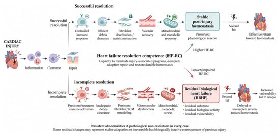
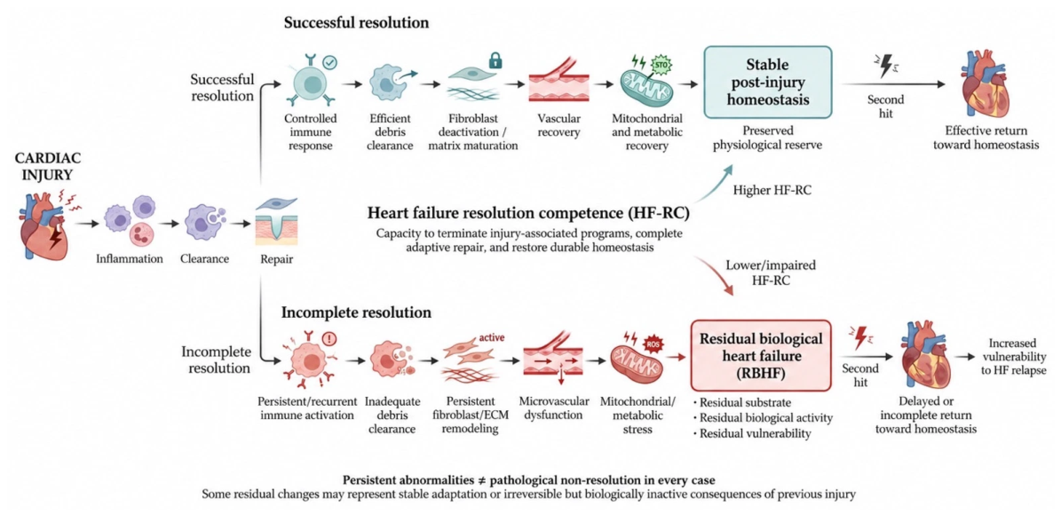
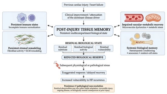
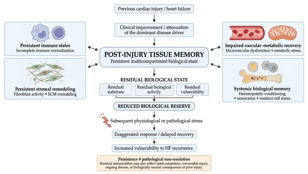
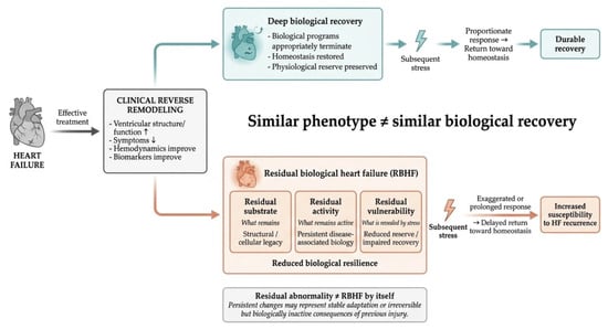
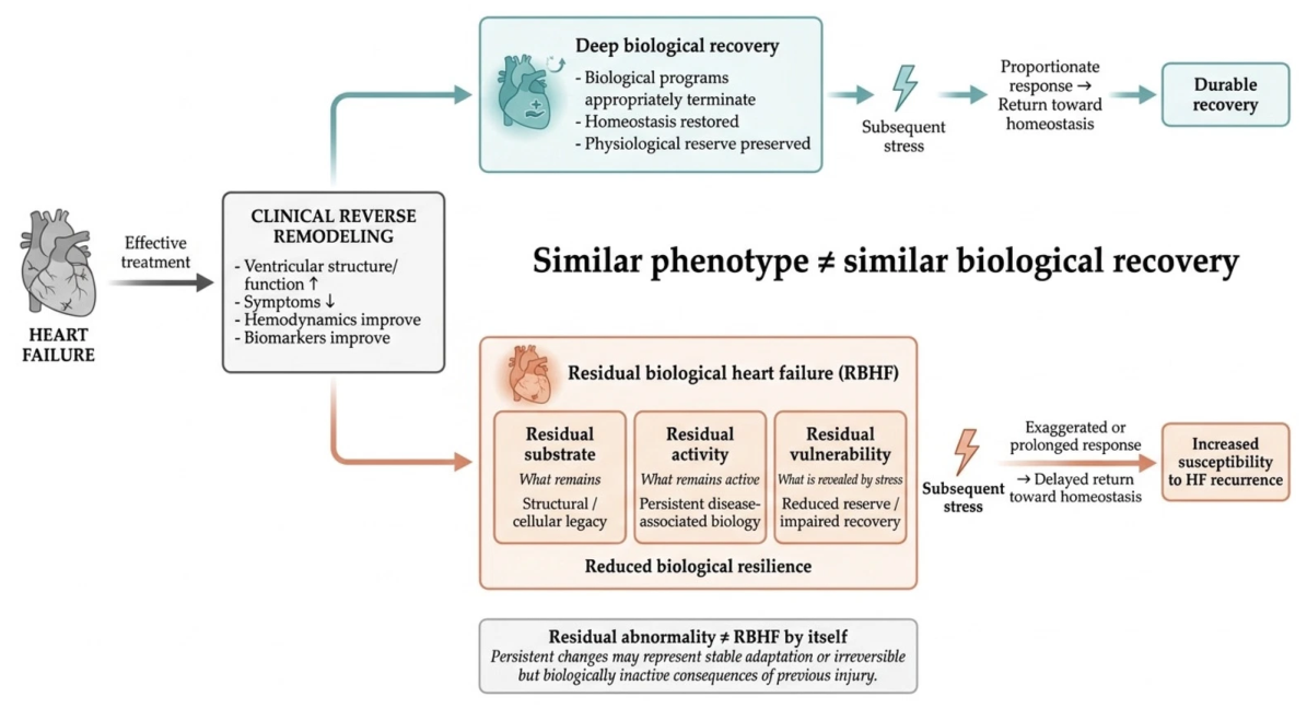
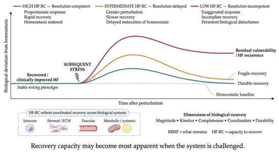
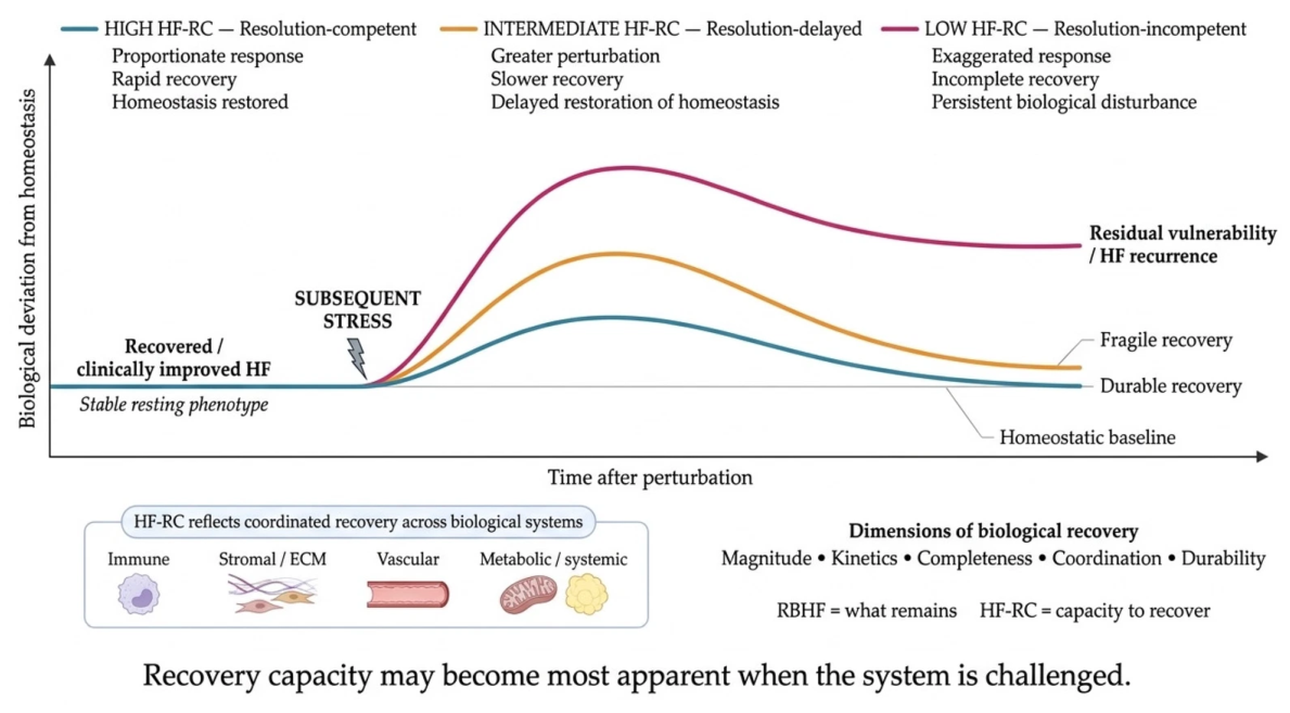

Disclaimer/Publisher’s Note: The statements, opinions and data contained in all publications are solely those of the individual author(s) and contributor(s) and not of MDPI and/or the editor(s). MDPI and/or the editor(s) disclaim responsibility for any injury to people or property resulting from any ideas, methods, instructions or products referred to in the content. |
© 2026 by the authors. Licensee MDPI, Basel, Switzerland. This article is an open access article distributed under the terms and conditions of the Creative Commons Attribution (CC BY) license.