


Highlights
What are the main findings?
- The ovarian tumor microenvironment (TME) consists of heterogenous dysfunctional cell subsets that interact with cancer cells through direct contact or by the elaboration of soluble molecules and confer rigid and complex immune suppression.
- In high-grade serous ovarian cancer (HGSOC), the cellular and structural architecture of the tumor stroma actively facilitates cancer initiation, progression, metastasis, and/or chemoresistance.
What are the implications of the main findings?
- The optimum therapy efficacy for HGSOC will likely be achieved with the use of combined therapies designed to simultaneously target multiple barriers to anti-tumor responses in the TME.
- Novel tools such as spatial imaging, spatial transcriptomics, and spatial proteomics are useful for profiling the HGSOC TME to reveal unique biomarkers in patients that will enable the design of effective precision medicine.
Abstract
Ovarian cancer is a lethal gynecologic malignancy with limited effective treatment options. It is expected that in 2026 there will be about 21,010 new cases of ovarian cancer diagnosed and about 12,450 deaths within the United States. Unfortunately, conventional therapies such as chemotherapy and radiation have limited efficacy for high-grade serous ovarian cancer (HGSOC) and also produce significant toxicity. HGSOC is characterized by an ovarian tumor microenvironment (TME) consisting of immune inhibitory T regulatory cells and inhibitory tumor-associated macrophages, including myeloid-derived suppressor cells (MDSCs) and M2 cells, which confer immune suppression. Immunocompetent T cells, tumor-suppressing M1 cells, natural killer cells, and other cellular subsets are also present. The TME is further shaped by soluble molecules, extracellular matrix, and other structural components, as well as the presence of blood vessels and ascitic fluid in the peritoneal cavity, which aid in the distribution of cancer cells to the omentum where more lesions soon develop. This review outlines the heterogenous immune cell subsets in the ovarian TME and other structural targets that promote or reduce cancer burden. Most single-agent immune strategies have shown limited efficacy in unselected HGSOC populations due to the complex immune-suppressive nature of the TME. Here, we also discuss novel combination treatments in clinical trials or in clinical practice for HGSOC. Advances in understanding the ovarian TME will reveal new immunotherapy and drug targets for translational medicine and aid in determining effective drug and immunotherapy combinations, which may ultimately lead to improved overall survival in HGSOC.
1. Introduction
Ovarian cancer is the leading cause of death from gynecologic cancers [1]. High-grade serous ovarian cancer (HGSOC) is discovered at advanced stages because of its non-specific symptoms and lack of effective screening tools. Platinum-based chemotherapy is initially effective for HGSOC patients, but these patients soon become resistant to these agents leading to disease recurrence and death [2,3,4,5].
In the quest to improve survival for this deadly cancer, newer treatments include immune checkpoint inhibitors (ICIs) and other immunotherapies, such as dendritic cell (DC) vaccine therapies, poly(ADP-ribose) polymerization (PARP) inhibitors, targeted therapies, and adoptive T-cell and natural killer (NK) cell therapies [6,7,8,9,10,11]. These and other strategies are being developed to target cancer cells, immune cells, or other suppressive components of the ovarian tumor microenvironment (TME). To date, most of these therapies have not reached the level of success needed to significantly impact HGSOC overall survival (O/S). However, one recently FDA-approved agent is Mirvetuximab Soravtansine (Elahere), a first-in-class antibody-drug conjugate (ADC) that targets folate receptor alpha (FRα) expressed on tumor cells. This agent that targets tumor cells in the TME is approved for HGSOC patients whose tumor cells have high FRα expression [12]. Another recently FDA-approved regimen is the use of pembrolizumab (Keytruda) (targeting immune checkpoints (ICs) in the TME) plus chemotherapy, an immunotherapy regimen paired with weekly paclitaxel (with or without bevacizumab) approved for patients with platinum-resistant disease, with PD-L1-expressing tumors [13]. A better understanding of the complexity of the ovarian TME will provide insight into designing best targeting strategies and the development of more effective treatment options.
Ovarian cancer is characterized by histological subtypes. Approximately ninety percent of ovarian cancer is of epithelial origin [14,15], including ovarian, fallopian tubes, and other sites in the pelvis. The other ten percent that are of non-epithelial origin are generally less aggressive than epithelial ovarian cancer. Epithelial ovarian cancer comprises mainly four groups: serous, endometroid, clear cell, and mucinous. Serous ovarian cancer is categorized as low grade (5 percent), mixed histology, or high grade (70 percent) (summarized in [15]). HGSOC is notorious for its late discovery and for developing early resistance to first-line therapies such as cytoreductive surgery and platinum-based chemotherapies [2,3,4,5].
The HGSOC TME consists of T regulatory cells, immunocompetent T cells, a myriad of macrophage subsets, NK cells, soluble factors, cancer cells, extracellular matrix components, vasculature, and other components coalescing into a series of networks, which favor immune suppression and cancer progression [8,15,16,17]. This review is a comprehensive summary of the role of the aforementioned TME elements as well as cells found in lower numbers in the HGSOC TME, such as gamma delta T cells, invariant NK T cells, and innate lymphoid cells. We discuss the potential of these TME components to hinder or advance HGSOC progression. We also discuss novel treatments for HGSOC, which are designed to overcome TME-suppressive mechanisms, as well as combinations of treatments that may be more effective than single therapy in improving disease prognosis and O/S. We conclude with a discussion of novel tools to study biomarkers in the HGSOC TME and how new drug strategies may be more precisely administered for the best outcome in precision medicine for this disease.
2. Immune Cells Regulate the Tumor Microenvironment
2.1. T Cells Tune the Ovarian Tumor Microenvironment
The increased presence of immune-competent tumor-infiltrating lymphocytes (TILs) in the HGSOC TME is reported by many to favor survival whereas T-cell exclusion is associated with worse prognosis [18,19,20]. In a study of 5500 patients of which 3196 were classified as HGSOC, TILs were associated with better outcomes regardless of the extent of surgical cytoreduction or chemotherapy regimens [21]. Tumors have previously been classified in a three-tier system based on the abundance and location of cytotoxic T cells. T-cell-inflamed tumors (also called hot tumors) are characterized by T-cell enrichment in the tumor deposits (islets) and in tumor-associated stroma, as well as increased PD-L1 expression. These hot tumors are more responsive to ICIs [22,23,24]. T-cell-excluded tumors (called altered tumors) are those in which CD8+ T cells are confined to the periphery and not infiltrated in the tumor mass. Non-inflamed tumors (called immune desert tumors or cold tumors) are the most T-cell-deficient tumors, both within the tumor as well as surrounding the tumor. Cold tumors are generally marked by the presence of immune-suppressive cells, including TAMs, MDSCs, and T regs [22,23,24].
A more standardized classification system for immune cells infiltrating ovarian cancer tumors was recently established by the use of immunohistochemistry (IHC) staining and evaluation, multiplex immunofluorescence (mIF) staining, and digital pathology analysis [25]. Five independent study cohorts were used for several cell subset studies. These cohorts consisted of a total of 697 tumor samples, of which 566 were of primary tumor origin and 131 from recurrent tumors [25].
These authors used an algorithm to capture the heterogeneity of CD8+ T-cell densities and spatial distribution in whole tissue slides. They converted the mean of five CD8+ T cells per high-power field (hpf) by IHC to a mean of 21 CD8+ T cells/mm2 on whole formalin-fixed paraffin-embedded (FFPE) slides stained by IF. Firstly, they tested the strength of this T-cell density model in primary tumors from two of the five clinical cohorts (IMCOL and UPENN cohorts) under study. It was found that tumors infiltrated by a mean of greater than 21 CD8+ T cells/mm2 had significantly longer survival than those with <21 CD8+ T cells/mm2 even after adjustments were made for optimal residual disease at the time of first surgery. These investigators identified four different immune phenotypes in the treatment of naïve ovarian cancer: purely inflamed, mixed inflamed, excluded, and desert tumors [25]. In this method, they developed a two-step algorithm to define a CD8+ T-cell-based immune classifier considering both cell density across whole tissue and heterogeneous CD8+ T-cell distribution within tumor/stroma compartments of each region of interest (ROI). The fraction of inflamed subregions (ROIs with >21 CD8 + T cells/mm2) in the tumor was categorized as purely inflamed if the percentage of inflamed ROIs was >70%. Tumor tissues with a percentage of inflamed ROIs between 50% and 70% were placed in the mixed inflamed group. When less than 50% of ROIs were inflamed in the tumor, they considered the stroma. The sample was placed in the excluded category if >10% of ROIs showed >21 CD8+ T cells/mm2 in the stroma. Finally, if <10% of ROIs showed >21 CD8+ T cells/mm2 in the stroma, the sample was placed in the desert category [25].
When the aforementioned CD8+ T-cell threshold was further applied to a third cohort (HiTide-UPENN), it was consistently found that long-term survivors were those with purely and mixed inflamed tumors when compared to those with excluded and desert tumors. They next used tissue microarrays (TMAs) from two large patient cohorts to confirm their findings. These cohorts were from the Nurses Health Study (NHS) and the NHSII and consisted of 418 patients. Even after several patient parameters were taken into account (such as tumor subtype, stage, and age), patients with mixed inflamed, excluded, and desert tumors had greater hazard ratios for death than patients with purely inflamed tumors [25]. Consistent with other studies, these authors also confirmed that samples with higher densities of CD3 T cells in both intratumoral and stromal locations were more beneficial to survival [18,19,20,21,26]. In a meta-analysis of 19 studies and 6004 patients CD3+, CD4+, CD8+, and CD103+ TILs were associated with longer O/S and progression free survival (P/FS) [27].
CD4+CD25+FoxP3 T regulatory cells (Tregs) in ovarian cancer have been the focus of several studies. They have been reported as a negative prognostic indicator by many investigators [28]. These cells may release pro-tumor cytokines, including interleukin-10 (IL-10) and transforming growth factor beta (TGF-β) by cell–cell contact mechanisms. It was found that when FoxP3+ cells were located within lymphoid aggregates surrounding tumors, these cells were significantly associated with reduced patient survival (p = 0.007) [29]. In mice, FoxP3+ T regs activated the Sirt1- ERK1/2- STAT-3 signaling pathway and suppressed NFK-β activity as a mechanism of promoting M2 macrophage polarization [30].
The classification of T regs encompasses many T-cell subsets, which suppress the responses of immune cells [31]. For example, whereas CD8+ T cells primarily kill tumor cells by recognizing tumor-specific peptides on MHC class I molecules, then releasing perforin and granzyme B or TNF-α to trigger death [32], in the immunosuppressive ovarian TME these CD8+ T cells may become dysfunctional and exhausted and upregulate inhibitory receptors such as the immune checkpoint (IC) receptor PD-1 [33,34,35]. T cells expressing high levels of IC are usually classified as negative immune regulators. ICs are generally but not exclusively expressed on T cells. Each IC usually has multiple ligands or binding sites on antigen-presenting, tumor, or other cells. Binding of ICs to ligand(s) usually results in reduced T-cell activity or lowered immune responses [33,34,35].
The most studied IC is programmed death-1 (PD-1; CD279). When PD-1 on T cells engages with programmed death ligand-1 (PD-L1; CD274) on macrophages or on tumor cells, this inhibits T-cell activity and leads to T-cell exhaustion. Antibodies blocking PD-1/PD-L1 linkage are FDA approved and show good efficacy in several solid cancers, such as melanoma, lung cancer, and renal cancer [36,37,38,39,40], but to date only show limited benefits in HGSOC [41,42,43,44].
Another IC, cytotoxic T-lymphocyte-associated protein-4 (CTLA-4), binds to receptor B7 on myeloid cells with higher affinity than CD28 binding to B7 (the latter gives a good anti-tumor response). The binding of CTLA-4 to B7 suppresses early T-cell activation leading to T-cell tolerance [36]. Other ICs in the ovarian TME include lymphocyte-activation gene-3 (LAG-3; CD223), which binds to MHC-II on antigen-presenting cells (APC) with higher affinity than CD4 binding to MHC-II, galectin-3, and other ligands. IC T-cell immunoreceptor with immunoglobulin and immunoreceptor tyrosine-based inhibition motif domain (TIGIT) binds to CD155 (PVR) on DCs and macrophages. T-cell immunoglobulin and mucin domain-containing protein 3 (TIM-3; CD366) binds to galectin-9. The presence of more than one IC on TILs may indicate an intense state of immune exhaustion and the use of combination ICI regimens may be a more effective therapeutic strategy in such cases [33,34,35,45].
The story of IC ligation activity in cancer is complex, such that there may be synergy between different IC pathways as well as with other immunosuppressive phenomena, such as tumor-associated macrophages (TAMs) and myeloid-derived suppressor cell (MDSC) aggregation. It is therefore likely that strategies designed to target IC axis activity in ovarian cancer will be more effective at reversing T-cell exhaustion if administered with other therapies.
2.2. Gamma Delta T Cells
The αβ T cell, the predominant T-cell population in the host, are critical for MHC-restricted antigen recognition. Another subset, gamma delta (γδ) T cells, makes up 1–10% of circulating T cells, but is found in higher percentages in tissues including the gut epithelium, spleen, liver, and other sites. In the normal host, γδ T cells are known to colonize the normal mucosa, where they protect against pathogens. These cells usually have different phenotypes and functions to their circulating counterparts and are crucial for local immune surveillance [46,47].
γδ T cells can be activated through the γδ receptor (T-cell receptor; TCR). On activation, they release perforin, granzymes, and IFN-γ, or kill through cell death ligands, like Fas and TNF-related apoptosis-inducing ligand (TRAIL)-R. These cells recognize antigens in an MHC-independent manner and, as such, respond more rapidly to stressed or transformed cells like in the TME, where they can recognize and attack malignant cells without the requirement for antigen processing and presentation [47]. The γδ T-cell subset can exert direct cytotoxicity against cancer cells and also boost anti-tumor effects through the release of cytokines, which influence other cells [46,48]. Human γδ T cells are also activated by NK cell receptors, a process not requiring TCR signaling. Activation through receptors NKp30, NKp44, and particularly NKG2D stimulates the cytotoxic activity of γδ T cells [49]. γδ T cells are also activated by butyrophilins and similar family molecules such as BTNL2 and BTN2A1, which is expressed in the uterus and fallopian tubes [46,50,51]. There is not yet a clear picture of the role of ovarian cancer-derived γδ T cells in HGSOC, but some investigators found that these cells were able to suppress the growth of ovarian cancer cells [52,53].
Over the past decade, several clinical trials on lung cancer, renal cancer, and multiple myeloma amongst other diseases have evaluated γδ-based therapies [46,48]. More evaluations need to be done on these cells in cancer, but it is anticipated that modifications of this cell subset may be a promising avenue for effective immunotherapy.
2.3. Invariant NKT Cells
NKT cells are a heterogenous group of cells that differ in phenotype and function [54,55]. The most studied group of these cells, invariant NKT (iNKT) cells, make up about 0.01%- 1% of T cells, and consists of several subgroups based on the cytokine profiles they secrete [55,56]. They are innate-like, non-conventional T cells that express TCR, NK receptors, and cytokine receptors, and are important in cancer surveillance [55,57]. Human iNKT cells express an invariant T-cell receptor, Vα24Jα18, as well as CD1d, a molecule related to MHC class-I, which presents self-lipids, glycolipids, and microbial antigens [55,58].
Activated iNKT cells can eliminate cancer cells in a CD1d-dependent manner. These cells recognize glycolipid antigens presented by CD1d molecules on tumor cells leading to direct cytotoxic responses [59]. They can kill tumor cells and can also regulate the TME to boost immune responses [58,60]. When activated, iNKT cells release great quantities of cytokines (IFN-γ, IL-12, IL-18, and IL-4 among others), which co-ordinate the activities of DCs, NK cells, and cytotoxic T cells. These cells can deplete TAMs and MDSCs and recruit effector cells to improve their function. They promote DC activation and maturation leading to more efficient T-cell stimulation [55,56,60,61]. Based on the aforementioned activities of these cells, they can potentially target ovarian cancer and the TME through several pathways, and can provide rapid multifaceted anti-tumor responses involving several immune cells. These potent cytotoxic cells are rare in vivo, but can be prepared on a larger scale under sterile in vitro conditions and are indeed promising as potent anti-cancer agents [62,63].
2.4. Opposing Roles of Macrophages in the Ovarian TME
Tumor-associated macrophages (TAMs) are the largest group of immune cells in the HGSOC TME [64,65]. One of the notable functions of these cells is that they can enhance ovarian cancer cell migration by secreting soluble molecules, such as TGF-β [66]. They are generally recruited from blood monocytes or sometimes originate from resident peritoneal macrophages. Both of these TAM groups exhibit phenotypes such as CD163 and CD206, as well as similar levels of genes for antigen-presenting cells and phagocytosis [17,67,68,69]. Importantly, TAMs in the TME exhibit higher levels of genes associated with extracellular matrix (ECM) remodeling [70]. TAMs mediate epithelial–mesenchymal transition (EMT), which promotes tumor progression. In this process, there is a downregulation of epithelial markers such as E-cadherin and upregulation of vimentin, snail, fibronectin, and alpha-smooth muscle actin. In EMT, epithelial cells change their phenotype to motile mesenchymal cells and aggressively migrate and invade tumors [71,72]. The process of EMT is fostered by several TAM products, including TGF-β, hepatocyte growth factor (HGF), and epidermal growth factor (EGF) [73].
It is reported that CD68+ and CD206+ TAMs are higher in patients with stage III/IV ovarian cancer than in patients with stage I/II diseases or patients with benign ovarian disease. These TAM groups (enriched for CD68 and CD206) were also higher in patients with lymphatic invasion than in those without lymphatic involvement [74]. The plasticity of TAMs is well-documented. For example, TAMs are high secretors of CCL18, a chemokine that promotes tumor cell migration and metastasis [75]. This chemokine can be blocked with interferon gamma (IFN-γ) secreted by CD4+ and CD8+ T cells, causing TAMs to switch to an anti-tumor phenotype [76].
IFN-γ in the TME promotes the emergence of M1 immunocompetent, classically activated macrophages, which are effective against microorganisms and cancer. M1 macrophages are secretors of IL-12, IL-23, and TNF-α, and are associated with improved survival in HGSOC patients [77]. M2 macrophages are a pro-cancer phenotype associated with poor outcomes in cancer patients [78]. A meta-analysis of 794 ovarian cancer patients evaluating 9 studies showed that a higher tumor M1/M2 ratio was indicative of improved ovarian cancer prognosis [79]. One ovarian cancer study found that suppressing T regs and blocking PD-1/PD-L1 linkage promoted the conversion of T regs to T helper cells, increased the ratio of M1 to M2 TAMs, and prolonged survival in mice [80].
In 246 primary ovarian tumors studied, inflamed tumors had the highest proportions of activated TAMs or myeloid cells expressing higher CD86 (co-stimulatory molecule) and pSTAT-1, suggesting the potential of greater anti-tumor immune responses in inflamed tumors [25]. When using RNA sequencing in 200 treatment-naïve ovarian cancer patient samples, it was found that in inflamed phenotype tumors (the highest T-cell index in tumors and stromas), there were highest levels of myeloid cell-related antigen presentation signatures, DC maturation signatures, and M1 macrophage signatures [25]. Signatures related to cancer progression and therapy resistance, such as EMT, ECM remodeling, and angiogenesis, were more prevalent in excluded and desert tumors where there were worse survival outcomes [25].
MDSCs (myeloid MDSCs) are a notorious type of TAMs known for HGSOC progression, increased tumor burden, metastasis, and resistance to immunotherapy [81,82,83]. In the ovarian cancer TME, there is a correlation between elevated levels of MDSCs and reduced survival. MDSCs migrate to the ovarian TME in the presence of CXCR4. Reports indicate that binding of the CXCR4 receptor to chemokine CXCL12 secreted under the influence of prostaglandin E2 (PGE2) is associated with HGSOC progression [84,85,86]. MDSCs also secrete several other molecules that abrogate cytotoxic T-cell responses. These include cyclooxygenase-2 (COX-2), an essential enzyme for PGE-2 synthesis, which facilitates the differentiation of DCs (CD1a+ cells) to MDSCs (CD14+CD33+CD34+ cells) [87].
Other negative immune regulators secreted by MDSCs include indoleamine 2,3-dioxygenase (IDO), arginase-1 (ARG-1), IL-10, nitric oxide synthase-2 (NOS-2), and vascular endothelial growth factor (VEGF) [17,88]. Figure 1 is a schematic representation of cell subsets that are associated with healthy individuals compared with those that favor cancer initiation and progression.
Figure 1.
Cell subsets which favor a healthy reproductive system versus ovarian cancer. The upper panel represents cells that maintain a normal-functioning immune state in individuals. CD8+ T cells of different phenotypes and CD4+ T cells elaborate Th1 cytokines (IFN-γ, TNF-α, and IL-2) and contribute to normal homeostasis and immunocompetence. Similarly, M1 TAMs enhance anti-tumor responses and mature DCs are efficient at recognizing foreign or altered cells and antigen processing and presentation to effector T cells, which elicit cytotoxic responses. N1 TANs aid in the recruitment of CD8+ effector T cells. NK cells are important for killing foreign or altered cells through several cytotoxic mechanisms. Endothelial cells line and maintain the integrity of blood vessels, whereas epithelial and mesothelial cells are crucial for defense and structural integrity in the abdominal cavity and other sites. Adipocytes are important for lipid storage and endocrine signaling, whilst fibroblasts respond to tissue injury. The lower panel shows altered and dysfunctional cell types, which foster progression and metastasis in HGSOC patients. Under the influence of soluble molecules elicited in cancer, cells become altered in morphology, phenotype, and function. CD8+PD1+ and CD4+FoxP3+ exhausted/regulatory immunosuppressive cells recruit other negative regulators such as M2 TAMs and MDSCs, which promote HGSOC progression, increased tumor burden, and resistance to therapy. Dysfunctional NK cells can no longer perform their cytotoxic functions to eliminate tumor cells. Neutrophil (N2) NET formation may occur through NETosis, releasing IL-10 and attracting T regs fostering immune suppression and limiting T-cell proliferation. CAFs remodel the ECM and release cytokines, which stimulate metastasis of tumor cells. Endothelial cells promote angiogenesis and mesothelial cells establish niches of tumor cells for metastasis. Epithelial cells change form and initiate the process of EMT, which is essential for cancer progression. Figure 1 was created with Biorender.com.
2.5. Control of the Ovarian TME by Dendritic Cells
In HGSOC and other cancer TMEs, DCs are few in number and mostly have an immature phenotype with a characteristic low expression of co-stimulatory molecules, and as such are capable of mounting only minimal immune responses [89]. DCs may present exogenously captured peptides to CD4 T cells through the MHC class II complex and endogenous peptide antigens to CD8 T cells through the MHC class I complex. DCs also present exogenously captured antigens as MHC class I-associated peptides (cross-presentation) [90,91,92,93].
For efficient antigen presentation, DCs must have three signals: a cognate antigen, co-stimulatory molecules on the DC cell surface (CD40, CD80/B7-1, and CD86/B7-2, the latter two that bind to CD28 on T cells), and thirdly, the presence of pro-inflammatory cytokines (IFN-γ and IL-12). If these three stages of activation can occur in sequence, T cells are driven to migrate, proliferate, and attack malignant cells [94,95].
Briefly, immature DCs express CXCR3, CXCR4, CCR1, CCR2, CCR5, and CCR6. The presence of antigen causes DCs to mature, upregulate co-stimulatory molecules, downregulate chemokine receptors associated with immature DCs, and upregulate CCR7, which guides DCs to lymph nodes to present antigens to immune cells (T cells). These mature DCs bearing high expression of CD40 crosslink with CD40 ligands, secrete IL-12 and efficiently stimulate T cells to proliferate and kill cancer cells [96,97,98]. Mature DCs initiate and control the amplitude of T-cell-dependent anti-tumor immunity. However, if the abovementioned processes do not occur in sequence, immune suppression mediated by T regs may be the outcome [93,94,95]. Tumors can also circumvent the abovementioned signals by loss of tumor antigens or by the release of soluble molecules and make DC function less efficient [99,100].
The inhibitory environment in the TME with TGF-β, IL-10, ARG-1, VEGF, and other molecules fosters the development of CD14+CD1a+ DCs (immature DCs), TAMs, and Tregs, which are associated with immune tolerance, immune exhaustion, and tumor growth. Preclinical studies showed that deletion of immature DCs (lacking co-stimulatory molecule CD80 and expressing low CD86) in mice bearing established ovarian tumors delayed tumor growth [101]. In translational medicine, the concept of harnessing anti-tumor DCs for improved HGSOC outcome has been recently described in clinical trial NCT00703105 [102] and in other vaccine trials using various DC modifications [7,103,104].
There are several DC types and classifications [93]. Here, in the context of cancer, it is noteworthy to mention the plasmacytoid DC (pDC; CD4+CD123+BDCA2+). These cells express CCR4 and migrate into the ovarian tumor drawn by CXCL12 (stromal-derived factor; SDF-1) and IL-10 [105]. These tolerogenic DCs release IDO, an enzyme that catalyzes tryptophan degradation [106,107]. IDO is associated with tumor angiogenesis, cancer metastasis, and immune-suppressive functions of TILs [108]. In a cohort of 44 ovarian cancer patients, pDCs in tumors were associated with early relapse [109]. In another study, higher pDC scores were independently associated with shorter PF/S [110].
2.6. Importance of Natural Killer Cells in Ovarian Cancer
Natural killer (NK) cells are known for their anti-cancer properties. However, ovarian cancer patients exhibit a reduction in NK cell numbers and limited function in the hostile TME and in peripheral blood [111,112,113,114,115]. The main surface antigens on NK cells are CD56, CD16, NKG2D, NKp30, NK44, and NK46, which help them to detect infected or tumor cells. CD56 bright NK cells are exceptional at producing cytokines, whereas CD56 dim NK cells have potent cytotoxic functions [114,115,116,117]. NK cells in ascites are enriched for CD56 bright cells, mostly exhibiting a cytokine secretory function rather than the CD56 low-cytotoxic NK cell subset [118,119].
NKp30, NK44, and NK46 are cytotoxicity receptors that trigger direct target killing [114]. CD16 (FcγRIIIa) plays an important role in antibody-dependent cellular cytotoxicity (ADCC) by binding IgG-coated targets [120]. In addition, NK cell activity is controlled by a balance of inhibitory or activating signals from lineage-encoded receptors, including two categories of MHC class-1-like binding receptors [111]. These receptors include a non-polymorphic C-type lectin-like receptor, such as killer cell-like receptor, K1(KLRK1), also called natural killer group 2 member D (NKG2D), which binds to stress-induced ligand, NKG2DL, on abnormal cells [121]. The NKG2DL consists of six members, MICA, and MICB, and six ULBP members, which induce activating signals leading to cytotoxicity [122]. Inhibitory receptors such as CD94/NKG2A mainly bind non-classical MHC-1b molecules such as human leukocyte antigen (HLA-E) [111,123], and inhibitory signals are released. These interactions are important to suppress NK cell activity during cytolysis. NK cells also express killer cell inhibitory receptors (KIRs), which recognize MHC class-1a molecules (HLA-A, HLA-B, and HLA-C). KIR and MHC-1 molecules binding on the surface of normal cells maintain immune tolerance in vivo and release inhibitory signals so that NK cells do not attack normal cells [111,124].
NK cells in the ovarian TME exhibit pro-cancer activities through several mechanisms [125]. For example, tumor-associated T and NK cells in ascites showed defective signaling proteins, including reduced TCR-zeta chain and p56 lck expression, as well as low levels of IFN-γ, IL-2, and IL-4 gene expression [126,127]. Ovarian cancer cells can successfully evade detection by NK cells by proteolytic shedding of molecules such as MICA and MICB (stress-induced ligands that activate the NKG2D receptor) from their surface. With this shedding activity, MICA/B can downregulate NKG2D on NK cells and lessen the anti-cancer activity of NK cells [122,128,129].
Efficient functioning NK cells have multiple anti-tumor mechanisms in cancer, including through the perforin (punches holes in tumor cell membranes)/granzyme pathway (lyse cancer cells). In ADCC, the Fc portions of their receptors bind to IgG antibodies attached to antigens on tumor cells, leading to granule release and cell lysis [116,120,130]. Cytokines such as IFN-γ and TNF-α elaborated from NK cells can limit tumor cell growth and sensitize tumor cells to conventional cancer treatments. Engineered NK cells can lyse ovarian cancer stem-like cells [113]. A rapidly growing field for clinical trials aimed at restoring NK cell function is chimeric antigen receptor (CAR) NK therapy, in which NK cells are genetically engineered with chimeric antigen receptors to recognize specific tumor antigens [114,131]. NK cell immunotherapy may have potential in ovarian and other hard-to-treat solid cancers.
2.7. Innate Lymphoid Cells
Innate lymphoid cells (ILCs) are a significant part of the innate immune system. The innate lymphoid cell (ILC) family is composed of NK cells, ILC1, ILC2, and ILC3 cells [132,133]. These three groups of ILCs were also revealed by sc-RNA-seq in a study profiling NK cells and ILCs in HGSOC ascites and healthy-donor peripheral blood [118]. ILC1 cells produce TNF-α and IFN-γ and share functional and transcriptional properties with NK cells [134]. ILC2 cells secrete IL-4, IL-5, and IL-15, and are associated with ovarian cancer progression [135]. There was an increased frequency of ILC2 cells with higher PD-1 expression levels in ascites from patients with HGSOC with shorter survival [118]. ILC3 cells produce IL-17 and IL-22 and contribute to mucosal immunity [133,136].
2.8. TANs in the TME
In 2009, investigators proposed that tumor-associated neutrophils (TANs) like TAMs could be categorized into N1 and N2 cells [137]. N1 TANs possess anti-tumor properties such as the release of immunostimulatory cytokines and chemokines and increased cytotoxicity against tumor cells [138]. N1 TANs recruit CD8+ T cells by releasing T-cell-attracting cytokines, such as CXCL9 and CXCL10, and Th1 cytokines, such as IL-12 [139]. N2 TANs secrete molecules including VEGF, ECM degrading enzymes (MMPs), IL-10, and TGF-β (Table 1), which aid in remodeling the TME and promoting tumor cell invasion and metastasis [140].
An interesting feature of neutrophils that play an important role in malignancy is neutrophil extracellular trap (NET) formation [141,142]. NETs were originally described as a mechanism whereby neutrophils trap and destroy micro-organisms but are now also known to be critical mediators of cancer-associated inflammation. NETs released by neutrophils in response to pathogens consist of fibrous decondensed chromatin bound with histones, myeloperoxidase (MPO), and cytoplasmic proteins such as elastase, cathepsin, and lactoferrin [64,143].
In tumors such as ovarian cancer, neutrophils infiltrate the omentum in early-stage ovarian cancer. Neutrophils differentiate into TANs in the presence of tumor-derived soluble molecules and hypoxia [144,145]. NET formation occurs via suicidal or NETosis pathways. Both of these pathways depend on the coordinated activity of neutrophil elastase (NE) MPO and protein–arginine deiminase 4 (PAD4) to drive chromatin decondensation and extracellular release of DNA fibers [144,145,146,147].
Table 1.
Main factors elaborated by immune cells in the TME.
In the ovarian TME, NET formation is fostered by high levels of cytokines such as IL-8, complement factors, and pathogen-associated molecular patterns (PAMPs) [169]. NETs can contribute to the recruitment of IL-10 secreting B cells through NET-induced CXCL13 expression. This can lead to an increase in T regs through the secretion of IL-10, a process which contributes to immune suppression limiting T-cell proliferation [144]. Additionally, ascites and the omental architecture provide an environment where MDSCs and TANs sustain NET production through CXCR1 and CXCR2 signaling. These NETs provide an adequate system for tumor cell adhesion, dissemination of tumor cells, ECM degradation, angiogenesis, and tumor cell evasion [170]. In HGSOC, NETs are also involved with chemotherapy resistance and suppression of immune responses, and are therefore adequate culprits to concurrently aid in disease progression and to decrease the efficacy of conventional and other therapy regimens [142,170]. Thus, through a very complex and multifactorial program, as previously described by others, NETs may act as key mediators of ovarian cancer progression. Targeting of NETs is therefore potentially a novel and relevant form of therapeutic intervention. A more detailed summary of factors elaborated by immune cells in the TME is shown in Table 1.
3. Other Players in the Ovarian Cancer TME
In addition to the immune cells detailed above, the ovarian TME is regulated by the ECM, fibroblasts, endothelial cells, blood vessels, mesothelial cells, adipocytes, and other components. The text below covers examples of how these lesser-mentioned elements modulate immune suppression in the TME [171].
3.1. Extracellular Matrix Involvement in Ovarian Cancer
The mesothelium, the squamous epithelium covering organs of the peritoneal cavity has a single layer of mesothelial cells, and below this a basement membrane. The ECM consists of collagen fibers that provide strength, proteoglycans that provide cell shelter, and glycoproteins such as laminin and fibronectin that connect collagen and proteoglycans such as integrins and hyaluronic acid receptors [172,173]. The ECM provides biochemical and biomechanical stimuli that alter the function of cells in the tumor by activation of receptors including integrins and syndecans [174]. Tumor cells have abnormal ECM deposition and this may increase mechanical stiffness of these cells leading to proliferation, invasion, and cell survival [175]. The ECM is negatively regulated by immunosuppressive soluble molecules (e.g., TGF-β) released by other cell types such as TAMs, M2 cells, and T regs [176,177]. In ovarian cancer, ECM remodeling facilitates the invasion of cancer cells into surrounding tissue and metastasis to various sites in patients [178].
3.2. Cancer-Associated Fibroblasts
Fibroblasts in the stroma regulate the pre-metastatic niche of ovarian cancer by remodeling the ECM and releasing cytokines. Under the influence of proliferative/activation signals, such as TGF-β, these fibroblasts differentiate into activated fibroblasts, known as cancer-associated fibroblasts (CAFs) [179,180]. CAFs are the main component of ovarian cancer stromal cells and are located in close proximity to ovarian cancer cells [181,182,183]. CAFs are spindle-shaped cells that are known for their response to tissue injury and secretion of several soluble molecules for endothelial cell recruitment during angiogenesis, including TGF-β, platelet-derived growth factor (PDGF), and fibroblast growth factor-2 (FGF-2). They release several cytokines that stimulate proliferation, invasion, and metastasis of tumor cells in their vicinity and indirectly regulate immune cells [179,184,185].
CAFs promote tumorigenesis by releasing high levels of mitogenic factors such as fibroblast growth factor-1 (FGF-1) and HGF [186,187]. Others showed that IL-8 secreted by CAFs and ovarian cancer cells promotes stemness through the activation of Notch-3-mediated signaling pathways [188]. CAF-secreted CXCL14 drives the expression of long noncoding RNA LINC00092 in ovarian cancer cells and promotes ovarian cancer growth and invasion [189].
A group of investigators subdivided HGSOC into four molecular subtypes. The mesenchymal HGSOC subtype was defined by stromal-related gene signatures and was associated with poor patient survival. These workers also identified four subsets of CAFs (CAF-S1-4) based on CD29, fibroblast activation protein (FAP), fibroblast-specific protein-1 (FSP-1), and smooth muscle actin (SMA) protein levels. CAF-S2 and CAF-S3 were classed as “non-activated” CAFs based on the lack of expression of SMA. CAF-S1 and CAF-S4 both expressed SMA and were classified as activated CAFs or myofibroblasts. CAF-S1 cells were one of the most frequent CAF subtypes in mesenchymal HGSOC. CXCL12α was expressed at similar levels in both CAF-S1 and CAF-S4 subsets. However, the beta isoform of chemokine CXCL12 (CXCL12β) specifically accumulated in the CAF-S1 cell subset but was almost undetectable in CAF-S4 cells. These CAF-S1 cells were highly immunosuppressive and enhanced the attraction, survival, differentiation and suppressive activity of CD25+FoxP3+ T cells. CXCL12β expression in CAF-S1 cells was crucial to the immune-suppressive capacity of these cells [190]. The identification of CAF subtypes crucial to ovarian cancer outcome will allow for more precise targeting of these cells in therapy design.
CAFs can exert compressive forces on micro-vessels and form physical barriers, which may make the delivery of chemotherapeutic agents less effective and lead to chemoresistance [191]. CAFs activate the Wnt/β-catenin pathway in ovarian cancer via the CXCL12/CCR4 linkage, thereby inducing EMT and cisplatin resistance [192]. CAFs, through matrix metalloproteinase (MMP) production, stimulate the degradation of the vascular basement membrane (a layer of the ECM). CAFs contribute to increased deposition of and alteration in the ECM and this creates a barrier that blocks efficient delivery of anti-cancer drugs and enhances chemoresistance. It is also known that fibroblasts diminish nuclear accumulation of platinum in ovarian cancer cells, enhancing resistance to platinum-based chemotherapy [183,187,193]. This resistance is partly due to glutathione and cysteine released by fibroblasts. However, CD8+ T cells can alter cystine and glutathione metabolism in CAFs via the JAK/STAT pathway, and this reduces CAF-induced resistance to platinum therapy [193].
3.3. Cancer-Associated Endothelial Cells and Angiogenesis
Endothelial cells line the inside of blood vessels. In cancer, these are the blood vessel-lining cells in the TME that supply nutrients and oxygen to the rapidly proliferating tumor cells [194]. Angiogenesis is the forming of new capillaries from existing blood vessels, a process that leads to the continued growth and survival of tumor cells by various mechanisms [194,195,196]. For example, CAFs through MMP production stimulate the degradation of the vascular basement membrane, leading to endothelial cell proliferation and migration. Pro-angiogenic factors such as vascular endothelial growth factor-A (VEGF-A) are normally in equilibrium with anti-angiogenic factors such as thrombospondin, and this maintains a state of tumor quiescence [196]. Under hypoxic conditions, hypoxia-inducible factors (HIFs) become active. With tumor hypoxia, HIF-1α is stabilized, leading to increased expression of pro-angiogenic factors including VEGF, PDGF, and FGF-2, and a definite suppression of anti-angiogenic factors [196]. HIF-1-induced VEGF upregulation along with MMP-9 increase leads to higher concentrations of soluble VEGF, followed by increased angiogenesis and metastasis [197]. The switch from an anti-angiogenic to a pro-angiogenic state creates an immune-suppressive TME and increases the abundance of pro-tumor TILs [195,198].
The importance of VEGF family members in action in the angiogenic process is presented in the subsequent text. VEGF binds to its receptors VEGFR-1 and VEGFR-2 on endothelial cells and trigger intracellular signals that cause endothelial cells to divide and increase vessel mass and produce new branches of vessels. It also prevents the apoptosis of endothelial cells and makes blood vessels more permeable to gases and nutrients that nourish the tumor cells [199]. VEGF-C in patient ascites is associated with FIGO stage, tumor grade, and lymph node metastasis, and a high concentration of VEGF is a predictor of reduced survival [200]. The angiogenesis inhibitor bevacizumab is a useful therapeutic option in ovarian cancer and acts by blocking VEGF-A and inhibiting blood vessel growth [201].
3.4. Cancer-Associated Mesothelial Cells
Mesothelial cells are supposed to be a defense barrier against ovarian cancer metastasis into the abdominal cavity but may also act adversely to establish niches of tumor cells that promote ovarian cancer metastasis [202,203,204,205]. CAMs secrete chemokines that promote peritoneal metastasis and chemoresistance in ovarian cancer. They also secrete high levels of IL-6 and FGF, which promote ovarian cancer progression [206,207]. Lipophosphatidic acid (LPA) secreted by CAMs enhanced adhesion, migration, and invasion of ovarian cancer cells [208,209].
Investigators performed sc-RNA analysis of cancer stroma-derived cells using 18,403 cells unbiasedly collected from seven untreated HGSOC tumors [210]. This allowed the identification of transcriptional markers specific to each stromal phenotype. From these markers, they estimated the prevalence of these phenotypes in conventional bulk data sets and characterized the relevance of these stromal phenotypes to patient prognosis. These workers determined six cellular phenotypes linked to prognosis as follows: mesothelial cells (FB_CALB2), myofibroblasts (FB_MYH11), TGF-β-driven cancer-associated fibroblasts (FB_COMP), tumor subcluster Tum_BAMBI, and lymphatic endothelial cells (EC_PROX1), which were associated with adverse effects on outcomes. Only the plasma cell subcluster (BC_IGHG1_PRDM1high) was associated with improved OS [210].
3.5. Cancer-Associated Adipocytes
The omentum, a prevalent site of ovarian cancer metastasis and recurrence, is rich in adipose tissue [211]. Under normal conditions, adipocytes are specialized for lipid storage and endocrine signaling. In cancer, these cells are dysregulated in function due to the immunosuppressive environment in which they exist [211]. Cancer-associated adipocytes (CAAs) induce ovarian cancer homing by secreting adipokines, leptin, adiponectin, IL-6, IL-8, MMP-11, and CCL5 [211]. Adipocyte-derived signals such as inflammatory mediators can enhance lipid uptake in ovarian cancer via STAT3/fatty acid-binding protein 4 (FABP4) signaling, leading to an increased proliferation of cancer cells [212]. An adipose-rich microenvironment fosters ovarian cancer growth, cell proliferation, and migration, as well as chemoresistance through several mechanisms [211,213,214].
4. Novel Treatment Strategies for Ovarian Cancer
The previous discussion underscores the diverse and intricate factors that confer potent immune suppression in the ovarian TME. It is therefore likely that therapeutic efficacy in HGSOC will be optimized if treatment agents are used together to better overcome the many barriers to anti-tumor immunity, which exists in the TME. Some of the recent advances in ovarian cancer therapies are discussed in the subsequent text.
4.1. Immune Checkpoint Inhibitors
For the greater part, immune checkpoint inhibitors have revolutionized cancer treatment [37,38,39]. Cancer cells circumvent immune surveillance through the activation of IC pathways, thereby inhibiting T-cell activation and limiting anti-tumor responses. Co-inhibitory molecules on the surface of tumor cells or antigen-presenting cells bind to T-cell co-inhibitory molecules and immune checkpoints [35]. The FDA approved the first of these ICI agents, Ipilimumab, in 2011, to target CTLA-4 in metastatic melanoma. Since then, other ICIs have been FDA-approved to target CTLA-4 as well as the PD-1/PD-L1 axis for a range of cancers such as renal cell carcinoma, colorectal cancer, hepatocellular carcinoma, non-small-cell lung cancer, and Hodgkin lymphoma amongst others [37,38,39,40].
Generally, ICIs have only yielded limited success in ovarian cancer [41,42,43,44,215,216]. However, notable progress in this area has been recently achieved. In 2026, following the evaluation of the KEYNOTE-B96 (NCT05116189) clinical trial (643 patients enrolled) [13], the FDA approved pembrolizumab (Keytruda) as well as pembrolizumab and berahyaluronidase alfa-pmph (Keytruda Qlex) in combination with paclitaxel, with or without bevacizumab, for adults with platinum-resistant epithelial ovarian, fallopian tube, or primary peritoneal carcinoma with tumors expressing PD-L1 (combined positive score; CPS ≥ 1), as determined by an FDA-approved test, and who have received one or two prior systemic therapy regimens.
There are several other clinical trials for ovarian cancer that are ICI-based [217,218], including NCT03602859 and NCT03737643, both testing anti-PD-1 and PARP inhibitors (PARPis), to mention a few. Additionally, a phase I clinical trial, NCT04503980, evaluates anti-PD-1 and mesothelin (MSLN)-directed CAR T cells.
4.2. Targeting TAMs in Ovarian Cancer
Therapies targeting M2 TAMs may be designed to shift them to M1 immunocompetent cell polarization. Potential agents to use are antibodies or inhibitors against colony stimulating factor-1 (CSF-1) receptor, against CCL-2 or against CD11b [219,220,221]. Triggering receptor expressed on myeloid cells (TREM)-1 found on MDSCs promotes tumor growth and suppresses anti-tumor T-cell responses predisposing T cells to T-cell exhaustion [222,223]. Effective inhibitors of TREM-1 may be designed for future clinical trials. Overall, testing of agents blocking TAM activity in cancer is predominantly in the pre-clinical phase [64,224].
4.3. Dendritic Cell-Based Immunotherapies for Ovarian Cancer
The use of DC vaccine therapy holds much promise in cancer treatment [225]. Typically, this involves the propagation of autologous myeloid-derived DCs from blood monocytes, pulsing them with tumor antigens and maturing them with cytokines, before infusion into patients for the generation of patient tumor-specific T-cell responses.
DC therapy has shown some efficacy in advanced ovarian cancer using several treatment regimens. For example, investigators administered a mature α-DC-1 vaccine (NCT00703105) to patients with relapsed/refractory ovarian cancer with minimal residual disease (MRD) after secondary debulking surgery. In the small cohort of 19 patients enrolled in the trial, this vaccine was found to be safe and in 26% of the patient cohort OS exceeded five years. Furthermore, administration of repeated vaccine administrations (six or more vaccines) was associated with a significant improvement in PFS [102]. Even though several other DC vaccines have been reported to show safety and some level efficacy in HGSOC [7,103,104], there is need for improvement in patient outcomes for DC vaccine therapy in all cancers tested. The immunogenicity of DC vaccines may be enhanced by combining this therapy with T-cell targeting therapies such as ICI or other agents.
Lastly, in the area of pDC targeting, in a phase I clinical trial, an IDO inhibitor, Epacadostat, was used in ovarian cancer patients (NCT02042430).
4.4. Inhibition of Angiogenesis
Bevacizumab (FDA-approved in 2014) is an anti-VEGF-A antibody, which inhibits angiogenesis and prevents the tumor from growing [226,227,228]. Apart from conventional treatment for HGSOC, bevacizumab in combination with platinum-based chemotherapy, followed by maintenance bevacizumab, became the first targeted therapy option for newly diagnosed HGSOC patients, based on the outcome of the GOG-0218 study [229,230,231,232,233].
Bevacizumab also shows efficacy when combined with, and as maintenance following, platinum-based chemotherapy in platinum-sensitive, recurrent ovarian cancer patients, and in combination with paclitaxel, pegylated liposomal doxorubicin, or topotecan in platinum-resistant, recurrent ovarian cancer patients [227,234]. After the PAOLA-1 trial analysis [235,236], bevacizumab combined with the PARP inhibitor (PARPi) olaparib was approved as maintenance therapy in patients with newly diagnosed advanced ovarian cancer with homologous recombination deficiency (HRD)-positive tumors (known as a BRCA1 and/or BRCA2 mutation and/or genomic instability) who achieved a clinical response after platinum-based chemotherapy plus bevacizumab [227]. Another ovarian cancer clinical trial designed to target angiogenesis includes phase III NCT00532194 targeting VEGFR1-3.
4.5. PARP Inhibitors
PARPis include agents such as olaparib, nivaparib, rucaparib, and others, which block the repair of single-stranded DNA breaks, leading to replication-associated double-stranded breaks (DSBs). In HRD ovarian cancer cells, these DSBs are not efficiently repaired, leading to cell death. PARPis have shown efficacy especially in patients with BRCA mutations or in HRD patients [9,237,238]. Clinical trials for PARPis, which show efficacy in ovarian cancer include phase III trials such as SOLO1 (evaluating olaparib as maintenance therapy in newly diagnosed patients), SOLO2 (evaluating olaparib for platinum-sensitive recurrent patients), and PRIMA (testing niraparib as a first-line maintenance therapy in patients with advanced ovarian cancer) [239,240,241,242,243]. Based on the outcome of the SOLO2 trial and Study 19 [244], olaparib received full approval as maintenance therapy in 2017 following the response to platinum-based chemotherapy. There are several other PARPi clinical trials that provide much-needed therapy options for ovarian cancer patients [9,237].
4.6. Targeting the TME with Antibody Drug Conjugates
Antibody drug conjugates (ADCs) deliver cytotoxic payloads using target antibodies. Important targets in ovarian cancer are folate receptor alpha (FR-α), human epidermal growth factor-2 (HER-2), MSLN, and cadherin-6 (CDH6) [245,246]. Miretuximab soravtansine (MIRV), an ADC that targets FR-α on tumor cells, received accelerated FDA approval (2022) for FR-α high, platinum-resistant ovarian cancer following 1–3 lines of therapy based on the outcome of the SORAYA trial (NCT04296890) [247]. This trial reported a confirmed objective response rate (ORR) of 32.4% in a biomarker-selected population. The confirming MIRASOL trial (NCT04209855) met its primary endpoint of PFS [12]. The phase III GLORIOSA trial (NCT05445778) evaluates MIRV plus bevacizumab as single maintenance therapy in FR-α high platinum-sensitive ovarian cancer.
4.7. Miscellaneous TME Targets
A phase III trial evaluated O/S with relacorilant and nab-paclitaxel in patients with platinum-resistant ovarian cancer (NCT05257408, ROSELLA) [248]. Relacorilant is a selective glucocorticoid receptor antagonist that increases the sensitivity to chemotherapy in several cancer types. This study’s cohort consisted of patients who had one-to-three lines of previous anti-cancer therapy and platinum-resistant disease. The use of relacorilant with nab-paclitaxel resulted in significantly longer O/S in patients with platinum-resistant ovarian cancer, without the need for biomarker selection [248].
A bispecific antibody, NI-1801 (targeting mesothelin and CD47), has also recently been evaluated with promising success. Study LCB-1801-001 is an open-label, phase 1 first-in-human clinical study of NI-1801 in patients with advanced, metastatic, or recurrent solid malignancies (including ovarian cancer) expressing mesothelin (MSLN). Unpublished meeting reports currently indicate that this agent in combination with pembrolizumab resulted in 40.2 percent of heavily pretreated ovarian cancer patients being alive at two years (NI-1801 Mesothelin x CD47 Bispecific Two-Year Ovarian Data), thereby indicating the potential success of strategies targeting mesothelial cells and ICs in the TME in this disease.
BCA101 is a bi-functional fusion antibody that simultaneously blocks EGFR and TGF-β signaling pathways in the TME to suppress tumor growth and enhance anti-tumor immune responses. Based on the findings of pre-clinical studies, clinical trials targeting CAFs include NCT04429542 using BCA101 [249] as single therapy or with ICI pembrolizumab in ovarian and other solid cancer patients. A phase 1 clinical trial (NCT03078400) evaluated the CD44 blocking peptide (targeting the ECM) plus chemotherapy. Evaluation of anti-MSLN (targeting CAMs), VEGFRi, and chemotherapy is in a phase I clinical trial (NCT04938583) for ovarian cancer patients.
4.8. Enhancement in T- and NK Cell Function in Ovarian Cancer
There are many types of adoptive T-cell therapies under development and being tested for ovarian cancer [250]. CAR T cells are MHC-unrestricted, directly binding to target antigens without the need for antigen presentation by peptide–MHC complexes. CARs are composed of an extracellular antigen-binding domain fused with the signaling and co-stimulatory domains of TCR. The extracellular domain is composed of a single-chain variable fragment (scFv), a fusion protein of the variable regions of immunoglobulin heavy and light chains, for the specificity of the treatment. The intracellular domains contain the primary CD3 signaling domain and secondary costimulatory domains, and some constructs add inducible cytokine expression to recruit bystander T cells for improved immune-mediated anti-tumor effects and clonal amplification [10,11,251]. CAR T-cell therapy works well in hematologic malignancies [252,253]. For ovarian cancer, this strategy is in the early phase of clinical testing. Clinical trial NCT04503980 is currently investigating MSLN-CAR T cells secreting PD-1 nanobodies in ovarian cancer.
There are several strategies in pre-clinical studies and in clinical trials to restore NK cell persistence and anti-tumor activity in ovarian cancer [114,254,255]. For example, a phase 1B trial (NCT06321484) is investigating patient-derived cytokine-induced memory-like NK cells (IL-12, IL-15, IL-18) with chemotherapy. CAR NK cells are genetically engineered to directly recognize tumor antigens. CAR NK cells can utilize mismatched allogeneic cells, and the risk of cytokine release syndrome and of graft versus host disease is low [114,256]. CAR T-cell and CAR NK cell therapy clinical trials for ovarian cancer are primarily in the first phase, and much work still needs to be done for the advancement of this field [257].
Table 2 provides a summary of selected clinical trials designed to target immune suppression in the ovarian TME.
Table 2.
Highlights of clinical trials targeting the ovarian TME.
5. Perspectives
The preceding text outlines a complex, heterogenous, and dynamic ovarian TME in which cells can alter phenotype and function depending on the concentration of cytokines, chemokines, or other soluble molecules in their environment. Generally, a paucity of immunocompetent T cells, DCs, and macrophages are found in the TME. On the contrary, there is an abundance of pro-tumor cell types, including MDSCs, M2 TAMs, N2 TANs, and Tregs, as well as other phenomena, such as loss of tumor cell surface antigens (such as MHC-I), TP53 mutations, or tumor cells altering specific signaling pathways, which favor tumorigenicity [128,262]. Cell types with pro-tumor propensity may secrete IL-10, TGF-β, VEGF, or CCL2 and other agents, which promote the recruitment of T regs, increase angiogenesis, metastasis, and drug resistance. It is therefore evident that there are many layers of immune suppression in the ovarian TME.
The inherent complex immunosuppression in the HGSOC TME presents challenges in designing effective therapy for this disease because such treatment agents are needed to address multiple factors, such as ECM remodeling, restoring function to the tumor vasculature, reducing T regs, restoring T-cell, DC, and/or NK cell function. To this end, there are many exciting clinical trials that are designed as combinatorial studies to target some of these immunosuppressive targets together [128,262] (Table 2). Several of these trials are testing the safety and efficacy of treatments, but they will also need to test the frequency and sequence of administration of agents in combination regimens, which will lead to optimum O/S.
Despite this aggressive female malignancy, there is hope on the horizon because of the many scientific advancements that are being made concurrent with clinical efforts, even though challenges persist. Interactions between the cancer and immune system vary between patients and are not currently understood, and this limits the development of effective combination therapies. Diagnostic tools such as spatial imaging, spatial genomics, and spatial transcriptomics will aid in the analysis of tumor cell subsets as well as detailed analysis of unique gene and protein features specific to individuals [263,264,265,266]. Spatial imaging can aid in revealing diverse immune niches, CD8+ T-cell densities, DC maturation state, and in general show immune cell localization, exhausted or dysfunctional T cells, MDSCs, and ICs such as PD-L1 in the tumor, and also the association with tumor stage and overall survival (O/S). This knowledge can be used to design combination therapy, which can reprogram cold tumors into hot tumors. High-resolution genomics, transcriptomics, and proteomics tools used to study the excised tumor can reveal region-specific proteins and RNA associated with patient survival. Newly revealed disease and treatment biomarkers as a result of these tools will make it easier to create innovative and more specific effective combination therapies to target the immune-suppressed ovarian TME. Such high-resolution TME investigations will direct patient selection, thereby narrowing treatments to cohorts that will respond best in specific clinical trials, and can also be used to more precisely measure response rates to treatments.
Improvements in delivery of treatment agents, such as by nanoparticle delivery of drugs or oncolytic viral approaches, may enhance drug stability and provide greater treatment efficacy [267,268]. Oncolytic viral immunotherapy uses naturally occurring or genetically modified viruses that can predominantly kill tumor cells as well as stimulate anti-tumor immune responses [269]. The viruses often used in preclinical or clinical systems are oncolytic adenovirus, human syncytial virus, and vaccinia virus [268]. These viruses replicate in tumor cells leading to tumor cell death, while normal cells are cleared of the virus. Pathogen-associated molecular patterns (PAMPs) and damage-associated molecular patterns (DAMPs), tumor antigens, and viral antigens are released, which can boost DC-T-cell priming, induce antigenicity, and lead to the recruitment of cytotoxic effector T cells, thereby reprogramming a cold tumor to a hot tumor, thus making the TME more responsive to immunotherapy, such as ICIs [11,270]. Oncolytic viral therapy is well-tolerated but, to date, the desired clinical efficacy has not been achieved in cancer therapy [271,272].
Nano-drug delivery systems may overcome some of the difficulties of specifically targeting tumor sites with treatment agents and reduce damage to normal tissues when therapy is administered. Nanoparticles and nanocapsules may be useful for the delivery of chemotherapy drugs and targeted therapies. In one case, a multi-drug loading nanosystem incorporating digoxigenin (Dig) enhanced the chemotherapeutic effect of ovarian cancer through chemotherapy and induced immunogenic cell death (ICD) [273]. It is anticipated that current and developing technological advances will lead to innovative therapies in the future, which will remodel the ovarian TME and significantly impact the O/S of HGSOC.
Author Contributions
P.J.S.: review and editing. C.M.C.: writing—review and editing. R.K.P.: review and editing. W.S.J.: review and editing. M.L.D.: conceptualization, writing—original draft, and writing—review and editing. All authors contributed to the manuscript preparation and final approval. All authors have read and agreed to the published version of the manuscript.
Funding
This research received no external funding.
Data Availability Statement
No new data were created or analyzed in this study. Data sharing is not applicable to this article.
Acknowledgments
This work was supported by the Perritt Charitable Foundation and the Burgess Foundation. The authors used www.Biorender.com for the creation of figure in this manuscript.
Conflicts of Interest
The authors declare no conflict of interest.
References
- Siegel, R.L.; Kratzer, T.B.; Wagle, N.S.; Sung, H.; Jemal, A. Cancer statistics, 2026. CA Cancer J. Clin. 2026, 76, e70043. [Google Scholar] [CrossRef] [Scilit] [PubMed]
- Zheng, L.; Cui, C.; Shi, O.; Lu, X.; Li, Y.K.; Wang, W.; Li, Y.; Wang, Q. Incidence and mortality of ovarian cancer at the global, regional, and national levels, 1990–2017. Gynecol. Oncol. 2020, 159, 239–247. [Google Scholar] [CrossRef] [Scilit] [PubMed]
- Liu, L.; Xiong, W. Effect of molecular targeted agents in chemotherapy for treating platinum-resistant recurrent ovarian cancer: A systematic review and meta-analysis. Medicine 2021, 32, e26849. [Google Scholar]
- Lheureux, S.; Braunstein, M.; Oza, A.M. Epithelial ovarian cancer: Evolution of management in the era of precision medicine. CA Cancer J. Clin. 2019, 69, 280–304. [Google Scholar] [CrossRef] [Scilit] [PubMed]
- Oronsky, B.; Ray, C.M.; Spira, A.I.; Trepel, J.B.; Carter, C.A.; Cottrill, H.M. A brief review of the management of platinum-resistant-platinum-refractory ovarian cancer. Med. Oncol. 2017, 34, 103. [Google Scholar] [CrossRef] [Scilit] [PubMed]
- Zam, W.; Ali, L. Immune Checkpoint Inhibitors in the Treatment of Cancer. Curr. Rev. Clin. Exp. Pharmacol. 2022, 17, 103–113. [Google Scholar] [CrossRef] [Scilit] [PubMed]
- Borges, F.; Laureano, R.S.; Vanmeerbeek, I.; Sprooten, J.; Demeulenaere, O.; Govaerts, J.; Kinget, L.; Saraswat, S.; Beuselinck, B.; De Vleeschouwer, S.; et al. Trial watch: Anticancer vaccination with dendritic cells. Oncoimmunology 2024, 13, 2412876. [Google Scholar] [CrossRef] [Scilit] [PubMed]
- Wang, Y.; Zhu, N.; Liu, J.; Chen, F.; Song, Y.; Ma, Y.; Yang, Z.; Wang, D. Role of tumor microenvironment in ovarian cancer metastasis and clinical advancements. J. Transl. Med. 2025, 23, 539. [Google Scholar] [CrossRef] [Scilit] [PubMed]
- Wang, L.; Zhang, Q.; Wang, X.; Dong, Z.; Liu, S.; Wang, Q.; Zhang, Z.; Xing, J. Therapeutic landscape of ovarian cancer: Recent advances and emerging therapies. Biomark. Res. 2025, 13, 103. [Google Scholar] [CrossRef] [Scilit] [PubMed]
- Alard, E.; Butnariu, A.B.; Grillo, M.; Kirkham, C.; Zinovkin, D.A.; Newnham, L.; Macciochi, J.; Pranjol, M.Z.I. Advances in Anti-Cancer Immunotherapy: Car-T Cell, Checkpoint Inhibitors, Dendritic Cell Vaccines, and Oncolytic Viruses, and Emerging Cellular and Molecular Targets. Cancers 2020, 12, 1826. [Google Scholar] [CrossRef] [Scilit] [PubMed]
- Rahman, M.A.; Jalouli, M.; Al-Zharani, M.; Harrath, A.H. Advances in Next-Generation Immunotherapies for Ovarian Cancer: Mechanisms of Immune Evasion and Novel Therapeutic Targets. Biomolecules 2026, 16, 246. [Google Scholar] [CrossRef] [Scilit] [PubMed]
- Moore, K.N.; Angelergues, A.; Konecny, G.E.; Garcia, Y.; Banerjee, S.; Lorusso, D.; Lee, J.Y.; Moroney, J.W.; Colombo, N.; Roszak, A.; et al. Mirvetuximab Soravtansine in FRalpha-Positive, Platinum-Resistant Ovarian Cancer. N Engl. J. Med. 2023, 389, 2162–2174. [Google Scholar] [CrossRef] [Scilit] [PubMed]
- Colombo, N.; Zsiros, E.; Parma, G.; Rulli, E.; Sebastianelli, A.; Bidzinski, M.; Gallardo, C.; Matanes, E.; Hasegawa, K.; Kose, F.; et al. Pembrolizumab plus weekly paclitaxel in platinum-resistant recurrent ovarian cancer (ENGOT-ov65/KEYNOTE-B96): A multicentre, randomised, double-blind, phase 3 study. Lancet 2026, 407, 1525–1537. [Google Scholar] [CrossRef] [Scilit] [PubMed]
- Arora, T.; Mullangi, S.; Vadakekut, E.S.; Lekkala, M.R. Epithelial Ovarian Cancer. In StatPearls; Copyright © 2024; StatPearls Publishing LLC: Treasure Island, FL, USA, 2024. [Google Scholar]
- Garlisi, B.; Lauks, S.; Aitken, C.; Ogilvie, L.M.; Lockington, C.; Petrik, D.; Eichhorn, J.S.; Petrik, J. The Complex Tumor Microenvironment in Ovarian Cancer: Therapeutic Challenges and Opportunities. Curr. Oncol. 2024, 31, 3826–3844. [Google Scholar] [CrossRef] [Scilit] [PubMed]
- Chap, B.S.; Rayroux, N.; Grimm, A.J.; Ghisoni, E.; Dangaj Laniti, D. Crosstalk of T cells within the ovarian cancer microenvironment. Trends Cancer 2024, 10, 1116–1130. [Google Scholar] [CrossRef] [Scilit] [PubMed]
- Drakes, M.L.; Stiff, P.J. Regulation of Ovarian Cancer Prognosis by Immune Cells in the Tumor Microenvironment. Cancers 2018, 10, 302. [Google Scholar] [CrossRef] [Scilit] [PubMed]
- Fridman, W.H.; Pagès, F.; Saut’s-Fridman, C.; Galon, J. The immune contexture in human tumours: Impact on clinical outcome. Nat. Rev. Cancer 2012, 12, 298–306. [Google Scholar] [CrossRef] [Scilit] [PubMed]
- Hwang, W.T.; Adams, S.F.; Tahirovic, E.; Hagemann, I.S.; Coukos, G. Prognostic significance of tumor-infiltrating T cells in ovarian cancer: A meta-analysis. Gynecol. Oncol. 2012, 124, 192–198. [Google Scholar] [CrossRef] [Scilit] [PubMed]
- Zhang, L.; Conejo-Garcia, J.; Katsaros, D.; Gimotty, P.A.; Massobrio, M.; Regnani, G.; Makrigiannakis, A.; Gray, H.; Schlienger, K.; Liebman, M.N.; et al. Intratumoral T cells, recurrence, and survival in epithelial ovarian cancer. N. Engl. J. Med. 2003, 348, 203–213. [Google Scholar] [CrossRef] [Scilit] [PubMed]
- Ovarian Tumor Tissue Analysis (OTTA) Consortium; Goode, E.L.; Block, M.S.; Kalli, K.R.; Vierkant, R.A.; Chen, W.; Fogarty, Z.C.; Gentry-Maharaj, A.; Toloczko, A.; Hein, A.; et al. Dose-Response Association of CD8+ Tumor-Infiltrating Lymphocytes and Survival Time in High-Grade Serous Ovarian Cancer. JAMA Oncol. 2017, 3, e173290. [Google Scholar] [CrossRef] [Scilit] [PubMed]
- Galon, J.; Bruni, D. Approaches to treat immune hot, altered and cold tumours with combination immunotherapies. Nat. Rev. Discov. 2019, 18, 197–218. [Google Scholar] [CrossRef] [Scilit] [PubMed]
- Khosravi, G.R.; Mostafavi, S.; Bastan, S.; Ebrahimi, N.; Gharibvand, R.S.; Eskandari, N. Immunologic tumor microenvironment modulators for turning cold tumors hot. Cancer Commun. 2024, 44, 521–553. [Google Scholar] [CrossRef] [Scilit] [PubMed]
- Wu, B.; Zhang, B.; Li, B.; Wu, H.; Jiang, M. Cold and hot tumors: From molecular mechanisms to targeted therapy. Signal Transduct. Target Ther. 2024, 9, 274. [Google Scholar] [CrossRef] [Scilit] [PubMed]
- Ghisoni, E.; Benedetti, F.; Minasyan, A.; Desbuisson, M.; Cunnea, P.; Grimm, A.J.; Fahr, N.; Capt, C.; Rayroux, N.; De Carlo, F.; et al. Myeloid cell networks govern re-establishment of original immune landscapes in recurrent ovarian cancer. Cancer Cell 2025, 43, 1568–1586 e10. [Google Scholar] [CrossRef] [Scilit] [PubMed]
- Santoiemma, P.P.; Powell, D.J. Tumor infiltrating lymphocytes in ovarian cancer. Cancer Biol. Ther. 2015, 16, 807–820. [Google Scholar] [CrossRef] [Scilit] [PubMed]
- Hao, J.; Yu, H.; Zhang, T.; An, R.; Xue, Y. Prognostic impact of tumor-infiltrating lymphocytes in high grade serous ovarian cancer: A systematic review and meta-analysis. Ther. Adv. Med. Oncol. 2020, 12, 1758835920967241. [Google Scholar] [CrossRef] [Scilit] [PubMed]
- Wolf, D.; Wolf, A.M.; Rumpold, H.; Fiegl, H.; Zeimet, A.G.; Muller-Holzner, E.; Deibl, M.; Gastl, G.; Gunsilius, E.; Marth, C. The expression of the regulatory T cell-specific forkhead box transcription factor FoxP3 is associated with poor prognosis in ovarian cancer. Clin. Cancer Res. 2005, 11, 8326–8331. [Google Scholar] [CrossRef] [Scilit] [PubMed]
- Hermans, C.; Anz, D.; Engel, J.; Kirchner, T.; Endres, S.; Mayr, D. Analysis of FoxP3+ T-regulatory cells and CD8+ T-cells in ovarian carcinoma: Location and tumor infiltration patterns are key prognostic markers. PLoS ONE 2014, 9, e111757. [Google Scholar] [CrossRef] [Scilit] [PubMed]
- Liang, N.; Chen, H.; Yang, Y.; Guo, B.; Xu, Z.; Li, L.; Jin, Y. Foxp3(+) Tregs Promote M2 Macrophage Polarization via Sirt1-ERK1/2- STAT3 Pathway in Ovarian Cancer Progression. Curr. Gene Ther. 2026, 26, E15665232417125. [Google Scholar] [CrossRef] [Scilit] [PubMed]
- Cassar, E.; Kartikasari, A.E.R.; Plebanski, M. Regulatory T Cells in Ovarian Carcinogenesis and Future Therapeutic Opportunities. Cancers 2022, 14, 5488. [Google Scholar] [CrossRef] [Scilit] [PubMed]
- Tuomela, K.; Ambrose, A.R.; Davis, D.M. Escaping Death: How Cancer Cells and Infected Cells Resist Cell-Mediated Cytotoxicity. Front. Immunol. 2022, 13, 867098. [Google Scholar] [CrossRef] [Scilit] [PubMed]
- Gaikwad, S.; Agrawal, M.Y.; Kaushik, I.; Ramachandran, S.; Srivastava, S.K. Immune checkpoint proteins: Signaling mechanisms and molecular interactions in cancer immunotherapy. Semin. Cancer Biol. 2022, 86, 137–150. [Google Scholar] [CrossRef] [Scilit] [PubMed]
- He, X.; Xu, C. Immune checkpoint signaling and cancer immunotherapy. Cell Res. 2020, 30, 660–669. [Google Scholar] [CrossRef] [Scilit] [PubMed]
- Zhang, M.; Xu, J.; Zheng, Z.; Guo, Z.; Wang, H.; Zhou, M.; Tang, H.; Zhu, L. The role of immune checkpoint molecules in cancers. Front. Immunol. 2025, 16, 1674818. [Google Scholar] [CrossRef] [Scilit] [PubMed]
- Park, J.; Skalhegg, B.S. Combination of PD-1/PD-L1 and CTLA-4 inhibitors in the treatment of cancer—A brief update. Front. Immunol. 2025, 16, 1680838. [Google Scholar] [CrossRef] [Scilit] [PubMed]
- Ye, H.; Liao, W.; Pan, J.; Shi, Y.; Wang, Q. Immune checkpoint blockade for cancer therapy: Current progress and perspectives. J. Zhejiang Univ. Sci. B 2025, 26, 203–226. [Google Scholar] [CrossRef] [Scilit] [PubMed]
- Kong, X.; Zhang, J.; Chen, S.; Wang, X.; Xi, Q.; Shen, H.; Zhang, R. Immune checkpoint inhibitors: Breakthroughs in cancer treatment. Cancer Biol. Med. 2024, 21, 451–472. [Google Scholar] [CrossRef] [Scilit] [PubMed]
- Twomey, J.D.; Zhang, B. Cancer Immunotherapy Update: FDA-Approved Checkpoint Inhibitors and Companion Diagnostics. AAPS J. 2021, 23, 39. [Google Scholar] [CrossRef] [Scilit] [PubMed]
- Vaddepally, R.K.; Kharel, P.; Pandey, R.; Garje, R.; Chandra, A.B. Review of Indications of FDA-Approved Immune Checkpoint Inhibitors per NCCN Guidelines with the Level of Evidence. Cancers 2020, 12, 738. [Google Scholar] [CrossRef] [Scilit] [PubMed]
- Zhu, J.; Yan, L.; Wang, Q. Efficacy of PD-1/PD-L1 inhibitors in ovarian cancer: A single-arm meta-analysis. J. Ovarian Res. 2021, 14, 112. [Google Scholar] [CrossRef] [Scilit] [PubMed]
- Dumitru, A.; Dobrica, E.C.; Croitoru, A.; Cretoiu, S.M.; Gaspar, B.S. Focus on PD-1/PD-L1 as a Therapeutic Target in Ovarian Cancer. Int. J. Mol. Sci. 2022, 23, 12067. [Google Scholar] [CrossRef] [Scilit] [PubMed]
- Matulonis, U.A.; Shapira-Frommer, R.; Santin, A.D.; Lisyanskaya, A.S.; Pignata, S.; Vergote, I.; Raspagliesi, F.; Sonke, G.S.; Birrer, M.; Provencher, D.M.; et al. Antitumor activity and safety of pembrolizumab in patients with advanced recurrent ovarian cancer: Results from the phase II KEYNOTE-100 study. Ann. Oncol. 2019, 30, 1080–1087. [Google Scholar] [CrossRef] [Scilit] [PubMed]
- Walsh, C.S.; Kamrava, M.; Rogatko, A.; Kim, S.; Li, A.; Cass, I.; Karlan, B.; Rimel, B.J. Phase II trial of cisplatin, gemcitabine and pembrolizumab for platinum-resistant ovarian cancer. PLoS ONE 2021, 16, e0252665. [Google Scholar] [CrossRef] [Scilit] [PubMed]
- Pawlowska, A.; Skiba, W.; Suszczyk, D.; Kurylo, W.; Jakubowicz-Gil, J.; Paduch, R.; Wertel, I. The Dual Blockade of the TIGIT and PD-1/PD-L1 Pathway as a New Hope for Ovarian Cancer Patients. Cancers 2022, 14, 5757. [Google Scholar] [CrossRef] [Scilit] [PubMed]
- Subhi-Issa, N.; Tovar Manzano, D.; Pereiro Rodriguez, A.; Sanchez Ramon, S.; Perez Segura, P.; Ocana, A. Gammadelta T Cells: Game Changers in Immune Cell Therapy for Cancer. Cancers 2025, 17, 1063. [Google Scholar] [CrossRef] [Scilit] [PubMed]
- Mensurado, S.; Blanco-Dominguez, R.; Silva-Santos, B. The emerging roles of gammadelta T cells in cancer immunotherapy. Nat. Rev. Clin. Oncol. 2023, 20, 178–191. [Google Scholar] [CrossRef] [Scilit] [PubMed]
- Hayday, A.; Dechanet-Merville, J.; Rossjohn, J.; Silva-Santos, B. Cancer immunotherapy by gammadelta T cells. Science 2024, 386, eabq7248. [Google Scholar] [CrossRef] [Scilit] [PubMed]
- Ribeiro, S.T.; Ribot, J.C.; Silva-Santos, B. Five Layers of Receptor Signaling in gammadelta T-Cell Differentiation and Activation. Front. Immunol. 2015, 6, 15. [Google Scholar] [CrossRef] [Scilit] [PubMed]
- Starick, L.; Riano, F.; Karunakaran, M.M.; Kunzmann, V.; Li, J.; Kreiss, M.; Amslinger, S.; Scotet, E.; Olive, D.; De Libero, G.; et al. Butyrophilin 3A (BTN3A, CD277)-specific antibody 20.1 differentially activates Vgamma9Vdelta2 TCR clonotypes and interferes with phosphoantigen activation. Eur. J. Immunol. 2017, 47, 982–992. [Google Scholar] [CrossRef] [Scilit] [PubMed]
- Rhodes, D.A.; Reith, W.; Trowsdale, J. Regulation of Immunity by Butyrophilins. Annu. Rev. Immunol. 2016, 34, 151–172. [Google Scholar] [CrossRef] [Scilit] [PubMed]
- Conejo-Garcia, J.R.; Anadon, C.M.; Lopez-Bailon, L.U.; Chaurio, R.A. Harnessing gammadelta T Cells against Human Gynecologic Cancers. Life 2024, 14, 325. [Google Scholar] [CrossRef] [Scilit] [PubMed]
- Mao, T.L.; Miao, C.H.; Liao, Y.J.; Chen, Y.J.; Yeh, C.Y.; Liu, C.L. Ex Vivo Expanded Human Vgamma9Vdelta2 T-Cells Can Suppress Epithelial Ovarian Cancer Cell Growth. Int. J. Mol. Sci. 2019, 20, 1139. [Google Scholar] [CrossRef] [Scilit] [PubMed]
- Liu, X.; Li, L.; Si, F.; Huang, L.; Zhao, Y.; Zhang, C.; Hoft, D.F.; Peng, G. NK and NKT cells have distinct properties and functions in cancer. Oncogene 2021, 40, 4521–4537. [Google Scholar] [CrossRef] [Scilit] [PubMed]
- Tognarelli, E.I.; Gutierrez-Vera, C.; Palacios, P.A.; Pasten-Ferrada, I.A.; Aguirre-Munoz, F.; Cornejo, D.A.; Gonzalez, P.A.; Carreno, L.J. Natural Killer T Cell Diversity and Immunotherapy. Cancers 2023, 15, 5737. [Google Scholar] [CrossRef] [Scilit] [PubMed]
- Wang, Z.; Zhang, G. CAR-iNKT cell therapy: Mechanisms, advantages, and challenges. Curr. Res. Transl. Med. 2025, 73, 103488. [Google Scholar] [CrossRef] [Scilit] [PubMed]
- Crosby, C.M.; Kronenberg, M. Tissue-specific functions of invariant natural killer T cells. Nat. Rev. Immunol. 2018, 18, 559–574. [Google Scholar] [CrossRef] [Scilit] [PubMed]
- Yang, Y.; Gu, J.; Ding, Y.; Li, Y.; Sun, Z. CAR-iNKT cells for cancer therapy: A comprehensive review of engineering, mechanisms, and clinical progress. Immunotherapy 2026, 18, 435–450. [Google Scholar] [CrossRef] [Scilit] [PubMed]
- Shyanti, R.K.; Haque, M.; Singh, R.; Mishra, M. Optimizing iNKT-driven immune responses against cancer by modulating CD1d in tumor and antigen presenting cells. Clin. Immunol. 2024, 269, 110402. [Google Scholar] [CrossRef] [Scilit] [PubMed]
- Diaz-Basabe, A.; Strati, F.; Facciotti, F. License to Kill: When iNKT Cells Are Granted the Use of Lethal Cytotoxicity. Int. J. Mol. Sci. 2020, 21, 3909. [Google Scholar] [CrossRef] [Scilit] [PubMed]
- Cui, G.; Abe, S.; Kato, R.; Ikuta, K. Insights into the heterogeneity of iNKT cells: Tissue-resident and circulating subsets shaped by local microenvironmental cues. Front. Immunol. 2024, 15, 1349184. [Google Scholar] [CrossRef] [Scilit] [PubMed]
- Niedzielska, M.; Chalmers, A.; Popis, M.C.; Altman-Sharoni, E.; Addis, S.; Beulen, R.; Rudqvist, N.P.; Chantzoura, E.; Purbhoo, M.A.; Chand, D.; et al. CAR-iNKT cells: Redefining the frontiers of cellular immunotherapy. Front. Immunol. 2025, 16, 1625426. [Google Scholar] [CrossRef] [Scilit] [PubMed]
- Delfanti, G.; Dellabona, P.; Casorati, G.; Fedeli, M. Adoptive Immunotherapy With Engineered iNKT Cells to Target Cancer Cells and the Suppressive Microenvironment. Front. Med. 2022, 9, 897750. [Google Scholar] [CrossRef] [Scilit] [PubMed]
- Cheng, K.C.; Lin, Y.H.; Wu, D.S.; Shih, I.M.; Wang, T.L. Macrophages and neutrophils in ovarian cancer microenvironment. Front. Immunol. 2025, 16, 1677441. [Google Scholar] [CrossRef] [Scilit] [PubMed]
- Schweer, D.; McAtee, A.; Neupane, K.; Richards, C.; Ueland, F.; Kolesar, J. Tumor-Associated Macrophages and Ovarian Cancer: Implications for Therapy. Cancers 2022, 14, 2220. [Google Scholar] [CrossRef] [Scilit] [PubMed]
- Steitz, A.M.; Steffes, A.; Finkernagel, F.; Unger, A.; Sommerfeld, L.; Jansen, J.M.; Wagner, U.; Graumann, J.; Muller, R.; Reinartz, S. Tumor-associated macrophages promote ovarian cancer cell migration by secreting transforming growth factor beta induced (TGFBI) and tenascin C. Cell Death Dis. 2020, 11, 249. [Google Scholar] [CrossRef] [Scilit] [PubMed]
- Elliott, L.A.; Doherty, G.A.; Sheahan, K.; Ryan, E.J. Human tumor-infiltrating myeloid cells: Phenotypic and functional diversity. Front. Immunol. 2017, 8, 86. [Google Scholar] [CrossRef] [Scilit] [PubMed]
- Liu, Y.; Cao, X. The origin and function of tumor-associated macrophages. Cell. Mol. Immunol. 2015, 12, 1–4. [Google Scholar] [CrossRef] [Scilit] [PubMed]
- Franklin, R.A.; Liao, W.; Sarkar, A.; Kim, M.V.; Bivona, M.R.; Liu, K.; Pamer, E.G.; Li, M.O. The cellular and molecular origin of tumor-associated macrophages. Science 2014, 344, 921–925. [Google Scholar] [CrossRef] [Scilit] [PubMed]
- Finkernagel, F.; Reinartz, S.; Lieber, S.; Adhikary, T.; Wortmann, A.; Hoffmann, N.; Bieringer, T.; Nist, A.; Stiewe, T.; Jansen, J.M.; et al. The transcriptional signature of human ovarian carcinoma macrophages is associated with extracellular matrix reorganization. Oncotarget 2016, 7, 75339–75352. [Google Scholar] [CrossRef] [Scilit] [PubMed]
- Huang, Y.; Hong, W.; Wei, X. The molecular mechanisms and therapeutic strategies of EMT in tumor progression and metastasis. J. Hematol. Oncol. 2022, 15, 129. [Google Scholar] [CrossRef] [Scilit] [PubMed]
- Zhang, Y.; Ding, X.; Zhang, X.; Li, Y.; Xu, R.; Li, H.J.; Zuo, D.; Chen, G. Unveiling the contribution of tumor-associated macrophages in driving epithelial-mesenchymal transition: A review of mechanisms and therapeutic Strategies. Front. Pharmacol. 2024, 15, 1404687. [Google Scholar] [CrossRef] [Scilit] [PubMed]
- Raza, A.; Baba, S.K.; El-Ella, D.M.A.; Mirza, S.; Nazir, A. Epithelial-mesenchymal transition (EMT) and cellular plasticity in Ovarian cancer. Pathol. Res. Pract. 2026, 280, 156398. [Google Scholar] [CrossRef] [Scilit] [PubMed]
- Ke, X.; Zhang, S.; Wu, M.; Lou, J.; Zhang, J.; Xu, T.; Huang, L.; Huang, P.; Wang, F.; Pan, S. Tumor-associated macrophages promote invasion via Toll-like receptors signaling in patients with ovarian cancer. Int. Immunopharmacol. 2016, 40, 184–195. [Google Scholar] [CrossRef] [Scilit] [PubMed]
- Lane, D.; Matte, I.; Laplante, C.; Garde-Granger, P.; Carignan, A.; Bessette, P.; Rancourt, C.; Piche, A. CCL18 from ascites promotes ovarian cancer cell migration through proline-rich tyrosine kinase 2 signaling. Mol. Cancer 2016, 15, 58–62. [Google Scholar] [CrossRef] [Scilit] [PubMed]
- Duluc, D.; Corvaisier, M.; Blanchard, S.; Catala, L.; Descamps, P.; Gamelin, E.; Ponsoda, S.; Delneste, Y.; Hebbar, M.; Jeannin, P. Interferon-gamma reverses the immunosuppressive and protumoral properties and prevents the generation of human tumor-associated macrophages. Int. J. Cancer 2009, 125, 367–373. [Google Scholar] [CrossRef] [Scilit] [PubMed]
- Xu, C.; Chen, J.; Tan, M.; Tan, Q. The role of macrophage polarization in ovarian cancer: From molecular mechanism to therapeutic potentials. Front. Immunol. 2025, 16, 1543096. [Google Scholar] [CrossRef] [Scilit] [PubMed]
- Wang, Y.; Ma, C.; Li, X.; Yang, F.; Wang, N.; Ji, G.; Liu, Q.; Zhu, H.; Xu, S.; Li, H. Unraveling the role of M2 TAMs in ovarian cancer dynamics: A systematic review. J. Transl. Med. 2025, 23, 623. [Google Scholar] [CrossRef] [Scilit] [PubMed]
- Yuan, X.; Zhang, J.; Li, D.; Mao, Y.; Mo, F.; Du, W.; Ma, X. Prognostic significance of tumor-associated macrophages in ovarian cancer: A meta-analysis. Gynecol. Oncol. 2017, 147, 181–187. [Google Scholar] [CrossRef] [Scilit] [PubMed]
- Zeng, Y.; Li, B.; Liang, Y.; Reeves, P.M.; Qu, X.; Ran, C.; Liu, Q.; Callahan, M.V.; Sluder, A.E.; Gelfand, J.A.; et al. Dual blockade of CXCL12-CXCR4 and PD-1-PD-L1 pathways prolongs survival of ovarian tumor-bearing mice by prevention of immunosuppression in the tumor microenvironment. FASEB J. 2019, 33, 6596–6608. [Google Scholar] [CrossRef] [Scilit] [PubMed]
- Baert, T.; Vankerckhoven, A.; Riva, M.; Van Hoylandt, A.; Thirion, G.; Holger, G.; Mathivet, T.; Vergote, I.; Coosemans, A. Myeloid Derived Suppressor Cells: Key Drivers of Immunosuppression in Ovarian Cancer. Front. Immunol. 2019, 10, 1273. [Google Scholar] [CrossRef] [Scilit] [PubMed]
- Mabuchi, S.; Sasano, T.; Komura, N. Targeting Myeloid-Derived Suppressor Cells in Ovarian Cancer. Cells 2021, 10, 329. [Google Scholar] [CrossRef] [Scilit] [PubMed]
- Cui, T.X.; Kryczek, I.; Zhao, L.; Zhao, E.; Kuick, R.; Roh, M.H.; Vatan, L.; Szeliga, W.; Mao, Y.; Thomas, D.G.; et al. Myeloid-derived suppressor cells enhance stemness of cancer cells by inducing microRNA101 and suppressing the corepressor CTBP2. Immunity 2013, 39, 611–621. [Google Scholar] [CrossRef] [Scilit] [PubMed]
- Obermajer, N.; Muthuswamy, R.; Odunsi, K.; Edwards, R.P.; Kalinski, P. PGE 2-induced CXCL 12 production and CXCR4 expression controls the accumulation of human MDSCs in ovarian cancer environment. Cancer Res. 2011, 71, 7463–7470. [Google Scholar] [CrossRef] [Scilit] [PubMed]
- Kajiyama, H.; Shibata, K.; Terauchi, M.; Ino, K.; Nawa, A.; Kikkawa, F. Involvement of SDF-1a/CXCR4 axis in the enhanced peritoneal metastasis of epithelial ovarian carcinoma. Int. J. Cancer 2008, 122, 91–99. [Google Scholar] [CrossRef] [Scilit] [PubMed]
- Righi, E.; Kashiwagi, S.; Yuan, J.; Santosuosso, M.; Leblanc, P.; Ingraham, R.; Forbes, B.; Edelblute, B.; Collette, B.; Xing, D.; et al. CXCL12/CXCR4 blockade induces multimodal antitumor effects that prolong survival in an immunocompetent mouse model of ovarian cancer. Cancer Res. 2011, 71, 5522–5534. [Google Scholar] [CrossRef] [Scilit] [PubMed]
- Obermajer, N.; Muthuswamy, R.; Lesnock, J.; Edwards, R.P.; Kalinski, P. Positive feedback between PGE2 and COX2 redirects the differentiation of human dendritic cells toward stable myeloid-derived suppressor cells. Blood 2011, 118, 5498–5505. [Google Scholar] [CrossRef] [Scilit] [PubMed]
- Li, R.; Mukherjee, M.B.; Lin, J. Coordinated Regulation of Myeloid-Derived Suppressor Cells by Cytokines and Chemokines. Cancers 2022, 14, 1236. [Google Scholar] [CrossRef] [Scilit] [PubMed]
- Bandola-Simon, J.; Roche, P.A. Dysfunction of antigen processing and presentation by dendritic cells in cancer. Mol. Immunol. 2019, 113, 31–37. [Google Scholar] [CrossRef] [Scilit] [PubMed]
- Moussion, C.; Delamarre, L. Antigen cross-presentation by dendritic cells: A critical axis in cancer immunotherapy. Semin. Immunol. 2024, 71, 101848. [Google Scholar] [CrossRef] [Scilit] [PubMed]
- Zhang, T.; Aipire, A.; Li, Y.; Guo, C.; Li, J. Antigen cross-presentation in dendric cells: From bench to bedside. BioMed. Pharmacother. 2023, 168, 115758. [Google Scholar] [CrossRef] [Scilit] [PubMed]
- Hilligan, K.L.; Ronchese, F. Antigen presentation by dendritic cells and their instruction of CD4+ T helper cell responses. Cell. Mol. Immunol. 2020, 17, 587–599. [Google Scholar] [CrossRef] [Scilit] [PubMed]
- Jin, F.; Xie, L.; Zhang, H.; Fan, X.; Tian, J.; Liu, W.; Xiao, Y.; Fan, X. Dendritic Cells: Origin, Classification, Development, Biological Functions, and Therapeutic Potential. MedComm 2025, 6, e70455. [Google Scholar] [CrossRef] [Scilit] [PubMed]
- Mempel, T.R.; Henrickson, S.E.; Von Andrian, U.H. T-cell priming by dendritic cells in lymph nodes occurs in three distinct phases. Nature 2004, 427, 154–159. [Google Scholar] [CrossRef] [Scilit] [PubMed]
- Henrickson, S.E.; Perro, M.; Loughhead, S.M.; Senman, B.; Stutte, S.; Quigley, M.; Alexe, G.; Iannacone, M.; Flynn, M.P.; Omid, S.; et al. Antigen availability determines CD8+ T cell-dendritic cell interaction kinetics and memory fate decisions. Immunity 2013, 39, 496–507. [Google Scholar] [CrossRef] [Scilit] [PubMed]
- Braun, A.; Worbs, T.; Moschovakis, G.L.; Halle, S.; Hoffmann, K.; Bolter, J.; Munk, A.; Forster, R. Afferent lymph-derived T cells and DCs use different chemokine receptor CCR7-dependent routes for entry into the lymph node and intranodal migration. Nat. Immunol. 2011, 12, 879–887. [Google Scholar] [CrossRef] [Scilit] [PubMed]
- Kim, M.K.; Kim, J. Properties of immature and mature dendritic cells: Phenotype, morphology, phagocytosis, and migration. RSC Adv. 2019, 9, 11230–11238. [Google Scholar] [CrossRef] [Scilit] [PubMed]
- Dudek, A.M.; Martin, S.; Garg, A.D.; Agostinis, P. Immature, Semi-Mature, and Fully Mature Dendritic Cells: Toward a DC-Cancer Cells Interface That Augments Anticancer Immunity. Front. Immunol. 2013, 4, 438. [Google Scholar] [CrossRef] [Scilit] [PubMed]
- Garrido, F.; Perea, F.; Bernal, M.; Sanchez-Palencia, A.; Aptsiauri, N.; Ruiz-Cabello, F. The Escape of Cancer from T Cell-Mediated Immune Surveillance: HLA Class I Loss and Tumor Tissue Architecture. Vaccines 2017, 5, 7. [Google Scholar] [CrossRef] [Scilit] [PubMed]
- Yang, D.H.; Park, J.S.; Jin, C.J.; Kang, H.K.; Nam, J.H.; Rhee, J.H.; Kim, Y.K.; Chung, S.Y.; Choi, S.J.; Kim, H.J.; et al. The dysfunction and abnormal signaling pathway of dendritic cells loaded by tumor antigen can be overcome by neutralizing VEGF in multiple myeloma. Leuk. Res. 2009, 33, 665–670. [Google Scholar] [CrossRef] [Scilit] [PubMed]
- Huarte, E.; Cubillos-Ruiz, J.R.; Nesbeth, Y.C.; Scarlett, U.K.; Martinez, D.G.; Buckanovich, R.J.; Benencia, F.; Stan, R.V.; Keler, T.; Sarobe, P.; et al. Depletion of dendritic cells delays ovarian cancer progression by boosting antitumor immunity. Cancer Res. 2008, 68, 7684–7691. [Google Scholar] [CrossRef] [Scilit] [PubMed]
- Stiff, P.J.; Czerlanis, C.M.; Potkul, R.K.; Liotta, M.; Yu, Z.; Pease, L.; Banerjee, S.; Mehrotra, S.; Winder, A.; Guevara, J.; et al. Outcomes of an Alpha-DC-1 Dendritic Cell-Based Vaccine Clinical Trial in Patients with Low-Tumor-Burden High-Risk Ovarian Carcinoma. Cancers 2026, 18, 1285. [Google Scholar] [CrossRef] [Scilit] [PubMed]
- Rob, L.; Cibula, D.; Knapp, P.; Mallmann, P.; Klat, J.; Minar, L.; Bartos, P.; Chovanec, J.; Valha, P.; Pluta, M.; et al. Safety and efficacy of dendritic cell-based immunotherapy DCVAC/OvCa added to first-line chemotherapy (carboplatin plus paclitaxel) for epithelial ovarian cancer: A phase 2, open-label, multicenter, randomized trial. J. Immunother. Cancer 2022, 10, e003190. [Google Scholar] [CrossRef] [Scilit] [PubMed]
- Tanyi, J.L.; Bobisse, S.; Ophir, E.; Tuyaerts, S.; Roberti, A.; Genolet, R.; Baumgartner, P.; Stevenson, B.J.; Iseli, C.; Dangaj, D.; et al. Personalized cancer vaccine effectively mobilizes antitumor T cell immunity in ovarian cancer. Sci. Transl. Med. 2018, 10, eaao5931. [Google Scholar] [CrossRef] [Scilit] [PubMed]
- Zoul, W.; Machelon, V.; Coulomb-L’Hermin, A.; Borvakz, J.; Nome, F.; Isaeva, T.; Wei, S.; Krzysieks, R.; Durand-Gasselin, I.; Gordon, A.; et al. Stromal-derived factor-1 in human tumors recruits and alters the function of plasmacytoid precursor dendritic cells. Nat. Med. 2001, 7, 1339–1346. [Google Scholar] [CrossRef] [Scilit] [PubMed]
- Ino, K. Indoleamine 2,3-dioxygenase and immune tolerance in ovarian cancer. Curr. Opin. Obstet. Gynecol. 2011, 23, 13–18. [Google Scholar] [CrossRef] [Scilit] [PubMed]
- Inaba, T.; Ino, K.; Kajiyama, H.; Yamamoto, E.; Shibata, K.; Nawa, A.; Nagasaka, T.; Akimoto, H.; Takikawa, O.; Kikkawa, F. Role of the immunosuppressive enzyme indoleamine 2,3-dioxygenase in the progression of ovarian carcinoma. Gynecol. Oncol. 2009, 115, 185–192. [Google Scholar] [CrossRef] [Scilit] [PubMed]
- Tanizaki, Y.; Kobayashi, A.; Toujima, S.; Shiro, M.; Mizoguchi, M.; Mabuchi, Y.; Yagi, S.; Minami, S.; Takikawa, O.; Ino, K. Indoleamine 2,3-dioxygenase promotes peritoneal metastasis of ovarian cancer by inducing an immunosuppressive environment. Cancer Sci. 2014, 105, 966–973. [Google Scholar] [CrossRef] [Scilit] [PubMed]
- Labidi-Galy, S.I.; Sisirak, V.; Meeus, P.; Gobert, M.; Treilleux, I.; Bajard, A.; Combes, J.D.; Faget, J.; Mithieux, F.; Cassignol, A.; et al. Quantitative and functional alterations of plasmacytoid dendritic cells contribute to immune tolerance in ovarian cancer. Cancer Res. 2011, 71, 5423–5434. [Google Scholar] [CrossRef] [Scilit] [PubMed]
- Steger, K.; Fiegl, H.; Rungger, K.; Leitner, K.; Tsibulak, I.; Feroz, B.; Ebner, C.; Marth, C.; Hackl, H.; Zeimet, A.G. The Role of Plasmacytoid Dendritic Cells in the Immune Contexture of TP53-Mutated High-Grade Serous Ovarian Cancer. Cancers 2025, 17, 3877. [Google Scholar] [CrossRef] [Scilit] [PubMed]
- Long, W.; Zhang, C.; Zhang, C.; Ni, J.; Xiong, C.; Wu, X.; Li, M.; Du, F.; Chen, Y.; Zhao, Y.; et al. Natural killer cells in ovarian cancer therapy: Mechanisms, evasion, and therapeutic innovations. Inflammopharmacology 2026, 34, 6413–6430. [Google Scholar] [CrossRef] [Scilit] [PubMed]
- Greppi, M.; Tabellini, G.; Patrizi, O.; Obino, V.; Bozzo, M.; Rutigliani, M.; Gorlero, F.; Di Luca, M.; Paleari, L.; Melaiu, O.; et al. PD-1(+) NK cell subsets in high grade serous ovarian cancer: An indicator of disease severity and a target for combined immune-checkpoint blockade. J. Exp. Clin. Cancer Res. 2025, 44, 258. [Google Scholar] [CrossRef] [Scilit] [PubMed]
- Huerta-Yepez, S.; Chen, P.C.; Kaur, K.; Jain, Y.; Singh, T.; Esedebe, F.; Liao, Y.J.; DiBernardo, G.; Moatamed, N.A.; Mei, A.; et al. Supercharged NK cells, unlike primary activated NK cells, effectively target ovarian cancer cells irrespective of MHC-class I expression. BMJ Oncol. 2025, 4, e000618. [Google Scholar] [CrossRef] [Scilit] [PubMed]
- Kaur, K. Natural Killer (NK) Cell-Based Therapies Have the Potential to Treat Ovarian Cancer Effectively by Targeting Diverse Tumor Populations and Reducing the Risk of Recurrence. Cancers 2025, 17, 3862. [Google Scholar] [CrossRef] [Scilit] [PubMed]
- Arachchige, P.M. Human NK cells: From development to effector functions. Innate Immun. 2021, 27, 212–229. [Google Scholar] [CrossRef] [Scilit] [PubMed]
- Cooper, M.A.; Fehniger, T.A.; Turner, S.C.; Chen, K.S.; Ghaheri, B.A.; Ghayur, T.; Carson, W.E.; Caligiuri, M.A. Human natural killer cells: A unique innate immunoregulatory role for the CD56(bright) subset. Blood 2001, 97, 3146–3151. [Google Scholar] [CrossRef] [Scilit] [PubMed]
- Mace, E.M. Human natural killer cells: Form, function, and development. J. Allergy Clin. Immunol. 2023, 151, 371–385. [Google Scholar] [CrossRef] [Scilit] [PubMed]
- Alashkar Alhamwe, B.; Alhamdan, F.; Ponath, V.; Hoffmann, N.; Meena, C.; Fingernagel, F.; Garn, H.; Pauk, K.; Keber, C.; Denkert, C.; et al. Ascites reprograms innate lymphoid immune cells in ovarian cancer by promoting ILC2 enrichment and dysfunctional NK-cell states. J. Immunother. Cancer 2026, 14, e015396. [Google Scholar] [CrossRef] [Scilit] [PubMed]
- Tonetti, C.R.; de Souza-Araujo, C.N.; Yoshida, A.; da Silva, R.F.; Alves, P.C.M.; Mazzola, T.N.; Derchain, S.; Fernandes, L.G.R.; Guimaraes, F. Ovarian Cancer-Associated Ascites Have High Proportions of Cytokine-Responsive CD56bright NK Cells. Cells 2021, 10, 1702. [Google Scholar] [CrossRef] [Scilit] [PubMed]
- Maskalenko, N.A.; Zahroun, S.; Tsygankova, O.; Anikeeva, N.; Sykulev, Y.; Campbell, K.S. The FcgammaRIIIA (CD16) L48-H/R Polymorphism Enhances NK Cell-Mediated Antibody-Dependent Cellular Cytotoxicity by Promoting Serial Killing. Cancer Immunol. Res. 2025, 13, 417–429. [Google Scholar] [CrossRef] [Scilit] [PubMed]
- Wang, J.; Dai, Z.; Miao, Y.; Zhao, T.; Gan, J.; Zhao, C.; Ran, J.; Guan, Q. Carbon ion ((12)C(6+)) irradiation induces the expression of Klrk1 in lung cancer and optimizes the tumor microenvironment based on the NKG2D/NKG2D-Ls pathway. Cancer Lett. 2021, 521, 178–195. [Google Scholar] [CrossRef] [Scilit] [PubMed]
- Ferrari de Andrade, L.; Tay, R.E.; Pan, D.; Luoma, A.M.; Ito, Y.; Badrinath, S.; Tsoucas, D.; Franz, B.; May, K.F., Jr.; Harvey, C.J.; et al. Antibody-mediated inhibition of MICA and MICB shedding promotes NK cell-driven tumor immunity. Science 2018, 359, 1537–1542. [Google Scholar] [CrossRef] [Scilit] [PubMed]
- Liu, X.; Song, J.; Zhang, H.; Liu, X.; Zuo, F.; Zhao, Y.; Zhao, Y.; Yin, X.; Guo, X.; Wu, X.; et al. Immune checkpoint HLA-E:CD94-NKG2A mediates evasion of circulating tumor cells from NK cell surveillance. Cancer Cell 2023, 41, 272–287.e9. [Google Scholar] [CrossRef] [Scilit] [PubMed]
- Debska-Zielkowska, J.; Moszkowska, G.; Zielinski, M.; Zielinska, H.; Dukat-Mazurek, A.; Trzonkowski, P.; Stefanska, K. KIR Receptors as Key Regulators of NK Cells Activity in Health and Disease. Cells 2021, 10, 1777. [Google Scholar] [CrossRef] [Scilit] [PubMed]
- Ning, J.; Yao, L. Natural killer cell exhaustion in ovarian cancer: From molecular suppression to therapeutic revival. Front. Immunol. 2025, 16, 1709075. [Google Scholar] [CrossRef] [Scilit] [PubMed]
- Lukesova, S.; Vroblova, V.; Tosner, J.; Kopecky, J.; Sedlakova, I.; Cermakova, E.; Vokurkova, D.; Kopecky, O. Comparative study of various subpopulations of cytotoxic cells in blood and ascites from patients with ovarian carcinoma. Contemp. Oncol. 2015, 19, 290–299. [Google Scholar] [CrossRef] [Scilit] [PubMed]
- Worzfeld, T.; Pogge von Strandmann, E.; Huber, M.; Adhikary, T.; Wagner, U.; Reinartz, S.; Muller, R. The Unique Molecular and Cellular Microenvironment of Ovarian Cancer. Front. Oncol. 2017, 7, 24. [Google Scholar] [CrossRef] [Scilit] [PubMed]
- Liu, C.; Yin, Q.; Wu, Z.; Li, W.; Huang, J.; Chen, B.; Yang, Y.; Zheng, X.; Zeng, L.; Wang, J. Inflammation and Immune Escape in Ovarian Cancer: Pathways and Therapeutic Opportunities. J. Inflamm. Res. 2025, 18, 895–909. [Google Scholar] [CrossRef] [Scilit] [PubMed]
- Xie, J.; Liu, M.; Li, Y.; Nie, Y.; Mi, Q.; Zhao, S. Ovarian tumor-associated microRNA-20a decreases natural killer cell cytotoxicity by downregulating MICA/B expression. Cell Mol. Immunol. 2014, 11, 495–502. [Google Scholar] [CrossRef] [Scilit] [PubMed]
- Coenon, L.; Geindreau, M.; Ghiringhelli, F.; Villalba, M.; Bruchard, M. Natural Killer cells at the frontline in the fight against cancer. Cell Death Dis. 2024, 15, 614. [Google Scholar] [CrossRef] [Scilit] [PubMed]
- Chen, T.; Lu, J.; Liu, H. Natural Killer Cell Therapy in Ovarian Cancer: From Preclinical Potentials to Clinical Applications and Combination Strategies. Int. J. Biol. Sci. 2026, 22, 3509–3521. [Google Scholar] [CrossRef] [Scilit] [PubMed]
- Das, A.; Ding, Y.; Harly, C.; Bhandoola, A. The development of innate lymphoid cells. Nat. Immunol. 2026, 27, 401–412. [Google Scholar] [CrossRef] [Scilit] [PubMed]
- Jacquelot, N.; Seillet, C.; Vivier, E.; Belz, G.T. Innate lymphoid cells and cancer. Nat. Immunol. 2022, 23, 371–379. [Google Scholar] [CrossRef] [Scilit] [PubMed]
- Spits, H.; Bernink, J.H.; Lanier, L. NK cells and type 1 innate lymphoid cells: Partners in host defense. Nat. Immunol. 2016, 17, 758–764. [Google Scholar] [CrossRef] [Scilit] [PubMed]
- Bie, Q.; Zhang, P.; Su, Z.; Zheng, D.; Ying, X.; Wu, Y.; Yang, H.; Chen, D.; Wang, S.; Xu, H. Polarization of ILC2s in peripheral blood might contribute to immunosuppressive microenvironment in patients with gastric cancer. J. Immunol. Res. 2014, 2014, 923135. [Google Scholar] [CrossRef] [Scilit] [PubMed]
- Li, M.; Wang, Z.; Jiang, W.; Lu, Y.; Zhang, J. The role of group 3 innate lymphoid cell in intestinal disease. Front. Immunol. 2023, 14, 1171826. [Google Scholar] [CrossRef] [Scilit] [PubMed]
- Fridlender, Z.G.; Sun, J.; Kim, S.; Kapoor, V.; Cheng, G.; Ling, L.; Worthen, G.S.; Albelda, S.M. Polarization of tumor-associated neutrophil phenotype by TGF-beta: “N1” versus “N2” TAN. Cancer Cell 2009, 16, 183–194. [Google Scholar] [CrossRef] [Scilit] [PubMed]
- Zhou, Y.; Shen, G.; Zhou, X.; Li, J. Therapeutic potential of tumor-associated neutrophils: Dual role and phenotypic plasticity. Signal Transduct. Target Ther. 2025, 10, 178. [Google Scholar] [CrossRef] [Scilit] [PubMed]
- Tecchio, C.; Cassatella, M.A. Neutrophil-derived chemokines on the road to immunity. Semin Immunol. 2016, 28, 119–128. [Google Scholar] [CrossRef] [Scilit] [PubMed]
- Yan, M.; Zheng, M.; Niu, R.; Yang, X.; Tian, S.; Fan, L.; Li, Y.; Zhang, S. Roles of tumor-associated neutrophils in tumor metastasis and its clinical applications. Front. Cell Dev. Biol. 2022, 10, 938289. [Google Scholar] [CrossRef] [Scilit] [PubMed]
- Demers, M.; Krause, D.S.; Schatzberg, D.; Martinod, K.; Voorhees, J.R.; Fuchs, T.A.; Scadden, D.T.; Wagner, D.D. Cancers predispose neutrophils to release extracellular DNA traps that contribute to cancer-associated thrombosis. Proc. Natl. Acad. Sci. USA 2012, 109, 13076–13081. [Google Scholar] [CrossRef] [Scilit] [PubMed]
- Zendehdel, H.; Esgandari, M.; Panahinia, P.; Fazeli, R.; Etezadi, A.; Rahimi, S. The neutrophil-NET Axis in ovarian cancer: Drivers of tumor microenvironment remodeling and therapeutic resistance. Int. Immunopharmacol. 2026, 168, 115826. [Google Scholar] [CrossRef] [Scilit] [PubMed]
- Brinkmann, V.; Reichard, U.; Goosmann, C.; Fauler, B.; Uhlemann, Y.; Weiss, D.S.; Weinrauch, Y.; Zychlinsky, A. Neutrophil extracellular traps kill bacteria. Science 2004, 303, 1532–1535. [Google Scholar] [CrossRef] [Scilit] [PubMed]
- Lee, W.; Ko, S.Y.; Akasaka, H.; Weigert, M.; Lengyel, E.; Naora, H. Neutrophil extracellular traps promote pre-metastatic niche formation in the omentum by expanding innate-like B cells that express IL-10. Cancer Cell 2025, 43, 69–85.e11. [Google Scholar] [CrossRef] [Scilit] [PubMed]
- Lee, W.; Ko, S.Y.; Mohamed, M.S.; Kenny, H.A.; Lengyel, E.; Naora, H. Neutrophils facilitate ovarian cancer premetastatic niche formation in the omentum. J. Exp. Med. 2019, 216, 176–194. [Google Scholar] [CrossRef] [Scilit] [PubMed]
- Papayannopoulos, V.; Metzler, K.D.; Hakkim, A.; Zychlinsky, A. Neutrophil elastase and myeloperoxidase regulate the formation of neutrophil extracellular traps. J. Cell Biol. 2010, 191, 677–691. [Google Scholar] [CrossRef] [Scilit] [PubMed]
- Sollberger, G.; Choidas, A.; Burn, G.L.; Habenberger, P.; Di Lucrezia, R.; Kordes, S.; Menninger, S.; Eickhoff, J.; Nussbaumer, P.; Klebl, B.; et al. Gasdermin D plays a vital role in the generation of neutrophil extracellular traps. Sci. Immunol. 2018, 3, eaar6689. [Google Scholar] [CrossRef] [Scilit] [PubMed]
- Kureshi, C.T.; Dougan, S.K. Cytokines in cancer. Cancer Cell 2025, 43, 15–35. [Google Scholar] [CrossRef] [Scilit] [PubMed]
- Lai, L.; Ran, S.; Li, Y.; Cui, J.; Zhang, X.; Yu, J.; Zou, Y.; Zhou, C.; Xia, J.; Wu, J. Cytotoxic CD4(+) T cells: Origin, biological functions, diseases and therapeutic targets. Signal Transduct. Target Ther. 2026, 11, 85. [Google Scholar] [CrossRef] [Scilit] [PubMed]
- Ozga, A.J.; Chow, M.T.; Luster, A.D. Chemokines and the immune response to cancer. Immunity 2021, 54, 859–874. [Google Scholar] [CrossRef] [Scilit] [PubMed]
- House, I.G.; Savas, P.; Lai, J.; Chen, A.X.Y.; Oliver, A.J.; Teo, Z.L.; Todd, K.L.; Henderson, M.A.; Giuffrida, L.; Petley, E.V.; et al. Macrophage-Derived CXCL9 and CXCL10 Are Required for Antitumor Immune Responses Following Immune Checkpoint Blockade. Clin. Cancer Res. 2020, 26, 487–504. [Google Scholar] [CrossRef] [Scilit] [PubMed]
- Jamali, M.C.; Al-Samawi, R.I.; Patel, A.A.; Mekkey, S.M.; Mallick, A.K.; Dawood, S.A.; Sadikan, M.Z.; Mustafa, A.E.M.; Mansuri, N.; Qureshi, M.A.; et al. Cytokine-mediated mechanisms of TAM in driving cancer progression. Int. Immunopharmacol. 2026, 186, 117125. [Google Scholar] [CrossRef] [Scilit] [PubMed]
- Xu, J.; Ding, L.; Mei, J.; Hu, Y.; Kong, X.; Dai, S.; Bu, T.; Xiao, Q.; Ding, K. Dual roles and therapeutic targeting of tumor-associated macrophages in tumor microenvironments. Signal Transduct. Target Ther. 2025, 10, 268. [Google Scholar] [CrossRef] [Scilit] [PubMed]
- Alli, R.S.; Khar, A. Interleukin-12 secreted by mature dendritic cells mediates activation of NK cell function. FEBS Lett. 2004, 559, 71–76. [Google Scholar] [CrossRef] [Scilit] [PubMed]
- Dong, M.; Lu, L.; Xu, H.; Ruan, Z. DC-derived CXCL10 promotes CTL activation to suppress ovarian cancer. Transl. Res. 2024, 272, 126–139. [Google Scholar] [CrossRef] [Scilit] [PubMed]
- Masucci, M.T.; Minopoli, M.; Carriero, M.V. Tumor Associated Neutrophils. Their Role in Tumorigenesis, Metastasis, Prognosis and Therapy. Front. Oncol. 2019, 9, 1146. [Google Scholar] [CrossRef] [Scilit] [PubMed]
- Xu, H.; Chen, X.; Lu, Y.; Sun, N.; Weisgerber, K.E.; Xu, M.; Bai, R.Y. Neutrophil Dynamics in Response to Cancer Therapies. Cancers 2025, 17, 2593. [Google Scholar] [CrossRef] [Scilit] [PubMed]
- Paul, S.; Lal, G. The Molecular Mechanism of Natural Killer Cells Function and Its Importance in Cancer Immunotherapy. Front. Immunol. 2017, 8, 1124. [Google Scholar] [CrossRef] [Scilit] [PubMed]
- Chaudhry, A.; Samstein, R.M.; Treuting, P.; Liang, Y.; Pils, M.C.; Heinrich, J.M.; Jack, R.S.; Wunderlich, F.T.; Bruning, J.C.; Muller, W.; et al. Interleukin-10 signaling in regulatory T cells is required for suppression of Th17 cell-mediated inflammation. Immunity 2011, 34, 566–578. [Google Scholar] [CrossRef] [Scilit] [PubMed]
- Wang, J.; Zhao, X.; Wan, Y.Y. Intricacies of TGF-beta signaling in Treg and Th17 cell biology. Cell. Mol. Immunol. 2023, 20, 1002–1022. [Google Scholar] [CrossRef] [Scilit] [PubMed]
- Zong, Y.; Deng, K.; Chong, W.P. Regulation of Treg cells by cytokine signaling and co-stimulatory molecules. Front. Immunol. 2024, 15, 1387975. [Google Scholar] [CrossRef] [Scilit] [PubMed]
- Vetsika, E.K.; Koukos, A.; Kotsakis, A. Myeloid-Derived Suppressor Cells: Major Figures that Shape the Immunosuppressive and Angiogenic Network in Cancer. Cells 2019, 8, 1647. [Google Scholar] [CrossRef] [Scilit] [PubMed]
- Kim, R.; Emi, M.; Tanabe, K. Functional roles of immature dendritic cells in impaired immunity of solid tumour and their targeted strategies for provoking tumour immunity. Clin. Exp. Immunol. 2006, 146, 189–196. [Google Scholar] [CrossRef] [Scilit] [PubMed]
- Thepmalee, C.; Panya, A.; Junking, M.; Chieochansin, T.; Yenchitsomanus, P.T. Inhibition of IL-10 and TGF-beta receptors on dendritic cells enhances activation of effector T-cells to kill cholangiocarcinoma cells. Hum. Vaccin Immunother. 2018, 14, 1423–1431. [Google Scholar] [CrossRef] [Scilit] [PubMed]
- Kahler, D.J.; Mellor, A.L. T cell regulatory plasmacytoid dendritic cells expressing indoleamine 2,3 dioxygenase. In Handb Exp Pharmacol; Springer: Berlin/Heidelberg, Germany, 2009; pp. 165–196. [Google Scholar] [CrossRef] [Scilit] [PubMed]
- Tecchio, C.; Micheletti, A.; Cassatella, M.A. Neutrophil-derived cytokines: Facts beyond expression. Front. Immunol. 2014, 5, 508. [Google Scholar] [CrossRef] [Scilit] [PubMed]
- Zhang, S.; Sun, L.; Zuo, J.; Feng, D. Tumor associated neutrophils governs tumor progression through an IL-10/STAT3/PD-L1 feedback signaling loop in lung cancer. Transl. Oncol. 2024, 40, 101866. [Google Scholar] [CrossRef] [Scilit] [PubMed]
- Zhu, D.; Lu, Y.; Wang, Y.; Wang, Y. PAD4 and Its Inhibitors in Cancer Progression and Prognosis. Pharmaceutics 2022, 14, 2414. [Google Scholar] [CrossRef] [Scilit] [PubMed]
- Amirlatifi, S.; Forouzin, S.; Sadati, E.; Moradpanah, S.; Fazeli, R.; Panahinia, P.; Zarifi, N.; Rahimi, S. Ovarian cancer, neutrophil hitchhiking, and NETs: Unraveling their role in pathogenesis and management. Med. Oncol. 2025, 42, 302. [Google Scholar] [CrossRef] [Scilit] [PubMed]
- Lorestani, K.; Ghorbanzade, S.; Rahimi, S.; Fazeli, R.; Khdakarim, N.; Hasanvand, A.; Hazari, V. The Role of Neutrophil Extracellular Traps (NETs) in the Pathogenesis of Ovarian Cancer: New Insight and Future Perspectives. Mol. Biotechnol. 2026, 68, 1667–1683. [Google Scholar] [CrossRef] [Scilit] [PubMed]
- Zhao, Y.; Shen, M.; Wu, L.; Yang, H.; Yao, Y.; Yang, Q.; Du, J.; Liu, L.; Li, Y.; Bai, Y. Stromal cells in the tumor microenvironment: Accomplices of tumor progression? Cell Death Dis. 2023, 14, 587. [Google Scholar] [CrossRef] [Scilit] [PubMed]
- Radisky, E.S. Extracellular proteolysis in cancer: Proteases, substrates, and mechanisms in tumor progression and metastasis. J. Biol. Chem. 2024, 300, 107347. [Google Scholar] [CrossRef] [Scilit] [PubMed]
- Walker, C.; Mojares, E.; Del Rio Hernandez, A. Role of Extracellular Matrix in Development and Cancer Progression. Int. J. Mol. Sci. 2018, 19, 3028. [Google Scholar] [CrossRef] [Scilit] [PubMed]
- Amos, S.E.; Choi, Y.S. The Cancer Microenvironment: Mechanical Challenges of the Metastatic Cascade. Front. Bioeng. Biotechnol. 2021, 9, 625859. [Google Scholar] [CrossRef] [Scilit] [PubMed]
- Sleeboom, J.J.F.; van Tienderen, G.S.; Schenke-Layland, K.; van der Laan, L.J.W.; Khalil, A.A.; Verstegen, M.M.A. The extracellular matrix as hallmark of cancer and metastasis: From biomechanics to therapeutic targets. Sci. Transl. Med. 2024, 16, eadg3840. [Google Scholar] [CrossRef] [Scilit] [PubMed]
- Puttock, E.H.; Tyler, E.J.; Manni, M.; Maniati, E.; Butterworth, C.; Burger Ramos, M.; Peerani, E.; Hirani, P.; Gauthier, V.; Liu, Y.; et al. Extracellular matrix educates an immunoregulatory tumor macrophage phenotype found in ovarian cancer metastasis. Nat. Commun. 2023, 14, 2514. [Google Scholar] [CrossRef] [Scilit] [PubMed]
- Valmiki, S.; Aid, M.A.; Chaitou, A.R.; Zahid, M.; Valmiki, M.; Fawzy, P.; Khan, S. Extracellular Matrix: A Treasure Trove in Ovarian Cancer Dissemination and Chemotherapeutic Resistance. Cureus 2021, 13, e13864. [Google Scholar] [CrossRef] [Scilit] [PubMed]
- Krawiec, A.; Pietrasik, J.; Pietrasik, Z.; Mikula-Pietrasik, J.; Ksiazek, K. Unveiling the role of extracellular matrix elements and regulators in shaping ovarian cancer growth and metastasis. Cell Signal 2025, 132, 111843. [Google Scholar] [CrossRef] [Scilit] [PubMed]
- Joshi, R.S.; Kanugula, S.S.; Sudhir, S.; Pereira, M.P.; Jain, S.; Aghi, M.K. The Role of Cancer-Associated Fibroblasts in Tumor Progression. Cancers 2021, 13, 1399. [Google Scholar] [CrossRef] [Scilit] [PubMed]
- Chen, Y.; McAndrews, K.M.; Kalluri, R. Clinical and therapeutic relevance of cancer-associated fibroblasts. Nat. Rev. Clin. Oncol. 2021, 18, 792–804. [Google Scholar] [CrossRef] [Scilit] [PubMed]
- Xu, Y.; Sun, D.; He, J.; Yao, Q. Cancer-associated fibroblasts in ovarian cancer: Research progress. Front. Oncol. 2025, 15, 1504762. [Google Scholar] [CrossRef] [Scilit] [PubMed]
- Yang, D.; Liu, J.; Qian, H.; Zhuang, Q. Cancer-associated fibroblasts: From basic science to anticancer therapy. Exp. Mol. Med. 2023, 55, 1322–1332. [Google Scholar] [CrossRef] [Scilit] [PubMed]
- Zhang, M.; Chen, Z.; Wang, Y.; Zhao, H.; Du, Y. The Role of Cancer-Associated Fibroblasts in Ovarian Cancer. Cancers 2022, 14, 2637. [Google Scholar] [CrossRef] [Scilit] [PubMed]
- Da Silva, A.C.; Jammal, M.P.; Crispim, P.C.A.; Murta, E.F.C.; Nomelini, R.S. The Role of Stroma in Ovarian Cancer. Immunol. Invest 2020, 49, 406–424. [Google Scholar] [CrossRef] [Scilit] [PubMed]
- McQuarter, A.A.; Cruz, J.; Yamauchi, C.R.; Chouchen, M.; Carter, C.S.; Webb, T.J.; Khan, S. The Role of Cancer-Associated Fibroblasts and Tumor-Associated Macrophages in the Tumor Microenvironment and Their Impact on Ovarian Cancer Survival and Therapy. Curr. Oncol. 2026, 33, 59. [Google Scholar] [CrossRef] [Scilit] [PubMed]
- Cirri, P.; Chiarugi, P. Cancer associated fibroblasts: The dark side of the coin. Am. J. Cancer Res. 2011, 1, 482–497. [Google Scholar] [PubMed]
- Deying, W.; Feng, G.; Shumei, L.; Hui, Z.; Ming, L.; Hongqing, W. CAF-derived HGF promotes cell proliferation and drug resistance by up-regulating the c-Met/PI3K/Akt and GRP78 signalling in ovarian cancer cells. Biosci. Rep. 2017, 37, BSR20160470. [Google Scholar] [CrossRef] [Scilit] [PubMed]
- Ji, Z.; Tian, W.; Gao, W.; Zang, R.; Wang, H.; Yang, G. Cancer-Associated Fibroblast-Derived Interleukin-8 Promotes Ovarian Cancer Cell Stemness and Malignancy Through the Notch3-Mediated Signaling. Front. Cell Dev. Biol. 2021, 9, 684505. [Google Scholar] [CrossRef] [Scilit] [PubMed]
- Zhao, L.; Ji, G.; Le, X.; Wang, C.; Xu, L.; Feng, M.; Zhang, Y.; Yang, H.; Xuan, Y.; Yang, Y.; et al. Long Noncoding RNA LINC00092 Acts in Cancer-Associated Fibroblasts to Drive Glycolysis and Progression of Ovarian Cancer. Cancer Res. 2017, 77, 1369–1382. [Google Scholar] [CrossRef] [Scilit] [PubMed]
- Givel, A.M.; Kieffer, Y.; Scholer-Dahirel, A.; Sirven, P.; Cardon, M.; Pelon, F.; Magagna, I.; Gentric, G.; Costa, A.; Bonneau, C.; et al. MiR200-regulated CXCL12beta promotes fibroblast heterogeneity and immunosuppression in ovarian cancers. Nat. Commun. 2018, 9, 1056. [Google Scholar] [CrossRef] [Scilit] [PubMed]
- Sarkar, M.; Nguyen, T.; Gundre, E.; Ogunlusi, O.; El-Sobky, M.; Giri, B.; Sarkar, T.R. Cancer-associated fibroblasts: The chief architect in the tumor microenvironment. Front. Cell Dev. Biol. 2023, 11, 1089068. [Google Scholar] [CrossRef] [Scilit] [PubMed]
- Zhang, F.; Cui, J.Y.; Gao, H.F.; Yu, H.; Gao, F.F.; Chen, J.L.; Chen, L. Cancer-associated fibroblasts induce epithelial-mesenchymal transition and cisplatin resistance in ovarian cancer via CXCL12/CXCR4 axis. Future Oncol. 2020, 16, 2619–2633. [Google Scholar] [CrossRef] [Scilit] [PubMed]
- Wang, W.; Kryczek, I.; Dostal, L.; Lin, H.; Tan, L.; Zhao, L.; Lu, F.; Wei, S.; Maj, T.; Peng, D.; et al. Effector T Cells Abrogate Stroma-Mediated Chemoresistance in Ovarian Cancer. Cell 2016, 165, 1092–1105. [Google Scholar] [CrossRef] [Scilit] [PubMed]
- Han, Y.; Zhu, B.; Meng, S. Endothelial cell in tumor angiogenesis: Origins, mechanisms, and therapeutic implication. Genes Dis. 2025, 12, 101611. [Google Scholar] [CrossRef] [Scilit] [PubMed]
- Gavalas, N.G.; Liontos, M.; Trachana, S.P.; Bagratuni, T.; Arapinis, C.; Liacos, C.; Dimopoulos, M.A.; Bamias, A. Angiogenesis-related pathways in the pathogenesis of ovarian cancer. Int. J. Mol. Sci. 2013, 14, 15885–15909. [Google Scholar] [CrossRef] [Scilit] [PubMed]
- Teleanu, R.I.; Chircov, C.; Grumezescu, A.M.; Teleanu, D.M. Tumor Angiogenesis and Anti-Angiogenic Strategies for Cancer Treatment. J. Clin. Med. 2019, 9, 84. [Google Scholar] [CrossRef] [Scilit] [PubMed]
- Winkler, J.; Abisoye-Ogunniyan, A.; Metcalf, K.J.; Werb, Z. Concepts of extracellular matrix remodelling in tumour progression and metastasis. Nat. Commun. 2020, 11, 5120. [Google Scholar] [CrossRef] [Scilit] [PubMed]
- Yu, P.; Wang, Y.; Yuan, D.; Sun, Y.; Qin, S.; Li, T. Vascular normalization: Reshaping the tumor microenvironment and augmenting antitumor immunity for ovarian cancer. Front. Immunol. 2023, 14, 1276694. [Google Scholar] [CrossRef] [Scilit] [PubMed]
- Shi, Z.; Kuai, M.; Li, B.; Akowuah, C.F.; Wang, Z.; Pan, Y.; Tang, M.; Yang, X.; Lu, P. The role of VEGF in Cancer angiogenesis and tumorigenesis: Insights for anti-VEGF therapy. Cytokine 2025, 189, 156908. [Google Scholar] [CrossRef] [Scilit] [PubMed]
- Liang, B.; Guo, Z.; Li, Y.; Liu, C. Elevated VEGF concentrations in ascites and serum predict adverse prognosis in ovarian cancer. Scand. J. Clin. Lab. Investig. 2013, 73, 309–314. [Google Scholar] [CrossRef] [Scilit] [PubMed]
- Zhang, M.; Zhu, J.; Bao, Y.; Ao, Q.; Mao, X.; Qiu, Z.; Zhang, Y.; Chen, Y.; Zhu, H.; Gao, J. Bevacizumab in ovarian cancer therapy: Current advances, clinical challenges, and emerging strategies. Front. Bioeng. Biotechnol. 2025, 13, 1589841. [Google Scholar] [CrossRef] [Scilit] [PubMed]
- Bajwa, P.; Kordylewicz, K.; Bilecz, A.; Lastra, R.R.; Wroblewski, K.; Rinkevich, Y.; Lengyel, E.; Kenny, H.A. Cancer-associated mesothelial cell-derived ANGPTL4 and STC1 promote the early steps of ovarian cancer metastasis. JCI Insight 2023, 8, 163019. [Google Scholar] [CrossRef] [Scilit] [PubMed]
- Jazwinska, D.E.; Kulawiec, D.G.; Zervantonakis, I.K. Cancer-mesothelial and cancer-macrophage interactions in the ovarian cancer microenvironment. Am. J. Physiol. Cell Physiol. 2023, 325, C721–C730. [Google Scholar] [CrossRef] [Scilit] [PubMed]
- Mogi, K.; Yoshihara, M.; Iyoshi, S.; Kitami, K.; Uno, K.; Tano, S.; Koya, Y.; Sugiyama, M.; Yamakita, Y.; Nawa, A.; et al. Ovarian Cancer-Associated Mesothelial Cells: Transdifferentiation to Minions of Cancer and Orchestrate Developing Peritoneal Dissemination. Cancers 2021, 13, 1352. [Google Scholar] [CrossRef] [Scilit] [PubMed]
- Uno, K.; Yoshihara, M.; Yamakita, Y.; Kitami, K.; Iyoshi, S.; Sugiyama, M.; Koya, Y.; Kanayama, T.; Sahara, H.; Nomura, S.; et al. Mesothelial cells promote peritoneal invasion and metastasis of ascites-derived ovarian cancer cells through spheroid formation. Sci. Adv. 2026, 12, eadu5944. [Google Scholar] [CrossRef] [Scilit] [PubMed]
- Yasui, H.; Kajiyama, H.; Tamauchi, S.; Suzuki, S.; Peng, Y.; Yoshikawa, N.; Sugiyama, M.; Nakamura, K.; Kikkawa, F. CCL2 secreted from cancer-associated mesothelial cells promotes peritoneal metastasis of ovarian cancer cells through the P38-MAPK pathway. Clin. Exp. Metastasis 2020, 37, 145–158. [Google Scholar] [CrossRef] [Scilit] [PubMed]
- Zheng, A.; Wei, Y.; Zhao, Y.; Zhang, T.; Ma, X. The role of cancer-associated mesothelial cells in the progression and therapy of ovarian cancer. Front. Immunol. 2022, 13, 1013506. [Google Scholar] [CrossRef] [Scilit] [PubMed]
- Klymenko, Y.; Bos, B.; Campbell, L.; Loughran, E.; Liu, Y.; Yang, J.; Kim, O.; Stack, M.S. Lysophosphatidic acid modulates ovarian cancer multicellular aggregate assembly and metastatic dissemination. Sci. Rep. 2020, 10, 10877. [Google Scholar] [CrossRef] [Scilit] [PubMed]
- Ren, J.; Xiao, Y.J.; Singh, L.S.; Zhao, X.; Zhao, Z.; Feng, L.; Rose, T.M.; Prestwich, G.D.; Xu, Y. Lysophosphatidic acid is constitutively produced by human peritoneal mesothelial cells and enhances adhesion, migration, and invasion of ovarian cancer cells. Cancer Res. 2006, 66, 3006–3014. [Google Scholar] [CrossRef] [Scilit] [PubMed]
- Olbrecht, S.; Busschaert, P.; Qian, J.; Vanderstichele, A.; Loverix, L.; Van Gorp, T.; Van Nieuwenhuysen, E.; Han, S.; Van den Broeck, A.; Coosemans, A.; et al. High-grade serous tubo-ovarian cancer refined with single-cell RNA sequencing: Specific cell subtypes influence survival and determine molecular subtype classification. Genome Med. 2021, 13, 111. [Google Scholar] [CrossRef] [Scilit] [PubMed]
- Chehade, H.; Tedja, R.; Ramos, H.; Bawa, T.S.; Adzibolosu, N.; Gogoi, R.; Mor, G.; Alvero, A.B. Regulatory Role of the Adipose Microenvironment on Ovarian Cancer Progression. Cancers 2022, 14, 2267. [Google Scholar] [CrossRef] [Scilit] [PubMed]
- Yu, C.; Niu, X.; Du, Y.; Chen, Y.; Liu, X.; Xu, L.; Iwakura, Y.; Ma, X.; Li, Y.; Yao, Z.; et al. IL-17A promotes fatty acid uptake through the IL-17A/IL-17RA/p-STAT3/FABP4 axis to fuel ovarian cancer growth in an adipocyte-rich microenvironment. Cancer Immunol. Immunother. 2020, 69, 115–126. [Google Scholar] [CrossRef] [Scilit] [PubMed]
- Santoro, A.; Angelico, G.; Piermattei, A.; Inzani, F.; Valente, M.; Arciuolo, D.; Spadola, S.; Mule, A.; Zorzato, P.; Fagotti, A.; et al. Pathological Chemotherapy Response Score in Patients Affected by High Grade Serous Ovarian Carcinoma: The Prognostic Role of Omental and Ovarian Residual Disease. Front. Oncol. 2019, 9, 778. [Google Scholar] [CrossRef] [Scilit] [PubMed]
- Dai, L.; Song, K.; Di, W. Adipocytes: Active facilitators in epithelial ovarian cancer progression? J. Ovarian Res. 2020, 13, 115. [Google Scholar] [CrossRef] [Scilit] [PubMed]
- Chen, K.; Wang, J.; Yang, M.; Deng, S.; Sun, L. Immunotherapy in Recurrent Ovarian Cancer. Biomedicines 2025, 13, 168. [Google Scholar] [CrossRef] [Scilit] [PubMed]
- Chen, Y.; Liu, X.; Hu, Y.; Xia, L. Efficacy and safety of PD-1/PD-L1 immune checkpoint inhibitors in the treatment of recurrent ovarian cancer: A systematic review and meta-analysis. Medicine 2024, 103, e38019. [Google Scholar] [CrossRef] [Scilit] [PubMed]
- Satora, M.; Kulak, K.; Zaremba, B.; Grunwald, A.; Swiechowska-Starek, P.; Tarkowski, R. New hopes and promises in the treatment of ovarian cancer focusing on targeted treatment-a narrative review. Front. Pharmacol. 2024, 15, 1416555. [Google Scholar] [CrossRef] [Scilit] [PubMed]
- Yue, S.; Wen, T.; Liu, X.; Tang, J.; Liu, Y.; Peng, S. Overcoming immune resistance in ovarian cancer: Checkpoint inhibitors, tumor microenvironment, and translational advances. Front. Oncol. 2026, 16, 1762619. [Google Scholar] [CrossRef] [Scilit] [PubMed]
- Khan, A.N.H.; Kolomeyevskaya, N.; Singel, K.L.; Grimm, M.J.; Moysich, K.B.; Daudi, S.; Grzankowski, K.S.; Lele, S.; Ylagan, L.; Webster, G.A.; et al. Targeting myeloid cells in the tumor microenvironment enhances vaccine efficacy in murine epithelial ovarian cancer. Oncotarget 2015, 6, 11310–11326. [Google Scholar] [CrossRef] [Scilit] [PubMed]
- Moughon, D.L.; He, H.; Schokrpur, S.; Jiang, Z.K.; Yaqoob, M.; David, J.; Lin, C.; Iruela-Arispe, M.L.; Dorigo, O.; Wu, L. Macrophage Blockade Using CSF1R Inhibitors Reverses the Vascular Leakage Underlying Malignant Ascites in Late-Stage Epithelial Ovarian Cancer. Cancer Res. 2015, 75, 4742–4752. [Google Scholar] [CrossRef] [Scilit] [PubMed]
- Brana, I.; Calles, A.; LoRusso, P.M.; Yee, L.K.; Puchalski, T.A.; Seetharam, S.; Zhong, B.; de Boer, C.J.; Tabernero, J.; Calvo, E. Carlumab, an anti-C-C chemokine ligand 2 monoclonal antibody, in combination with four chemotherapy regimens for the treatment of patients with solid tumors: An open-label, multicenter phase 1b study. Target. Oncol. 2015, 10, 111–123. [Google Scholar] [CrossRef] [Scilit] [PubMed]
- Li, C.; Cai, C.; Xu, D.; Chen, X.; Song, J. TREM1: Activation, signaling, cancer and therapy. Pharmacol. Res. 2024, 204, 107212. [Google Scholar] [CrossRef] [Scilit] [PubMed]
- Tang, C.; Ye, D.; He, Q.; He, Q.; Zhou, W.; Lin, L.; Jiang, C.; Huang, D.; Zhou, J. Expressional and prognostic value of TREM1 in ovarian cancer: A comprehensive study based on bioinformatics and clinical analysis validation. J. Cancer 2025, 16, 577–589. [Google Scholar] [CrossRef] [Scilit] [PubMed]
- Liu, Y.; Tan, H.; Dai, J.; Lin, J.; Zhao, K.; Hu, H.; Zhong, C. Targeting macrophages in cancer immunotherapy: Frontiers and challenges. J. Adv. Res. 2025, 76, 695–713. [Google Scholar] [CrossRef] [Scilit] [PubMed]
- Zhang, S.; Mo, S.; Huang, W.; Zhong, D.; Yang, X.; Xie, S.; Liu, A.; Mo, F.; Huang, X.; Liu, H.; et al. Dendritic cell vaccines: Current research progress, challenges, and opportunities. Genes Dis. 2026, 13, 101913. [Google Scholar] [CrossRef] [Scilit] [PubMed]
- Chitoran, E.; Rotaru, V.; Stefan, D.C.; Gullo, G.; Simion, L. Blocking Tumoral Angiogenesis VEGF/VEGFR Pathway: Bevacizumab-20 Years of Therapeutic Success and Controversy. Cancers 2025, 17, 1126. [Google Scholar] [CrossRef] [Scilit] [PubMed]
- Monk, B.J.; Lorusso, D.; Fujiwara, K.; Sehouli, J. Optimal bevacizumab treatment strategy in advanced ovarian cancer: A review. Cancer Treat. Rev. 2025, 137, 102945. [Google Scholar] [CrossRef] [Scilit] [PubMed]
- Wang, Z.; Li, J.; Guo, J.; Wei, P. Direct antitumor activity of bevacizumab: An overlooked mechanism? Front Pharmacol. 2024, 15, 1394878. [Google Scholar] [CrossRef] [Scilit] [PubMed]
- Oza, A.M.; Cook, A.D.; Pfisterer, J.; Embleton, A.; Ledermann, J.A.; Pujade-Lauraine, E.; Kristensen, G.; Carey, M.S.; Beale, P.; Cervantes, A.; et al. Standard chemotherapy with or without bevacizumab for women with newly diagnosed ovarian cancer (ICON7): Overall survival results of a phase 3 randomised trial. Lancet Oncol. 2015, 16, 928–936. [Google Scholar] [CrossRef] [Scilit] [PubMed]
- Perren, T.J.; Swart, A.M.; Pfisterer, J.; Ledermann, J.A.; Pujade-Lauraine, E.; Kristensen, G.; Carey, M.S.; Beale, P.; Cervantes, A.; Kurzeder, C.; et al. A phase 3 trial of bevacizumab in ovarian cancer. N. Engl. J. Med. 2011, 365, 2484–2496. [Google Scholar] [CrossRef] [Scilit] [PubMed]
- Pignata, S.; Lorusso, D.; Joly, F.; Gallo, C.; Colombo, N.; Sessa, C.; Bamias, A.; Salutari, V.; Selle, F.; Frezzini, S.; et al. Carboplatin-based doublet plus bevacizumab beyond progression versus carboplatin-based doublet alone in patients with platinum-sensitive ovarian cancer: A randomised, phase 3 trial. Lancet Oncol. 2021, 22, 267–276. [Google Scholar] [CrossRef] [Scilit] [PubMed]
- Tewari, K.S.; Burger, R.A.; Enserro, D.; Norquist, B.M.; Swisher, E.M.; Brady, M.F.; Bookman, M.A.; Fleming, G.F.; Huang, H.; Homesley, H.D.; et al. Final Overall Survival of a Randomized Trial of Bevacizumab for Primary Treatment of Ovarian Cancer. J. Clin. Oncol. 2019, 37, 2317–2328. [Google Scholar] [CrossRef] [Scilit] [PubMed]
- Burger, R.A.; Brady, M.F.; Bookman, M.A.; Fleming, G.F.; Monk, B.J.; Huang, H.; Mannel, R.S.; Homesley, H.D.; Fowler, J.; Greer, B.E.; et al. Incorporation of bevacizumab in the primary treatment of ovarian cancer. N. Engl. J. Med. 2011, 365, 2473–2483. [Google Scholar] [CrossRef] [Scilit] [PubMed]
- Pfisterer, J.; Shannon, C.M.; Baumann, K.; Rau, J.; Harter, P.; Joly, F.; Sehouli, J.; Canzler, U.; Schmalfeldt, B.; Dean, A.P.; et al. Bevacizumab and platinum-based combinations for recurrent ovarian cancer: A randomised, open-label, phase 3 trial. Lancet Oncol. 2020, 21, 699–709. [Google Scholar] [CrossRef] [Scilit] [PubMed]
- Ray-Coquard, I.; Leary, A.; Pignata, S.; Cropet, C.; Gonzalez-Martin, A.; Marth, C.; Nagao, S.; Vergote, I.; Colombo, N.; Maenpaa, J.; et al. Olaparib plus bevacizumab first-line maintenance in ovarian cancer: Final overall survival results from the PAOLA-1/ENGOT-ov25 trial. Ann. Oncol. 2023, 34, 681–692. [Google Scholar] [CrossRef] [Scilit] [PubMed]
- Ray-Coquard, I.; Pautier, P.; Pignata, S.; Perol, D.; Gonzalez-Martin, A.; Berger, R.; Fujiwara, K.; Vergote, I.; Colombo, N.; Maenpaa, J.; et al. Olaparib plus Bevacizumab as First-Line Maintenance in Ovarian Cancer. N. Engl. J. Med. 2019, 381, 2416–2428. [Google Scholar] [CrossRef] [Scilit] [PubMed]
- O’Malley, D.M.; Krivak, T.C.; Kabil, N.; Munley, J.; Moore, K.N. PARP Inhibitors in Ovarian Cancer: A Review. Target Oncol. 2023, 18, 471–503. [Google Scholar] [CrossRef] [Scilit] [PubMed]
- Apelian, S.; Martincuks, A.; Whittum, M.; Yasukawa, M.; Nguy, L.; Mathyk, B.; Andikyan, V.; Anderson, M.L.; Rutherford, T.; Cristea, M.; et al. PARP Inhibitors in Ovarian Cancer: Resistance Mechanisms, Clinical Evidence, and Evolving Strategies. Biomedicines 2025, 13, 1126. [Google Scholar] [CrossRef] [Scilit] [PubMed]
- Banerjee, S.; Moore, K.N.; Colombo, N.; Scambia, G.; Kim, B.G.; Oaknin, A.; Friedlander, M.; Lisyanskaya, A.; Floquet, A.; Leary, A.; et al. Maintenance olaparib for patients with newly diagnosed advanced ovarian cancer and a BRCA mutation (SOLO1/GOG 3004): 5-year follow-up of a randomised, double-blind, placebo-controlled, phase 3 trial. Lancet Oncol. 2021, 22, 1721–1731. [Google Scholar] [CrossRef] [Scilit] [PubMed]
- DiSilvestro, P.; Banerjee, S.; Colombo, N.; Scambia, G.; Kim, B.G.; Oaknin, A.; Friedlander, M.; Lisyanskaya, A.; Floquet, A.; Leary, A.; et al. Overall Survival With Maintenance Olaparib at a 7-Year Follow-Up in Patients With Newly Diagnosed Advanced Ovarian Cancer and a BRCA Mutation: The SOLO1/GOG 3004 Trial. J. Clin. Oncol. 2023, 41, 609–617. [Google Scholar] [CrossRef] [Scilit] [PubMed]
- Moore, K.; Colombo, N.; Scambia, G.; Kim, B.G.; Oaknin, A.; Friedlander, M.; Lisyanskaya, A.; Floquet, A.; Leary, A.; Sonke, G.S.; et al. Maintenance Olaparib in Patients with Newly Diagnosed Advanced Ovarian Cancer. N. Engl. J. Med. 2018, 379, 2495–2505. [Google Scholar] [CrossRef] [Scilit] [PubMed]
- Poveda, A.; Floquet, A.; Ledermann, J.A.; Asher, R.; Penson, R.T.; Oza, A.M.; Korach, J.; Huzarski, T.; Pignata, S.; Friedlander, M.; et al. Olaparib tablets as maintenance therapy in patients with platinum-sensitive relapsed ovarian cancer and a BRCA1/2 mutation (SOLO2/ENGOT-Ov21): A final analysis of a double-blind, randomised, placebo-controlled, phase 3 trial. Lancet Oncol. 2021, 22, 620–631. [Google Scholar] [CrossRef] [Scilit] [PubMed]
- Pujade-Lauraine, E.; Ledermann, J.A.; Selle, F.; Gebski, V.; Penson, R.T.; Oza, A.M.; Korach, J.; Huzarski, T.; Poveda, A.; Pignata, S.; et al. Olaparib tablets as maintenance therapy in patients with platinum-sensitive, relapsed ovarian cancer and a BRCA1/2 mutation (SOLO2/ENGOT-Ov21): A double-blind, randomised, placebo-controlled, phase 3 trial. Lancet Oncol. 2017, 18, 1274–1284. [Google Scholar] [CrossRef] [Scilit] [PubMed]
- Ledermann, J.; Harter, P.; Gourley, C.; Friedlander, M.; Vergote, I.; Rustin, G.; Scott, C.; Meier, W.; Shapira-Frommer, R.; Safra, T.; et al. Olaparib maintenance therapy in platinum-sensitive relapsed ovarian cancer. N. Engl. J. Med. 2012, 366, 1382–1392. [Google Scholar] [CrossRef] [Scilit] [PubMed]
- Noronha, M.M.; Costa de Almeida, L.F.; Costa Filho, V.O.; Venegas, L.; Gouveia, M.C.; Veneziani, A.C. Antibody-drug conjugates and beyond in platinum-resistant ovarian cancer: A comprehensive review of emergent strategies. Ther. Adv. Med. Oncol. 2026, 18, 17588359261480350. [Google Scholar] [CrossRef] [Scilit] [PubMed]
- Narayana, R.V.L.; Gupta, R. Exploring the therapeutic use and outcome of antibody-drug conjugates in ovarian cancer treatment. Oncogene 2025, 44, 2343–2356. [Google Scholar] [CrossRef] [Scilit] [PubMed]
- Coleman, R.L.; Lorusso, D.; Oaknin, A.; Cecere, S.C.; Denys, H.; Colombo, N.; van Gorp, T.; Konner, J.A.; Romeo Marin, M.; Harter, P.; et al. Mirvetuximab soravtansine in folate receptor alpha (FRalpha)-high platinum-resistant ovarian cancer: Final overall survival and post hoc sequence of therapy subgroup results from the SORAYA trial. Int. J. Gynecol. Cancer 2024, 34, 1119–1125. [Google Scholar] [CrossRef] [Scilit] [PubMed]
- Lorusso, D.; Gladieff, L.; O’Malley, D.M.; Kim, J.W.; Garbaos, G.; Fagotti, A.; Gilbert, L.; Mileshkin, L.; Quesada, S.; Hopp, E.; et al. Overall survival with relacorilant and nab-paclitaxel in patients with platinum-resistant ovarian cancer (ROSELLA): A phase 3 randomised controlled trial. Lancet 2026, 407, 1513–1524. [Google Scholar] [CrossRef] [Scilit] [PubMed]
- Boreddy, S.R.; Nair, R.; Pandey, P.K.; Kuriakose, A.; Marigowda, S.B.; Dey, C.; Banerjee, A.; Kulkarni, H.; Sagar, M.; Krishn, S.R.; et al. BCA101 Is a Tumor-Targeted Bifunctional Fusion Antibody That Simultaneously Inhibits EGFR and TGFbeta Signaling to Durably Suppress Tumor Growth. Cancer Res. 2023, 83, 1883–1904. [Google Scholar] [CrossRef] [Scilit] [PubMed]
- Zhao, N.; An, Y.; Guo, S.; Xu, Z.; Shi, H.; Zhang, Z. Advances in adoptive cell therapy for ovarian cancer. Front. Immunol. 2026, 17, 1794716. [Google Scholar] [CrossRef] [Scilit] [PubMed]
- Lei, Y.; Liu, N.; Qin, D.; Liu, M.; Wang, Y. Next-generation CAR-T cells design: Leveraging tumor features for enhanced efficacy. Mol. Cancer 2025, 25, 20. [Google Scholar] [CrossRef] [Scilit] [PubMed]
- Hernandez-Idarraga, K.J.; Arias-Rozo, A.J.; Arango-Rodriguez, M.L.; Sossa, C.L.; Becerra-Bayona, S.M. CAR T-cells in hematologic malignancies: Advances, challenges, and future directions. iScience 2026, 29, 115213. [Google Scholar] [CrossRef] [Scilit] [PubMed]
- Janes, M.J.; Zhao, L.; Bruton, L.G.; Schmidt, A.A.; Parker, J.M.; Wakefield, M.R.; Fang, Y. Rewiring immunity: CAR-T cells as a paradigm shift in hematologic oncology. Crit. Rev. Oncol. Hematol. 2026, 222, 105281. [Google Scholar] [CrossRef] [Scilit] [PubMed]
- Tarannum, M.; Dinh, K.; Vergara, J.; Birch, G.; Abdulhamid, Y.Z.; Kaplan, I.E.; Ay, O.; Maia, A.; Beaver, O.; Sheffer, M.; et al. CAR memory-like NK cells targeting the membrane proximal domain of mesothelin demonstrate promising activity in ovarian cancer. Sci. Adv. 2024, 10, eadn0881. [Google Scholar] [CrossRef] [Scilit] [PubMed]
- Azani, A.; Hasani, S.; Sharafi, M.; Gharedaghi, H.; Doachi, R.; Ahangari, F.; Asadi, F.; Ghasrsaz, H.; Foroozand, H.; Baradaran, M.; et al. Current Developments of CAR-T and CAR-NK Cell Therapies for Ovarian Cancer. Stem Cell Rev. Rep. 2026, 22, 707–736. [Google Scholar] [CrossRef] [Scilit] [PubMed]
- Xu, Z.; Li, Y.; Zhou, Y.; Ye, C.; Kuang, J.; Yin, C.; Zhao, D.; Guo, H.; Chellamani, N.; Shanmuganathan, M.; et al. Advancements and challenges in CAR-NK cell therapy for cancer treatment. Trends Biotechnol. 2026, 44, 2511–2529. [Google Scholar] [CrossRef] [Scilit] [PubMed]
- Ahmed, N.; Jabeen, J.; Rehman, M.; Noor, S.; Tahseen, S.; Qamar, A.; Anas, M.; Khalid, M.M.; Ahmad, T.; Wang, W.; et al. Efficacy of chimeric antigen receptor natural killer cells in treatment of ovarian cancer. A meta-analysis of pre-clinical studies. Immunol. Res. 2026, 74, 17. [Google Scholar] [CrossRef] [Scilit] [PubMed]
- Harter, P.; Trillsch, F.; Okamoto, A.; Reuss, A.; Kim, J.W.; Rubio-Perez, M.J.; Vardar, M.A.; Scambia, G.; Tredan, O.; Nyvang, G.B.; et al. Durvalumab with carboplatin/paclitaxel and bevacizumab followed by durvalumab and bevacizumab with or without olaparib maintenance in newly diagnosed non-BRCA-mutated advanced ovarian cancer. Ann. Oncol. 2026, 37, 503–520. [Google Scholar] [CrossRef] [Scilit] [PubMed]
- Matulonis, U.A.; Lorusso, D.; Oaknin, A.; Pignata, S.; Dean, A.; Denys, H.; Colombo, N.; Van Gorp, T.; Konner, J.A.; Marin, M.R.; et al. Efficacy and Safety of Mirvetuximab Soravtansine in Patients With Platinum-Resistant Ovarian Cancer With High Folate Receptor Alpha Expression: Results From the SORAYA Study. J. Clin. Oncol. 2023, 41, 2436–2445. [Google Scholar] [CrossRef] [Scilit] [PubMed]
- Mirza, M.R.; Monk, B.J.; Herrstedt, J.; Oza, A.M.; Mahner, S.; Redondo, A.; Fabbro, M.; Ledermann, J.A.; Lorusso, D.; Vergote, I.; et al. Niraparib Maintenance Therapy in Platinum-Sensitive, Recurrent Ovarian Cancer. N. Engl. J. Med. 2016, 375, 2154–2164. [Google Scholar] [CrossRef] [Scilit] [PubMed]
- Coleman, R.L.; Oza, A.M.; Lorusso, D.; Aghajanian, C.; Oaknin, A.; Dean, A.; Colombo, N.; Weberpals, J.I.; Clamp, A.; Scambia, G.; et al. Rucaparib maintenance treatment for recurrent ovarian carcinoma after response to platinum therapy (ARIEL3): A randomised, double-blind, placebo-controlled, phase 3 trial. Lancet 2017, 390, 1949–1961. [Google Scholar] [CrossRef] [Scilit] [PubMed]
- Taylor, B.C.; Balko, J.M. Mechanisms of MHC-I Downregulation and Role in Immunotherapy Response. Front. Immunol. 2022, 13, 844866. [Google Scholar] [CrossRef] [Scilit] [PubMed]
- Chai, C.; Liang, L.; Mikkelsen, N.S.; Wang, W.; Zhao, W.; Sun, C.; Bak, R.O.; Li, H.; Lin, L.; Wang, F.; et al. Single-cell transcriptome analysis of epithelial, immune, and stromal signatures and interactions in human ovarian cancer. Commun. Biol. 2024, 7, 131. [Google Scholar] [CrossRef] [Scilit] [PubMed]
- Liu, Z.; Xun, J.; Liu, S.; Wang, B.; Zhang, A.; Zhang, L.; Wang, X.; Zhang, Q. Imaging mass cytometry: High-dimensional and single-cell perspectives on the microenvironment of solid tumours. Prog. Biophys. Mol. Biol. 2022, 175, 140–146. [Google Scholar] [CrossRef] [Scilit] [PubMed]
- Mateiou, C.; Lokhande, L.; Diep, L.H.; Knulst, M.; Carlsson, E.; Ek, S.; Sundfeldt, K.; Gerdtsson, A. Spatial tumor immune microenvironment phenotypes in ovarian cancer. npj Precis. Oncol. 2024, 8, 148. [Google Scholar] [CrossRef] [Scilit] [PubMed]
- Olalekan, S.; Xie, B.; Back, R.; Eckart, H.; Basu, A. Characterizing the tumor microenvironment of metastatic ovarian cancer by single-cell transcriptomics. Cell Rep. 2021, 35, 109165. [Google Scholar] [CrossRef] [Scilit] [PubMed]
- Saripilli, R.; Sharma, D.K. Nanotechnology-based drug delivery system for the diagnosis and treatment of ovarian cancer. Discov. Oncol. 2025, 16, 422. [Google Scholar] [CrossRef] [Scilit] [PubMed]
- Xiao, D.; Zhang, H.; Liu, Y.; Li, Y.; Li, G.; Ning, Y. Oncolytic viruses: Advanced strategies in cancer therapy. Signal Transduct. Target Ther. 2026, 11, 45. [Google Scholar] [CrossRef] [Scilit] [PubMed]
- Shi, G.; Shi, P.; Yu, Y.; Xu, J.; Ma, J.; Zhang, Y.; Dong, Z.; Shen, L.; Dai, L.; Cheng, L.; et al. Oncolytic adenovirus inhibits malignant ascites of advanced ovarian cancer by reprogramming the ascitic immune microenvironment. Mol. Ther. Oncolytics 2021, 23, 488–500. [Google Scholar] [CrossRef] [Scilit] [PubMed]
- Ma, J.; Ramachandran, M.; Jin, C.; Quijano-Rubio, C.; Martikainen, M.; Yu, D.; Essand, M. Characterization of virus-mediated immunogenic cancer cell death and the consequences for oncolytic virus-based immunotherapy of cancer. Cell Death Dis. 2020, 11, 48. [Google Scholar] [CrossRef] [Scilit] [PubMed]
- Chaurasiya, S.; Fong, Y.; Warner, S.G. Oncolytic Virotherapy for Cancer: Clinical Experience. Biomedicines 2021, 9, 419. [Google Scholar] [CrossRef] [Scilit] [PubMed]
- Rezaei, R.; Esmaeili Gouvarchin Ghaleh, H.; Farzanehpour, M.; Dorostkar, R.; Ranjbar, R.; Bolandian, M.; Mirzaei Nodooshan, M.; Ghorbani Alvanegh, A. Combination therapy with CAR T cells and oncolytic viruses: A new era in cancer immunotherapy. Cancer Gene Ther. 2022, 29, 647–660. [Google Scholar] [CrossRef] [Scilit] [PubMed]
- Ling, X.; Han, W.; Jiang, X.; Chen, X.; Rodriguez, M.; Zhu, P.; Wu, T.; Lin, W. Point-source burst of coordination polymer nanoparticles for tri-modality cancer therapy. Biomaterials 2021, 270, 120690. [Google Scholar] [CrossRef] [Scilit] [PubMed]
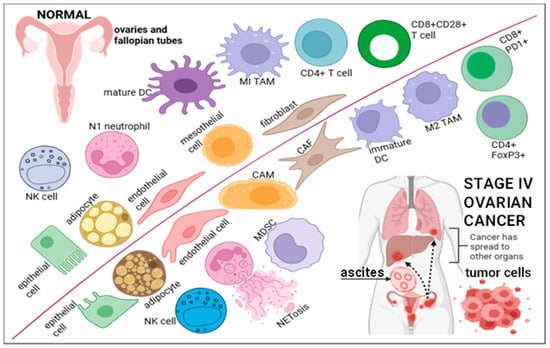
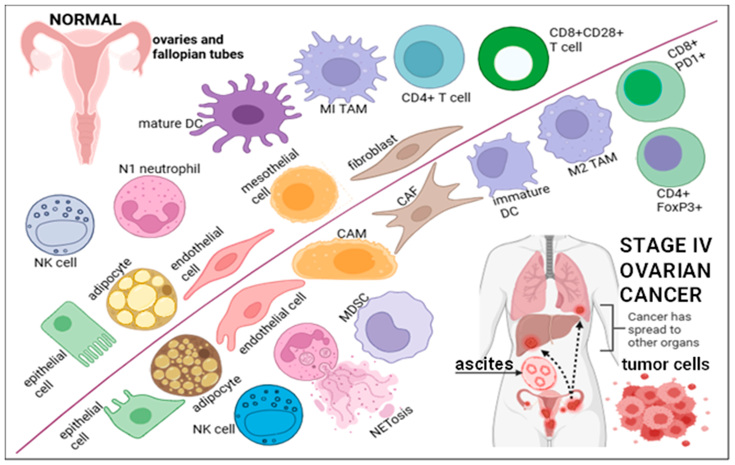

Disclaimer/Publisher’s Note: The statements, opinions and data contained in all publications are solely those of the individual author(s) and contributor(s) and not of MDPI and/or the editor(s). MDPI and/or the editor(s) disclaim responsibility for any injury to people or property resulting from any ideas, methods, instructions or products referred to in the content. |
© 2026 by the authors. Licensee MDPI, Basel, Switzerland. This article is an open access article distributed under the terms and conditions of the Creative Commons Attribution (CC BY) license.