


Abstract
Colorectal cancer (CRC) remains one of the most common malignancies worldwide and a leading cause of cancer-related mortality. Recent advances in CRC molecular biology have focused on biomarkers for early detection, prognostic stratification, treatment selection, and disease monitoring. Clinically validated biomarkers, including microsatellite instability (MSI), RAS and BRAF mutations, HER2 amplification, and carcinoembryonic antigen (CEA), guide targeted therapy and predict treatment response. However, tumor heterogeneity, clonal evolution, and microenvironmental influences limit the predictive value of single biomarkers. Recent molecular diagnostics therefore increasingly focus on dynamic and minimally invasive approaches. Circulating tumor DNA (ctDNA) is a promising biomarker for detecting minimal residual disease, monitoring recurrence, and assessing tumor evolution in real time. Multi-omics profiling, spatial transcriptomics, and artificial intelligence further expand tumor characterization except for conventional genomic alterations. Our review integrates common CRC biomarkers with developing liquid biopsy, multi-omics, spatial transcriptomics, and artificial intelligence approaches across early detection, prognosis, treatment selection, and longitudinal monitoring. Future biomarker strategies will likely rely on integrated molecular models combined with longitudinal liquid-biopsy assessment. However, most new biomarkers remain at the investigational stage, and assay heterogeneity, limited standardization, and insufficient prospective clinical validation currently restrict their routine clinical use.
1. Introduction
Biomarkers are widely known as measurable indicators of biological or pathological processes that can mirror the presence and progression of the disease as well as the response to therapeutic interventions. Depending on the clinical context they reflect, biomarkers can be classified as diagnostic, prognostic, or predictive. According to the FDA–NIH Biomarker Working Group, a biomarker constitutes “a defined characteristic that is measured as an indicator of normal biological processes, pathogenic processes, or responses to an exposure or intervention” [1,2]. In oncology, biomarkers play a significant role in early detection, prognostic stratification, and prediction of therapeutic response, laying the foundation for precision medicine. When it comes to their role in colorectal cancer (CRC), the third most common type of cancer in the world that is also infamous for being the second leading cause of cancer-related mortality, the identification and clinical implementation of reliable biomarkers have become crucial for disease management and therapeutic decision-making (Figure 1) [3,4].
Figure 1.
Biomarkers in the colorectal cancer (CRC) clinical pathway. Biomarkers may have overlapping diagnostic, prognostic, predictive, and monitoring roles depending on the clinical context [5,6,7,8,9,10,11,12,13,14,15]. Abbreviations: dMMR, deficient mismatch repair; HER2, human epidermal growth factor receptor 2; MSI-H, microsatellite instability-high. Created in BioRender. Kulbacka, J. (2026) https://BioRender.com/wuj7ja7.
CRC represents a major global health burden, with more than 1.9 million new cases and nearly 900,000 deaths reported worldwide in 2022 [4]. By 2040, the burden of CRC is predicted to increase with 3.2 million new cases and 1.6 million deaths [16]. Disturbingly, the incidence of CRC diagnosed before the age of 50, referred to as early-onset CRC (EOCRC), has been increasing worldwide in recent years and is often associated with more advanced disease at diagnosis and poorer survival outcomes. From a biomarker perspective, this trend underscores the need to characterize EOCRC’s distinct molecular features and identify biomarkers for earlier detection, risk stratification, and treatment selection [17,18]. The incidence and mortality of CRC vary considerably across geographic regions, with the highest rates observed in developed countries, although increasing trends have recently been reported in many low- and middle-income regions. These differences largely reflect variations in lifestyle, environmental exposures, and access to screening programs. Established risk factors include aging, obesity, insufficient physical activity, smoking, alcohol consumption, and diets rich in processed or red meat. Although highly penetrant hereditary syndromes, including Lynch syndrome (LS) and familial adenomatous polyposis (FAP), account for approximately 5% of CRC cases, familial aggregation studies indicate that inherited factors contribute more broadly to CRC susceptibility. In the Nordic Twin Study of Cancer, genetic factors were estimated to account for 40% (95% CI, 33–48%) of the variation in CRC susceptibility, although estimates were lower for colon and rectal cancer considered separately. A substantial proportion of familial CRC risk therefore remains unexplained by currently recognized Mendelian syndromes and may reflect the combined effects of polygenic risk, rare variants, and shared environmental factors [19,20]. Five-year survival among patients with stage I CRC is 91%, but it declines to approximately 15% in stage IV disease [21]. Thus, early diagnosis is one of the most important determinants of prognosis [22]. Difficulty arises from the fact that the vast majority of patients are diagnosed with CRC once they become symptomatic, and the presence of symptoms, such as rectal bleeding, anemia, and change in the rhythm of bowel movements, is most commonly an indicator of late-stage disease [23,24]. Thus, early diagnosis in asymptomatic patients is one of the main ways to achieve favorable survival outcomes. Although colonoscopy remains the gold standard for CRC detection, patient reluctance and financial constraints limit its use; fecal immunochemical testing (FIT), in turn, is limited by imperfect sensitivity and specificity, resulting in false-positive and false-negative findings [13,25]. The sensitivity of FIT for detecting CRC, using a 100 ng/mL cutoff, reaches 73.8%, compared with 92.3% for a stool-based DNA assay assessing KRAS mutations and aberrant NDRG4 and BMP3 methylation [12,13]. Regarding advanced precancerous lesions, the sensitivity of the discussed tests reaches 28.8% and 42.4% [12], which emphasizes the limitations of the current screening strategy as well as the unmet need for non-invasive, reliable biomarkers. Emerging research addresses this problem and identifies promising markers with diagnostic accuracy, such as circulating tumor DNA (ctDNA) and microRNAs, suggesting that panels combining multiple biomarkers could significantly improve early CRC detection [14,26]. Apart from their usefulness in diagnostics, biomarkers also play a crucial role in both predicting the course of the disease and guiding therapeutic strategies. Prognostic markers, like mutations of KRAS and BRAF as well as ctDNA, can provide information on the risk of occurrence and overall survival (OS), whereas predictive biomarkers enable the selection of targeted therapies and help optimize treatment efficacy while minimizing toxicity [10,11,22,26]. Therefore, the identification and validation of reliable molecular biomarkers remain essential for developing personalized management strategies for CRC.
Recent reviews have addressed biomarkers in gastrointestinal cancers, CRC diagnosis, or selected applications of liquid biopsy and minimal residual disease assessment [5,9,27,28,29]. However, an updated CRC-specific framework integrating established tissue- and blood-based biomarkers with emerging liquid biopsy, multi-omics, spatial transcriptomics, and artificial intelligence approaches remains lacking. The added value of the present review lies in linking individual biomarkers to their principal clinical roles in early detection, prognostic assessment, treatment selection, and longitudinal disease monitoring, while also discussing their current level of clinical validation and the barriers to clinical implementation, including assay standardization, tumor heterogeneity, and the need for prospective validation.
Review Methodology
This narrative review was based on a structured search of PubMed/MEDLINE, Scopus, and Web of Science Core Collection. The literature search prioritized studies published during the preceding 10 years. However, we included selected older publications when they provided essential historical or methodological foundations, reported landmark biomarker discoveries, described pivotal clinical trials, or established diagnostic and prognostic principles that remain clinically relevant. The search strategy combined terms related to colorectal cancer (“colorectal cancer,” “colon cancer,” “rectal cancer”) with terms related to biomarkers and their clinical applications, including “biomarker,” “molecular marker,” “liquid biopsy,” “circulating tumor DNA,” “minimal residual disease,” “microsatellite instability,” “mismatch repair,” “KRAS,” “NRAS,” “BRAF,” “HER2,” “CEA,” “microRNA,” “DNA methylation,” “multi-omics,” “spatial transcriptomics,” and “artificial intelligence.” The reference list of key publications was also screened to identify relevant milestone studies and current guidelines. We included English-language human studies, clinical trials, observational studies, systematic reviews, narrative reviews, meta-analyses, and clinical guidelines or consensus statements addressing CRC biomarkers or their clinical implementation. Publications unrelated to CRC biomarkers, duplicate records, conference abstracts without sufficient data, and studies lacking relevant clinical or translational information were excluded. Titles and abstracts were screened for relevance, followed by full-text assessment of potentially eligible publications. Because this was a narrative review, publications were selected purposively according to their clinical relevance, methodological quality, and contribution to the objectives of the review.
2. Established Clinical Biomarkers
Several molecular biomarkers are already incorporated into routine clinical practice for CRC management. These markers provide valuable information regarding tumor biology, prognosis, and potential response to targeted therapies, thereby supporting personalized treatment strategies. Current clinical guidelines recommend assessing specific genomic alterations and molecular characteristics to guide therapeutic decisions, particularly in advanced disease. The most widely recognized and clinically validated biomarkers in CRC include microsatellite instability (MSI), mutations in the RAS and BRAF genes, HER2 amplification, and serum carcinoembryonic antigen (CEA) levels [10,15,22].
2.1. MSI/Deficient Mismatch Repair (dMMR)
DNA mismatch repair (MMR) and MSI status are invaluable biomarkers that guide clinical management of CRC patients [30]. MMR proteins recognize and repair base–base mismatches and insertion–deletion loops generated during DNA replication, thereby maintaining genomic stability. In humans, mismatch recognition is mediated by two MutS heterodimers: MutSα (MSH2–MSH6), which preferentially recognizes base–base mismatches and small insertion–deletion loops, and MutSβ (MSH2–MSH3), which preferentially recognizes larger insertion–deletion loops. Following mismatch recognition, MutLα (MLH1–PMS2) is recruited to the MutS–DNA complex and coordinates downstream repair. Its PMS2 subunit provides mismatch-activated endonuclease activity that introduces nicks into the newly synthesized strand, thereby enabling excision of the error-containing strand and subsequent DNA resynthesis [31]. Alterations in genes involved in the MMR pathway are frequently observed in sporadic CRC. Moreover, germline mutations in MMR genes are associated with LS, the most common hereditary form of CRC. dMMR constitutes a consequence of the inactivation of one or more core MMR genes, which may occur through germline variants, as observed in LS, or through somatic loss-of-function mutations followed by a second hit leading to biallelic gene inactivation. In addition, MMR deficiency may arise from epigenetic silencing caused by MLH1 promoter hypermethylation. The loss of functional MMR proteins leads to the accumulation of replication errors in microsatellite regions, resulting in MSI. Depending on the extent of instability detected across microsatellite loci, MSI can be classified as microsatellite stable (MSS), MSI-low (MSI-L), and MSI-high (MSI-H) (Figure 2) [31,32]. MMR/MSI status is used not only to identify LS, but also to predict immune checkpoint inhibitors (ICIs) treatment response [33]. MSI/dMMR status can be assessed by immunohistochemistry for MLH1, PMS2, MSH2, and MSH6; PCR-based MSI testing; or a validated NGS assay. Universal tumor testing is recommended for newly diagnosed CRC [31]. Loss of MLH1/PMS2 should be followed by BRAF V600E and/or MLH1 promoter methylation testing to exclude a sporadic tumor. Patients with abnormal MSH2, MSH6, or PMS2 expression, or with MLH1/PMS2 loss without a sporadic explanation, should be referred for genetic counseling and germline testing for Lynch syndrome [34].
Figure 2.
Mechanisms of deficient mismatch repair (dMMR) and microsatellite instability (MSI). Symbols: crosses (X) over the MSH2–MSH6 and MLH1–PMS2 heterodimers indicate that these MMR protein complexes are dysfunctional or absent (dMMR). Abbreviations: MMR, mismatch repair; dMMR, deficient mismatch repair; MSI, microsatellite instability [30,31,32]. Created in BioRender. Kulbacka, J. (2026) https://BioRender.com/p54lwcb.
Clinically, dMMR/MSI-H CRC tumors usually present as poorly differentiated mucinous adenocarcinomas and are more frequently located in the proximal colon [35,36]. In the sporadic setting, BRAF V600E commonly occurs within the serrated neoplasia pathway and is strongly associated with CpG island methylator phenotype-high (CIMP-high) tumors, which are characterized by widespread promoter CpG-island hypermethylation. In a substantial proportion of these cancers, hypermethylation of the MLH1 promoter causes transcriptional silencing of MLH1, resulting in dMMR and the MSI-H phenotype [36,37]. dMMR/MSI-H is more common in early-stage CRC, likely because high tumor mutational burden (TMB) generates neoantigens that promote CD8+ T-cell infiltration and an immunogenic tumor microenvironment (TME). However, in metastatic disease, dMMR/MSI-H is associated with poorer prognosis, which may partly reflect the higher prevalence of the BRAF V600E mutation rather than the MSI status itself [36,38]. Regarding therapeutic implications, although CRC patients with MSI-H generally respond favorably to ICIs, treatment response depends on specific modifying factors. First, disruptions in the gut microbiota ecosystem caused by broad-spectrum antibiotics can negatively affect the therapeutic potential of ICIs, whereas appropriate modulation of the microbiome may improve immunotherapy outcomes [39]. For instance, although Fusobacterium nucleatum is widely regarded as a pro-tumorigenic and immunosuppressive microorganism in CRC and has been associated with adverse clinical outcomes, some studies suggest it may enhance responses to programmed death-ligand 1 (PD-L1) blockade in specific experimental contexts [40]. Secondly, peritoneal metastases accompanied by ascites noticeably reduce the effectiveness of ICIs, probably because of the combined effects of malignant ascites and immunosuppressive TME [41]. These findings question the use of MSI-H as a standalone predictive biomarker, underscoring the need for a composite biomarker approach.
Neoadjuvant immunotherapy has also produced practice-changing results in localized dMMR/MSI-H CRC. In the NICHE-2 study, neoadjuvant nivolumab plus ipilimumab produced pathological responses in 98% of patients and a pathological complete response in 68%, with no recurrences reported at a median follow-up of 26 months [42]. In locally advanced dMMR rectal cancer, neoadjuvant dostarlimab has also enabled organ-preservation strategies in patients achieving a complete clinical response [43]. Nevertheless, surgery should currently be omitted only in carefully selected patients managed within structured surveillance protocols or clinical trials.
In contrast to MSI-H CRC, MSS CRC, in which microsatellite length and composition remain stable during replication, is typically associated with proficient mismatch repair (pMMR). MSS/pMMR tumors are characterized by low TMB and limited infiltration of immune cells within the TME, which results in poor response to ICI monotherapy [44]. Importantly, MSS CRC accounts for approximately 85–90% of colorectal tumors. It is usually located in the left colon and rectum and presents as well-differentiated or moderately differentiated adenocarcinomas. The immune landscape of MSS tumors is generally dominated by immunosuppressive cell populations, including regulatory T cells and M2 macrophages, and is characterized by relatively low PD-L1 expression, which collectively facilitates immune evasion [45].
From a prognostic perspective, outcomes in MSS CRC are strongly influenced by tumor stage and the characteristics of the TME. While survival outcomes in early-stage MSS disease may be comparable to MSI-H tumors, patients with advanced MSS CRC often demonstrate significantly poorer survival, likely due to both chemotherapy resistance and an immunosuppressive TME. Interestingly, recent studies highlight the prognostic importance of tumor-infiltrating lymphocyte (TIL) density in MSS CRC. Even within the MSS group, patients with high TIL infiltration (MSS-TIL-H) have shown significantly improved overall and disease-free survival (DFS) compared with those with low TIL levels, indicating that immune contexture remains an important determinant of prognosis [46]. Given the limited efficacy of immunotherapy in MSS CRC, current research focuses on combination therapies, such as combining vascular endothelial growth factor receptor (VEGFR) inhibitors with PD-1 blockade and dual immune checkpoint inhibition, which have shown encouraging activity in refractory MSS metastatic CRC (mCRC). In parallel, increasing attention is being directed toward biomarker-driven therapeutic strategies, including the identification of predictive markers such as TIL density, POLE/POLD mutations, and TMB, which may help identify subgroups of MSS CRC patients more likely to benefit from immunotherapeutic approaches [46,47,48]. Recent advances in digital pathology and artificial intelligence (AI) have also improved MMR/MSI status evaluation in CRC. Several studies have shown that deep learning (DL) algorithms applied to digitized whole-slide images can accurately predict dMMR/MSI-H status, offering a rapid, scalable approach to tumor classification [49,50]. In addition to MSI detection, DL-based models can extract clinically relevant prognostic and predictive features from routine hematoxylin and eosin (H&E)-stained slides, enabling the development of computational prognostic models that may outperform traditional morphological and molecular markers [51]. These tools have shown consistent performance across tumor and nodal stages and may support personalized treatment decisions, including identifying patients who may benefit from intensified therapy or, conversely, those who may avoid overtreatment [52]. Moreover, machine learning (ML) approaches integrating clinical, pathological, and transcriptomic data are increasingly being explored to predict immunotherapy responses in CRC, highlighting the growing potential of AI-assisted diagnostics in precision oncology [53,54].
MSI/dMMR status is an established biomarker of sensitivity to immune-checkpoint inhibitors. In clinical practice, MSI/dMMR testing identifies patients who may benefit from pembrolizumab or nivolumab-based immunotherapy in advanced CRC [55,56]. Neoadjuvant immunotherapy may also support organ-preservation strategies in carefully selected patients with localized dMMR/MSI-H disease [42,43]. POLE/POLD1 alterations may identify additional hypermutated tumors with potential sensitivity to immune-checkpoint inhibitors, but the available evidence remains limited, and these alterations should not be considered equivalent to MSI-H/dMMR [57]. Other immune biomarkers, including PD-1/Tim-3 coexpression, HLA class I expression, tumor-infiltrating lymphocytes, MCL-1, and CCR5, have been associated with prognosis or immunotherapy response in exploratory cohorts. However, these markers lack standardized assays, validated thresholds, and prospective evidence showing that biomarker-guided selection improves outcomes and should therefore remain investigational [46,58,59,60,61].
2.2. RAS Mutations
Mutations in the RAS superfamily have been validated as drivers of tumor initiation and survival in numerous studies [62]. RAS proteins function as GTPase molecular switches that alternate between inactive GDP-bound and active GTP-bound states. In their active conformation, they interact with downstream effector proteins to initiate signaling pathways. In CRC, KRAS mutations occur in approximately 40–45% of tumors, whereas NRAS mutations are much less frequent, affecting about 3–5% of cases, and HRAS mutations are exceedingly rare [63]. KRAS mutations, which usually involve alterations in codons 12 and 13, with the most common variants including G12D, G12V, and G13D, are associated with poor tumor differentiation, distant metastases, and shorter recurrence-free survival (RFS) and OS [63,64,65,66]. Nevertheless, the prognostic significance of KRAS mutations may vary depending on the specific mutation subtype and disease stage. For instance, while codon 12 mutations are often associated with poorer overall survival and DFS, the prognostic impact of codon 13 alterations remains less clear. Furthermore, in stage II–III CRC, KRAS mutation status has not consistently demonstrated significant prognostic relevance [67]. Heterogeneity in KRAS mutation status between primary tumors and distant metastases is generally uncommon, as it occurs in less than 10% of cases [68,69]. However, pulmonary metastases demonstrate substantially higher discordance rates, reaching up to 30% in comparison with other metastatic sites [70,71]. This finding supports testing metastatic tissue whenever feasible, particularly in pulmonary metastases, and repeating RAS testing when progression occurs or when molecular results from different disease sites are discordant. Moreover, both wild-type-to-mutant and mutant-to-wild-type mutations have been reported in metastatic lesions [72]. Discussed findings suggest that KRAS-mutant subclones may be present in both primary and metastatic tumors, potentially contributing to therapeutic response [73]. Analyses of ctDNA have demonstrated that previously undetected KRAS mutations can emerge during therapy, reflecting the expansion of resistant tumor clones [74]. Conversely, some patients initially classified as RAS-mutant may exhibit loss of detectable mutations in plasma during disease progression, suggesting potential therapeutic implications for liquid biopsy–based reassessment of RAS status during treatment [75,76]. From a molecular perspective, KRAS consists of two principal structural components: the G-domain and the C-terminal hypervariable region. The G-domain includes the switch I, switch II, and P-loop regions, which undergo conformational changes depending on whether GDP or GTP is bound [65,77]. Under physiological conditions, epidermal growth factor receptor (EGFR) activation leads to RAS activation via GTP binding, which then triggers downstream signaling through the MAPK pathway, a key regulator of cell proliferation, differentiation, and survival [66,78]. Once a KRAS mutation occurs, the protein remains constitutively bound to GTP, persistently activating this signaling cascade independent of upstream EGFR stimulation (Figure 3). This promotes uncontrolled tumor growth.
Figure 3.
EGFR–RAS–RAF–MEK–ERK (MAPK) signaling in colorectal cancer and the effect of KRAS mutation. With ligand-bound EGFR, RAS cycles between an inactive GDP-bound and an active GTP-bound state, and RAS-GTP activates RAF, which sequentially phosphorylates MEK and ERK. Without ligand, RAS remains GDP-bound and signaling stops. Mutant KRAS is locked in the GTP-bound state and activates RAF constitutively, independently of upstream EGFR, so anti-EGFR antibodies fail to switch off the cascade [62,66,78,79]. Symbols: the red cross (X) indicates that inactive, GDP-bound RAS does not activate RAF; the red arrow indicates constitutive, EGFR-independent activation of RAF by mutant KRAS-GTP, which persists despite EGFR blockade by anti-EGFR antibodies. Created in BioRender. Kulbacka, J. (2026) https://BioRender.com/bpevgui.
RAS status is therefore a core predictive biomarker for anti-EGFR therapy. Activating KRAS or NRAS mutations indicate primary resistance to cetuximab and panitumumab, whereas RAS wild-type status identifies patients who may be eligible for anti-EGFR treatment [11,15]. The dedicated Section ”Anti-EGFR Therapy: Predictive Biomarkers and Resistance Mechanisms” on mechanisms of resistance discusses acquired resistance mechanisms, including emergent RAS or BRAF alterations, HER2 activation, PIK3CA/PTEN dysregulation, and EGFR extracellular-domain mutations [75,80,81]. In previously treated patients with KRAS G12C-mutated mCRC, targeted combinations such as adagrasib plus cetuximab or sotorasib plus panitumumab represent additional treatment options [82,83].
2.3. BRAF Mutations
BRAF, a serine/threonine kinase, plays a central role in the MAPK signaling cascade, regulating cell proliferation and survival. Activating BRAF mutations occur in about 8–10% of CRC cases and constitutively activate the MAPK pathway, promoting tumor growth and progression [84]. The vast majority of BRAF alterations are the V600E mutations, which account for about 90–95% of BRAF-mutant CRC cases and exhibit markedly increased kinase activity compared with wild-type BRAF [85]. Clinically, BRAF V600E mutations are associated with rapid disease progression and poor prognosis, especially in mCRC. They usually occur in the right colon, show mucinous histology, and are more common in older patients and women [84]. Regarding interactions with other biomarkers, BRAF mutations are detected in a substantial proportion of sporadic MSI-H CRC, particularly those with MLH1 promoter hypermethylation [86]. MSI-H status might partially mitigate the otherwise adverse prognostic impact of BRAF mutations. In contrast, MSS BRAF-mutant tumors exhibit particularly poor survival outcomes [84,86,87]. BRAF mutations are divided into functional classes: class I (V600), class II (RAS-independent non-V600), and class III (RAS-dependent). Class II mutations are typically resistant to anti-EGFR therapy, whereas class III mutations may retain sensitivity to EGFR inhibitors [88]. BRAF V600E can also be assessed with the VE1 mutation-specific immunohistochemical assay, which provides rapid, tissue-based screening. However, its diagnostic performance in CRC depends on the staining platform, antibody interpretation, tumor heterogeneity, and positivity threshold. Therefore, molecular testing should confirm confusing or discordant results. VE1 detects the V600E protein and does not identify non-V600E BRAF variants [89]. Importantly, targeted therapeutic strategies have emerged for CRC with BRAF V600E mutations. Combination therapy with the BRAF inhibitor encorafenib and the anti-EGFR antibody cetuximab has demonstrated improved OS, progression-free survival (PFS), and response rates in previously treated mCRC patients [90]. In addition to targeted therapy, BRAF mutation status also influences response to immunotherapy. Clinical trials such as KEYNOTE-177 and CheckMate-142 have demonstrated that ICIs, including pembrolizumab and nivolumab (alone or combined with ipilimumab), provide significant clinical benefit in MSI-H mCRC, including cases harboring BRAF mutations [55,56]. Consequently, BRAF mutation status represents both an important prognostic biomarker and a predictive marker for targeted and immunotherapeutic strategies in CRC.
2.4. HER-2 Amplification
ERBB2, also known as CD340 and HER-2, is a protein tyrosine kinase receptor, the locus of which is localized on chromosome 17q12 [91]. HER-2 alterations, including gene amplification and activating point mutations, are detected in a small subset of CRCs. Gene amplification occurs in about 2–5% of cases and is more frequent in RAS wild-type tumors, whereas the association with higher tumor mutational burden appears to depend on the specific ERBB2 alteration and molecular context [92]. HER2 overexpression is more frequently detected in tumors of the left colon and rectum [93]. Within the complex molecular interactions between HER-2 and other biomarkers, MSI-H was not found in tumors with HER-2 amplification, whereas the correlation between point mutations in HER-2 and MSI-H CRC is positive [94]. The prognostic significance of the discussed biomarker remains controversial. According to some studies, HER-2 amplification is associated with increased invasive potential of the tumor and poorer survival outcomes [95,96,97], whereas others claim that either survival is improved or there is no association at all [98,99]. These differences may reflect anatomical and molecular heterogeneity in CRC, differences in assay platforms (including immunohistochemistry, in situ hybridization, and sequencing), variation in interpretation cut-offs, and differences in study populations and clinical settings. As a predictive biomarker, HER-2 alterations predict resistance to anti-EGFR therapy in CRC. Both HER-2 amplification and heregulin-mediated HER-3 activation sustain ERK1/2 signaling and continued tumor growth. In patients with wild-type KRAS/NRAS/BRAF/PIK3CA, HER2 amplification has been associated with shorter response duration to EGFR inhibitors and poorer prognosis, while emerging data also highlight HER2 as a therapeutic target for HER2-directed agents such as antibody–drug conjugates (ADCs) [100]. Currently, ADCs, monoclonal antibodies, and tyrosine kinase inhibitors (TKIs), with combination regimens such as trastuzumab–pertuzumab or trastuzumab-based therapies, are under investigation and show promising activity in HER2-positive, RAS wild-type mCRC [100,101,102]. Most importantly, tucatinib plus trastuzumab is the only CRC-specific FDA-approved HER2-directed regimen for RAS wild-type, HER2-positive unresectable or metastatic CRC that has progressed after fluoropyrimidine-, oxaliplatin-, and irinotecan-based therapy. This approval was based on the MOUNTAINEER trial [103].
2.5. CEA
CEA represents one of the most widely recognized tumor markers across multiple cancer types. However, a high rate of false positives limits its usefulness for CRC diagnosis when assessed without broader molecular context [104]. Nevertheless, multimarker serum panels may improve diagnostic performance compared with single markers. In a study of 1027 samples, a six-marker panel comprising CEA, CYFRA 21-1, ferritin, osteopontin, anti-p53, and seprase achieved 69.6% sensitivity at 95% specificity, compared with 43.9% for CEA alone [105]. Interestingly, recent research suggests that for both early-stage CEA and even the precancerous stage of disease, fecal CEA may be a more sensitive biomarker than serum CEA, offering improved diagnostic precision through a non-invasive approach [106]. Although CEA’s diagnostic potential remains limited, it plays a significant role in detecting CRC metastases. Integrating CEA interpretation with magnetic resonance imaging (MRI) increases the accuracy of liver metastasis diagnosis [107]. Given CEA’s prognostic value, it is widely used to predict the risk of CRC recurrence after surgery. An increase in the initial level of this biomarker is associated with a greater risk of CRC recurrence, whereas an elevation at recurrence is related to lower five-year DFS rates [108,109]. However, serum CEA alone cannot confirm or exclude tumor recurrence and should be interpreted together with clinical assessment and imaging.
2.6. Other Clinically Investigated Biomarkers
Researchers have investigated additional biomarkers for potential clinical utility in CRC. Mutations in the TP53 tumor suppressor gene occur in approximately 50–60% of colorectal tumors and are associated with genomic instability and disease progression [110]. Similarly, PIK3CA mutations, which activate the PI3K/AKT signaling pathway, have been implicated in tumor growth and may influence the response to targeted therapies [111]. Protein-based biomarkers such as vascular endothelial growth factor (VEGF) and the proliferation marker Ki-67 have also been extensively studied. VEGF plays a key role in tumor angiogenesis and may predict response to antiangiogenic therapies such as bevacizumab [112], while Ki-67 expression reflects tumor proliferative activity and has been associated with aggressive tumor behavior [113]. None of these markers currently has a routine guideline-supported role in treatment selection. TP53, PIK3CA, VEGF, and Ki-67 should therefore be regarded as exploratory or research-oriented biomarkers, in contrast to established biomarkers such as MSI/dMMR, RAS, BRAF, and HER2, which inform guideline-based treatment decisions in appropriate clinical settings [15,114,115].
3. Emerging Biomarkers
Despite the important role of established biomarkers in guiding CRC management, their clinical utility remains limited by their restricted sensitivity, tumor heterogeneity, and inability to fully capture the disease’s dynamic molecular evolution. Advances in molecular technologies have therefore stimulated the search for novel biomarkers that could improve early detection, enable real-time monitoring of tumor burden, and refine prognostic and predictive stratification. Among these emerging approaches, liquid biopsy has attracted considerable attention because it is minimally invasive and can reflect the tumor’s current molecular landscape by analyzing circulating tumor-derived components such as ctDNA, RNA molecules, and circulating tumor cells (CTCs) [6,116,117]. Despite liquid biopsy and multi-omics, genome-wide methylation profiles, circulating miRNAs, telomere length, germline polymorphisms, serum proteins, and chromosomal alterations have been associated with prognosis or treatment response in CRC [118,119]. However, most available evidence comes from retrospective or single-study cohorts, and assay platforms, thresholds, patient populations, and clinical endpoints vary substantially. None of these markers is currently sufficient for routine clinical decision-making or guideline-based treatment selection [120,121,122,123,124,125]. They should therefore be considered investigational. Future studies should use standardized assays, prospective multicenter validation, and predefined clinical endpoints, while demonstrating added value beyond established clinicopathological and molecular factors.
3.1. Liquid Biopsy in CRC: Current Evidence and Clinical Scope
3.1.1. Biological Background
Liquid biopsy encompasses several tumor-derived components detectable in blood. ctDNA is released into circulation through apoptosis, necrosis, and active secretion, reflecting the tumor’s current biological state. Its molecular profile retains carcinogenic alterations, including changes in the APC, TP53, and KRAS genes, which are among the most clonal alterations in advanced CRC [80]. This feature allows ctDNA to serve as a dynamic sensor of disease, often providing information before radiological progression. Other circulating molecules, including miRNAs and long non-coding RNAs (lncRNAs), are also being investigated. Serum miR-21 levels have been associated with CRC aggressiveness and decrease after tumor resection, supporting their potential diagnostic and prognostic value [126]. Epigenetic studies further suggest that lncRNAs and histone modifications may contribute to CRC biology and could be incorporated into future liquid-biopsy panels [127]. Nevertheless, ctDNA remains the liquid-biopsy biomarker with the highest level of clinical validation [117].
Other circulating tumor-derived components, including CTCs and extracellular vesicles, may provide complementary information on metastatic potential, tumor heterogeneity, and treatment response (Figure 4) [26,116]. CTCs provide complementary prognostic information. Plastin 3 (PLS3)-positive CTCs are associated with poor prognosis, particularly in patients with stage II–III CRC [128]. Specific CTC features also relate to prognosis: PLS3-positive CTCs are associated with poor outcomes; a six-gene assay (CEA, EpCAM, CK19, MUC1, EGFR, and c-MET) improves disease monitoring and correlates with PFS [129]. High CTC counts and mesenchymal phenotype (M+ CTCs) correlate with metastasis and shorter PFS and OS [130]. Akt-2, ALDH1, or PI3Kα expression in CTCs identifies patients with poorer outcomes [131], while COX-2–positive CTCs may indicate more aggressive disease [132]. Extracellular vesicles, particularly tumor-derived exosomes, contain proteins, DNA fragments, and regulatory RNAs that may influence intercellular communication and serve as non-invasive indicators of tumor activity [133].
Figure 4.
Major liquid-biopsy components in colorectal cancer (CRC). Liquid biopsy enables the analysis of circulating tumor DNA (ctDNA), circulating tumor cells (CTCs), circulating microRNAs and other non-coding RNAs, and tumor-derived extracellular vesicles. These analytes may support early detection, prognostic assessment, treatment monitoring, and minimal residual disease (MRD) evaluation. Abbreviations: ctDNA, circulating tumor DNA; CTCs, circulating tumor cells; MRD, minimal residual disease [6,7,8,26,116,117,133]. Created in BioRender. Kulbacka, J. (2026) https://BioRender.com/93g7mtg.
3.1.2. ctDNA in Minimal Residual Disease (MRD) Assessment
In patients with localized colon and rectal cancer, the presence of ctDNA after resection of the primary tumor has been shown to be one of the strongest known risk factors for recurrence and death, surpassing clinical-pathological and imaging parameters. Studies by Tie and colleagues have shown that ctDNA-negative patients after stage II surgery have a very low risk of recurrence, while ctDNA-positive patients have a significantly higher risk [134,135]. A meta-analysis by Chidharla et al. confirms that ctDNA after treatment is associated with significantly shorter RFS and OS [136]. Similar observations were made in rectal cancer after neoadjuvant therapy, where ctDNA strongly differentiated prognosis even before surgery, supporting the interpretation of ctDNA as a molecular equivalent of oncological margins. An important extension of this concept is using ctDNA in oligometastatic disease treated with radical intent. In this group, classic prognostic parameters such as the number of metastases, their location, or margin status have proven insufficiently accurate for predicting recurrence. Research by Marmorino and colleagues has shown that detectable ctDNA after resection of liver metastases in CRC is an independent and strong marker of residual disease, predicting both time to recurrence and survival [137]. Similarly, O’Donnell and co-authors observe that ctDNA after completion of locoregional treatment (resections, ablations, stereotactic radiotherapy) enables earlier and more accurate identification of patients at risk of recurrence than traditional clinical variables [138]. The commentary by Yan et al. synthesizes data showing that liquid biopsy not only allows for the detection of disease, but above all, for the dynamic assessment of MRD, risk of recurrence, and response to treatment at various stages of CRC [116]. The prognostic role of post-treatment ctDNA in oligometastatic CRC is discussed in greater detail below.
In clinical practice, these findings support using ctDNA as a dynamic marker of recurrence risk and for treatment monitoring. In the clinical setting, postoperative ctDNA positivity is associated with a higher risk of recurrence, while serial testing may detect molecular relapse before radiological progression [136,139]. Prospective randomized evidence supports ctDNA-guided adjuvant decisions in selected stage II colon cancer settings, but the broader clinical role remains assay-, timing-, and disease-setting dependent [139,140]. ctDNA should therefore complement, rather than replace, clinicopathological assessment and imaging until prospective studies demonstrate that acting on ctDNA results improves patient outcomes [140,141].
3.1.3. Epigenetic Signals in Liquid Biopsy
Liquid-biopsy components have different potential clinical applications and should not be considered interchangeable. Mutational ctDNA analysis is primarily suited to tracing clonal evolution, quantifying tumor burden, and detecting emerging mechanisms of therapeutic resistance, including the re-emergence of RAS-mutant clones during anti-EGFR therapy [117]. Plasma DNA methylation markers reflect recurrent epigenetic changes and may be particularly useful for blood-based screening and broad detection of tumor-derived signals. Methylated SEPT9 is a representative example; however, its sensitivity remains insufficient to replace FIT or colonoscopy, particularly for detecting advanced precancerous lesions [142,143].
Circulating miRNAs and lncRNAs reflect regulatory pathways associated with proliferation, epithelial–mesenchymal transition, invasion, and treatment response. Nevertheless, the current evidence remains predominantly retrospective, single-study, or laboratory-based, and these markers have not been validated for routine clinical decision-making in CRC [126,144].
Integrated plasma assays combining mutation and methylation signals, such as Guardant Reveal, may provide tumor-naïve MRD assessment without prior tumor sequencing. However, their clinical performance remains assay-specific, and results obtained from different MRD platforms should not be considered directly interchangeable [144,145].
3.1.4. Oligometastatic Disease
In a prospective single-center study of 25 patients with oligometastatic CRC treated with definitive local therapy, detectable ctDNA after treatment was associated with worse overall survival (HR 11.28; 95% CI, 1.31–97.05), whereas baseline ctDNA was not significantly prognostic. Serial ctDNA patterns were also associated with recurrence risk. However, only 19 patients were evaluable, and the wide confidence interval and single-center design indicate substantial uncertainty. These findings should therefore be considered hypothesis-generating and require validation in larger prospective multicenter studies before ctDNA can guide treatment decisions in oligometastatic CRC. It also should not be used alone to select, escalate, or de-escalate treatment outside a prospective clinical study [146].
More generally, the clinical value of liquid biopsy in CRC depends on both the assay used and the clinical setting. As discussed by Malla et al., different ctDNA platforms have distinct strengths and weaknesses depending on the intended use, while Parikh et al. note that tumor-informed approaches may better distinguish tumor-derived alterations from non-tumor signals, such as clonal hematopoiesis [147]. Tumor-informed assays require sequencing of the resected tumor and subsequently track patient-specific variants in plasma [139,148]. They generally provide high analytical specificity but require available tumor tissue [139]. Tumor-naïve or plasma-only assays infer tumor-derived signals directly from plasma genomic and methylation patterns without prior tumor sequencing [145,147]. They are logistically simpler and can be used when tissue is unavailable, but their sensitivity may be lower at very low ctDNA fractions, and they require rigorous validation to distinguish tumor-derived signals from clonal hematopoiesis [139,145]. Consequently, results from different MRD platforms should not be considered interchangeable. Another important issue is the timing of blood collection, because testing performed too early after surgery may reduce sensitivity due to the higher levels of postoperative cfDNA. This helps explain why the performance of particular liquid-biopsy markers differs across settings, from the high specificity but limited sensitivity of mSEPT9 in screening to the stronger value of serial ctDNA measurements in MRD assessment [117,142,146,147]. Table 1 shows an overview of the main biomarker classes, their clinical applications, and current evidence and guideline status.
Table 1.
Comparison of biomarker approaches discussed in this section a [117,142,146,147].
Blood-based screening has progressed from methylated SEPT9 testing [142] to cell-free DNA assays [143] and multi-cancer early detection (MCED) tests [149]. In the prospective ECLIPSE study, a cfDNA blood test detected 83.1% of colorectal cancers but only 13.2% of advanced precancerous lesions, indicating that blood-based testing may improve screening participation but cannot replace colonoscopy or FIT for prevention of precancerous lesions [143]. MCED assays such as PATHFINDER 2 are promising for detecting cancer signals from multiple organs, but their effect on CRC-specific mortality and overdiagnosis remains unproven [149].
Beyond screening, prospective ctDNA-guided management has expanded beyond the DYNAMIC trial [139]. NRG-GI005/COBRA evaluated a plasma-only, tumor-naïve assay in low-risk stage II colon cancer [150], whereas the CIRCULATE-Japan platform includes the observational GALAXY registry and the randomized phase III VEGA and ALTAIR trials [145,151]. VEGA evaluates treatment de-escalation in ctDNA-negative patients, while ALTAIR evaluates treatment escalation at molecular relapse in ctDNA-positive patients [151,152]. The UK TRACC program evaluates plasma-only ctDNA-guided adjuvant treatment [153,154], whereas BESPOKE is a prospective observational study based on a tumor-informed assay [148]. These studies address different clinical questions and should not be considered interchangeable (Table 2).
Table 2.
Clinical scenarios, biomarkers, evidence level, and guideline status [27,28,29,139,142,145,148].
3.2. Multi-Omics and Spatial Profiling
Traditional genomic approaches do not fully describe the complexity of CRC, as DNA changes do not always translate into changes in protein expression. In response, multi-layered strategies involving genomics, transcriptomics, proteomics, and phosphoproteomics have been developed. Guo et al. performed whole-exome, transcriptomic, proteomic, and phosphoproteomic analyses on paired tumor and normal-adjacent samples from 144 CRC patients and identified three molecular subtypes associated with differences in prognosis and TME. Importantly, the authors emphasize that the CMS classification remains useful, but does not fully reflect CRC heterogeneity because it is based on mRNA data alone. In this setting, multi-omics can complement CMS by adding proteomic and phosphoproteomic information and by identifying biologically distinct groups, including the S_I subtype, which was associated with the worst prognosis, a higher proportion of early-onset CRC, and a higher stromal score [157]. However, the S_I subtype still requires replication in independent external cohorts and has not been incorporated into routine CRC molecular classification systems [157]. From this perspective, multi-omics is not only a broader descriptive approach but also a way to link clinically relevant tumor behavior to layers of biology not captured by DNA alterations alone. In the study by Guo et al., the integration of genomic, transcriptomic, proteomic, and phosphoproteomic data refined molecular stratification and showed that differences between tumors are related not only to mutation patterns but also to the state of signaling pathways and the TME. In turn, Wu et al. describe single-cell multi-omics and spatial methods as approaches that make it possible to study biomolecules in their tissue context and to better capture biological heterogeneity. Guo et al. likewise note that single-cell and spatial transcriptomic approaches may further improve the characterization of cellular composition and spatial organization in CRC. Taken together, these data suggest that future biomarker models may combine longitudinal blood-based MRD assessment with tissue-based molecular classification and spatially resolved tumor features [157,158].
Building on these approaches, integrated biomarker models combine genomic, epigenetic, immune, and clinical features to characterize CRC heterogeneity, treatment response, and resistance [159]. However, multi-omics integration does not by itself establish clinical utility; such models require external validation, calibration, reproducibility, and demonstration of benefit beyond established clinicopathological and molecular factors [155,156,159]. Individual findings involving PTEN/EGFR, ARID1A, chemokine pathways, or aspirin response should therefore be treated as component-level or hypothesis-generating evidence rather than as validated integrated biomarkers [160,161,162,163].
The quickly growing amount of biomedical data is too much for traditional analysis methods. Because of this, more focus is going to artificial intelligence (AI) and machine learning (ML) techniques. These approaches can analyze large genomic and clinical datasets and identify patterns that may remain undetectable using conventional analytical methods [155,156].
4. Clinical Applications
The previous sections described the biological basis and diagnostic relevance of individual CRC biomarkers. This section focuses on how clinicians use these biomarkers in decision-making, including treatment selection, resistance monitoring, and longitudinal disease assessment. Treatment selection for metastatic or recurrent disease is increasingly guided by molecular profiling. According to the ESMO and NCCN guidelines, first-line therapy depends on molecular status and tumor location [15,114,115]. In left-sided, RAS (KRAS/NRAS) and BRAF wild-type, MSS adenocarcinomas, the recommended approach combines EGFR inhibitors (cetuximab or panitumumab) with chemotherapy. For previously untreated BRAF V600E–mutated mCRC, encorafenib plus cetuximab combined with mFOLFOX6 is supported by the BREAKWATER trial [164], whereas the encorafenib–cetuximab doublet is an established option for previously treated mCRC based on the BEACON study [90]. Tumors harboring KRAS or NRAS mutations are generally treated with chemotherapy combined with an antiangiogenic agent such as bevacizumab in the first-line setting, whereas aflibercept plus FOLFIRI is an established second-line option after prior oxaliplatin-based therapy [165]. In later lines, targeted therapies include cetuximab with adagrasib for KRAS G12C, trastuzumab-based regimens for HER2-positive disease, and larotrectinib for NTRK fusions [15,114,115,166].
Biomarker-related heterogeneity also affects antiangiogenic therapy. c-MET overexpression has been associated with shorter overall and progression-free survival during bevacizumab treatment, whereas high MCL-1 expression has been associated with more favorable outcomes; however, neither marker is sufficiently validated for routine treatment selection [60,167]. Their potential role in resistance to antiangiogenic therapy remains investigational and requires prospective validation.
Anti-EGFR Therapy: Predictive Biomarkers and Resistance Mechanisms
Anti-EGFR monoclonal antibodies (mAbs) such as cetuximab and panitumumab show benefit in RAS wild-type mCRC, yet responses vary markedly. Several biomarkers refine patient selection. High EGFR gene copy number (GCN ≥ 4) and increased EGFR expression correlate with improved clinical benefit, PFS, and OS [168,169]. In addition to EGFR copy number and expression, researchers are also looking at other possible markers such as ligand and co-receptor activity, epigenetic changes, circulating RNA profiles, and telomere-related features. These markers have been associated with anti-EGFR response mainly in exploratory translational or retrospective studies. RAS status remains the principal guideline-supported biomarker for anti-EGFR treatment selection, whereas EGFR gene copy number, EGFR expression, and the other candidate markers described above are not validated standalone criteria for routine treatment selection [15,115]. However, assay methods, thresholds, patient populations, and clinical endpoints vary substantially. These biomarkers are therefore not currently sufficient for routine treatment selection and should be regarded as hypothesis-generating. Prospective multicenter validation using standardized assays and clinically relevant endpoints is required before they can be incorporated into treatment guidelines [118,121,122,170,171,172,173,174,175,176,177].
Resistance to anti-EGFR therapy can be grouped into three interacting categories. First, target-gene alterations include acquired mutations in KRAS, NRAS, and BRAF, as well as mutations in the EGFR extracellular domain that may reactivate downstream signaling or impair antibody binding [178,179]. Second, compensatory pathway activation involves HER2/ERBB2, PIK3CA/PTEN dysregulation, and parallel PI3K/AKT or MAPK signaling, allowing tumor cells to maintain proliferative signaling despite EGFR blockade [81,180]. Third, tumor-microenvironment-mediated resistance may involve amphiregulin and TGF-α signaling, angiogenic pathways, stromal interactions, and immune-contexture effects [160,181,182]. Most of these mechanisms remain investigational and should not be used as standalone biomarkers for routine treatment selection. Serial ctDNA may help detect emerging resistance alterations, but its results should be interpreted together with clinical and radiological findings [160,181,183].
5. Future Perspectives
5.1. From Single Biomarkers to Integrated Molecular Profiles
CRC biomarker research is moving from isolated single-marker analyses toward integrated molecular profiles that combine genomic, transcriptomic, epigenetic, proteomic, spatial, and clinical information [159]. The priority is to demonstrate reproducible clinical value and benefit beyond established clinicopathological and molecular factors through external validation and prospective assessment of patient outcomes. Key questions include which biomarker combinations provide clinically meaningful information despite established markers, where they are most useful, and whether implementing them improves patient outcomes.
5.2. Molecular Tumor Evolution and Adaptive Treatment Strategies
Molecular tumor evolution creates a rationale for adaptive biomarker-guided treatment, but detecting a molecular change does not by itself prove that changing therapy improves outcomes [145,184]. Adaptive strategies should therefore be evaluated in prospective interventional studies with predefined sampling intervals, actionable thresholds, clinically relevant endpoints, and assessment of patient benefit [140,184].
5.3. Challenges in Clinical Translation and Standardization of Advanced CRC Biomarkers
Clinical translation requires standardized preanalytical and analytical procedures across ctDNA, circulating tumor cell, microRNA, methylation, and tissue-based assays, as well as assay-specific thresholds, quality control, and evidence that results are comparable across platforms [7,126,140]. Preanalytical variables matter equally: the interval between surgery and blood collection, postoperative cfDNA release, and the timing of serial blood draws can materially affect ctDNA readouts. Therefore, postoperative sampling windows should be predefined and harmonized across studies before ctDNA testing can be widely adopted [140]. Intratumoral heterogeneity, including variation in EGFR gene copy number, further complicates biomarker interpretation [168]. Regulatory approval, guideline inclusion, analytical performance, and prognostic association should be considered separate levels of evidence [140]. For ctDNA-based approaches, implementation also depends on the clinical setting, tissue availability, assay design, and the clinical action triggered by the result [140,148]. Prospective studies must demonstrate that biomarker-guided decisions improve patient outcomes and are clinically and economically justified before routine implementation [141,185]. For example, in the randomized phase 2/3 DYNAMIC-III trial in stage III colon cancer, ctDNA-guided de-escalation reduced oxaliplatin exposure in ctDNA-negative patients but did not meet the non-inferiority margin for RFS, whereas treatment escalation in ctDNA-positive patients did not improve RFS [186].
5.4. Future Directions
Future research should prioritize clinically actionable, scenario-specific biomarker strategies rather than discovery-only panels. Some of the most important questions include which biomarker combinations add value beyond established clinicopathological and molecular factors, how to integrate spatial and longitudinal information, and whether AI-supported models improve clinical decision-making and patient outcomes. Single-cell, spatial, liquid-biopsy, and AI-based approaches should therefore be evaluated in externally validated prospective studies with predefined clinical endpoints and transparent reporting [155,156,157,158]. Biomarkers should complement, rather than replace, established pathology, imaging, and clinical assessment.
6. Conclusions
Conventional biomarkers, including MSI/dMMR, RAS, BRAF, HER2, and CEA, remain the cornerstone of current CRC clinical practice because they provide the most established basis for treatment selection, surveillance, and prognostic assessment. Liquid biopsy, particularly ctDNA, offers substantial promise for minimal residual disease detection, recurrence monitoring, and longitudinal assessment of tumor evolution. However, assay heterogeneity, preanalytical variability, tumor shedding, clonal hematopoiesis, and the need to demonstrate benefit from ctDNA-guided interventions continue to limit routine implementation. Multi-omics, spatial transcriptomics, and artificial-intelligence-driven approaches represent important future directions for characterizing tumor heterogeneity and treatment response. Nevertheless, these technologies require harmonized methods, external validation, and large-scale prospective studies demonstrating improved patient outcomes before routine clinical deployment. Biomarkers should therefore complement, rather than replace, established pathology, imaging, and clinical assessment.
Author Contributions
Conceptualization, K.H.; methodology, K.H., A.H., S.S. and J.C.; validation and formal analysis, J.R.-R. and J.K.; investigation, K.H., A.H., S.S. and J.C.; writing—original draft preparation, K.H., A.H., S.S. and J.C.; writing—review and editing, O.M., J.R.-R. and J.K.; visualization, K.H. and J.K.; supervision and project administration, J.R.-R. and J.K., investigation: O.M. All authors have read and agreed to the published version of the manuscript.
Funding
The study was supported by the Statutory Subsidy Funds of the Department of Molecular and Cellular Biology no. SUBZ.D260.26.013.
Institutional Review Board Statement
Not applicable.
Informed Consent Statement
Not applicable.
Data Availability Statement
No new data were created or analyzed in this study. Data sharing is not applicable to this article.
Conflicts of Interest
The authors declare no conflicts of interest.
References
- Califf, R.M. Biomarker definitions and their applications. Exp. Biol. Med. 2018, 243, 213. [Google Scholar] [CrossRef] [Scilit]
- FDA-NIH Biomarker Working Group. BEST (Biomarkers, EndpointS, and Other Tools) Resource; Food and Drug Administration: Silver Spring, MD, USA; National Institutes of Health: Bethesda, MD, USA, 2016. Available online: https://www.ncbi.nlm.nih.gov/books/NBK326791/ (accessed on 3 October 2026).
- Siegel, R.L.; Miller, K.D.; Fuchs, H.E.; Jemal, A. Cancer statistics, 2022. CA Cancer J. Clin. 2022, 72, 7–33. [Google Scholar] [CrossRef] [Scilit]
- Bray, F.; Laversanne, M.; Sung, H.; Ferlay, J.; Siegel, R.L.; Soerjomataram, I.; Jemal, A. Global cancer statistics 2022: GLOBOCAN estimates of incidence and mortality worldwide for 36 cancers in 185 countries. CA Cancer J. Clin. 2024, 74, 229–263. [Google Scholar] [CrossRef] [Scilit]
- Han, D.M.; Wakefield, M.R.; Fang, Y. Beyond Detection: Conventional and Emerging Biomarkers in Gastrointestinal Cancers. Cancers 2025, 17, 2725. [Google Scholar] [CrossRef] [Scilit]
- Henriksen, T.; Demuth, C.; Frydendahl, A.; Nors, J.; Nesic, M.; Rasmussen, M.; Reinert, T.; Larsen, O.; Jaensch, C.; Løve, U.; et al. Unraveling the potential clinical utility of circulating tumor DNA detection in colorectal cancer-evaluation in a nationwide Danish cohort. Ann. Oncol. 2024, 35, 229–239. [Google Scholar] [CrossRef] [Scilit]
- Peach, G.; Kim, C.; Zacharakis, E.; Purkayastha, S.; Ziprin, P. Prognostic significance of circulating tumour cells following surgical resection of colorectal cancers: A systematic review. Br. J. Cancer 2010, 102, 1327–1334. [Google Scholar] [CrossRef] [Scilit]
- Taieb, J.; Taly, V.; Vernerey, D.; Bourreau, C.; Bennouna, J.; Faroux, R.; Desrame, J.; Bouche, O.; Borg, C.; Egreteau, J.; et al. Analysis of circulating tumour DNA (ctDNA) from patients enrolled in the IDEA-FRANCE phase III trial: Prognostic and predictive value for adjuvant treatment duration. Ann. Oncol. 2019, 30, v867. [Google Scholar] [CrossRef] [Scilit]
- Kwan, A.S.H.; Uwishema, O.; Mshaymesh, S.; Choudhary, K.; Salem, F.K.; Sengar, A.S.; Patel, R.P.; Kazan, Z.; Wellington, J. Advances in the diagnosis of colorectal cancer: The application of molecular biomarkers and imaging techniques: A literature review. Ann. Med. Surg. 2025, 87, 192. [Google Scholar] [CrossRef] [Scilit]
- Yokota, T. Are KRAS/BRAF mutations potent prognostic and/or predictive biomarkers in colorectal cancers? Anticancer Agents Med. Chem. 2012, 12, 163–171. [Google Scholar] [CrossRef] [Scilit]
- Karapetis, C.S.; Khambata-Ford, S.; Jonker, D.J.; O’Callaghan, C.J.; Tu, D.; Tebbutt, N.C.; Simes, R.J.; Chalchal, H.; Shapiro, J.D.; Robitaille, S.; et al. K-ras mutations and benefit from cetuximab in advanced colorectal cancer. N. Engl. J. Med. 2008, 359, 1757–1765. [Google Scholar] [CrossRef] [Scilit]
- Imperiale, T.F.; Ransohoff, D.F.; Itzkowitz, S.H.; Levin, T.R.; Lavin, P.; Lidgard, G.P.; Ahlquist, D.A.; Berger, B.M. Multitarget stool DNA testing for colorectal-cancer screening. N. Engl. J. Med. 2014, 370, 1287–1297. [Google Scholar] [CrossRef] [Scilit]
- Gupta, S. Screening for Colorectal Cancer. Hematol. Oncol. Clin. N. Am. 2022, 36, 393–414. [Google Scholar] [CrossRef] [Scilit]
- Cohen, J.D.; Li, L.; Wang, Y.; Thoburn, C.; Afsari, B.; Danilova, L.; Douville, C.; Javed, A.A.; Wong, F.; Mattox, A.; et al. Detection and localization of surgically resectable cancers with a multi-analyte blood test. Science 2018, 359, 926–930. [Google Scholar] [CrossRef] [Scilit]
- Cremolini, C.; Chalabi, M.; Elez, E.; Fassan, M.; Gelli, M.; Goéré, D.; Modest, D.; Montagut, C.; Osterlund, P.; Ricke, J.; et al. Metastatic colorectal cancer: ESMO Clinical Practice Guideline for diagnosis, treatment and follow-up. Ann. Oncol. 2026, 37, 759–776. [Google Scholar] [CrossRef] [Scilit]
- Morgan, E.; Arnold, M.; Gini, A.; Lorenzoni, V.; Cabasag, C.J.; Laversanne, M.; Vignat, J.; Ferlay, J.; Murphy, N.; Bray, F. Global burden of colorectal cancer in 2020 and 2040: Incidence and mortality estimates from GLOBOCAN. Gut 2023, 72, 338–344. [Google Scholar] [CrossRef] [Scilit]
- Akimoto, N.; Ugai, T.; Zhong, R.; Hamada, T.; Fujiyoshi, K.; Giannakis, M.; Wu, K.; Cao, Y.; Ng, K.; Ogino, S. Rising incidence of early-onset colorectal cancer—A call to action. Nat. Rev. Clin. Oncol. 2021, 18, 230–243. [Google Scholar] [CrossRef] [Scilit]
- Sinicrope, F.A. Increasing Incidence of Early-Onset Colorectal Cancer. N. Engl. J. Med. 2022, 386, 1547–1558. [Google Scholar] [CrossRef] [Scilit]
- Schubert, S.A.; Morreau, H.; de Miranda, N.F.C.C.; van Wezel, T. The missing heritability of familial colorectal cancer. Mutagenesis 2020, 35, 221–231. [Google Scholar] [CrossRef] [Scilit]
- Graff, R.E.; Möller, S.; Passarelli, M.N.; Witte, J.S.; Skytthe, A.; Christensen, K.; Tan, Q.; Adami, H.-O.; Czene, K.; Harris, J.R.; et al. Familial Risk and Heritability of Colorectal Cancer in the Nordic Twin Study of Cancer. Clin. Gastroenterol. Hepatol. 2017, 15, 1256–1264. [Google Scholar] [CrossRef] [Scilit]
- Siegel, R.L.; Kratzer, T.B.; Giaquinto, A.N.; Sung, H.; Jemal, A. Cancer statistics, 2025. CA Cancer J. Clin. 2025, 75, 10–45. [Google Scholar] [CrossRef] [Scilit]
- Zlobec, I.; Lugli, A. Prognostic and predictive factors in colorectal cancer. Postgrad. Med. J. 2008, 84, 403–411. [Google Scholar] [CrossRef] [Scilit]
- Smith, D.; Ballal, M.; Hodder, R.; Soin, G.; Selvachandran, S.N.; Cade, D. Symptomatic Presentation of Early Colorectal Cancer. Ann. R. Coll. Surg. Engl. 2006, 88, 185–190. [Google Scholar] [CrossRef] [Scilit]
- Aslam, M.I.; Chaudhri, S.; Singh, B.; Jameson, J.S. The ‘two-week wait’ referral pathway is not associated with improved survival for patients with colorectal cancer. Int. J. Surg. 2017, 43, 181–185. [Google Scholar] [CrossRef] [Scilit]
- Vleugels, J.L.A.; Van Lanschot, M.C.J.; Dekker, E. Colorectal cancer screening by colonoscopy: Putting it into perspective. Dig. Endosc. 2016, 28, 250–259. [Google Scholar] [CrossRef] [Scilit]
- Heitzer, E.; Haque, I.S.; Roberts, C.E.S.; Speicher, M.R. Current and future perspectives of liquid biopsies in genomics-driven oncology. Nat. Rev. Genet. 2019, 20, 71–88. [Google Scholar] [CrossRef] [Scilit]
- Lișcu, H.-D.; Verga, N.; Atasiei, D.-I.; Badiu, D.-C.; Dumitru, A.V.; Ultimescu, F.; Pavel, C.; Stefan, R.-E.; Manole, D.-C.; Ionescu, A.-I. Biomarkers in Colorectal Cancer: Actual and Future Perspectives. Int. J. Mol. Sci. 2024, 25, 11535. [Google Scholar] [CrossRef] [Scilit]
- Hoseini, S.H.; Enayati, P.; Nazari, M.; Babakhanzadeh, E.; Rastgoo, M.; Sohrabi, N.B. Biomarker Profile of Colorectal Cancer: Current Findings and Future Perspective. J. Gastrointest. Cancer 2024, 55, 497–510. [Google Scholar] [CrossRef] [Scilit]
- Patelli, G.; Lazzari, L.; Crisafulli, G.; Sartore-Bianchi, A.; Bardelli, A.; Siena, S.; Marsoni, S. Clinical utility and future perspectives of liquid biopsy in colorectal cancer. Commun. Med. 2025, 5, 137. [Google Scholar] [CrossRef] [Scilit]
- Dekker, E.; Tanis, P.J.; Vleugels, J.L.A.; Kasi, P.M.; Wallace, M.B. Colorectal cancer. Lancet 2019, 394, 1467–1480. [Google Scholar] [CrossRef] [Scilit]
- Bateman, A.C. DNA mismatch repair proteins: Scientific update and practical guide. J. Clin. Pathol. 2021, 74, 264–268. [Google Scholar] [CrossRef] [Scilit]
- Li, S.K.H.; Martin, A. Mismatch Repair and Colon Cancer: Mechanisms and Therapies Explored. Trends Mol. Med. 2016, 22, 274–289. [Google Scholar] [CrossRef] [Scilit]
- Marcus, L.; Fashoyin-Aje, L.A.; Donoghue, M.; Yuan, M.; Rodriguez, L.; Gallagher, P.S.; Philip, R.; Ghosh, S.; Theoret, M.R.; Beaver, J.A.; et al. FDA Approval Summary: Pembrolizumab for the Treatment of Tumor Mutational Burden-High Solid Tumors. Clin. Cancer Res. 2021, 27, 4685–4689. [Google Scholar] [CrossRef] [Scilit]
- Bartley, A.N.; Mills, A.M.; Konnick, E.; Overman, M.; Ventura, C.B.; Souter, L.; Colasacco, C.; Stadler, Z.K.; Kerr, S.; Howitt, B.E.; et al. Mismatch Repair and Microsatellite Instability Testing for Immune Checkpoint Inhibitor Therapy: Guideline from the College of American Pathologists in Collaboration with the Association for Molecular Pathology and Fight Colorectal Cancer. Arch. Pathol. Lab. Med. 2022, 146, 1194–1210. [Google Scholar] [CrossRef] [Scilit]
- Kalady, M.F.; Shawki, S. Recent advances in understanding Lynch syndrome. F1000Research 2016, 5, 2889. [Google Scholar] [CrossRef] [Scilit]
- Ganesh, K.; Stadler, Z.K.; Cercek, A.; Mendelsohn, R.B.; Shia, J.; Segal, N.H.; Diaz, L.A. Immunotherapy in colorectal cancer: Rationale, challenges and potential. Nat. Rev. Gastroenterol. Hepatol. 2019, 16, 361–375. [Google Scholar] [CrossRef] [Scilit]
- Yaeger, R.; Corcoran, R.B. Targeting Alterations in the RAF-MEK Pathway. Cancer Discov. 2019, 9, 329–341. [Google Scholar] [CrossRef] [Scilit]
- Cohen, R.; Taieb, J.; Fiskum, J.; Yothers, G.; Goldberg, R.; Yoshino, T.; Alberts, S.; Allegra, C.; de Gramont, A.; Seitz, J.-F.; et al. Microsatellite Instability in Patients with Stage III Colon Cancer Receiving Fluoropyrimidine with or Without Oxaliplatin: An ACCENT Pooled Analysis of 12 Adjuvant Trials. J. Clin. Oncol. 2021, 39, 642–651. [Google Scholar] [CrossRef] [Scilit]
- Elkrief, A.; Derosa, L.; Kroemer, G.; Zitvogel, L.; Routy, B. The negative impact of antibiotics on outcomes in cancer patients treated with immunotherapy: A new independent prognostic factor? Ann. Oncol. 2019, 30, 1572–1579. [Google Scholar] [CrossRef] [Scilit]
- Gao, Y.; Bi, D.; Xie, R.; Li, M.; Guo, J.; Liu, H.; Guo, X.; Fang, J.; Ding, T.; Zhu, H.; et al. Fusobacterium nucleatum enhances the efficacy of PD-L1 blockade in colorectal cancer. Signal Transduct. Target. Ther. 2021, 6, 398, Correction in Signal Transduct. Target. Ther. 2021, 6, 434. https://doi.org/10.1038/s41392-021-00840-9. [Google Scholar] [CrossRef] [Scilit]
- Fucà, G.; Cohen, R.; Lonardi, S.; Shitara, K.; Elez, M.E.; Fakih, M.; Chao, J.; Klempner, S.J.; Emmett, M.; Jayachandran, P.; et al. Ascites and resistance to immune checkpoint inhibition in dMMR/MSI-H metastatic colorectal and gastric cancers. J. Immunother. Cancer 2022, 10, e004001. [Google Scholar] [CrossRef] [Scilit]
- Chalabi, M.; Verschoor, Y.L.; Tan, P.B.; Balduzzi, S.; Van Lent, A.U.; Grootscholten, C.; Dokter, S.; Büller, N.V.; Grotenhuis, B.A.; Kuhlmann, K.; et al. Neoadjuvant Immunotherapy in Locally Advanced Mismatch Repair-Deficient Colon Cancer. N. Engl. J. Med. 2024, 390, 1949–1958. [Google Scholar] [CrossRef] [Scilit]
- Cercek, A.; Lumish, M.; Sinopoli, J.; Weiss, J.; Shia, J.; Lamendola-Essel, M.; El Dika, I.H.; Segal, N.; Shcherba, M.; Sugarman, R.; et al. PD-1 Blockade in Mismatch Repair-Deficient, Locally Advanced Rectal Cancer. N. Engl. J. Med. 2022, 386, 2363–2376. [Google Scholar] [CrossRef] [Scilit]
- Guven, D.C.; Kavgaci, G.; Erul, E.; Syed, M.P.; Magge, T.; Saeed, A.; Yalcin, S.; Sahin, I.H. The Efficacy of Immune Checkpoint Inhibitors in Microsatellite Stable Colorectal Cancer: A Systematic Review. Oncologist 2024, 29, e580–e600. [Google Scholar] [CrossRef] [Scilit]
- Lin, K.X.; Istl, A.C.; Quan, D.; Skaro, A.; Tang, E.; Zheng, X. PD-1 and PD-L1 inhibitors in cold colorectal cancer: Challenges and strategies. Cancer Immunol. Immunother. 2023, 72, 3875–3893. [Google Scholar] [CrossRef] [Scilit]
- Wankhede, D.; Yuan, T.; Kloor, M.; Halama, N.; Brenner, H.; Hoffmeister, M. Clinical significance of combined tumour-infiltrating lymphocytes and microsatellite instability status in colorectal cancer: A systematic review and network meta-analysis. Lancet Gastroenterol. Hepatol. 2024, 9, 609–619. [Google Scholar] [CrossRef] [Scilit]
- Bullock, A.J.; Schlechter, B.L.; Fakih, M.G.; Tsimberidou, A.M.; Grossman, J.E.; Gordon, M.S.; Wilky, B.A.; Pimentel, A.; Mahadevan, D.; Balmanoukian, A.S.; et al. Botensilimab plus balstilimab in relapsed/refractory microsatellite stable metastatic colorectal cancer: A phase 1 trial. Nat. Med. 2024, 30, 2558–2567. [Google Scholar] [CrossRef] [Scilit]
- Yang, X.; Yin, X.; Qu, X.; Guo, G.; Zeng, Y.; Liu, W.; Jagielski, M.; Liu, Z.; Zhou, H. Efficacy, safety, and predictors of fruquintinib plus anti-programmed death receptor-1 (PD-1) antibody in refractory microsatellite stable metastatic colorectal cancer in a real-world setting: A retrospective cohort study. J. Gastrointest. Oncol. 2023, 14, 2425–2435. [Google Scholar] [CrossRef] [Scilit]
- Feng, X.; Yin, W.; Ye, Q.; Chi, Y.; Wen, H.; Sun, Y.; Zheng, J.; Wang, Q.; Wang, Q.; Zhao, M.; et al. Deepath-MSI: A clinic-ready deep learning model for microsatellite instability detection in colorectal cancer using whole-slide imaging. npj Precis. Oncol. 2025, 9, 302. [Google Scholar] [CrossRef] [Scilit]
- Echle, A.; Laleh, N.G.; Schrammen, P.L.; West, N.P.; Trautwein, C.; Brinker, T.J.; Gruber, S.B.; Buelow, R.D.; Boor, P.; Grabsch, H.I.; et al. Deep learning for the detection of microsatellite instability from histology images in colorectal cancer: A systematic literature review. ImmunoInformatics 2021, 3–4, 100008. [Google Scholar] [CrossRef] [Scilit]
- Pai, R.K.; Banerjee, I.; Shivji, S.; Jain, S.; Hartman, D.; Buchanan, D.D.; Jenkins, M.A.; Schaeffer, D.F.; Rosty, C.; Como, J.; et al. Quantitative Pathologic Analysis of Digitized Images of Colorectal Carcinoma Improves Prediction of Recurrence-Free Survival. Gastroenterology 2022, 163, 1531–1546.e8. [Google Scholar] [CrossRef] [Scilit]
- Fa’ak, F.; Coudray, N.; Jour, G.; Ibrahim, M.; Illa-Bochaca, I.; Qiu, S.; Claudio Quiros, A.; Yuan, K.; Johnson, D.B.; Rimm, D.L.; et al. Artificial Intelligence Algorithm Predicts Response to Immune Checkpoint Inhibitors. Clin. Cancer Res. 2025, 31, 3526–3536. [Google Scholar] [CrossRef] [Scilit]
- Yang, J.; Huang, J.; Han, D.; Ma, X. Artificial Intelligence Applications in the Treatment of Colorectal Cancer: A Narrative Review. Clin. Med. Insights Oncol. 2024, 18, 11795549231220320. [Google Scholar] [CrossRef] [Scilit]
- Liu, Z.; Yu, D.; Xia, P.; Wang, S. Machine learning approach to predict prognosis and immunotherapy responses in colorectal cancer patients. hLife 2025, 3, 172–186. [Google Scholar] [CrossRef] [Scilit]
- André, T.; Shiu, K.-K.; Kim, T.W.; Jensen, B.V.; Jensen, L.H.; Punt, C.; Smith, D.; Garcia-Carbonero, R.; Benavides, M.; Gibbs, P.; et al. Pembrolizumab in Microsatellite-Instability–High Advanced Colorectal Cancer. N. Engl. J. Med. 2020, 383, 2207–2218. [Google Scholar] [CrossRef] [Scilit]
- Lenz, H.-J.; Van Cutsem, E.; Luisa Limon, M.; Wong, K.Y.M.; Hendlisz, A.; Aglietta, M.; García-Alfonso, P.; Neyns, B.; Luppi, G.; Cardin, D.B.; et al. First-Line Nivolumab Plus Low-Dose Ipilimumab for Microsatellite Instability-High/Mismatch Repair-Deficient Metastatic Colorectal Cancer: The Phase II CheckMate 142 Study. J. Clin. Oncol. 2022, 40, 161–170. [Google Scholar] [CrossRef] [Scilit]
- Forgó, E.; Gomez, A.J.; Steiner, D.; Zehnder, J.; Longacre, T.A. Morphological, immunophenotypical and molecular features of hypermutation in colorectal carcinomas with mutations in DNA polymerase ε (POLE). Histopathology 2020, 76, 366–374. [Google Scholar] [CrossRef] [Scilit]
- Kuai, W.; Xu, X.; Yan, J.; Zhao, W.; Li, Y.; Wang, B.; Yuan, N.; Li, Z.; Jia, Y. Prognostic Impact of PD-1 and Tim-3 Expression in Tumor Tissue in Stage I-III Colorectal Cancer. BioMed Res. Int. 2020, 2020, 5294043. [Google Scholar] [CrossRef] [Scilit]
- Kawazu, M.; Ueno, T.; Saeki, K.; Sax, N.; Togashi, Y.; Kanaseki, T.; Chida, K.; Kishigami, F.; Sato, K.; Kojima, S.; et al. HLA Class I Analysis Provides Insight Into the Genetic and Epigenetic Background of Immune Evasion in Colorectal Cancer with High Microsatellite Instability. Gastroenterology 2022, 162, 799–812. [Google Scholar] [CrossRef] [Scilit]
- Mittal, P.; Battaglin, F.; Baca, Y.; Xiu, J.; Farrell, A.; Soni, S.; Lo, J.H.; Torres-Gonzalez, L.; Algaze, S.; Jayachandran, P.; et al. Comprehensive characterization of MCL-1 in patients with colorectal cancer: Expression, molecular profiles, and outcomes. Int. J. Cancer 2025, 156, 1583–1593. [Google Scholar] [CrossRef] [Scilit]
- Suarez-Carmona, M.; Chaorentong, P.; Kather, J.N.; Rothenheber, R.; Ahmed, A.; Berthel, A.; Heinzelmann, A.; Moraleda, R.; Valous, N.A.; Kosaloglu, Z.; et al. CCR5 status and metastatic progression in colorectal cancer. Oncoimmunology 2019, 8, e1626193. [Google Scholar] [CrossRef] [Scilit]
- Hobbs, G.A.; Der, C.J.; Rossman, K.L. RAS isoforms and mutations in cancer at a glance. J. Cell Sci. 2016, 129, 1287–1292. [Google Scholar] [CrossRef] [Scilit]
- Taieb, J.; Le Malicot, K.; Shi, Q.; Penault-Lorca, F.; Bouché, O.; Tabernero, J.; Mini, E.; Goldberg, R.M.; Folprecht, G.; Luc Van Laethem, J.; et al. Prognostic Value of BRAF and KRAS Mutations in MSI and MSS Stage III Colon Cancer. J. Natl. Cancer Inst. 2017, 109, djw272. [Google Scholar] [CrossRef] [Scilit]
- Dienstmann, R.; Mason, M.; Sinicrope, F.; Phipps, A.; Tejpar, S.; Nesbakken, A.; Danielsen, S.; Sveen, A.; Buchanan, D.; Clendenning, M.; et al. Prediction of overall survival in stage II and III colon cancer beyond TNM system: A retrospective, pooled biomarker study. Ann. Oncol. 2017, 28, 1023–1031. [Google Scholar] [CrossRef] [Scilit]
- Gao, J.; Aksoy, B.A.; Dogrusoz, U.; Dresdner, G.; Gross, B.; Sumer, S.O.; Sun, Y.; Jacobsen, A.; Sinha, R.; Larsson, E.; et al. Integrative analysis of complex cancer genomics and clinical profiles using the cBioPortal. Sci. Signal. 2013, 6, pl1. [Google Scholar] [CrossRef] [Scilit]
- Zhu, G.; Pei, L.; Xia, H.; Tang, Q.; Bi, F. Role of oncogenic KRAS in the prognosis, diagnosis and treatment of colorectal cancer. Mol. Cancer 2021, 20, 143. [Google Scholar] [CrossRef] [Scilit]
- Roth, A.D.; Tejpar, S.; Delorenzi, M.; Yan, P.; Fiocca, R.; Klingbiel, D.; Dietrich, D.; Biesmans, B.; Bodoky, G.; Barone, C.; et al. Prognostic role of KRAS and BRAF in stage II and III resected colon cancer: Results of the translational study on the PETACC-3, EORTC 40993, SAKK 60-00 trial. J. Clin. Oncol. 2010, 28, 466–474. [Google Scholar] [CrossRef] [Scilit]
- Knijn, N.; Mekenkamp, L.J.M.; Klomp, M.; Vink-Börger, M.E.; Tol, J.; Teerenstra, S.; Meijer, J.W.R.; Tebar, M.; Riemersma, S.; van Krieken, J.H.J.M.; et al. KRAS mutation analysis: A comparison between primary tumours and matched liver metastases in 305 colorectal cancer patients. Br. J. Cancer 2011, 104, 1020–1026. [Google Scholar] [CrossRef] [Scilit]
- Watanabe, T.; Kobunai, T.; Yamamoto, Y.; Matsuda, K.; Ishihara, S.; Nozawa, K.; Iinuma, H.; Shibuya, H.; Eshima, K. Heterogeneity of KRAS status may explain the subset of discordant KRAS status between primary and metastatic colorectal cancer. Dis. Colon Rectum 2011, 54, 1170–1178. [Google Scholar] [CrossRef] [Scilit]
- Italiano, A.; Hostein, I.; Soubeyran, I.; Fabas, T.; Benchimol, D.; Evrard, S.; Gugenheim, J.; Becouarn, Y.; Brunet, R.; Fonck, M.; et al. KRAS and BRAF mutational status in primary colorectal tumors and related metastatic sites: Biological and clinical implications. Ann. Surg. Oncol. 2010, 17, 1429–1434. [Google Scholar] [CrossRef] [Scilit]
- Santini, D.; Loupakis, F.; Vincenzi, B.; Floriani, I.; Stasi, I.; Canestrari, E.; Rulli, E.; Maltese, P.E.; Andreoni, F.; Masi, G.; et al. High Concordance of KRAS Status Between Primary Colorectal Tumors and Related Metastatic Sites: Implications for Clinical Practice. Oncologist 2008, 13, 1270–1275. [Google Scholar] [CrossRef] [Scilit]
- Albanese, I.; Scibetta, A.G.; Migliavacca, M.; Russo, A.; Bazan, V.; Tomasino, R.M.; Colomba, P.; Tagliavia, M.; La Farina, M. Heterogeneity within and between primary colorectal carcinomas and matched metastases as revealed by analysis of Ki-ras and p53 mutations. Biochem. Biophys. Res. Commun. 2004, 325, 784–791. [Google Scholar] [CrossRef] [Scilit]
- Kim, M.-J.; Lee, H.S.; Kim, J.H.; Kim, Y.J.; Kwon, J.H.; Lee, J.-O.; Bang, S.-M.; Park, K.U.; Kim, D.-W.; Kang, S.-B.; et al. Different metastatic pattern according to the KRAS mutational status and site-specific discordance of KRAS status in patients with colorectal cancer. BMC Cancer 2012, 12, 347. [Google Scholar] [CrossRef] [Scilit]
- Mao, C.; Wu, X.-Y.; Yang, Z.-Y.; Threapleton, D.E.; Yuan, J.-Q.; Yu, Y.-Y.; Tang, J.-L. Concordant analysis of KRAS, BRAF, PIK3CA mutations, and PTEN expression between primary colorectal cancer and matched metastases. Sci. Rep. 2015, 5, 8065. [Google Scholar] [CrossRef] [Scilit]
- Misale, S.; Yaeger, R.; Hobor, S.; Scala, E.; Janakiraman, M.; Liska, D.; Valtorta, E.; Schiavo, R.; Buscarino, M.; Siravegna, G.; et al. Emergence of KRAS mutations and acquired resistance to anti-EGFR therapy in colorectal cancer. Nature 2012, 486, 532–536. [Google Scholar] [CrossRef] [Scilit]
- Diaz, L.A., Jr.; Williams, R.T.; Wu, J.; Kinde, I.; Hecht, J.R.; Berlin, J.; Allen, B.; Bozic, I.; Reiter, J.G.; Nowak, M.A.; et al. The molecular evolution of acquired resistance to targeted EGFR blockade in colorectal cancers. Nature 2012, 486, 537–540. [Google Scholar] [CrossRef] [Scilit]
- Cerami, E.; Gao, J.; Dogrusoz, U.; Gross, B.E.; Sumer, S.O.; Aksoy, B.A.; Jacobsen, A.; Byrne, C.J.; Heuer, M.L.; Larsson, E.; et al. The cBio Cancer Genomics Portal: An Open Platform for Exploring Multidimensional Cancer Genomics Data. Cancer Discov. 2012, 2, 401, Correction in Cancer Discov. 2012, 2, 960. https://doi.org/10.1158/2159-8290.CD-12-0326. [Google Scholar] [CrossRef] [Scilit]
- Song, Y.; Bi, Z.; Liu, Y.; Qin, F.; Wei, Y.; Wei, X. Targeting RAS-RAF-MEK-ERK signaling pathway in human cancer: Current status in clinical trials. Genes Dis. 2023, 10, 76–88. [Google Scholar] [CrossRef] [Scilit]
- Ma, Q.; Zhang, W.; Wu, K.; Shi, L. The roles of KRAS in cancer metabolism, tumor microenvironment and clinical therapy. Mol. Cancer 2025, 24, 14. [Google Scholar] [CrossRef] [Scilit]
- Topham, J.T.; O’Callaghan, C.J.; Feilotter, H.; Kennecke, H.F.; Lee, Y.S.; Li, W.; Banks, K.C.; Quinn, K.; Renouf, D.J.; Jonker, D.J.; et al. Circulating Tumor DNA Identifies Diverse Landscape of Acquired Resistance to Anti-Epidermal Growth Factor Receptor Therapy in Metastatic Colorectal Cancer. J. Clin. Oncol. 2023, 41, 485–496. [Google Scholar] [CrossRef] [Scilit]
- Arena, S.; Bellosillo, B.; Siravegna, G.; Martínez, A.; Cañadas, I.; Lazzari, L.; Ferruz, N.; Russo, M.; Misale, S.; González, I.; et al. Emergence of multiple EGFR extracellular mutations during cetuximab treatment in colorectal cancer. Clin. Cancer Res. 2015, 21, 2157–2166. [Google Scholar] [CrossRef] [Scilit]
- Yaeger, R.; Weiss, J.; Pelster, M.S.; Spira, A.I.; Barve, M.; Ou, S.-H.I.; Leal, T.A.; Bekaii-Saab, T.S.; Paweletz, C.P.; Heavey, G.A.; et al. Adagrasib with or without Cetuximab in Colorectal Cancer with Mutated KRAS G12C. N. Engl. J. Med. 2023, 388, 44–54. [Google Scholar] [CrossRef] [Scilit]
- Fakih, M.G.; Salvatore, L.; Esaki, T.; Modest, D.P.; Lopez-Bravo, D.P.; Taieb, J.; Karamouzis, M.V.; Ruiz-Garcia, E.; Kim, T.-W.; Kuboki, Y.; et al. Sotorasib plus Panitumumab in Refractory Colorectal Cancer with Mutated KRAS G12C. N. Engl. J. Med. 2023, 389, 2125–2139. [Google Scholar] [CrossRef] [Scilit]
- Sahin, I.H.; Klostergaard, J. BRAF Mutations as Actionable Targets: A Paradigm Shift in the Management of Colorectal Cancer and Novel Avenues. JCO Oncol. Pract. 2021, 17, 723–730. [Google Scholar] [CrossRef] [Scilit]
- Liu, M.; Liu, Q.; Hu, K.; Dong, Y.; Sun, X.; Zou, Z.; Ji, D.; Liu, T.; Yu, Y. Colorectal cancer with BRAF V600E mutation: Trends in immune checkpoint inhibitor treatment. Crit. Rev. Oncol. Hematol. 2024, 204, 104497. [Google Scholar] [CrossRef] [Scilit]
- Grassi, E.; Corbelli, J.; Papiani, G.; Barbera, M.A.; Gazzaneo, F.; Tamberi, S. Current Therapeutic Strategies in BRAF-Mutant Metastatic Colorectal Cancer. Front. Oncol. 2021, 11, 601722. [Google Scholar] [CrossRef] [Scilit]
- Jones, J.C.; Renfro, L.A.; Al-Shamsi, H.O.; Schrock, A.B.; Rankin, A.; Zhang, B.Y.; Kasi, P.M.; Voss, J.S.; Leal, A.D.; Sun, J.; et al. Non-V600BRAF mutations define a clinically distinct molecular subtype of metastatic colorectal cancer. J. Clin. Oncol. 2017, 35, 2624–2630. [Google Scholar] [CrossRef] [Scilit]
- Yaeger, R.; Kotani, D.; Mondaca, S.; Parikh, A.R.; Bando, H.; Van Seventer, E.E.; Taniguchi, H.; Zhao, H.; Thant, C.N.; de Stanchina, E.; et al. Response to Anti-EGFR Therapy in Patients with BRAF non-V600-Mutant Metastatic Colorectal Cancer. Clin. Cancer Res. 2019, 25, 7089–7097. [Google Scholar] [CrossRef] [Scilit]
- Estrella, J.S.; Tetzlaff, M.T.; Bassett, R.L.; Patel, K.P.; Williams, M.D.; Curry, J.L.; Rashid, A.; Hamilton, S.R.; Broaddus, R.R. Assessment of BRAF V600E Status in Colorectal Carcinoma: Tissue-Specific Discordances between Immunohistochemistry and Sequencing. Mol. Cancer Ther. 2015, 14, 2887–2895. [Google Scholar] [CrossRef] [Scilit]
- Tabernero, J.; Grothey, A.; Van Cutsem, E.; Yaeger, R.; Wasan, H.; Yoshino, T.; Desai, J.; Ciardiello, F.; Loupakis, F.; Hong, Y.S.; et al. Encorafenib Plus Cetuximab as a New Standard of Care for Previously Treated BRAF V600E-Mutant Metastatic Colorectal Cancer: Updated Survival Results and Subgroup Analyses from the BEACON Study. J. Clin. Oncol. 2021, 39, 273–284. [Google Scholar] [CrossRef] [Scilit]
- Iqbal, N.; Iqbal, N. Human Epidermal Growth Factor Receptor 2 (HER2) in Cancers: Overexpression and Therapeutic Implications. Mol. Biol. Int. 2014, 2014, 852748. [Google Scholar] [CrossRef] [Scilit]
- Ross, J.S.; Fakih, M.; Ali, S.M.; Elvin, J.A.; Schrock, A.B.; Suh, J.; Vergilio, J.; Ramkissoon, S.; Severson, E.; Daniel, S.; et al. Targeting HER2 in colorectal cancer: The landscape of amplification and short variant mutations in ERBB2 and ERBB3. Cancer 2018, 124, 1358–1373. [Google Scholar] [CrossRef] [Scilit]
- Raghav, K.; Loree, J.M.; Morris, J.S.; Overman, M.J.; Yu, R.; Meric-Bernstam, F.; Menter, D.; Korphaisarn, K.; Kee, B.; Muranyi, A.; et al. Validation of HER2 Amplification as a Predictive Biomarker for Anti–Epidermal Growth Factor Receptor Antibody Therapy in Metastatic Colorectal Cancer. JCO Precis. Oncol. 2019, 3, 1800226. [Google Scholar] [CrossRef] [Scilit]
- Qiu, M.-Z.; He, C.-Y.; Yang, X.-H.; Yang, L.-Q.; Lin, J.-Z.; Zhou, D.-L.; Long, Y.-K.; Guan, W.-L.; Jin, Y.; Li, Y.-H.; et al. Relationship of HER2 Alteration and Microsatellite Instability Status in Colorectal Adenocarcinoma. Oncologist 2021, 26, e1161–e1170. [Google Scholar] [CrossRef] [Scilit]
- Ingold Heppner, B.; Behrens, H.-M.; Balschun, K.; Haag, J.; Krüger, S.; Becker, T.; Röcken, C. HER2/neu testing in primary colorectal carcinoma. Br. J. Cancer 2014, 111, 1977–1984. [Google Scholar] [CrossRef] [Scilit]
- Yagisawa, M.; Sawada, K.; Nakamura, Y.; Fujii, S.; Yuki, S.; Komatsu, Y.; Yoshino, T.; Sakamoto, N.; Taniguchi, H. Prognostic Value and Molecular Landscape of HER2 Low-Expressing Metastatic Colorectal Cancer. Clin. Colorectal Cancer 2021, 20, 113–120.e1. [Google Scholar] [CrossRef] [Scilit]
- Pilati, C.; Soulabaille, A.; Gallois, C.; Blons, H.; Cayre, A.; Sroussi, M.; Le Corre, D.; Mouillet-Richard, S.; Mulot, C.; Le Malicot, K.; et al. ERBB2 Comprehensive Profiling and Prognostication in Stage III Colon Cancer: Findings from PETACC8 and IDEA-France Cohorts. Gastroenterology 2025, 168, 714–724.e4. [Google Scholar] [CrossRef] [Scilit]
- Conradi, L.-C.; Styczen, H.; Sprenger, T.; Wolff, H.A.; Rödel, C.; Nietert, M.; Homayounfar, K.; Gaedcke, J.; Kitz, J.; Talaulicar, R.; et al. Frequency of HER-2 positivity in rectal cancer and prognosis. Am. J. Surg. Pathol. 2013, 37, 522–531. [Google Scholar] [CrossRef] [Scilit]
- Richman, S.D.; Southward, K.; Chambers, P.; Cross, D.; Barrett, J.; Hemmings, G.; Taylor, M.; Wood, H.; Hutchins, G.; Foster, J.M.; et al. HER2 overexpression and amplification as a potential therapeutic target in colorectal cancer: Analysis of 3256 patients enrolled in the QUASAR, FOCUS and PICCOLO colorectal cancer trials. J. Pathol. 2016, 238, 562–570. [Google Scholar] [CrossRef] [Scilit]
- Sawada, K.; Nakamura, Y.; Yamanaka, T.; Kuboki, Y.; Yamaguchi, D.; Yuki, S.; Yoshino, T.; Komatsu, Y.; Sakamoto, N.; Okamoto, W.; et al. Prognostic and Predictive Value of HER2 Amplification in Patients with Metastatic Colorectal Cancer. Clin. Colorectal Cancer 2018, 17, 198–205. [Google Scholar] [CrossRef] [Scilit]
- Yoshino, T.; Di Bartolomeo, M.; Raghav, K.; Masuishi, T.; Loupakis, F.; Kawakami, H.; Yamaguchi, K.; Nishina, T.; Wainberg, Z.; Elez, E.; et al. Final results of DESTINY-CRC01 investigating trastuzumab deruxtecan in patients with HER2-expressing metastatic colorectal cancer. Nat. Commun. 2023, 14, 3332. [Google Scholar] [CrossRef] [Scilit]
- Raghav, K.P.S.; Guthrie, K.A.; Tan, B.; Denlinger, C.S.; Fakih, M.; Overman, M.J.; Dasari, N.A.; Corum, L.R.; Hicks, L.G.; Patel, M.S.; et al. Trastuzumab Plus Pertuzumab Versus Cetuximab Plus Irinotecan in Patients with RAS/BRAF Wild-Type, HER2-Positive, Metastatic Colorectal Cancer (S1613): A Randomized Phase II Trial. J. Clin. Oncol. 2025, 43, 1348–1357. [Google Scholar] [CrossRef] [Scilit]
- Strickler, J.H.; Cercek, A.; Siena, S.; André, T.; Ng, K.; Van Cutsem, E.; Wu, C.; Paulson, A.S.; Hubbard, J.M.; Coveler, A.L.; et al. Tucatinib plus trastuzumab for chemotherapy-refractory, HER2-positive, RAS wild-type unresectable or metastatic colorectal cancer (MOUNTAINEER): A multicentre, open-label, phase 2 study. Lancet Oncol. 2023, 24, 496–508. [Google Scholar] [CrossRef] [Scilit]
- Litvak, A.; Cercek, A.; Segal, N.; Reidy-Lagunes, D.; Stadler, Z.K.; Yaeger, R.D.; Kemeny, N.E.; Weiser, M.R.; Pessin, M.S.; Saltz, L. False-positive elevations of carcinoembryonic antigen in patients with a history of resected colorectal cancer. J. Natl. Compr. Canc. Netw. 2014, 12, 907–913. [Google Scholar] [CrossRef] [Scilit]
- Wild, N.; Andres, H.; Rollinger, W.; Krause, F.; Dilba, P.; Tacke, M.; Karl, J. A combination of serum markers for the early detection of colorectal cancer. Clin. Cancer Res. 2010, 16, 6111–6121. [Google Scholar] [CrossRef] [Scilit]
- Li, L.; Xing, S.; Wu, M.; Ao, Y.; Zheng, X.; Cai, R.; Han, R.; Li, J.; Li, X.; Zeng, Q. Fecal CEA Has an Advantage in the Diagnosis of Colorectal Cancer at Early Stage. Cancer Control 2021, 28, 10732748211048292. [Google Scholar] [CrossRef] [Scilit]
- Zhu, H.-Q.; Wang, D.-Y.; Xu, L.-S.; Chen, J.-L.; Chu, E.-W.; Zhou, C.-J. Diagnostic value of an enhanced MRI combined with serum CEA, CA19-9, CA125 and CA72-4 in the liver metastasis of colorectal cancer. World J. Surg. Oncol. 2022, 20, 401, Correction in World J. Surg. Oncol. 2022, 21, 92. https://doi.org/10.1186/s12957-023-02980-4. [Google Scholar] [CrossRef] [Scilit]
- Ramphal, W.; Boeding, J.R.; van Iwaarden, M.; Schreinemakers, J.M.; Rutten, H.J.; Crolla, R.M.; Gobardhan, P.D. Serum carcinoembryonic antigen to predict recurrence in the follow-up of patients with colorectal cancer. Int. J. Biol. Markers 2019, 34, 60–68. [Google Scholar] [CrossRef] [Scilit]
- Lee, T.H.; Kim, J.S.; Baek, S.J.; Kwak, J.M.; Kim, J. Diagnostic Accuracy of Carcinoembryonic Antigen (CEA) in Detecting Colorectal Cancer Recurrence Depending on Its Preoperative Level. J. Gastrointest. Surg. 2023, 27, 1694–1701. [Google Scholar] [CrossRef] [Scilit]
- Tornesello, M.L. TP53 mutations in cancer: Molecular features and therapeutic opportunities (Review). Int. J. Mol. Med. 2025, 55, 7. [Google Scholar] [CrossRef] [Scilit]
- Liao, X.; Lochhead, P.; Nishihara, R.; Morikawa, T.; Kuchiba, A.; Yamauchi, M.; Imamura, Y.; Qian, Z.R.; Baba, Y.; Shima, K.; et al. Aspirin Use, Tumor PIK3CA Mutation, and Colorectal-Cancer Survival. N. Engl. J. Med. 2012, 367, 1596–1606. [Google Scholar] [CrossRef] [Scilit]
- Hurwitz, H.; Fehrenbacher, L.; Novotny, W.; Cartwright, T.; Hainsworth, J.; Heim, W.; Berlin, J.; Baron, A.; Griffing, S.; Holmgren, E.; et al. Bevacizumab plus Irinotecan, Fluorouracil, and Leucovorin for Metastatic Colorectal Cancer. N. Engl. J. Med. 2004, 350, 2335–2342. [Google Scholar] [CrossRef] [Scilit]
- Jansson, A.; Sun, X.F. Ki-67 expression in relation to clinicopathological variables and prognosis in colorectal adenocarcinomas. APMIS 1997, 105, 730–734. [Google Scholar] [CrossRef] [Scilit]
- National Comprehensive Cancer Network. NCCN Clinical Practice Guidelines in Oncology: Rectal Cancer; National Comprehensive Cancer Network: Plymouth Meeting, PA, USA, 2026. [Google Scholar]
- National Comprehensive Cancer Network. NCCN Clinical Practice Guidelines in Oncology: Colon Cancer, Version 2.2026; National Comprehensive Cancer Network: Plymouth Meeting, PA, USA, 2026. [Google Scholar]
- Yan, M.; Funatsu, N.; Watanabe, M.; Uetake, H. Efficacy of liquid biopsy in colorectal cancer. J. Gastrointest. Oncol. 2025, 16, 1785–1788. [Google Scholar] [CrossRef] [Scilit]
- Malla, M.; Loree, J.M.; Kasi, P.M.; Parikh, A.R. Using Circulating Tumor DNA in Colorectal Cancer: Current and Evolving Practices. J. Clin. Oncol. 2022, 40, 2846–2857. [Google Scholar] [CrossRef] [Scilit]
- Ouchi, K.; Takahashi, S.; Yamada, Y.; Tsuji, S.; Tatsuno, K.; Takahashi, H.; Takahashi, N.; Takahashi, M.; Shimodaira, H.; Aburatani, H.; et al. DNA methylation status as a biomarker of anti-epidermal growth factor receptor treatment for metastatic colorectal cancer. Cancer Sci. 2015, 106, 1722–1729. [Google Scholar] [CrossRef] [Scilit]
- Pichler, M.; Winter, E.; Ress, A.L.; Bauernhofer, T.; Gerger, A.; Kiesslich, T.; Lax, S.; Samonigg, H.; Hoefler, G. miR-181a is associated with poor clinical outcome in patients with colorectal cancer treated with EGFR inhibitor. J. Clin. Pathol. 2014, 67, 198–203. [Google Scholar] [CrossRef] [Scilit]
- Lee, K.-W.; Lee, S.S.; Kim, S.-B.; Sohn, B.H.; Lee, H.-S.; Jang, H.-J.; Park, Y.-Y.; Kopetz, S.; Kim, S.S.; Oh, S.C.; et al. Significant association of oncogene YAP1 with poor prognosis and cetuximab resistance in colorectal cancer patients. Clin. Cancer Res. 2015, 21, 357–364. [Google Scholar] [CrossRef] [Scilit]
- Jaka, A.; Gutiérrez-Rivera, A.; Ormaechea, N.; Blanco, J.; La Casta, A.; Sarasqueta, C.; Izeta, A.; Tuneu, A. Association between EGFR gene polymorphisms, skin rash and response to anti-EGFR therapy in metastatic colorectal cancer patients. Exp. Dermatol. 2014, 23, 751–753. [Google Scholar] [CrossRef] [Scilit]
- Augustine, T.A.; Baig, M.; Sood, A.; Budagov, T.; Atzmon, G.; Mariadason, J.M.; Aparo, S.; Maitra, R.; Goel, S. Telomere length is a novel predictive biomarker of sensitivity to anti-EGFR therapy in metastatic colorectal cancer. Br. J. Cancer 2015, 112, 313–318. [Google Scholar] [CrossRef] [Scilit]
- Takahashi, N.; Yamada, Y.; Furuta, K.; Honma, Y.; Iwasa, S.; Takashima, A.; Kato, K.; Hamaguchi, T.; Shimada, Y. Serum levels of hepatocyte growth factor and epiregulin are associated with the prognosis on anti-EGFR antibody treatment in KRAS wild-type metastatic colorectal cancer. Br. J. Cancer 2014, 110, 2716–2727. [Google Scholar] [CrossRef] [Scilit]
- Stahler, A.; Heinemann, V.; Neumann, J.; Crispin, A.; Schalhorn, A.; Stintzing, S.; Giessen-Jung, C.; Fischer von Weikersthal, L.; Vehling-Kaiser, U.; Stauch, M.; et al. Prevalence and influence on outcome of HER2/neu, HER3 and NRG1 expression in patients with metastatic colorectal cancer. Anticancer Drugs 2017, 28, 717–722. [Google Scholar] [CrossRef] [Scilit]
- Joosse, S.A.; Souche, F.-R.; Babayan, A.; Gasch, C.; Kerkhoven, R.M.; Ramos, J.; Fabre, J.-M.; Riethdorf, S.; König, A.; Wikman, H.; et al. Chromosomal Aberrations Associated with Sequential Steps of the Metastatic Cascade in Colorectal Cancer Patients. Clin. Chem. 2018, 64, 1505–1512. [Google Scholar] [CrossRef] [Scilit]
- Toiyama, Y.; Takahashi, M.; Hur, K.; Nagasaka, T.; Tanaka, K.; Inoue, Y.; Kusunoki, M.; Boland, C.R.; Goel, A. Serum miR-21 as a diagnostic and prognostic biomarker in colorectal cancer. J. Natl. Cancer Inst. 2013, 105, 849–859. [Google Scholar] [CrossRef] [Scilit]
- Wang, G.; Ma, Q.; Zhu, N.; Yan, Y.; Zhong, C.; Hong, X.; Lai, Y.; Chen, X.; Li, N.; Yu, W. Long non-coding RNAs promote colorectal cancer development through other epigenetic modifications. Gastroenterol. Rep. 2025, 13, goaf081. [Google Scholar] [CrossRef] [Scilit]
- Yokobori, T.; Iinuma, H.; Shimamura, T.; Imoto, S.; Sugimachi, K.; Ishii, H.; Iwatsuki, M.; Ota, D.; Ohkuma, M.; Iwaya, T.; et al. Plastin3 is a novel marker for circulating tumor cells undergoing the epithelial-mesenchymal transition and is associated with colorectal cancer prognosis. Cancer Res. 2013, 73, 2059–2069. [Google Scholar] [CrossRef] [Scilit]
- Shou, X.; Li, Y.; Hu, W.; Ye, T.; Wang, G.; Xu, F.; Sui, M.; Xu, Y. Six-gene Assay as a new biomarker in the blood of patients with colorectal cancer: Establishment and clinical validation. Mol. Oncol. 2019, 13, 781–791. [Google Scholar] [CrossRef] [Scilit]
- Wang, W.; Wan, L.; Wu, S.; Yang, J.; Zhou, Y.; Liu, F.; Wu, Z.; Cheng, Y. Mesenchymal marker and LGR5 expression levels in circulating tumor cells correlate with colorectal cancer prognosis. Cell. Oncol. 2018, 41, 495–504. [Google Scholar] [CrossRef] [Scilit]
- Ning, Y.; Zhang, W.; Hanna, D.L.; Yang, D.; Okazaki, S.; Berger, M.D.; Miyamoto, Y.; Suenaga, M.; Schirripa, M.; El-Khoueiry, A.; et al. Clinical relevance of EMT and stem-like gene expression in circulating tumor cells of metastatic colorectal cancer patients. Pharmacogenom. J. 2018, 18, 29–34. [Google Scholar] [CrossRef] [Scilit]
- Cai, J.; Huang, L.; Huang, J.; Kang, L.; Lin, H.; Huang, P.; Zhu, P.; Wang, J.; Dong, J.; Wang, L.; et al. Associations between the cyclooxygenase-2 expression in circulating tumor cells and the clinicopathological features of patients with colorectal cancer. J. Cell. Biochem. 2019, 120, 4935–4941. [Google Scholar] [CrossRef] [Scilit]
- Kalluri, R.; LeBleu, V.S. The biology, function, and biomedical applications of exosomes. Science 2020, 367, eaau6977. [Google Scholar] [CrossRef] [Scilit]
- Tie, J.; Wang, Y.; Tomasetti, C.; Li, L.; Springer, S.; Kinde, I.; Silliman, N.; Tacey, M.; Wong, H.-L.; Christie, M.; et al. Circulating tumor DNA analysis detects minimal residual disease and predicts recurrence in patients with stage II colon cancer. Sci. Transl. Med. 2016, 8, 346ra92. [Google Scholar] [CrossRef] [Scilit]
- Tie, J.; Cohen, J.D.; Lahouel, K.; Lo, S.N.; Wang, Y.; Kosmider, S.; Wong, R.; Shapiro, J.; Lee, M.; Harris, S.; et al. Circulating Tumor DNA Analysis Guiding Adjuvant Therapy in Stage II Colon Cancer. N. Engl. J. Med. 2022, 386, 2261–2272. [Google Scholar] [CrossRef] [Scilit]
- Chidharla, A.; Rapoport, E.; Agarwal, K.; Madala, S.; Linares, B.; Sun, W.; Chakrabarti, S.; Kasi, A. Circulating Tumor DNA as a Minimal Residual Disease Assessment and Recurrence Risk in Patients Undergoing Curative-Intent Resection with or without Adjuvant Chemotherapy in Colorectal Cancer: A Systematic Review and Meta-Analysis. Int. J. Mol. Sci. 2023, 24, 10230. [Google Scholar] [CrossRef] [Scilit]
- Marmorino, F.; Prisciandaro, M.; Giordano, M.; Ortolan, E.; Crucitta, S.; Manca, P.; Antoniotti, C.; Valenti, M.M.; Danesi, R.; Conca, V.; et al. Circulating Tumor DNA as a Marker of Minimal Residual Disease After Radical Resection of Colorectal Liver Metastases. JCO Precis. Oncol. 2022, 6, e2200244. [Google Scholar] [CrossRef] [Scilit]
- O’Donnell, C.D.J.; Naleid, N.; Siripoon, T.; Zablonski, K.G.; Storandt, M.H.; Selfridge, J.E.; Hallemeier, C.L.; Conces, M.L.; Jethwa, K.R.; Bajor, D.L.; et al. Circulating Tumor DNA Predicts Early Recurrence Following Locoregional Therapy for Oligometastatic Colorectal Cancer. Cancers 2024, 16, 2407. [Google Scholar] [CrossRef] [Scilit]
- Tie, J.; Wang, Y.; Lo, S.N.; Lahouel, K.; Cohen, J.D.; Wong, R.; Shapiro, J.D.; Harris, S.J.; Khattak, A.; Burge, M.E.; et al. Circulating tumor DNA analysis guiding adjuvant therapy in stage II colon cancer: 5-year outcomes of the randomized DYNAMIC trial. Nat. Med. 2025, 31, 1509–1518. [Google Scholar] [CrossRef] [Scilit]
- Caughey, B.A.; Parikh, A.R. ctDNA/MRD Testing for Colon Cancer: A Work in Progress or Ready for Prime-Time Standard of Care? J. Natl. Compr. Canc. Netw. 2024, 22, e247049. [Google Scholar] [CrossRef] [Scilit]
- Barrett, L.G.; Gangi, A.; Hendifar, A.; Osipov, A.; Atkins, K.; Zaghiyan, K.; Nasseri, Y.; Murrell, Z.; Waters, K.; Mehta, P.; et al. The evolving role of ctDNA in colorectal cancer: Guidance of adjuvant therapy or surveillance strategies? Transl. Gastroenterol. Hepatol. 2026, 11, 2. [Google Scholar] [CrossRef] [Scilit]
- Church, T.R.; Wandell, M.; Lofton-Day, C.; Mongin, S.J.; Burger, M.; Payne, S.R.; Castaños-Vélez, E.; Blumenstein, B.A.; Rösch, T.; Osborn, N.; et al. Prospective evaluation of methylated SEPT9 in plasma for detection of asymptomatic colorectal cancer. Gut 2014, 63, 317–325. [Google Scholar] [CrossRef] [Scilit]
- Chung, D.C.; Gray, D.M.; Singh, H.; Issaka, R.B.; Raymond, V.M.; Eagle, C.; Hu, S.; Chudova, D.I.; Talasaz, A.; Greenson, J.K.; et al. A Cell-free DNA Blood-Based Test for Colorectal Cancer Screening. N. Engl. J. Med. 2024, 390, 973–983. [Google Scholar] [CrossRef] [Scilit]
- Pająk, W.; Kleinrok, J.; Pec, J.; Michno, K.; Wojtas, J.; Badach, M.; Teresińska, B.; Baj, J. Micro RNA in Colorectal Cancer-Potential Diagnostic and Prognostic Markers-An Updated Review. Int. J. Mol. Sci. 2025, 26, 8615. [Google Scholar] [CrossRef] [Scilit]
- Nakamura, Y.; Kaneva, K.; Lo, C.; Neems, D.; Freaney, J.E.; Boulos, H.; Hyun, S.W.; Islam, F.; Yamada-Hanff, J.; Driessen, T.M.; et al. A Tumor-Naïve ctDNA Assay Detects Minimal Residual Disease in Resected Stage II or III Colorectal Cancer and Predicts Recurrence: Subset Analysis from the GALAXY Study in CIRCULATE-Japan. Clin. Cancer Res. 2025, 31, 328–338. [Google Scholar] [CrossRef] [Scilit]
- Sanz-Garcia, E.; Peinado, P.; Peláez, A.; Lopez-Aranda, E.G.; Álvarez-Gallego, R.; Muñoz, C.; Toledano, C.; Mihic, L.; Ortega, J.; Lazaro, A.; et al. Circulating tumor DNA using a plasma-only assay predicts survival in patients with oligometastatic colorectal cancer after definitive therapy. J. Gastrointest. Oncol. 2025, 16, 580–590. [Google Scholar] [CrossRef] [Scilit]
- Parikh, A.R.; Van Seventer, E.E.; Siravegna, G.; Hartwig, A.V.; Jaimovich, A.; He, Y.; Kanter, K.; Fish, M.G.; Fosbenner, K.D.; Miao, B.; et al. Minimal Residual Disease Detection using a Plasma-only Circulating Tumor DNA Assay in Patients with Colorectal Cancer. Clin. Cancer Res. 2021, 27, OF1–OF9. [Google Scholar] [CrossRef] [Scilit]
- Kasi, P.M.; Sawyer, S.; Guilford, J.; Munro, M.; Ellers, S.; Wulff, J.; Hook, N.; Krinshpun, S.; Koyen Malashevich, A.; Malhotra, M.; et al. BESPOKE study protocol: A multicentre, prospective observational study to evaluate the impact of circulating tumour DNA guided therapy on patients with colorectal cancer. BMJ Open 2021, 11, e047831. [Google Scholar] [CrossRef] [Scilit]
- Nabavizadeh, N.; McDonnell, C.; Kurbegov, D.; Matrana, M.; Gadgeel, S.; Kim, R.H.; Stipec, G.; Oeffinger, K.C.; Demeure, M.J.; Lillard, J.W.; et al. Performance and safety of a multi-cancer early detection test: The PATHFINDER 2 study. Nat. Med. 2026. Epub ahead of printing. [Google Scholar] [CrossRef] [Scilit]
- Morris, V.K.; Yothers, G.; Kopetz, S.; Puhalla, S.L.; Lucas, P.C.; Iqbal, A.; Boland, P.M.; Deming, D.A.; Scott, A.J.; Lim, H.J.; et al. Phase II results of circulating tumor DNA as a predictive biomarker in adjuvant chemotherapy in patients with stage II colon cancer: NRG-GI005 (COBRA) phase II/III study. J. Clin. Oncol. 2024, 42, 5. [Google Scholar] [CrossRef] [Scilit]
- Taniguchi, H.; Nakamura, Y.; Kotani, D.; Yukami, H.; Mishima, S.; Sawada, K.; Shirasu, H.; Ebi, H.; Yamanaka, T.; Aleshin, A.; et al. CIRCULATE-Japan: Circulating tumor DNA-guided adaptive platform trials to refine adjuvant therapy for colorectal cancer. Cancer Sci. 2021, 112, 2915–2920. [Google Scholar] [CrossRef] [Scilit]
- Bando, H.; Watanabe, J.; Takahashi, Y.; Kotaka, M.; Matsuhashi, N.; Oki, E.; Komatsu, Y.; Shiozawa, M.; Hirata, K.; Miyamoto, Y.; et al. Post-adjuvant chemotherapy in ctDNA-positive patients with resected colorectal cancer: A randomized phase 3 trial. Nat. Med. 2026, 32, 2473–2480. [Google Scholar] [CrossRef] [Scilit]
- Slater, S.; Bryant, A.; Chen, H.-C.; Begum, R.; Rana, I.; Aresu, M.; Peckitt, C.; Zhitkov, O.; Lazaro-Alcausi, R.; Borja, V.; et al. ctDNA guided adjuvant chemotherapy versus standard of care adjuvant chemotherapy after curative surgery in patients with high risk stage II or stage III colorectal cancer: A multi-centre, prospective, randomised control trial (TRACC Part C). BMC Cancer 2023, 23, 257. [Google Scholar] [CrossRef] [Scilit]
- Slater, S.; Bryant, A.; Aresu, M.; Begum, R.; Chen, H.-C.; Peckitt, C.; Lazaro-Alcausi, R.; Carter, P.; Anandappa, G.; Khakoo, S.; et al. Tissue-Free Liquid Biopsies Combining Genomic and Methylation Signals for Minimal Residual Disease Detection in Patients with Early Colorectal Cancer from the UK TRACC Part B Study. Clin. Cancer Res. 2024, 30, 3459–3469. [Google Scholar] [CrossRef] [Scilit]
- Radhakrishnan, S.K.; Nath, D.; Russ, D.; Merodio, L.B.; Lad, P.; Daisi, F.K.; Acharjee, A. Machine learning-based identification of proteomic markers in colorectal cancer using UK Biobank data. Front. Oncol. 2024, 14, 1505675. [Google Scholar] [CrossRef] [Scilit]
- Javali, P.S.; Thirumurugan, K. Artificial intelligence driven multi-omics framework identifies COL6A3 as a diagnostic biomarker and a putative gene target modulated by Embelin in Colorectal cancer. Front. Oncol. 2026, 16, 1711079. [Google Scholar] [CrossRef] [Scilit]
- Guo, X.; Tian, S.; Li, X.; Zhang, H.; Wang, A.; Jin, Y.; Bian, C.; Lin, J.; Liu, S.; Tang, M.; et al. Integrated Multi-Omics Profiling to Characterize Molecular Subtypes and Reveal Potential Therapeutic Strategies for Colorectal Cancer. MedComm 2025, 6, e70492. [Google Scholar] [CrossRef] [Scilit]
- Wu, R.; Veličković, M.; Burnum-Johnson, K.E. From single cell to spatial multi-omics: Unveiling molecular mechanisms in dynamic and heterogeneous systems. Curr. Opin. Biotechnol. 2024, 89, 103174. [Google Scholar] [CrossRef] [Scilit]
- Ullah, I.; Yang, L.; Yin, F.-T.; Sun, Y.; Li, X.-H.; Li, J.; Wang, X.-J. Multi-Omics Approaches in Colorectal Cancer Screening and Diagnosis, Recent Updates and Future Perspectives. Cancers 2022, 14, 5545. [Google Scholar] [CrossRef] [Scilit]
- Johnson, R.M.; Qu, X.; Lin, C.-F.; Huw, L.-Y.; Venkatanarayan, A.; Sokol, E.; Ou, F.-S.; Ihuegbu, N.; Zill, O.A.; Kabbarah, O.; et al. ARID1A mutations confer intrinsic and acquired resistance to cetuximab treatment in colorectal cancer. Nat. Commun. 2022, 13, 5478. [Google Scholar] [CrossRef] [Scilit]
- Razis, E.; Pentheroudakis, G.; Rigakos, G.; Bobos, M.; Kouvatseas, G.; Tzaida, O.; Makatsoris, T.; Papakostas, P.; Bai, M.; Goussia, A.; et al. EGFR gene gain and PTEN protein expression are favorable prognostic factors in patients with KRAS wild-type metastatic colorectal cancer treated with cetuximab. J. Cancer Res. Clin. Oncol. 2014, 140, 737–748. [Google Scholar] [CrossRef] [Scilit]
- Suenaga, M.; Zhang, W.; Mashima, T.; Schirripa, M.; Cao, S.; Okazaki, S.; Berger, M.D.; Miyamoto, Y.; Barzi, A.; Yamaguchi, T.; et al. Potential Molecular Cross Talk Among CCR5 Pathway Predicts Regorafenib Responsiveness in Metastatic Colorectal Cancer Patients. Cancer Genom. Proteom. 2021, 18, 317–324. [Google Scholar] [CrossRef] [Scilit]
- Martling, A.; Hed Myrberg, I.; Nilbert, M.; Grönberg, H.; Granath, F.; Eklund, M.; Öresland, T.; Iversen, L.H.; Haapamäki, C.; Janson, M.; et al. Low-Dose Aspirin for PI3K-Altered Localized Colorectal Cancer. N. Engl. J. Med. 2025, 393, 1051–1064. [Google Scholar] [CrossRef] [Scilit]
- Elez, E.; Yoshino, T.; Shen, L.; Lonardi, S.; Van Cutsem, E.; Eng, C.; Kim, T.W.; Wasan, H.S.; Desai, J.; Ciardiello, F.; et al. Encorafenib, Cetuximab, and mFOLFOX6 in BRAF-Mutated Colorectal Cancer. N. Engl. J. Med. 2025, 392, 2425–2437. [Google Scholar] [CrossRef] [Scilit]
- Van Cutsem, E.; Tabernero, J.; Lakomy, R.; Prenen, H.; Prausová, J.; Macarulla, T.; Ruff, P.; van Hazel, G.A.; Moiseyenko, V.; Ferry, D.; et al. Addition of aflibercept to fluorouracil, leucovorin, and irinotecan improves survival in a phase III randomized trial in patients with metastatic colorectal cancer previously treated with an oxaliplatin-based regimen. J. Clin. Oncol. 2012, 30, 3499–3506. [Google Scholar] [CrossRef] [Scilit]
- Qi, C.; Shen, L.; Andre, T.; Chung, H.C.; Cannon, T.L.; Garralda, E.; Italiano, A.; Rieke, D.T.; Liu, T.; Burcoveanu, D.-I.; et al. Efficacy and safety of larotrectinib in patients with TRK fusion gastrointestinal cancer. Eur. J. Cancer 2025, 220, 115338. [Google Scholar] [CrossRef] [Scilit]
- Lee, S.J.; Lee, J.; Park, S.H.; Park, J.O.; Lim, H.Y.; Kang, W.K.; Park, Y.S.; Kim, S.T. c-MET Overexpression in Colorectal Cancer: A Poor Prognostic Factor for Survival. Clin. Colorectal Cancer 2018, 17, 165–169. [Google Scholar] [CrossRef] [Scilit]
- Ålgars, A.; Avoranta, T.; Österlund, P.; Lintunen, M.; Sundström, J.; Jokilehto, T.; Ristimäki, A.; Ristamäki, R.; Carpén, O. Heterogeneous EGFR Gene Copy Number Increase Is Common in Colorectal Cancer and Defines Response to Anti-EGFR Therapy. PLoS ONE 2014, 9, e99590. [Google Scholar] [CrossRef] [Scilit]
- Ålgars, A.; Lintunen, M.; Carpén, O.; Ristamäki, R.; Sundström, J. EGFR gene copy number assessment from areas with highest EGFR expression predicts response to anti-EGFR therapy in colorectal cancer. Br. J. Cancer 2011, 105, 255–262. [Google Scholar] [CrossRef] [Scilit][Green Version]
- Yonesaka, K.; Takegawa, N.; Satoh, T.; Ueda, H.; Yoshida, T.; Takeda, M.; Shimizu, T.; Chiba, Y.; Okamoto, I.; Nishio, K.; et al. Combined Analysis of Plasma Amphiregulin and Heregulin Predicts Response to Cetuximab in Metastatic Colorectal Cancer. PLoS ONE 2015, 10, e0143132. [Google Scholar] [CrossRef] [Scilit]
- Seligmann, J.F.; Elliott, F.; Richman, S.D.; Jacobs, B.; Hemmings, G.; Brown, S.; Barrett, J.H.; Tejpar, S.; Quirke, P.; Seymour, M.T. Combined Epiregulin and Amphiregulin Expression Levels as a Predictive Biomarker for Panitumumab Therapy Benefit or Lack of Benefit in Patients with RAS Wild-Type Advanced Colorectal Cancer. JAMA Oncol. 2016, 2, 633–642. [Google Scholar] [CrossRef] [Scilit]
- Cushman, S.M.; Jiang, C.; Hatch, A.J.; Shterev, I.; Sibley, A.B.; Niedzwiecki, D.; Venook, A.P.; Owzar, K.; Hurwitz, H.I.; Nixon, A.B. Gene expression markers of efficacy and resistance to cetuximab treatment in metastatic colorectal cancer: Results from CALGB 80203 (Alliance). Clin. Cancer Res. 2015, 21, 1078–1086. [Google Scholar] [CrossRef] [Scilit]
- Wu, X.; He, X.; Chen, Y.; Yuan, R.; Zeng, Y.; Lian, L.; Zou, Y.; Lan, N.; Wu, X.; Lan, P. High expression of CD73 as a poor prognostic biomarker in human colorectal cancer. J. Surg. Oncol. 2012, 106, 130–137. [Google Scholar] [CrossRef] [Scilit]
- Sebio, A.; Páez, D.; Salazar, J.; Berenguer-Llergo, A.; Paré-Brunet, L.; Lasa, A.; del Río, E.; Tobeña, M.; Martín-Richard, M.; Baiget, M.; et al. Intergenic polymorphisms in the amphiregulin gene region as biomarkers in metastatic colorectal cancer patients treated with anti-EGFR plus irinotecan. Pharmacogenom. J. 2014, 14, 256–262. [Google Scholar] [CrossRef] [Scilit]
- Inoue, Y.; Hazama, S.; Iwamoto, S.; Miyake, Y.; Matsuda, C.; Tsunedomi, R.; Okayama, N.; Hinoda, Y.; Yamasaki, T.; Suehiro, Y.; et al. FcγR and EGFR Polymorphisms as Predictive Markers of Cetuximab Efficacy in Metastatic Colorectal Cancer. Mol. Diagn. Ther. 2014, 18, 541–548. [Google Scholar] [CrossRef] [Scilit]
- Suto, T.; Yokobori, T.; Yajima, R.; Morita, H.; Fujii, T.; Yamaguchi, S.; Altan, B.; Tsutsumi, S.; Asao, T.; Kuwano, H. MicroRNA-7 expression in colorectal cancer is associated with poor prognosis and regulates cetuximab sensitivity via EGFR regulation. Carcinogenesis 2015, 36, 338–345. [Google Scholar] [CrossRef] [Scilit]
- Zhou, J.; Lv, L.; Lin, C.; Hu, G.; Guo, Y.; Wu, M.; Tian, B.; Li, X. Combinational treatment with microRNA-133b and cetuximab has increased inhibitory effects on the growth and invasion of colorectal cancer cells by regulating EGFR. Mol. Med. Rep. 2015, 12, 5407–5414. [Google Scholar] [CrossRef] [Scilit]
- Therkildsen, C.; Bergmann, T.K.; Henrichsen-Schnack, T.; Ladelund, S.; Nilbert, M. The predictive value of KRAS, NRAS, BRAF, PIK3CA and PTEN for anti-EGFR treatment in metastatic colorectal cancer: A systematic review and meta-analysis. Acta Oncol. 2014, 53, 852–864. [Google Scholar] [CrossRef] [Scilit]
- Mao, C.; Yang, Z.Y.; Hu, X.F.; Chen, Q.; Tang, J.L. PIK3CA exon 20 mutations as a potential biomarker for resistance to anti-EGFR monoclonal antibodies in KRAS wild-type metastatic colorectal cancer: A systematic review and meta-analysis. Ann. Oncol. 2012, 23, 1518–1525. [Google Scholar] [CrossRef] [Scilit]
- Kavuri, S.M.; Jain, N.; Galimi, F.; Cottino, F.; Leto, S.M.; Migliardi, G.; Searleman, A.C.; Shen, W.; Monsey, J.; Trusolino, L.; et al. HER2 Activating Mutations Are Targets for Colorectal Cancer Treatment. Cancer Discov. 2015, 5, 832–841. [Google Scholar] [CrossRef] [Scilit]
- Hobor, S.; Van Emburgh, B.O.; Crowley, E.; Misale, S.; Di Nicolantonio, F.; Bardelli, A. TGFα and amphiregulin paracrine network promotes resistance to EGFR blockade in colorectal cancer cells. Clin. Cancer Res. 2014, 20, 6429–6438. [Google Scholar] [CrossRef] [Scilit]
- Saridaki, Z.; Tzardi, M.; Papadaki, C.; Sfakianaki, M.; Pega, F.; Kalikaki, A.; Tsakalaki, E.; Trypaki, M.; Messaritakis, I.; Stathopoulos, E.; et al. Impact of KRAS, BRAF, PIK3CA Mutations, PTEN, AREG, EREG Expression and Skin Rash in ≥2nd Line Cetuximab-Based Therapy of Colorectal Cancer Patients. PLoS ONE 2011, 6, e15980. [Google Scholar] [CrossRef] [Scilit]
- Derangère, V.; Fumet, J.D.; Boidot, R.; Bengrine, L.; Limagne, E.; Chevriaux, A.; Vincent, J.; Ladoire, S.; Apetoh, L.; Rébé, C.; et al. Does bevacizumab impact anti-EGFR therapy efficacy in metastatic colorectal cancer? Oncotarget 2016, 7, 9309–9321. [Google Scholar] [CrossRef] [Scilit]
- Oh, C.R.; Lee, J.-Y.; Kim, J.E.; Jun, H.R.; Kim, S.Y.; Kim, T.W.; Kim, J.; Chun, S.-M.; Hong, Y.S. Clinical utility of longitudinal circulating tumor DNA analysis in metastatic colorectal cancer patients receiving palliative chemotherapy. Clin. Transl. Oncol. 2026, 28, 2273–2285. [Google Scholar] [CrossRef] [Scilit]
- Negro, S.; Pulvirenti, A.; Trento, C.; Indraccolo, S.; Ferrari, S.; Scarpa, M.; Urso, E.D.L.; Bergamo, F.; Pucciarelli, S.; Deidda, S.; et al. Circulating Tumor DNA as a Real-Time Biomarker for Minimal Residual Disease and Recurrence Prediction in Stage II Colorectal Cancer: A Systematic Review and Meta-Analysis. Int. J. Mol. Sci. 2025, 26, 2486. [Google Scholar] [CrossRef] [Scilit]
- Tie, J.; Wang, Y.; Loree, J.M.; Cohen, J.D.; Wong, R.; Price, T.; Tebbutt, N.C.; Gebski, V.; Espinoza, D.; Burge, M.; et al. Circulating tumor DNA-guided adjuvant therapy in locally advanced colon cancer: The randomized phase 2/3 DYNAMIC-III trial. Nat. Med. 2025, 31, 4291–4300. [Google Scholar] [CrossRef] [Scilit]
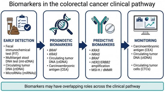
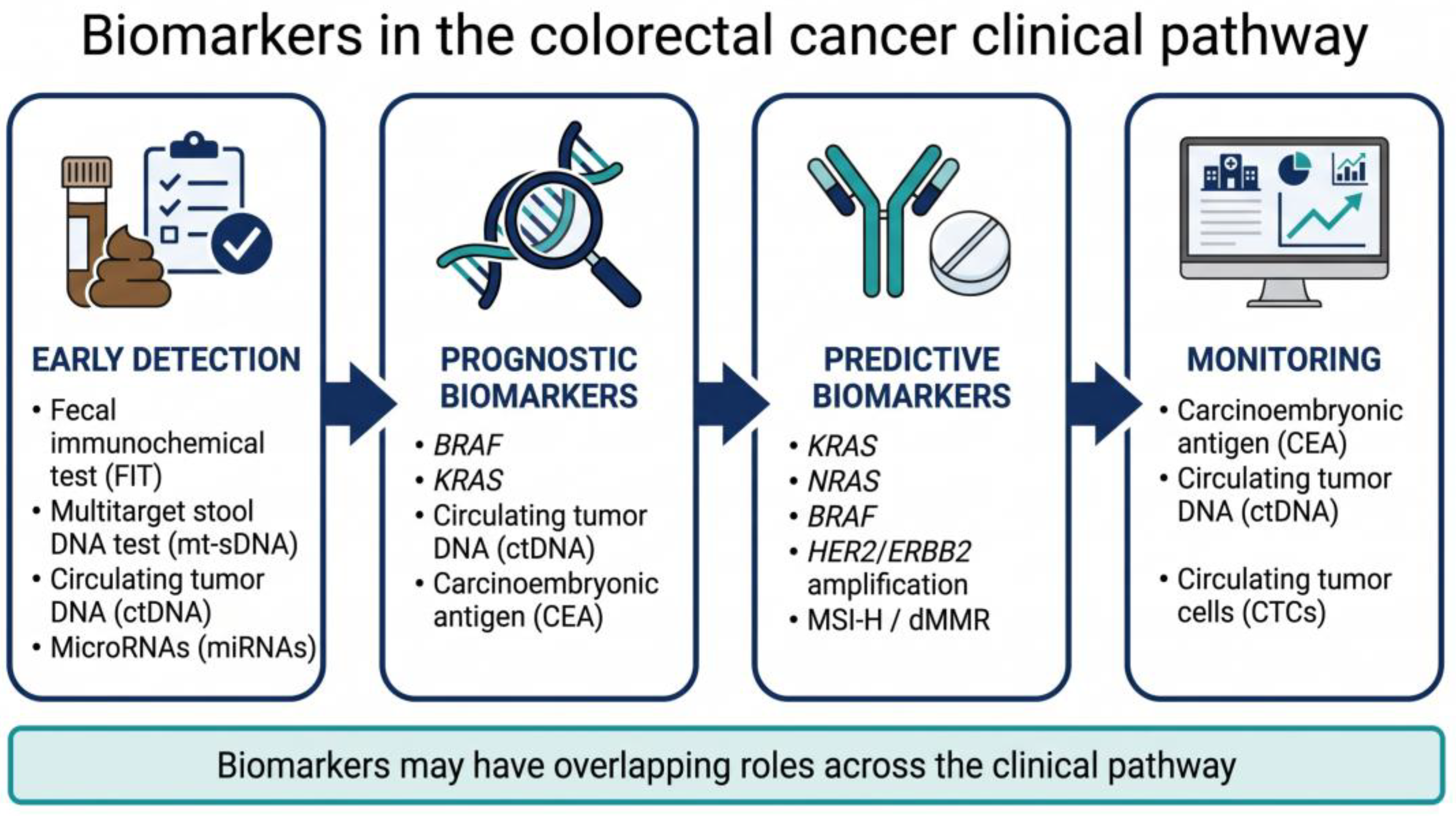
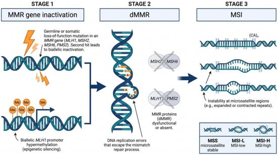
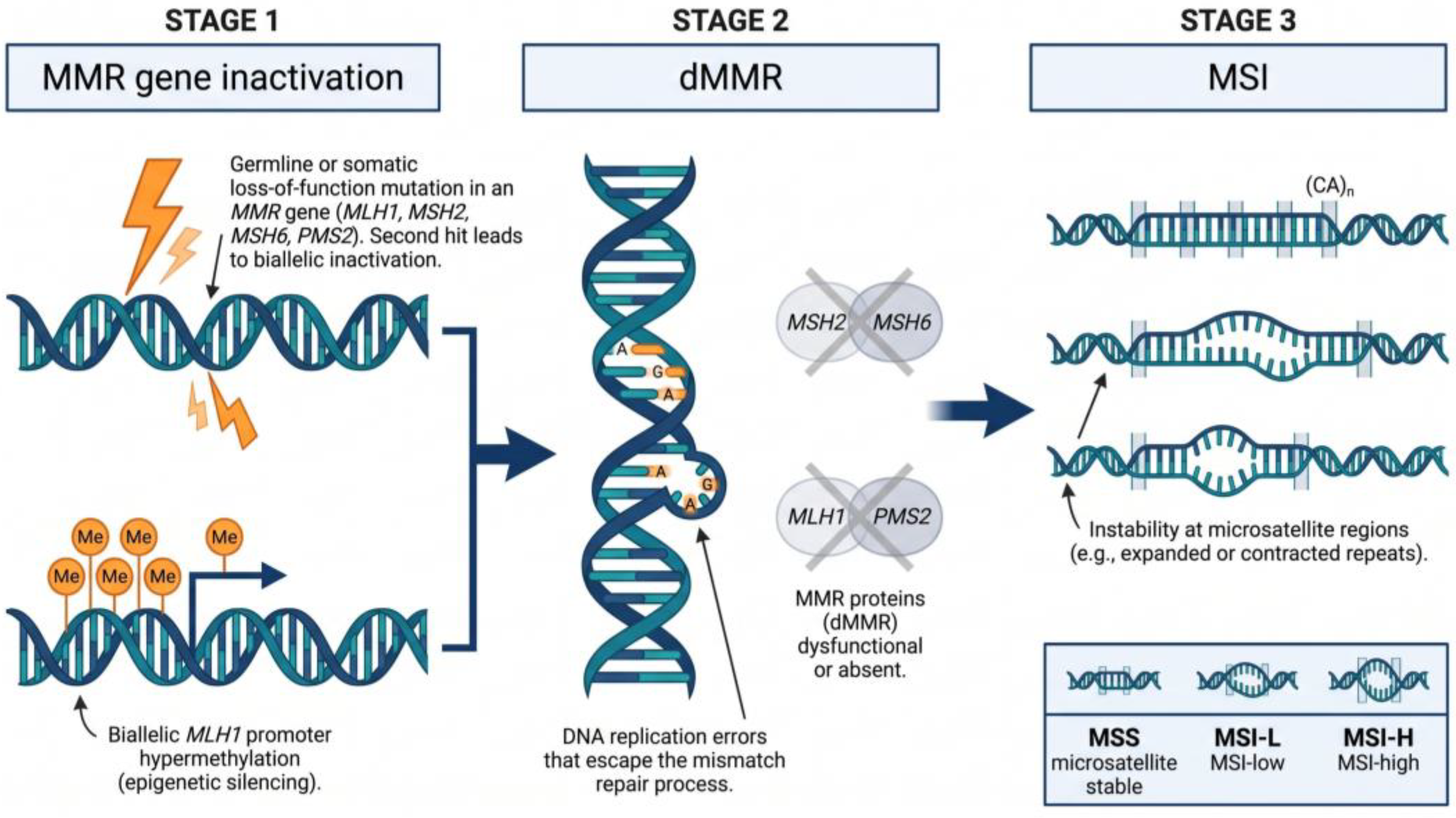
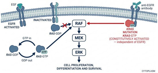
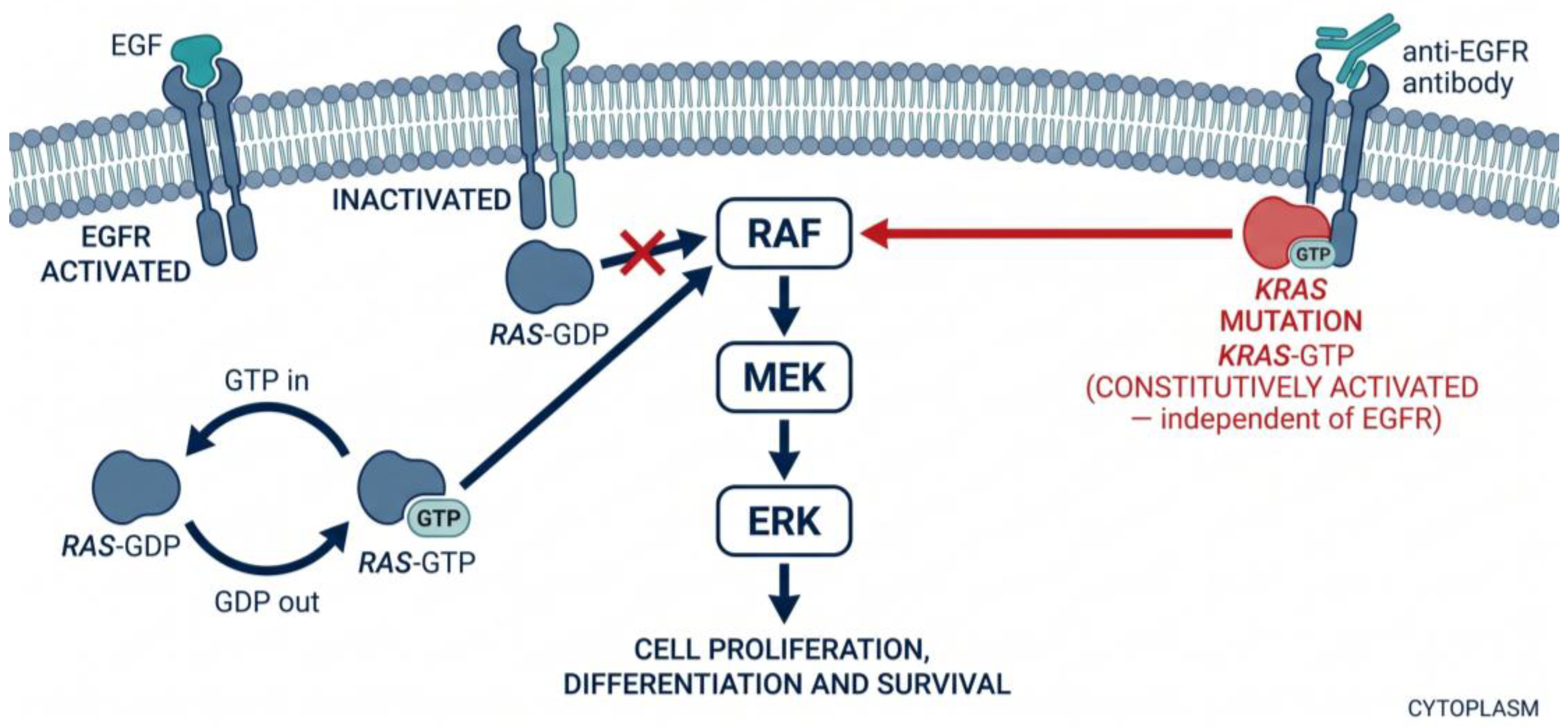
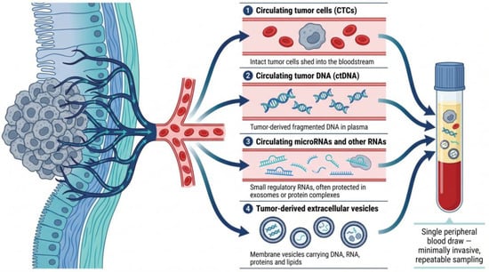
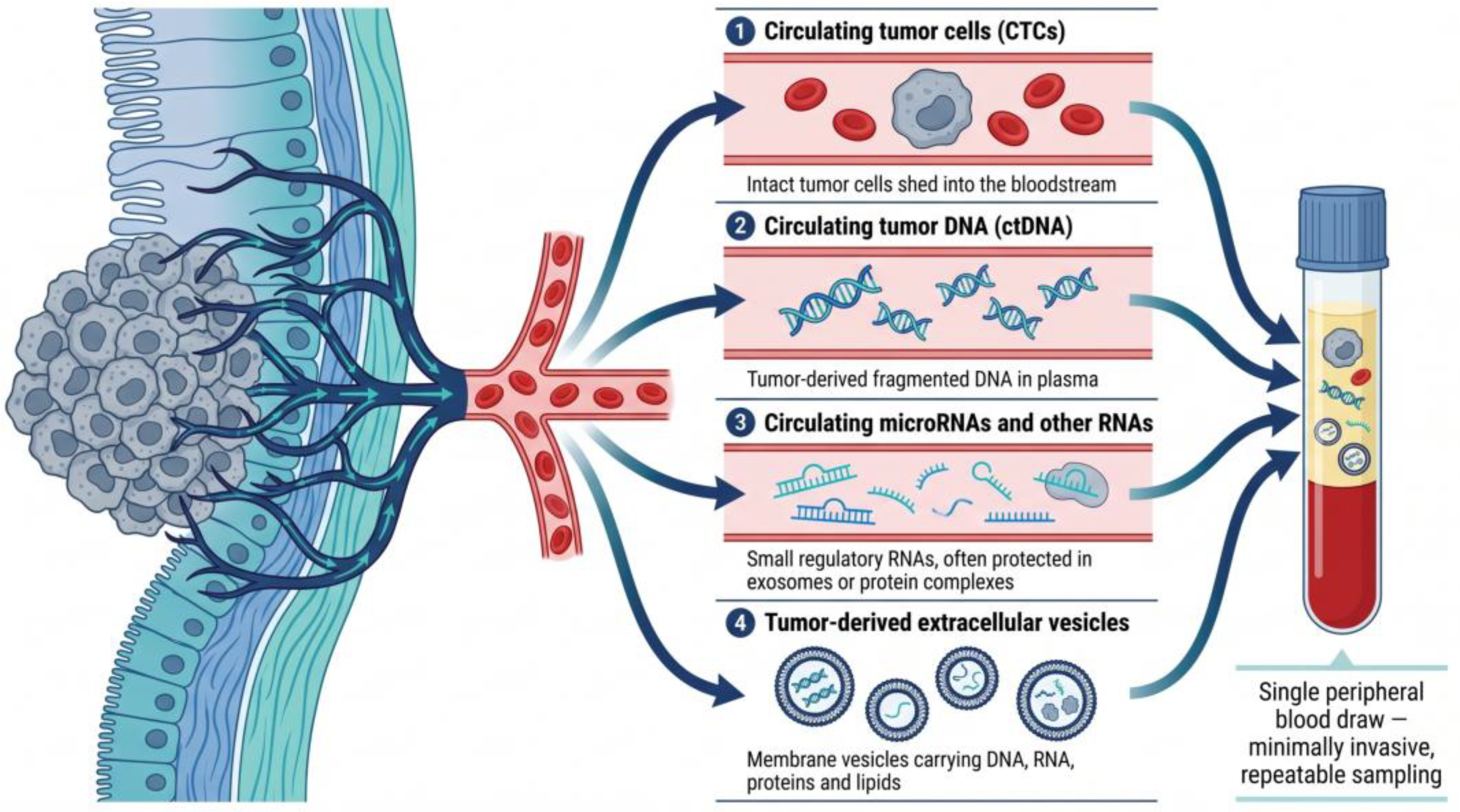

Disclaimer/Publisher’s Note: The statements, opinions and data contained in all publications are solely those of the individual author(s) and contributor(s) and not of MDPI and/or the editor(s). MDPI and/or the editor(s) disclaim responsibility for any injury to people or property resulting from any ideas, methods, instructions or products referred to in the content. |
© 2026 by the authors. Licensee MDPI, Basel, Switzerland. This article is an open access article distributed under the terms and conditions of the Creative Commons Attribution (CC BY) license.