


Highlights
What are the main findings?
- Fibroblast-derived PLA2G12A promotes IgE/antigen-dependent mast cell activation and anaphylactic responses in vivo.
- PLA2G12A hydrolyzes phospholipids in the extracellular vesicle-enriched fraction to generate lysophospholipids that potentiate mast cell activation.
What are the implications of the main findings?
- Secreted phospholipase A2-driven extracellular phospholipid remodeling represents a mechanism for stromal regulation of mast cell responsiveness.
- Extracellular vesicle-enriched fractions contain potential phospholipid substrates for PLA2G12A.
Abstract
Extracellular vesicles (EVs) mediate intercellular communication, yet whether extracellular phospholipid metabolism on EV membranes regulates immune activation remains incompletely understood. Through comparative phenotypic analyses of multiple secreted phospholipase A2 (sPLA2)-deficient mouse strains, we identified PLA2G12A as a stromal regulator of extracellular lipid metabolism that promotes IgE/antigen-dependent mast cell activation. Pla2g12a deficiency attenuated passive cutaneous and systemic anaphylaxis and selectively impaired IgE-dependent, but not IgE-independent, mast cell responses. Single-cell RNA-sequencing of mouse and human skin revealed that PLA2G12A is expressed in broad subsets of stromal fibroblasts, including papillary fibroblasts adjacent to mast cells, implicating a specialized stromal niche in mast cell regulation. Consistently, mast cell-intrinsic PLA2G12A was dispensable, whereas fibroblast-targeted deletion of Pla2g12a phenocopied global Pla2g12a deficiency. Lipidomic analyses during IgE-mediated anaphylaxis revealed a significant reduction in lysophospholipids, accompanied by minimal changes in oxylipins, in Pla2g12a-deficient skin. Mechanistically, fibroblast-derived PLA2G12A hydrolyzed phospholipids in EV-enriched fractions to generate multiple lysophospholipids, and treatment with either the PLA2G12A-treated EV-enriched fraction or exogenous lysophospholipids restored impaired IgE-dependent anaphylactic responses in Pla2g12a-deficient mice. Collectively, these findings establish sPLA2-driven extracellular phospholipid remodeling as a mechanism by which stromal niches regulate allergic inflammation.
1. Introduction
Allergic diseases, including asthma and atopic dermatitis, have become increasingly prevalent in recent decades. Environmental and lifestyle factors that disrupt epithelial barrier integrity and immune homeostasis promote IgE production and the activation of mast cells (MCs) via crosslinking of the high-affinity IgE receptor FcεRI by IgE and antigen (Ag) [1]. As tissue-resident immune cells, MCs orchestrate immediate hypersensitivity reactions and contribute to type 2 inflammatory diseases [2]. In addition to IgE/Ag-dependent FcεRI signaling, MC activation is profoundly influenced by the tissue microenvironment, which comprises stromal cells, extracellular matrix components, cytokines and lipid mediators [3,4]. Increasing evidence indicates that the stromal niche actively regulates MC activation and responsiveness in peripheral tissues.
Extracellular vesicles (EVs) have emerged as important mediators of intercellular communication in tissue microenvironments. EVs transport proteins, nucleic acids, and lipids that influence the behavior of recipient cells [5]. Since MCs rapidly release EVs upon IgE/Ag stimulation, EV-mediated signaling has been thought to modulate allergic responses [6,7]. Beyond their role as carriers of protein and nucleic acid cargos, EV membranes are enriched in phospholipids that can serve as substrates for extracellular lipid-metabolizing enzymes [8,9]. Recent studies have revealed that EV-associated phospholipids are not merely structural components of vesicle membranes but also serve as precursors for active mediators of intercellular communication. Extracellular hydrolysis of EV phospholipids generates bioactive lipid mediators, including lysophospholipids and fatty acid-derived metabolites (oxylipins), which ultimately regulate diverse biological processes such as immune and inflammatory responses, tumor progression, MC maturation, and pathogenic T cell differentiation [10,11,12,13,14,15].
Lipid mediators are critically involved in the regulation of MC biology and allergic inflammation. Upon activation, MCs produce arachidonic acid (AA)-derived oxylipins called eicosanoids, including prostaglandin D2 (PGD2) and leukotriene C4 (LTC4), which amplify type 2 immune responses and allergic inflammation [16,17,18]. Phospholipase A2 (PLA2) enzymes hydrolyze membrane phospholipids at the sn-2 position to release free fatty acids and lysophospholipids, thereby initiating diverse lipid signaling pathways [19,20]. The production of these eicosanoids in activated MCs is initiated by cytosolic phospholipase A2α (cPLA2α; PLA2G4A), which selectively hydrolyzes AA-containing phospholipids to supply the substrate for eicosanoid biosynthesis [20,21,22,23]. In addition, MCs express platelet-activating factor acetylhydrolase II (PAFAH2; PLA2G7B), an intracellular PLA2 preferentially hydrolyzing oxidized phospholipids to liberate ω3 polyunsaturated fatty acid (PUFA)-derived epoxy lipids, which optimize FcεRI-dependent MC activation and immediate allergic responses [24]. Beyond serving as a source of lipid mediators, MCs are themselves regulated by diverse extracellular lipid signals, including PUFAs, lysophospholipids, sphingolipids, and their metabolites, which control multiple aspects of MC biology, including homing, maturation and activation [12,25,26,27,28,29].
Among the secreted phospholipase A2 (sPLA2) family, which comprises 11 isoforms with distinct tissue distributions and substrate preferences, we have identified group III sPLA2 (PLA2G3), the sole mammalian homolog of bee venom PLA2, as a critical regulator of MC maturation [12,18,29,30,31]. MC-secreted PLA2G3 acts on EV membrane phospholipids and, through functional coupling with neighboring fibroblasts, promotes fibroblast-dependent lysophosphatidic acid (LPA) signaling, thereby driving MC maturation and associated anaphylactic responses [12,29,31]. These findings highlight that sPLA2-driven extracellular phospholipid metabolism is an important regulator of allergic inflammation. Notably, several sPLA2 isoforms, when overexpressed or added exogenously at high concentrations, have been reported to promote MC activation in vitro [29,32,33,34]. However, beyond the regulation of MC maturation by the sPLA2-III–EV axis as mentioned above [12,29,31], it remains unclear whether extracellular lipid metabolism mediated by endogenous sPLA2s would contribute to MC activation in vivo. Furthermore, although several lysophospholipids, such as lysophosphatidylserine (LysoPS), have been shown to potentiate FcεRI-dependent MC degranulation [25,35], it remains unknown whether sPLA2-dependent lysophospholipid production constitutes a physiological pathway regulating MC activation, and if so, which sPLA2 isoform and lysophospholipid species are responsible for this process. In particular, whether EV membrane phospholipids could serve as functional substrates for an endogenous sPLA2 to regulate MC activation during allergic responses has not yet been explored.
Here, we identify PLA2G12A, an atypical sPLA2 isoform [36,37], as a stromal regulator of IgE-dependent MC activation, but not MC maturation. Through comprehensive phenotypic analyses of multiple sPLA2-deficient mouse strains, followed by lipidomics, EV reconstitution, and fibroblast-targeted gene deletion, we provide evidence that fibroblast-derived PLA2G12A facilitates IgE-dependent MC activation and immediate hypersensitivity responses through extracellular phospholipid remodeling.
2. Materials and Methods
2.1. Mice
Various PLA2-deficient mouse strains on a C57BL/6 background have been described previously [14,38,39,40,41,42,43,44]. MC-deficient KitW-sh/W-sh mice (JAX stock #005051) were purchased from the Jackson Laboratory (Bar Harbor, ME, USA) [45]. Pla2g12a floxed mice [46] were crossed with Col1a2-Cre transgenic mice (Resource No. nbio228; Laboratory Animal Resource Bank, National Institutes of Biomedical Innovation, Health and Nutrition, Ibaraki, Osaka, Japan) [47] to generate fibroblast-targeted Pla2g12a conditional knockout mice (Pla2g12afl/fl Col1a2-Cre). Male mice aged 8–12 weeks were used in all experiments, except for an additional experiment performed using female mice of the same age range. Mice were housed under specific pathogen-free conditions on a 12-h light-dark cycle with free access to food and water. Animal experiments were performed at the Tokyo Metropolitan Institute of Medical Science and the University of Tokyo. All animal experiments were approved by the institutional animal care and use committees and conducted in accordance with the Japanese Guide for the Care and Use of Laboratory Animals.
2.2. Anaphylaxis
IgE-dependent passive cutaneous (PCA) or systemic (PSA) anaphylaxis induced by Ag and IgE-independent cutaneous anaphylaxis induced by compound 48/80 (C48/80) were performed as described previously [12,29,31]. For PCA, mice were sensitized by intradermal injection of anti-DNP IgE (SPE-7, Sigma-Aldrich, St. Louis, MO, USA; 30 ng) into the ear pinna. Twenty-four hours later, mice were challenged intravenously with dinitrophenylated human serum albumin (DNP-HSA, Sigma-Aldrich; 60 µg) and Evans blue (FUJIFILM Wako, Osaka, Japan; 1 mg per mouse). For IgE-independent cutaneous anaphylaxis, C48/80 (Sigma-Aldrich; 250 ng) was injected intradermally into the ear, followed immediately by intravenous Evans blue administration. Ear swelling was assessed by measuring ear thickness before and 30 min after challenge using a micrometer (Mitutoyo, Kawasaki, Japan). Vascular permeability was quantified by spectrophotometric measurement at 620 nm of Evans blue extracted from IgE-sensitized or phosphate-buffered saline (PBS)-treated ears collected 30 min after Ag challenge. For PSA, mice were sensitized intravenously with anti-DNP IgE (3 µg per mouse) and challenged the following day with DNP-HSA (500 µg per mouse). Rectal temperature was measured over time using a digital thermometer with a rectal probe (Physitemp Instruments, Clifton, NJ, USA).
2.3. Measurement of Histamine
Histamine levels in sera and tissues were measured by LC-MS/MS with minor modifications of a previously described method [12]. Tissues were homogenized using a Precellys Homogenizer (Bertin Technologies, Montigny-le-Bretonneux, France) in Hank’s balanced salt solution (FUJIFILM Wako) containing 0.5% (v/v) Triton X-100, followed by perchloric acid extraction. Serum samples were mixed with methanol and centrifuged to remove precipitated proteins. Deuterium-labeled histamine·2HCl (α,α,β,β-D4) (Cambridge Isotope Laboratories, Tewksbury, MA, USA) was used as an internal standard for quantification.
2.4. Measurement of MC Protease Activities
MC protease activity in tissue extracts was measured as described previously [48]. Tissue extracts prepared for histamine measurement (see above) were used to evaluate protease activity. Chymotrypsin-like, trypsin-like, and carboxypeptidase A (CPA) activities were measured using the chromogenic substrates S-2586 (MeO-Suc-Arg-Pro-Tyr-pNA; DiaPharma, West Chester, OH, USA), S-2288 (H-D-Ile-Pro-Arg-pNA-2HCl; DiaPharma), and M-2245 (N-(4-methoxyphenylazoformyl)-Phe-OH potassium salt; Bachem, Bubendorf, Switzerland), respectively. Absorbance at 405 nm was measured before and after incubation at 37 °C, and protease activities were calculated from the change in absorbance per minute. Each activity was normalized to total protein quantity.
2.5. Histology
Ear pinnae were collected and fixed in 10% (v/v) Neutral Buffered Formalin (FUJIFILM Wako). Ear tissues were embedded in paraffin using an automated tissue processor (Tissue-Tek VIP 5 Jr; Sakura Finetek Japan, Tokyo, Japan) and sectioned at 4 μm using a sliding microtome (REM-710; YAMATO KOHKI, Niiza, Saitama, Japan). The sections were stained with 0.05% (w/v) toluidine blue (pH 0.5) (FUJIFILM Wako), and images were acquired using an All-in-One Fluorescence Microscope (BZ-X710; KEYENCE, Osaka, Japan). Toluidine blue-positive metachromatic cells were quantified as MCs.
2.6. Preparation, Maturation, and Activation of Bone Marrow-Derived Mast Cells
Bone marrow-derived mast cells (BMMCs) were generated and activated with minor modifications of a previously described method [12,29,49]. Briefly, bone marrow cells isolated from mouse femurs were cultured in Dulbecco’s modified Eagle’s medium (DMEM) containing 10% (v/v) fetal bovine serum (FBS; Biosera, Cholet, France) and baculovirus-derived recombinant mouse IL-3 (10 ng/mL) [50]. For maturation into connective tissue-type mast cell (CTMC)-like cells, BMMCs were cocultured with Swiss 3T3 fibroblasts (JCRB9019, Japanese Collection of Research Bioresources, Ibaraki, Osaka, Japan) in the presence of stem cell factor (SCF, 100 ng/mL; PeproTech, Cranbury, NJ, USA) for 4 days, with half of the culture medium replaced every 2 days. MCs were then separated from fibroblasts by replating or magnetic sorting using anti-CD117 (c-Kit) microbeads (Miltenyi Biotec, Bergisch Gladbach, Germany). MC maturation was assessed by histamine content, expression of mast cell marker genes, and cell-surface expression of FcεRIα and c-Kit. For activation assays, BMMCs (106 cells) were sensitized with anti-DNP IgE (1 µg/mL) for 2 h, washed, and stimulated with DNP-BSA (100 ng/mL; Sigma-Aldrich) at 37 °C. Alternatively, the cells were stimulated with C48/80 (10 µg/mL).
2.7. β-Hexosaminidase Release Assay
IgE-sensitized BMMCs were stimulated with DNP-BSA at 37 °C for 10 min. Culture supernatants and cell pellets were collected separately, and β-hexosaminidase activity was measured using p-nitrophenyl N-acetyl-β-D-glucosaminide (FUJIFILM Wako) as a substrate. β-Hexosaminidase release was calculated as S/(S + P) × 100, where S and P represent β-hexosaminidase activities in the culture supernatant and the cell pellet fractions, respectively.
2.8. Measurement of Lipid Mediators and Cytokines
IgE-sensitized BMMCs were stimulated with DNP-BSA at 37 °C. For lipid mediator analysis, culture supernatants were collected after 10 min, and PGD2 and LTC4 levels were measured using the PGD2-MOX ELISA Kit and LTC4 ELISA Kit (both from Cayman Chemical, Ann Arbor, MI, USA), respectively. For cytokine analysis, culture supernatants were collected after 4 h, and TNF-α levels were quantified using a TNF-α ELISA Kit (R&D Systems, Minneapolis, MN, USA) according to the manufacturer’s instructions.
2.9. Reverse Transcription-Quantitative PCR (RT-qPCR)
For stimulation experiments, IgE-sensitized BMMCs were stimulated with DNP-BSA at 37 °C for 30 min before RNA isolation. For coculture experiments, BMMCs were cocultured with Swiss 3T3 fibroblasts in the presence of SCF for 4 days as described above. Total RNA was extracted from tissues and cultured cells using TRIzol Reagent (Invitrogen, Carlsbad, CA, USA). cDNA was synthesized using the High-Capacity cDNA Reverse Transcription Kit (Applied Biosystems, Foster City, CA, USA). Quantitative PCR (qPCR) was performed using the TaqMan Gene Expression Master Mix and predesigned TaqMan probe-primer assays on the StepOnePlus Real-Time PCR System (Applied Biosystems). Detailed information on the primer-probe sets used is provided in Table S1. Gene expression levels were normalized to Gapdh (Mouse GAPDH Endogenous Control; Applied Biosystems) using the 2−ΔΔCt method and are presented relative to the control group. Where indicated, mast cell-associated genes were further normalized to Kit expression to account for differences in MC abundance.
2.10. MC Reconstitution
BMMCs (106 cells) were injected intradermally into the ears of 6-week-old MC-deficient KitW-sh/W-sh mice for MC reconstitution [12,29,31,45]. Mice were analyzed 6 weeks after cell transfer.
2.11. Preparation of Single-Cell Suspensions from Ear Skin
Ear skin was minced and treated with Accutase (Innovative Cell Technologies, San Diego, CA, USA), followed by digestion with Liberase TM (250 µg/mL; Roche, Mannheim, Germany) and DNase I (2 Kunitz units/mL; Sigma-Aldrich) in RPMI-1640 containing 10 mM HEPES and 5% FBS. Liberase/DNase digestion was performed twice. Cells were enriched by 25% (v/v) Percoll density-gradient centrifugation and passed through a 40-µm cell strainer to obtain single-cell suspensions. Single-cell suspensions were used for flow cytometry, RT-qPCR, or CD140a+ cell enrichment using the mouse CD140a (PDGFRα) MicroBead Kit (Miltenyi Biotec, Bergisch Gladbach, Germany).
2.12. Flow Cytometry
Tissue-derived single-cell suspensions and cultured BMMCs were resuspended in a FACS buffer consisting of Ca2+/Mg2+-free PBS containing 1% (w/v) BSA and 2 mM EDTA. Cells were incubated with TruStain FcX (anti-mouse CD16/32; BioLegend, San Diego, CA, USA) on ice for 10 min prior to antibody staining. The cells were then stained with fluorochrome-conjugated monoclonal antibodies against mouse CD117 (c-Kit) FITC (2B8; BD Pharmingen, San Diego, CA, USA), FcεRIα PE (MAR-1; eBioscience, San Diego, CA, USA), and CD45 Brilliant Violet 421 (30-F11; BioLegend). Corresponding isotype controls were used where indicated. Flow cytometry was performed using a FACSMelody Cell Sorter (BD Biosciences, San Jose, CA, USA), and data were analyzed using FlowJo v10.10.1 software (BD Biosciences). MCs were defined as CD45+CD117+FcεRIα+ cells in skin-derived single-cell suspensions and as CD117+FcεRIα+ cells in cultured BMMCs.
2.13. Single-Cell RNA-Sequencing (scRNA-Seq) Analysis
The mouse skin scRNA-seq dataset generated in our previous study [12] and deposited in Gene Expression Omnibus (GEO) under accession number GSE262558 was reanalyzed to assess Pla2g12a expression across skin cell populations. Fibroblast subclustering and visualization were performed using Seurat (v4.2.1). A published human skin scRNA-seq dataset (GSE241132) [51,52] was reanalyzed to examine PLA2G12A expression in fibroblast subsets. Fibroblasts were isolated, subclustered, and annotated on the basis of canonical marker-gene expression using Scanpy (v1.11.3).
2.14. Preparation of the EV-Enriched Fraction
EV-enriched fractions were prepared from fibroblast–MC coculture supernatants as described previously [12,53]. Because fibroblast–BMMC coculture in the presence of SCF recapitulates the niche environment that supports CTMC maturation and activation [4,12,54], EV-enriched fractions were prepared directly from coculture supernatants without additional IgE/Ag stimulation. Briefly, conditioned media were collected after 2 days of culture, filtered through 0.22-μm PVDF filters (Merck Millipore, Burlington, MA, USA), and concentrated using Vivaspin 20 Centrifugal Concentrators (Sartorius, Göttingen, Germany). Concentrated samples were mixed with ExoQuick-TC reagent (System Biosciences, Palo Alto, CA, USA) and incubated overnight at 4 °C. EV-enriched fractions were recovered by centrifugation at 6000× g for 30 min, washed once with Ca2+/Mg2+-free PBS, and collected by an additional centrifugation step at 6000× g for 15 min. Pellets were resuspended in the indicated buffer and subjected to subsequent analyses. As this isolation procedure relies on polymer-based precipitation rather than classical density-gradient or size-exclusion purification, the resulting material is referred to as the EV-enriched fraction throughout this study.
2.15. PLA2 Assay Using the EV-Enriched Fraction as a Substrate
For PLA2 reactions, the EV-enriched fraction corresponding to 2 × 10⁶ BMMC equivalents was incubated with recombinant human PLA2G12A protein, generously provided by Dr. Gérard Lambeau (Centre National de la Recherche Scientifique (CNRS), France), in reaction buffer containing 100 mM Tris-HCl (pH 7.5) and 5 mM CaCl2 in a total volume of 150 µL. After incubation at 37 °C for 30 min, reactions were terminated by the addition of methanol containing internal standards. Lipids were extracted by the Bligh and Dyer method [55] and analyzed by liquid chromatography–tandem mass spectrometry (LC-MS/MS) as previously described [12] using a 4000 QTRAP triple quadrupole linear ion trap mass spectrometer (SCIEX, Marlborough, MA, USA) coupled to a Nexera UPLC system (Shimadzu, Kyoto, Japan). Phospholipids, lysophospholipids, and free fatty acids were quantified by multiple reaction monitoring.
2.16. In Vivo Rescue Experiments with the PLA2G12A-Treated EV-Enriched Fraction and Lysophospholipids
For in vivo rescue experiments, the EV-enriched fraction was incubated with recombinant mouse PLA2G12A (300 ng/mL), and 30 µL of the preparation containing 8 or 18 µg of the EV-enriched fraction was administered intradermally into the ear immediately before Ag challenge. Since PLA2G12A was not removed or inactivated before administration and was therefore carried over, equivalent amounts of PLA2G12A alone and the EV-enriched fraction without PLA2G12A pretreatment were injected into the ear as controls. For rescue experiments using pure lysophospholipids, each of lysophosphatidylcholine (LPC), lysophosphatidylethanolamine (LPE), LysoPS, or LPA, all having an 18:1 acyl chain at the sn-1 position (Avanti Polar Lipids, Alabaster, AL, USA), was administered intradermally into the ear at doses of 0, 15, 150, or 300 pmol per ear immediately before Ag challenge.
2.17. Lipidomic Analysis
For in vivo lipidomic analysis, ear skin samples were collected from Pla2g12a+/+ and Pla2g12a−/− mice before and 2 min after Ag challenge in the PCA model. Individual molecular species of LPC, LPE, LysoPS, LPA, LPG, and LPI were quantified by LC–MS/MS, as described previously [56]. Free fatty acid and oxylipin analyses were performed by the Laboratory for Metabolomics, RIKEN Center for Integrative Medical Sciences, using previously published methods [57,58].
2.18. Statistical Analysis
Statistical analyses were performed using GraphPad Prism software (v11.1.0; GraphPad Software, Boston, MA, USA). Data are presented as mean ± s.e.m. or as indicated in the figure legends. Animals were allocated to experimental groups without randomization, and investigators were not blinded during outcome assessment unless otherwise indicated. Comparisons between two groups were performed using an unpaired two-tailed Student’s t-test or Mann–Whitney U test, as appropriate. Comparisons among multiple groups were performed using ordinary one-way or two-way ANOVA followed by Šídák’s or Tukey’s multiple-comparisons test, or Brown–Forsythe and Welch ANOVA followed by Dunnett’s T3 multiple-comparisons test, as indicated in the figure legends. For lipidomic analyses involving multiple lipid species, p values were adjusted for multiple testing using the Benjamini–Hochberg false discovery rate (FDR) procedure, as indicated in the figure legends. A p value < 0.05 was considered statistically significant.
3. Results
3.1. PLA2G12A Promotes IgE/Ag-Dependent MC Activation In Vivo
To assess whether sPLA2 isoforms would be involved in IgE/Ag-dependent MC activation in vivo, we performed PCA-based phenotypic screening using multiple sPLA2-deficient mouse strains, with Evans blue extravasation as a readout. In addition to the expected reduction in dye extravasation in Pla2g3−/− mice, consistent with the established role of PLA2G3 in MC maturation [12,29,31], Pla2g12a−/− mice, but not mice lacking other sPLA2s (Pla2g1b−/−, Pla2g2d−/−, Pla2g2e−/−, Pla2g2f−/−, Pla2g5−/− and Pla2g10−/−), also exhibited a markedly attenuated PCA response (Figure 1A). IgE/Ag-induced Evans blue extravasation was significantly reduced in male Pla2g12a−/− mice compared with littermate Pla2g12a+/+ controls (Figure 1B), as shown in a representative gross image of the ears 30 min after Ag challenge (Figure S1A). Female Pla2g12a−/− mice also exhibited an attenuated PCA response compared with Pla2g12a+/+ mice (Figure S1B). Histological analysis of toluidine blue-stained ear sections revealed that, despite comparable dermal MC abundance between Pla2g12a+/+ and Pla2g12a−/− mice, IgE/Ag-induced MC degranulation was significantly reduced in Pla2g12a−/− mice (Figure 1C,D), indicating that PLA2G12A deficiency impairs MC activation without altering MC abundance. We next assessed systemic anaphylactic responses using an IgE-mediated PSA model. Following systemic Ag challenge, IgE-sensitized Pla2g12a−/− mice exhibited attenuated hypothermia and reduced serum histamine levels compared with littermate Pla2g12a+/+ mice (Figure 1E,F), indicating an attenuated systemic anaphylactic response. By contrast, IgE-independent dye extravasation induced by C48/80, which acts through the G protein-coupled receptor MRGPRB2 [59], was preserved in Pla2g12a−/− mice (Figure 1G), suggesting that PLA2G12A preferentially contributes to IgE/Ag-dependent MC activation. Unlike Pla2g3 deficiency, which impaired MC maturation [12,29,31], dermal MCs in Pla2g12a−/− mice exhibited normal expression of c-Kit, FcεRIα, and several other canonical MC marker genes, together with unaltered tissue histamine content and MC protease activities (Figure 1H–K and Figure S2), indicating that Pla2g12a deficiency does not influence MC maturation. Collectively, these findings support a role for PLA2G12A in amplifying FcεRI-dependent MC activation and anaphylactic responses in vivo, without affecting MC abundance, maturation, or MRGPRB2-dependent responses.
Figure 1.
PLA2G12A deficiency attenuates IgE/Ag-dependent MC activation and anaphylactic responses in vivo. (A) Relative IgE/Ag-induced PCA responses in mouse strains deficient in individual sPLA2 isoforms. Vascular permeability was assessed by Evans blue extravasation. Colors indicate the responses of individual knockout mice (n = 3 per genotype) relative to those of littermate controls (+/+), which were set to 100%. (B) Evans blue extravasation under IgE/Ag (–) and IgE/Ag (+) PCA conditions in Pla2g12a+/+ and Pla2g12a−/− mice. (C) Representative toluidine blue-stained ear sections from Pla2g12a+/+ and Pla2g12a−/− mice under IgE/Ag (–) and IgE/Ag (+) conditions. Blue and red arrowheads indicate intact and degranulated MCs, respectively. Insets show higher-magnification views. Scale bar, 25 µm. (D) Quantification of total (left) and degranulated (right) MCs in ear sections shown in (C). (E,F) IgE/Ag-induced PSA in Pla2g12a+/+ and Pla2g12a−/− mice. Changes in rectal temperature following Ag challenge (E) and serum histamine levels (F) are shown. n = 5 per genotype for IgE/Ag (–) and n = 9 per genotype for IgE/Ag (+) (E). (G) Evans blue extravasation in C48/80-induced cutaneous anaphylaxis in Pla2g12a+/+ and Pla2g12a−/− mice. (H) Flow cytometric analysis of dermal MCs from ear skin of Pla2g12a+/+ and Pla2g12a−/− mice. Representative contour plots (left) and quantification of surface c-Kit and FcεRIα expression (right) are shown. (I) Relative expression of mast cell marker genes in ear tissues from Pla2g12a+/+ and Pla2g12a−/− mice. Expression levels in Pla2g12a+/+ mice were set to 1. (J,K) Histamine content (J) and chymotrypsin-like, trypsin-like, and CPA activities (K) in ear tissue extracts from Pla2g12a+/+ and Pla2g12a−/− mice. Data are presented as mean ± s.e.m. Representative images from two independent experiments are shown in (C). Data are pooled from one (H–K), two (D) or three (A,B,E–G) independent experiments. Statistical analysis was performed using two-tailed Mann–Whitney U test (A,H–K), Brown–Forsythe and Welch ANOVA followed by Dunnett’s T3 multiple comparisons test (B,D,F,G), or two-way ANOVA followed by Tukey’s multiple comparisons test (E). **, p < 0.01; NS, not significant.
3.2. Fibroblast-Derived PLA2G12A Augments IgE/Ag-Dependent MC Activation In Vivo
We next examined whether PLA2G12A regulates MC function in a cell-autonomous manner. BMMCs prepared from Pla2g12a−/− mice proliferated normally and expressed comparable levels of c-Kit and FcεRIα to those from Pla2g12a+/+ mice (Figure 2A,B). IgE/Ag-induced degranulation, production of lipid mediators (PGD2 and LTC4), and secretion of TNF-α were comparable between Pla2g12a+/+ and Pla2g12a−/− BMMCs (Figure 2C–F). Likewise, induction of inflammatory gene expression (e.g., Tnf, Il6, Ccl2, and Ccl3) following IgE/Ag stimulation was unaffected by Pla2g12a deficiency (Figure 2G,H). Moreover, Pla2g12a−/− BMMCs underwent normal maturation following coculture with Swiss3T3 fibroblasts, as assessed by the expression of maturation-inducible genes, including Hdc, Mcpt4, and Hpgds (Figure 2I) [12,29]. These results indicate that MC-intrinsic PLA2G12A is dispensable for both MC activation and maturation.
Figure 2.
PLA2G12A deficiency does not impair MC activation or fibroblast-dependent maturation in vitro. (A) IL-3-dependent proliferation of Pla2g12a+/+ and Pla2g12a−/− BMMCs during in vitro culture. (B) Flow cytometric analysis of c-Kit and FcεRIα expression in Pla2g12a+/+ and Pla2g12a−/− BMMCs. (C–F) IgE/Ag-induced activation of Pla2g12a+/+ and Pla2g12a−/− BMMCs. β-hexosaminidase (β-HEX) release (C), PGD2 production (D), LTC4 production (E), and TNF-α secretion (F) were assessed with or without IgE/Ag stimulation. (G,H) RT-qPCR analysis of cytokine- and chemokine-associated genes in IgE/Ag-stimulated Pla2g12a+/+ and Pla2g12a−/− BMMCs. Expression levels were normalized to unstimulated Pla2g12a+/+ BMMCs, which were set to 1. (I) RT-qPCR analysis of MC maturation-associated genes in Pla2g12a+/+ and Pla2g12a−/− BMMCs cultured for 4 days with or without Swiss 3T3 fibroblasts in the presence of SCF. Expression levels were normalized to immature Pla2g12a+/+ BMMCs, which were set to 1. Data are presented as mean ± s.e.m. Representative flow cytometry plots from one of two independent experiments are shown in (B). Quantitative data are pooled from two independent experiments. Statistical analysis was performed using a two-tailed Mann–Whitney U test (B), Brown–Forsythe and Welch ANOVA followed by Dunnett’s T3 multiple comparisons test (C–I), or two-way ANOVA followed by Šídák’s multiple comparisons test (A). **, p < 0.01.
These observations prompted us to investigate whether PLA2G12A expressed in non-MCs in tissue microenvironments would be responsible for the regulation of MC responses. Pla2g12a was the most abundant sPLA2 isoform in Swiss 3T3 fibroblasts (Figure 3A) and was expressed at higher levels than in BMMCs (Figure 3B). Notably, Pla2g12a expression in fibroblasts was further increased after coculture with BMMCs (Figure 3B). Consistent with this, Pla2g12a expression was enriched in CD140a+ dermal fibroblasts compared with CD140a− cells in mouse skin, where enrichment of the fibroblast marker Col1a1 in the CD140a+ fraction confirmed successful fibroblast isolation (Figure 3C). To identify the fibroblast population expressing Pla2g12a in vivo, we reanalyzed our previously reported mouse skin scRNA-seq dataset [12]. Pla2g12a was broadly expressed in various fibroblast subsets, including a papillary fibroblast subset (Fp) characterized by high expression of Kitl, Lpar1, and Enpp2, consistent with our previous identification of this niche-associated fibroblast population as a regulator of dermal MC maturation and activation (Figure 3D,E) [12]. Furthermore, reanalysis of a published human skin scRNA-seq dataset [51,52] revealed a similar expression pattern, with PLA2G12A broadly expressed across multiple fibroblast subsets, including a papillary fibroblast subset characterized by high expression of KITLG, LPAR1, and ENPP2 (Figure 3F,G). These findings indicate that PLA2G12A is broadly expressed across fibroblast subsets, including the MC-associated papillary fibroblast population, in both mouse and human skin.
Figure 3.
PLA2G12A is broadly expressed in various fibroblast subsets in mouse and human skin. (A) Relative mRNA expression of sPLA2 isoforms in Swiss 3T3 fibroblasts, as determined by RT-qPCR. Expression levels were normalized to Gapdh. (B) RT-qPCR analysis of Pla2g12a expression in BMMCs and Swiss 3T3 fibroblasts cultured alone or in coculture. Expression levels were normalized to Gapdh. (C) RT-qPCR analysis of Pla2g12a and the fibroblast marker Col1a1 in CD140a+ and CD140a− fractions isolated from mouse ear skin. Expression levels were normalized to Gapdh. (D) UMAP visualization of annotated fibroblast subsets from our previously reported mouse skin scRNA-seq data (GSE262558), together with feature plots showing expression of Pla2g12a, Kitl, Lpar1, and Enpp2. Fibroblast subsets are annotated as follows: Fp, papillary fibroblast; Fr, reticular fibroblast; iFr, activated inflammatory reticular dermal fibroblast; Fdp, follicular dermal papilla/arrector pili-associated fibroblast; Fds, follicular dermal sheath fibroblast; HpF, hypodermal preadipocyte-like fibroblast. (E) Dot plot showing expression of fibroblast subset marker genes across the mouse fibroblast subsets shown in (D). (F) UMAP visualization of fibroblast subsets from a published human skin scRNA-seq data (GSE241132), together with feature plots showing expression of PLA2G12A, KITLG, LPAR1, and ENPP2. (G) Dot plot showing expression of fibroblast subset marker genes across the human fibroblast subsets shown in (F). Data are presented as mean ± s.e.m. Data are pooled from one (D,E), two (A,C), or three (B) independent experiments. Human scRNA-seq data shown in (F,G) were generated by reanalyzing a published dataset. Statistical analysis was performed using two-tailed Student’s t-test (B). *, p < 0.05.
To distinguish the relative contributions of MC-intrinsic and stromal-derived PLA2G12A in vivo, we performed MC reconstitution experiments using KitW-sh/W-sh mice [45] crossed onto either Pla2g12a+/+ or Pla2g12a−/− background (Figure 4A). These recipient mice were engrafted with either Pla2g12a+/+ or Pla2g12a−/− BMMCs. Six weeks after engraftment, toluidine blue staining confirmed comparable dermal MC reconstitution across all groups (Figure 4B). In Pla2g12a+/+KitW-sh/W-sh recipients (+/+Wsh), transfer of Pla2g12a+/+ or Pla2g12a−/− BMMCs restored PCA responses to comparable levels, indicating that MC-intrinsic PLA2G12A is dispensable for IgE/Ag-dependent MC activation in vivo. By contrast, PCA responses were markedly reduced in Pla2g12a−/−KitW-sh/W-sh recipients (–/–Wsh) reconstituted with either Pla2g12a+/+ or Pla2g12a−/− BMMCs to a similar level (Figure 4C). These findings indicate that stromal PLA2G12A, rather than MC-intrinsic PLA2G12A, plays a dominant role in promoting IgE/Ag-dependent MC activation in vivo.
Figure 4.
Fibroblast-derived PLA2G12A promotes IgE/Ag-dependent MC activation in vivo. (A) Schematic overview of MC reconstitution experiments using Pla2g12a+/+KitW-sh/W-sh (+/+Wsh) and Pla2g12a−/−Wsh (–/–Wsh) recipient mice engrafted with Pla2g12a+/+ or Pla2g12a−/− BMMCs. Blue crosses indicate PLA2G12A deficiency. (B) Quantification of toluidine blue-positive dermal MCs in ear skin 6 weeks after BMMC engraftment. (C) IgE/Ag-induced PCA responses, as assessed by Evans blue extravasation, in MC-reconstituted mice. (D) RT-qPCR analysis of Pla2g12a expression in ear skin from Pla2g12afl/fl (fl/fl) and fibroblast-targeted Pla2g12a-deficient (F-KO) mice generated using Col1a2-Cre mice. Expression levels were normalized to Gapdh. (E) IgE/Ag-induced PCA responses, as assessed by Evans blue extravasation, in fl/fl and F-KO mice. Data are presented as mean ± s.e.m. Data are pooled from one (B), two (D) or three (C,E) independent experiments. Statistical analysis was performed using Mann–Whitney U test (D), or Brown–Forsythe and Welch ANOVA followed by Dunnett’s T3 multiple comparisons test (B,C,E). **, p < 0.01; NS, not significant.
To directly test the contribution of fibroblast-derived PLA2G12A, we generated fibroblast-targeted Pla2g12a-deficient mice by crossing Pla2g12afl/fl mice with Col1a2-Cre mice, in which Cre recombinase is expressed broadly in fibroblasts. Efficient deletion was confirmed by markedly reduced Pla2g12a expression in ear skin (Figure 4D). Furthermore, Col1a2-Cre-mediated deletion of Pla2g12a significantly attenuated PCA responses, phenocopying the defect observed in global Pla2g12a−/− mice (Figure 4E). Collectively, these findings demonstrate that fibroblast-derived PLA2G12A is mainly responsible for augmenting IgE/Ag-dependent MC activation in vivo.
3.3. Comprehensive Lipidomic Profiling Reveals PLA2G12A-Dependent Changes in Lysophospholipids During IgE/Ag-Induced Anaphylaxis
Given the emerging view that sPLA2s act on extracellular phospholipids to mobilize bioactive lipid mediators in tissue microenvironments, we attempted to identify such lipid mediators potentially regulated downstream of PLA2G12A during IgE/Ag-induced MC activation. To this end, we performed comprehensive lipidomic analyses of ear skin. Because the effects of Pla2g12a deficiency became evident only after Ag challenge, we focused our lipidomic analysis on the immediate phase of the PCA response. Lipids were extracted from ear skin just before or 2 min after Ag challenge, corresponding to the onset of rapid MC degranulation, and subjected to comprehensive analyses of lipid metabolites potentially involved in PLA2-dependent phospholipid metabolism, including lysophospholipids, free fatty acids, and their oxygenated metabolites (oxylipins). Volcano plots revealed an overall tendency toward lower levels of various lipid metabolites in Pla2g12a−/− mice than in Pla2g12a+/+ mice following IgE/Ag challenge (Figure 5A,B). Although several lipid species showed nominal differences between genotypes (p < 0.05), none remained significant after FDR correction for multiple testing. Among the oxylipins, several AA metabolites (eicosanoids), including the MC-derived lipid mediators PGD2 and LTD4, showed lower levels in Ag-challenged Pla2g12a−/− skin compared with Pla2g12a+/+ skin (Figure 5A). These changes, even though relatively modest, are consistent with the attenuated MC activation observed in Pla2g12a−/− mice and may contribute partly to the reduced vascular permeability during PCA. Compared to these oxylipins, various lysophospholipid species were more profoundly affected by PLA2G12A deficiency (Figure 5B). Heatmap analysis showed that Ag challenge induced coordinated increases in multiple lysophospholipid species in Pla2g12a+/+ mice, whereas these responses were broadly attenuated in Pla2g12a−/− mice (Figure 5C). Quantitative analyses further showed that, despite their high background levels in tissues, several LPC, LPE, and LPA species were elevated in Pla2g12a+/+ skin following Ag challenge, whereas these increases were attenuated in Pla2g12a−/− skin (Figure 5D). Among the other lysophospholipids, LysoPS 18:1 also tended to be lower in Pla2g12a−/− skin after and even before Ag challenge, while lysophosphatidylglycerol (LPG) and lysophosphatidylinositol (LPI) species were largely unaffected by Pla2g12a deficiency (Figure 5C,D). These results suggest that one or more of these lysophospholipids may act downstream of PLA2G12A during IgE/Ag-induced anaphylaxis to amplify MC activation.
Figure 5.
PLA2G12A deficiency reduces lysophospholipid accumulation during IgE/Ag-induced anaphylaxis. (A,B) Volcano plots showing differential abundance of free fatty acids and oxylipins (A) and lysophospholipids (B) in ear skin from Pla2g12a+/+ and Pla2g12a−/− mice 2 min after Ag challenge during PCA. Each dot represents an individual lipid species. The x-axis indicates the abundance ratio of Pla2g12a+/+ relative to Pla2g12a−/− mice, with values > 1 indicating higher abundance in Pla2g12a+/+ mice and values < 1 indicating higher abundance in Pla2g12a−/− mice. The vertical and horizontal lines indicate a ratio of 1 and a nominal p value of 0.05, respectively. (C) Heatmap showing the relative abundance of lysophospholipid species in ear skin from Pla2g12a+/+ and Pla2g12a−/− mice immediately before or 2 min after Ag challenge. Colors indicate the relative abundance of individual lysophospholipid species, normalized to the corresponding level in unstimulated Pla2g12a+/+ mice. Numbers (e.g., 16:0, 18:0, and 18:1) indicate the sn-1 acyl chain of each lysophospholipid species. (D) Quantification of individual lysophospholipid species in ear skin under the indicated conditions. Insets show enlarged views of LysoPS 18:1. Data are presented as mean ± s.e.m. Data are representative of two independent experiments with similar results. For (A,B), nominal p values were calculated using unpaired two-tailed Student’s t-test and adjusted separately for multiple testing in the oxylipin and lysophospholipid datasets using the Benjamini–Hochberg FDR procedure. No lipid species remained significant after FDR correction. For (D), statistical analysis was performed using two-way ANOVA to evaluate the effects of genotype, IgE/Ag challenge, and their interaction, followed by Šídák’s multiple-comparisons test. *, p < 0.05; **, p < 0.01; NS, not significant.
3.4. PLA2G12A Generates Extracellular Lysophospholipids from Phospholipids in EV-Enriched Fractions and Restores Impaired PCA Responses in Pla2g12a-Deficient Mice
We have recently shown that PLA2G12A hydrolyzes phospholipids in EVs released from activated T cells, resulting in the extracellular generation of lysophospholipids that promote Th17 differentiation [14]. To determine whether a similar mechanism operates in MC microenvironments, EV-enriched fractions were prepared from SCF-dependent cocultures of BMMCs and Swiss 3T3 fibroblasts using an EV isolation kit and incubated with recombinant PLA2G12A. Lipidomic analysis revealed dose-dependent generation of multiple lysophospholipid species following PLA2G12A treatment, with LPC and LPE representing the major products, whereas other lysophospholipid classes were generated at lower absolute levels despite showing dose-dependent increases (Figure 6A). Concomitantly, PLA2G12A treatment increased nonselective release of multiple free fatty acids (Figure 6B). These results confirm that PLA2G12A hydrolyzes phospholipids in EV-enriched fractions, particularly phosphatidylcholine (PC) and phosphatidylethanolamine (PE), generating multiple lysophospholipid species, including LPC and LPE.
Figure 6.
PLA2G12A generates multiple lysophospholipids from EV-enriched fractions and restores impaired PCA responses in Pla2g12a-deficient mice. (A,B) Lipidomic analysis of lysophospholipids (A) and free fatty acids (B) generated in EV-enriched fractions prepared from the supernatants of SCF-dependent cocultures of BMMCs and Swiss 3T3 fibroblasts following incubation with recombinant PLA2G12A at the indicated concentrations. In (A), 16:0, 18:0, and 18:1 indicate the sn-1 acyl chain of individual lysophospholipid species, whereas in (B), 16:0, 18:0, 18:1, 18:2, 20:4, 20:5, and 22:6 indicate the corresponding free fatty acid species. Net LC–MS peak areas (AUC) are shown. (C) IgE/Ag-induced PCA responses in Pla2g12a+/+ and Pla2g12a−/− mice following intradermal administration of PLA2G12A alone or EV-enriched fractions, with (+) or without (−) PLA2G12A pretreatment just before Ag challenge. Vascular permeability was quantified by Evans blue dye extravasation. (D) IgE/Ag-induced PCA responses in Pla2g12a+/+ and Pla2g12a−/− mice following intradermal administration of the indicated doses of LPC 18:1, LPE 18:1, LPA 18:1, or LysoPS 18:1 immediately before Ag challenge. Evans blue dye extravasation was quantified as an index of vascular permeability. Data are presented as mean ± s.e.m. Data are representative of two independent experiments with similar results (A,B) or pooled from two independent experiments (C,D). Statistical analysis was performed using ordinary one-way ANOVA followed by Šídák’s multiple comparisons test (A,B) or Brown–Forsythe and Welch ANOVA followed by Dunnett’s T3 multiple comparisons test (C,D). *, p < 0.05; **, p < 0.01; NS, not significant.
We next examined whether PLA2G12A-modified EV-enriched fractions could restore the impaired PCA response observed in Pla2g12a−/− mice. While intradermal administration of PLA2G12A alone restored IgE/Ag-induced vascular leakage only partially in Pla2g12a−/− mice, PLA2G12A-treated EV-enriched fractions restored it much more efficiently in a dose-dependent manner to levels comparable to those in Pla2g12a+/+ mice (Figure 6C). PLA2G12A-untreated EV-enriched fractions did not significantly restore the response at the corresponding amounts. Of note, although the same amount (9 ng) of PLA2G12A was carried over in different doses of the PLA2G12A-treated EV groups, they showed a greater and synergistic rescue, particularly at a higher dose. These observations suggest that lysophospholipids generated through PLA2G12A-mediated hydrolysis of phospholipids in EV-enriched fractions contribute to the restoration of the impaired PCA response in Pla2g12a−/− mice. To determine whether the major lysophospholipid species identified by lipidomic analyses indeed contributed to this activity, the lysophospholipid species generated following PLA2G12A treatment of EV-enriched fractions were administered intradermally immediately before Ag challenge. We injected 15–300 pmol of lysophospholipids, with the doses selected based on their endogenous amounts detected in the skin. When these lysophospholipids were administered to Pla2g12a+/+ mice, LysoPS augmented the PCA response most effectively, showing its activity at concentrations of as low as 150 pmol, followed by LPE and LPC, which were active only at 300 pmol. The attenuated PCA response in Pla2g12a−/− mice was restored to levels comparable to Pla2g12a+/+ mice by LysoPS and LPE (at 150 pmol) and by LPC (at 300 pmol). None of these lysophospholipids increased dye extravasation when administered alone. Thus, all of these lysophospholipids augmented the IgE/Ag-induced PCA response, with a potency order of LysoPS > LPE > LPC (Figure 6D). LPA displayed a distinct activity profile, increasing vascular leakage irrespective of Pla2g12a genotype and even in the absence of Ag challenge, resulting in only partial normalization of the attenuated PCA response in Pla2g12a−/− mice (Figure 6D). These results suggest that individual lysophospholipid species have the capacity to augment the PCA response with distinct potencies, collectively contributing to the amplification of IgE/Ag-induced anaphylactic responses downstream of PLA2G12A-mediated hydrolysis of phospholipids in EV-enriched fractions. Considering the mass abundance and selectivity of individual lysophospholipids generated by PLA2G12A in tissues (Figure 5D) and EV-enriched fractions (Figure 6A), LPE and/or LPC are likely to make major contributions to this response. Given that the amounts of lysophospholipids generated by PLA2G12A pretreatment were estimated to be only a few pmol per 10 μg of EV-enriched fractions, the simultaneous generation of multiple lysophospholipid species in EV membranes appears to be far more effective than lysophospholipids alone in augmenting MC activation. These results collectively support PLA2G12A-dependent extracellular phospholipid remodeling as a mechanism that amplifies IgE/Ag-induced MC activation.
4. Discussion
In the present study, we identify PLA2G12A as a previously unrecognized stromal regulator of IgE/Ag-dependent MC activation. Whereas previous studies have shown that several sPLA2 isoforms can enhance MC degranulation when overexpressed or added exogenously at relatively high concentrations in vitro [12,29,33,34,60], the in vivo relevance of these observations had remained elusive. Our present findings have demonstrated that endogenous PLA2G12A secreted by stromal fibroblasts physiologically regulates immediate allergic responses in vivo. Mechanistically, PLA2G12A hydrolyzes extracellular phospholipids, likely EV membrane phospholipids, thereby generating bioactive lysophospholipids that enhance IgE/Ag-dependent MC activation. These findings provide an additional line of evidence that EV membrane phospholipids serve as physiologically relevant extracellular substrates for sPLA2s (in this case PLA2G12A), although other extracellular phospholipid structures possibly present in the EV-enriched fraction, such as lipoproteins, dying cells, and membrane debris, may also serve as a hydrolytic platform for sPLA2s in vivo [43,61,62,63,64]. Collectively, our study establishes stromal PLA2G12A-dependent extracellular phospholipid remodeling as a distinct lipid-driven mechanism through which tissue stromal niches fine-tune MC responsiveness during immediate allergic reactions and expands our understanding of the general role of sPLA2-dependent EV modification in various pathophysiological events [10,11,12,13,14,15].
One of the most notable findings of this study is that PLA2G12A regulated FcεRI-dependent, but not MRGPRB2-dependent, MC activation. This property is reminiscent of the responsiveness of rodent CTMCs to lysophospholipids, particularly LysoPS, which augments IgE/Ag-induced MC activation rather than directly inducing it [65,66]. This selectivity provides an important clue to the underlying mechanism of lysophospholipid action on MCs. Unlike MRGPRB2, which primarily activates Gαq/PLCβ-dependent Ca2+ signaling [67], FcεRI signaling critically depends on the assembly of LAT-centered signaling complexes within cholesterol-rich membrane microdomains before PLCγ activation and downstream degranulation [68,69]. We therefore speculate that extracellular lysophospholipids generated downstream of PLA2G12A do not function as direct MC agonists by acting on their cognate GPCRs, but instead lower the activation threshold of FcεRI signaling. Owing to their cone-shaped amphipathic structures, lysophospholipids can modify membrane curvature, lipid packing, and the lateral organization of the plasma membrane [70,71]. These biophysical changes could influence the dynamics of these membrane microdomains, thereby facilitating the assembly or stabilization of FcεRI-associated signaling complexes and lowering the threshold for Ag-induced MC activation, while exerting relatively little influence on GPCR-mediated MRGPRB2 signaling. In addition to these membrane biophysical effects, lysophospholipids may also potentiate FcεRI signaling by enhancing store-operated Ca2+ entry (SOCE). Sustained Ca2+ influx through STIM1/Orai1-dependent SOCE is indispensable for FcεRI-dependent MC degranulation, whereas C48/80-induced (i.e., MRGPRB2-dependent) MC activation depends solely on Ca2+ release from the intracellular store and does not require extracellular Ca2+ influx [72,73,74]. Consistent with this possibility, current studies have demonstrated that lysophospholipids generated by PLA2G6 (iPLA2β), a member of the patatin-like phospholipase domain-containing (PNPLA) family, facilitate Orai1-dependent SOCE in muscles and neurons [75,76,77], suggesting that lysophospholipids themselves can promote sustained Ca2+ influx. Although it remains unknown whether SOCE in MCs is similarly regulated by lysophospholipids supplied extracellularly by PLA2G12A, this mechanism provides an attractive hypothesis linking sPLA2-mediated extracellular production of lysophospholipids to FcεRI signaling.
Reportedly, the enhancement of MC activation by lysophospholipids is dependent on FcεRI, but not MRGPRB2, and occurs specifically in CTMCs rather than BMMCs, recapitulating the action of PLA2G12A observed in this study. Although LysoPS has long been recognized as the most potent lysophospholipid to augment CTMC activation [25], several lysophospholipid species other than LysoPS, including LPC and LPA, can also potentiate MC activation [12,26,78], and our present reevaluation, employing multiple lysophospholipids, has delineated that various lysophospholipids have the capacity to augment the PCA response downstream of PLA2G12A, with the potency order of LysoPS > LPE > LPC. Exceptionally, the action of LPA is unique, showing low PLA2G12A dependence. Taken together, our results support a model in which multiple lysophospholipid species generated by stromal PLA2G12A collectively amplify FcεRI signaling. Notably, the robust rescue achieved with PLA2G12A-treated EV-enriched fractions, despite the relatively small amounts of lysophospholipids estimated to be generated, suggests that PLA2G12A-dependent phospholipid remodeling within EVs or related extracellular membrane components can enhance MC activation more efficiently than freely administered lysophospholipids. Consistent with our previous findings [12,14], lysophospholipids newly generated by sPLA2 may remain associated with extracellular lipid-containing structures, including EV membranes, thereby facilitating and eventually enabling their coordinated presentation to target cells, MCs in this case, in the local stromal niche while limiting their diffusion into the extracellular milieu. EV membranes may therefore serve not only as physiologically relevant extracellular substrates for PLA2G12A but also as platforms that facilitate the presentation of newly generated lysophospholipids to target MCs.
The present findings also highlight the functional diversity of sPLA2 family members in regulating MC biology. Our previous studies demonstrated that MC-derived PLA2G3 hydrolyzes phospholipids in EVs constitutively supplied from maturing MCs, initiating extracellular phospholipid metabolism that leads to fibroblast-derived PGD2 production and supports MC maturation [12,29]. In contrast, fibroblast-derived PLA2G12A regulates the activation threshold of mature MCs through the local generation of lysophospholipids. Since PLA2G12A deficiency cancels the increase in lysophospholipids following IgE/Ag challenge, it is conceivable that PLA2G12A secreted from stromal fibroblasts acts on phospholipid substrates, likely those in EV membranes released from activated MCs in the local niche. Indeed, activated MCs release EVs abundantly, which contribute to the regulation of MCs themselves and neighboring cells [6,7]. Although both the PLA2G3- and PLA2G12A-dependent pathways depend on extracellular lysophospholipid generation and intercellular communication between MCs and fibroblasts, they are distinguished by the cellular source and temporal context of their secretion, the direction of intercellular signaling, and the stage of MC biology (maturation and activation) that they regulate. Collectively, these observations suggest that the biological functions of sPLA2s are determined not only by their intrinsic substrate specificity, but also by the spatiotemporal context of their secretion and the lipid composition of their target membranes. This concept supports a broader conceptual framework in which extracellular phospholipid metabolism orchestrates distinct stages and mechanisms of tissue MC biology (Figure 7 and Table 1).
Figure 7.
Distinct roles of extracellular phospholipid metabolism in MC biology mediated by PLA2G3 and PLA2G12A. (A) MC-derived PLA2G3 hydrolyzes phospholipids in EVs constitutively supplied by maturing MCs (and possibly fibroblasts), generating lysophospholipid intermediates, including LPC and LPE. Through sequential metabolism involving autotaxin (ATX, also known as ENPP2), this lysophospholipid pathway culminates in LPA generation, which activates LPA1 receptor on fibroblasts to coordinate multiple maturation-associated signals, including PGD2–DP1 signaling, thereby promoting MC maturation over an extended time scale. (B) Fibroblast-derived PLA2G12A hydrolyzes phospholipids in EVs supplied by activated MCs to rapidly generate bioactive lysophospholipids, including LPE, LPC, and LysoPS. These lysophospholipids act within minutes to lower the activation threshold of mature MCs, thereby potentiating IgE/FcεRI-dependent activation and degranulation.
Table 1.
Comparison of the PLA2G3- and PLA2G12A-mediated pathways regulating MC biology through extracellular phospholipid metabolism. The table summarizes the distinct characteristics of the PLA2G3- and PLA2G12A-mediated pathways, including the cellular source of sPLA2, physiological timing, extracellular substrates, lipid metabolites, target cells, signaling mechanisms, and biological functions. These pathways illustrate how extracellular phospholipid metabolism, together with the intrinsic substrate specificity, the spatiotemporal context of distinct sPLA2s, and the availability and lipid composition of target EVs, regulates different stages of tissue MC biology.
Several limitations and important questions remain to be addressed. Although dermal MC abundance and FcεRI expression were comparable between Pla2g12a+/+ and Pla2g12a−/− mice, these parameters were not systematically assessed in other tissues relevant to systemic anaphylaxis. Thus, we do not fully rule out the possibility that the differences in MC abundance or FcεRI expression in these extra-skin tissues might contribute to the attenuated PSA response in Pla2g12a−/− mice. Although our whole-tissue lipidomic analyses revealed PLA2G12A-dependent changes in lysophospholipid levels, these measurements cannot resolve the concentrations or spatial distribution of extracellular versus intracellular lysophospholipids in local MC–fibroblast niches. The mechanisms through which individual lysophospholipids regulate MC activation remain incompletely understood. It remains to be determined whether lysophospholipids enhance FcεRI signaling by modulating plasma-membrane microdomains, STIM1/Orai1-dependent SOCE, or additional receptor-dependent or receptor-independent pathways. LPA, a distinct lysophospholipid mediator, may act through distinct MC-dependent and MC-independent mechanisms, including its effects on vascular endothelial functions via six LPA receptors. The identity and physiological relevance of the extracellular phospholipid substrates of PLA2G12A remain to be firmly established. Because the EV-enriched fractions used here were obtained by polymer-based precipitation, our data do not necessarily indicate that EV membrane phospholipids indeed serve as specific substrates for PLA2G12A. Whether EVs released from different MC subsets or in response to distinct activation stimuli are similarly metabolized, and whether PLA2G12A-mediated hydrolysis alters EV cargo delivery or EV biological activities or behaviors, remain unknown. Because recombinant PLA2G12A was not removed or inactivated before administration of PLA2G12A-treated EV-enriched fractions, the contribution of lysophospholipids generated during the ex vivo incubation cannot be distinguished from that of continued PLA2G12A-dependent phospholipid hydrolysis after in vivo administration. Therefore, the rescue observed with these fractions cannot be attributed specifically to pre-generated lysophospholipids. Although PLA2G12A-treated EV-enriched fractions enhanced the PCA response in a dose-dependent manner, whether they directly enhanced MC degranulation or involved some additional mechanisms remains to be determined. Although we specifically focused on anaphylaxis in this study, the functional relevance of PLA2G12A-mediated MC regulation in broader pathophysiological events, including host defense against Staphylococcus aureus or parasites, venom detoxification, type 2 immune responses, and other MC-associated tissue homeostasis and disorders, remains to be established. Finally, whether PLA2G12A-dependent extracellular phospholipid hydrolysis similarly regulates MC activation and allergic diseases in humans warrants further investigation. Addressing these questions should provide important insights into the physiological and pathological significance of stromal regulation of MC activation in human tissues and provide opportunities for the development of PLA2G12A-targeted inhibitors or antibodies to treat MC-associated diseases.
5. Conclusions
In summary, this work provides a conceptual framework for understanding how stromal regulation of extracellular phospholipid remodeling fine-tunes tissue MC responsiveness. Our findings further highlight a role for fibroblast-derived PLA2G12A in amplifying IgE/Ag-dependent MC activation and immediate allergic responses through extracellular phospholipid remodeling.
Supplementary Materials
The following supporting information can be downloaded at: https://www.mdpi.com/article/10.3390/cells15201848/s1, Figure S1. Additional characterization of IgE/Ag-induced PCA in Pla2g12a-deficient mice. Figure S2. Flow cytometric gating strategy for identification of dermal c-Kit+FcεRIα+ mast cells from mouse ear skin. Table S1. TaqMan Gene Expression Assays used for RT-qPCR.
Author Contributions
Conceptualization, Y.T. and M.M.; methodology, Y.T., M.M., J.A., and M.A.; validation, Y.T.; formal analysis, Y.T., Y.M., T.H., and Y.N.; investigation, Y.T., Y.M., and T.H.; resources, K.K., J.A., and M.A.; data curation, Y.T.; writing—original draft preparation, Y.T.; writing—review and editing, Y.T. and M.M.; visualization, Y.T.; supervision, M.M.; project administration, Y.T. and M.M.; funding acquisition, Y.T. and M.M. All authors have read and agreed to the published version of the manuscript.
Funding
This work was supported by the Japan Society for the Promotion of Science (JSPS) KAKENHI grants to Y.T. (JP18K06624, JP25K02222) and M.M. (JP20H05691, JP25H01017), a Grant-in-Aid for Transformative Research Areas to M.M. (JP25H01845), AMED-PRIME from the Japan Agency for Medical Research and Development (AMED) to Y.T. (JP25gm6910026), and AMED-CREST to M.M. (JP20gm1210013, JP25gm2110005).
Institutional Review Board Statement
All animal procedures were approved by the Institutional Animal Care and Use Committees of the Tokyo Metropolitan Institute of Medical Science and The University of Tokyo (approval numbers 13045, 14060, 15801, 16074, 17005, M-P17-032, and M-P21-120) and were conducted in accordance with the Japanese Guide for the Care and Use of Laboratory Animals.
Informed Consent Statement
Not applicable.
Data Availability Statement
Publicly available scRNA-seq datasets analyzed in this study are cited in the Materials and Methods. All other data supporting the findings of this study are available from the corresponding authors upon reasonable request.
Acknowledgments
We thank Gérard Lambeau (Centre National de la Recherche Scientifique, France) for generously providing recombinant human PLA2G12A protein used in this study. We thank Anna Sunakawa for her support in facilitating this research project.
Conflicts of Interest
The authors declare no conflicts of interest. The funders had no role in the design of the study; in the collection, analyses, or interpretation of data; in the writing of the manuscript; or in the decision to publish the results.
Abbreviations
The following abbreviations are used in this manuscript:
| AA | Arachidonic acid |
| Ag | Antigen |
| ATX | Autotaxin |
| BMMC | Bone marrow-derived mast cell |
| CPA | Carboxypeptidase A |
| cPLA2α | Cytosolic phospholipase A2α |
| CTMC | Connective tissue-type mast cell |
| FDR | False discovery rate |
| EV | Extracellular vesicle |
| FcεRI | High-affinity IgE receptor |
| Fdp | Follicular dermal papilla/arrector pili-associated fibroblast |
| Fds | Follicular dermal sheath fibroblast |
| Fp | Papillary fibroblast |
| HpF | Hypodermal preadipocyte-like fibroblast |
| iFr | Activated inflammatory reticular dermal fibroblast |
| LC-MS/MS | Liquid chromatography–tandem mass spectrometry |
| LPA | Lysophosphatidic acid |
| LPC | Lysophosphatidylcholine |
| LPE | Lysophosphatidylethanolamine |
| LPG | Lysophosphatidylglycerol |
| LPI | Lysophosphatidylinositol |
| LT | Leukotriene |
| LysoPS | Lysophosphatidylserine |
| MC | Mast cell |
| MRGPRB2 | Mas-related G protein-coupled receptor B2 |
| PAFAH2 | Platelet-activating factor acetylhydrolase II |
| PC | Phosphatidylcholine |
| PCA | Passive cutaneous anaphylaxis |
| PE | Phosphatidylethanolamine |
| PG | Prostaglandin |
| PSA | Passive systemic anaphylaxis |
| PUFA | Polyunsaturated fatty acid |
| RT-qPCR | Reverse transcription-quantitative polymerase chain reaction |
| SCF | Stem cell factor |
| scRNA-seq | Single-cell RNA sequencing |
| SOCE | Store-operated Ca2+ entry |
| sPLA2 | Secreted phospholipase A2 |
References
- Akdis, C.A. Does the epithelial barrier hypothesis explain the increase in allergy, autoimmunity and other chronic conditions? Nat. Rev. Immunol. 2021, 21, 739–751. [Google Scholar] [CrossRef] [Scilit]
- Galli, S.J.; Tsai, M. IgE and mast cells in allergic disease. Nat. Med. 2012, 18, 693–704. [Google Scholar] [CrossRef] [Scilit]
- Metcalfe, D.D.; Baram, D.; Mekori, Y.A. Mast cells. Physiol. Rev. 1997, 77, 1033–1079. [Google Scholar] [CrossRef] [Scilit]
- Tsai, M.; Valent, P.; Galli, S.J. KIT as a master regulator of the mast cell lineage. J. Allergy Clin. Immunol. 2022, 149, 1845–1854. [Google Scholar] [CrossRef] [Scilit]
- Kalluri, R. The biology and function of extracellular vesicles in immune response and immunity. Immunity 2024, 57, 1752–1768. [Google Scholar] [CrossRef] [Scilit]
- Lecce, M.; Molfetta, R.; Milito, N.D.; Santoni, A.; Paolini, R. FcεRI signaling in the modulation of allergic response: Role of mast cell-derived exosomes. Int. J. Mol. Sci. 2020, 21, 5464. [Google Scholar] [CrossRef] [Scilit]
- Shefler, I.; Salamon, P.; Mekori, Y.A. Extracellular vesicles as emerging players in intercellular communication: Relevance in mast cell-mediated pathophysiology. Int. J. Mol. Sci. 2021, 22, 9176. [Google Scholar] [CrossRef] [Scilit]
- Murakami, M. Extracellular vesicles as a hydrolytic platform of secreted phospholipase A2. Biochim. Biophys. Acta Mol. Cell Biol. Lipids 2024, 1869, 159536. [Google Scholar] [CrossRef] [Scilit]
- Murakami, M. Secreted phospholipase A2 regulates intercellular communications by coordinating extracellular phospholipid metabolism. Int. Immunol. 2025, 37, 599–610. [Google Scholar] [CrossRef] [Scilit]
- Duchez, A.C.; Boudreau, L.H.; Naika, G.S.; Bollinger, J.; Belleannee, C.; Cloutier, N.; Laffont, B.; Mendoza-Villarroel, R.E.; Levesque, T.; Rollet-Labelle, E.; et al. Platelet microparticles are internalized in neutrophils via the concerted activity of 12-lipoxygenase and secreted phospholipase A2-IIA. Proc. Natl. Acad. Sci. USA 2015, 112, E3564–E3573. [Google Scholar] [CrossRef] [Scilit]
- Kudo, K.; Miki, Y.; Carreras, J.; Nakayama, S.; Nakamoto, Y.; Ito, M.; Nagashima, E.; Yamamoto, K.; Higuchi, H.; Morita, S.Y.; et al. Secreted phospholipase A2 modifies extracellular vesicles and accelerates B cell lymphoma. Cell Metab. 2022, 34, 615–633 e618. [Google Scholar] [CrossRef] [Scilit]
- Taketomi, Y.; Higashi, T.; Kano, K.; Miki, Y.; Mochizuki, C.; Toyoshima, S.; Okayama, Y.; Nishito, Y.; Nakae, S.; Tanaka, S.; et al. Lipid-orchestrated paracrine circuit coordinates mast cell maturation and anaphylaxis through functional interaction with fibroblasts. Immunity 2024, 57, 1828–1847.e1811. [Google Scholar] [CrossRef] [Scilit]
- Dore, E.; Berube, E.; Greffard, K.; Morozumi, S.; Chenard, V.; Lemaire, G.; Allaeys, I.; Levesque, T.; Rudkowska, I.; Brisson, A.R.; et al. Secretory phospholipase A2-IIA targets bacterial extracellular vesicles to modulate immune signaling. Commun. Biol. 2025, 8, 1634. [Google Scholar] [CrossRef] [Scilit]
- Mochizuki-Ono, C.; Taketomi, Y.; Irie, A.; Kano, K.; Nagasaki, Y.; Miki, Y.; Ono, T.; Nishito, Y.; Nakajima, T.; Tomabechi, Y.; et al. PLA2G12A-driven extracellular vesicle-lipid signaling amplifies pathogenic T cell responses in inflammatory diseases. Cell Rep. 2026, 45, 117391. [Google Scholar] [CrossRef] [Scilit]
- Nakayama, S.; Kanamori, A.; Kamakura, T.; Kamiya, K.; Kudo, K.; Yanagiya, R.; Shimizu, T.; Miki, Y.; Nagasaki, Y.; Araki, M.; et al. sPLA2-reacted extracellular vesicles (SPLEVs) as a therapeutic modality for cytokine storm syndromes. Sci. Adv. 2026, 12, eadr9135. [Google Scholar] [CrossRef] [Scilit]
- Boyce, J.A. Mast cells and eicosanoid mediators: A system of reciprocal paracrine and autocrine regulation. Immunol. Rev. 2007, 217, 168–185. [Google Scholar] [CrossRef] [Scilit]
- Kitaura, J.; Murakami, M. Positive and negative roles of lipids in mast cells and allergic responses. Curr. Opin. Immunol. 2021, 72, 186–195. [Google Scholar] [CrossRef] [Scilit]
- Taketomi, Y.; Murakami, M. Regulatory roles of phospholipase A2 enzymes and bioactive lipids in mast cell biology. Front. Immunol. 2022, 13, 923265. [Google Scholar] [CrossRef] [Scilit]
- Murakami, M.; Sato, H.; Taketomi, Y. Modulation of immunity by the secreted phospholipase A2 family. Immunol. Rev. 2023, 317, 42–70. [Google Scholar] [CrossRef] [Scilit]
- Murakami, M. The phospholipase A2 superfamily as a central hub of bioactive lipids and beyond. Pharmacol. Ther. 2023, 244, 108382. [Google Scholar] [CrossRef] [Scilit]
- Fujishima, H.; Sanchez Mejia, R.O.; Bingham, C.O., 3rd; Lam, B.K.; Sapirstein, A.; Bonventre, J.V.; Austen, K.F.; Arm, J.P. Cytosolic phospholipase A2 is essential for both the immediate and the delayed phases of eicosanoid generation in mouse bone marrow-derived mast cells. Proc. Natl. Acad. Sci. USA 1999, 96, 4803–4807. [Google Scholar] [CrossRef] [Scilit]
- Nakatani, N.; Uozumi, N.; Kume, K.; Murakami, M.; Kudo, I.; Shimizu, T. Role of cytosolic phospholipase A2 in the production of lipid mediators and histamine release in mouse bone-marrow-derived mast cells. Biochem. J. 2000, 352, 311–317. [Google Scholar] [CrossRef]
- Ueno, N.; Taketomi, Y.; Yamamoto, K.; Hirabayashi, T.; Kamei, D.; Kita, Y.; Shimizu, T.; Shinzawa, K.; Tsujimoto, Y.; Ikeda, K.; et al. Analysis of two major intracellular phospholipases A2 (PLA2) in mast cells reveals crucial contribution of cytosolic PLA2α, not Ca2+-independent PLA2β, to lipid mobilization in proximal mast cells and distal fibroblasts. J. Biol. Chem. 2011, 286, 37249–37263. [Google Scholar] [CrossRef] [Scilit]
- Shimanaka, Y.; Kono, N.; Taketomi, Y.; Arita, M.; Okayama, Y.; Tanaka, Y.; Nishito, Y.; Mochizuki, T.; Kusuhara, H.; Adibekian, A.; et al. Omega-3 fatty acid epoxides are autocrine mediators that control the magnitude of IgE-mediated mast cell activation. Nat. Med. 2017, 23, 1287–1297. [Google Scholar] [CrossRef] [Scilit]
- Martin, T.W.; Lagunoff, D. Interactions of lysophospholipids and mast cells. Nature 1979, 279, 250–252. [Google Scholar] [CrossRef] [Scilit]
- Marquardt, D.L.; Walker, L.L. Lysophosphatidylcholine induces mast cell secretion and protein kinase C activation. J. Allergy Clin. Immunol. 1991, 88, 721–730. [Google Scholar] [CrossRef] [Scilit]
- Jolly, P.S.; Bektas, M.; Olivera, A.; Gonzalez-Espinosa, C.; Proia, R.L.; Rivera, J.; Milstien, S.; Spiegel, S. Transactivation of sphingosine-1-phosphate receptors by FcεRI triggering is required for normal mast cell degranulation and chemotaxis. J. Exp. Med. 2004, 199, 959–970. [Google Scholar] [CrossRef] [Scilit]
- Kay, L.J.; Yeo, W.W.; Peachell, P.T. Prostaglandin E2 activates EP2 receptors to inhibit human lung mast cell degranulation. Br. J. Pharmacol. 2006, 147, 707–713. [Google Scholar] [CrossRef] [Scilit]
- Taketomi, Y.; Ueno, N.; Kojima, T.; Sato, H.; Murase, R.; Yamamoto, K.; Tanaka, S.; Sakanaka, M.; Nakamura, M.; Nishito, Y.; et al. Mast cell maturation is driven via a group III phospholipase A2-prostaglandin D2-DP1 receptor paracrine axis. Nat. Immunol. 2013, 14, 554–563. [Google Scholar] [CrossRef] [Scilit]
- Valentin, E.; Ghomashchi, F.; Gelb, M.H.; Lazdunski, M.; Lambeau, G. Novel human secreted phospholipase A2 with homology to the group III bee venom enzyme. J. Biol. Chem. 2000, 275, 7492–7496. [Google Scholar] [CrossRef] [Scilit]
- Taketomi, Y.; Endo, Y.; Higashi, T.; Murase, R.; Ono, T.; Taya, C.; Kobayashi, T.; Murakami, M. Mast cell-specific deletion of group III secreted phospholipase A2 impairs mast cell maturation and functions. Cells 2021, 10, 1691. [Google Scholar] [CrossRef] [Scilit]
- Murakami, M.; Hara, N.; Kudo, I.; Inoue, K. Triggering of degranulation in mast cells by exogenous type II phospholipase A2. J. Immunol. 1993, 151, 5675–5684. [Google Scholar] [CrossRef] [Scilit]
- Enomoto, A.; Murakami, M.; Valentin, E.; Lambeau, G.; Gelb, M.H.; Kudo, I. Redundant and segregated functions of granule-associated heparin-binding group II subfamily of secretory phospholipases A2 in the regulation of degranulation and prostaglandin D2 synthesis in mast cells. J. Immunol. 2000, 165, 4007–4014. [Google Scholar] [CrossRef] [Scilit]
- Granata, F.; Staiano, R.I.; Loffredo, S.; Petraroli, A.; Genovese, A.; Marone, G.; Triggiani, M. The role of mast cell-derived secreted phospholipases A2 in respiratory allergy. Biochimie 2010, 92, 588–593. [Google Scholar] [CrossRef] [Scilit]
- Khandelwal, N.; Shaikh, M.; Mhetre, A.; Singh, S.; Sajeevan, T.; Joshi, A.; Balaji, K.N.; Chakrapani, H.; Kamat, S.S. Fatty acid chain length drives lysophosphatidylserine-dependent immunological outputs. Cell Chem. Biol. 2021, 28, 1169–1179.e1166. [Google Scholar] [CrossRef] [Scilit]
- Gelb, M.H.; Valentin, E.; Ghomashchi, F.; Lazdunski, M.; Lambeau, G. Cloning and recombinant expression of a structurally novel human secreted phospholipase A2. J. Biol. Chem. 2000, 275, 39823–39826. [Google Scholar] [CrossRef] [Scilit]
- Ho, I.C.; Arm, J.P.; Bingham, C.O., 3rd; Choi, A.; Austen, K.F.; Glimcher, L.H. A novel group of phospholipase A2s preferentially expressed in type 2 helper T cells. J. Biol. Chem. 2001, 276, 18321–18326. [Google Scholar] [CrossRef] [Scilit]
- Richmond, B.L.; Boileau, A.C.; Zheng, S.; Huggins, K.W.; Granholm, N.A.; Tso, P.; Hui, D.Y. Compensatory phospholipid digestion is required for cholesterol absorption in pancreatic phospholipase A2-deficient mice. Gastroenterology 2001, 120, 1193–1202. [Google Scholar] [CrossRef] [Scilit]
- Satake, Y.; Diaz, B.L.; Balestrieri, B.; Lam, B.K.; Kanaoka, Y.; Grusby, M.J.; Arm, J.P. Role of group V phospholipase A2 in zymosan-induced eicosanoid generation and vascular permeability revealed by targeted gene disruption. J. Biol. Chem. 2004, 279, 16488–16494. [Google Scholar] [CrossRef] [Scilit]
- Fujioka, D.; Saito, Y.; Kobayashi, T.; Yano, T.; Tezuka, H.; Ishimoto, Y.; Suzuki, N.; Yokota, Y.; Nakamura, T.; Obata, J.E.; et al. Reduction in myocardial ischemia/reperfusion injury in group X secretory phospholipase A2-deficient mice. Circulation 2008, 117, 2977–2985. [Google Scholar] [CrossRef] [Scilit]
- Sato, H.; Taketomi, Y.; Isogai, Y.; Miki, Y.; Yamamoto, K.; Masuda, S.; Hosono, T.; Arata, S.; Ishikawa, Y.; Ishii, T.; et al. Group III secreted phospholipase A2 regulates epididymal sperm maturation and fertility in mice. J. Clin. Investig. 2010, 120, 1400–1414. [Google Scholar] [CrossRef] [Scilit]
- Miki, Y.; Yamamoto, K.; Taketomi, Y.; Sato, H.; Shimo, K.; Kobayashi, T.; Ishikawa, Y.; Ishii, T.; Nakanishi, H.; Ikeda, K.; et al. Lymphoid tissue phospholipase A2 group IID resolves contact hypersensitivity by driving antiinflammatory lipid mediators. J. Exp. Med. 2013, 210, 1217–1234. [Google Scholar] [CrossRef] [Scilit]
- Sato, H.; Taketomi, Y.; Ushida, A.; Isogai, Y.; Kojima, T.; Hirabayashi, T.; Miki, Y.; Yamamoto, K.; Nishito, Y.; Kobayashi, T.; et al. The adipocyte-inducible secreted phospholipases PLA2G5 and PLA2G2E play distinct roles in obesity. Cell Metab. 2014, 20, 119–132. [Google Scholar] [CrossRef] [Scilit]
- Yamamoto, K.; Miki, Y.; Sato, M.; Taketomi, Y.; Nishito, Y.; Taya, C.; Muramatsu, K.; Ikeda, K.; Nakanishi, H.; Taguchi, R.; et al. The role of group IIF-secreted phospholipase A2 in epidermal homeostasis and hyperplasia. J. Exp. Med. 2015, 212, 1901–1919. [Google Scholar] [CrossRef] [Scilit]
- Grimbaldeston, M.A.; Chen, C.C.; Piliponsky, A.M.; Tsai, M.; Tam, S.Y.; Galli, S.J. Mast cell-deficient W-sash c-kit mutant KitW-sh/W-sh mice as a model for investigating mast cell biology in vivo. Am. J. Pathol. 2005, 167, 835–848. [Google Scholar] [CrossRef] [Scilit]
- Endo, Y.; Kanno, T.; Nakajima, T.; Ikeda, K.; Taketomi, Y.; Yokoyama, S.; Sasamoto, S.; Asou, H.K.; Miyako, K.; Hasegawa, Y.; et al. 1-Oleoyl-lysophosphatidylethanolamine stimulates RORγt activity in TH17 cells. Sci. Immunol. 2023, 8, eadd4346. [Google Scholar] [CrossRef] [Scilit]
- Florin, L.; Alter, H.; Grone, H.J.; Szabowski, A.; Schutz, G.; Angel, P. Cre recombinase-mediated gene targeting of mesenchymal cells. Genesis 2004, 38, 139–144. [Google Scholar] [CrossRef] [Scilit]
- Forsberg, E.; Pejler, G.; Ringvall, M.; Lunderius, C.; Tomasini-Johansson, B.; Kusche-Gullberg, M.; Eriksson, I.; Ledin, J.; Hellman, L.; Kjellen, L. Abnormal mast cells in mice deficient in a heparin-synthesizing enzyme. Nature 1999, 400, 773–776. [Google Scholar] [CrossRef] [Scilit]
- Ogasawara, T.; Murakami, M.; Suzuki-Nishimura, T.; Uchida, M.K.; Kudo, I. Mouse bone marrow-derived mast cells undergo exocytosis, prostanoid generation, and cytokine expression in response to G protein-activating polybasic compounds after coculture with fibroblasts in the presence of c-kit ligand. J. Immunol. 1997, 158, 393–404. [Google Scholar] [CrossRef] [Scilit]
- Murakami, M.; Austen, K.F.; Bingham, C.O., 3rd; Friend, D.S.; Penrose, J.F.; Arm, J.P. Interleukin-3 regulates development of the 5-lipoxygenase/leukotriene C4 synthase pathway in mouse mast cells. J. Biol. Chem. 1995, 270, 22653–22656. [Google Scholar] [CrossRef] [Scilit]
- Bian, X.; Piipponen, M.; Liu, Z.; Luo, L.; Geara, J.; Chen, Y.; Sangsuwan, T.; Maselli, M.; Diaz, C.; Bain, C.A.; et al. Epigenetic memory of radiotherapy in dermal fibroblasts impairs wound repair capacity in cancer survivors. Nat. Commun. 2024, 15, 9286. [Google Scholar] [CrossRef] [Scilit]
- Liu, Z.; Bian, X.; Luo, L.; Bjorklund, A.K.; Li, L.; Zhang, L.; Chen, Y.; Guo, L.; Gao, J.; Cao, C.; et al. Spatiotemporal single-cell roadmap of human skin wound healing. Cell Stem Cell 2025, 32, 479–498.e478. [Google Scholar] [CrossRef] [Scilit]
- Toyoshima, S.; Sakamoto-Sasaki, T.; Kurosawa, Y.; Hayama, K.; Matsuda, A.; Watanabe, Y.; Terui, T.; Gon, Y.; Matsumoto, K.; Okayama, Y. miR103a-3p in extracellular vesicles from FcεRI-aggregated human mast cells enhances IL-5 production by group 2 innate lymphoid cells. J. Allergy Clin. Immunol. 2021, 147, 1878–1891. [Google Scholar] [CrossRef] [Scilit]
- Gurish, M.F.; Austen, K.F. Developmental origin and functional specialization of mast cell subsets. Immunity 2012, 37, 25–33. [Google Scholar] [CrossRef] [Scilit]
- Bligh, E.G.; Dyer, W.J. A rapid method of total lipid extraction and purification. Can. J. Biochem. Physiol. 1959, 37, 911–917. [Google Scholar] [CrossRef] [Scilit]
- Okudaira, M.; Inoue, A.; Shuto, A.; Nakanaga, K.; Kano, K.; Makide, K.; Saigusa, D.; Tomioka, Y.; Aoki, J. Separation and quantification of 2-acyl-1-lysophospholipids and 1-acyl-2-lysophospholipids in biological samples by LC-MS/MS. J. Lipid Res. 2014, 55, 2178–2192. [Google Scholar] [CrossRef] [Scilit]
- Okahashi, N.; Ueda, M.; Yasuda, S.; Tsugawa, H.; Arita, M. Global profiling of gut microbiota-associated lipid metabolites in antibiotic-treated mice by LC-MS/MS-based analyses. STAR Protoc. 2021, 2, 100492. [Google Scholar] [CrossRef] [Scilit]
- Miyata, J.; Sunata, K.; Sasaki, H.; Kawashima, Y.; Otsu, Y.; Onozato, R.; Matsuyama, E.; Okuzumi, S.; Mochimaru, T.; Masaki, K.; et al. Multiomics insights into retinoic acid-mediated regulation of eosinophils in severe asthma. J. Allergy Clin. Immunol. 2026, 157, 531–539. [Google Scholar] [CrossRef] [Scilit]
- McNeil, B.D.; Pundir, P.; Meeker, S.; Han, L.; Undem, B.J.; Kulka, M.; Dong, X. Identification of a mast-cell-specific receptor crucial for pseudo-allergic drug reactions. Nature 2015, 519, 237–241. [Google Scholar] [CrossRef] [Scilit]
- Murakami, M.; Koduri, R.S.; Enomoto, A.; Shimbara, S.; Seki, M.; Yoshihara, K.; Singer, A.; Valentin, E.; Ghomashchi, F.; Lambeau, G.; et al. Distinct arachidonate-releasing functions of mammalian secreted phospholipase A2s in human embryonic kidney 293 and rat mastocytoma RBL-2H3 cells through heparan sulfate shuttling and external plasma membrane mechanisms. J. Biol. Chem. 2001, 276, 10083–10096. [Google Scholar] [CrossRef] [Scilit]
- Sato, H.; Kato, R.; Isogai, Y.; Saka, G.; Ohtsuki, M.; Taketomi, Y.; Yamamoto, K.; Tsutsumi, K.; Yamada, J.; Masuda, S.; et al. Analyses of group III secreted phospholipase A2 transgenic mice reveal potential participation of this enzyme in plasma lipoprotein modification, macrophage foam cell formation, and atherosclerosis. J. Biol. Chem. 2008, 283, 33483–33497. [Google Scholar] [CrossRef] [Scilit]
- Boudreau, L.H.; Duchez, A.C.; Cloutier, N.; Soulet, D.; Martin, N.; Bollinger, J.; Pare, A.; Rousseau, M.; Naika, G.S.; Levesque, T.; et al. Platelets release mitochondria serving as substrate for bactericidal group IIA-secreted phospholipase A2 to promote inflammation. Blood 2014, 124, 2173–2183. [Google Scholar] [CrossRef] [Scilit]
- Watanabe, K.; Taketomi, Y.; Miki, Y.; Kugiyama, K.; Murakami, M. Group V secreted phospholipase A2 plays a protective role against aortic dissection. J. Biol. Chem. 2020, 295, 10092–10111. [Google Scholar] [CrossRef] [Scilit]
- Nakamura, A.; Sakai, S.; Taketomi, Y.; Tsuyama, J.; Miki, Y.; Hara, Y.; Arai, N.; Sugiura, Y.; Kawaji, H.; Murakami, M.; et al. PLA2G2E-mediated lipid metabolism triggers brain-autonomous neural repair after ischemic stroke. Neuron 2023, 111, 2995–3010 e2999. [Google Scholar] [CrossRef] [Scilit]
- Murakami, M.; Kudo, I.; Fujimori, Y.; Suga, H.; Inoue, K. Group II phospholipase A2 inhibitors suppressed lysophosphatidylserine-dependent degranulation of rat peritoneal mast cells. Biochem. Biophys. Res. Commun. 1991, 181, 714–721. [Google Scholar] [CrossRef] [Scilit]
- Murakami, M.; Umeda, M.; Kudo, I.; Inoue, K. Change in sensitivity to lysophosphatidylserine of mouse bone marrow-derived mast cells during cultivation with fibroblasts. Int. Arch. Allergy Appl. Immunol. 1991, 96, 156–160. [Google Scholar] [CrossRef] [Scilit]
- Galli, S.J.; Gaudenzio, N.; Tsai, M. Mast cells in inflammation and disease: Recent progress and ongoing concerns. Annu. Rev. Immunol. 2020, 38, 49–77. [Google Scholar] [CrossRef] [Scilit]
- Sengupta, P.; Baird, B.; Holowka, D. Lipid rafts, fluid/fluid phase separation, and their relevance to plasma membrane structure and function. Semin. Cell Dev. Biol. 2007, 18, 583–590. [Google Scholar] [CrossRef] [Scilit]
- Kalesnikoff, J.; Galli, S.J. New developments in mast cell biology. Nat. Immunol. 2008, 9, 1215–1223. [Google Scholar] [CrossRef] [Scilit]
- Silvius, J.R. Role of cholesterol in lipid raft formation: Lessons from lipid model systems. Biochim. Biophys. Acta 2003, 1610, 174–183. [Google Scholar] [CrossRef] [Scilit]
- McMahon, H.T.; Gallop, J.L. Membrane curvature and mechanisms of dynamic cell membrane remodelling. Nature 2005, 438, 590–596. [Google Scholar] [CrossRef] [Scilit]
- Baba, Y.; Nishida, K.; Fujii, Y.; Hirano, T.; Hikida, M.; Kurosaki, T. Essential function for the calcium sensor STIM1 in mast cell activation and anaphylactic responses. Nat. Immunol. 2008, 9, 81–88. [Google Scholar] [CrossRef] [Scilit]
- Vig, M.; DeHaven, W.I.; Bird, G.S.; Billingsley, J.M.; Wang, H.; Rao, P.E.; Hutchings, A.B.; Jouvin, M.H.; Putney, J.W.; Kinet, J.P. Defective mast cell effector functions in mice lacking the CRACM1 pore subunit of store-operated calcium release-activated calcium channels. Nat. Immunol. 2008, 9, 89–96. [Google Scholar] [CrossRef] [Scilit]
- Holowka, D.; Wilkes, M.; Stefan, C.; Baird, B. Roles for Ca2+ mobilization and its regulation in mast cell functions: Recent progress. Biochem. Soc. Trans. 2016, 44, 505–509. [Google Scholar] [CrossRef] [Scilit]
- Smani, T.; Zakharov, S.I.; Csutora, P.; Leno, E.; Trepakova, E.S.; Bolotina, V.M. A novel mechanism for the store-operated calcium influx pathway. Nat. Cell Biol. 2004, 6, 113–120. [Google Scholar] [CrossRef] [Scilit]
- Csutora, P.; Zarayskiy, V.; Peter, K.; Monje, F.; Smani, T.; Zakharov, S.I.; Litvinov, D.; Bolotina, V.M. Activation mechanism for CRAC current and store-operated Ca2+ entry: Calcium influx factor and Ca2+-independent phospholipase A2β-mediated pathway. J. Biol. Chem. 2006, 281, 34926–34935. [Google Scholar] [CrossRef] [Scilit]
- Xu, H.; Bhaskaran, S.; Brown, J.; de Sousa, L.G.O.; Georgescu, C.; Duggan, E.; Kneuper, K.; Bell, A.; Thomason, J.; Gamez-Rico, A.; et al. iPLA2β: A novel store-operated calcium entry modulator contributing to muscle dysfunction during denervation. Sci. Adv. 2026, 12, eaed1646. [Google Scholar] [CrossRef] [Scilit]
- Hashimoto, T.; Ohata, H.; Honda, K. Lysophosphatidic acid (LPA) induces plasma exudation and histamine release in mice via LPA receptors. J. Pharmacol. Sci. 2006, 100, 82–87. [Google Scholar] [CrossRef] [Scilit]
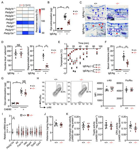
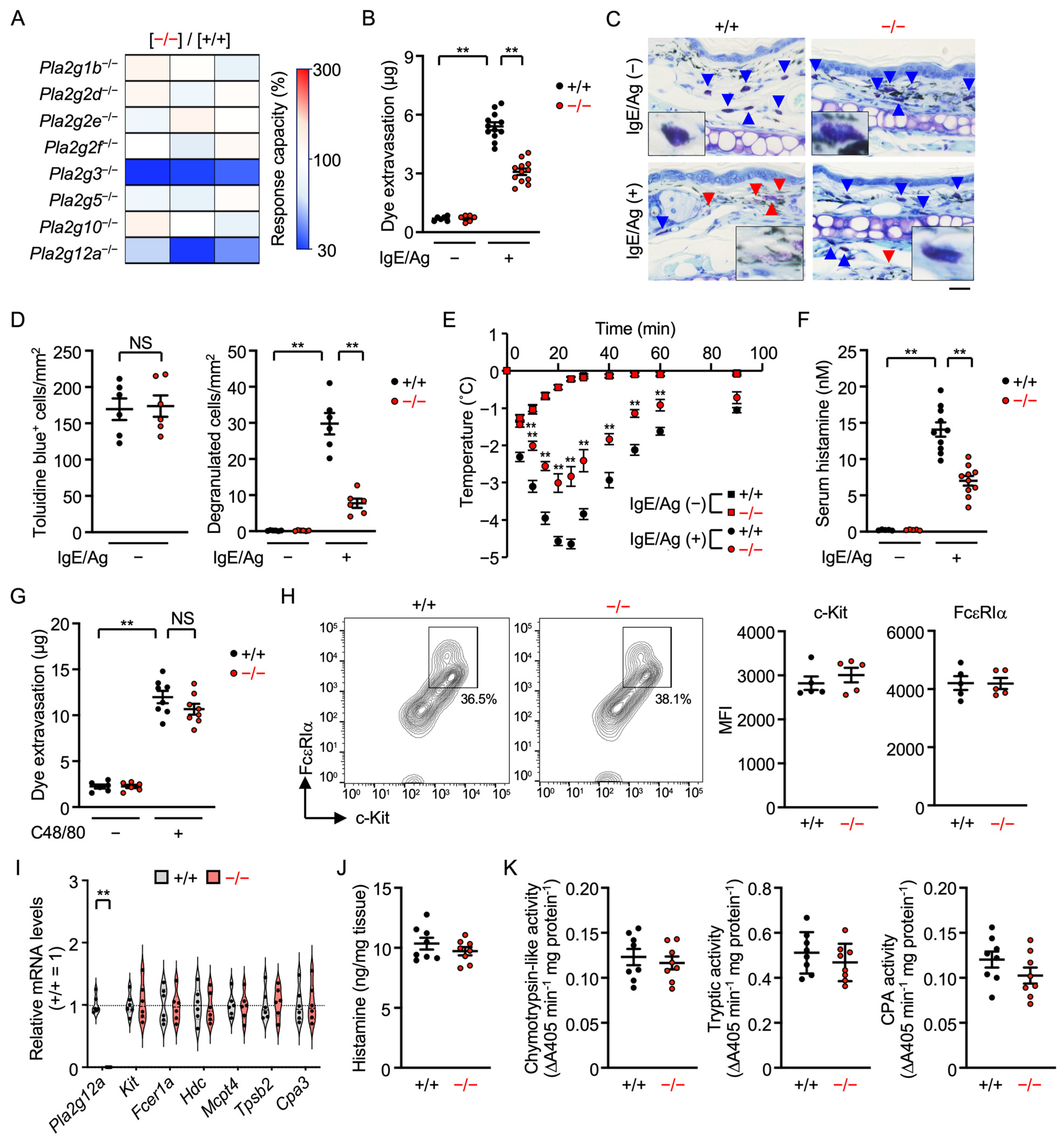
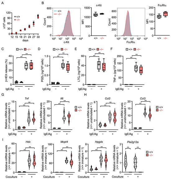
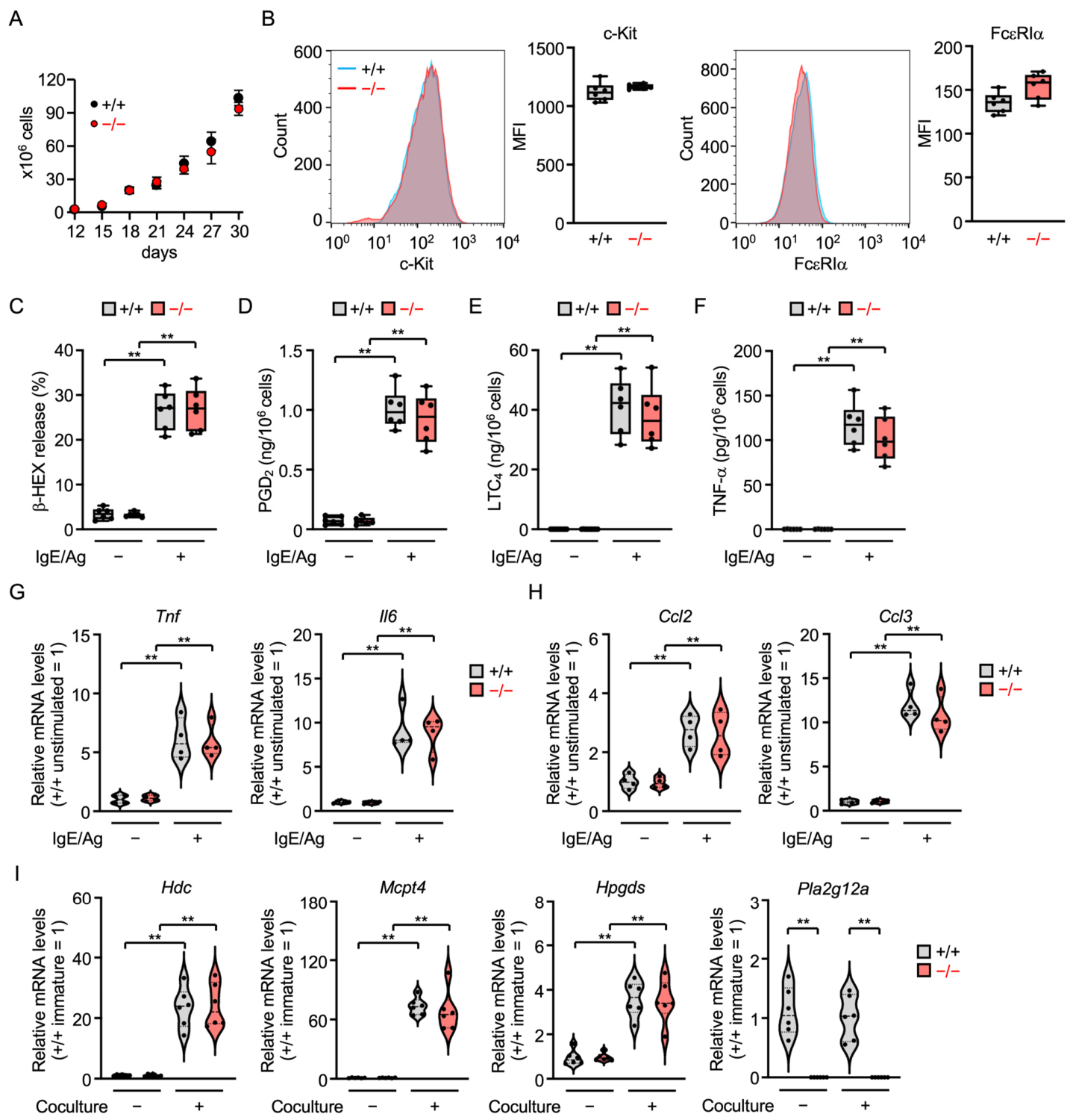
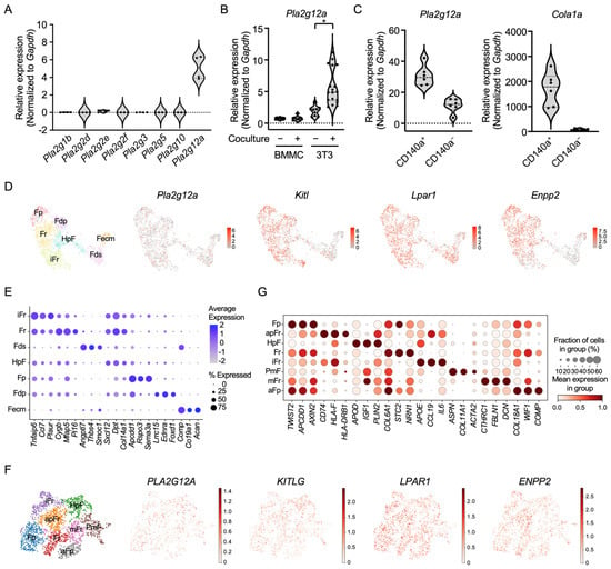
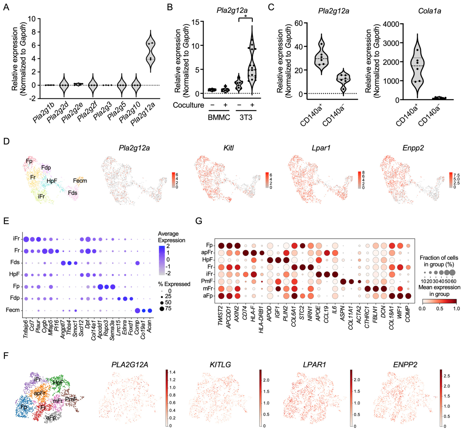
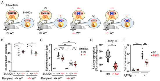
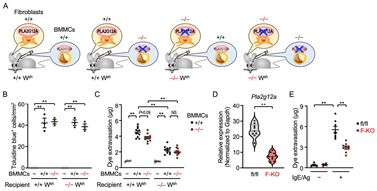
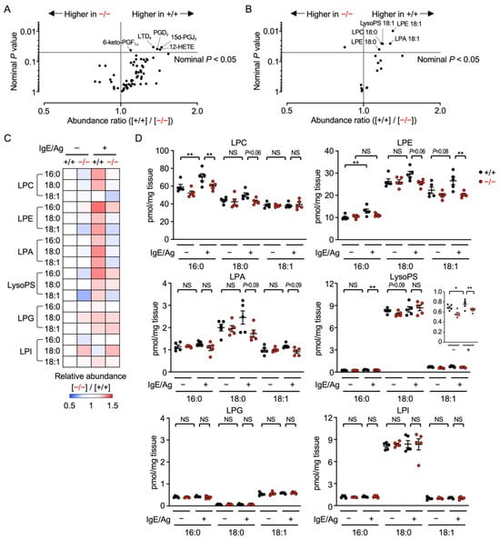
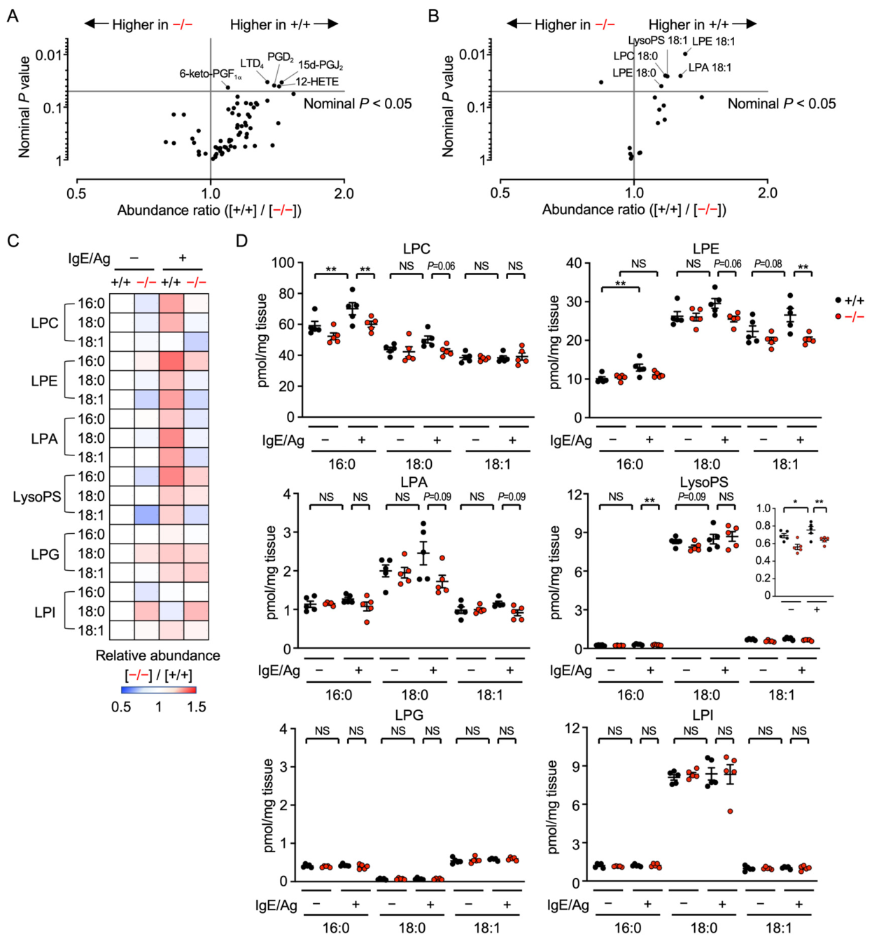
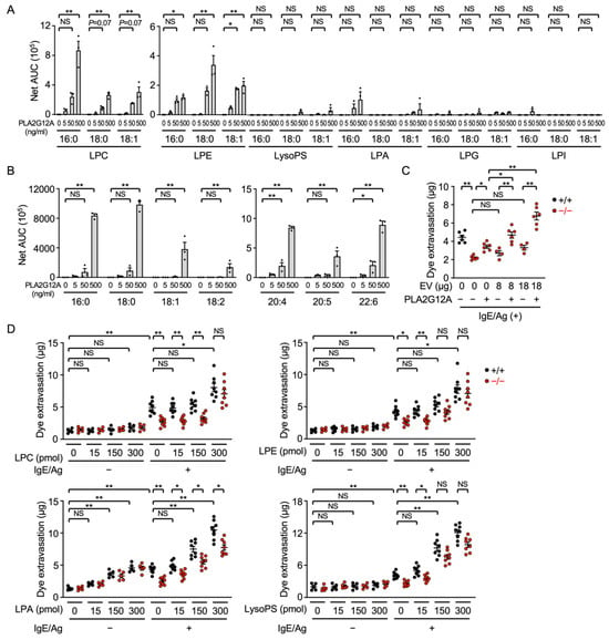
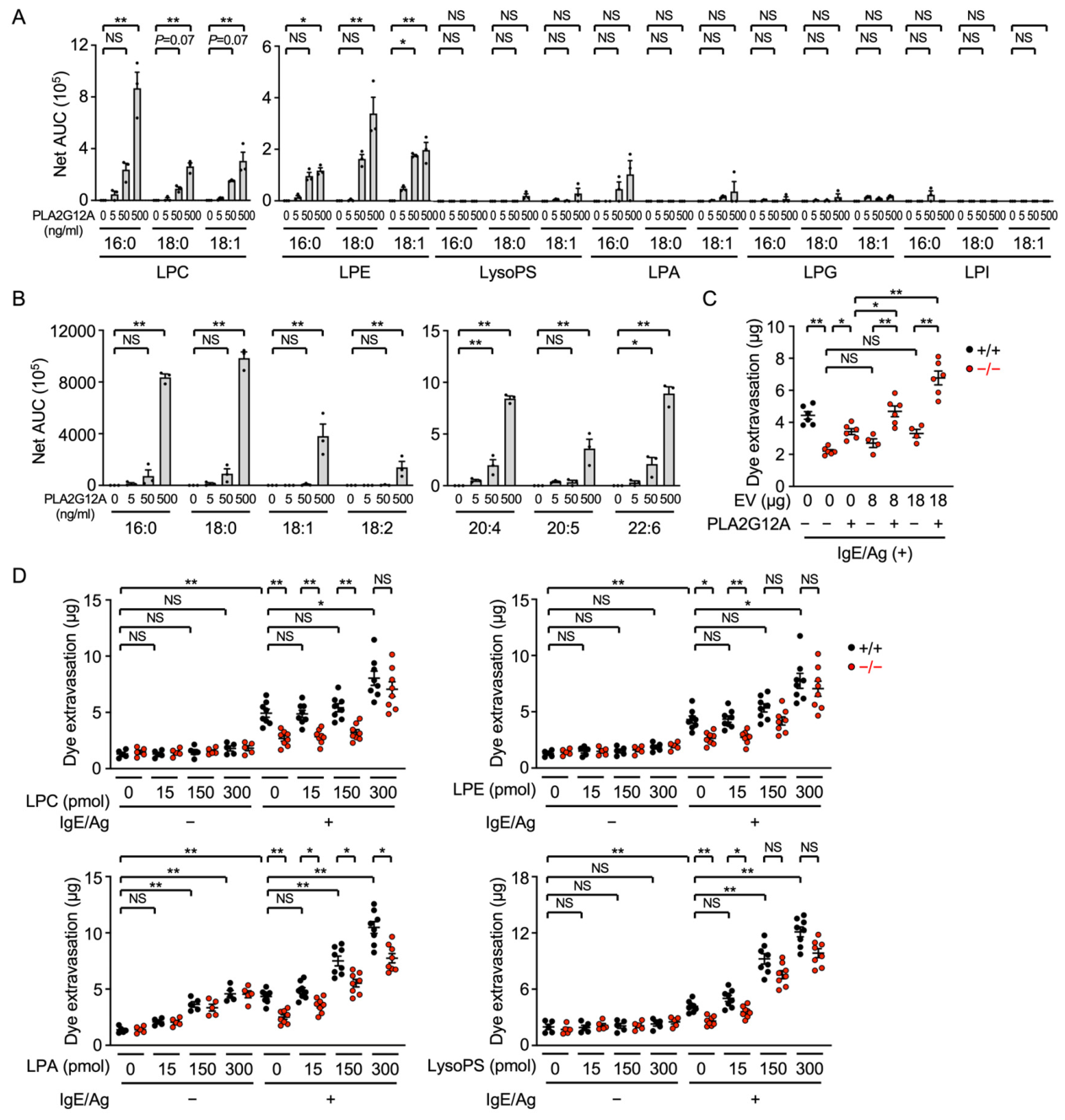
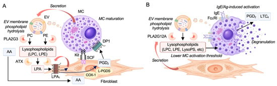
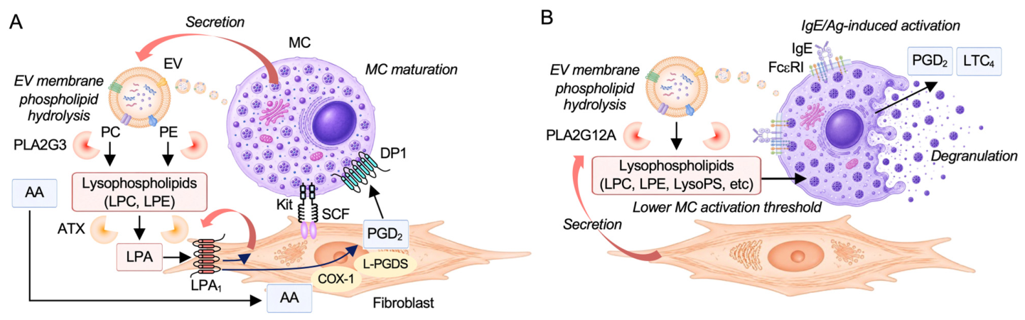

Disclaimer/Publisher’s Note: The statements, opinions and data contained in all publications are solely those of the individual author(s) and contributor(s) and not of MDPI and/or the editor(s). MDPI and/or the editor(s) disclaim responsibility for any injury to people or property resulting from any ideas, methods, instructions or products referred to in the content. |
© 2026 by the authors. Licensee MDPI, Basel, Switzerland. This article is an open access article distributed under the terms and conditions of the Creative Commons Attribution (CC BY) license.