Next Issue
Volume 12, July-2
Previous Issue
Volume 12, June-2


Cells, Volume 12, Issue 13 (July-1 2023) – 138 articles

Cover Story (view full-size image): 
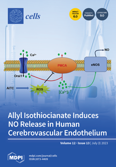
Allyl isothiocyanate (AITC) is a major active constituent of cruciferous vegetables that can activate vasorelaxing pathways in rodent brain microvessels. Herein, we showed that AITC causes the release of nitric oxide (NO), the most important vasorelaxing mediator in human brains, from human cerebrovascular endothelial cells. AITC inhibits the plasma membrane Ca2+-ATPase (PMCA) through the production of reactive oxygen species, thereby attenuating Ca2+ extrusion across the plasma membrane and causing a slow increase in [Ca2+]i. Moreover, PMCA inhibition by AITC unmasks constitutive Ca2+ entry through Orai channels. The resulting increase in [Ca2+]i leads to robust AITC-induced NO release, thereby providing the proof-of-concept that dietary supplementation of AITC represents a promising strategy to reduce the burden of brain disorders. View this paper
  • Issues are regarded as officially published after their release is announced to the table of contents alert mailing list.
  • You may sign up for e-mail alerts to receive table of contents of newly released issues.
  • PDF is the official format for papers published in both, html and pdf forms. To view the papers in pdf format, click on the "PDF Full-text" link, and use the free Adobe Reader to open them.
Order results
Result details
Section
Select all
Export citation of selected articles as:
20 pages, 6802 KiB  
Article
Tissue Inhibitor of Metalloproteinases-1 Overexpression Mediates Chemoresistance in Triple-Negative Breast Cancer Cells
by Lisa Agnello, Annachiara d’Argenio, Alessandra Caliendo, Roberto Nilo, Antonella Zannetti, Monica Fedele, Simona Camorani and Laura Cerchia
Cells 2023, 12(13), 1809; https://doi.org/10.3390/cells12131809 - 07 Jul 2023
Cited by 3 | Viewed by 1356
Abstract
Triple-negative breast cancer (TNBC) is among the most aggressive breast cancer subtypes. Despite being initially responsive to chemotherapy, patients develop drug-resistant and metastatic tumors. Tissue inhibitor of metalloproteinases-1 (TIMP-1) is a secreted protein with a tumor suppressor function due to its anti-proteolytic activity. [...] Read more.
Triple-negative breast cancer (TNBC) is among the most aggressive breast cancer subtypes. Despite being initially responsive to chemotherapy, patients develop drug-resistant and metastatic tumors. Tissue inhibitor of metalloproteinases-1 (TIMP-1) is a secreted protein with a tumor suppressor function due to its anti-proteolytic activity. Nevertheless, evidence indicates that TIMP-1 binds to the CD63 receptor and activates noncanonical oncogenic signaling in several cancers, but its role in mediating TNBC chemoresistance is still largely unexplored. Here, we show that mesenchymal-like TNBC cells express TIMP-1, whose levels are further increased in cells generated to be resistant to cisplatin (Cis-Pt-R) and doxorubicin (Dox-R). Moreover, public dataset analyses indicate that high TIMP-1 levels are associated with a worse prognosis in TNBC subjected to chemotherapy. Knock-down of TIMP-1 in both Cis-Pt-R and Dox-R cells reverses their resistance by inhibiting AKT activation. Consistently, TNBC cells exposed to recombinant TIMP-1 or TIMP-1-enriched media from chemoresistant cells, acquire resistance to both cisplatin and doxorubicin. Importantly, released TIMP-1 reassociates with plasma membrane by binding to CD63 and, in the absence of CD63 expression, TIMP-1-mediated chemoresistance is blocked. Thus, our results identify TIMP-1 as a new biomarker of TNBC chemoresistance and lay the groundwork for evaluating whether blockade of TIMP-1 signal is a viable treatment strategy. Full article
Show Figures

Figure 1

18 pages, 1119 KiB  
Review
Single-Cell RNA Sequencing: Opportunities and Challenges for Studies on Corneal Biology in Health and Disease
by Julian A. Arts, Camille Laberthonnière, Dulce Lima Cunha and Huiqing Zhou
Cells 2023, 12(13), 1808; https://doi.org/10.3390/cells12131808 - 07 Jul 2023
Cited by 1 | Viewed by 1617
Abstract
The structure and major cell types of the multi-layer human cornea have been extensively studied. However, various cell states in specific cell types and key genes that define the cell states are not fully understood, hindering our comprehension of corneal homeostasis, related diseases, [...] Read more.
The structure and major cell types of the multi-layer human cornea have been extensively studied. However, various cell states in specific cell types and key genes that define the cell states are not fully understood, hindering our comprehension of corneal homeostasis, related diseases, and therapeutic discovery. Single-cell RNA sequencing is a revolutionary and powerful tool for identifying cell states within tissues such as the cornea. This review provides an overview of current single-cell RNA sequencing studies on the human cornea, highlighting similarities and differences between them, and summarizing the key genes that define corneal cell states reported in these studies. In addition, this review discusses the opportunities and challenges of using single-cell RNA sequencing to study corneal biology in health and disease. Full article
Show Figures

Figure 1

34 pages, 2536 KiB  
Review
Genetic Primary Microcephalies: When Centrosome Dysfunction Dictates Brain and Body Size
by Sarah Farcy, Hassina Hachour, Nadia Bahi-Buisson and Sandrine Passemard
Cells 2023, 12(13), 1807; https://doi.org/10.3390/cells12131807 - 07 Jul 2023
Cited by 2 | Viewed by 1689
Abstract
Primary microcephalies (PMs) are defects in brain growth that are detectable at or before birth and are responsible for neurodevelopmental disorders. Most are caused by biallelic or, more rarely, dominant mutations in one of the likely hundreds of genes encoding PM proteins, i.e., [...] Read more.
Primary microcephalies (PMs) are defects in brain growth that are detectable at or before birth and are responsible for neurodevelopmental disorders. Most are caused by biallelic or, more rarely, dominant mutations in one of the likely hundreds of genes encoding PM proteins, i.e., ubiquitous centrosome or microtubule-associated proteins required for the division of neural progenitor cells in the embryonic brain. Here, we provide an overview of the different types of PMs, i.e., isolated PMs with or without malformations of cortical development and PMs associated with short stature (microcephalic dwarfism) or sensorineural disorders. We present an overview of the genetic, developmental, neurological, and cognitive aspects characterizing the most representative PMs. The analysis of phenotypic similarities and differences among patients has led scientists to elucidate the roles of these PM proteins in humans. Phenotypic similarities indicate possible redundant functions of a few of these proteins, such as ASPM and WDR62, which play roles only in determining brain size and structure. However, the protein pericentrin (PCNT) is equally required for determining brain and body size. Other PM proteins perform both functions, albeit to different degrees. Finally, by comparing phenotypes, we considered the interrelationships among these proteins. Full article
(This article belongs to the Special Issue Cellular Mechanisms of Microcephaly)
Show Figures

Figure 1

11 pages, 3156 KiB  
Communication
Exploring the Process of Neutrophil Transendothelial Migration Using Scanning Ion-Conductance Microscopy
by Svetlana N. Pleskova, Nikolay A. Bezrukov, Ekaterina N. Gorshkova, Sergey Z. Bobyk and Ekaterina V. Lazarenko
Cells 2023, 12(13), 1806; https://doi.org/10.3390/cells12131806 - 07 Jul 2023
Cited by 2 | Viewed by 1282
Abstract
The dynamics of neutrophil transendothelial migration was investigated in a model of experimental septicopyemia. Scanning ion-conductance microscopy allowed us to determine changes in morphometric characteristics of endothelial cells during this process. In the presence of a pyogenic lesion simulated by Staphylococcus aureus, [...] Read more.
The dynamics of neutrophil transendothelial migration was investigated in a model of experimental septicopyemia. Scanning ion-conductance microscopy allowed us to determine changes in morphometric characteristics of endothelial cells during this process. In the presence of a pyogenic lesion simulated by Staphylococcus aureus, such migration was accompanied by both compensatory reactions and alteration of both neutrophils and endothelial cells. Neutrophils demonstrated crawling along the contact sites between endothelial cells, swarming phenomenon, as well as anergy and formation of neutrophil extracellular traps (NETs) as a normergic state. Neutrophil swarming was accompanied by an increase in the intercellular spaces between endothelial cells. Endothelial cells decreased the area of adhesion to the substrate, which was determined by a decrease in the cell projection area, and the cell membrane was smoothed. However, endothelial cell rigidity was paradoxically unchanged compared to the control. Over time, neutrophil migration led to a more significant alteration of endothelial cells: first, shallow perforations in the membrane were formed, which were repaired rather quickly, then stress fibrils were formed, and finally, endothelial cells died and multiple perforations were formed on their membrane. Full article
(This article belongs to the Special Issue Advances in Scanning Probe Microscopy in Cell Biology)
Show Figures

Figure 1

17 pages, 2983 KiB  
Article
Soluble Collectin 11 (CL-11) Acts as an Immunosuppressive Molecule Potentially Used by Stem Cell-Derived Retinal Epithelial Cells to Modulate T Cell Response
by Giorgia Fanelli, Marco Romano, Giovanna Lombardi and Steven H. Sacks
Cells 2023, 12(13), 1805; https://doi.org/10.3390/cells12131805 - 07 Jul 2023
Cited by 1 | Viewed by 1211
Abstract
Retinal pigment epithelium (RPE) cell allotransplantation is seen as a possible solution to retinal diseases. However, the RPE-complement system triggered by the binding of collectin-11 (CL-11) is a potential barrier for RPE transplantation as the complement-mediated inflammatory response may promote T cell recognition. [...] Read more.
Retinal pigment epithelium (RPE) cell allotransplantation is seen as a possible solution to retinal diseases. However, the RPE-complement system triggered by the binding of collectin-11 (CL-11) is a potential barrier for RPE transplantation as the complement-mediated inflammatory response may promote T cell recognition. To address this, we investigated the role of CL-11 on T cell immuno-response. We confirmed that RPE cells up-regulated MHC class I and expressed MHC class II molecules in an inflammatory setting. Co-cultures of RPE cells with T cells led to the inhibition of T cell proliferation. We found that CL-11 was partially responsible for this effect as T cell binding of CL-11 inhibited T cell proliferation in association with the downregulation of CD28. We also found that the suppressive action of CL-11 was abrogated in the presence of the RGD peptide given to block the T cell binding of CL-11 by its collagen-like domain. Because RPE cells can bind and secrete CL-11 under stress conditions, we postulate that soluble CL-11 contributes to the immunosuppressive properties of RPE cells. The investigation of this dual biological activity of CL-11, namely as a trigger of the complement cascade and a modulator of T cell responses, may provide additional clues about the mechanisms that orchestrate the immunogenic properties of RPE cells. Full article
(This article belongs to the Special Issue The Role of Immune Cells in Ocular Diseases)
Show Figures

Figure 1

27 pages, 5318 KiB  
Article
Dysregulation of Immature Sertoli Cell Functions by Exposure to Acetaminophen and Genistein in Rodent Cell Models
by Maia Corpuz-Hilsabeck, Nicole Mohajer and Martine Culty
Cells 2023, 12(13), 1804; https://doi.org/10.3390/cells12131804 - 07 Jul 2023
Cited by 1 | Viewed by 1567
Abstract
Sertoli cells are essential for germ cell development and function. Their disruption by endocrine disrupting chemicals (EDCs) or drugs could jeopardize spermatogenesis, contributing to male infertility. Perinatal exposure to EDCs and acetaminophen (APAP) disrupts male reproductive functions in animals and humans. Infants can [...] Read more.
Sertoli cells are essential for germ cell development and function. Their disruption by endocrine disrupting chemicals (EDCs) or drugs could jeopardize spermatogenesis, contributing to male infertility. Perinatal exposure to EDCs and acetaminophen (APAP) disrupts male reproductive functions in animals and humans. Infants can be exposed simultaneously to the dietary soy phytoestrogen genistein (GEN) and APAP used for fever or pain relief. Our goal was to determine the effects of 10–100 µM APAP and GEN, alone or mixed, on immature Sertoli cells using mouse TM4 Sertoli cell line and postnatal-day 8 rat Sertoli cells, by measuring cell viability, proliferation, prostaglandins, genes and protein expression, and functional pathways. A value of 50 µM APAP decreased the viability, while 100 µM APAP and GEN decreased the proliferation. Sertoli cell and eicosanoid pathway genes were affected by GEN and mixtures, with downregulation of Sox9, Cox1, Cox2, and genes relevant for Sertoli cell function, while genes involved in inflammation were increased. RNA-seq analysis identified p53 and TNF signaling pathways as common targets of GEN and GEN mixture in both cell types. These results suggest that APAP and GEN dysregulate immature Sertoli cell function and may aid in elucidating novel EDC and drug targets contributing to the etiology of male infertility. Full article
Show Figures

Figure 1

17 pages, 3661 KiB  
Article
Progranulin Protects against Hyperglycemia-Induced Neuronal Dysfunction through GSK3β Signaling
by Cass Dedert, Lyuba Salih and Fenglian Xu
Cells 2023, 12(13), 1803; https://doi.org/10.3390/cells12131803 - 07 Jul 2023
Viewed by 1162
Abstract
Type II diabetes affects over 530 million individuals worldwide and contributes to a host of neurological pathologies. Uncontrolled high blood glucose (hyperglycemia) is a major factor in diabetic pathology, and glucose regulation is a common goal for maintenance in patients. We have found [...] Read more.
Type II diabetes affects over 530 million individuals worldwide and contributes to a host of neurological pathologies. Uncontrolled high blood glucose (hyperglycemia) is a major factor in diabetic pathology, and glucose regulation is a common goal for maintenance in patients. We have found that the neuronal growth factor progranulin protects against hyperglycemic stress in neurons, and although its mechanism of action is uncertain, our findings identified Glycogen Synthase Kinase 3β (GSK3β) as being potentially involved in its effects. In this study, we treated mouse primary cortical neurons exposed to high-glucose conditions with progranulin and a selective pharmacological inhibitor of GSK3β before assessing neuronal health and function. Whole-cell and mitochondrial viability were both improved by progranulin under high-glucose stress in a GSK3β—dependent manner. This extended to autophagy flux, indicated by the expressions of autophagosome marker Light Chain 3B (LC3B) and lysosome marker Lysosome-Associated Membrane Protein 2A (LAMP2A), which were affected by progranulin and showed heterogeneous changes from GSK3β inhibition. Lastly, GSK3β inhibition attenuated downstream calcium signaling and neuronal firing effects due to acute progranulin treatment. These data indicate that GSK3β plays an important role in progranulin’s neuroprotective effects under hyperglycemic stress and serves as a jumping-off point to explore progranulin’s protective capabilities in other neurodegenerative models. Full article
(This article belongs to the Collection Advances in Neurodegenerative Disease)
Show Figures

Figure 1

12 pages, 3116 KiB  
Perspective
Platelet Metabolic Flexibility: A Matter of Substrate and Location
by Silvia Ravera, Maria Grazia Signorello and Isabella Panfoli
Cells 2023, 12(13), 1802; https://doi.org/10.3390/cells12131802 - 07 Jul 2023
Cited by 1 | Viewed by 1256
Abstract
Platelets are cellular elements that are physiologically involved in hemostasis, inflammation, thrombotic events, and various human diseases. There is a link between the activation of platelets and their metabolism. Platelets possess considerable metabolic versatility. Although the role of platelets in hemostasis and inflammation [...] Read more.
Platelets are cellular elements that are physiologically involved in hemostasis, inflammation, thrombotic events, and various human diseases. There is a link between the activation of platelets and their metabolism. Platelets possess considerable metabolic versatility. Although the role of platelets in hemostasis and inflammation is known, our current understanding of platelet metabolism in terms of substrate preference is limited. Platelet activation triggers an oxidative metabolism increase to sustain energy requirements better than aerobic glycolysis alone. In addition, platelets possess extra-mitochondrial oxidative phosphorylation, which could be one of the sources of chemical energy required for platelet activation. This review aims to provide an overview of flexible platelet metabolism, focusing on the role of metabolic compartmentalization in substrate preference, since the metabolic flexibility of stimulated platelets could depend on subcellular localization and functional timing. Thus, developing a detailed understanding of the link between platelet activation and metabolic changes is crucial for improving human health. Full article
(This article belongs to the Special Issue Recent Advances in Metabolism and Oxidative Stress in Human Diseases)
Show Figures

Figure 1

28 pages, 2375 KiB  
Review
Neurochemical Basis of Inter-Organ Crosstalk in Health and Obesity: Focus on the Hypothalamus and the Brainstem
by Dhanush Haspula and Zhenzhong Cui
Cells 2023, 12(13), 1801; https://doi.org/10.3390/cells12131801 - 07 Jul 2023
Cited by 2 | Viewed by 2877
Abstract
Precise neural regulation is required for maintenance of energy homeostasis. Essential to this are the hypothalamic and brainstem nuclei which are located adjacent and supra-adjacent to the circumventricular organs. They comprise multiple distinct neuronal populations which receive inputs not only from other brain [...] Read more.
Precise neural regulation is required for maintenance of energy homeostasis. Essential to this are the hypothalamic and brainstem nuclei which are located adjacent and supra-adjacent to the circumventricular organs. They comprise multiple distinct neuronal populations which receive inputs not only from other brain regions, but also from circulating signals such as hormones, nutrients, metabolites and postprandial signals. Hence, they are ideally placed to exert a multi-tier control over metabolism. The neuronal sub-populations present in these key metabolically relevant nuclei regulate various facets of energy balance which includes appetite/satiety control, substrate utilization by peripheral organs and glucose homeostasis. In situations of heightened energy demand or excess, they maintain energy homeostasis by restoring the balance between energy intake and expenditure. While research on the metabolic role of the central nervous system has progressed rapidly, the neural circuitry and molecular mechanisms involved in regulating distinct metabolic functions have only gained traction in the last few decades. The focus of this review is to provide an updated summary of the mechanisms by which the various neuronal subpopulations, mainly located in the hypothalamus and the brainstem, regulate key metabolic functions. Full article
(This article belongs to the Special Issue Hypothalamic Hormonal Secretion and Metabolism)
Show Figures

Figure 1

19 pages, 2407 KiB  
Review
Mitochondrial Control for Healthy and Autoimmune T Cells
by Li Jia, Lei Zhang, Mengdi Liu, Huiyan Ji, Zhenke Wen and Chunhong Wang
Cells 2023, 12(13), 1800; https://doi.org/10.3390/cells12131800 - 07 Jul 2023
Cited by 3 | Viewed by 2081
Abstract
T cells are critical players in adaptive immunity, driving the tissue injury and organ damage of patients with autoimmune diseases. Consequently, investigations on T cell activation, differentiation, and function are valuable in uncovering the disease pathogenesis, thus exploring promising therapeutics for autoimmune diseases. [...] Read more.
T cells are critical players in adaptive immunity, driving the tissue injury and organ damage of patients with autoimmune diseases. Consequently, investigations on T cell activation, differentiation, and function are valuable in uncovering the disease pathogenesis, thus exploring promising therapeutics for autoimmune diseases. In recent decades, accumulating studies have pinpointed immunometabolism as the fundamental determinant in controlling T cell fate. Specifically, mitochondria, as a hub of intracellular metabolism, connect glucose, lipid, and amino acid metabolic pathways. Herein, we summarize metabolic adaptations of mitochondrial oxidative phosphorylation and the relevant glucose, lipid, and amino acid metabolism during T cell activation, differentiation, and function. Further, we focused on current updates of the molecular bases for metabolic reprogramming in autoimmune T cells and advances in exploring metabolic-targeted therapeutics against autoimmune diseases. This might facilitate the in-depth understanding of autoimmune pathogeneses and the clinical management of autoimmune diseases. Full article
Show Figures

Figure 1

16 pages, 15971 KiB  
Article
LRRK2 Kinase Inhibition Attenuates Neuroinflammation and Cytotoxicity in Animal Models of Alzheimer’s and Parkinson’s Disease-Related Neuroinflammation
by Veronica Mutti, Giulia Carini, Alice Filippini, Stefania Castrezzati, Lorena Giugno, Massimo Gennarelli and Isabella Russo
Cells 2023, 12(13), 1799; https://doi.org/10.3390/cells12131799 - 06 Jul 2023
Cited by 6 | Viewed by 1567
Abstract
Chronic neuroinflammation plays a crucial role in the progression of several neurodegenerative diseases (NDDs), including Parkinson’s disease (PD) and Alzheimer’s disease (AD). Intriguingly, in the last decade, leucine-rich repeat kinase-2 (LRRK2), a gene mutated in familial and sporadic PD, was revealed [...] Read more.
Chronic neuroinflammation plays a crucial role in the progression of several neurodegenerative diseases (NDDs), including Parkinson’s disease (PD) and Alzheimer’s disease (AD). Intriguingly, in the last decade, leucine-rich repeat kinase-2 (LRRK2), a gene mutated in familial and sporadic PD, was revealed as a key mediator of neuroinflammation. Therefore, the anti-inflammatory properties of LRRK2 inhibitors have started to be considered as a disease-modifying treatment for PD; however, to date, there is little evidence on the beneficial effects of targeting LRRK2-related neuroinflammation in preclinical models. In this study, we further validated LRRK2 kinase modulation as a pharmacological intervention in preclinical models of AD- and PD-related neuroinflammation. Specifically, we reported that LRRK2 kinase inhibition with MLi2 and PF-06447475 (PF) molecules attenuated neuroinflammation, gliosis and cytotoxicity in mice with intracerebral injection of Aβ1-42 fibrils or α-syn preformed fibrils (pffs). Moreover, for the first time in vivo, we showed that LRRK2 kinase activity participates in AD-related neuroinflammation and therefore might contribute to AD pathogenesis. Overall, our findings added evidence on the anti-inflammatory effects of LRRK2 kinase inhibition in preclinical models and indicate that targeting LRRK2 activity could be a disease-modifying treatment for NDDs with an inflammatory component. Full article
(This article belongs to the Collection LRRK2-Dependent Neurodegeneration in Parkinson’s Disease)
Show Figures

Figure 1

24 pages, 6432 KiB  
Article
SHIP1 Is Present but Strongly Downregulated in T-ALL, and after Restoration Suppresses Leukemia Growth in a T-ALL Xenotransplantation Mouse Model
by Patrick Ehm, Ruth Rietow, Wiebke Wegner, Lara Bußmann, Malte Kriegs, Kevin Dierck, Stefan Horn, Thomas Streichert, Martin Horstmann and Manfred Jücker
Cells 2023, 12(13), 1798; https://doi.org/10.3390/cells12131798 - 06 Jul 2023
Viewed by 1367
Abstract
Acute lymphoblastic leukemia (ALL) is the most common cause of cancer-related death in children. Despite significantly increased chances of cure, especially for high-risk ALL patients, it still represents a poor prognosis for a substantial fraction of patients. Misregulated proteins in central switching points [...] Read more.
Acute lymphoblastic leukemia (ALL) is the most common cause of cancer-related death in children. Despite significantly increased chances of cure, especially for high-risk ALL patients, it still represents a poor prognosis for a substantial fraction of patients. Misregulated proteins in central switching points of the cellular signaling pathways represent potentially important therapeutic targets. Recently, the inositol phosphatase SHIP1 (SH2-containing inositol 5-phosphatase) has been considered as a tumor suppressor in leukemia. SHIP1 serves as an important negative regulator of the PI3K/AKT signaling pathway, which is frequently constitutively activated in primary T-ALL. In contrast to other reports, we show for the first time that SHIP1 has not been lost in T-ALL cells, but is strongly downregulated. Reduced expression of SHIP1 leads to an increased activation of the PI3K/AKT signaling pathway. SHIP1-mRNA expression is frequently reduced in primary T-ALL samples, which is recapitulated by the decrease in SHIP1 expression at the protein level in seven out of eight available T-ALL patient samples. In addition, we investigated the change in the activity profile of tyrosine and serine/threonine kinases after the restoration of SHIP1 expression in Jurkat T-ALL cells. The tyrosine kinase receptor subfamilies of NTRK and PDGFR, which are upregulated in T-ALL subgroups with low SHIP1 expression, are significantly disabled after SHIP1 reconstitution. Lentiviral-mediated reconstitution of SHIP1 expression in Jurkat cells points to a decreased cellular proliferation upon transplantation into NSG mice in comparison to the control cohort. Together, our findings will help to elucidate the complex network of cell signaling proteins, further support a functional role for SHIP1 as tumor suppressor in T-ALL and, much more importantly, show that full-length SHIP1 is expressed in T-ALL samples. Full article
(This article belongs to the Section Cell Signaling)
Show Figures

Figure 1

22 pages, 3817 KiB  
Systematic Review
Maxillary Sinus Augmentation Using Autologous Platelet Concentrates (Platelet-Rich Plasma, Platelet-Rich Fibrin, and Concentrated Growth Factor) Combined with Bone Graft: A Systematic Review
by Giuseppina Malcangi, Assunta Patano, Giulia Palmieri, Chiara Di Pede, Giulia Latini, Alessio Danilo Inchingolo, Denisa Hazballa, Elisabetta de Ruvo, Grazia Garofoli, Francesco Inchingolo, Gianna Dipalma, Elio Minetti and Angelo Michele Inchingolo
Cells 2023, 12(13), 1797; https://doi.org/10.3390/cells12131797 - 06 Jul 2023
Cited by 3 | Viewed by 2001
Abstract
Background: The current review aims to provide an overview of the most recent research on the potentials of concentrated growth factors used in the maxillary sinus lift technique. Materials and methods: “PRP”, “PRF”, “L-PRF”, “CGF”, “oral surgery”, “sticky bone”, “sinus lift” were the [...] Read more.
Background: The current review aims to provide an overview of the most recent research on the potentials of concentrated growth factors used in the maxillary sinus lift technique. Materials and methods: “PRP”, “PRF”, “L-PRF”, “CGF”, “oral surgery”, “sticky bone”, “sinus lift” were the search terms utilized in the databases Scopus, Web of Science, and Pubmed, with the Boolean operator “AND” and “OR”. Results: Of these 1534 studies, 22 publications were included for this review. Discussion: The autologous growth factors released from platelet concentrates can help to promote bone remodeling and cell proliferation, and the application of platelet concentrates appears to reduce the amount of autologous bone required during regenerative surgery. Many authors agree that growth factors considerably enhance early vascularization in bone grafts and have a significantly positive pro-angiogenic influence in vivo when combined with alloplastic and xenogeneic materials, reducing inflammation and postoperative pain and stimulating the regeneration of injured tissues and accelerating their healing. Conclusions: Even if further studies are still needed, the use of autologous platelet concentrates can improve clinical results where a large elevation of the sinus is needed by improving bone height, thickness and vascularization of surgical sites, and post-operative healing. Full article
(This article belongs to the Special Issue Recent Advances in Regenerative Dentistry)
Show Figures

Figure 1

26 pages, 2475 KiB  
Review
Atrial Cardiomyopathy in Valvular Heart Disease: From Molecular Biology to Clinical Perspectives
by Andrea Ágnes Molnár, Attila Sánta, Dorottya Tímea Pásztor and Béla Merkely
Cells 2023, 12(13), 1796; https://doi.org/10.3390/cells12131796 - 06 Jul 2023
Cited by 5 | Viewed by 3271
Abstract
This review discusses the evolving topic of atrial cardiomyopathy concerning valvular heart disease. The pathogenesis of atrial cardiomyopathy involves multiple factors, such as valvular disease leading to atrial structural and functional remodeling due to pressure and volume overload. Atrial enlargement and dysfunction can [...] Read more.
This review discusses the evolving topic of atrial cardiomyopathy concerning valvular heart disease. The pathogenesis of atrial cardiomyopathy involves multiple factors, such as valvular disease leading to atrial structural and functional remodeling due to pressure and volume overload. Atrial enlargement and dysfunction can trigger atrial tachyarrhythmia. The complex interaction between valvular disease and atrial cardiomyopathy creates a vicious cycle of aggravating atrial enlargement, dysfunction, and valvular disease severity. Furthermore, atrial remodeling and arrhythmia can predispose to atrial thrombus formation and stroke. The underlying pathomechanism of atrial myopathy involves molecular, cellular, and subcellular alterations resulting in chronic inflammation, atrial fibrosis, and electrophysiological changes. Atrial dysfunction has emerged as an essential determinant of outcomes in valvular disease and heart failure. Despite its predictive value, the detection of atrial fibrosis and dysfunction is challenging and is not included in the clinical routine. Transthoracic echocardiography and cardiac magnetic resonance imaging are the main diagnostic tools for atrial cardiomyopathy. Recently published data have revealed that both left atrial volumes and functional parameters are independent predictors of cardiovascular events in valvular disease. The integration of atrial function assessment in clinical practice might help in early cardiovascular risk estimation, promoting early therapeutic intervention in valvular disease. Full article
(This article belongs to the Special Issue Molecular Biology of Atrial Myocardium)
Show Figures

Figure 1

21 pages, 6564 KiB  
Article
A YAP/TAZ–ARHGAP29–RhoA Signaling Axis Regulates Podocyte Protrusions and Integrin Adhesions
by Manuel Rogg, Jasmin I. Maier, Martin Helmstädter, Alena Sammarco, Felix Kliewe, Oliver Kretz, Lisa Weißer, Clara Van Wymersch, Karla Findeisen, Anna L. Koessinger, Olga Tsoy, Jan Baumbach, Markus Grabbert, Martin Werner, Tobias B. Huber, Nicole Endlich, Oliver Schilling and Christoph Schell
Cells 2023, 12(13), 1795; https://doi.org/10.3390/cells12131795 - 06 Jul 2023
Cited by 1 | Viewed by 1732
Abstract
Glomerular disease due to podocyte malfunction is a major factor in the pathogenesis of chronic kidney disease. Identification of podocyte-specific signaling pathways is therefore a prerequisite to characterizing relevant disease pathways and developing novel treatment approaches. Here, we employed loss of function studies [...] Read more.
Glomerular disease due to podocyte malfunction is a major factor in the pathogenesis of chronic kidney disease. Identification of podocyte-specific signaling pathways is therefore a prerequisite to characterizing relevant disease pathways and developing novel treatment approaches. Here, we employed loss of function studies for EPB41L5 (Yurt) as a central podocyte gene to generate a cell type-specific disease model. Loss of Yurt in fly nephrocytes caused protein uptake and slit diaphragm defects. Transcriptomic and proteomic analysis of human EPB41L5 knockout podocytes demonstrated impaired mechanotransduction via the YAP/TAZ signaling pathway. Further analysis of specific inhibition of the YAP/TAZ-TEAD transcription factor complex by TEADi led to the identification of ARGHAP29 as an EPB41L5 and YAP/TAZ-dependently expressed podocyte RhoGAP. Knockdown of ARHGAP29 caused increased RhoA activation, defective lamellipodia formation, and increased maturation of integrin adhesion complexes, explaining similar phenotypes caused by loss of EPB41L5 and TEADi expression in podocytes. Detection of increased levels of ARHGAP29 in early disease stages of human glomerular disease implies a novel negative feedback loop for mechanotransductive RhoA—YAP/TAZ signaling in podocyte physiology and disease. Full article
Show Figures

Figure 1

32 pages, 2639 KiB  
Article
Inversely Regulated Inflammation-Related Processes Mediate Anxiety–Obesity Links in Zebrafish Larvae and Adults
by Hila Yehuda, Nimrod Madrer, Doron Goldberg, Hermona Soreq and Ari Meerson
Cells 2023, 12(13), 1794; https://doi.org/10.3390/cells12131794 - 06 Jul 2023
Viewed by 1560
Abstract
Anxiety and metabolic impairments are often inter-related, but the underlying mechanisms are unknown. To seek RNAs involved in the anxiety disorder–metabolic disorder link, we subjected zebrafish larvae to caffeine-induced anxiety or high-fat diet (HFD)-induced obesity followed by RNA sequencing and analyses. Notably, differentially [...] Read more.
Anxiety and metabolic impairments are often inter-related, but the underlying mechanisms are unknown. To seek RNAs involved in the anxiety disorder–metabolic disorder link, we subjected zebrafish larvae to caffeine-induced anxiety or high-fat diet (HFD)-induced obesity followed by RNA sequencing and analyses. Notably, differentially expressed (DE) transcripts in these larval models and an adult zebrafish caffeine-induced anxiety model, as well as the transcript profiles of inherently anxious versus less anxious zebrafish strains and high-fat diet-fed versus standard diet-fed adult zebrafish, revealed inversely regulated DE transcripts. In both larval anxiety and obesity models, these included long noncoding RNAs and transfer RNA fragments, with the overrepresented immune system and inflammation pathways, e.g., the “interleukin signaling pathway” and “inflammation mediated by chemokine and cytokine signaling pathway”. In adulthood, overrepresented immune system processes included “T cell activation”, “leukocyte cell-cell adhesion”, and “antigen processing and presentation”. Furthermore, unlike adult zebrafish, obesity in larvae was not accompanied by anxiety-like behavior. Together, these results may reflect an antagonistic pleiotropic phenomenon involving a re-adjusted modulation of the anxiety–metabolic links with an occurrence of the acquired immune system. Furthermore, the HFD potential to normalize anxiety-upregulated immune-related genes may reflect the high-fat diet protection of anxiety and neurodegeneration reported by others. Full article
Show Figures

Figure 1

22 pages, 6453 KiB  
Article
Adult Multipotent Cardiac Progenitor-Derived Spheroids: A Reproducible Model of In Vitro Cardiomyocyte Commitment and Specification
by Mariangela Scalise, Fabiola Marino, Luca Salerno, Nunzia Amato, Claudia Quercia, Chiara Siracusa, Andrea Filardo, Antonio Chiefalo, Loredana Pagano, Giuseppe Misdea, Nadia Salerno, Antonella De Angelis, Konrad Urbanek, Giuseppe Viglietto, Daniele Torella and Eleonora Cianflone
Cells 2023, 12(13), 1793; https://doi.org/10.3390/cells12131793 - 05 Jul 2023
Cited by 1 | Viewed by 1775
Abstract
Background: Three-dimensional cell culture systems hold great promise for bridging the gap between in vitro cell-based model systems and small animal models to study tissue biology and disease. Among 3D cell culture systems, stem-cell-derived spheroids have attracted significant interest as a strategy to [...] Read more.
Background: Three-dimensional cell culture systems hold great promise for bridging the gap between in vitro cell-based model systems and small animal models to study tissue biology and disease. Among 3D cell culture systems, stem-cell-derived spheroids have attracted significant interest as a strategy to better mimic in vivo conditions. Cardiac stem cell/progenitor (CSC)-derived spheroids (CSs) provide a relevant platform for cardiac regeneration. Methods: We compared three different cell culture scaffold-free systems, (i) ultra-low attachment plates, (ii) hanging drops (both requiring a 2D/3D switch), and (iii) agarose micro-molds (entirely 3D), for CSC-derived CS formation and their cardiomyocyte commitment in vitro. Results: The switch from a 2D to a 3D culture microenvironment per se guides cell plasticity and myogenic differentiation within CS and is necessary for robust cardiomyocyte differentiation. On the contrary, 2D monolayer CSC cultures show a significant reduced cardiomyocyte differentiation potential compared to 3D CS culture. Forced aggregation into spheroids using hanging drop improves CS myogenic differentiation when compared to ultra-low attachment plates. Performing CS formation and myogenic differentiation exclusively in 3D culture using agarose micro-molds maximizes the cardiomyocyte yield. Conclusions: A 3D culture system instructs CS myogenic differentiation, thus representing a valid model that can be used to study adult cardiac regenerative biology. Full article
(This article belongs to the Special Issue Recent Advances in Cardiac Repair and Regeneration)
Show Figures

Figure 1

17 pages, 4047 KiB  
Article
Interplay between Protein Kinase C Epsilon and Reactive Oxygen Species during Myogenic Differentiation
by Giulia Pozzi, Valentina Presta, Elena Masselli, Giancarlo Condello, Samuele Cortellazzi, Maria Luisa Arcari, Cristina Micheloni, Marco Vitale, Giuliana Gobbi, Prisco Mirandola and Cecilia Carubbi
Cells 2023, 12(13), 1792; https://doi.org/10.3390/cells12131792 - 05 Jul 2023
Cited by 3 | Viewed by 1415
Abstract
Reactive oxygen species (ROS) are currently recognized as a key driver of several physiological processes. Increasing evidence indicates that ROS levels can affect myogenic differentiation, but the molecular mechanisms still need to be elucidated. Protein kinase C (PKC) epsilon (PKCe) promotes muscle stem [...] Read more.
Reactive oxygen species (ROS) are currently recognized as a key driver of several physiological processes. Increasing evidence indicates that ROS levels can affect myogenic differentiation, but the molecular mechanisms still need to be elucidated. Protein kinase C (PKC) epsilon (PKCe) promotes muscle stem cell differentiation and regeneration of skeletal muscle after injury. PKCs play a tissue-specific role in redox biology, with specific isoforms being both a target of ROS and an up-stream regulator of ROS production. Therefore, we hypothesized that PKCe represents a molecular link between redox homeostasis and myogenic differentiation. We used an in vitro model of a mouse myoblast cell line (C2C12) to study the PKC–redox axis. We demonstrated that the transition from a myoblast to myotube is typified by increased PKCe protein content and decreased ROS. Intriguingly, the expression of the antioxidant enzyme superoxide dismutase 2 (SOD2) is significantly higher in the late phases of myogenic differentiation, mimicking PKCe protein content. Furthermore, we demonstrated that PKCe inhibition increases ROS and reduces SOD2 protein content while SOD2 silencing did not affect PKCe protein content, suggesting that the kinase could be an up-stream regulator of SOD2. To support this hypothesis, we found that in C2C12 cells, PKCe interacts with Nrf2, whose activation induces SOD2 transcription. Overall, our results indicate that PKCe is capable of activating the antioxidant signaling preventing ROS accumulation in a myotube, eventually promoting myogenic differentiation. Full article
(This article belongs to the Topic Stem Cell Differentiation and Applications)
Show Figures

Figure 1

21 pages, 1289 KiB  
Review
Genetic and Molecular Mechanisms in Brugada Syndrome
by Errol Moras, Kruti Gandhi, Bharat Narasimhan, Ramon Brugada, Josep Brugada, Pedro Brugada and Chayakrit Krittanawong
Cells 2023, 12(13), 1791; https://doi.org/10.3390/cells12131791 - 05 Jul 2023
Cited by 3 | Viewed by 2631
Abstract
Brugada syndrome is a rare hereditary arrhythmia disorder characterized by a distinctive electrocardiogram pattern and an elevated risk of ventricular arrhythmias and sudden cardiac death in young adults. Despite recent advances, it remains a complex condition, encompassing mechanisms, genetics, diagnosis, arrhythmia risk stratification, [...] Read more.
Brugada syndrome is a rare hereditary arrhythmia disorder characterized by a distinctive electrocardiogram pattern and an elevated risk of ventricular arrhythmias and sudden cardiac death in young adults. Despite recent advances, it remains a complex condition, encompassing mechanisms, genetics, diagnosis, arrhythmia risk stratification, and management. The underlying electrophysiological mechanism of Brugada syndrome requires further investigation, with current theories focusing on abnormalities in repolarization, depolarization, and current-load match. The genetic basis of the syndrome is strong, with mutations found in genes encoding subunits of cardiac sodium, potassium, and calcium channels, as well as genes involved in channel trafficking and regulation. While the initial discovery of mutations in the SCN5A gene provided valuable insights, Brugada syndrome is now recognized as a multifactorial disease influenced by several loci and environmental factors, challenging the traditional autosomal dominant inheritance model. This comprehensive review aims to provide a current understanding of Brugada syndrome, focusing on its pathophysiology, genetic mechanisms, and novel models of risk stratification. Advancements in these areas hold the potential to facilitate earlier diagnosis, improve risk assessments, and enable more targeted therapeutic interventions. Full article
Show Figures

Figure 1

11 pages, 2359 KiB  
Article
Chronic Mg2+ Deficiency Does Not Impair Insulin Secretion in Mice
by Noushafarin Khajavi, Klea Riçku, Pascale C. F. Schreier, Tanja Gentz, Philipp Beyerle, Emmanuel Cruz, Andreas Breit, Peter S. Reinach and Thomas Gudermann
Cells 2023, 12(13), 1790; https://doi.org/10.3390/cells12131790 - 05 Jul 2023
Viewed by 988
Abstract
Magnesium is an essential mediator of a vast number of critical enzymatic cellular reactions in the human body. Some clinical epidemiological studies suggest that hypomagnesemia accounts for declines in insulin secretion in patients with type 2 diabetes (T2D); however, the results of various [...] Read more.
Magnesium is an essential mediator of a vast number of critical enzymatic cellular reactions in the human body. Some clinical epidemiological studies suggest that hypomagnesemia accounts for declines in insulin secretion in patients with type 2 diabetes (T2D); however, the results of various experimental studies do not support this notion. To address this discrepancy, we assessed the short- and long-term effects of hypomagnesemia on β-cell function and insulin secretion in primary mouse islets of Langerhans and in a mouse model of hypomagnesemia known as Trpm6Δ17 /fl;Villin1-Cre mice. We found that lowering the extracellular Mg2+ concentration from 1.2 mM to either 0.6 or 0.1 mM remarkably increased glucose-induced insulin secretion (GIIS) in primary islets isolated from C57BL/6 mice. Similarly, both the plasma insulin levels and GIIS rose in isolated islets of Trpm6Δ17 /fl;Villin1-Cre mice. We attribute these rises to augmented increases in intracellular Ca2+ oscillations in pancreatic β-cells. However, the glycemic metabolic profile was not impaired in Trpm6Δ17 /fl;Villin1-Cre mice, suggesting that chronic hypomagnesemia does not lead to insulin resistance. Collectively, the results of this study suggest that neither acute nor chronic Mg2+ deficiency suppresses glucose-induced rises in insulin secretion. Even though hypomagnesemia can be symptomatic of T2D, such deficiency may not account for declines in insulin release in this disease. Full article
(This article belongs to the Section Cell Signaling)
Show Figures

Graphical abstract

13 pages, 2281 KiB  
Article
Repeated Social Defeat Stress Induces HMGB1 Nuclear Export in Prefrontal Neurons, Leading to Social Avoidance in Mice
by Shiho Kitaoka, Ayaka Tomohiro, Shinya Ukeshima, Keyue Liu, Hidenori Wake, Shinya H. Kimura, Yasuhiko Yamamoto, Masahiro Nishibori and Tomoyuki Furuyashiki
Cells 2023, 12(13), 1789; https://doi.org/10.3390/cells12131789 - 05 Jul 2023
Cited by 5 | Viewed by 1555
Abstract
Inflammation has been associated with depression, and innate immune receptors, such as the Toll-like receptor (TLR) 2/4 in the medial prefrontal cortex (mPFC), are crucial for chronic stress-induced depression-related behaviors in mice. HMGB1, a putative ligand for TLR2/4, has been suggested to promote [...] Read more.
Inflammation has been associated with depression, and innate immune receptors, such as the Toll-like receptor (TLR) 2/4 in the medial prefrontal cortex (mPFC), are crucial for chronic stress-induced depression-related behaviors in mice. HMGB1, a putative ligand for TLR2/4, has been suggested to promote depression-related behaviors under acute stress. However, the roles of endogenous HMGB1 under chronic stress remain to be investigated. Here, we found that the cerebroventricular infusion of HMGB1 proteins blocked stress-induced social avoidance and that HMGB1-neutralizing antibodies augmented repeated social defeat stress-induced social avoidance in mice, suggesting the antidepressive-like effect of HMGB1 in the brain. By contrast, the infusion of HMGB1-neutralizing antibodies to the mPFC and HMGB1 knockout in α-CaMKII-positive forebrain neurons attenuated the social avoidance, suggesting the pro-depressive-like effect of HMGB1 released from prefrontal neurons under chronic stress. In addition, repeated social defeat stress induced HMGB1 nuclear export selectively in mPFC neurons, which was abolished in the mice lacking RAGE, one of HMGB1 receptors, suggesting the positive feedback loop of HMGB1-RAGE signaling under chronic stress. These findings pave the way for identifying multiple roles of HMGB1 in the brain for chronic stress and depression. Full article
(This article belongs to the Special Issue High Mobility Group Box-1 (HMGB1) in a Neuroimmune Crosstalk)
Show Figures

Figure 1

16 pages, 546 KiB  
Article
Drug Regulatory-Compliant Validation of a qPCR Assay for Bioanalysis Studies of a Cell Therapy Product with a Special Focus on Matrix Interferences in a Wide Range of Organ Tissues
by Hannes M. Schröder, Elke Niebergall-Roth, Alexandra Norrick, Jasmina Esterlechner, Christoph Ganss, Markus H. Frank and Mark A. Kluth
Cells 2023, 12(13), 1788; https://doi.org/10.3390/cells12131788 - 05 Jul 2023
Cited by 1 | Viewed by 1794
Abstract
Quantitative polymerase chain reaction (qPCR) has emerged as an important bioanalytical method for assessing the pharmacokinetics of human-cell-based medicinal products after xenotransplantation into immunodeficient mice. A particular challenge in bioanalytical qPCR studies is that the different tissues of the host organism can affect [...] Read more.
Quantitative polymerase chain reaction (qPCR) has emerged as an important bioanalytical method for assessing the pharmacokinetics of human-cell-based medicinal products after xenotransplantation into immunodeficient mice. A particular challenge in bioanalytical qPCR studies is that the different tissues of the host organism can affect amplification efficiency and amplicon detection to varying degrees, and ignoring these matrix effects can easily cause a significant underestimation of the true number of target cells in a sample. Here, we describe the development and drug regulatory-compliant validation of a TaqMan® qPCR assay for the quantification of mesenchymal stromal cells in the range of 125 to 20,000 cells/200 µL lysate via the amplification of a human-specific, highly repetitive α-satellite DNA sequence of the chromosome 17 centromere region HSSATA17. An assessment of matrix effects in 14 different mouse tissues and blood revealed a wide range of spike recovery rates across the different tissue types, from 11 to 174%. Based on these observations, we propose performing systematic spike-and-recovery experiments during assay validation and correcting for the effects of the different tissue matrices on cell quantification in subsequent bioanalytical studies by multiplying the back-calculated cell number by tissue-specific factors derived from the inverse of the validated percent recovery rate. Full article
(This article belongs to the Special Issue Stromal Cells—Structure, Function and Therapeutics Development)
Show Figures

Figure 1

21 pages, 2133 KiB  
Review
Lighting Up the Fire in the Microenvironment of Cold Tumors: A Major Challenge to Improve Cancer Immunotherapy
by Alice Benoit, Guillaume Vogin, Caroline Duhem, Guy Berchem and Bassam Janji
Cells 2023, 12(13), 1787; https://doi.org/10.3390/cells12131787 - 05 Jul 2023
Cited by 9 | Viewed by 2798
Abstract
Immunotherapy includes immune checkpoint inhibitors (ICI) such as antibodies targeting cytotoxic T-lymphocyte-associated protein 4 (CTLA-4) or the programmed cell death protein/programmed death ligand 1 (PD-1/PD-L1) axis. Experimental and clinical evidence show that immunotherapy based on immune checkpoint inhibitors (ICI) provides long-term survival benefits [...] Read more.
Immunotherapy includes immune checkpoint inhibitors (ICI) such as antibodies targeting cytotoxic T-lymphocyte-associated protein 4 (CTLA-4) or the programmed cell death protein/programmed death ligand 1 (PD-1/PD-L1) axis. Experimental and clinical evidence show that immunotherapy based on immune checkpoint inhibitors (ICI) provides long-term survival benefits to cancer patients in whom other conventional therapies have failed. However, only a minority of patients show high clinical benefits via the use of ICI alone. One of the major factors limiting the clinical benefits to ICI can be attributed to the lack of immune cell infiltration within the tumor microenvironment. Such tumors are classified as “cold/warm” or an immune “desert”; those displaying significant infiltration are considered “hot” or inflamed. This review will provide a brief summary of different tumor properties contributing to the establishment of cold tumors and describe major strategies that could reprogram non-inflamed cold tumors into inflamed hot tumors. More particularly, we will describe how targeting hypoxia can induce metabolic reprogramming that results in improving and extending the benefit of ICI. Full article
(This article belongs to the Collection Tumor Microenvironment: Interaction and Metabolism)
Show Figures

Figure 1

21 pages, 4104 KiB  
Article
Astroglial Cell-to-Cell Interaction with Autoreactive Immune Cells in Experimental Autoimmune Encephalomyelitis Involves P2X7 Receptor, β3-Integrin, and Connexin-43
by Katarina D. Milicevic, Danijela B. Bataveljic, Jelena J. Bogdanovic Pristov, Pavle R. Andjus and Ljiljana M. Nikolic
Cells 2023, 12(13), 1786; https://doi.org/10.3390/cells12131786 - 05 Jul 2023
Cited by 1 | Viewed by 1485
Abstract
In multiple sclerosis (MS), glial cells astrocytes interact with the autoreactive immune cells that attack the central nervous system (CNS), which causes and sustains neuroinflammation. However, little is known about the direct interaction between these cells when they are in close proximity in [...] Read more.
In multiple sclerosis (MS), glial cells astrocytes interact with the autoreactive immune cells that attack the central nervous system (CNS), which causes and sustains neuroinflammation. However, little is known about the direct interaction between these cells when they are in close proximity in the inflamed CNS. By using an experimental autoimmune encephalomyelitis (EAE) model of MS, we previously found that in the proximity of autoreactive CNS-infiltrated immune cells (CNS-IICs), astrocytes respond with a rapid calcium increase that is mediated by the autocrine P2X7 receptor (P2X7R) activation. We now reveal that the mechanisms regulating this direct interaction of astrocytes and CNS-IICs involve the coupling between P2X7R, connexin-43, and β3-integrin. We found that P2X7R and astroglial connexin-43 interact and concentrate in the immediate proximity of the CNS-IICs in EAE. P2X7R also interacts with β3-integrin, and the block of astroglial αvβ3-integrin reduces the P2X7R-dependent calcium response of astrocytes upon encountering CNS-IICs. This interaction was dependent on astroglial mitochondrial activity, which regulated the ATP-driven P2X7R activation and facilitated the termination of the astrocytic calcium response evoked by CNS-IICs. By further defining the interactions between the CNS and the immune system, our findings provide a novel perspective toward expanding integrin-targeting therapeutic approaches for MS treatment by controlling the cell–cell interactions between astrocytes and CNS-IICs. Full article
(This article belongs to the Special Issue Astroglial (Patho)Physiology)
Show Figures

Graphical abstract

15 pages, 3897 KiB  
Article
Translation of Experimental Findings from Animal to Human Biology: Identification of Neuronal Mineralocorticoid and Glucocorticoid Receptors in a Sectioned Main Nerve Trunk of the Leg
by Sascha Tafelski, Jan D. Wandrey, Mohammed Shaqura, Xueqi Hong, Antje Beyer, Michael Schäfer and Shaaban A. Mousa
Cells 2023, 12(13), 1785; https://doi.org/10.3390/cells12131785 - 05 Jul 2023
Viewed by 932
Abstract
The activation of the mineralocorticoid (MR) and glucocorticoid (GR) receptors on peripheral sensory neurons seems to modify pain perception through both direct non-genomic and indirect genomic pathways. These distinct subpopulations of sensory neurons are not known for peripheral human nerves. Therefore, we examined [...] Read more.
The activation of the mineralocorticoid (MR) and glucocorticoid (GR) receptors on peripheral sensory neurons seems to modify pain perception through both direct non-genomic and indirect genomic pathways. These distinct subpopulations of sensory neurons are not known for peripheral human nerves. Therefore, we examined MR and GR on subpopulations of sensory neurons in sectioned human and rat peripheral nerves. Real-time PCR (RT-PCR) and double immunofluorescence confocal analysis of MR and GR with the neuronal markers PGP9.5, neurofilament 200 (NF200), and the potential pain signaling molecules CGRP, Nav1.8, and TRPV1 were performed in human and rat nerve tissue. We evaluated mechanical hyperalgesia after intrathecal administration of GR and MR agonists. We isolated MR- and GR-specific mRNA from human peripheral nerves using RT-PCR. Our double immunofluorescence analysis showed that the majority of GR colocalized with NF200 positive, myelinated, mechanoreceptive A-fibers and, to a lesser extent, with peripheral peptidergic CGRP-immunoreactive sensory nerve fibers in humans and rats. However, the majority of MR colocalized with CGRP in rat as well as human nerve tissue. Importantly, there was an abundant colocalization of MR with the pain signaling molecules TRPV1, CGRP, and Nav1.8 in human as well as rat nerve tissue. The intrathecal application of the GR agonist reduced, and intrathecal administration of an MR agonist increased, mechanical hyperalgesia in rats. Altogether, these findings support a translational approach in mammals that aims to explain the modulation of sensory information through MR and GR activation. Our findings show a significant overlap between humans and rats in MR and GR expression in peripheral sensory neurons. Full article
(This article belongs to the Special Issue The Signaling and Cellular Mechanisms of Pain)
Show Figures

Figure 1

23 pages, 7647 KiB  
Article
Single-Cell Analysis of ADSC Interactions with Fibroblasts and Endothelial Cells in Scleroderma Skin
by Marvin L. Frommer, Benjamin J. Langridge, Laura Awad, Sara Jasionowska, Christopher P. Denton, David J. Abraham, Jeries Abu-Hanna and Peter E. M. Butler
Cells 2023, 12(13), 1784; https://doi.org/10.3390/cells12131784 - 05 Jul 2023
Cited by 2 | Viewed by 2531
Abstract
Adipose-derived stem cells (ADSCs) as part of autologous fat grafting have anti-fibrotic and anti-inflammatory effects, but the exact mechanisms of action remain unknown. By simulating the interaction of ADSCs with fibroblasts and endothelial cells (EC) from scleroderma (SSc) skin in silico, we aim [...] Read more.
Adipose-derived stem cells (ADSCs) as part of autologous fat grafting have anti-fibrotic and anti-inflammatory effects, but the exact mechanisms of action remain unknown. By simulating the interaction of ADSCs with fibroblasts and endothelial cells (EC) from scleroderma (SSc) skin in silico, we aim to unravel these mechanisms. Publicly available single-cell RNA sequencing data from the stromal vascular fraction of 3 lean patients and biopsies from the skin of 10 control and 12 patients with SSc were obtained from the GEO and analysed using R and Seurat. Differentially expressed genes were used to compare the fibroblast and EC transcriptome between controls and SSc. GO and KEGG functional enrichment was performed. Ligand–receptor interactions of ADSCs with fibroblasts and ECs were explored with LIANA. Pro-inflammatory and extracellular matrix (ECM) interacting fibroblasts were identified in SSc. Arterial, capillary, venous and lymphatic ECs showed a pro-fibrotic and pro-inflammatory transcriptome. Most interactions with both cell types were based on ECM proteins. Differential interactions identified included NTN1, VEGFD, MMP2, FGF2, and FNDC5. The ADSC secretome may disrupt vascular and perivascular inflammation hubs in scleroderma by promoting angiogenesis and especially lymphangiogenesis. Key phenomena observed after fat grafting remain unexplained, including modulation of fibroblast behaviour. Full article
(This article belongs to the Special Issue The Role of Epithelial Cells in Scleroderma)
Show Figures

Figure 1

16 pages, 1000 KiB  
Review
The Polyvalent Role of CD30 for Cancer Diagnosis and Treatment
by Adrian Vasile Dumitru, Dana Antonia Țăpoi, Georgian Halcu, Octavian Munteanu, David-Ioan Dumitrascu, Mihail Constantin Ceaușu and Ancuța-Augustina Gheorghișan-Gălățeanu
Cells 2023, 12(13), 1783; https://doi.org/10.3390/cells12131783 - 05 Jul 2023
Cited by 3 | Viewed by 1977
Abstract
CD30, also known as TNFRSF8 (tumor necrosis factor receptor superfamily member 8), is a protein receptor that is heavily glycosylated inside the Golgi apparatus, as well as a tumor marker that is found on the surface of specific cells in the body, including [...] Read more.
CD30, also known as TNFRSF8 (tumor necrosis factor receptor superfamily member 8), is a protein receptor that is heavily glycosylated inside the Golgi apparatus, as well as a tumor marker that is found on the surface of specific cells in the body, including certain immune cells and cancer ones. This review aims to shed light on the critical importance of CD30, from its emergence in the cell to its position in diagnosing various diseases, including Hodgkin lymphoma, where it is expressed on Hodgkin and Reed–Sternberg cells, as well as embryonal carcinoma, anaplastic large cell lymphoma (ALCL), and cutaneous T-cell lymphoma (CTCL). In addition to its role in positive diagnosis, targeting CD30 has been a promising approach treating CD30-positive lymphomas, and there is ongoing research into the potential use of CD30-targeted therapies for autoimmune disorders. We aim to elaborate on CD30’s roles as a tumor marker, supporting thus the hypothesis that this receptor might be the aim of cytostatic treatment. Full article
Show Figures

Figure 1

18 pages, 4653 KiB  
Article
Actin Cytoskeleton Polymerization and Focal Adhesion as Important Factors in the Pathomechanism and Potential Targets of Mucopolysaccharidosis Treatment
by Lidia Gaffke, Estera Rintz, Karolina Pierzynowska and Grzegorz Węgrzyn
Cells 2023, 12(13), 1782; https://doi.org/10.3390/cells12131782 - 05 Jul 2023
Cited by 2 | Viewed by 1187
Abstract
The main approach used in the current therapy of mucopolysaccharidosis (MPS) is to reduce the levels of glycosaminoglycans (GAGs) in cells, the deposits considered to be the main cause of the disease. Previous studies have revealed significant differences in the expression of genes [...] Read more.
The main approach used in the current therapy of mucopolysaccharidosis (MPS) is to reduce the levels of glycosaminoglycans (GAGs) in cells, the deposits considered to be the main cause of the disease. Previous studies have revealed significant differences in the expression of genes encoding proteins involved in many processes, like those related to actin filaments, in MPS cells. Since the regulation of actin filaments is essential for the intracellular transport of specific molecules, the process which may affect the course of MPSs, the aim of this study was to evaluate the changes that occur in the actin cytoskeleton and focal adhesion in cells derived from patients with this disease, as well as in the MPS I mouse model, and to assess whether they could be potential therapeutic targets for different MPS types. Western-blotting, flow cytometry and transcriptomic analyses were employed to address these issues. The levels of the key proteins involved in the studied processes, before and after specific treatment, were assessed. We have also analyzed transcripts whose levels were significantly altered in MPS cells. We identified genes whose expressions were changed in the majority of MPS types and those with particularly highly altered expression. For the first time, significant changes in the expression of genes involved in the actin cytoskeleton structure/functions were revealed which may be considered as an additional element in the pathogenesis of MPSs. Our results suggest the possibility of using the actin cytoskeleton as a potential target in therapeutic approaches for this disease. Full article
(This article belongs to the Special Issue Advances in Cytoskeleton Research: From Structure to Function)
Show Figures

Graphical abstract

11 pages, 2838 KiB  
Communication
Development of Woolly Hair and Hairlessness in a CRISPR−Engineered Mutant Mouse Model with KRT71 Mutations
by Tao Zhang, Hongwu Yao, Hejun Wang and Tingting Sui
Cells 2023, 12(13), 1781; https://doi.org/10.3390/cells12131781 - 05 Jul 2023
Viewed by 1221
Abstract
Hypotrichosis simplex (HS) and woolly hair (WH) are rare and monogenic disorders of hair loss. HS, characterized by a diffuse loss of hair, usually begins in early childhood and progresses into adulthood. WH displays strong coiled hair involving a localized area of the [...] Read more.
Hypotrichosis simplex (HS) and woolly hair (WH) are rare and monogenic disorders of hair loss. HS, characterized by a diffuse loss of hair, usually begins in early childhood and progresses into adulthood. WH displays strong coiled hair involving a localized area of the scalp or covering the entire side. Mutations in the keratin K71(KRT71) gene have been reported to underlie HS and WH. Here, we report the generation of a mouse model of HS and WH by the co−injection of Cas9 mRNA and sgRNA, targeting exon6 into mouse zygotes. The Krt71−knockout (KO) mice displayed the typical phenotypes, including Krt71 protein expression deletion and curly hair in their full body. Moreover, we found that mice in 3–5 weeks showed a new phenomenon of the complete shedding of hair, which was similar to nude mice. However, we discovered that the mice exhibited no immune deficiency, which was a typical feature of nude mice. To our knowledge, this novel mouse model generated by the CRISPR/Cas9 system mimicked woolly hair and could be valuable for hair disorder studies. Full article
(This article belongs to the Special Issue A Vertebrate Model for Human Molecular Genetics)
Show Figures

Figure 1

35 pages, 1494 KiB  
Review
The Microenvironment of the Pathogenesis of Cardiac Hypertrophy
by Farhad Bazgir, Julia Nau, Saeideh Nakhaei-Rad, Ehsan Amin, Matthew J. Wolf, Jeffry J. Saucerman, Kristina Lorenz and Mohammad Reza Ahmadian
Cells 2023, 12(13), 1780; https://doi.org/10.3390/cells12131780 - 04 Jul 2023
Cited by 7 | Viewed by 3000
Abstract
Pathological cardiac hypertrophy is a key risk factor for the development of heart failure and predisposes individuals to cardiac arrhythmia and sudden death. While physiological cardiac hypertrophy is adaptive, hypertrophy resulting from conditions comprising hypertension, aortic stenosis, or genetic mutations, such as hypertrophic [...] Read more.
Pathological cardiac hypertrophy is a key risk factor for the development of heart failure and predisposes individuals to cardiac arrhythmia and sudden death. While physiological cardiac hypertrophy is adaptive, hypertrophy resulting from conditions comprising hypertension, aortic stenosis, or genetic mutations, such as hypertrophic cardiomyopathy, is maladaptive. Here, we highlight the essential role and reciprocal interactions involving both cardiomyocytes and non-myocardial cells in response to pathological conditions. Prolonged cardiovascular stress causes cardiomyocytes and non-myocardial cells to enter an activated state releasing numerous pro-hypertrophic, pro-fibrotic, and pro-inflammatory mediators such as vasoactive hormones, growth factors, and cytokines, i.e., commencing signaling events that collectively cause cardiac hypertrophy. Fibrotic remodeling is mediated by cardiac fibroblasts as the central players, but also endothelial cells and resident and infiltrating immune cells enhance these processes. Many of these hypertrophic mediators are now being integrated into computational models that provide system-level insights and will help to translate our knowledge into new pharmacological targets. This perspective article summarizes the last decades’ advances in cardiac hypertrophy research and discusses the herein-involved complex myocardial microenvironment and signaling components. Full article
(This article belongs to the Special Issue Cardiomyopathy: Pathogenesis, Diagnosis, and Treatment)
Show Figures

Figure 1

Previous Issue
Next Issue
Back to TopTop