Next Issue
Volume 10, June
Previous Issue
Volume 10, April


Cells, Volume 10, Issue 5 (May 2021) – 306 articles

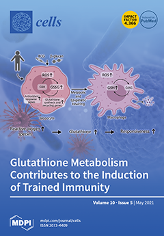
Cover Story (view full-size image): The innate immune system displays memory characteristics characterized by stronger responses to a secondary challenge. This is termed trained immunity and relies on epigenetic and metabolic rewiring. We hypothesized that the redox molecule glutathione plays a role in the induction of trained immunity. Here we show that the modulation of glutathione levels reduced pro-inflammatory cytokine production in human monocytes. Single nucleotide polymorphisms (SNPs) in genes involved in glutathione metabolism were associated with changes in pro-inflammatory cytokine production capacity upon trained immunity. Also, plasma glutathione concentration was positively associated with pro-inflammatory cytokine production. In conclusion, glutathione metabolism is involved in the induction of trained immunity, and future studies are warranted to explore its functional consequences in human diseases. View this paper
  • Issues are regarded as officially published after their release is announced to the table of contents alert mailing list.
  • You may sign up for e-mail alerts to receive table of contents of newly released issues.
  • PDF is the official format for papers published in both, html and pdf forms. To view the papers in pdf format, click on the "PDF Full-text" link, and use the free Adobe Reader to open them.
Order results
Result details
Section
Select all
Export citation of selected articles as:
25 pages, 912 KiB  
Review
Structure and Immune Function of Afferent Lymphatics and Their Mechanistic Contribution to Dendritic Cell and T Cell Trafficking
by Jorge Arasa, Victor Collado-Diaz and Cornelia Halin
Cells 2021, 10(5), 1269; https://doi.org/10.3390/cells10051269 - 20 May 2021
Cited by 27 | Viewed by 5643
Abstract
Afferent lymphatic vessels (LVs) mediate the transport of antigen and leukocytes to draining lymph nodes (dLNs), thereby serving as immunologic communication highways between peripheral tissues and LNs. The main cell types migrating via this route are antigen-presenting dendritic cells (DCs) and antigen-experienced T [...] Read more.
Afferent lymphatic vessels (LVs) mediate the transport of antigen and leukocytes to draining lymph nodes (dLNs), thereby serving as immunologic communication highways between peripheral tissues and LNs. The main cell types migrating via this route are antigen-presenting dendritic cells (DCs) and antigen-experienced T cells. While DC migration is important for maintenance of tolerance and for induction of protective immunity, T cell migration through afferent LVs contributes to immune surveillance. In recent years, great progress has been made in elucidating the mechanisms of lymphatic migration. Specifically, time-lapse imaging has revealed that, upon entry into capillaries, both DCs and T cells are not simply flushed away with the lymph flow, but actively crawl and patrol and even interact with each other in this compartment. Detachment and passive transport to the dLN only takes place once the cells have reached the downstream, contracting collecting vessel segments. In this review, we describe how the anatomy of the lymphatic network supports leukocyte trafficking and provide updated knowledge regarding the cellular and molecular mechanisms responsible for lymphatic migration of DCs and T cells. In addition, we discuss the relevance of DC and T cell migration through afferent LVs and its presumed implications on immunity. Full article
Show Figures

Figure 1

19 pages, 3131 KiB  
Article
Establishing a 3D In Vitro Hepatic Model Mimicking Physiologically Relevant to In Vivo State
by Hyun Kyoung Kang, Madina Sarsenova, Da-Hyun Kim, Min Soo Kim, Jin Young Lee, Eun-Ah Sung, Myung Geun Kook, Nam Gyo Kim, Soon Won Choi, Vyacheslav Ogay and Kyung-Sun Kang
Cells 2021, 10(5), 1268; https://doi.org/10.3390/cells10051268 - 20 May 2021
Cited by 25 | Viewed by 3939
Abstract
Three-dimensional (3D) bioprinting is a promising technology to establish a 3D in vitro hepatic model that holds great potential in toxicological evaluation. However, in current hepatic models, the central area suffers from hypoxic conditions, resulting in slow and weak metabolism of drugs and [...] Read more.
Three-dimensional (3D) bioprinting is a promising technology to establish a 3D in vitro hepatic model that holds great potential in toxicological evaluation. However, in current hepatic models, the central area suffers from hypoxic conditions, resulting in slow and weak metabolism of drugs and toxins. It remains challenging to predict accurate drug effects in current bioprinted hepatic models. Here, we constructed a hexagonal bioprinted hepatic construct and incorporated a spinning condition with continuous media stimuli. Under spinning conditions, HepG2 cells in the bioprinted hepatic construct exhibited enhanced proliferation capacity and functionality compared to those under static conditions. Additionally, the number of spheroids that play a role in boosting drug-induced signals and responses increased in the bioprinted hepatic constructs cultured under spinning conditions. Moreover, HepG2 cells under spinning conditions exhibited intensive TGFβ-induced epithelial-to-mesenchymal transition (EMT) and increased susceptibility to acetaminophen (APAP)-induced hepatotoxicity as well as hepatotoxicity prevention by administration of N-acetylcysteine (NAC). Taken together, the results of our study demonstrate that the spinning condition employed during the generation of bioprinted hepatic constructs enables the recapitulation of liver injury and repair phenomena in particular. This simple but effective culture strategy facilitates bioprinted hepatic constructs to improve in vitro modeling for drug effect evaluation. Full article
Show Figures

Figure 1

18 pages, 538 KiB  
Review
Alzheimer and Purinergic Signaling: Just a Matter of Inflammation?
by Stefania Merighi, Tino Emanuele Poloni, Anna Terrazzan, Eva Moretti, Stefania Gessi and Davide Ferrari
Cells 2021, 10(5), 1267; https://doi.org/10.3390/cells10051267 - 20 May 2021
Cited by 19 | Viewed by 4115
Abstract
Alzheimer’s disease (AD) is a widespread neurodegenerative pathology responsible for about 70% of all cases of dementia. Adenosine is an endogenous nucleoside that affects neurodegeneration by activating four membrane G protein-coupled receptor subtypes, namely P1 receptors. One of them, the A2A subtype, [...] Read more.
Alzheimer’s disease (AD) is a widespread neurodegenerative pathology responsible for about 70% of all cases of dementia. Adenosine is an endogenous nucleoside that affects neurodegeneration by activating four membrane G protein-coupled receptor subtypes, namely P1 receptors. One of them, the A2A subtype, is particularly expressed in the brain at the striatal and hippocampal levels and appears as the most promising target to counteract neurological damage and adenosine-dependent neuroinflammation. Extracellular nucleotides (ATP, ADP, UTP, UDP, etc.) are also released from the cell or are synthesized extracellularly. They activate P2X and P2Y membrane receptors, eliciting a variety of physiological but also pathological responses. Among the latter, the chronic inflammation underlying AD is mainly caused by the P2X7 receptor subtype. In this review we offer an overview of the scientific evidence linking P1 and P2 mediated purinergic signaling to AD development. We will also discuss potential strategies to exploit this knowledge for drug development. Full article
Show Figures

Graphical abstract

16 pages, 637 KiB  
Review
The Complexity of FGF23 Effects on Cardiomyocytes in Normal and Uremic Milieu
by Andreja Figurek, Merita Rroji and Goce Spasovski
Cells 2021, 10(5), 1266; https://doi.org/10.3390/cells10051266 - 20 May 2021
Cited by 5 | Viewed by 4635
Abstract
Fibroblast growth factor-23 (FGF23) appears to be one of the most promising biomarkers and predictors of cardiovascular risk in patients with heart disease and normal kidney function, but moreover in those with chronic kidney disease (CKD). This review summarizes the current knowledge of [...] Read more.
Fibroblast growth factor-23 (FGF23) appears to be one of the most promising biomarkers and predictors of cardiovascular risk in patients with heart disease and normal kidney function, but moreover in those with chronic kidney disease (CKD). This review summarizes the current knowledge of FGF23 mechanisms of action in the myocardium in the physiological and pathophysiological state of CKD, as well as its cross-talk to other important signaling pathways in cardiomyocytes. In this regard, current therapeutic possibilities and future perspectives are also discussed. Full article
(This article belongs to the Special Issue Organization and Function of Cellular Structural Networks)
Show Figures

Figure 1

26 pages, 1546 KiB  
Review
Having an Old Friend for Dinner: The Interplay between Apoptotic Cells and Efferocytes
by Austin Le Lam and Bryan Heit
Cells 2021, 10(5), 1265; https://doi.org/10.3390/cells10051265 - 20 May 2021
Cited by 11 | Viewed by 4191
Abstract
Apoptosis, the programmed and intentional death of senescent, damaged, or otherwise superfluous cells, is the natural end-point for most cells within multicellular organisms. Apoptotic cells are not inherently damaging, but if left unattended, they can lyse through secondary necrosis. The resulting release of [...] Read more.
Apoptosis, the programmed and intentional death of senescent, damaged, or otherwise superfluous cells, is the natural end-point for most cells within multicellular organisms. Apoptotic cells are not inherently damaging, but if left unattended, they can lyse through secondary necrosis. The resulting release of intracellular contents drives inflammation in the surrounding tissue and can lead to autoimmunity. These negative consequences of secondary necrosis are avoided by efferocytosis—the phagocytic clearance of apoptotic cells. Efferocytosis is a product of both apoptotic cells and efferocyte mechanisms, which cooperate to ensure the rapid and complete removal of apoptotic cells. Herein, we review the processes used by apoptotic cells to ensure their timely removal, and the receptors, signaling, and cellular processes used by efferocytes for efferocytosis, with a focus on the receptors and signaling driving this process. Full article
(This article belongs to the Special Issue New Phase of Phagocytosis and a Bite of Trogocytosis)
Show Figures

Figure 1

26 pages, 5274 KiB  
Article
Mycobacterium tuberculosis Binds Human Serum Amyloid A, and the Interaction Modulates the Colonization of Human Macrophages and the Transcriptional Response of the Pathogen
by Malwina Kawka, Anna Brzostek, Katarzyna Dzitko, Jakub Kryczka, Radosław Bednarek, Renata Płocińska, Przemysław Płociński, Dominik Strapagiel, Justyna Gatkowska, Jarosław Dziadek and Bożena Dziadek
Cells 2021, 10(5), 1264; https://doi.org/10.3390/cells10051264 - 20 May 2021
Cited by 12 | Viewed by 3172
Abstract
As a very successful pathogen with outstanding adaptive properties, Mycobacterium tuberculosis (Mtb) has developed a plethora of sophisticated mechanisms to subvert host defenses and effectively enter and replicate in the harmful environment inside professional phagocytes, namely, macrophages. Here, we demonstrated the [...] Read more.
As a very successful pathogen with outstanding adaptive properties, Mycobacterium tuberculosis (Mtb) has developed a plethora of sophisticated mechanisms to subvert host defenses and effectively enter and replicate in the harmful environment inside professional phagocytes, namely, macrophages. Here, we demonstrated the binding interaction of Mtb with a major human acute phase protein, namely, serum amyloid A (SAA1), and identified AtpA (Rv1308), ABC (Rv2477c), EspB (Rv3881c), TB 18.6 (Rv2140c), and ThiC (Rv0423c) membrane proteins as mycobacterial effectors responsible for the pathogen-host protein interplay. SAA1-opsonization of Mtb prior to the infection of human macrophages favored bacterial entry into target phagocytes accompanied by a substantial increase in the load of intracellularly multiplying and surviving bacteria. Furthermore, binding of human SAA1 by Mtb resulted in the up- or downregulation of the transcriptional response of tubercle bacilli. The most substantial changes were related to the increased expression level of the genes of two operons encoding mycobacterial transporter systems, namely, mmpL5/mmpS5 (rv0676c), and rv1217c, rv1218c. Therefore, we postulate that during infection, Mtb-SAA1 binding promotes the infection of host macrophages by tubercle bacilli and modulates the functional response of the pathogen. Full article
Show Figures

Figure 1

21 pages, 309 KiB  
Review
Hepatotoxicity of Contemporary Antiretroviral Drugs: A Review and Evaluation of Published Clinical Data
by Ashley O. Otto, Christina G. Rivera, John D. Zeuli and Zelalem Temesgen
Cells 2021, 10(5), 1263; https://doi.org/10.3390/cells10051263 - 20 May 2021
Cited by 21 | Viewed by 7042
Abstract
Contemporary antiretroviral agents afford enhanced potency and safety for patients living with HIV. Newer antiretroviral drugs are often better tolerated than those initially approved in the early stages of the HIV epidemic. While the safety profile has improved, adverse drug reactions still occur. [...] Read more.
Contemporary antiretroviral agents afford enhanced potency and safety for patients living with HIV. Newer antiretroviral drugs are often better tolerated than those initially approved in the early stages of the HIV epidemic. While the safety profile has improved, adverse drug reactions still occur. We have segregated the antiretroviral agents used in contemporary practice into class groupings based on their mechanism of antiviral activity (non-nucleoside reverse transcriptase inhibitors, nucleoside reverse transcriptase inhibitors, integrase inhibitors, protease inhibitors, and entry inhibitors) while providing a review and discussion of the hepatoxicity seen in the most relevant clinical literature published to date. Clinical literature for individual agents is discussed and agent comparisons afforded within each group in tabular format. Our review will provide a summative overview of the incidence and medications associated with hepatic adverse reactions linked to the use of contemporary antiretroviral drugs. Full article
(This article belongs to the Special Issue Anti-HIV Therapy and the Development of Chronic Liver Diseases)
19 pages, 1987 KiB  
Review
A Potential Role for HUWE1 in Modulating Cisplatin Sensitivity
by Stijn Wenmaekers, Bastiaan J. Viergever, Gunjan Kumar, Onno Kranenburg, Peter C. Black, Mads Daugaard and Richard P. Meijer
Cells 2021, 10(5), 1262; https://doi.org/10.3390/cells10051262 - 20 May 2021
Cited by 11 | Viewed by 5431
Abstract
Cisplatin is a widely used antineoplastic agent, whose efficacy is limited by primary and acquired therapeutic resistance. Recently, a bladder cancer genome-wide CRISPR/Cas9 knock-out screen correlated cisplatin sensitivity to multiple genetic biomarkers. Among the screen’s top hits was the HECT domain-containing ubiquitin E3 [...] Read more.
Cisplatin is a widely used antineoplastic agent, whose efficacy is limited by primary and acquired therapeutic resistance. Recently, a bladder cancer genome-wide CRISPR/Cas9 knock-out screen correlated cisplatin sensitivity to multiple genetic biomarkers. Among the screen’s top hits was the HECT domain-containing ubiquitin E3 ligase (HUWE1). In this review, HUWE1 is postulated as a therapeutic response modulator, affecting the collision between platinum-DNA adducts and the replication fork, the primary cytotoxic action of platins. HUWE1 can alter the cytotoxic response to platins by targeting essential components of the DNA damage response including BRCA1, p53, and Mcl-1. Deficiency of HUWE1 could lead to enhanced DNA damage repair and a dysfunctional apoptotic apparatus, thereby inducing resistance to platins. Future research on the relationship between HUWE1 and platins could generate new mechanistic insights into therapy resistance. Ultimately, HUWE1 might serve as a clinical biomarker to tailor cancer treatment strategies, thereby improving cancer care and patient outcomes. Full article
Show Figures

Figure 1

17 pages, 2147 KiB  
Article
PI3K/mTOR Dual Inhibitor PF-04691502 Is a Schedule-Dependent Radiosensitizer for Gastroenteropancreatic Neuroendocrine Tumors
by Zeta Chow, Jeremy Johnson, Aman Chauhan, Tadahide Izumi, Michael Cavnar, Heidi Weiss, Courtney M. Townsend, Jr., Lowell Anthony, Carrigan Wasilchenko, Matthew L. Melton, Jörg Schrader, B. Mark Evers and Piotr Rychahou
Cells 2021, 10(5), 1261; https://doi.org/10.3390/cells10051261 - 20 May 2021
Cited by 14 | Viewed by 4380
Abstract
Patients with advanced-stage gastroenteropancreatic neuroendocrine tumors (GEP-NETs) have a poor overall prognosis despite chemotherapy and radiotherapy (e.g., peptide receptor radionuclide therapy (PRRT)). Better treatment options are needed to improve disease regression and patient survival. The purpose of this study was to examine a [...] Read more.
Patients with advanced-stage gastroenteropancreatic neuroendocrine tumors (GEP-NETs) have a poor overall prognosis despite chemotherapy and radiotherapy (e.g., peptide receptor radionuclide therapy (PRRT)). Better treatment options are needed to improve disease regression and patient survival. The purpose of this study was to examine a new treatment strategy by combining PI3K/mTOR dual inhibition and radiotherapy. First, we assessed the efficacy of two PI3K/mTOR dual inhibitors, PF-04691502 and PKI-402, to inhibit pAkt and increase apoptosis in NET cell lines (BON and QGP-1) and patient-derived tumor spheroids as single agents or combined with radiotherapy (XRT). Treatment with PF-04691502 decreased pAkt (Ser473) expression for up to 72 h compared with the control; in contrast, decreased pAkt expression was noted for less than 24 h with PKI-402. Simultaneous treatment with PF-04691502 and XRT did not induce apoptosis in NET cells; however, the addition of PF-04691502 48 h after XRT significantly increased apoptosis compared to PF-04691502 or XRT treatment alone. Our results demonstrate that schedule-dependent administration of a PI3K/mTOR inhibitor, combined with XRT, can enhance cytotoxicity by promoting the radiosensitivity of NET cells. Moreover, our findings suggest that radiotherapy, in combination with timed PI3K/mTOR inhibition, may be a promising therapeutic regimen for patients with GEP-NET. Full article
Show Figures

Figure 1

21 pages, 4108 KiB  
Review
Siglec Ligands
by Anabel Gonzalez-Gil and Ronald L. Schnaar
Cells 2021, 10(5), 1260; https://doi.org/10.3390/cells10051260 - 20 May 2021
Cited by 58 | Viewed by 9013
Abstract
A dense and diverse array of glycans on glycoproteins and glycolipids decorate all cell surfaces. In vertebrates, many of these carry sialic acid, in a variety of linkages and glycan contexts, as their outermost sugar moiety. Among their functions, glycans engage complementary glycan [...] Read more.
A dense and diverse array of glycans on glycoproteins and glycolipids decorate all cell surfaces. In vertebrates, many of these carry sialic acid, in a variety of linkages and glycan contexts, as their outermost sugar moiety. Among their functions, glycans engage complementary glycan binding proteins (lectins) to regulate cell physiology. Among the glycan binding proteins are the Siglecs, sialic acid binding immunoglobulin-like lectins. In humans, there are 14 Siglecs, most of which are expressed on overlapping subsets of immune system cells. Each Siglec engages distinct, endogenous sialylated glycans that initiate signaling programs and regulate cellular responses. Here, we explore the emerging science of Siglec ligands, including endogenous sialoglycoproteins and glycolipids and synthetic sialomimetics. Knowledge in this field promises to reveal new molecular pathways controlling cell physiology and new opportunities for therapeutic intervention. Full article
(This article belongs to the Special Issue Sugars on Cell Surfaces and Their Biological Purposes)
Show Figures

Figure 1

14 pages, 2476 KiB  
Article
Knockout of the CMP–Sialic Acid Transporter SLC35A1 in Human Cell Lines Increases Transduction Efficiency of Adeno-Associated Virus 9: Implications for Gene Therapy Potency Assays
by Antje Banning, Anna Zakrzewicz, Xin Chen, Steven J. Gray and Ritva Tikkanen
Cells 2021, 10(5), 1259; https://doi.org/10.3390/cells10051259 - 19 May 2021
Cited by 6 | Viewed by 5177
Abstract
Recombinant adeno-associated viruses (AAV) have emerged as an important tool for gene therapy for human diseases. A prerequisite for clinical approval is an in vitro potency assay that can measure the transduction efficiency of each virus lot produced. The AAV serotypes are typical [...] Read more.
Recombinant adeno-associated viruses (AAV) have emerged as an important tool for gene therapy for human diseases. A prerequisite for clinical approval is an in vitro potency assay that can measure the transduction efficiency of each virus lot produced. The AAV serotypes are typical for gene therapy bind to different cell surface structures. The binding of AAV9 on the surface is mediated by terminal galactose residues present in the asparagine-linked carbohydrates in glycoproteins. However, such terminal galactose residues are rare in cultured cells. They are masked by sialic acid residues, which is an obstacle for the infection of many cell lines with AAV9 and the respective potency assays. The sialic acid residues can be removed by enzymatic digestion or chemical treatment. Still, such treatments are not practical for AAV9 potency assays since they may be difficult to standardize. In this study, we generated human cell lines (HEK293T and HeLa) that become permissive for AAV9 transduction after a knockout of the CMP–sialic acid transporter SLC35A1. Using the human aspartylglucosaminidase (AGA) gene, we show that these cell lines can be used as a model system for establishing potency assays for AAV9-based gene therapy approaches for human diseases. Full article
(This article belongs to the Special Issue Lysosomal Storage Disorders)
Show Figures

Graphical abstract

19 pages, 3078 KiB  
Review
The Autophagy Machinery in Human-Parasitic Protists; Diverse Functions for Universally Conserved Proteins
by Hirokazu Sakamoto, Kumiko Nakada-Tsukui and Sébastien Besteiro
Cells 2021, 10(5), 1258; https://doi.org/10.3390/cells10051258 - 19 May 2021
Cited by 15 | Viewed by 4798
Abstract
Autophagy is a eukaryotic cellular machinery that is able to degrade large intracellular components, including organelles, and plays a pivotal role in cellular homeostasis. Target materials are enclosed by a double membrane vesicle called autophagosome, whose formation is coordinated by autophagy-related proteins (ATGs). [...] Read more.
Autophagy is a eukaryotic cellular machinery that is able to degrade large intracellular components, including organelles, and plays a pivotal role in cellular homeostasis. Target materials are enclosed by a double membrane vesicle called autophagosome, whose formation is coordinated by autophagy-related proteins (ATGs). Studies of yeast and Metazoa have identified approximately 40 ATGs. Genome projects for unicellular eukaryotes revealed that some ATGs are conserved in all eukaryotic supergroups but others have arisen or were lost during evolution in some specific lineages. In spite of an apparent reduction in the ATG molecular machinery found in parasitic protists, it has become clear that ATGs play an important role in stage differentiation or organelle maintenance, sometimes with an original function that is unrelated to canonical degradative autophagy. In this review, we aim to briefly summarize the current state of knowledge in parasitic protists, in the light of the latest important findings from more canonical model organisms. Determining the roles of ATGs and the diversity of their functions in various lineages is an important challenge for understanding the evolutionary background of autophagy. Full article
(This article belongs to the Special Issue Proteins in Autophagic Machinery)
Show Figures

Figure 1

18 pages, 5737 KiB  
Article
Regional Differences in Heat Shock Protein 25 Expression in Brain and Spinal Cord Astrocytes of Wild-Type and SOD1 G93A Mice
by Rebecca San Gil, Benjamin E. Clarke, Heath Ecroyd, Bernadett Kalmar and Linda Greensmith
Cells 2021, 10(5), 1257; https://doi.org/10.3390/cells10051257 - 19 May 2021
Cited by 3 | Viewed by 3133
Abstract
Heterogeneity of glia in different CNS regions may contribute to the selective vulnerability of neuronal populations in neurodegenerative conditions such as amyotrophic lateral sclerosis (ALS). Here, we explored regional variations in the expression of heat shock protein 25 in glia under conditions of [...] Read more.
Heterogeneity of glia in different CNS regions may contribute to the selective vulnerability of neuronal populations in neurodegenerative conditions such as amyotrophic lateral sclerosis (ALS). Here, we explored regional variations in the expression of heat shock protein 25 in glia under conditions of acute and chronic stress. Hsp27 (Hsp27; murine orthologue: Hsp25) fulfils a number of cytoprotective functions and may therefore be a possible therapeutic target in ALS. We identified a subpopulation of astrocytes in primary murine mixed glial cultures that expressed Hsp25. Under basal conditions, the proportion of Hsp25-positive astrocytes was twice as high in spinal cord cultures than in cortical cultures. To explore the physiological role of the elevated Hsp25 expression in spinal cord astrocytes, we exposed cortical and spinal cord glia to acute stress, using heat stress and pro-inflammatory stimuli. Surprisingly, we observed no stress-induced increase in Hsp25 expression in either cortical or spinal cord astrocytes. Similarly, exposure to endogenous stress, as modelled in glial cultures from SOD1 G93A-ALS mice, did not increase Hsp25 expression above that observed in astrocytes from wild-type mice. In vivo, Hsp25 expression was greater under conditions of chronic stress present in the spinal cord of SOD1 G93A mice than in wild-type mice, although this increase in expression is likely to be due to the extensive gliosis that occurs in this model. Together, these results show that there are differences in the expression of Hsp25 in astrocytes in different regions of the central nervous system, but Hsp25 expression is not upregulated under acute or chronic stress conditions. Full article
Show Figures

Figure 1

10 pages, 706 KiB  
Perspective
Chromosome Instability, Aging and Brain Diseases
by Ivan Y. Iourov, Yuri B. Yurov, Svetlana G. Vorsanova and Sergei I. Kutsev
Cells 2021, 10(5), 1256; https://doi.org/10.3390/cells10051256 - 19 May 2021
Cited by 26 | Viewed by 4631
Abstract
Chromosome instability (CIN) has been repeatedly associated with aging and progeroid phenotypes. Moreover, brain-specific CIN seems to be an important element of pathogenic cascades leading to neurodegeneration in late adulthood. Alternatively, CIN and aneuploidy (chromosomal loss/gain) syndromes exhibit accelerated aging phenotypes. Molecularly, cellular [...] Read more.
Chromosome instability (CIN) has been repeatedly associated with aging and progeroid phenotypes. Moreover, brain-specific CIN seems to be an important element of pathogenic cascades leading to neurodegeneration in late adulthood. Alternatively, CIN and aneuploidy (chromosomal loss/gain) syndromes exhibit accelerated aging phenotypes. Molecularly, cellular senescence, which seems to be mediated by CIN and aneuploidy, is likely to contribute to brain aging in health and disease. However, there is no consensus about the occurrence of CIN in the aging brain. As a result, the role of CIN/somatic aneuploidy in normal and pathological brain aging is a matter of debate. Still, taking into account the effects of CIN on cellular homeostasis, the possibility of involvement in brain aging is highly likely. More importantly, the CIN contribution to neuronal cell death may be responsible for neurodegeneration and the aging-related deterioration of the brain. The loss of CIN-affected neurons probably underlies the contradiction between reports addressing ontogenetic changes of karyotypes within the aged brain. In future studies, the combination of single-cell visualization and whole-genome techniques with systems biology methods would certainly define the intrinsic role of CIN in the aging of the normal and diseased brain. Full article
(This article belongs to the Special Issue Aging and Disease)
Show Figures

Figure 1

18 pages, 2722 KiB  
Review
The Role of Trogocytosis in the Modulation of Immune Cell Functions
by Kensuke Miyake and Hajime Karasuyama
Cells 2021, 10(5), 1255; https://doi.org/10.3390/cells10051255 - 19 May 2021
Cited by 49 | Viewed by 14135
Abstract
Trogocytosis is an active process, in which one cell extracts the cell fragment from another cell, leading to the transfer of cell surface molecules, together with membrane fragments. Recent reports have revealed that trogocytosis can modulate various biological responses, including adaptive and innate [...] Read more.
Trogocytosis is an active process, in which one cell extracts the cell fragment from another cell, leading to the transfer of cell surface molecules, together with membrane fragments. Recent reports have revealed that trogocytosis can modulate various biological responses, including adaptive and innate immune responses and homeostatic responses. Trogocytosis is evolutionally conserved from protozoan parasites to eukaryotic cells. In some cases, trogocytosis results in cell death, which is utilized as a mechanism for antibody-dependent cytotoxicity (ADCC). In other cases, trogocytosis-mediated intercellular protein transfer leads to both the acquisition of novel functions in recipient cells and the loss of cellular functions in donor cells. Trogocytosis in immune cells is typically mediated by receptor–ligand interactions, including TCR–MHC interactions and Fcγ receptor-antibody-bound molecule interactions. Additionally, trogocytosis mediates the transfer of MHC molecules to various immune and non-immune cells, which confers antigen-presenting activity on non-professional antigen-presenting cells. In this review, we summarize the recent advances in our understanding of the role of trogocytosis in immune modulation. Full article
(This article belongs to the Special Issue New Phase of Phagocytosis and a Bite of Trogocytosis)
Show Figures

Figure 1

15 pages, 9600 KiB  
Article
Human Umbilical Cord-Derived Mesenchymal Stem Cells Promote Corneal Epithelial Repair In Vitro
by Santhosh Kacham, Tejal Sunil Bhure, Sindhuja D. Eswaramoorthy, Gaurav Naik, Subha Narayan Rath, Sreenivasa Rao Parcha, Sayan Basu, Virender Singh Sangwan and Sachin Shukla
Cells 2021, 10(5), 1254; https://doi.org/10.3390/cells10051254 - 19 May 2021
Cited by 24 | Viewed by 6024
Abstract
Corneal injuries are among the leading causes of blindness and vision impairment. Trauma, infectious keratitis, thermal and chemical (acids and alkali burn) injuries may lead to irreversible corneal scarring, neovascularization, conjunctivalization, and limbal stem cell deficiency. Bilateral blindness constitutes 12% of total global [...] Read more.
Corneal injuries are among the leading causes of blindness and vision impairment. Trauma, infectious keratitis, thermal and chemical (acids and alkali burn) injuries may lead to irreversible corneal scarring, neovascularization, conjunctivalization, and limbal stem cell deficiency. Bilateral blindness constitutes 12% of total global blindness and corneal transplantation remains a stand-alone treatment modality for the majority of end-stage corneal diseases. However, global shortage of donor corneas, the potential risk of graft rejection, and severe side effects arising from long-term use of immunosuppressive medications, demands alternative therapeutic approaches. Umbilical cord-derived mesenchymal stem cells can be isolated in large numbers using a relatively less invasive procedure. However, their role in injury induced corneal repair is largely unexplored. Here, we isolated, cultured and characterized mesenchymal stem cells from human umbilical cord, and studied the expression of mesenchymal (CD73, CD90, CD105, and CD34), ocular surface and epithelial (PAX6, WNT7A, and CK-8/18) lineage markers through immunofluorescence. The cultured human limbal and corneal epithelial cells were used as controls. Scratch assay was used to study the corneal epithelial repair potential of umbilical cord-derived mesenchymal stem cells, in vitro. The in vitro cultured umbilical cord-derived mesenchymal stem cells were plastic adherent, showed trilineage differentiation and expressed: mesenchymal markers CD90, CD105, CD73; epithelial marker CK-8/18, and ocular lineage developmental markers PAX6 and WNT-7A. Our findings suggest that umbilical cord-derived mesenchymal stem cells promote repair of the injured corneal epithelium by stimulating the proliferation of corneal epithelial cells, in vitro. They may serve as a potential non-ocular source of stem cells for treating injury induced bilateral corneal diseases. Full article
(This article belongs to the Section Stem Cells)
Show Figures

Figure 1

25 pages, 8647 KiB  
Article
Transcription Factor β-Catenin Plays a Key Role in Fluid Flow Shear Stress-Mediated Glomerular Injury in Solitary Kidney
by Tarak Srivastava, Daniel P. Heruth, R. Scott Duncan, Mohammad H. Rezaiekhaligh, Robert E. Garola, Lakshmi Priya, Jianping Zhou, Varun C. Boinpelly, Jan Novak, Mohammed Farhan Ali, Trupti Joshi, Uri S. Alon, Yuexu Jiang, Ellen T. McCarthy, Virginia J. Savin, Ram Sharma, Mark L. Johnson and Mukut Sharma
Cells 2021, 10(5), 1253; https://doi.org/10.3390/cells10051253 - 19 May 2021
Cited by 6 | Viewed by 3043
Abstract
Increased fluid flow shear stress (FFSS) in solitary kidney alters podocyte function in vivo. FFSS-treated cultured podocytes show upregulated AKT-GSK3β-β-catenin signaling. The present study was undertaken to confirm (i) the activation of β-catenin signaling in podocytes in vivo using unilaterally nephrectomized (UNX) [...] Read more.
Increased fluid flow shear stress (FFSS) in solitary kidney alters podocyte function in vivo. FFSS-treated cultured podocytes show upregulated AKT-GSK3β-β-catenin signaling. The present study was undertaken to confirm (i) the activation of β-catenin signaling in podocytes in vivo using unilaterally nephrectomized (UNX) TOPGAL mice with the β-galactosidase reporter gene for β-catenin activation, (ii) β-catenin translocation in FFSS-treated mouse podocytes, and (iii) β-catenin signaling using publicly available data from UNX mice. The UNX of TOPGAL mice resulted in glomerular hypertrophy and increased the mesangial matrix consistent with hemodynamic adaptation. Uninephrectomized TOPGAL mice showed an increased β-galactosidase expression at 4 weeks but not at 12 weeks, as assessed using immunofluorescence microscopy (p < 0.001 at 4 weeks; p = 0.16 at 12 weeks) and X-gal staining (p = 0.008 at 4 weeks; p = 0.65 at 12 weeks). Immunofluorescence microscopy showed a significant increase in phospho-β-catenin (Ser552, p = 0.005) at 4 weeks but not at 12 weeks (p = 0.935) following UNX, and the levels of phospho-β-catenin (Ser675) did not change. In vitro FFSS caused a sustained increase in the nuclear translocation of phospho-β-catenin (Ser552) but not phospho-β-catenin (Ser675) in podocytes. The bioinformatic analysis of the GEO dataset, #GSE53996, also identified β-catenin as a key upstream regulator. We conclude that transcription factor β-catenin mediates FFSS-induced podocyte (glomerular) injury in solitary kidney. Full article
Show Figures

Figure 1

31 pages, 2363 KiB  
Review
Role of Glycans on Key Cell Surface Receptors That Regulate Cell Proliferation and Cell Death
by Yin Gao, Xue Luan, Jacob Melamed and Inka Brockhausen
Cells 2021, 10(5), 1252; https://doi.org/10.3390/cells10051252 - 19 May 2021
Cited by 26 | Viewed by 7404
Abstract
Cells undergo proliferation and apoptosis, migration and differentiation via a number of cell surface receptors, most of which are heavily glycosylated. This review discusses receptor glycosylation and the known roles of glycans on the functions of receptors expressed in diverse cell types. We [...] Read more.
Cells undergo proliferation and apoptosis, migration and differentiation via a number of cell surface receptors, most of which are heavily glycosylated. This review discusses receptor glycosylation and the known roles of glycans on the functions of receptors expressed in diverse cell types. We included growth factor receptors that have an intracellular tyrosine kinase domain, growth factor receptors that have a serine/threonine kinase domain, and cell-death-inducing receptors. N- and O-glycans have a wide range of functions including roles in receptor conformation, ligand binding, oligomerization, and activation of signaling cascades. A better understanding of these functions will enable control of cell survival and cell death in diseases such as cancer and in immune responses. Full article
(This article belongs to the Special Issue Sugars on Cell Surfaces and Their Biological Purposes)
Show Figures

Graphical abstract

19 pages, 281 KiB  
Review
Regulatory Effects of Cannabidiol on Mitochondrial Functions: A Review
by John Zewen Chan and Robin Elaine Duncan
Cells 2021, 10(5), 1251; https://doi.org/10.3390/cells10051251 - 19 May 2021
Cited by 43 | Viewed by 5144
Abstract
Cannabidiol (CBD) is part of a group of phytocannabinoids derived from Cannabissativa. Initial work on CBD presumed the compound was inactive, but it was later found to exhibit antipsychotic, anti-depressive, anxiolytic, and antiepileptic effects. In recent decades, evidence has indicated a role [...] Read more.
Cannabidiol (CBD) is part of a group of phytocannabinoids derived from Cannabissativa. Initial work on CBD presumed the compound was inactive, but it was later found to exhibit antipsychotic, anti-depressive, anxiolytic, and antiepileptic effects. In recent decades, evidence has indicated a role for CBD in the modulation of mitochondrial processes, including respiration and bioenergetics, mitochondrial DNA epigenetics, intrinsic apoptosis, the regulation of mitochondrial and intracellular calcium concentrations, mitochondrial fission, fusion and biogenesis, and mitochondrial ferritin concentration and mitochondrial monoamine oxidase activity regulation. Despite these advances, current data demonstrate contradictory findings with regard to not only the magnitude of effects mediated by CBD, but also to the direction of effects. For example, there are data indicating that CBD treatment can increase, decrease, or have no significant effect on intrinsic apoptosis. Differences between studies in cell type, cell-specific response to CBD, and, in some cases, dose of CBD may help to explain differences in outcomes. Most studies on CBD and mitochondria have utilized treatment concentrations that exceed the highest recorded plasma concentrations in humans, suggesting that future studies should focus on CBD treatments within a range observed in pharmacokinetic studies. This review focuses on understanding the mechanisms of CBD-mediated regulation of mitochondrial functions, with an emphasis on findings in neural cells and tissues and therapeutic relevance based on human pharmacokinetics. Full article
(This article belongs to the Collection Determinants of Neuronal Susceptibility to Mitochondrial Disease)
Show Figures

Graphical abstract

22 pages, 4807 KiB  
Article
Modulation of Cyclic AMP Levels in Fallopian Tube Cells by Natural and Environmental Estrogens
by Marinella Rosselli, Barbara P. S. Cometti, Brigitte Leeners, Marta Ewa Szutkowska, Edwin K. Jackson and Raghvendra K. Dubey
Cells 2021, 10(5), 1250; https://doi.org/10.3390/cells10051250 - 19 May 2021
Cited by 4 | Viewed by 3302
Abstract
Autocrine/paracrine factors generated in response to 17β-estradiol (E2) within the fallopian tube (FT) facilitate fertilization and early embryo development for implantation. Since cyclic AMP (cAMP) plays a key role in reproduction, regulation of its synthesis by E2 may be of biological/pathophysiological relevance. Herein, [...] Read more.
Autocrine/paracrine factors generated in response to 17β-estradiol (E2) within the fallopian tube (FT) facilitate fertilization and early embryo development for implantation. Since cyclic AMP (cAMP) plays a key role in reproduction, regulation of its synthesis by E2 may be of biological/pathophysiological relevance. Herein, we investigated whether cAMP production in FT cells (FTCs) is regulated by E2 and environmental estrogens (EE’s; xenoestrogens and phytoestrogens). Under basal conditions, low levels of extracellular cAMP were detectable in bovine FTCs (epithelial cells and fibroblasts; 1:1 ratio). Treatment of FTCs with forskolin (AC; adenylyl cyclase activator), isoproterenol (β-adrenoceptor agonist) and IBMX (phosphodiesterase (PDE) inhibitor) dramatically (>10 fold) increased cAMP; whereas LRE1 (sAC; soluble AC inhibitor) and 2’,5’-dideoxyadenosine (DDA; transmembrane AC (tmAC)) inhibitor decreased cAMP. Comparable changes in basal and stimulated intracellular cAMP were also observed. Ro-20-1724 (PDE-IV inhibitor), but not milrinone (PDE-III inhibitor) nor mmIBMX (PDE-I inhibitor), augmented forskolin-stimulated cAMP levels, suggesting that PDE-IV dominates in FTCs. E2 increased cAMP levels and CREB phosphorylation in FTCs, and these effects were mimicked by EE’s (genistein, 4-hydroxy-2’,4’,6’-trichlorobiphenyl, 4-hydroxy-2’,4’,6’-dichlorobiphenyl). Moreover, the effects of E2 and EE were blocked by the tmAC inhibitor DDA, but not by the ERα/β antagonist ICI182780. Moreover, BAPTA-AM (intracellular-Ca2+ chelator) abrogated the effects of E2, but not genistein, on cAMP suggesting differential involvement of Ca2+. Treatment with non-permeable E2-BSA induced cAMP levels and CREB-phosphorylation; moreover, the stimulatory effects of E2 and EEs on cAMP were blocked by G15, a G protein-coupled estrogen receptor (GPER) antagonist. E2 and IBMX induced cAMP formation was inhibited by LRE1 and DDA suggesting involvement of both tmAC and sAC. Our results provide the first evidence that in FTCs, E2 and EE’s stimulate cAMP synthesis via GPER. Exposure of the FT to EE’s and PDE inhibitors may result in abnormal non-cyclic induction of cAMP levels which may induce deleterious effects on reproduction. Full article
Show Figures

Graphical abstract

17 pages, 6422 KiB  
Article
Fibrous Demineralized Bone Matrix (DBM) Improves Bone Marrow Mononuclear Cell (BMC)-Supported Bone Healing in Large Femoral Bone Defects in Rats
by René D. Verboket, Tanja Irrle, Yannic Busche, Alexander Schaible, Katrin Schröder, Jan C. Brune, Ingo Marzi, Christoph Nau and Dirk Henrich
Cells 2021, 10(5), 1249; https://doi.org/10.3390/cells10051249 - 19 May 2021
Cited by 10 | Viewed by 3123
Abstract
Regeneration of large bone defects is a major objective in trauma surgery. Bone marrow mononuclear cell (BMC)-supported bone healing was shown to be efficient after immobilization on a scaffold. We hypothesized that fibrous demineralized bone matrix (DBM) in various forms with BMCs is [...] Read more.
Regeneration of large bone defects is a major objective in trauma surgery. Bone marrow mononuclear cell (BMC)-supported bone healing was shown to be efficient after immobilization on a scaffold. We hypothesized that fibrous demineralized bone matrix (DBM) in various forms with BMCs is superior to granular DBM. A total of 65 male SD rats were assigned to five treatment groups: syngenic cancellous bone (SCB), fibrous demineralized bone matrix (f-DBM), fibrous demineralized bone matrix densely packed (f-DBM 120%), DBM granules (GDBM) and DBM granules 5% calcium phosphate (GDBM5%Ca2+). BMCs from donor rats were combined with different scaffolds and placed into 5 mm femoral bone defects. After 8 weeks, bone mineral density (BMD), biomechanical stability and histology were assessed. Similar biomechanical properties of f-DBM and SCB defects were observed. Similar bone and cartilage formation was found in all groups, but a significantly bigger residual defect size was found in GDBM. High bone healing scores were found in f-DBM (25) and SCB (25). The application of DBM in fiber form combined with the application of BMCs shows promising results comparable to the gold standard, syngenic cancellous bone. Denser packing of fibers or higher amount of calcium phosphate has no positive effect. Full article
(This article belongs to the Special Issue Cellular Mechanisms of Bone Regeneration)
Show Figures

Graphical abstract

17 pages, 73151 KiB  
Article
IL-10 Mediated Immunomodulation Limits Subepithelial Fibrosis and Repairs Airway Epithelium in Rejecting Airway Allografts
by Mohammad Afzal Khan, Ghazi Abdulmalik Ashoor, Talal Shamma, Fatimah Alanazi, Abdullah Altuhami, Shadab Kazmi, Hala Abdalrahman Ahmed, Abdullah Mohammed Assiri and Dieter Clemens Broering
Cells 2021, 10(5), 1248; https://doi.org/10.3390/cells10051248 - 19 May 2021
Cited by 6 | Viewed by 3288
Abstract
Interleukin-10 plays a vital role in maintaining peripheral immunotolerance and favors a regulatory immune milieu through the suppression of T effector cells. Inflammation-induced microvascular loss has been associated with airway epithelial injury, which is a key pathological source of graft malfunctioning and subepithelial [...] Read more.
Interleukin-10 plays a vital role in maintaining peripheral immunotolerance and favors a regulatory immune milieu through the suppression of T effector cells. Inflammation-induced microvascular loss has been associated with airway epithelial injury, which is a key pathological source of graft malfunctioning and subepithelial fibrosis in rejecting allografts. The regulatory immune phase maneuvers alloimmune inflammation through various regulatory modulators, and thereby promotes graft microvascular repair and suppresses the progression of fibrosis after transplantation. The present study was designed to investigate the therapeutic impact of IL-10 on immunotolerance, in particular, the reparative microenvironment, which negates airway epithelial injury, and fibrosis in a mouse model of airway graft rejection. Here, we depleted and reconstituted IL-10, and serially monitored the phase of immunotolerance, graft microvasculature, inflammatory cytokines, airway epithelium, and subepithelial collagen in rejecting airway transplants. We demonstrated that the IL-10 depletion suppresses FOXP3+ Tregs, tumor necrosis factor-inducible gene 6 protein (TSG-6), graft microvasculature, and establishes a pro-inflammatory phase, which augments airway epithelial injury and subepithelial collagen deposition while the IL-10 reconstitution facilitates FOXP3+ Tregs, TSG-6 deposition, graft microvasculature, and thereby favors airway epithelial repair and subepithelial collagen suppression. These findings establish a potential reparative modulation of IL-10-associated immunotolerance on microvascular, epithelial, and fibrotic remodeling, which could provide a vital therapeutic option to rescue rejecting transplants in clinical settings. Full article
(This article belongs to the Collection Advances in Immune Monitoring)
Show Figures

Figure 1

21 pages, 2954 KiB  
Article
Maternal Exercise Mediates Hepatic Metabolic Programming via Activation of AMPK-PGC1α Axis in the Offspring of Obese Mothers
by Philipp Kasper, Saida Breuer, Thorben Hoffmann, Christina Vohlen, Ruth Janoschek, Lisa Schmitz, Sarah Appel, Gregor Fink, Christoph Hünseler, Alexander Quaas, Münevver Demir, Sonja Lang, Hans-Michael Steffen, Anna Martin, Christoph Schramm, Martin Bürger, Esther Mahabir, Tobias Goeser, Jörg Dötsch, Eva Hucklenbruch-Rother and Inga Bae-Gartzadd Show full author list remove Hide full author list
Cells 2021, 10(5), 1247; https://doi.org/10.3390/cells10051247 - 19 May 2021
Cited by 17 | Viewed by 3705
Abstract
Maternal obesity is associated with an increased risk of hepatic metabolic dysfunction for both mother and offspring and targeted interventions to address this growing metabolic disease burden are urgently needed. This study investigates whether maternal exercise (ME) could reverse the detrimental effects of [...] Read more.
Maternal obesity is associated with an increased risk of hepatic metabolic dysfunction for both mother and offspring and targeted interventions to address this growing metabolic disease burden are urgently needed. This study investigates whether maternal exercise (ME) could reverse the detrimental effects of hepatic metabolic dysfunction in obese dams and their offspring while focusing on the AMP-activated protein kinase (AMPK), representing a key regulator of hepatic metabolism. In a mouse model of maternal western-style-diet (WSD)-induced obesity, we established an exercise intervention of voluntary wheel-running before and during pregnancy and analyzed its effects on hepatic energy metabolism during developmental organ programming. ME prevented WSD-induced hepatic steatosis in obese dams by alterations of key hepatic metabolic processes, including activation of hepatic ß-oxidation and inhibition of lipogenesis following increased AMPK and peroxisome-proliferator-activated-receptor-γ-coactivator-1α (PGC-1α)-signaling. Offspring of exercised dams exhibited a comparable hepatic metabolic signature to their mothers with increased AMPK-PGC1α-activity and beneficial changes in hepatic lipid metabolism and were protected from WSD-induced adipose tissue accumulation and hepatic steatosis in later life. In conclusion, this study demonstrates that ME provides a promising strategy to improve the metabolic health of both obese mothers and their offspring and highlights AMPK as a potential metabolic target for therapeutic interventions. Full article
(This article belongs to the Section Intracellular and Plasma Membranes)
Show Figures

Graphical abstract

19 pages, 1558 KiB  
Review
Store Operated Calcium Entry in Cell Migration and Cancer Metastasis
by Ayat S. Hammad and Khaled Machaca
Cells 2021, 10(5), 1246; https://doi.org/10.3390/cells10051246 - 19 May 2021
Cited by 34 | Viewed by 6002
Abstract
Ca2+ signaling is ubiquitous in eukaryotic cells and modulates many cellular events including cell migration. Directional cell migration requires the polarization of both signaling and structural elements. This polarization is reflected in various Ca2+ signaling pathways that impinge on cell movement. [...] Read more.
Ca2+ signaling is ubiquitous in eukaryotic cells and modulates many cellular events including cell migration. Directional cell migration requires the polarization of both signaling and structural elements. This polarization is reflected in various Ca2+ signaling pathways that impinge on cell movement. In particular, store-operated Ca2+ entry (SOCE) plays important roles in regulating cell movement at both the front and rear of migrating cells. SOCE represents a predominant Ca2+ influx pathway in non-excitable cells, which are the primary migrating cells in multicellular organisms. In this review, we summarize the role of Ca2+ signaling in cell migration with a focus on SOCE and its diverse functions in migrating cells and cancer metastasis. SOCE has been implicated in regulating focal adhesion turnover in a polarized fashion and the mechanisms involved are beginning to be elucidated. However, SOCE is also involved is other aspects of cell migration with a less well-defined mechanistic understanding. Therefore, much remains to be learned regarding the role and regulation of SOCE in migrating cells. Full article
(This article belongs to the Collection STIM and Orai Communication in Health and Disease)
Show Figures

Figure 1

20 pages, 2142 KiB  
Article
TILLING-by-Sequencing+ Reveals the Role of Novel Fatty Acid Desaturases (GmFAD2-2s) in Increasing Soybean Seed Oleic Acid Content
by Naoufal Lakhssassi, Valéria Stefania Lopes-Caitar, Dounya Knizia, Mallory A. Cullen, Oussama Badad, Abdelhalim El Baze, Zhou Zhou, Mohamed G. Embaby, Jonas Meksem, Aicha Lakhssassi, Pengyin Chen, Amer AbuGhazaleh, Tri D. Vuong, Henry T. Nguyen, Tarek Hewezi and Khalid Meksem
Cells 2021, 10(5), 1245; https://doi.org/10.3390/cells10051245 - 19 May 2021
Cited by 17 | Viewed by 3705
Abstract
Soybean is the second largest source of oil worldwide. Developing soybean varieties with high levels of oleic acid is a primary goal of the soybean breeders and industry. Edible oils containing high level of oleic acid and low level of linoleic acid are [...] Read more.
Soybean is the second largest source of oil worldwide. Developing soybean varieties with high levels of oleic acid is a primary goal of the soybean breeders and industry. Edible oils containing high level of oleic acid and low level of linoleic acid are considered with higher oxidative stability and can be used as a natural antioxidant in food stability. All developed high oleic acid soybeans carry two alleles; GmFAD2-1A and GmFAD2-1B. However, when planted in cold soil, a possible reduction in seed germination was reported when high seed oleic acid derived from GmFAD2-1 alleles were used. Besides the soybean fatty acid desaturase (GmFAD2-1) subfamily, the GmFAD2-2 subfamily is composed of five members, including GmFAD2-2A, GmFAD2-2B, GmFAD2-2C, GmFAD2-2D, and GmFAD2-2E. Segmental duplication of GmFAD2-1A/GmFAD2-1B, GmFAD2-2A/GmFAD2-2C, GmFAD2-2A/GmFAD2-2D, and GmFAD2-2D/GmFAD2-2C have occurred about 10.65, 27.04, 100.81, and 106.55 Mya, respectively. Using TILLING-by-Sequencing+ technology, we successfully identified 12, 8, 10, 9, and 19 EMS mutants at the GmFAD2-2A, GmFAD2-2B, GmFAD2-2C, GmFAD2-2D, and GmFAD2-2E genes, respectively. Functional analyses of newly identified mutants revealed unprecedented role of the five GmFAD2-2A, GmFAD2-2B, GmFAD2-2C, GmFAD2-2D, and GmFAD2-2E members in controlling the seed oleic acid content. Most importantly, unlike GmFAD2-1 members, subcellular localization revealed that members of the GmFAD2-2 subfamily showed a cytoplasmic localization, which may suggest the presence of an alternative fatty acid desaturase pathway in soybean for converting oleic acid content without substantially altering the traditional plastidial/ER fatty acid production. Full article
(This article belongs to the Section Cellular Metabolism)
Show Figures

Figure 1

15 pages, 13344 KiB  
Article
Polyamine Metabolism in Scots Pine Embryogenic Cells under Potassium Deficiency
by Riina Muilu-Mäkelä, Jaana Vuosku, Hely Häggman and Tytti Sarjala
Cells 2021, 10(5), 1244; https://doi.org/10.3390/cells10051244 - 18 May 2021
Cited by 2 | Viewed by 2554
Abstract
Polyamines (PA) have a protective role in maintaining growth and development in Scots pine during abiotic stresses. In the present study, a controlled liquid Scots pine embryogenic cell culture was used for studying the responses of PA metabolism related to potassium deficiency. The [...] Read more.
Polyamines (PA) have a protective role in maintaining growth and development in Scots pine during abiotic stresses. In the present study, a controlled liquid Scots pine embryogenic cell culture was used for studying the responses of PA metabolism related to potassium deficiency. The transcription level regulation of PA metabolism led to the accumulation of putrescine (Put). Arginine decarboxylase (ADC) had an increased expression trend under potassium deficiency, whereas spermidine synthase (SPDS) expression decreased. Generally, free spermidine (Spd) and spermine (Spm)/ thermospermine (t-Spm) contents were kept relatively stable, mostly by the downregulation of polyamine oxidase (PAO) expression. The low potassium contents in the culture medium decreased the potassium content of the cells, which inhibited cell mass growth, but did not affect cell viability. The reduced growth was probably caused by repressed metabolic activity and cell division, whereas there were no signs of H2O2-induced oxidative stress or increased cell death. The low intracellular content of K+ decreased the content of Na+. The decrease in the pH of the culture medium indicated that H+ ions were pumped out of the cells. Altogether, our findings emphasize the specific role(s) of Put under potassium deficiency and strict developmental regulation of PA metabolism in Scots pine. Full article
(This article belongs to the Special Issue Plant Polyamines in Plant Stress Tolerance)
Show Figures

Figure 1

11 pages, 698 KiB  
Article
Segmental Evaluation of Thoracic Aortic Calcium and Their Relations with Cardiovascular Risk Factors in the Brazilian Longitudinal Study of Adult Health (ELSA-Brasil)
by Jesiana Ferreira Pedrosa, Luisa Campos Caldeira Brant, Stephanie Alves de Aquino, Antonio Luiz Ribeiro and Sandhi Maria Barreto
Cells 2021, 10(5), 1243; https://doi.org/10.3390/cells10051243 - 18 May 2021
Cited by 5 | Viewed by 2744
Abstract
Thoracic aortic calcium (TAC) appears to be a subclinical marker of cardiovascular disease (CVD) and to predict cardiovascular (CV) mortality. However, studies on TAC use tomographic scans obtained for coronary artery calcium (CAC) score, which does not include the aortic arch. This study [...] Read more.
Thoracic aortic calcium (TAC) appears to be a subclinical marker of cardiovascular disease (CVD) and to predict cardiovascular (CV) mortality. However, studies on TAC use tomographic scans obtained for coronary artery calcium (CAC) score, which does not include the aortic arch. This study evaluates TAC prevalence in aortic arch (AAC), ascending (ATAC) and descending thoracic aorta (DTAC) and verify whether they are associated with the same CV risk factors. Cross-sectional analysis, including 2427 participants (mean age 55.6 ± 8.7; 54.1% women) of the ELSA-Brasil cohort. Nonenhanced ECG-gated tomographies were performed in 2015–2016. Multivariable logistic regression estimated the CV risk factors associated with calcium in each segment. Overall prevalence of ATAC, AAC and DTAC was, 23.1%, 62.1%, and 31.2%, respectively. About 90.4% of the individuals with TAC had AAC and only 19.5% had calcium in all segments. In the multivariable analysis, increasing age, lower levels of schooling, current smoking, higher body mass index, and hypertension remained associated with calcium in all segments. No sex or race/ethnicity differences were found in any aortic segment. Diabetes and dyslipidemia were associated with ATAC and DTAC, but not with AAC, suggesting that AAC may reflect an overlap of mechanisms that impact vascular health, including atherosclerosis. Full article
(This article belongs to the Special Issue Research on Vascular Calcification in Cardiovascular Disease)
Show Figures

Figure 1

19 pages, 3116 KiB  
Article
Preliminary Phytochemical Analysis and Evaluation of the Biological Activity of Leonotis nepetifolia (L.) R. Br Transformed Roots Extracts Obtained through Rhizobium rhizogenes-Mediated Transformation
by Tomasz Kowalczyk, Anna Merecz-Sadowska, Patricia Rijo, Vera M. S. Isca, Laurent Picot, Marzena Wielanek, Tomasz Śliwiński and Przemysław Sitarek
Cells 2021, 10(5), 1242; https://doi.org/10.3390/cells10051242 - 18 May 2021
Cited by 18 | Viewed by 4388
Abstract
According to the present knowledge, this is the first report on establishing transformed root cultures of Leonotis nepetifolia after Rhizobium rhizogenes-mediated transformation. The preliminary phytochemical analysis showed differences in the content of phenols and flavonoids in transformed and nontransformed roots. The dominant [...] Read more.
According to the present knowledge, this is the first report on establishing transformed root cultures of Leonotis nepetifolia after Rhizobium rhizogenes-mediated transformation. The preliminary phytochemical analysis showed differences in the content of phenols and flavonoids in transformed and nontransformed roots. The dominant compounds in the analyzed extracts were (+)-catechin (5464 and 6808 µg/g DW), p-coumaric acid (2549 and 4907 µg/g DW), m-coumaric acid (1508 and 2048 µg/g DW) and rosmarinic acid (1844 and 2643 µg/g DW) for nontransformed (LNNR) and transformed (LNTR4) roots, respectively. Initial biological studies carried out on LNNR, and LNTR4 extracts showed a cytotoxic effect on the A549 lung, HCC1937 breast and leukemia NALM-6 cell lines, antioxidants, as well as repair and protection against DNA damage induced by H2O2 in HUVEC cells. Due to the stronger effect of the LNTR4 root extract, which can be a relatively efficient and cheap source of bioactive secondary metabolites, further biological analyses are needed to discover in detail their potentially valuable biological properties. Full article
Show Figures

Graphical abstract

16 pages, 2414 KiB  
Communication
foxm1 Modulates Cell Non-Autonomous Response in Zebrafish Skeletal Muscle Homeostasis
by Fábio J. Ferreira, Leonor Carvalho, Elsa Logarinho and José Bessa
Cells 2021, 10(5), 1241; https://doi.org/10.3390/cells10051241 - 18 May 2021
Cited by 8 | Viewed by 4835
Abstract
foxm1 is a master regulator of the cell cycle, contributing to cell proliferation. Recent data have shown that this transcription factor also modulates gene networks associated with other cellular mechanisms, suggesting non-proliferative functions that remain largely unexplored. In this study, we used CRISPR/Cas9 [...] Read more.
foxm1 is a master regulator of the cell cycle, contributing to cell proliferation. Recent data have shown that this transcription factor also modulates gene networks associated with other cellular mechanisms, suggesting non-proliferative functions that remain largely unexplored. In this study, we used CRISPR/Cas9 to disrupt foxm1 in the zebrafish terminally differentiated fast-twitching muscle cells. foxm1 genomic disruption increased myofiber death and clearance. Interestingly, this contributed to non-autonomous satellite cell activation and proliferation. Moreover, we observed that Cas9 expression alone was strongly deleterious to muscle cells. Our report shows that foxm1 modulates a muscle non-autonomous response to myofiber death and highlights underreported toxicity to high expression of Cas9 in vivo. Full article
Show Figures

Figure 1

15 pages, 820 KiB  
Review
Targeting the VEGF Pathway in Osteosarcoma
by Tarek Assi, Sarah Watson, Bachar Samra, Elie Rassy, Axel Le Cesne, Antoine Italiano and Olivier Mir
Cells 2021, 10(5), 1240; https://doi.org/10.3390/cells10051240 - 18 May 2021
Cited by 39 | Viewed by 4405
Abstract
Osteosarcoma is the most common primary tumor of the bones affecting mainly young adults. Despite the advances in the field of systemic anticancer therapy, the prognosis of relapsed of metastatic osteosarcoma patients remain dismal with very short survival. However, the better understanding of [...] Read more.
Osteosarcoma is the most common primary tumor of the bones affecting mainly young adults. Despite the advances in the field of systemic anticancer therapy, the prognosis of relapsed of metastatic osteosarcoma patients remain dismal with very short survival. However, the better understanding of the pathophysiology of this subtype of sarcoma has led to the identification of new targeted agents with significant activity. In fact, increased angiogenesis plays a major role in the tumor growth and survival of osteosarcoma patients. Several targeted agents have demonstrated a significant anti-tumor activity including multi-kinase inhibitors. In this review, we will discuss the pathophysiology, rationale, and role of targeting angiogenesis via the VEGF pathway in patients with osteosarcoma with emphasis on the published clinical trials and future directions. Full article
(This article belongs to the Special Issue Research Advances and Therapy of Human Osteosarcoma)
Show Figures

Figure 1

Previous Issue
Next Issue
Back to TopTop