


Abstract
High-dispersion nanocomposite materials based on cerium(III) orthophosphate stabilized with different forms of cellulose (methylcellulose (MC), carboxymethylcellulose (CMC) and sodium carboxymethylcellulose (CMCNa)) with the addition of citric acid were synthesized by chemical precipitation. Experimental samples modified with metronidazole were obtained. A comprehensive analysis of their phase composition, morphology, and physicochemical characteristics was performed using X-ray diffraction, transmission electron microscopy and IR spectroscopy. According to the results of physicochemical studies all synthesized nanocomposites contained nanocrystals with a bimodal size distribution (width 3–6 nm, length 15–200 nm), depending on the matrix composition, and diffraction peaks characteristic of CePO4. IR spectroscopy suggested a mechanism for the formation of chemical bonds: cellulose polymer chains are retained on the CePO4 surface mainly through a branched network of weak hydrogen bonds, while the carboxylate groups of citric acid form coordination bonds with Ce3+. A positive effect of citric acid on limiting the linear dimensions of CePO4 crystallites in the presence of a cellulose matrix was established.
1. Introduction
The development of nanocomposite materials based on biocompatible polymer matrices is one of the fastest-growing fields in polymer science, biomedicine and pharmaceuticals [1,2,3]. A promising and actively developing direction is the creation of innovative hybrid materials combining the catalytic and biochemical functionality of rare earth element (REE) compounds with the high specific surface area, lightness and biodegradability of biopolymers [4,5,6,7]. Cerium nanocompounds (in particular, their oxides and phosphates) are of great interest to researchers in the field of tissue regeneration due to their unique antioxidant, bacteriostatic and immunomodulatory properties [5,6,7,8,9,10,11]. The use of uncoated nanoparticles is often limited due to their tendency to aggregate [11,12,13]. The introduction (encapsulation) of nanoparticles into three-dimensional macromolecular polymer matrices allows not only their stabilization due to spatial effects, but also regulation of the release kinetics of therapeutic agents, providing a prolonged effect [1,2,6,12,13]. The construction of organic-inorganic systems, where natural polysaccharides serve as matrices and rare earth element compounds act as functional fillers, is of practical interest to researchers in various fields of science and technology. Lanthanides impart unique catalytic and optical properties to such composites, while the polymer framework provides mechanical strength, biodegradability and delayed release kinetics due to the controlled porosity of the obtained materials [4,5,6,7,13,14].
The basic structural element for the creation of such matrices is cellulose [4,5,6,15,16,17,18,19]. Cellulose is a linear polysaccharide consisting of anhydroglucose units (AGU) linked by β-(1→4)-glycosidic bonds, with a degree of polymerization (DP) of 50–5000 and more. The developed system of intra- and intermolecular hydrogen bonds determines the high crystallinity and insolubility of natural cellulose, which requires its conversion to water-soluble ethers. Chemical modification of AGU hydroxyl groups allows the production of polyelectrolytes, which can be either nonionic (methylcellulose, MC) or anionic (carboxymethylcellulose, CMC) [20,21,22]. Cellulose derivatives are actively used in various fields, including the development of biomedical products and drugs [22,23,24,25,26]. The introduction of inorganic salts into CMC solutions significantly changes the conformation of polymer chains due to the shielding of carboxylate groups. While monovalent cations preserve phase stability, multivalent ions (including lanthanides, in particular, Ce3+/Ce4+) cause ionic cross-linking of macromolecules. From the point of view of nanomaterial synthesis, coordination of CMC functional groups with metal ions limits their diffusion, localizing nucleation processes and preventing sedimentation and aggregation of the resulting nanocrystals [4,13,27,28,29,30].
Macroporous three-dimensional hydrogels and cryogels based on collagen, alginate and gum arabic are capable of acquiring similarity to the extracellular matrix, retaining moisture and providing gas exchange [31,32,33,34,35,36,37]. Modification of such hydrogel matrices with cerium oxide and phosphate nanoparticles makes it possible to reduce oxidative stress in the wound area due to the pronounced antioxidant activity of the filler [28,37,38,39,40,41,42,43]. Alternatively, bacterial cellulose (BC) can be used, which demonstrates high rehydration capacity and stability of the three-dimensional network [17,23,25,42,44].
During the formation of heterogeneous polymer-inorganic systems, the main chemical processes occur at the phase interface. During the synthesis of CeO2/CePO4 nanocomposites, the polymer matrix acts as a template, limiting the spatial growth of grains, while charge transfer in the redox cycle Ce3+ → Ce4+ on the matrix surface provides catalytic oxidase and phosphatase activity of the system [45,46,47,48], blocking pathogen metabolism [8,10,11,28,43,48,49,50]. Similar synthesis principles are used during the mineralization of calcium phosphate phases on a bacterial cellulose matrix (BC-Ce:CaP) [51] and the creation of cement composites for osteogenesis [52,53,54,55]. The incorporation of the rare earth element cerium (Ce) into the lattice of doped hydroxyapatite (GA) changes the parameters of the crystal lattice and the specific surface area of the polymer-mineral framework (Ce-GA), which allows its use for prolonged desorption of doxorubicin [55,56]. At the same time, the macromolecular environment forms a so-called “protein corona” on the nanoparticle surface, determining biological compatibility in the organism’s liquid media [57].
The kinetics of controlled drug release from hydrogels directly depends on the cross-linking density of the polymer network and the degree of its swelling [28]. A comparative analysis of antibiotic diffusion from composite matrices “chitosan–collagen–Ce-GA” showed that the polymer polyelectrolyte network acts as a diffusion barrier, prolonging drug release compared with unmodified mineral carriers. Thus, hydroxyapatite releases 90% of gentamicin within the first 10 min, whereas chitosan–collagen–cerium hydroxyapatite nanocomposites release 80% of gentamicin after two hours [58]. In this regard, the widely known antimicrobial and antiprotozoal drug metronidazole [59,60,61], which is compatible with various excipients, such as mannitol, various forms of cellulose and gums [60,62], can be considered promising.
This work is a continuation of studies on the synthesis methodology of nanosized cerium compounds [38,43,50,63,64,65], modified with cellulose derivatives, citric acid and the antimicrobial agent metronidazole.
The aim of the present study is the chemical co-precipitation of highly dispersed nanocomposite materials based on cerium(III) orthophosphate, stabilized with various forms of cellulose (MC, CMC, CMCNa) with the addition of citric acid, as well as the preparation of experimental samples modified with metronidazole, followed by a comprehensive analysis of their phase composition, morphology and physicochemical characteristics using X-ray diffraction (XRD), transmission electron microscopy (TEM) and IR spectroscopy methods.
The main scientific novelty of the work lies in establishing the regularities of the influence of the nature and concentration of various water-soluble forms of cellulose and tricarboxylic acids on the geometric parameters and aggregative stability of cerium phosphate nanoparticles during their co-precipitation and simultaneous introduction of the drug agent.
2. Materials and Methods
2.1. Reagents and Materials
The following starting materials and reagents were used in this study: cerium(III) nitrate hexahydrate, Ce(NO3)3·6H2O (99.99%, LANHIT LLC, Moscow, Russia), as the matrix precursor; ammonium dihydrogen phosphate, NH4H2PO4 (“chemically pure for analysis”, 99.5%, Vekton, Moscow, Russia), as the precipitating agent. Water-soluble cellulose derivatives were used as organic modifiers and structure-directing agents: nonionic methylcellulose (MC) with a degree of substitution (DS) of 1.4–2.2, anionic carboxymethyl cellulose (CMC), and its sodium salt (CMCNa) with a degree of substitution of 1.5. Citric acid (Cit) was used to further functionalize the phosphate matrix, while metronidazole was employed as a commercial antibacterial agent. All solutions were prepared using double-distilled water.
2.2. Synthesis of CePO4 Nanocomposites Modified with Cellulose Derivatives
Highly dispersed composite materials based on cerium(III) orthophosphate were synthesized by the precipitation method at room temperature. This method has been used repeatedly and described in detail in our previous works [10,43,64,65].
To ensure reproducibility of the synthesis conditions, the total concentration of components in the reaction medium was maintained constant, calculated as CeO2 (12.5 g/L) and NH4H2PO4 (25 g/L). The concentration of the introduced polymer (MC, CMC, or Na-CMC) was used as the variable parameter and was 2.5, 1.25, 0.625, 0.3125, or 0.156 g/L.
A typical synthesis procedure was as follows. Under continuous stirring (stirring rate of 350 rpm), 100 mL of the initial Ce(NO3)3 solution (125 g/L calculated as CeO2) and the calculated volume of an aqueous solution of the corresponding cellulose derivative (stock solution containing 5 g in 500 mL of water: 250, 125, or 62.5 mL) were successively introduced into the reaction vessel. The total volume was then adjusted to 875 mL with distilled water. Subsequently, 125 mL of NH4H2PO4 solution (100 g/L) was uniformly added above the surface of the resulting mixture by spray dosing over 15 min using a spray nozzle. The pH of the reaction medium during precipitation was approximately 3.0 and was not additionally adjusted.
Immediately after completion of the precipitation process, the resulting cerium (III) phosphate suspension was transferred into graduated cylinders for fractional separation of nanoparticles according to their size in the mother liquor. The upper sol fraction, containing the most stable composite nanoparticles, was separated and used for subsequent physicochemical characterization. To obtain powder samples, the materials were dried. In addition, part of the CMC-based sample series was subjected to thermal treatment (annealing) at 400 °C for 3 h to investigate thermal stability and phase evolution.
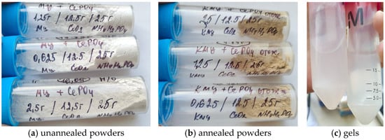
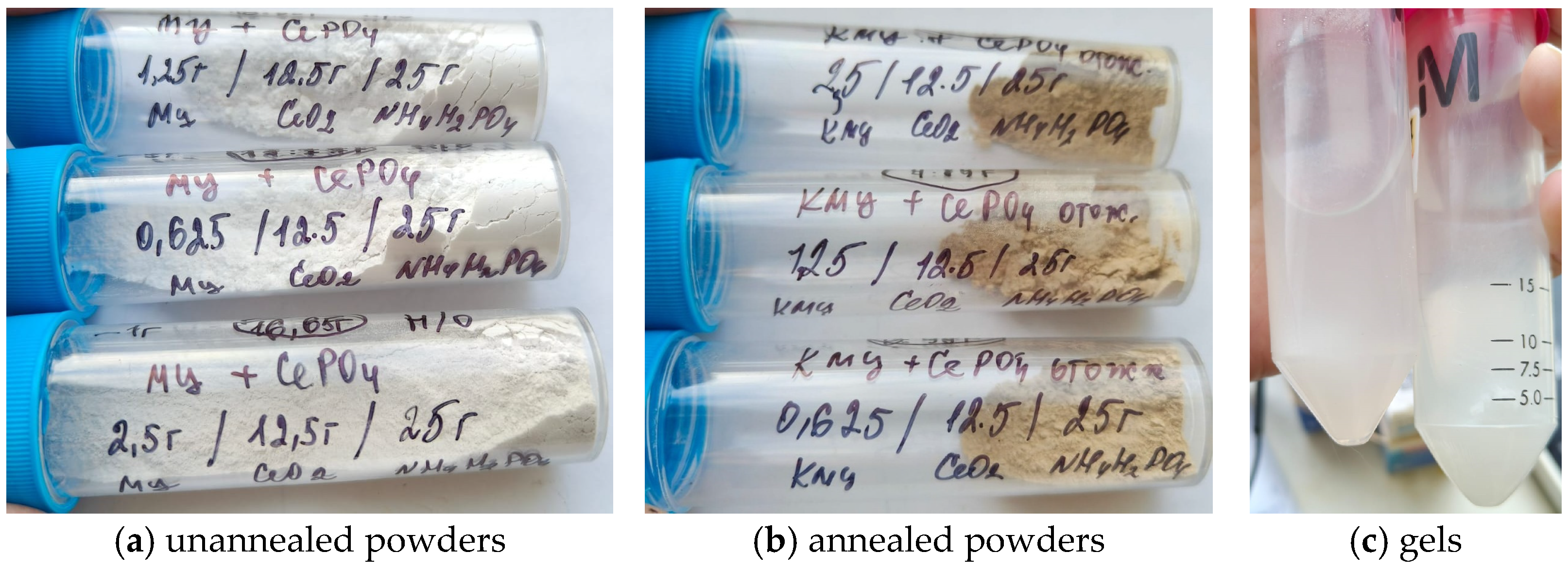
The appearance of the unannealed and annealed samples of nanocomposite powders is shown in Figure 1.
Figure 1.
Photographs of nanocomposite samples sent for analysis: (a) unannealed powders (methylcellulose used, from top to bottom: 1.25 g, 0.625 g, 2.5 g); (b) annealed powders (carboxymethyl cellulose used, from top to bottom: 2.5 g, 1.25 g, 0.625 g); (c) gels (from (a) on the left, from (b) on the right).
2.3. Synthesis of Multicomponent CePO4 Nanocomposites, Additionally Incorporating Citric Acid and Metronidazole
To obtain multicomponent nanocomposites, the synthesis was carried out in a total reaction volume of 500 mL using a modified procedure with thermal activation. Citric acid (final concentration 12.5 g/L) was introduced into the reaction system containing the cerium precursor and cellulose matrix solutions (final polymer concentration 0.625 g/L) as a modifier of the phosphate matrix, together with a commercial metronidazole preparation in an amount of 500 mg per batch. Precipitation with ammonium dihydrogen phosphate was performed at an elevated temperature (38 °C) under continuous stirring, providing efficient cocrystallization and encapsulation of the drug within the hydrophilic organic–inorganic matrix.
2.4. Physicochemical Characterization of the Materials
Transmission electron microscopy (TEM) was performed at the Collective Use Center “Materials Science and Metallurgy” of the National University of Science and Technology MISIS. The morphology, particle size, and degree of nanoparticle agglomeration were investigated using a JEM 2100 high-resolution transmission electron microscope (JEOL, Akishima, Tokyo, Japan) equipped with a LaB6 cathode and operated at an accelerating voltage of 200 kV. To ensure high spatial coherence and image resolution, condenser (50 μm) and objective (40 μm) apertures were used. The average electron beam diameter was approximately 100 nm (Spot size 3). Images were acquired after a 2 h thermal stabilization of the instrument. The local crystalline structure was confirmed by electron diffraction (ring diffraction patterns).
X-ray diffraction (XRD). The qualitative and quantitative phase composition, as well as the crystal structure parameters of the obtained nanopowders, were determined by X-ray diffraction using a Bruker D8 diffractometer (Bruker AXS, Karlsruhe, Germany) with monochromatized CuKα radiation (λ = 0.154 nm). Diffraction patterns were recorded in the θ − 2\θ geometry at an operating voltage of 40 kV and a tube current of 40 mA. The crystallite size (coherent scattering domain size) was estimated from the broadening of the diffraction peaks.
Fourier-transform infrared spectroscopy (FTIR, IR) of annealed powders: Intermolecular interactions, the functional composition of the composites, and the incorporation of the therapeutic agent were investigated by Fourier-transform infrared spectroscopy over the wavenumber range of 400–4000 cm−1. The measurements were performed using a Bruker VERTEX 70 FTIR spectrometer (Bruker Optik GmbH, Ettlingen, Germany) equipped with a PIKE MIRacle attenuated total reflectance (ATR) accessory (PIKE Technologies, Cottonwood, AZ, USA) with a diamond crystal mounted on a zinc selenide substrate. The spectra were recorded under the following conditions: 32 scans, spectral resolution of 4 cm−1, spectral range of 4000–600 cm−1, and background spectrum collected immediately before each measurement.
3. Results
3.1. Phase Composition and Crystal Structure
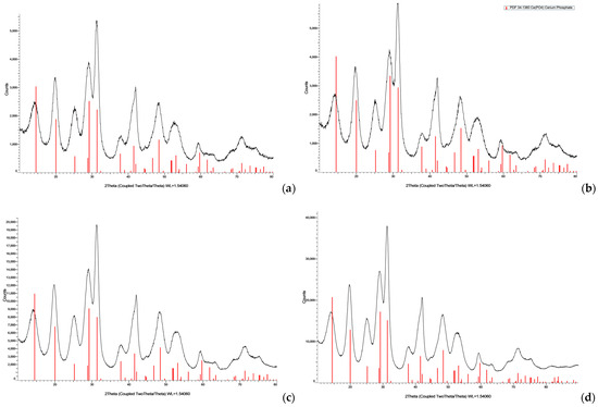
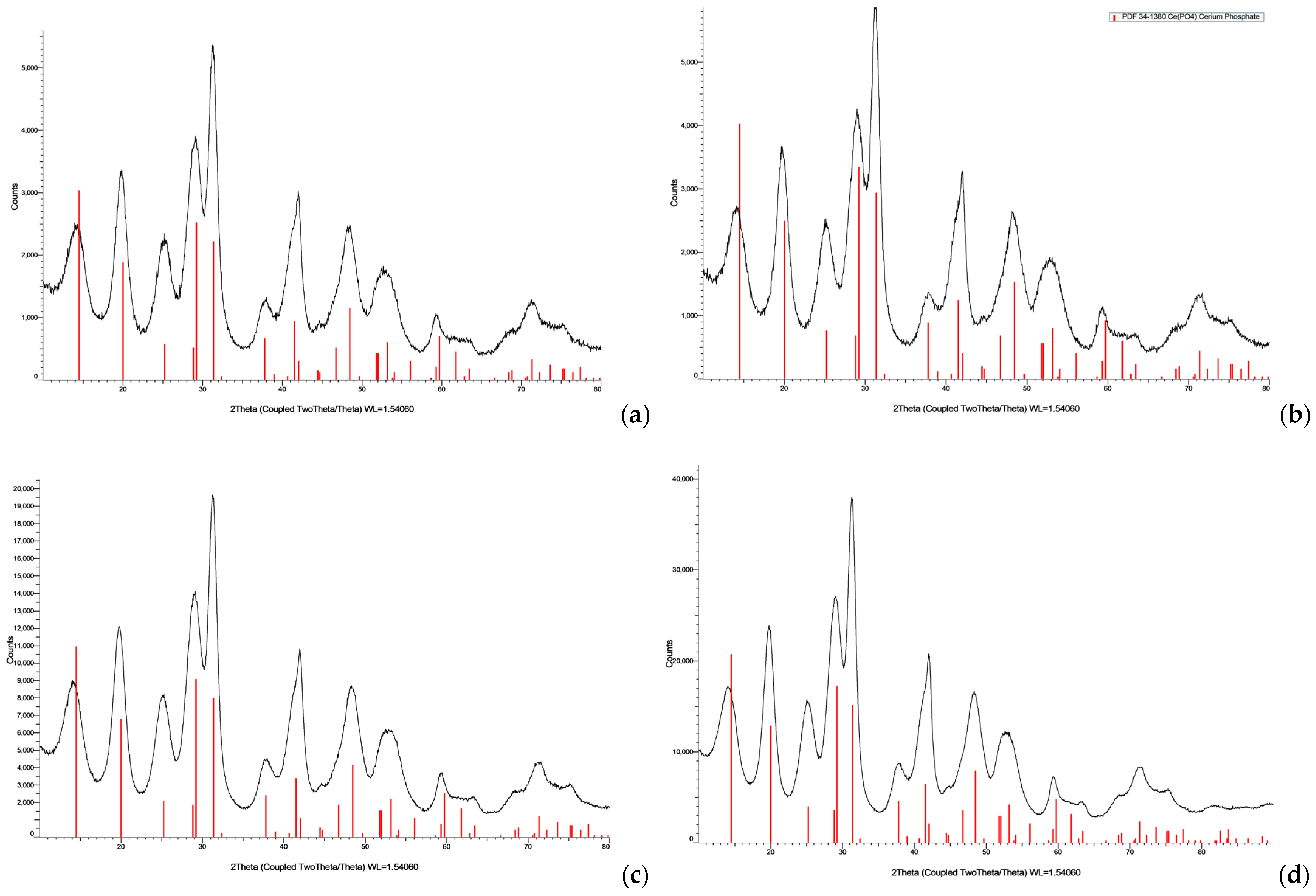
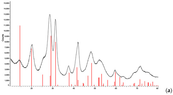
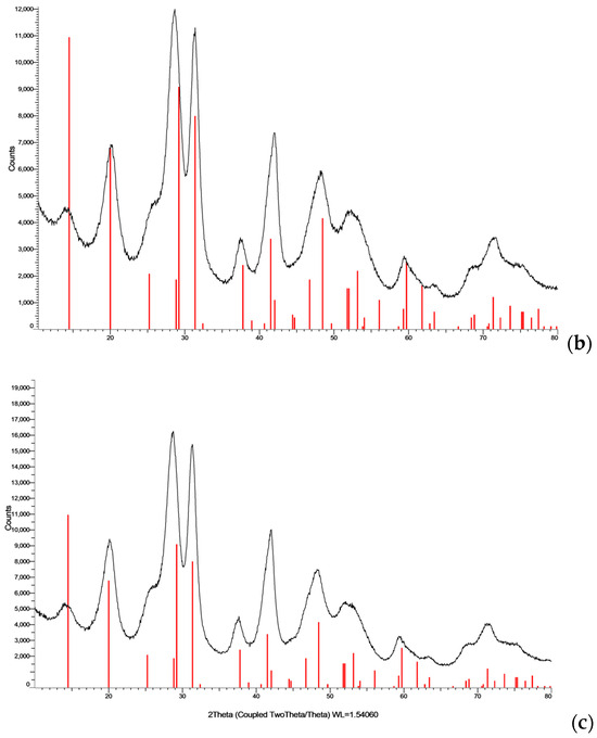
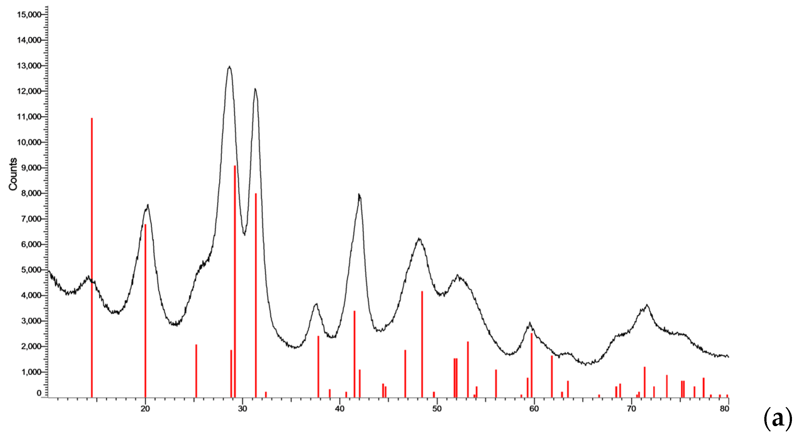
X-ray diffraction (XRD) analysis of the powders obtained from all the investigated multicomponent systems showed that all samples consisted of single-phase, finely dispersed cerium(III) phosphate. Because the diffraction patterns obtained for the samples were identical, representative XRD patterns for each experimental series are presented in the figures.
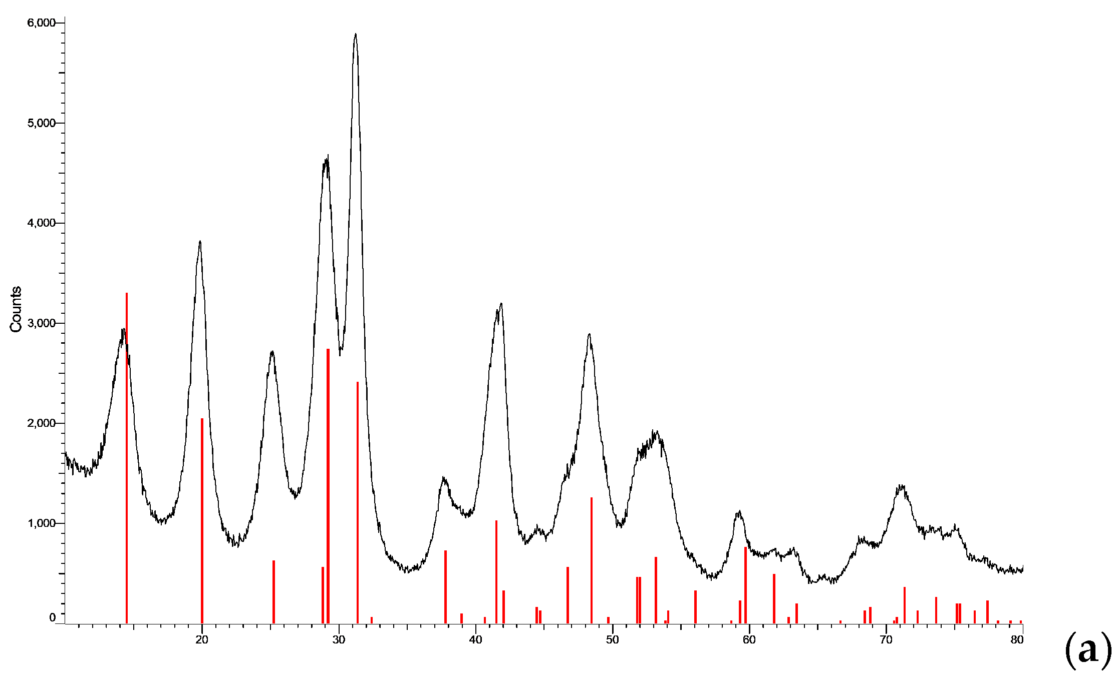
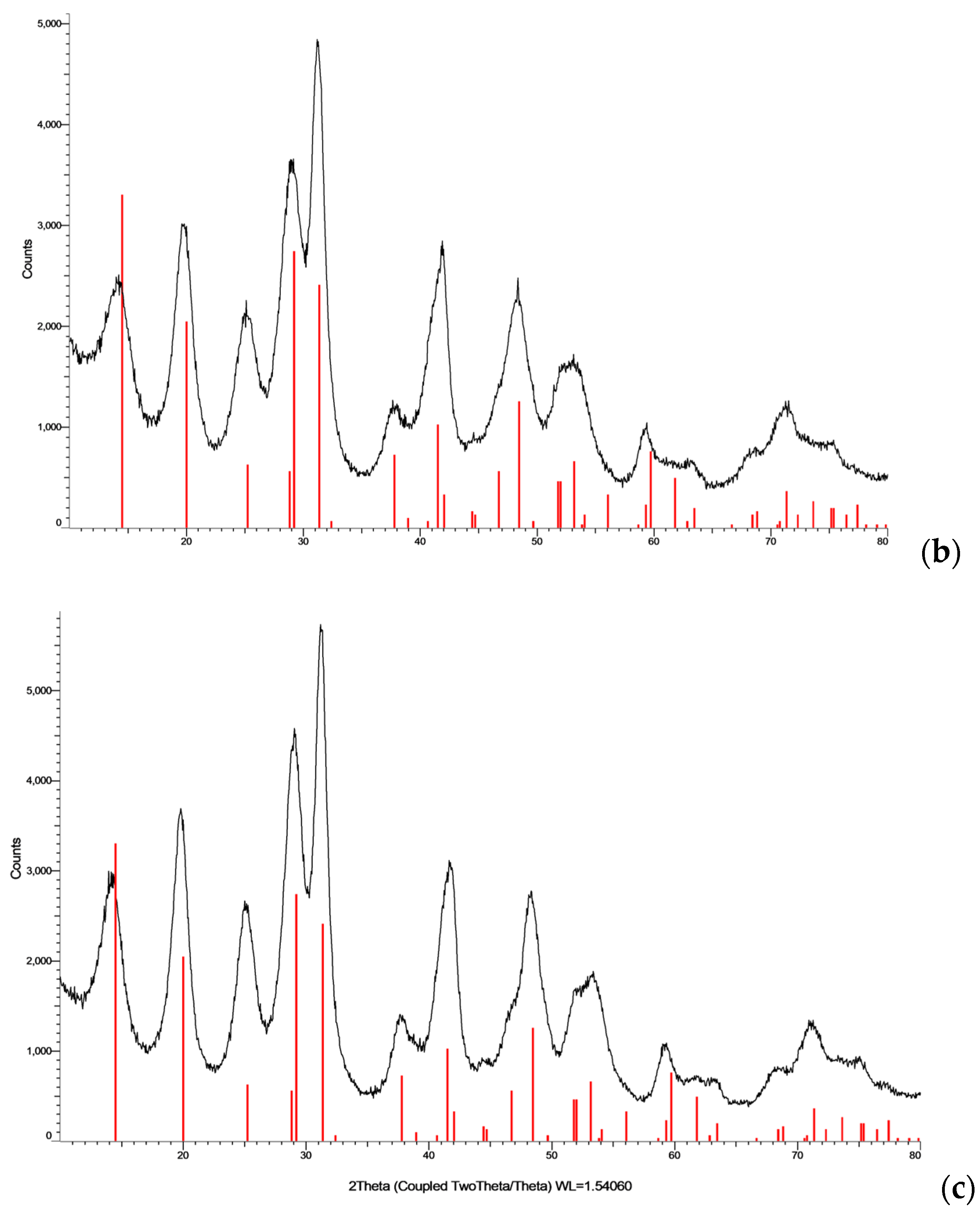
The diffraction peaks shown in Figure 2, Figure 3 and Figure 4 are in full agreement with the crystalline phase of cerium phosphate (CePO4). A comparison of the standard CePO4 peak widths (according to JCPDS/PDF card No. 34-1380, hexagonal phase with a rhabdophane-type structure) with the peaks widths obtained for the compounds under investigation shows that the peaks are approximately two to three times broader, both at the base and in the central region.
Figure 2.
XRD patterns of cerium phosphate nanoparticle samples: (a) MC 0.625, (b) CMC 0.625, (c) CMCNa 0.625, and (d) CePO4 obtained from a phosphate buffer solution.
Figure 3.
XRD patterns of annealed CePO4–CMCNa nanocomposite samples: (a) CMCNa 1.25; (b) CMCNa 0.625; and (c) CMCNa 0.625/6.25.
Figure 4.
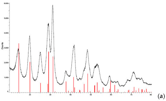
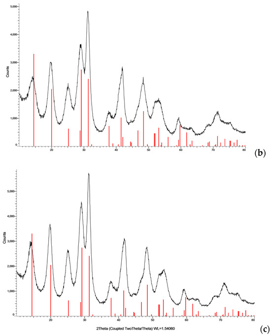
XRD patterns of cerium phosphate/polymer nanocomposites CePO4–Cellulose–Citrate: (a) MC + Cit 0.625/12.5; (b) CMC + Cit 0.625/12.5; and (c) CMCNa + Cit 0.625/12.5.
The pronounced broadening of the diffraction peaks is primarily attributed to the small size of the coherent scattering domains, reflecting the nanocrystalline nature of the crystallites. The slight broadening of the peak profiles and the elevated background are most likely associated with an amorphous polymer phase on the surface of the phosphate core. The initial, non-annealed CMCNa-basedsamples modified with metronidazole consisted of X-ray amorphous prior to drying and thermal treatment, indicating a high degree of structural disorder in the hydrated polymer networks, and the dried samples were the same single-phase cerium phosphate.
X-ray diffraction analysis of the CMCNa series samples annealed at 400 °C (Figure 3) also revealed the presence of a single cerium phosphate phase, despite the theoretically expected formation of cerium–sodium cerium–sodium phosphates. Compared with the as-prepared samples, the diffraction peaks became narrower, indicating an increase in crystallinity. In addition, an inversion in the relative intensities of the two main CePO4 reflections was observed, most likely resulting from texturing (preferred orientation) of the anisotropic crystallites induced by thermal treatment.
Analysis of the diffraction patterns of the powders synthesized from the initial solutions containing citric acid (Figure 4) confirmed the preservation of the target phase purity/ The incorporation of citric acid and metronidazole into the multicomponent systems also did not lead to a change in the phase composition of the cerium component or the formation of cerium citrate complexes.
3.2. Morphology, Particle Size, and Aggregation Stability of the Nanoparticles (TEM)
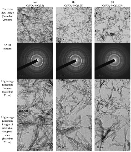
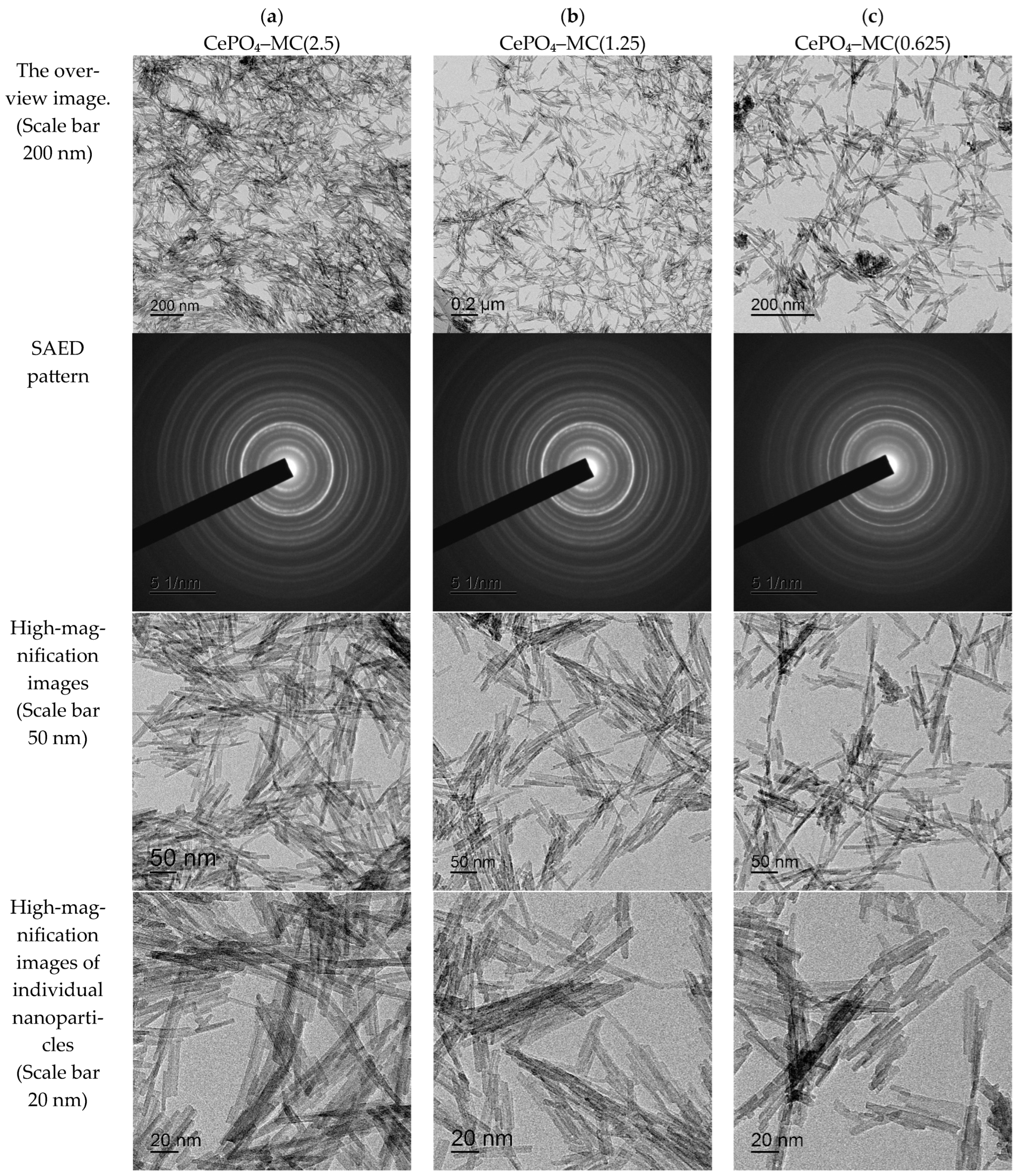
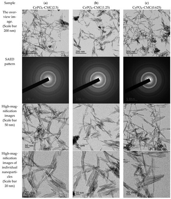
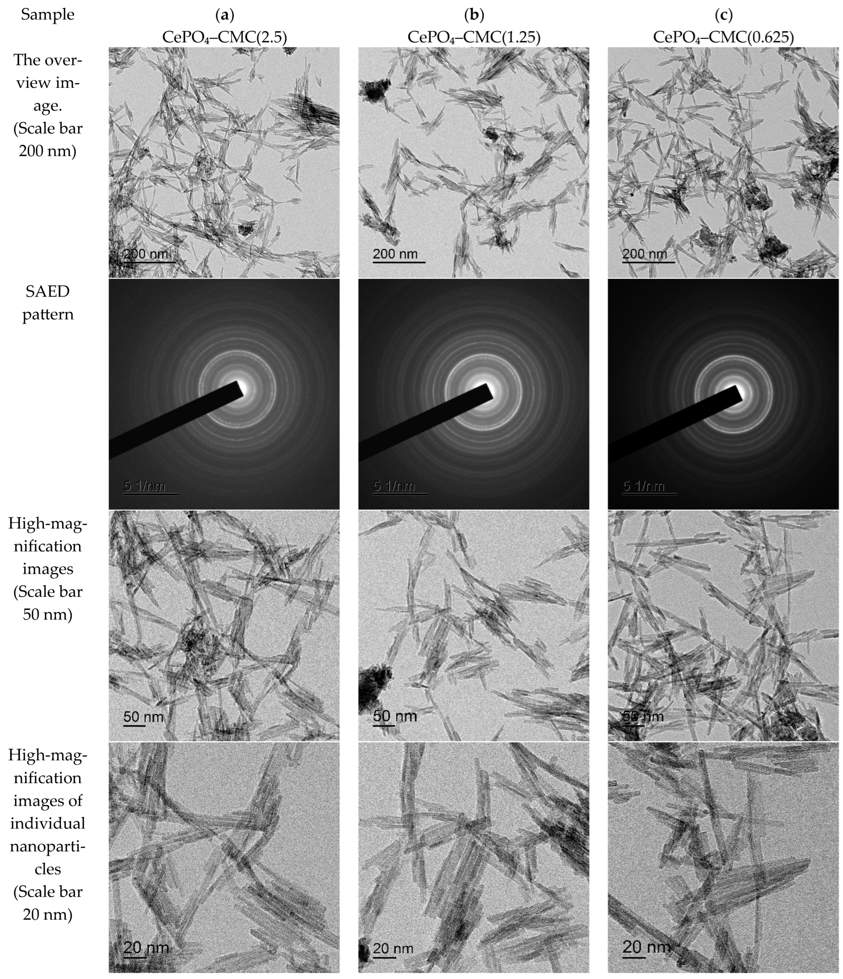
Comparison of TEM images showed the dependence of the morphology and size of the nanocomposites on the nature of the cellulose polymer used. In all cases, the resulting nanoparticles exhibited a pronounced anisotropic (rod-like or needle-like) morphology, which is probably due to the selective adsorption of polysaccharide macromolecules on specific crystallographic faces of the CePO4 nuclei, thereby restricting their lateral growth.
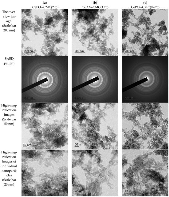
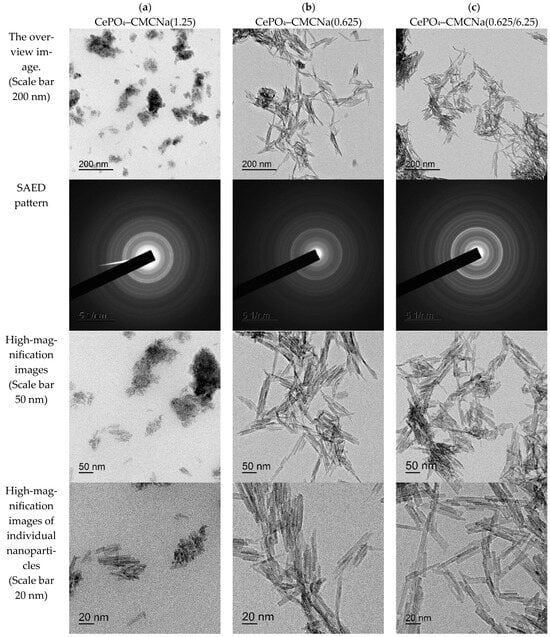
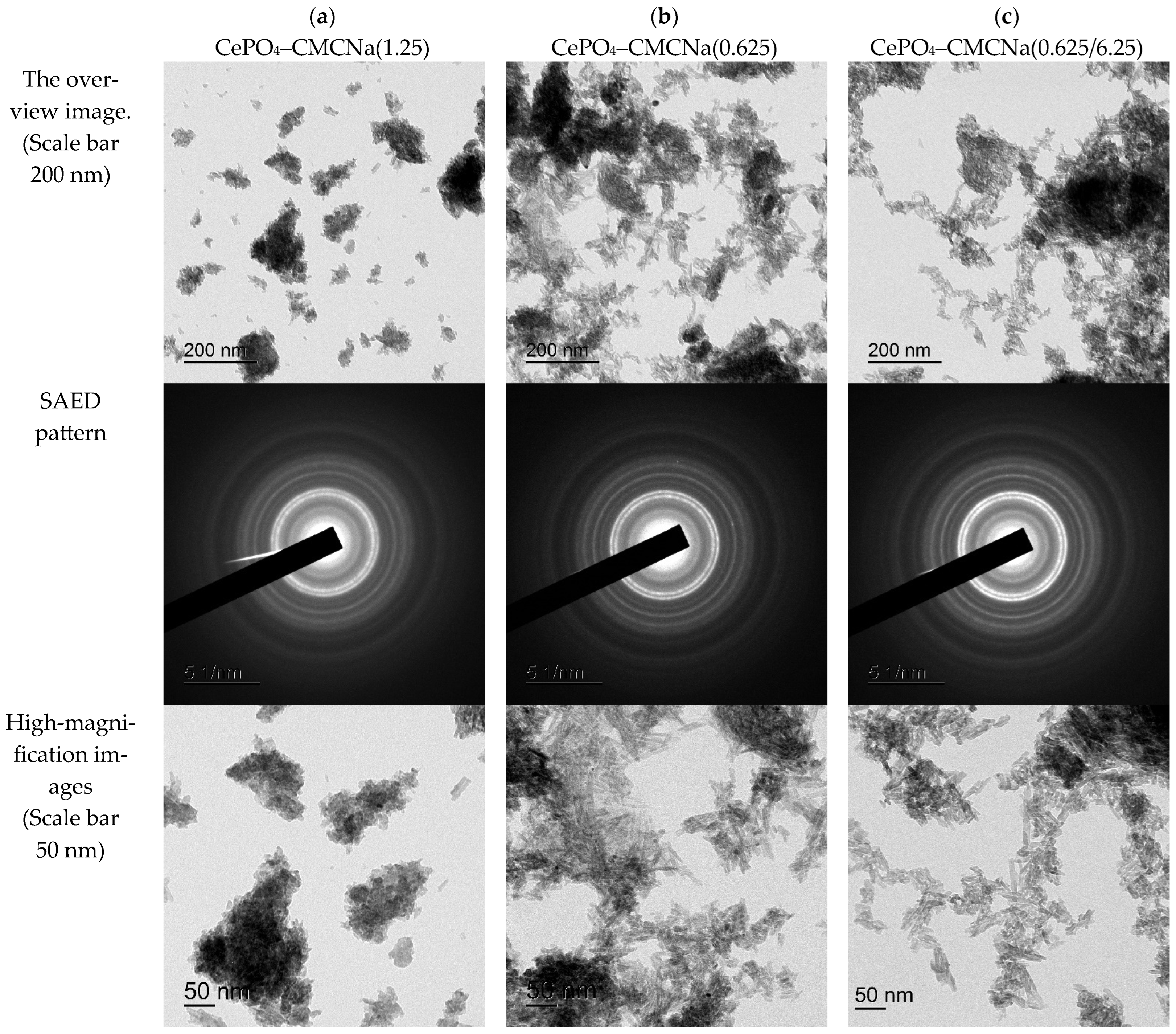
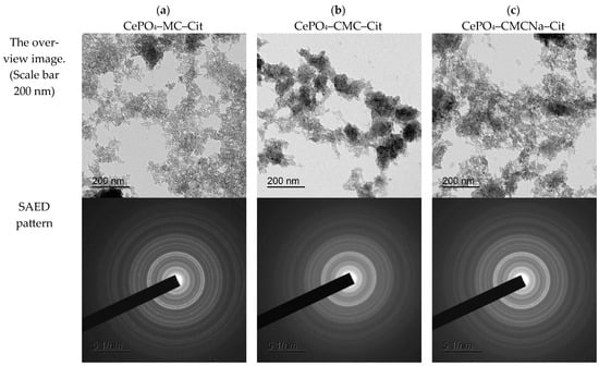
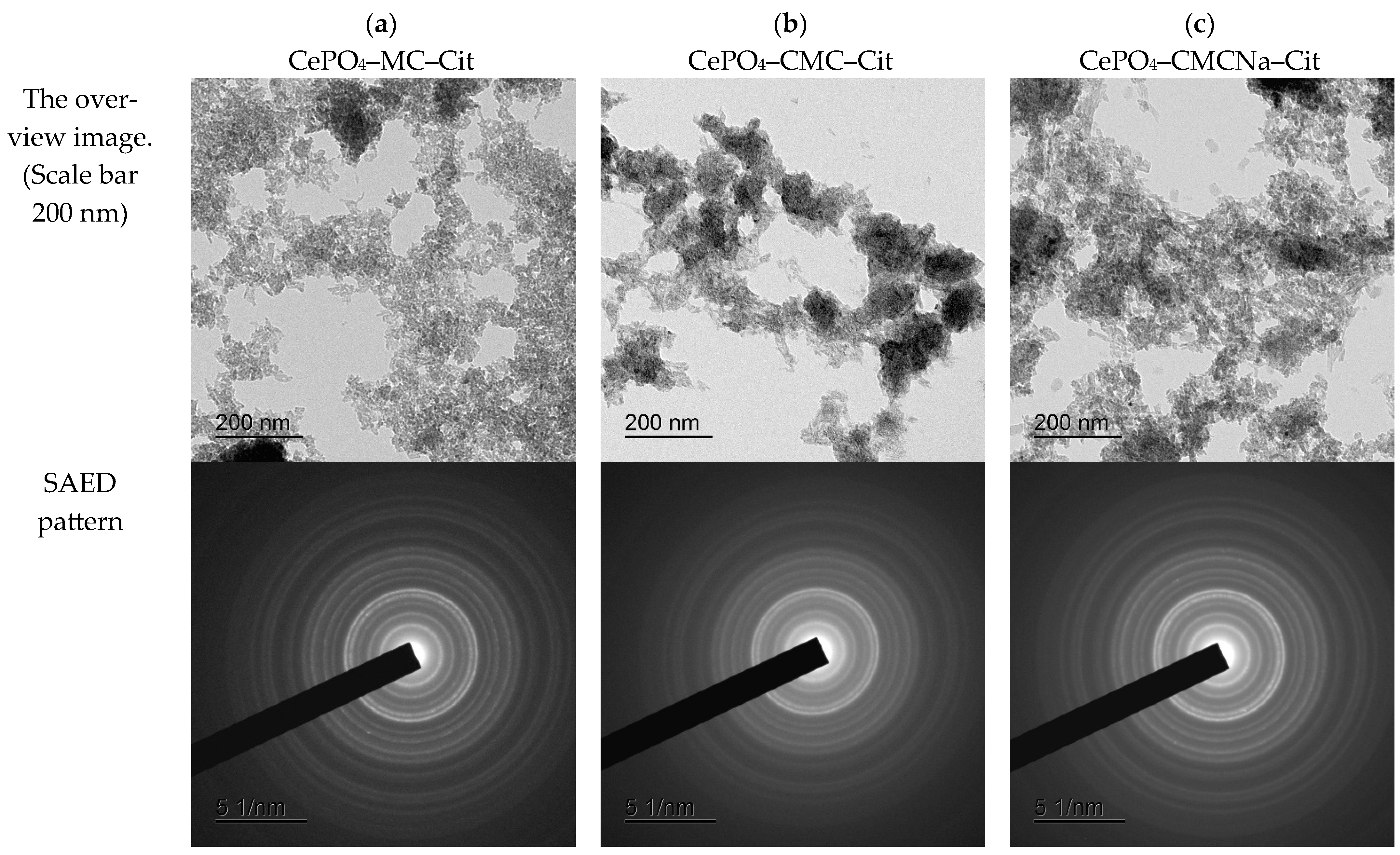
For all investigated systems, analysis of the selected area electron diffraction (SAED) ring patterns confirmed their complete correspondence to the hexagonal CePO4 phase.
Figure 5 shows TEM images of the nanocomposite particles in cerium(III) phosphate samples modified with methylcellulose (MC). The samples are moderately agglomerated and consist of loosely packed, randomly distributed aggregates of fine particles. The nanoparticles exhibit a rod-like morphology, with widths ranging from 3 to 6 nm and lengths varying from 100 to 200 nm. High-resolution images clearly reveal the crystal lattice of the individual nanorods. Variation of the MC concentration (2.5, 1.25, and 0.625 g/L) does not significantly affect the geometry of the individual crystallites but acts as a regulator of their secondary aggregation.
Figure 5.
TEM images of nanocomposite particles in CePO4-methylcellulose (MC) samples: (a) MC 2.5, (b) MC 1.25, and (c) MC 0.625.
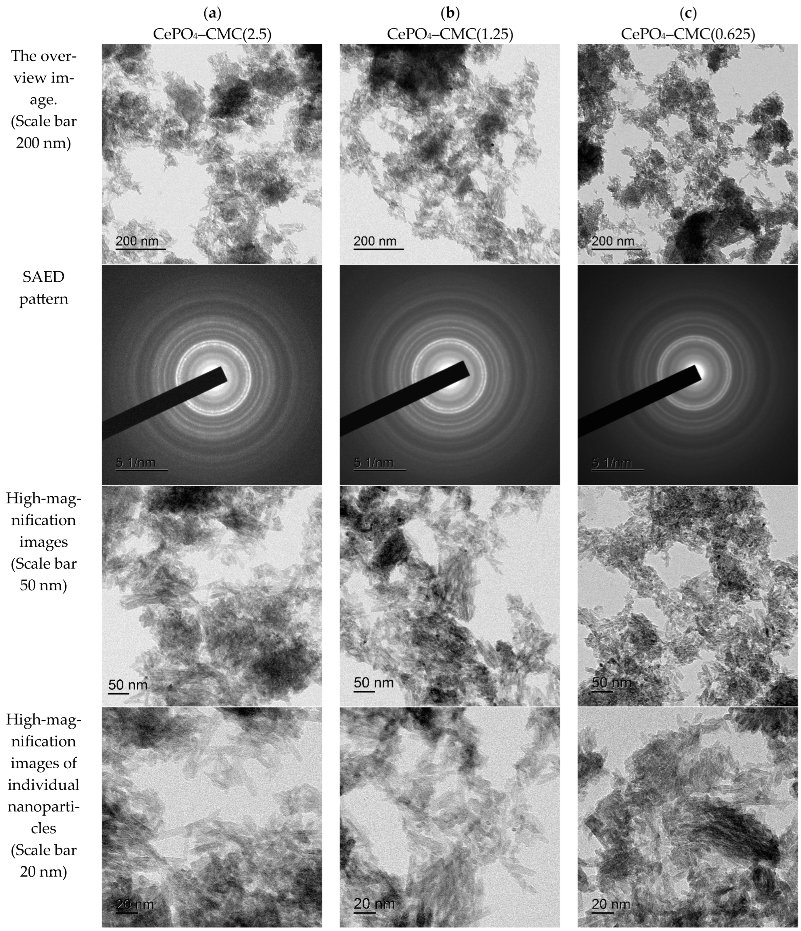
The use of CMC as a modifier (Figure 6) results in the formation of nanoparticles with average dimensions slightly smaller than those obtained using nonionic MC. We assume that this is attributed to the stronger polyelectrolyte interaction between the anionic carboxylate groups of the polymer (-COO−-) and (Ce3+) ions during the nucleation stage.
Figure 6.
TEM images of nanocomposite particles in CePO4/carboxymethyl cellulose (CMC) samples: (a) CMC 2.5, (b) CMC 1.25, and (c) CMC 0.625.
For the samples containing CMC, MC and CMCNa, this temperature is insufficient; calcination at 400 °C leads to significant burnout of the organic component and slightly changes the particle morphology (Figure 7). After calcination, particle aggregation increases significantly, and conglomerates with sizes of 200 nm and larger are formed, consisting of individual crystallites approximately 5 nm in width and with a maximum length of 80 nm.
Figure 7.
TEM images of annealed nanocomposite particles in CePO4/carboxymethyl cellulose (CMC) samples: (a) CMC 2.5, (b) CMC 1.25, (c) CMC 0.625.
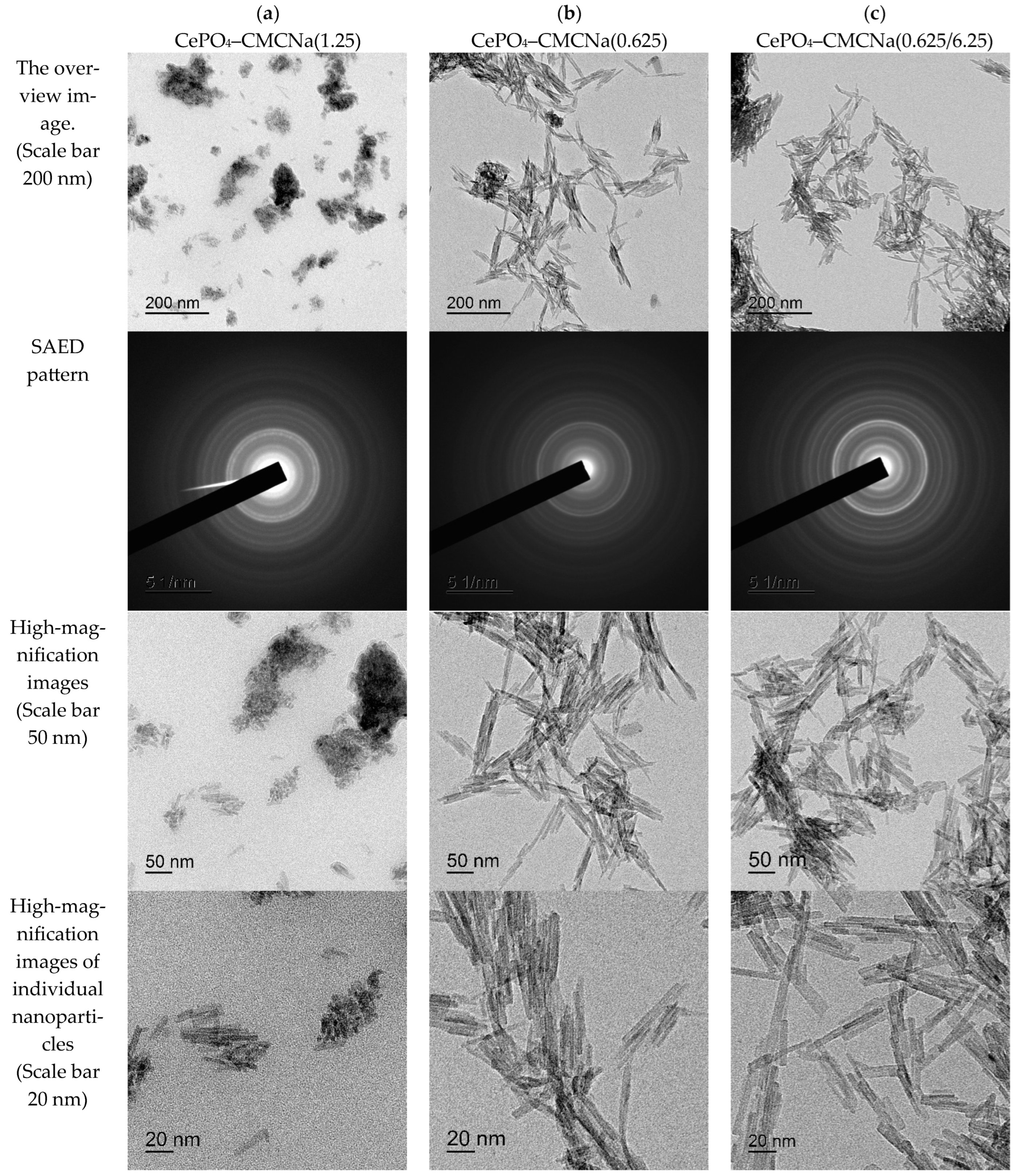
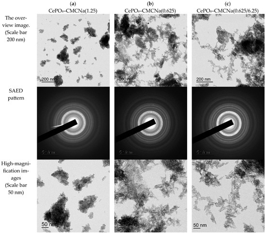
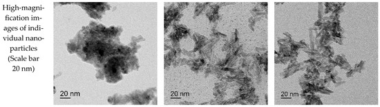
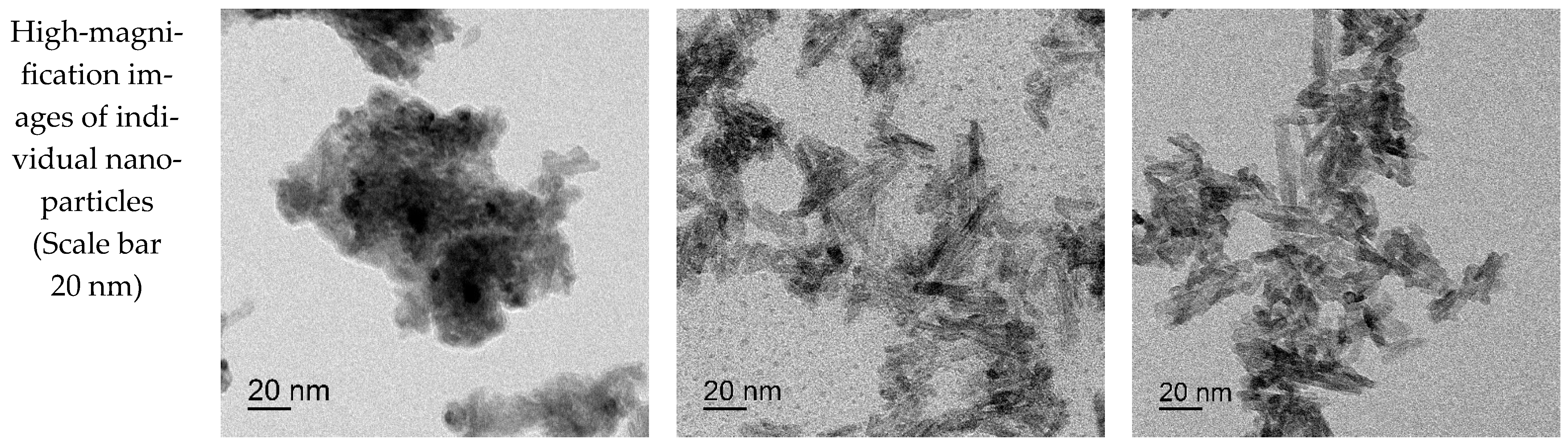
Figure 8 shows TEM images of nanocomposite particles in non-annealed cerium(III) phosphate samples modified with CMCNa. In all samples, agglomerates with sizes of approximately 200 nm or larger were observed, consisting of randomly distributed clusters of fine composite particles. The nanoparticles exhibit an anisotropic morphology, with widths remaining nearly constant at approximately 3–5 nm, whereas their lengths vary over a wide range. Individual particles reach lengths of up to 100 nm.
Figure 8.
TEM images of nanocomposite particles in CePO4/sodium carboxymethyl cellulose (CMCNa) samples: (a) CMCNa 1.25, (b) CMCNa 0.625, and (c) CMCNa 0.625/6.25.
The nanoparticles synthesized from CMCNa-containing solutions exhibit characteristics similar to those of the CMC-derived samples. The obtained results demonstrate that variations in the concentrations of the initial solutions influence both the particle dimensions and the degree of agglomeration. A higher CMCNa concentration concentration increases the viscosity of the reaction medium, which prevents the linear growth of nanocomposites. Conversely, a decrease in the concentration of the cerium component results in a slight reduction in both the width and the length of the cerium phosphate nanoparticles.
To further investigate the effect of annealing on the dimensions of the CMC-CePO4 nanocomposites, the obtained powders were also annealed at 400 °C. TEM images acquired after annealing (Figure 9) show that all samples contain agglomerates with sizes of approximately 200 nm or larger, consisting of randomly distributed clusters of fine composite particles. The particles forming these agglomerates retain an anisotropic morphology, with widths remaining nearly constant at approximately 3–5 nm, whereas their lengths range from 15 to 30 nm.
Figure 9.
TEM images of annealed nanocomposite particles in CePO4/sodium carboxymethyl cellulose (CMCNa) samples: (a) CMCNa 1.25, (b) CMCNa 0.625, and (c) CMCNa 0.625/6.25.
The annealed CePO4 nanoparticles, obtained in the presence of any form of the cellulose matrix, have a shorter length than the particles in the corresponding non-annealed samples. This indirectly confirms the assumption that, in all cases of phosphate synthesis from solutions containing both CMC and CMCNa, crystallization of fine phosphate particles occurs within the space of the cellulose matrix, between the cellulose fibers. Removal of cellulose during calcination does not lead to linear growth of cerium phosphate crystals but increases their agglomeration.
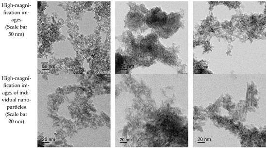
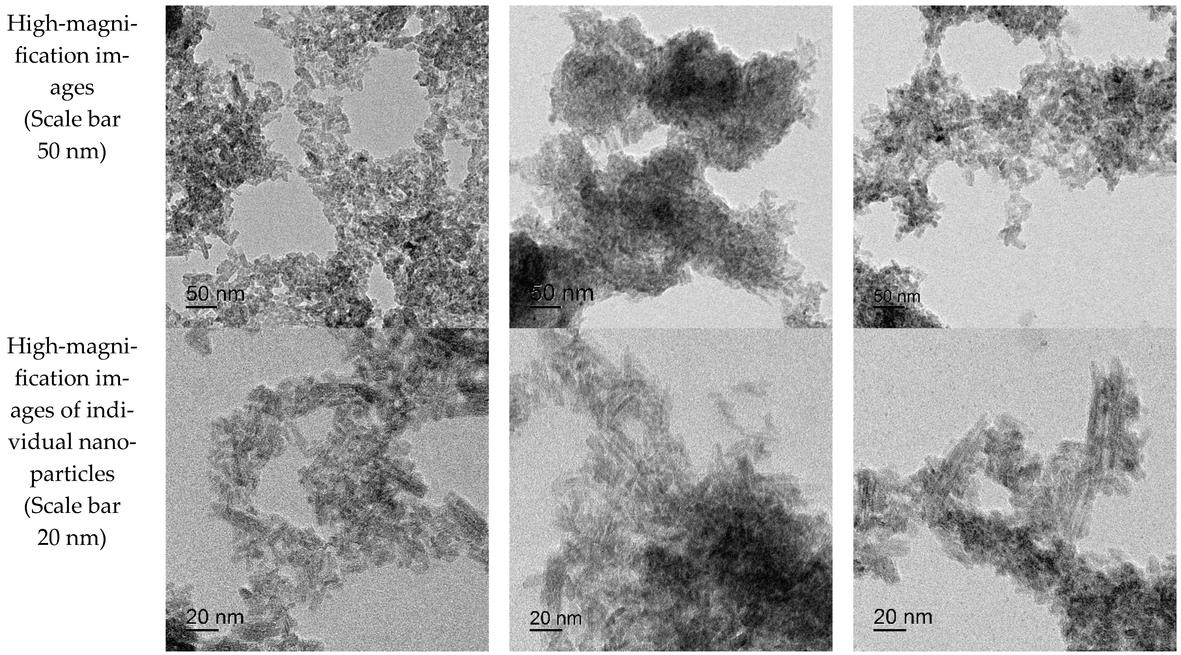
TEM micrographs of the samples modified with citric acid (Figure 10) confirm its effect on limiting nanoparticle growth: their length decreases by tens of times (some crystallites are only 5–7 nm long with a width of 2–5 nm; individual rods reach 25 nm), which once again confirms our observations regarding the role of citric acid as a limiter of the linear growth of crystals (compared with crystals obtained in aqueous [43,65] and cellulose-containing media [64]). Similar results have also been reported in other studies [66,67,68].
Figure 10.
TEM images of nanocomposite particles in (a) CePO4 + MC + Citrate, (b) CePO4 + CMC + Citrate, and (c) CePO4 + CMCNa + Citrate samples.
3.3. Investigation of Intermolecular Interactions (FTIR Spectroscopy)
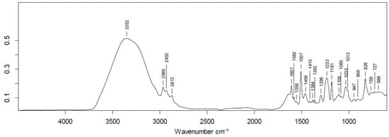
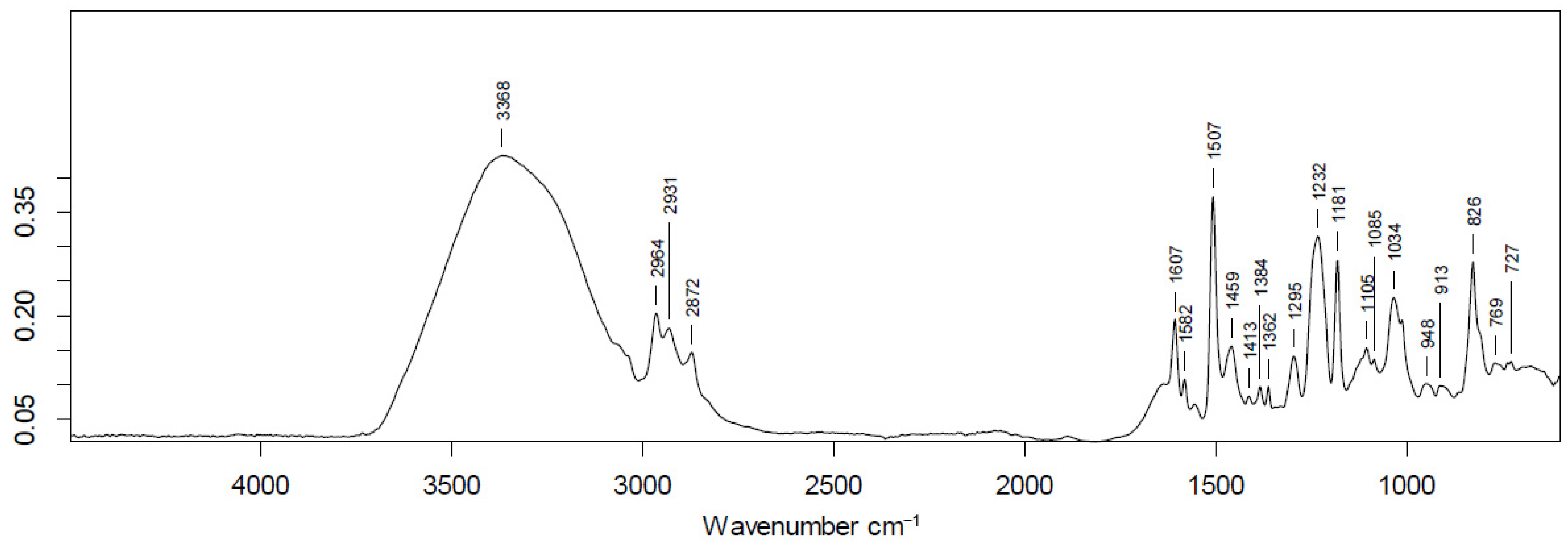
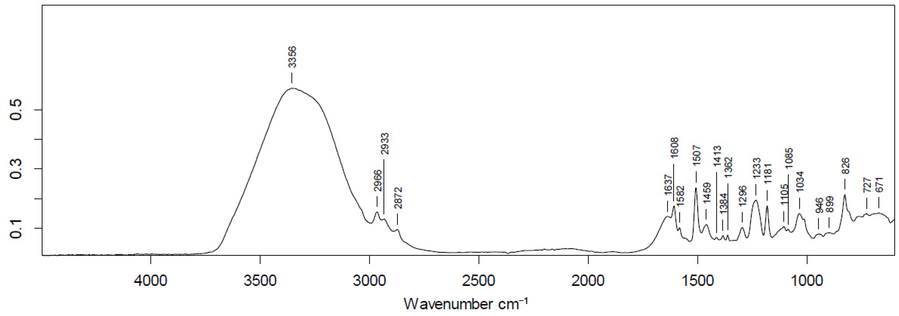
Based on the FTIR spectra of the starting components and the obtained composites (Figure 11, Figure 12 and Figure 13 and Table 1, Table 2 and Table 3 with assignments of the FTIR absorption bands), several conclusions can be drawn regarding the coordination mode, intermolecular interactions, and the successful formation of the hybrid materials.
Figure 11.
FTIR spectrum of the CePO4–citrate–CMCNa–metronidazole composite.
Figure 12.
FTIR spectrum of the CePO4–MC composite.
Figure 13.
FTIR spectrum of the CePO4–MC–citric acid composite.
Table 1.
Assignment of the FTIR absorption bands of the CePO4–citric acid–CMCNa–metronidazole composite.
Table 2.
Assignment of the FTIR absorption bands of the CePO4–MC composite.
Table 3.
Assignment of the FTIR absorption bands of the CePO4–MC–citric acid composite.
The spectrum of free citric acid exhibits intense absorption bands corresponding to the stretching vibrations of carbonyl (C=O) groups in the 1722–1686 cm−1 region. Sodium carboxymethyl cellulose (CMCNa) displays a band at 1588 cm−1 assigned to the asymmetric stretching vibrations of the carboxylate anion (COO−).
In the spectra of all composites containing CMC and citric acid, the high-frequency absorption bands of free carboxylic groups (1722–1686 cm−1) disappear completely. Instead, an intense band appears at 1607–1608 cm−1. This indicates complete deprotonation of the carboxyl groups of citric acid and CMCNa, as well as their chelation or bridging coordination with Ce3+ ions. The shift of the COO− absorption band to approximately 1607 cm−1 is characteristic of metal–ligand coordination.
For nonionic methylcellulose (MC), which contains no carboxyl groups, the band at approximately 1607 cm−1 is assigned to the bending vibrations of coordinated (bound) water molecules. Analysis of the spectra of the binary CePO4–MC system shows that the polymer macromolecules are retained on the surface of the inorganic phase predominantly through an extended network of weak hydrogen bonds. This is evidenced by the broadening and high-wavenumber shift of the O–H stretching band from 3241–3173 cm−1 in the initial polymers to 3352–3368 cm−1 in the composites. Instead of strong polymer–polymer interactions, an interconnected hydrogen-bonding network is formed between the cellulose hydroxyl groups, water molecules, and phosphate centers.
The band at 1507 cm−1 is observed in all three materials. Most probably, it corresponds to the deformation vibrations of bound water molecules. The overtone of the deformation vibrations of water molecules is usually observed at approximately 1630–1640 cm−1. However, for water molecules that have formed a system of strong hydrogen bonds with the hydroxyl (-OH) and methoxy (-OCH3) groups of the cellulose framework, this band may exhibit a pronounced shift to the lower-frequency region (1500–1520 cm−1).
The three bands (1295, 1232 and 1181 cm−1) can be assigned to vibrations of cellulose fragments of the compounds. Thus, the bands observed at 1295 and 1232 cm−1 correspond to the in-plane deformation vibrations of hydroxyl groups and the stretching vibrations of pyranose rings in polysaccharides, whereas the band at 1181 cm−1 is attributed to the asymmetric stretching vibrations of the C-O-C glycosidic bond connecting glucose units.
Band at 826 cm−1. This band is assigned either to deformation vibrations of Ce–O bonds or to deformation vibrations of coordinated organic molecules, confirming the formation of a stable coordination framework.
All composite samples exhibit a broad, complex absorption band in the 1033–1034 cm−1 region, which includes the asymmetric stretching vibrations of P–O bonds in phosphate anions (PO43−) together with vibrations involving Ce–O bonds.
These observations confirm the successful precipitation and formation of the crystalline or amorphous cerium orthophosphate (CePO4) phase, which is firmly integrated into the polymer matrix (MC/CMCNa).
The -OH stretching bands of the initial polymers (MC: 3241 cm−1; CMCNa: 3173 cm−1) become broader and shift toward higher wavenumbers (3352–3368 cm−1) in the composite materials.
This indicates a rearrangement of the hydrogen-bonding network. Instead of strong intermolecular polymer–polymer interactions, an extended hydrogen-bonding network is formed between the hydroxyl groups of methylcellulose or CMC, coordinated water molecules, and phosphate centers.
Analysis of the IR spectra of the two-component systems (CePO4–MC and CePO4–CMC) showed the absence of characteristic bands that could indicate the formation of strong coordination or covalent bonds between the functional groups of cellulose ethers and defects in the cerium phosphate lattice. The polymer macromolecules are retained on the surface of the inorganic nanorods by weak hydrogen bonds. The main role of MC and CMC is limited to rheological control of the precipitation medium and spatial restriction of ion migration.
In the spectra of the samples synthesized with the addition of citric acid, a shift of the absorption bands corresponding to the skeletal stretching vibrations of the C–C bonds of citric acid was observed. This shift confirms the formation of a stable CePO4–citric acid nanocomposite due to the coordination of carboxylate groups to surface cerium ions. At the same time, no direct spectral evidence of interaction between the cellulose matrix (MC/CMC) and citric acid was detected.
The introduction of metronidazole into the system complicates the IR spectral profile in the 3100–3600 cm−1 region because of the multiple superposition and overlap of intense broad stretching vibration bands of the hydroxyl groups (OH−) of residual water, cellulose units and citric acid, which somewhat complicates their assignment. However, the combination of the obtained data, preservation of the phase according to XRD, and the presence of the combined organic and inorganic bands in the IR spectra indirectly indicate the successful incorporation of metronidazole into the nanocomposite.
4. Discussion
In recent decades, nanomaterials have become transformative tools in pharmacotherapy and biomedicine, making it possible to overcome the key limitations of conventional dosage forms, such as low bioavailability, insufficient solubility, rapid systemic clearance, and pronounced side effects [1,2,3,4,5]. One of the most promising directions is the development of new nanomaterials based on rare earth elements possessing regenerative properties and pronounced enzyme-mimetic activity (oxidase-, peroxidase-, catalase-, and phosphatase-like) [5,6,7], especially based on cerium compound nanozymes capable of effectively destroying bacterial biofilms and protecting cellular structures from destructive oxidative stress, thereby creating optimal conditions for tissue regeneration and wound healing [8,9,28,38,39,40,41,42,50]. However, while the beneficial biological effects of cerium oxide nanoparticles have been studied for a long time by numerous researchers from various countries, research into the potential applications of cerium phosphate nanoparticles in biomedical fields is relatively new [64,65] Water-soluble cellulose derivatives—methylcellulose (MC) and carboxymethylcellulose (CMC)—have been successfully used as biopolymer matrices for stabilizing and directing the formation of these nanostructures [20,21,22,23,24,25,26]. Acting as rheological modifiers and stabilizers, these cellulose ethers not only promote the preferential precipitation of small, isolated crystallites but also provide the moisture retention required in the wound area [4,31].
The present study is a continuation of a series of studies devoted to the development of a new generation of pharmaceuticals based on redox-active cerium oxide and cerium phosphate nanoparticles in biopolymer matrices with antimicrobial activity for accelerating wound regeneration [10,38,40,43,50,63,64,65]. Initially, we studied the possibility of synthesizing core-less cerium phosphate nanoparticles, in particular, trivalent cerium phosphate [43]; in our latest study, we investigated the possibility of creating a CePO4-citrate nanocomposite by selecting optimal dosages of citric acid [65]. Unlike our previous studies, in this work we have, for the first time, carried out a comprehensive series of syntheses of nanocomposites based on cerium(III) orthophosphate stabilized with various forms of cellulose (MC, CMC, CMCNa) at different concentrations with the addition of different concentrations of citric acid. Characterization of the synthesized novel hybrid nanocomposites made it possible to reveal the regularities of the influence of the nature and concentration of various forms of cellulose and tricarboxylic acids on the morphology, dispersity, and aggregative stability of the nanoparticles and, ultimately, to select the optimal nanocomposite formulation and demonstrate the possibility of metronidazole encapsulation in the CePO4–citrate–cellulose system developed by us.
The obtained results confirm the working hypothesis regarding the possibility of directed regulation of the morphology of a nanosized cerium(III) orthophosphate composite by varying the nature of the biopolymer matrix and the addition of organic components. The formation of an exclusively hexagonal phase CePO4 (rhabdophane structural type), detected by X-ray diffraction in all synthesized samples, is fully consistent with the regularities of low-temperature precipitation of rare earth phosphates. The high dispersity of the phosphate phase is retained even after calcination at 400 °C, whereas, in the absence of the polymer matrix, nanoparticle coarsening occurs.
Analysis of the transmission electron microscopy data makes it possible to propose a mechanism for the influence of organic polymer additives on the geometry and habit of the nanoparticles. The constant width of the nanocrystals (3–6 nm) suggests that the growth of the lateral faces is limited by steric factors. The decrease in the length of the nanorods from 200 to 15 nm in the presence of citric acid confirms its role as a selective regulator of linear dimensions. Citric acid exhibits a high affinity for cerium ions—the overall stability constants (logβ) for the citrate complexes of rare earth elements formed in aqueous solutions range from 9.1 to 11.2, depending on pH and ionic strength (taking into account both average monocomplexes and protonated species of the type ([M(HCit)]+) [69,70]. We may assume that citric acid molecules are selectively coordinated on the terminal faces of the growing crystals, thereby restricting their further growth.
The difference in the effect of nonionic MC and the anionic forms CMC and CMCNa on the size of the resulting particles is apparently explained by the different surface charge density of the polymer chains and the contribution of electrostatic interactions. It follows that changing the composition of the cellulose matrix will alter the morphology of the nanocomposites.
The observed shift of the characteristic absorption bands of the functional groups in the IR spectra indicates that the cellulose polymer chains are retained on the surface CePO4 predominantly through an extensive network of weak hydrogen bonds. In contrast, the carboxylate groups of citric acid exhibit a pronounced shift characteristic of the formation of coordination bonds with surface Ce3+ centers.
The addition of metronidazole demonstrates the possibility of creating multifunctional materials for regenerative medicine. The combination of the bacteriostatic action of metronidazole and the enzyme-mimetic properties of cerium orthophosphate would make it possible to simultaneously address the suppression of pathogenic microflora and the reduction of excessive oxidative stress in the wound area.
Limitations and Prospects
The main objective of this study was to investigate the effect of various forms of cellulose on the morphology of the nanoscale citrate complex of cerium phosphate, to assess its stability, and to verify the possibility of incorporating (encapsulating) the medicinal drug into the structure of the nanocomposite. This article does not discuss the dosage of the drug, but only the potential possibility of creating a hybrid nanocomposite. Our next studies will focus on the dosages and effectiveness of nanocomposites for biomedical applications. In addition, our future studies will be aimed at obtaining cellulose films and hydrogels containing cerium nanocompounds (phosphate or oxide), a complexing agent (carboxylic acid), and an affordable antibiotic (including metronidazole). We hope that the developed nanocomposites will have good prospects for application in biomedicine as prolonged wound-healing and antibacterial agents.
5. Conclusions
- A method for the synthesis of cerium(III) phosphate nanocomposite powders modified with various water-soluble forms of cellulose and citric acid has been developed and optimized, allowing control of the morphology and dispersity of the resulting systems.
- The influence of the nature and concentration of cellulose ethers on the properties of the nanoparticles has been established: nonionic methylcellulose promotes the formation of elongated rod-like particles (100–200 nm) with moderate loose agglomeration, whereas anionic carboxymethylcellulose (CMC) and its sodium salt form shorter crystallites prone to the formation of bulky and loose nanoaggregates.
- The positive effect of citric acid on limiting the linear dimensions of cerium(III) phosphate crystallites in the presence of a cellulose matrix has been demonstrated.
- X-ray diffraction confirmed the formation of single-phase cerium(III) orthophosphate in all experimental series. This structure is retained after calcination at 400 °C, which is apparently due to the shielding effect of the polymer matrix.
- According to transmission electron microscopy data, the individual nanoparticles exhibit pronounced anisotropy with a fixed width of 3–6 nm and a length ranging from 15 to 200 nm, depending on the rheological parameters of the medium and the nature of the coordination of the stabilizing agents.
- Experimental samples of cerium phosphate nanocomposites modified with metronidazole were obtained for the first time during the synthesis process. It appears that the stability of the composite is ensured by hydrogen bonding between cellulose molecules and the coordination of citric acid with surface cerium ions.
Author Contributions
Conceptualization, E.V.S., E.L.C. and V.A.S.; methodology, E.L.C., O.I.A. and A.A.G.; validation, E.V.S., O.I.A. and A.A.G.; formal analysis, E.L.C., O.I.A., E.V.S., V.A.P., V.A.S., N.E.M. and A.A.G.; investigation, O.I.A., E.L.C. and A.A.G.; data curation, A.A.G., E.L.C. and E.V.S.; visualization, O.I.A., V.A.S. and V.A.P.; supervision, E.V.S., N.E.M. and E.L.C.; project administration, E.V.S., E.L.C. and V.A.S.; writing—original draft preparation, E.L.C. and O.I.A.; writing—review and editing, A.A.G., E.V.S., N.E.M., V.A.P. and V.A.S. funding acquisition, E.V.S. All authors have read and agreed to the published version of the manuscript.
Funding
The research was funded by the Russian Science Foundation, interdisciplinary grant No. 23-65-10040, https://rscf.ru/project/23-65-10040/ (accessed on 19 August 2026). The funders had no role in the design of the study; in the collection, analyses, or interpretation of data; in the writing of the manuscript; or in the decision to publish the results.
Institutional Review Board Statement
Not applicable for studies not involving humans or animals.
Data Availability Statement
Data available on request due to restrictions. The data presented in this study are available on request from the corresponding author.
Conflicts of Interest
Author Elena L. Chuvilina (E.L.C.), Olga I. Andreeva (O.I.A.) and Akhmedali A. Gasanov (A.A.G.) were employed by the company LANHIT LLC. The remaining authors declare that the research was conducted in the absence of any commercial or financial relationships that could be construed as a potential conflict of interest. E.L.C, A.A.G. and O.I.A. are affiliated with the company “LANHIT”. E.L.C, A.A.G. and O.I.A. have PhD degrees (chemical, technical), which contributed to the professional scientific development of the grant topic. O.I.A. and E.L.C were members of the grant project team No. 23-65-100-40 (the following authors of the article are also members of the scientific team of the grant project: S.E.V., N.E.M., D.D.T. and V.A.S.). The company «LANHIT» did not sponsor this study and did not participate in the further instrumental analyses of obtained materials that were not performed on the basis of LANHIT. LLC «LANHIT» has been a manufacturer of high- and special-purity inorganic compounds since 1991, including rare-earth metal compounds. In addition, instrumental studies were conducted within the framework of non-financial agreements (cooperation agreement) between the institutions indicated in the affiliation of the article, independently of each other. In particular, there is a scientific cooperation agreement between I.M. Sechenov First Moscow State Medical University (Sechenov University) and LANHIT.
References
- Parvin, N.; Aslam, M.; Alam, M.N.; Mandal, T.K. Nanotechnology Driven Innovations in Modern Pharmaceutics: Therapeutics, Imaging, and Regeneration. Nanomaterials 2025, 15, 1733. [Google Scholar] [CrossRef] [Scilit] [PubMed]
- Kumar, M.; Mahmood, S.; Chopra, S.; Bhatia, A. Biopolymer based nanoparticles and their therapeutic potential in wound healing—A review. Int. J. Biol. Macromol. 2024, 267, 131335. [Google Scholar] [CrossRef] [Scilit] [PubMed]
- De, R.; Mahata, M.K.; Kim, K.T. Structure-Based Varieties of Polymeric Nanocarriers and Influences of Their Physicochemical Properties on Drug Delivery Profiles. Adv. Sci. 2022, 9, e2105373. [Google Scholar] [CrossRef] [Scilit] [PubMed]
- Pegoraro, G.M.; Carriello, G.M.; Alves, L.R.; Lopes, H.S.M.; Filho, J.F.; Barros, M.F.J.; de Menezes, A.J.; Mambrini, G.P. Nanocellulose and rare earth elements: A literature review. Cellulose 2025, 32, 1393–1421. [Google Scholar] [CrossRef] [Scilit]
- Titova, S.A.; Kruglova, M.P.; Stupin, V.A.; Manturova, N.E.; Silina, E.V. Potential Applications of Rare Earth Metal Nanoparticles in Biomedicine. Pharmaceuticals 2025, 18, 154. [Google Scholar] [CrossRef] [Scilit] [PubMed]
- Salfate, G.; Sánchez, J. Rare earth elements uptake by synthetic polymeric and cellulose-based materials: A review. Polymers 2022, 14, 4786. [Google Scholar] [CrossRef] [Scilit] [PubMed]
- Luong, A.D.; Maruthapandi, M.; Gedanken, A.; Luong, J.H.T. Rare-Earth Oxide Nanoparticles: A New Weapon Against Multidrug-Resistant Pathogens with Potential Wound Healing Treatment. Nanomaterials 2025, 15, 1862. [Google Scholar] [CrossRef] [Scilit] [PubMed]
- Dhall, A.; Self, W. Cerium Oxide Nanoparticles: A Brief Review of Their Synthesis Methods and Biomedical Applications. Antioxidants 2018, 7, 97. [Google Scholar] [CrossRef] [Scilit] [PubMed]
- Nosrati, H.; Heydari, M.; Khodaei, M. Cerium oxide nanoparticles: Synthesis methods and applications in wound healing. Mater. Today Bio 2023, 23, 100823. [Google Scholar] [CrossRef] [Scilit] [PubMed]
- Titova, S.A.; Stupin, V.A.; Manturova, N.E.; Chuvilina, E.L.; Gasanov, A.A.; Parfenov, V.A.; Silina, E.V. Nanosized Cerium Phosphate: Synthesis Methods, Morphology, and Potential Applications in Biomedicine. Biomedicines 2026, 14, 1337. [Google Scholar] [CrossRef] [Scilit] [PubMed]
- Cai, Y.; Ren, W.; Du, C.; Zhang, J.; Zhao, Y.; Yu, J.; Du, H.; Kang, H.; Ge, X.; Lu, M.; et al. Cerium Oxide Nanozymes: Exploring Synthesis, Catalytic Activity, and Biomedical Potential. Small 2025, 21, e2504436. [Google Scholar] [CrossRef] [Scilit] [PubMed]
- Shrestha, S.; Wang, B.; Dutta, P. Nanoparticle processing: Understanding and controlling aggregation. Adv. Colloid Interface Sci. 2020, 279, 102162. [Google Scholar] [CrossRef] [Scilit] [PubMed]
- Titova, S.A.; Kruglova, M.P.; Stupin, V.A.; Manturova, N.E.; Achar, R.R.; Deshpande, G.; Parfenov, V.A.; Silina, E.V. Excipients for Cerium Dioxide Nanoparticle Stabilization in the Perspective of Biomedical Applications. Molecules 2025, 30, 1210. [Google Scholar] [CrossRef] [Scilit] [PubMed]
- Magdalane, C.M.; Kaviyarasu, K.; Vijaya, J.J.; Siddhardha, B.; Jeyaraj, B. Photocatalytic activity of binary metal oxide nanocomposites of CeO2/CdO nanospheres: Investigation of optical and antimicrobial activity. J. Photochem. Photobiol. B 2016, 163, 77–86. [Google Scholar] [CrossRef] [Scilit] [PubMed]
- Vatansever, E.; Arslan, D.; Nofar, M. Polylactide cellulose-based nanocomposites. Int. J. Biol. Macromol. 2019, 137, 912–938. [Google Scholar] [CrossRef] [Scilit] [PubMed]
- Saddique, A.; Lee, H.M.; Kim, J.C.; Bae, J.; Cheong, I.W. Cellulose nanocrystal nanocomposites capable of low-temperature and fast self-healing performance. Carbohydr. Polym. 2022, 296, 119973. [Google Scholar] [CrossRef] [Scilit] [PubMed]
- Revin, V.V.; Liyaskina, E.V.; Parchaykina, M.V.; Kuzmenko, T.P.; Kurgaeva, I.V.; Revin, V.D.; Ullah, M.W. Bacterial Cellulose-Based Polymer Nanocomposites: A Review. Polymers 2022, 14, 4670. [Google Scholar] [CrossRef] [Scilit] [PubMed]
- Yuvaraj, G.; Ramesh, M.; Rajeshkumar, L. Carbon and Cellulose-Based Nanoparticle-Reinforced Polymer Nanocomposites: A Critical Review. Nanomaterials 2023, 13, 1803. [Google Scholar] [CrossRef] [Scilit] [PubMed]
- Oyewo, O.A.; Elemike, E.E.; Onwudiwe, D.C.; Onyango, M.S. Metal oxide-cellulose nanocomposites for the removal of toxic metals and dyes from wastewater. Int. J. Biol. Macromol. 2020, 164, 2477–2496. [Google Scholar] [CrossRef] [Scilit] [PubMed]
- Hynninen, V.; Patrakka, J.; Nonappa. Methylcellulose-Cellulose Nanocrystal Composites for Optomechanically Tunable Hydrogels and Fibers. Materials 2021, 14, 5137. [Google Scholar] [CrossRef] [Scilit] [PubMed]
- Kasle, P.; Bains, A.; Goksen, G.; Dhull, S.B.; Ali, N.; Nagarik, R.; Fareed, M.; Chawla, P. Exploring the Properties of Carboxymethyl Cellulose Blended with Other Polymeric Compounds for the Formulation of Biodegradable Packaging Films and Edible Coatings: A Review. Compr. Rev. Food Sci. Food Saf. 2025, 24, e70215. [Google Scholar] [CrossRef] [Scilit] [PubMed]
- Oprea, M.; Voicu, S.I. Recent advances in composites based on cellulose derivatives for biomedical applications. Carbohydr. Polym. 2020, 247, 116683. [Google Scholar] [CrossRef] [Scilit] [PubMed]
- Wahid, F.; Huang, L.-H.; Zhao, X.-Q.; Li, W.-C.; Wang, Y.-Y.; Jia, S.-R.; Zhong, C. Bacterial cellulose and its potential for biomedical applications. Biotechnol. Adv. 2021, 53, 107856. [Google Scholar] [CrossRef] [Scilit] [PubMed]
- Abdelhamid, H.N.; Mathew, A.P. Cellulose-Based Nanomaterials Advance Biomedicine: A Review. Int. J. Mol. Sci. 2022, 23, 5405. [Google Scholar] [CrossRef] [Scilit] [PubMed]
- Liu, W.; Du, H.; Zheng, T.; Si, C. Biomedical Applications of Bacterial Cellulose based Composite Hydrogels. Curr. Med. Chem. 2021, 28, 8319–8332. [Google Scholar] [CrossRef] [Scilit] [PubMed]
- Fuster, M.G.; Moulefera, I.; Muñoz, M.N.; Montalbán, M.G.; Víllora, G. Synthesis of Cellulose Nanoparticles from Ionic Liquid Solutions for Biomedical Applications. Polymers 2023, 15, 382. [Google Scholar] [CrossRef] [Scilit] [PubMed]
- Isogai, A. Emerging Nanocellulose Technologies: Recent Developments. Adv. Mater. 2021, 33, e2000630. [Google Scholar] [CrossRef] [Scilit] [PubMed]
- Orhan, B.; Karadeniz, D.; Kalaycıoğlu, Z.; Kaygusuz, H.; Torlak, E.; Erim, F.B. Foam-based antibacterial hydrogel composed of carboxymethyl cellulose/polyvinyl alcohol/cerium oxide nanoparticles for potential wound dressing. Int. J. Biol. Macromol. 2025, 291, 138924. [Google Scholar] [CrossRef] [Scilit] [PubMed]
- Spiridonov, V.V.; Sybachin, A.V.; Pigareva, V.A.; Afanasov, M.I.; Musoev, S.A.; Knotko, A.V.; Zezin, S.B. One-Step Low Temperature Synthesis of CeO2 Nanoparticles Stabilized by Carboxymethylcellulose. Polymers 2023, 15, 1437. [Google Scholar] [CrossRef] [Scilit] [PubMed]
- Alotaibi, R.; Alawad, M.O.; Khalil, K.D.; Al-Rafai, H.; Bashal, A.H.; Alotaibi, A.A.; Zaidi, B. Tailoring the electrical properties of cerium oxide-supported carboxymethyl cellulose composites for electronics and energy storage: Insights from density functional theory and molecular dynamics simulations. Int. J. Biol. Macromol. 2025, 305, 141132. [Google Scholar] [CrossRef] [Scilit] [PubMed]
- Bîrcă, A.C.; Minculescu, M.A.; Niculescu, A.-G.; Hudiță, A.; Holban, A.M.; Alberts, A.; Grumezescu, A.M. Nanoparticle-Enhanced Collagen Hydrogels for Chronic Wound Management. J. Funct. Biomater. 2025, 16, 91. [Google Scholar] [CrossRef] [Scilit] [PubMed]
- Stupin, V.A.; Gabitov, R.B.; Sinelnikova, T.G.; Silina, E.V. Biological Mechanisms of Chronic Wound and Diabetic Foot Healing: The Role of Collagen. Serbian J. Exp. Clin. Res. 2018, 19, 373–382. [Google Scholar] [CrossRef] [Scilit]
- Chen, J.; Xia, Y.; Lan, Q.; Hu, M.; Xu, Y.; Wu, Q.; Liu, X.; Liu, Y. Alginate based photothermal cryogels boost ferrous-supply for enhanced antibacterial chemodynamic therapy and accelerated wound healing. Int. J. Biol. Macromol. 2023, 232, 123473. [Google Scholar] [CrossRef] [Scilit] [PubMed]
- Kapoor, D.U.; Pareek, A.; Sharma, S.; Prajapati, B.G.; Thanawuth, K.; Sriamornsak, P. Alginate gels: Chemistry, gelation mechanisms, and therapeutic applications with a focus on GERD treatment. Int. J. Pharm. 2025, 675, 125570. [Google Scholar] [CrossRef] [Scilit] [PubMed]
- Peng, W.; Li, D.; Dai, K.; Wang, Y.; Song, P.; Li, H.; Tang, P.; Zhang, Z.; Li, Z.; Zhou, Y.; et al. Recent progress of collagen, chitosan, alginate and other hydrogels in skin repair and wound dressing applications. Int. J. Biol. Macromol. 2022, 208, 400–408. [Google Scholar] [CrossRef] [Scilit] [PubMed]
- La Monica, F.; Campora, S.; Ghersi, G. Collagen-Based Scaffolds for Chronic Skin Wound Treatment. Gels 2024, 10, 137. [Google Scholar] [CrossRef] [Scilit] [PubMed]
- Donati, I.; Christensen, B.E. Alginate-metal cation interactions: Macromolecular approach. Carbohydr. Polym. 2023, 321, 121280. [Google Scholar] [CrossRef] [Scilit] [PubMed]
- Silina, E.V.; Manturova, N.E.; Sevastianov, V.I.; Perova, N.V.; Gladchenko, M.P.; Kryukov, A.A.; Ivanov, A.V.; Dudka, V.T.; Prazdnova, E.V.; Emelyantsev, S.A.; et al. Development of a Collagen–Cerium Oxide Nanohydrogel for Wound Healing: In Vitro and In Vivo Evaluation. Biomedicines 2025, 13, 2623. [Google Scholar] [CrossRef] [Scilit] [PubMed]
- Feraru, A.; Tóth, Z.-R.; Magyari, K.; Baia, M.; Gyulavári, T.; Páll, E.; Licarete, E.; Costinas, C.; Cadar, O.; Papuc, I.; et al. The effect of nanoceria on the alginate-gum arabic crosslinking mechanism and in vitro behavior as a wound dressing. Int. J. Biol. Macromol. 2025, 288, 138569. [Google Scholar] [CrossRef] [Scilit] [PubMed]
- Silina, E.V.; Manturova, N.E.; Vasin, V.I.; Artyushkova, E.B.; Khokhlov, N.V.; Ivanov, A.V.; Stupin, V.A. Efficacy of A Novel Smart Polymeric Nanodrug in the Treatment of Experimental Wounds in Rats. Polymers 2020, 12, 1126. [Google Scholar] [CrossRef] [Scilit] [PubMed]
- Zubairi, W.; Tehseen, S.; Nasir, M.; Anwar Chaudhry, A.; Ur Rehman, I.; Yar, M. A study of the comparative effect of cerium oxide and cerium peroxide on stimulation of angiogenesis: Design and synthesis of pro-angiogenic chitosan/collagen hydrogels. J. Biomed. Mater. Res. B Appl. Biomater. 2022, 110, 2751–2762. [Google Scholar] [CrossRef] [Scilit] [PubMed]
- Melnikova, N.; Malygina, D.; Korokin, V.; Al-Azzawi, H.; Zdorova, D.; Mokshin, E.; Liyaskina, E.; Kurgaeva, I.; Revin, V. Synthesis of Cerium Oxide Nanoparticles in a Bacterial Nanocellulose Matrix and the Study of Their Oxidizing and Reducing Properties. Molecules 2023, 28, 2604. [Google Scholar] [CrossRef] [Scilit] [PubMed]
- Silina, E.V.; Stupin, V.A.; Manturova, N.E.; Chuvilina, E.L.; Gasanov, A.A.; Andreeva, O.I.; Korobko, E.V.; Andreeva, N.V.; Dodonova, S.A.; Tkachenko, D.D.; et al. Cerium Phosphate Nanoparticles: Synthesis, Characterization, Biocompatibility, Regenerative Potential, and Antioxidant Activity. Molecules 2025, 30, 3916. [Google Scholar] [CrossRef] [Scilit] [PubMed]
- Fischer, M.R.; Garcia, M.C.F.; Nogueira, A.L.; Porto, L.M.; Schneider, A.L.d.S.; Pezzin, A.P.T. Biossíntese e caracterização de nanocelulose bacteriana para engenharia de tecidos. Matéria 2018, 22, e11934. [Google Scholar] [CrossRef] [Scilit]
- Noor, M.; Al Mamun, M.A.; Atique Ullah, A.K.M.; Matsuda, A.; Kawamura, G.; Hakim, M.A.; Islam, M.F.; Matin, M.A. Physics of Ce3+↔Ce4+ electronic transition in phytosynthesized CeO2/CePO4 nanocomposites and its antibacterial activities. J. Phys. Chem. Solids 2021, 148, 109751. [Google Scholar] [CrossRef] [Scilit]
- Vinotha Sre, V.; Danushri, S.; Khan, S.S. Catalysis Beyond Enzymes: Ceria Nanozyme as a Smart Platform for Biocatalysis, Anti-oxidant Defense, and Biosensing. Top. Curr. Chem. 2025, 383, 49. [Google Scholar] [CrossRef] [Scilit] [PubMed]
- Peera, S.G.; Kim, S.W. Rare Earth Ce/CeO2 Electrocatalysts: Role of High Electronic Spin State of Ce and Ce3+/Ce4+ Redox Couple on Oxygen Reduction Reaction. Nanomaterials 2025, 15, 600. [Google Scholar] [CrossRef] [Scilit] [PubMed]
- Zeng, Y.; Wang, Y.; Zhang, S.; Zhong, Q.; Rong, W.; Li, X. One-pot synthesis of ceria and cerium phosphate (CeO2-CePO4) nanorod composites for selective catalytic reduction of NO with NH3: Active sites and reaction mechanism. J. Colloid. Interface Sci. 2018, 524, 8–15. [Google Scholar] [CrossRef] [Scilit] [PubMed]
- Huang, L.; Sun, D.-W.; Pu, H.; Zhang, C.; Zhang, D. Nanocellulose-based polymeric nanozyme as bioinspired spray coating for fruit preservation. Food Hydrocoll. 2023, 135, 108138. [Google Scholar] [CrossRef] [Scilit]
- Silina, E.V.; Ivanova, O.S.; Manturova, N.E.; Medvedeva, O.A.; Shevchenko, A.V.; Vorsina, E.S.; Achar, R.R.; Parfenov, V.A.; Stupin, V.A. Antimicrobial Activity of Citrate-Coated Cerium Oxide Nanoparticles. Nanomaterials 2024, 14, 354. [Google Scholar] [CrossRef] [Scilit] [PubMed]
- Sousa, R.B.; Dametto, A.C.; Sábio, R.M.; de Carvalho, R.A.; Vieira, E.G.; Oliveira, A.F.A.; Ribeiro, L.K.; Barud, H.S.; Silva-Filho, E.C. Cerium-doped calcium phosphates precipitated on bacterial cellulose platform by mineralization. Ceram. Int. 2020, 46, 26985–26990. [Google Scholar] [CrossRef] [Scilit]
- Chen, Z.; Zhou, X.; Mo, M.; Hu, X.; Liu, J.; Chen, L. Systematic review of the osteogenic effect of rare earth nanomaterials and the underlying mechanisms. J. Nanobiotechnol. 2024, 22, 185. [Google Scholar] [CrossRef] [Scilit] [PubMed]
- Windarti, T.; Prasetya, N.B.A.; Siahaan, P.; Abid, M.B.; Nurhasanah, I.; Nulandaya, L. Development of calcium phosphate cement composed of silica-modified hydroxyapatite, cerium oxide, and silica as an osteoporotic bone filler. Ceram. Int. 2024, 50, 45957–45968. [Google Scholar] [CrossRef] [Scilit]
- Petrakova, N.V.; Zobkova, Y.O.; Komlev, V.S.; Ashmarin, A.A.; Lysenkov, A.S.; Volchenkova, V.A.; Barinov, S.M.; Sadovnikova, M.A.; Murzakhanov, F.F.; Gafurov, M.R.; et al. Synthesis and characterization of luminescent cerium-doped hydroxyapatite. Ceram. Int. 2024, 50, 20905–20916. [Google Scholar] [CrossRef] [Scilit]
- Mahmoud, E.M.; Rehab, T.A.; Soliman, A.A.; Said, I.G.; Naga, S.M. Production of an eco-friendly, effective antimicrobial and antifungal from cerium/natural bio-hydroxyapatite coasting for titanium plates suitable for bone augmentation. Ceram. Int. 2024, 50, 6979–6994. [Google Scholar] [CrossRef] [Scilit]
- Moaness, M.; Mousa, S.M.; Abo-Elfadl, M.T.; El-Bassyouni, G.T. Doxorubicin loaded cerium substituted hydroxyapatite nanoparticles: A promising new therapeutic approach for bone regeneration, doxorubicin delivery, and cancer treatment. Int. J. Pharm. 2024, 654, 123969. [Google Scholar] [CrossRef] [Scilit] [PubMed]
- Ta, K.M.; Neal, C.J.; Coathup, M.J.; Seal, S.; Phillips, R.M.; Molinari, M. The interaction of phosphate species with cerium oxide: The known, the ambiguous and the unexplained. Biomater. Adv. 2025, 166, 214063. [Google Scholar] [CrossRef] [Scilit] [PubMed]
- Quilumbango, A.; Briceño, S.; Ponce, J.F.; Vizuete, K.; Debut, A.; Botas, J.A.; González, G. Chitosan-collagen-cerium hydroxyapatite nanocomposites for In-vitro gentamicin drug delivery and antibacterial properties. Carbon. Trends 2024, 16, 100392. [Google Scholar] [CrossRef] [Scilit]
- Leitsch, D. A review on metronidazole: An old warhorse in antimicrobial chemotherapy. Parasitology 2019, 146, 1167–1178. [Google Scholar] [CrossRef] [Scilit] [PubMed]
- Suvarna, V.; Murahari, M.; Pawar, S. Metronidazole-An Old Drug for Structure Optimization and Repurposing. Chem. Biodivers. 2025, 22, e03389. [Google Scholar] [CrossRef] [Scilit] [PubMed]
- Chikwe, T.N.; Chikwe, T.N.; Abayeh, O.J.; Nwaka, C. Antibacterial evaluation of metronidazole and its derivative in the treatment of Escherichia coli and Staphylococus aureus. Dutse J. Pure Appl. Sci. 2025, 11, 58–65. [Google Scholar] [CrossRef] [Scilit]
- Mahmoud, A.; Maged, N.; Abdalwali, S. Metronidazole-excipient compatibility studies for medicated chewing gum delivery systems development. Eur. J. Pharm. Med. Res. 2025, 12, 567–589. [Google Scholar]
- Silina, E.V.; Stupin, V.A.; Manturova, N.E.; Chuvilina, E.L.; Gasanov, A.A.; Ostrovskaya, A.A.; Andreeva, O.I.; Tabachkova, N.Y.; Abakumov, M.A.; Nikitin, A.A.; et al. Development of Technology for the Synthesis of Nanocrystalline Cerium Oxide Under Production Conditions with the Best Regenerative Activity and Biocompatibility for Further Creation of Wound-Healing Agents. Pharmaceutics 2024, 16, 1365. [Google Scholar] [CrossRef] [Scilit] [PubMed]
- Dodonova, S.A.; Kryukov, A.A.; Stupin, V.A.; Manturova, N.E.; Chuvilina, E.L.; Gasanov, A.A.; Angreeva, O.I.; Artyushkova, E.B.; Silina, E.V. Potential biomedical use of nanosized cerium phosphate compounds in wound healing. Bull. Russ. Mil. Med. Acad. 2025, 27, 535–546. [Google Scholar] [CrossRef] [Scilit]
- Silina, E.V.; Manturova, N.E.; Chuvilina, E.L.; Gasanov, A.A.; Andreeva, O.I.; Artyushkova, E.B.; Gladchenko, M.P.; Ivanov, A.V.; Dudka, V.T.; Mironov, S.Y.; et al. Novel cerium phosphate–citric acid nanocomposites for biomedical applications in regenerative medicine: In vitro and in vivo study. Int. J. Mol. Sci. 2026, 27, 7476. [Google Scholar] [CrossRef] [Scilit]
- Selvaraj, S.; Chauhan, A.; Radhakrishnan, A.; Rana, G.; Dutta, V.; Batoo, K.M.; Ghotakar, S. Cerium Oxide Nanoparticles and Their Polymeric Composites: Advancements in Biomedical Applications. J. Inorg. Organomet. Polym. 2024, 34, 5691–5717. [Google Scholar] [CrossRef] [Scilit]
- Duangsa, K.; Tangtrakarn, A.; Mongkolkachit, C.; Aungkavattana, P.; Moolsarn, K. The Effect of Tartaric Acid and Citric Acid as a Complexing Agent on Defect Structure and Conductivity of Copper Samarium Co-Doped Ceria Prepared by a Sol-Gel Auto-Combustion Method. Adv. Mater. Sci. Eng. 2021, 2021, 5592437. [Google Scholar] [CrossRef] [Scilit]
- Krukowski, S.; Karasiewicz, M.; Kolodziejski, W. Convenient UV-spectrophotometric determination of citrates in aqueous solutions with applications in the pharmaceutical analysis of oral electrolyte formulations. J. Food Drug Anal. 2017, 25, 717–722. [Google Scholar] [CrossRef] [Scilit] [PubMed]
- Ohyoshi, A.; Ohyoshi, E.; Ono, H.; Yamakawa, S. A study of citrate complexes of several lanthanides. J. Inorg. Nucl. Chem. 1972, 34, 1955–1960. [Google Scholar] [CrossRef] [Scilit]
- Wen, Y.; Liu, P.; Wang, Q.; Zhao, S.; Tang, Y. Organic Ligand-Mediated Dissolution and Fractionation of Rare-Earth Elements (REEs) from Carbonate and Phosphate Minerals. ACS Earth Space Chem. 2024, 8, 1048–1061. [Google Scholar] [CrossRef] [Scilit] [PubMed]

Disclaimer/Publisher’s Note: The statements, opinions and data contained in all publications are solely those of the individual author(s) and contributor(s) and not of MDPI and/or the editor(s). MDPI and/or the editor(s) disclaim responsibility for any injury to people or property resulting from any ideas, methods, instructions or products referred to in the content. |
© 2026 by the authors. Licensee MDPI, Basel, Switzerland. This article is an open access article distributed under the terms and conditions of the Creative Commons Attribution (CC BY) license.