


Abstract
Background: Titanium implant osseointegration is hierarchically governed by surface properties directing protein adsorption, cell recognition, immune modulation, and bone formation. Photofunctionalization creates ultrahydrophilic surfaces by removing hydrocarbons. To integrate it with 3D-printed architectures requires systematic synthesis. Problem: The classical static view of osseointegration obscures its dynamic, multiscale nature. How photofunctionalization-induced ultrahydrophilicity modulates the surface–protein–cell–bone interface as a continuous, hierarchical system remains unclear. Scope: This review synthesizes evidence on how photofunctionalized ultrahydrophilic titanium surfaces control protein adsorption, integrin-mediated mechanotransduction, immune responses, and in vivo osseointegration, with an emphasis on 3D-printed porous architectures. Conclusions: Photofunctionalization enhances protein adsorption, preserves bioactive conformation, and stabilizes protein layers, selectively engaging osteogenic integrins and amplifying FAK–Src/YAP–TAZ signaling. In 3D-printed implants, ultrahydrophilicity enables capillary-driven fluid infiltration and uniform bone ingrowth. Through this review, knowledge gaps—in surface aging and limited in situ characterization—are identified, and an interface-informed design integrating surface chemistry, architecture, and biological timing is proposed.
1. Introduction
Titanium is highly suitable for orthopedic, dental, and maxillofacial reconstruction because of its favorable mechanical properties, corrosion resistance, and general biocompatibility. Adequate clinical performance depends on successful long-term fixation to the surrounding bone, a concept known as osseointegration [1,2]. The original—classical—definition of osseointegration is direct structural and functional contact between the living bone and the surface of the load-bearing implant. According to this definition, successful osseointegration is strongly related to close bone–implant contact without the deposition of intervening fibrous tissue [3], a description that is too static and simple for a process that is dynamic and biologically complex.
Osseointegration is increasingly recognized as a time-dependent, dynamic, and multistage process. In particular, events that happen immediately after loading the implant are critical; that is, after loading, the implant surface contacts blood and interstitial fluid and immediately adsorbs plasma proteins to create a provisional biological interface on the implant surface. The identity, conformation, and distribution of such adsorbed proteins determine downstream cell recognition, immune sensitization, and osteogenic recruitment [4,5]. Even small variations in the surface chemistry, wettability, and surface energy of the implant during this early incubation period may drive a shift in biology down an irreversible path to either successful and stable bone formation or a “pathologically inflamed” condition [6,7].
Despite significant progress in implant design, achieving osseointegration in the short term and with predictable healing timelines remains a desirable outcome [8]. Osseointegration bonds the implant surface with the bone and is more likely to have minimal micromotion and delayed loading. It has been assumed to be dependent on the patients’ systemic state, which may be compromised by osteoporosis, advanced aging, diabetes, chronic inflammatory diseases, etc., conditions which increase the risk of osseointegration failure due to adverse dosing and molecular immunity before implantation [9,10]. Titanium-based implants are essentially inert relative to the biological responses at the interface and cell competence [11,12].
Additive manufacturing technologies enable engineers to control implant macro-, micro-, and nanoarchitecture and geometry and mechanical properties to previously unattainable levels [13]. 3D-printed titanium implants can anatomically replace bone, create artificial bone tissue via the organism’s biological processes, encourage hypertrophy, and achieve physiological load transfer to the host’s local bone even if the implant is structurally porous [14]. These advantageous features do not, however, fully correlate to biologically predictable favorable outcomes. Due to surface heterogeneity and oxide variations, the surface energy of titanium-based implants with a porous architecture decreases due to substantial increases in the exposed outer surface area. This limits reproduced bioactivity at their surface and renders their further sinterability dependent on shell subunit behavior [15]. In this respect, bioactive behavior, including protein adsorption and early biological chaos, is vital for achieving timely results at passive biological interfaces.
Many implant surface modification techniques are used for these purposes and can be divided into two categories. The first is the improvement of mechanical interlocking by surface coating or anodization [16,17]. The second is adding supplementary layers that cannot actively participate in the early interface environment under physiological and interfacial conditions. A new approach to reducing hydrocarbon load and increasing titanium’s intrinsic reactivity, potentially to zero, is photofunctionalization [18].
Thus, entirely different approaches should be taken when defining titanium-based implants. Instead of the current focus on passive interfaces, the implants’ ability to influence interfacial activity should be considered, as osseointegration begins at the material surface level and extends hierarchically to protein adsorption, cell recognition, immune response, and bone formation. By establishing an interface-governed conceptual framework, this review systematically elaborates how ultrahydrophilic photofunctional 3D-printed titanium regulates the early biological microenvironment of the implant surface and promotes stable osseointegration [19].
Literature Search and Selection Methodology
We searched three mainstream academic databases: PubMed, Scopus, and Web of Science. The search period was set from 2010 to 2025. Core keywords were photofunctionalization, UV treatment, ultrahydrophilic titanium, 3D-printed titanium, and osseointegration. We included peer-reviewed original articles, in vitro studies, in vivo animal experiments, and clinical studies. Non-English articles, conference abstracts, duplicated papers, and studies without valid biological outcomes were excluded. A total of 187 studies were initially retrieved, and 144 papers were finally selected for review. The literature retrieval and screening rules applied in this review are summarized in detail in Table 1.
Table 1.
Literature selection criteria.
In this review, two types of content are clearly distinguished to ensure evidence traceability:
- (1)
- Author-generated conceptual models: The schematic diagrams and comparative summary tables are comprehensive conceptual frameworks independently organized and drawn by the authors, which integrate multi-literature research results for systematic display. Each schematic diagram (Figure 1, Figure 2, Figure 3, Figure 4, Figure 5, Figure 6 and Figure 7) and comparative summary table (Table 2 and Table 3) clearly lists the supporting reference interval in captions or table columns.
- (2)
- Evidence-supported conclusions: All molecular mechanisms, in vitro/in vivo experimental laws and clinical phenomena described in the main text are directly derived from peer-reviewed original research, with real-time reference citations marked after each argument.
2. Photofunctionalization and Ultrahydrophilicity of Titanium Surfaces
Photofunctionalization has never been a simple “surface treatment” that makes surfaces appear “fresh rather than old”; it operates as a first and selective intervention at the first decisive moment when the interface, which is naturally affected by aging, confronts host anatomy. Photofunctionalization does not use surface morphology “alteration” or “coating”. Instead, it affects the physicochemical surface state just prior to biological interaction [20]. It restores the ability of the implant’s native surface chemistry to interact with the host anatomy at previously “inert” times, targeting the local biological factors responsible for osseointegration [21].
Referring to Figure 1 in the above information, photofunctionalization “sets back” the biological input interface of an implant prior to the following interfacial events. It does not govern the subsequent events in series by merely improving surface properties but instead represents the first communication of information to interfacial organisms [22]. This section first describes the biophysical basis of photofunctionalization, followed by an explanation of how ultrahydrophilicity, which affects biological decision-making processes, develops [23]. The physicochemical mechanisms of titanium photofunctionalization are illustrated in Figure 2.
Figure 1.
Interface-governed osseointegration of photofunctionalized 3D-printed titanium implants. Synthesized conceptual schematic by the authors, integrated and adapted based on Refs. [6,18,24]. Photofunctionalization eliminates hydrocarbon contaminants to restore the intrinsic surface energy and ultrahydrophilicity of titanium, reprogramming the initial material–biological interface. This fundamental physicochemical state hierarchically modulates subsequent protein adsorption, cell recognition, immune regulation and ultimate bone formation, rendering the implant surface an active regulatory interface rather than an inert passive substrate. All biological cascade conclusions summarized in this schematic are supported by validated in vitro and in vivo findings from the cited references.
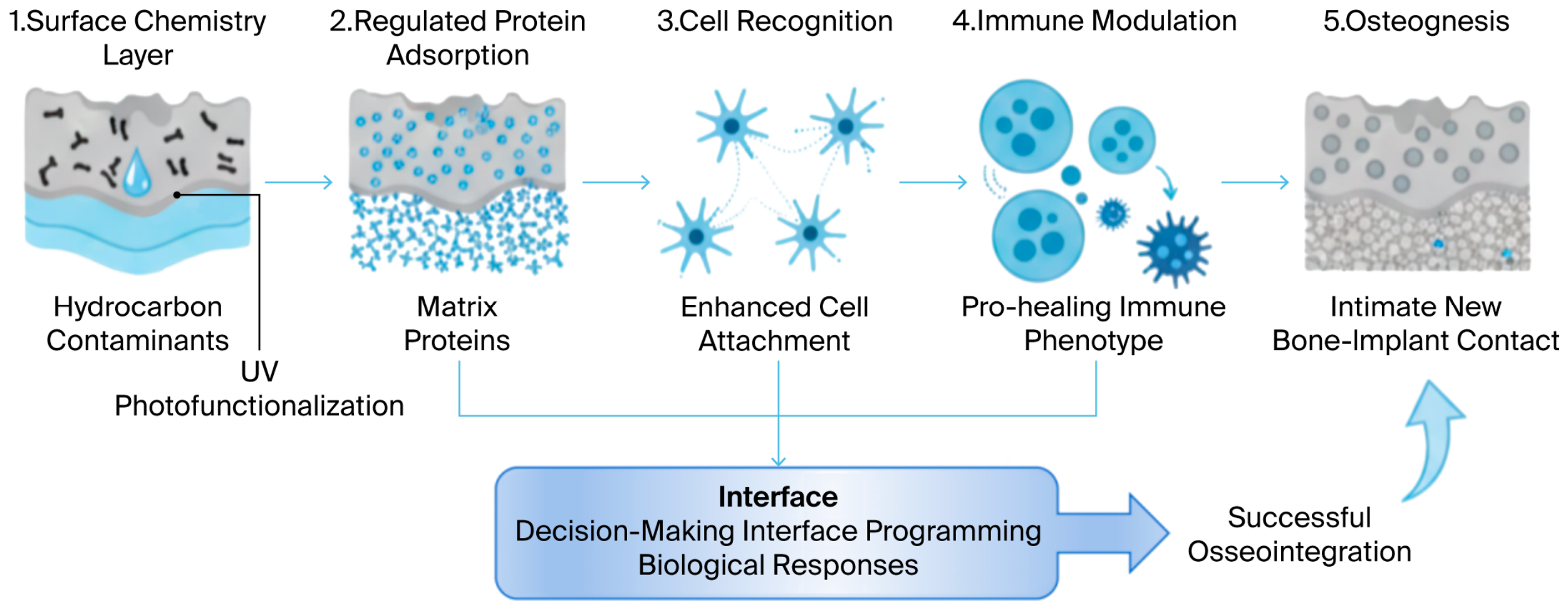
Figure 2.
Physicochemical mechanisms underlying titanium photofunctionalization. Synthesized conceptual schematic by the authors, integrated and adapted based on Refs. [18,25,26]. Ultraviolet irradiation activates the TiO2 surface layer via photocatalytic reactions, decomposing adsorbed extrinsic hydrocarbons and exposing reactive hydroxyl groups. This treatment restores ultrahydrophilicity, elevates surface energy and modulates surface charge, synergistically improving titanium interfacial reactivity prior to biological contact. The photocatalytic pathway and surface aging trends summarized in this schematic are validated by XPS and XRD characterizations reported in the cited references.
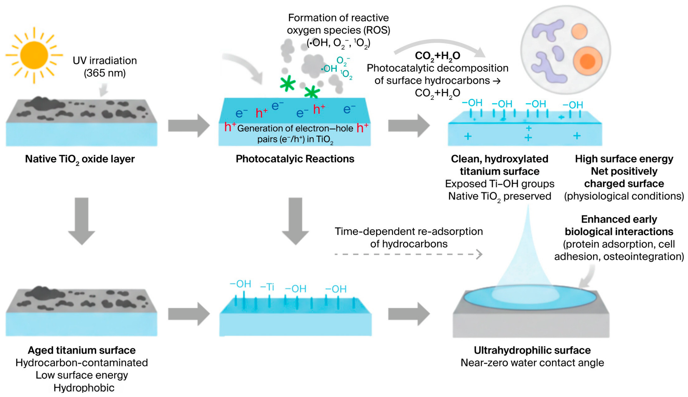
Among multiple UV wavebands commonly adopted for titanium photofunctionalization, 365 nm near-ultraviolet light is the most widely applied option. This wavelength matches the intrinsic band-gap excitation characteristic of the TiO2 oxide layer, efficiently generating electron–hole pairs to trigger the photocatalytic degradation of surface hydrocarbons. It also avoids excessive deep ultraviolet-induced substrate damage and does not pose any obvious cytotoxic risks, thereby exhibiting a superior balance between modification efficiency and biosafety for subsequent biological experiments and clinical application.
2.1. Principles of Photofunctionalization
2.1.1. UV-Induced Removal of Surface Hydrocarbon Contaminants
Titanium surfaces age rapidly due to the accumulation of adventitious carbon derived from both packaging and the environment. Reaction of ambient hydrocarbons with titanium oxide, either from experimental handling or preparation, gradually coats the surface of titanium with hydrocarbons, covering the reactive surface and suppressing surface energy. This is an important process in the initial setting of titanium with biological fluids [27].
Ultraviolet light can photocatalytically activate aged titanium surfaces. Ultraviolet light generates electron–hole pairs inside the oxide layer, leading to the creation of strong oxidizing oxidative species on the interface between the TiO2 layer and hydrocarbon films. The surface hydrocarbon film is easily decomposed into H2O and CO2 that adsorb on the surface [25,28]. In principle, no leaching of chemical residues occurs during pollutant removal. From an aging point of view, this process alters no aspect of the physical structure or dimensions of the surface [29].
Clearing these hydrocarbons reveals chemically active sites that were hidden beneath the hydrocarbon layer. The hydroxyl groups play a significant role in the implant’s interaction with water molecules, ions, and proteins. From this point of view, UV facilitates the creation of titanium without creating a completely new surface with photofunctionalization [30].
2.1.2. Generation of Ultrahydrophilic and Positively Charged Surfaces
Photofunctionalization instantly converts the titanium surface from a hydrophobic or weakly hydrophilic state to being ultrahydrophilic. Ultrahydrophilicity is defined as a state in which the water contact angle is virtually zero or an indeterminate state that approaches virtual zero, with water droplets stabilized through the surface free energy of the implant. This is an extremely important state in implants that come into contact with biological fluids soon after placement [31,32].
In addition to surface wettability, photofunctionalization also changes the surface charge state of the Ti implant. Under physiological conditions, the photodegradation of organic layers leads to full exposure of the Ti-OH groups to the solution, which, in turn, determines the sign and magnitude of the positive charge of the surface. This outcome has an impact on later blood and plasma protein adsorption in terms of both the quantities of proteins adsorbed and their structure and orientation [33,34].
The transformed surface is highly unstable, and hydrocarbons reattach after photofunctionalization, followed by a rapid decrease in surface energy. This means that the aging process, although slow, will affect any photofunctionalized surface and is not limited to the “old” materials. Thus, it is important to UV-treat the surface at the right time and with the correct clinical photofunctionalization procedure [35].
According to XPS and XRD characterization results reported in the published literature, UV photofunctionalization removes adventitious hydrocarbon contaminants adsorbed on the titanium surface via photocatalytic degradation without altering the intrinsic crystal lattice structure, crystallinity, and bulk chemical composition of the titanium substrate. XPS data verify the proportion of elevated hydroxyl groups (Ti–OH) on the treated surface, indicating a rearranged surface charge distribution and a transition to an ultrahydrophilic state. Relevant spectral evidence further reveal the generation principle of electron–hole pairs on the TiO2 layer and the underlying photocatalytic reaction pathway driving the surface physicochemical transformation.
2.2. Photofunctionalization of 3D-Printed Titanium: Multiscale and Geometry-Dependent Effects
Critical mechanisms have been identified regarding the photofunctionalization of titanium surfaces. It can be assumed that the photofunctionalization of 3D-printed implants will encounter challenges dictating which effect of photofunctionalization is significant and which is prominent on such implants [24]. This section details the multiscale effects expected when applying photofunctionalization to complex geometries produced by additive manufacturing [36].
2.2.1. Interaction Between Photofunctionalization and Hierarchical Surface Topography
By definition, 3D-printed surfaces have multiscale micro- and nano-roughness, given the powder-based layered approach and partial melting and resolidification of powder particle portions during processing [37]. Micro/nano-roughness then variably affects oxide growth, including oxide crystallinity, the degree of mixing with substrate titanium, oxide defectivity, and, ultimately, hydrocarbon adsorption. In contrast, photofunctionalization allows for the activation of reactive sites during aging at all levels of the newly introduced oxide layer topographies [38].
UV radiation penetration within porous structures predicts an inhomogeneous response, with surface chemistry restoration in the sample’s most interior regions, even when treated conformally [39]. Interestingly, while UV radiation alone is sometimes insufficient to activate surfaces, partial photofunctionalization of surfaces has proved to be very efficient in increasing the overall activation potential of the treated surface, with some degree of selectivity [40]. Compared to a planar–substrate situation in which the creation of an active surface is the only expected benefit of photofunctionalization, the micro- and nanoscale topographies are expected to drive the activation phenomena toward derived synergisms [41].
2.2.2. Amplification of Ultrahydrophilicity in Porous and Lattice Structures
In porous or lattice implants, the concept of ultrahydrophilicity, which is linked to the extreme degree of surface energy of the interface upon titanium photofunctionalization, can be extrapolated to the fluid dynamics throughout the implant itself [42]. Owing to its intrinsic surface energy, the interface displays an important capillarity property, which allows for rapid displacement of fluids in the implant’s pore throat. Compared to hydrophobic interfaces, water and wet blood will quickly flow through a porous ultrahydrophilic titanium implant due to capillary suction [43].
Improved diffusion kinetics of proteins and cells favors the contact of solid–fluid interfaces with any solid surface which the blood will come into contact with (i.e., the inner and outer solid surfaces of the implant) during early blood clot formation [44]. Upon first contact of blood with an implant, the ultra-low contact angle and hydrophilic property of photofunctionalized titanium will first direct blood plasma into the pores via capillary suction [45].
2.2.3. Distinct Photofunctionalization Responses Compared with Conventionally Manufactured Titanium
Compared to traditionally fabricated titanium, the metrics of the photocatalytic response of additively manufactured titanium to ultraviolets differ as a function of the thickness, crystallinity, and defectivity of the titanium oxide layer formed upon laser- or electron-beam-based processing [46].
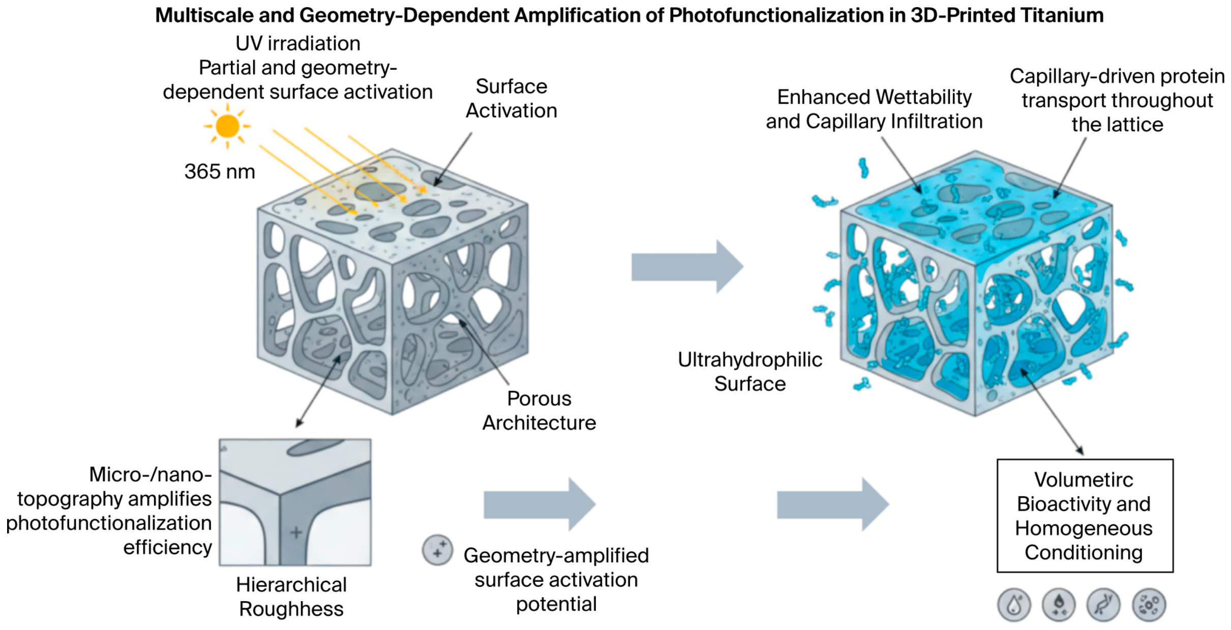
These differences affect the rate at which a photofunctionalized, photocatalytic surface is achieved and how that state is maintained. For example, altered chemistry could change the production of reactive species during UV irradiation, while altered microstructure and defectivity could change the rate of contamination by re-adsorption of hydrocarbons once out of the UV-irradiated environment [47]. These effects must be considered when developing photofunctionalization protocols for flat titanium and scaling up to more complex 3D-printed implants to achieve similar biological outcomes [48]. The geometry-dependent amplification of photofunctionalization’s effects in 3D-printed titanium architectures is illustrated in Figure 3.
Figure 3.
Geometry-dependent amplification of photofunctionalization’s effects in 3D-printed titanium architectures. Synthesized conceptual schematic by the authors, integrated and adapted based on Refs. [24,36,42]. Cumulative recent research evidence indicates that the hierarchical micro/nano-roughness and interconnected porous architecture of additively manufactured titanium implants amplify the biological benefits of photofunctionalization. Ultraviolet activation strengthens capillary-driven fluid infiltration, extends protein adsorption spatially into internal porous networks, and facilitates uniform bioactivation across the entire implant scaffold. The capillary fluid transport and porous tissue ingrowth mechanisms summarized herein are verified by fluid dynamic tests and in vitro penetration experiments reported in the cited studies.
2.3. Stability, Reproducibility, and Practical Considerations
2.3.1. Time-Dependent Loss of Ultrahydrophilicity and Surface Reactivity
Photofunctionalization leads to a very typical time-based loss for ultrafunctions that occurs much faster on Ti surfaces, wherein surface aging after UV photofunctionalization depends primarily on environmental hydrocarbons re-adsorbing on the Ti surfaces [26]. This process is accelerated by factors such as temperature, humidity, and environmental cleanliness upon exposure to airborne particulates, even under aseptic conditions. The increased surface-to-volume ratio in 3D-printed implants significantly contributes to this [49].
This is a great advantage for the hundreds of times one might like to reproduce this concept clinically.
2.3.2. Strategies to Preserve or Reactivate Photofunctionalized Surfaces
A chairside device enabling UV treatment constitutes the best means of halting and overcoming aging processes. This is the only way to reestablish optimized bioactive surface conditions when biological interaction begins at implantation; in situ, it can refine the photofunctionalization approach just prior to implant insertion and makes its effects more predictable by yielding results not affected by ambient contamination from the environment [50].
For these reasons, photofunctionalization of titanium and what one may call surface bioactivation will be based on the slight modification and minor development of translated technology. This seems immediately clinically feasible when one considers the introduction of compact UV lamps that can be adapted to support the introduction of 3D-printed titanium implants [51].
The superhydrophilicity induced by photofunctionalization is not permanent. With exposure to ambient air, hydrocarbon contaminants will gradually re-adsorb onto the treated titanium surface, resulting in hydrophobic recovery over time. The effective duration of the ultrahydrophilic state is affected by ambient temperature, relative humidity, storage atmosphere, surface area, and the pore structure of the 3D-printed implant. Existing studies have reported different attenuation cycles of hydrophilic performance under diverse preservation conditions, which provides a practical reference for determining the frequency of UV chairside treatment.
To intuitively compare the advantages and inherent drawbacks of photofunctionalization relative to mainstream surface modification approaches for titanium implants, we summarize typical modification strategies in Table 2.
Table 2.
Comparison of surface modification strategies for titanium implants.
By horizontal comparison, photofunctionalization exhibits unique advantages of simple operation, no risk of coating shedding, and limited biological regulation, though the inevitable surface aging problem restricts its long-term clinical application prospects.
3. Surface–Protein Interactions at the Implant Interface
Protein adsorption is the first biological “interpretive” layer by which the physicochemical state of a material’s surface is translated into what the cells “see”. Although the underlying titanium is immediately covered with a thin layer of adsorbed proteins from blood and interstitial fluid, this layer presents a different biological ‘face’ of the interface [54]. Cells do not interface directly with the titanium material itself. Instead, they interact with an interfacial protein matrix whose composition, conformation, orientation, and stability over time are the determining factors in subsequent cellular event signaling [55].
Adsorption of proteins onto surfaces is not a passive process affecting which proteins are interfacially coupled with the surface but rather a regulator of downstream osteogenic material–integrin binding, focal-adhesion formation, and related events [56].
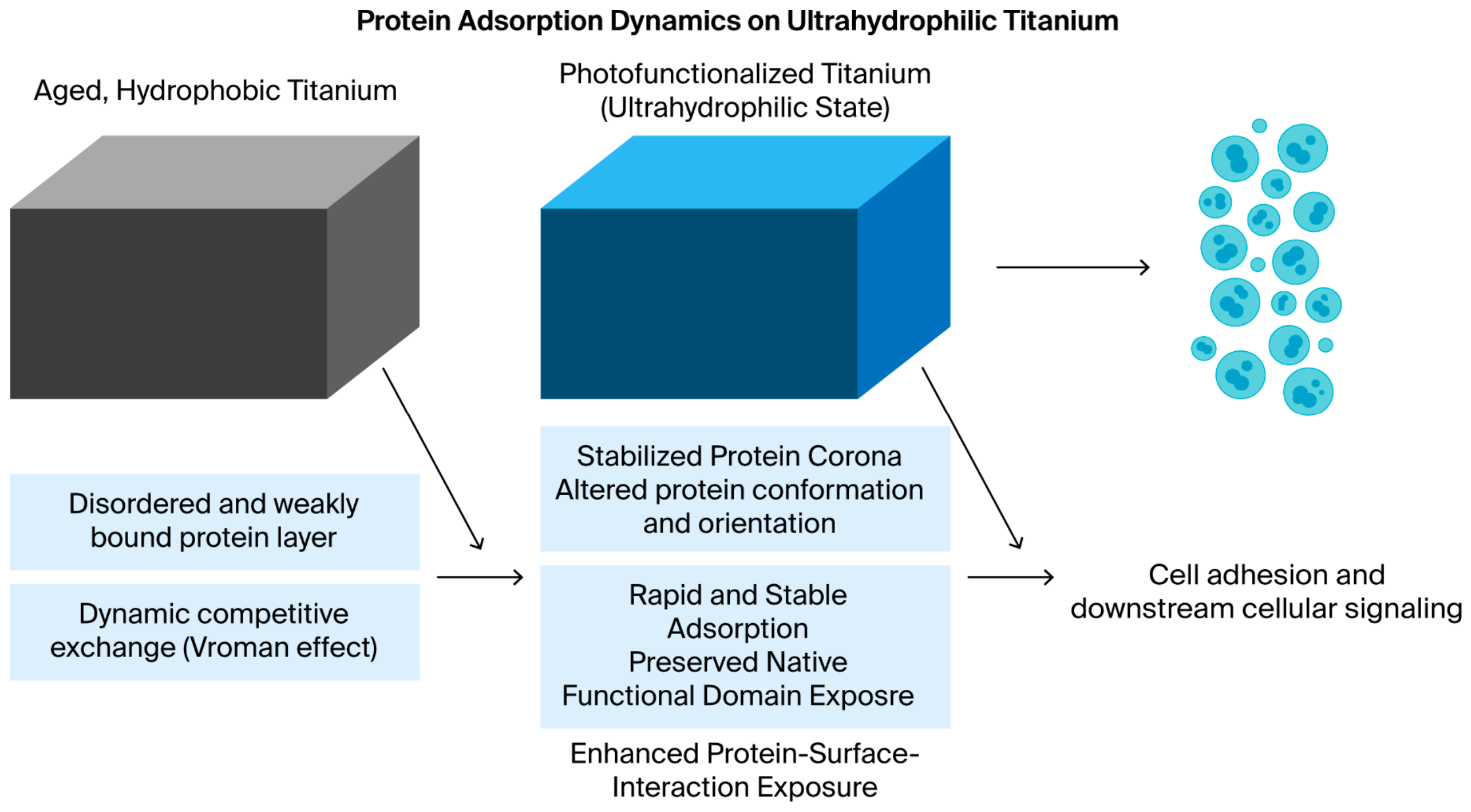
Although the interface is conditioned by surface-active molecular species other than proteins, such as lipids and metabolites, which can provide additional specific signals to cells, practically all material–cell interactions are recognized to originate with proteins already presented on the surface [57]. By inference, the third level of interpretation implies how the ultrahydrophilic state of Ti leads to the formation of a protein film with altered properties, laying the cell signaling foundation required for optimal biocompatibility [58]. The formation of this protein-mediated biological interface on an ultrahydrophilic titanium surface is depicted in Figure 4.
Figure 4.
Formation of the protein-mediated biological interface on ultrahydrophilic titanium surfaces Synthesized conceptual schematic by the authors, integrated and adapted based on Refs. [18,24,26]. Photofunctionalization of titanium surfaces facilitates rapid wetting and extensive adsorption of plasma proteins, leading to the formation of a continuous protein layer at the implant interface. This protein-mediated interface constitutes the first biological boundary encountered by cells and serves as the molecular basis for subsequent cell recognition and adhesion. The Vroman effect attenuation and stabilized protein conformation phenomena summarized here are derived from multi-protein competitive adsorption assays in cited literature.
3.1. Characteristics of Adsorbed Protein Layers on Ultrahydrophilic Titanium Surfaces
3.1.1. Quantity, Conformation, and Orientation of Adsorbed Proteins
As a rule, photofunctionalized ultrahydrophilic Ti adsorbs more protein than hydrophobized or non-photofunctionalized surfaces. The high surface energy of the surface leads to the instantaneous and unrivaled spreading of biological fluids, resulting in a large degree of contact between proteins and the titanium oxide surface immediately after implantation [59]. Nevertheless, the quantity of proteins adsorbed does not, by itself, offer a substantial explanation for the bioactivity of ultrahydrophilic Ti.
Ultrahydrophilic Ti differs qualitatively from other surfaces in the preserved functionality of the adsorbed proteins. While more traditional hydrophobic surfaces induce protein denaturation or (at a minimum) significant conformational change, which masks cell-interacting domains, an ultrahydrophilic surface may increase the possibility of preserving the protein in its active state upon adsorption. This difference is increased with adhesive proteins, such as fibronectin and vitronectin, the function of which depends on the amount of active domain available for interaction [60].
Ultrahydrophilicity may also contribute to orientation changes in the adhesion proteins. This allows for an exposure geometry that favors interaction of the RGD motif with integrins expressed by osteoblastic cells [61]. In summary, conformation and functional bioavailability may be more associated with cell–material recognition than the amount of adsorbed adhesive proteins on the ultrahydrophilic surface.
Green-wavelength laser-induced photocatalytic cleaning uniquely preserves the original composition of the titanium oxide surface, restores intrinsic high surface free energy, and results in more strongly and consistently observed changes in protein-related events after surface conditioning than many alternative approaches to achieving hydrophilicity [62].
3.1.2. Reduced Competitive Adsorption and Improved Protein Stability
Protein adsorption at biomaterial interfaces is inherently dynamic and competitive, as described by the classical Vroman effect. In complex biological fluids, abundant low-affinity proteins are initially adsorbed and subsequently displaced by higher-affinity species [63]. Although this exchange is physiologically unavoidable, excessive protein turnover can destabilize early biological signaling and compromise cell adhesion.
Ultrahydrophilic titanium surfaces modulate this competitive adsorption process by strengthening protein–surface interactions. Increased surface energy and favorable electrostatic interactions reduce premature protein displacement, resulting in preferential retention of osteogenic proteins such as fibronectin, vitronectin, and bone-associated growth factors [64]. Consequently, the adsorbed protein layer exhibits greater temporal stability during the critical early healing phase.
This stabilization has direct biological implications. A persistent and osteogenic protein interface supports robust early cell adhesion, promotes focal adhesion assembly, and facilitates activation of intracellular signaling pathways associated with osteogenic differentiation [65]. By attenuating excessive competitive adsorption, ultrahydrophilic surfaces indirectly regulate early cell signaling events that shape long-term osseointegration.
3.2. Spatial and Temporal Regulation of Protein Layers in 3D-Printed Titanium Implants
Protein adsorption on surfaces is a relatively simple physicochemical process; however, applying this process to 3D printing, especially under flow conditions, quickly leads to complications. Extension of protein–surface interactions should be considered not only as the surface interaction of a planar solid but also as a surface interaction in space and through time [66].
3.2.1. Spatial Distribution of Adsorbed Proteins Within Porous Architectures
As shown by ample evidence on protein adsorption for nonporous Ti, proteins will adsorb on all surfaces of 3D-printed structures and not only their exteriors. Hydrophilicity, particularly when critical, maintains contact between the surface and fluids; capillary forces promote further penetration into and travel through the 3D-printed structure to facilitate contact between the inside surfaces and proteinaceous molecules, which would hardly occur otherwise for an air- or fluid-filled porous microenvironment with surfaces that are hydrophobized by non-photofunctionalized Ti [67].
In other words, proteins will not only be found on the surfaces of such implants but also inside them; the extent to which they are present depends on how fast and how far the surface can be wetted via the pores and the size of the pores themselves [68]. Larger pores facilitate greater penetration, and a larger surface energy will increase the rate of penetration through the pores, as well as the contact surface area. Therefore, ultrahydrophilic porous implants will be coated/covered much more generously by proteins than what would normally be achievable with porous hydrophobic implants, in which case the coating would be much more surface-associated [69].
In the case of 3D-printed Ti, although direct evidence for penetration within very deep porous regions remains scarce, experimental observations so far allow for positive extrapolation regarding the increased penetration and distribution of proteins within these implants compared to hydrophobic Ti. Such a spatially uniform deposition and distribution of proteins might provide homogeneous biochemical guidance within an implant that supports coherent cell migration supported by procession, as indicated by cell ingrowth and/or vascularization [70].
3.2.2. Temporal Evolution of the Protein Corona Under Physiological Conditions
Under physiological conditions, protein-based unit layers are not static in time. Interfacial blood flow, tissue fluid exchange, and, in general, mechanical loading conditions cause temporal dynamics in the adsorption, desorption, and reorganization of biomolecules at the interface [71].
Under the influence of ultrahydrophilicity, protein molecules are much more likely to be strongly associated with the surface, leading to considerably low and limited protein turnover or preliminary remodeling [72]. This means that such a corona could maintain the protein’s biological activity, mirroring processes of desorption/adsorption while simultaneously responding to external stimuli presented through the implant surface. Desorption of adsorbed proteins from such unstable layers would most likely impair cell adhesion and delay tissue integration [73].
Turning to the retention of proteins at the interface by physiological mechanical loading, the molecules in contact with the implant surface will also be subjected to fluid shear, orientation changes, residence time, molecular rearrangement, etc. Loading conditions will impact these interfacial events, further linking mechanical forces to biochemistry. Studying the dynamics of protein corona formation under physiological conditions would provide added value for understanding the in vivo data [74].
3.2.3. Interplay Between Surface Chemistry, Protein Adsorption, and Downstream Cell Recognition
Protein adsorption serves as the molecular intermediary between surface chemistry and cellular recognition. Ultrahydrophilicity couples physicochemical surface properties with selective protein presentation, dictating how cells interpret and respond to the implant interface [75].
Through protein-mediated integrin engagement, the adsorbed layer initiates focal adhesion formation and cytoskeletal organization, thereby linking extracellular cues to intracellular mechanotransduction pathways. In this hierarchically coupled system, surface chemistry governs protein behavior, which in turn directs cellular signaling. These protein-mediated interfacial events establish the molecular foundation upon which integrin signaling, cytoskeletal organization, and osteogenic cell fate decisions—discussed in the following section—are built [76].
3.3. Methodological Considerations and Current Knowledge Gaps
3.3.1. Experimental Models for Studying Protein Adsorption on 3D-Printed Surfaces
Various methods of surface modification have been evaluated in almost all studies mentioned in this review. However, these experimental models were not able to reproduce the phenomenology of 3D-printed titanium, which should display better protein deposition by having an overall larger surface area and a colonized substructure within the pores of the implant [77].
3.3.2. Unresolved Questions in Protein–Surface Interactions
Due to the nature of the tools available, there are opportunities to study cell behaviors within deep, porous regions of 3D-printed implants because these tools can easily characterize the conformation, orientation, and stability of the proteins adsorbed onto their surfaces. The overall measurable bioactivity of cells relies on the contributions of and synergies between the surface chemistry, wettability, and roughness of the implant surfaces, which are mediated by their interfaced proteins [78].
Studies focused on the effect of Ti surface morphology and wettability on protein–surface interactions have covered protein synthetic peptide/protein adsorption, unfolding, the adsorption rate, monolayer/biolayer assembly, orientation, stability, interaction, bioactive sites, and 1-D cell-based functions [79].
We further evaluated the reliability and limitations of existing research regarding protein adsorption behavior on titanium surfaces. Reviewing the published literature revealed that inconsistent results on the initial adsorption quantity, conformational stability and desorption tendency of proteins can be explained primarily by discrepancies in surface topography, residual hydrocarbon contamination level, UV treatment parameters, and cell incubation settings between different experiments. Moreover, most relevant conclusions were obtained from simplified in vitro single-protein or blended-protein culture systems, which cannot reproduce the competitive adsorption dynamics of various proteins within native blood plasma in vivo. Accordingly, there are inevitable restrictions when extrapolating these in vitro findings to the complex biological environment surrounding clinical implants.
4. Cell–Material Interactions and Osteogenic Cell Fate
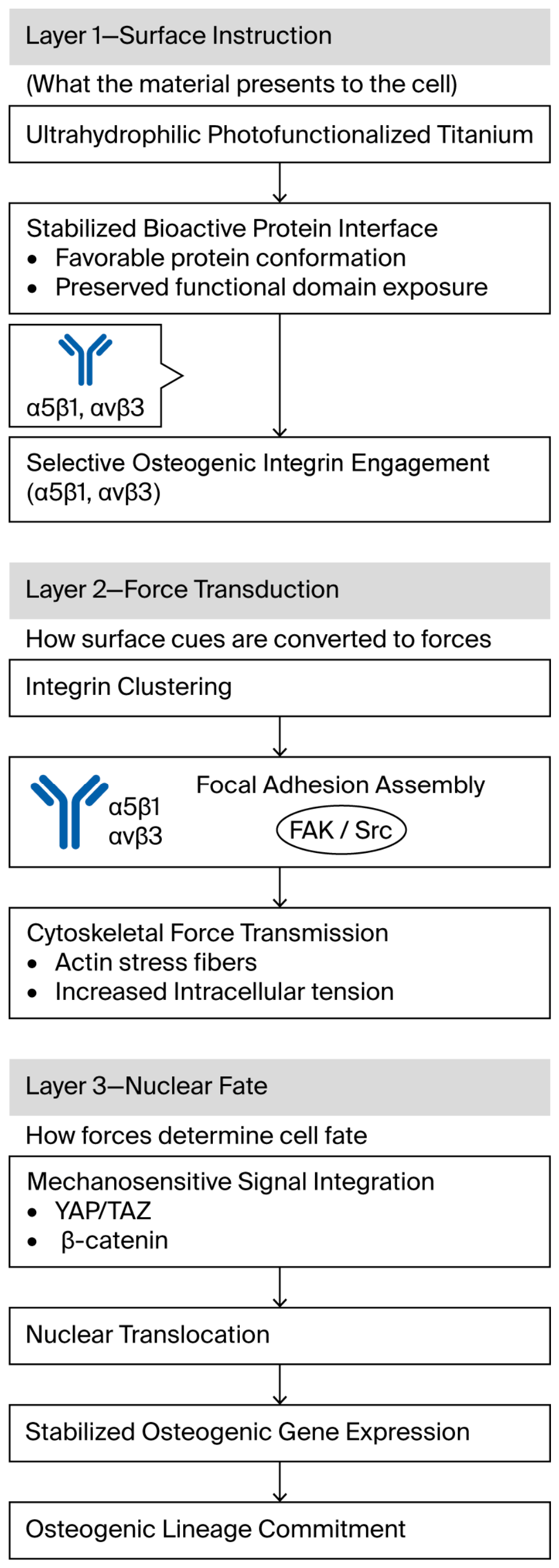
When cells are confronted with adsorbed proteins and interfacial conditioning, their response represents the biological stage at which surface-derived information determines their fate. At this point, interactions between cells and materials no longer record reversible adhesion events but instead an unavoidable process in which biochemical signals, mechanical inputs, and immune balance delimit cell lineage commitment. Thus, osteogenic differentiation on ultrahydrophilic titanium is less a sum of discrete signaling processes and more the net vector of early mechanosensing balanced by the sustained propagation of signals modulated by microenvironmental cues [80]. This idea is supported by considering the responses of cells at proteinaceous interfaces in terms of how material chemistry dictates osteogenic fate—first at molecular and cellular scales and then at 3D tissue scales. Integrin-mediated mechanotransduction and osteogenic cell fate on photofunctionalized titanium are illustrated in Figure 5.
Figure 5.
Integrin-mediated mechanotransduction and osteogenic cell fate on photofunctionalized titanium. Synthesized conceptual schematic by the authors, integrated and adapted based on Ref. [36]. Favorable protein presentation on ultrahydrophilic titanium selectively engages osteogenic integrins, enhancing focal adhesion formation and cytoskeletal tension. These mechanical cues activate downstream signaling pathways, including FAK–Src, Wnt/β-catenin, and YAP/TAZ, leading to stabilized osteogenic differentiation. All mechanotransduction signaling cascades shown were verified via immunofluorescence and qPCR detection in the cited in vitro mechanistic studies.
4.1. Cell Recognition and Adhesion on Protein-Mediated Ultrahydrophilic Interfaces
4.1.1. Integrin-Mediated Cell Recognition of Adsorbed Protein Layers
Idealized models of cultured cells responding to cell substrates overlook the fact that the cells responding to tissue-integrated implants do not directly recognize material at all. The first layer that contacting cells interact with is the adsorbed proteinaceous layer formed immediately upon impregnation of the implants [81]. Thus, the interface recognized by the cell is that formed by the proteins locally adsorbed by the underlying material surface, encoding the biological identity of the interface. As discussed in Section 2, photofunctionalized ultrahydrophilic titanium favors conformations and orientations of adsorbing proteins that enhance integrin recognition [82].
The ultrahydrophilic titanium enhances the availability of RGD and other integrin recognition sites for binding with corresponding membrane receptors and thereby supports highly efficient and selective cell–material recognition. Importantly, this selectivity is not randomly distributed among integrins but is physically and functionally biased toward a restricted set of osteogenic integrins (i.e., α5/β1 and αv/β3) [83]. These integrins mediate osteoblast cell adhesion, survival, differentiation, and function, thus coupling early cell recognition to the osteogenic bias of subsequent cell–material interactions. Through this selection, ultrahydrophilic titanium surfaces function as molecular filters that constrain cellular interpretations toward bone formation [84].
4.1.2. Focal Adhesion Formation and Cytoskeletal Organization
Many of the adhesion-associated motors with actin cytoskeletons are implicated in the initiation of this cellular process, and increased spreading results in bundling or stress fibers, producing a high level of cytoskeletal tension [85].
Such structures and levels, at this early stage of mechanotransduction, are likely to profoundly affect responses directly and indirectly—starting from the phenotype of these cells toward their differentiation. On ultrahydrophilic surfaces, the early mechanotransduction process appears to potentiate the selection of osteogenic lineages over alternative phenotypes [86].
4.2. Mechanotransduction and Osteogenic Signaling Pathways Governing Cell Fate
4.2.1. Focal Adhesion-Driven Mechanotransduction and Signal Propagation
Through integrin clustering and recruitment of FA proteins, FAs form a major physical connection between the extracellular protein layer and the intracellular actin cytoskeleton [87]. Within this context, the improved formation of mature FAs on photofunctionalized hydrophilic titanium is likely to facilitate more efficient force transmission from the interface adhesion sites through the cytoskeleton. In parallel with FA formation, a large family of mechanoresponsive pathways is mobilized, with the FAK–Src complex among the first to be activated [88]. This has led to the concept of focal contacts acting as mechanosensitive hubs that couple mechanical inputs to the onset of biochemical signaling cascades.
Reorganization of the actin cytoskeleton and the levels of various actin-associated regulators are broadly integrin-specific and involve a predominantly Src family kinase-mediated propagation network that rapidly propagates into the cell and includes Rho GTPases [89]. All these signaling events downstream of integrins are likely to be guided, if not strictly determined, by the implant’s surface features, including its topography and chemistry.
4.2.2. Regulation of Osteogenic Gene Expression and Lineage Commitment
The integration of adhesion-mediated signaling and mechanotransduction finally results in a massive activation of osteogenic transcriptional programs. Highly hydrophilic titanium surfaces correlate with increased activation of the Wnt/β-catenin and MAPK pathways, which both predominate in driving osteoblast differentiation [90]. Ultimately, these pathways converge to key osteogenic transcription factors, RUNX2 and osterix, committing cells to the osteogenic lineage.
Thus, signaling timing and duration are predictors of fate stability. An early and prolonged activation of osteogenic pathways reduces cellular plasticity and reinforces lineage commitment. In this respect, ultrahydrophilic interfaces do not just initiate osteogenic signaling; they stabilize differentiation trajectories by aligning early adhesion cues with long-term transcriptional regulation [91].
Despite the consistent trend of hydrophilic surfaces triggering enhanced osteogenic signaling cascades, considerable inconsistencies can be noticed concerning the activation intensity and sustained duration of the Wnt/β-catenin and YAP/TAZ mechanotransduction pathways across the published literature. Such divergences originate from differences in the 2D flat titanium substrates, porous 3D scaffold structures, cell seeding densities, and detection time points adopted in different research studies. It is noteworthy that most mechanistic investigations rely heavily on monolayer mesenchymal stem cell culturing in vitro; the crosstalk between these signaling axes in complex in vivo microenvironments, as well as the interference from immune cells and extracellular matrix remodeling, remains insufficiently characterized, restricting the translational interpretation of these molecular regulatory mechanisms.
4.3. Immunomodulatory and Three-Dimensional Microenvironmental Regulation of Cellular Responses
4.3.1. Modulation of Innate Immune Responses at Ultrahydrophilic Interfaces
Compared with hydrophobic interfaces, ultrahydrophilic interfaces reduce the adhesion, activation, and polarization of macrophages. These surfaces also reduce or counter tissue macrophage activation and curtail the duration of their proinflammatory and inflammatory responses [92].
Aside from the part proportional to the high level of immune modulation exerted by ultrahydrophilic interfaces, the phenotype of the net immune response after restraint activation is, in effect, both noninflammatory and proregenerative in nature, and it favors tissue regeneration [93,94].
4.3.2. Cell Migration, Spatial Organization, and Crosstalk in 3D-Printed Architectures
3D-printed Ti implants can further shape the osteogenic cell response to surface micro- and nanotopography and surface chemistry through architectural and geometric constraints. Ultrahydrophilic surfaces enhance adhesion and spreading of cells seeded on the outermost surfaces of the implants, thus facilitating slow transitions of cells from the implant body’s external surfaces to its internal surfaces via interconnected pores [95].
It therefore appears that ultrahydrophilicity should be considered an additional driver of the migration of cells into spatial coordinates, bringing osteogenic cells close to immune cells. Notably, such proximity may lead to fine paracrine processes of mutual regulation between immune and osteogenic cells and the cross-integration of immune, mechanical, and biochemical signals [96]. These circumstances facilitate a population response and promote oriented behavior in the tissue. In contrast, surface chemical, mechanosensing, and spatial signaling act together to better focus the osteogenic response of the 3D-printed ultrahydrophilic Ti implants toward the cells and reconstruct their organization [97].
This coordinated crosstalk between osteogenic lineage cells and resident immune cells lays a critical biological foundation for subsequent surface-mediated immunomodulatory behaviors of photofunctionalized 3D-printed titanium implants.
Conflicting outcomes have been reported concerning the immunomodulatory effect of photofunctionalized titanium surfaces on macrophage polarization. Several studies have demonstrated that ultrahydrophilic surfaces facilitate anti-inflammatory M2-type macrophage polarization to favor osteoinflammation and bone regeneration, whereas a small number of investigations observed negligible or even opposite immune phenotypes under specific storage and pre-culture conditions. We propose that these contradictions are predominantly driven by the degree of surface aging following UV irradiation, the initial surface contamination level, the macrophage source, and the serum composition in culture medium. Existing immune-related research is also limited by short-term in vitro detection and lacks long-term in vivo immune microenvironment tracking to validate the persistent immunoregulatory property of photofunctionalized implants.
5. Bone–Implant Integration and In Vivo Osseointegration
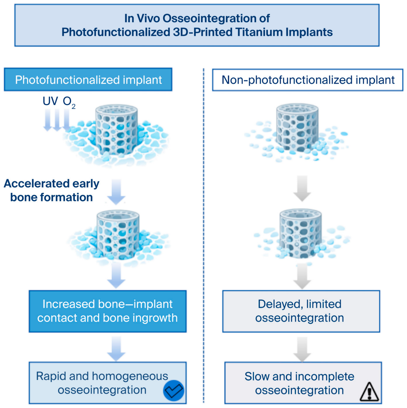
While in vitro studies have undoubtedly provided critical mechanistic details on multilayered biological regulation on implant surfaces, the in vivo osseointegration of an implant represents the ultimate result of the overall behavior at the interface. In this regard, an implant with bone integrated on its surface is no longer a simple interaction between the external forces of the surface and the bone but an irreversible residual state of pre-existing surface–protein–cell responses interacting with the physiological environment of mechanical loading, biological stimuli, and inflammatory response [98]. Thus, photofunctionalized superhydrophilic titanium surfaces present no new biological pathway in vivo but simply represent a stronger commitment to the early interfacial events that they generate, events that eventually spread across the hierarchies of time and space. In the case of 3D-printed porous titanium implants, this can biologically manifest as coherent bone formation, long-term stability, and mechanical competence, which confirm an implant-governed interface process with direct biological evidence presented above [99]. The in vivo osseointegration characteristics of photofunctionalized 3D-printed titanium implants are illustrated in Figure 6.
5.1. Early Bone Formation and Bone–Implant Contact
5.1.1. Accelerated Early Bone Deposition at Ultrahydrophilic Interfaces
Earlier osteoid formation and higher rates of mineralized tissue deposition for ultrahydrophilic 3D-printed titanium implants than for conventional ones have been histologically observed in diverse animal models. Sensitivity is greatest during the early postimplantation days due to increased micromotion and the possibility of fibrous encapsulation. Early matrix deposition and mineralization after ultrahydrophilic surface treatment can shorten this critical period [100].
The principal advantage in vivo for any form of surface bioactivation, therefore, is not just a matter of increased bone formation but also involves accelerated coupling between surface events and subsequent larger scale, longer-time matrix mineralization [101].
Figure 6.
In vivo osseointegration of photofunctionalized 3D-printed titanium implants. Synthesized conceptual schematic by the authors, integrated and adapted based on Refs. [24,36]. Photofunctionalized implants exhibit accelerated early osteoid deposition and mineralization, increased direct bone–implant contact, and enhanced, spatially uniform bone ingrowth throughout interconnected porous architectures. These effects promote accelerated functional fixation and mechanically competent load transfer, supporting stable in vivo osseointegration. Histomorphometric and biomechanical test data from rabbit, sheep and canine animal models in cited references support the bone regeneration trends illustrated.
5.1.2. Improved Bone–Implant Contact and Interfacial Maturation
The acceleration of early bone formation was accompanied by improvements in bone–implant contact, a histomorphometric indicator representing the extent of direct contact with bone and osseointegration quality. The higher bone–implant contact that photofunctionalized implants demonstrated in various in vivo models reflects substantial direct bone contact on the titanium surface [102].
For superhydrophilic surfaces, morphometric analyses showed that mineralized bone, but not fibrous tissue, touched the titanium surface. In this regard, the close interface favors interfacial maturation from woven to lamellar bone and potentiates long-term stability with diminishing micromovement [103]. Superior fibrous tissue encapsulation retains micromotion and relative motion at the interface.
Photofunctionalization thus also affects the quality and speed of osseointegration.
5.2. Bone Ingrowth and Structural Integration in 3D-Printed Titanium Implants
5.2.1. Enhanced Bone Ingrowth Within Interconnected Porous Architectures
Unlike standard machined surfaces, 3D-printed titanium implants feature interconnected porous lattices that eliminate the geometric effects that usually confound attempts at osseointegration. Superhydrophilicity again optimizes pore penetration by fluids and thereby the transport of proteins and cells to the inner regions [104].
Generally, in vivo studies show deep and uniform bone ingrowth in photofunctionalized porous implants. Thus, while the interconnectedness of pores determines whether cells can physically pass through, biofunctionality controls their ability to attach to those surfaces [105].
The bone development is therefore not limited to the outer surface lattice but extends deep inside the scaffold, forming a structure that can contain the bone–implant unit and firmly fix it within the host tissue [106].
5.2.2. Functional Load Transfer and Mechanical Stability
The enhancement of mechanical properties through increased bone ingrowth represent an improvement in ultimate mechanical performance in photofunctionalized 3D-printed implants relative to controls.
Earlier loading reduces risk since the bone functionalizes earlier, with less stress concentrated around the implant. In the long term, better stress distribution also reduces stress shielding, which favors the viral component of bone remodeling, thus improving longevity [107].
Thus, ultrahydrophilic 3D-printed titanium implants can improve not only biological fixation but also functional fixation.
5.3. Translational Evidence, Limitations, and In Vivo Considerations
5.3.1. In Vivo Models and Evidence Supporting Enhanced Osseointegration
Enhanced osseointegration due to photofunctionalized ultrahydrophilic titanium surfaces has been demonstrated in small and large animals. Small animals are generally suitable for analyzing osseointegration over time and localizing osteogenesis at the microstructural level, while large animals can be used to mimic clinical implantation in terms of both general behavior and loading conditions [108].
Indeed, most of the microstructural and mechanical/fixation parameters quantified in the literature that positively correlate surface bioactivation with osseointegration have been validated in several animal models [109]. However, considering the differences between the model parameters and the morphology of implants, it can be reasonably concluded that positive trends have been widely reported, even if the data are not strictly unchanged across studies [110].
5.3.2. Current Limitations and Challenges for Clinical Translation
Retentive success in Australia and Japan has been tempered by time-dependent hydrocarbon regrowth under ambient—and physiological—conditions, so for every activated surface, there should be some degree of control over storage and packaging, as well as the timing of use [111].
However, from a practical standpoint, clinical, ergonomic, and manufacturing considerations are not always taken into account when developing photofunctionalization methods for new generations of implants [112]. Standardized criteria for describing osseointegration and comparison of the techniques used to functionalize surfaces should be created with an eye toward using 3D printing to develop innovative surface bioactivation technologies [113].
To systematically examine current research progress regarding photofunctionalized titanium implants, representative investigations covering in vitro experiments, in vivo animal studies, and clinical observations are summarized in Table 3.
Table 3.
Summary of representative landmark studies on titanium photofunctionalization (Synthesized and expanded by authors based on Refs. [19,24,31,36]).
6. Current Limitations, Controversies, and Knowledge Gaps
With promising mechanistic insight and successful in vivo validation, the study of photofunctionalized ultrahydrophilic 3D-printed titanium implants will reveal predominant failure modes of interface-governed osseointegration [114]. These limits give no cause to question the biological significance of surface bioactivation; rather, they define the limits within which interfacial control can still be exercised with reproducibility and reliability in clinical settings [115]. From this point of view, issues related to surface stability, conclusive mechanistic attribution, and translational modeling should not be considered just technical deficiencies; they represent serious, fundamental challenges concerning surface bioactivity and its interpretation under physiological and clinical constraints. Thorough consideration of such issues and the related state of flux should position photofunctionalization not simply as a promising surface bioactivation strategy but as a genuinely robust and predictable clinical technology.
Notably, most existing mechanistic conclusions regarding titanium photofunctionalization were reached based on conventionally machined titanium substrates, while targeted investigations exploring photomodification rules specific to 3D-printed porous titanium scaffolds remain insufficient. Distinct and inherent discrepancies in native oxide layer structure, pore geometry, spontaneous hydrocarbon adsorption kinetics, and UV-triggered photoresponse behavior exist between additively manufactured titanium and conventional wrought titanium materials. For this reason, relevant research outcomes obtained from traditional titanium cannot be directly extrapolated to their 3D-printed counterparts without dedicated validation, highlighting an urgent demand for systematic research tailored to the unique interfacial properties of 3D-printed implant architectures [116,117]. Inconsistent research outputs can be attributed to variable experimental parameters, material surface topography differences, inconsistent detection methods, and cell sources used in independent studies, which fosters uncertainty in a unified interpretation of the relevant regulatory mechanisms.
6.1. Stability and Practicality of Photofunctionalized Ultrahydrophilic Surfaces
Temporal Decay of Ultrahydrophilicity and Surface Bioactivity
The temporal decay of the ultra-hydrophilicity of photofunctionalized titanium surfaces is the first and most recurrent drawback suggested by researchers. A titanium surface, after UV activation, slowly readsorbs hydrocarbons from the atmosphere and biological fluids, thereby progressively reducing its surface energy and wettability. The “aging” process ruins the uniqueness of such surfaces, which has been reported to evoke enhanced protein adsorption and cell adhesion.
Results of laboratory studies on surface stability often contradict observed in vivo behavior. Laboratory conditions can allow surfaces to retain ultrahydrophilicity for long periods. In contrast, in vivo, it can be affected by protein fouling, ions interacting fluidically with the surface, and body movements, which are generally more aggressive in modifying a surface [118]. For this reason, current consensus in this field is that direct extrapolation of in vitro surface characterization to in vivo biological outcomes is generally precluded.
While photofunctionalization restores surface bioactivity, its time course under ideal physiologic conditions remains poorly characterized.
In addition to biological stability, these aspects represent relevant clinical translation issues. Storage and sterilization modalities will considerably change the surface and its wettability [119]. Common sterilization protocols—that is, gamma radiation, autoclaving, and plasma treatment—may change surface oxidation and induce early surface contamination, thereby reducing or eliminating ultrahydrophilic behavior [120].
To resolve these issues, periodical chair-light photofunctionalization was proposed to reduce surface aging, but the protocols published thus far suffer from variability in UV wavelength, exposure time, the geometry of the emitting device, and the operator’s position. Indeed, the eventual incorporation of these steps into everyday surgical settings increases complexity and time.
Highly diverse institution-based clinical conditions also lead to heterogeneity, which further decreases the possibility of standardization. These practical issues and related solutions underpin the need for reproducible bioactivity preservation or restoration, which could be applied at the point of care and integrated into the clinical workflow [121].
6.2. Incomplete Understanding of Surface–Protein–Cell Coupling Mechanisms
6.2.1. Limited Insight into In Situ Protein Behavior at Implant Interfaces
Although protein adsorption is accepted as the first biological event occurring at the cell–material interface, there is little information regarding the fate of proteins at implant interfaces in vivo. Most mechanistic understanding is based on simplified in vitro models of isolated proteins or static absorption conditions that do not capture many of the complexities of physiological fluids [122].
Probing directly into protein conformation, orientation, and movement at the implant/soft-tissue interface surface, especially in deep, porous regions of 3D-printed structures, poses exceptional technical difficulties. Inferentially, such information plays a key role in making sense of the current state of the art and leading factors in health signaling [123].
The above-mentioned difficulties may compromise studies of how specific biological responses can be linked to protein–surface interactions and valve strategies for the rational design of surface bioactivation.
6.2.2. Unresolved Mechanistic Links Between Protein Presentation and Cell Fate
Another issue involves determining the relative contributions of surface chemistry, wettability, and topography to cellular responses. Though it is evident that surface ultrahydrophilicity changes protein adsorption, there are contradictory reports on the effect of wettability alone on osteogenic differentiation and in combination with micro- and nanoscale topographic features [124].
The problem lies in decoupling these factors; changes in surface chemistry typically affect surface roughness and oxidation state. Therefore, establishing a causative link between specific protein presentations and cell fate decisions is not straightforward and may require further development of multiscale mechanistic models of surface physicochemistry–protein dynamics–cell signaling interphases [125].
6.3. Translational Gaps Between Experimental Models and Clinical Reality
6.3.1. Limitations of Current In Vitro and In Vivo Models
Most experimental models for osseointegration have thus far been based on simplified cell culture systems devoid of immune reactions, applied mechanical stresses, and patient variability in biological responses. These experimental systems might be suitable when trying to elucidate mechanisms since they necessarily lead to an overestimation of actual biological effects in vivo under operative conditions [126].
Animal models, especially those using small animals, provide a first step in in vivo validation, though they have little relevance to the final long-term performance of an implanted bone structure under realistic loading conditions. More importantly, the physiological difference in the rate of bone remodeling and immune response, as well as the mechanical environment, does not allow for translation at the human clinical level. Typical categorizations, such as patient age, bone quality or systemic disease, and drugs used, cannot be reflected in these preclinical studies [127].
6.3.2. Lack of Standardization and Long-Term Clinical Evidence
Thus, at present, truly translational impediments relate to the lack of a standard protocol for surface treatment and outcome measurement, varying implant geometry, pore architecture, photofunctionalization parameters, and outcome metrics [128].
Second, there is little long-term, controlled clinical evidence-based research on photofunctionalized 3D-printed titanium implants. Apart from this, there are regulatory and manufacturing issues related to the broad clinical validation of any surface bioactivated devices made by additive manufacturing [129].
In the absence of benchmarks and well-documented long-term clinical evidence, establishing standards for new technologies tested in preclinical studies must be prioritized.
All mechanistic contradictions and unresolved research gaps summarized in this chapter are fully supported by inconsistent experimental data recorded in the cited literature; the conceptual improvement strategies proposed in Section 7 belong to forward-looking author-integrated deductions based on existing research evidence.
7. Future Perspectives and Design Strategies
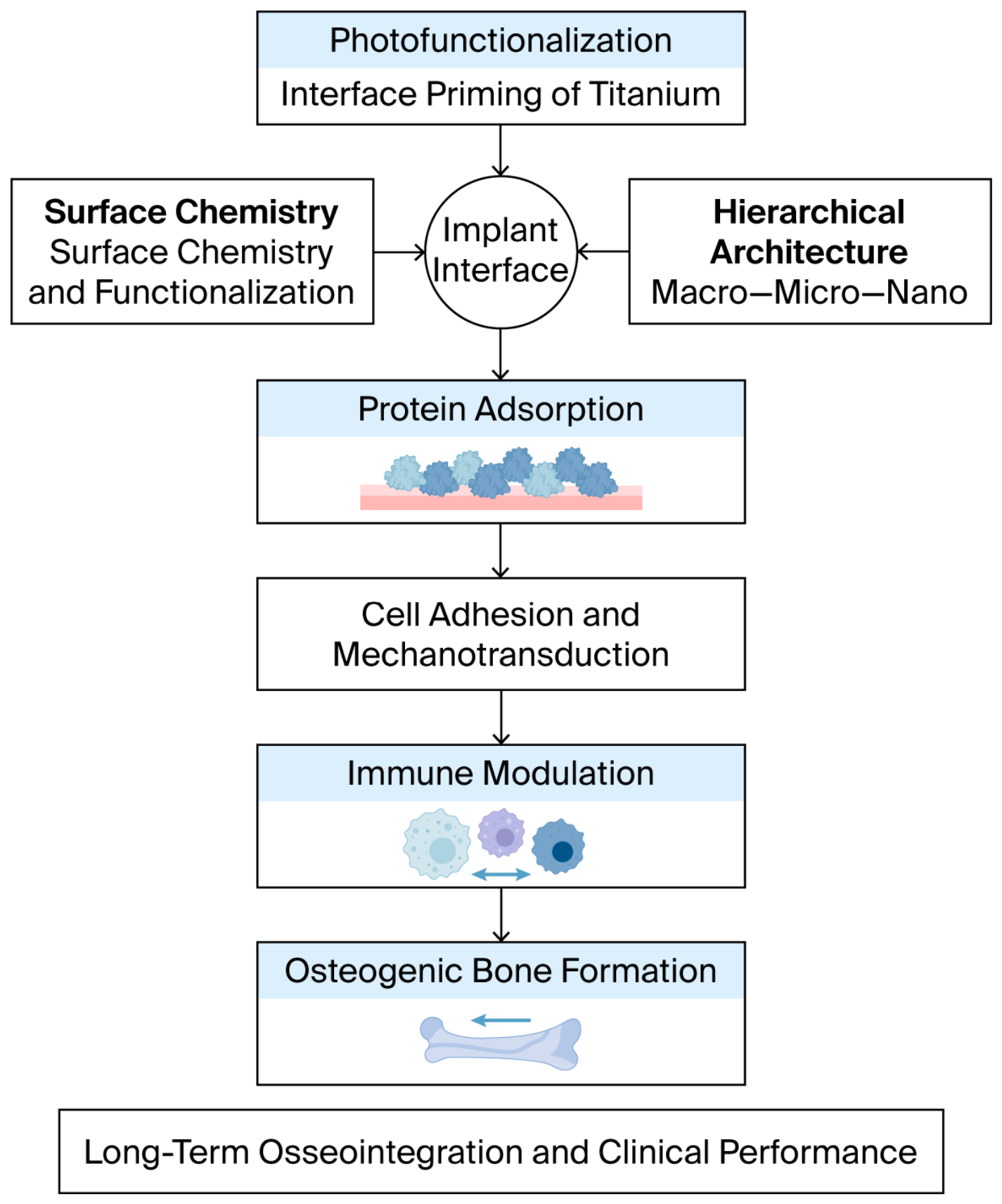
The above evidence reveals that 3D-printed titanium implants with photofunctionalized ultrahydrophilic surface chemistry demonstrate a pure example of interface-governed osseointegration [130]. However, the failure limits discussed in Section 5 suggest that surface-based bioactivation is far from universal. Needless to say, implant design beyond photofunctionalization will inevitably contrast with straightforward optimization in monodimensional parameters in favor of interface-informed systemic strategies to control biological decision-making in multidimensional time and space scales [131]. From this perspective, surface chemistry, structural geometry, biological relevance, and clinical relevance cease to be separate aspects of design and become the axes of a multivariate problem. Thus, photofunctionalization, as an add-on technology, is not the endpoint; it is one step among many toward the full realization of a decision-controllable implant interface [132]. The interface-informed design paradigm for next-generation 3D-printed titanium implants is depicted in Figure 7.
7.1. Advanced Surface Engineering Strategies Beyond Conventional Photofunctionalization
7.1.1. Interface-Integrated Bioactivated Surfaces
Photofunctionalization treatment ensures the restoration of ultrahigh hydrophilicity and activation energy and interface permissiveness but does not guarantee any specific level of biological information or specificity at the molecular level [133]. Hence, future challenges consist primarily of combining photofunctionalization treatment with other existing treatments of implant biomaterial surfaces capable of conferring specific biological information, as noted in [134]. Such combined treatments would produce 3D-printed implant surfaces that both demonstrate flexibility and guide osseointegration.
Figure 7.
Interface-informed design paradigm for next-generation 3D-printed titanium implants Synthesized conceptual schematic by the authors, integrated and adapted based on Refs. [6,116]. Future implant design should integrate surface chemistry, hierarchical architecture, and biological timing into a unified interface-governed framework. Photofunctionalization serves as a foundational strategy for programming early interfacial decisions that dictate long-term osseointegration and clinical performance. This multi-scale integrated design framework is a comprehensive author-proposed conceptual model, built upon the cumulative translational research evidence from cited references.
On this basis, an ideal design could synergize ultrahigh hydrophilicity with calcium phosphate coatings, enhance calcium phosphate-like biomimetic mineral phases, promote early nonspecific protein adsorption, and consistently confer classical osteoconductive mineral coating properties [52]. Alternatively, or analogously, it would enhance ultrahigh hydrophilicity with thin organic deposition treatments, whether through admixed monolayers or covalent grafting approaches. Still, coatings of this nature can only have, at best, permissive effects; selective attachment of specific moieties, such as peptides, growth factors, or ECM proteins, supplies surfaces with ultrahigh hydrophilicity with information at the integrin level or higher and are therefore informationally competent [53]. In this scenario, the choice of coating composition should also consider the best large-area conformation accessible for adhesive domains. This coating would be most efficiently delivered on large areas of very hydrophilic surfaces. One should expect synergistic activity from double coatings/concepts that would coordinate the best biologically relevant interface specificity and not the maximal bioactivity given a loss of functional specificity by selective protein adsorption or cells [135].
7.1.2. Interface Surfaces with Temporally and Spatially Programmed Biological Activity
Conventional photofunctionalization should overcome titanium aging by reinstating the ultrahigh hydrophilicity of the oxide layer that makes the implant surface bioactive. As a result, a primary challenge relating to the surface chemistry, shifted via treatment to ultrahigh hydrophilicity, is its transient nature, which relates to the horizon of attainable stability. Hence, this section presents options to extend the state of ultrahigh hydrophilicity past the treatment to more complicated surfaces with spatial and temporal variation in their interface state [136]. There are many design strategies that could modulate the lifetime of the interface bioactive state, either by controlling its surface chemistry in the presence of, after the realization, or considering its reactivation by the very same treatment. One attractive strategy would be to create an interface state that already demonstrates ultrahigh hydrophilicity or occasionally reactivate it in vivo using light or biochemical input, refunctionalizing its surface wettability toward interacting with biological fluids within an extended critical window from implantation through healing [137]. All interface-active states to be used in ultrahigh hydrophilicity will have to be created so that time and space extremes correspond to the modalities of surface programmatic physiology in the body, where metabolism is not constant but is modulated in a spatiotemporal manner. Here, surface programmatic responses will be necessary to maintain long-term implant integration in vivo [138].
7.2. Interface-Informed Design of 3D-Printed Titanium Implants
7.2.1. Coupling Surface Chemistry with Hierarchical Structural Design
Given the added freedom offered by 3D printing in choosing the outer geometry of an implant, future implant design should couple surface chemistry with architecture at multiple scale levels [139]. Material architecture at the scale of pores and pore networks will need to be purposely shaped and dimensioned to allow efficient multiscalar photofunctionalization. The implant design should allow for the coherence of coatings providing a defined mechanical environment at the scale of single pores with biological guidance provided through locally varying cell behavior and tissue generation. In other words, physical, chemical, topographical, and architecturally multiscalar trends in structure and function will have to be considered, replacing bottom-up principles based on general geometry and biomaterial substitution [140].
7.2.2. Data-Driven and Mechanism-Guided Implant Optimization
With this added layer of complexity in implant design and optimization, one can anticipate a complete in silico modeling of the geometrically and microstructurally complex implant flow, protein transport, solid mechanics, and cellular response. On a mechanistic level, one predicts a significant response in a given surface–protein–cell–bone-binding event by coupling all prior knowledge with multiscale data [141].
7.3. Translational Pathways Toward Clinical Implementation
7.3.1. Bridging Experimental Discoveries and Clinically Relevant Models
Future implant designs might result in significant variations in dynamic interfacial fluids in combination with immune cells due to implant size and/or load. Hence, new feasibility concepts will need to incorporate new immune cell models, experimental dynamic loading, complex biofluids, and in vitro experimental setups [142]. A large animal model is undoubtedly needed in the long term for assessing quality and the service conditions of novel implant designs. In the meantime, their clinical relevance and translatability will be enhanced via factors that increase variability with short-term aging, such as bone quality, age dependence, and comorbidity of patients, into further due diligence in concept proof at the translational level [143].
7.3.2. Standardization, Regulatory Considerations, and Clinical Adoption
Considering the potential for clinical translation of photofunctionalized 3D-printed titanium implants, the research community should work to overcome multiple challenges. As standardization to boost efficiency across the board and reduce product batch-to-batch variation is the means by which a technology is replicated, photofunctionalization must include recent documentation on the optimization of a batch, treatment wavelength, treatment time, storage, and expected outcome [144].
From the perspective of clinical translation, chairside UV photofunctionalization faces practical challenges, including standardized operation parameters, intraoperative processing timing, and portable equipment configuration. Further optimization of irradiation intensity, processing duration, and aseptic operation specifications is required to produce unified clinical guidelines. Meanwhile, long-term clinical cohort observation remains necessary to verify the actual clinical benefit and cost-effectiveness of this in situ surface modification strategy.
8. Conclusions
Osseointegration should no longer be considered a mere biomechanical problem of surface roughness and mechanical fixation. Rather, available evidence provides robust arguments and considers it to be an interface-driven biological decision-making process determined by rapid physicochemical cues at the implant surface that topographically order proteins, which, in turn, determine how the cells lie on the surface to drive immune and bone responses. The next few hours or days of bone contact are highly decisive for long-term performance.
Accordingly, photofunctionalization of 3D-printed titanium implants enables intervention at this critical interface level by eliminating hydrocarbons and restoring the chemical activity and surface energy of the titanium surface. In this process, photofunctionalization dramatically modifies protein adsorption to titanium surfaces, stabilizes the bioactive conformation of the adsorbed proteins, and selects integrin-mediated cell recognition. These effects create a gentle shape-setting effect upstream of the cellular and tissue-level responses of the biological regulatory system. 3D-printed titanium implants exhibit additional lag effects of surface bioactivities. These implants possess a hierarchical porous architecture and high specific surface area so they may be clinically reproducibly manufactured; implants loaded with biologically active fluids, and then eventually cells or bone tissue, can infiltrate throughout the bulk of the implant to structurally distribute the surface bioactivity.
This review found that surface–protein–cell–bone interaction is not independent or additive but rather a single continuous event; it is not a series of interactions but a connected and hierarchical system with feedback and feedforward control. Perturbations at the interfacial level echo at molecular, cellular, and tissue levels, and they modify responses to specific responses and very severely limit later stages of the process. In other words, there will be a neutral, substantial change to later potential optimizations if the interfacial environment is unfavorable. Effects related to surface topography minimally influence the early interface and are therefore unlikely to predict late-occurring biological responses.
In summary, surface modifications to balance topographical roughness characteristics only or associated bioactive coatings are unlikely to lead to progress for both conventionally machined titanium implants and 3D-printed titanium implants. It is most likely that in the future, both conventionally machined and 3D-printed titanium implants will be designed according to an interface-informed strategy that considers surface chemical control, regulated attachment of proteins to surfaces, cell physical behavior related to matrix interactions, and immune response. Only representational models that reproduce the in vivo process’s real spatial and temporal parameters can yield clinically relevant insights.
In this regard, photofunctionalization will become a surface modification rather than a method to control early surface-mediated biological decisions that affect the following process. This is a new perspective on the implant surface as a “governing” substrate that is able to contribute to integrating the structural and biologic events occurring around 3D-printed reconstructive titanium implants with a high degree of design freedom, dedicated clinical strategies, and dependable long-term performance.
Author Contributions
B.W.: writing—original draft, visualization, and investigation. X.W.: conceptualization, funding acquisition, writing—review and editing, and supervision. Y.Y.: investigation and validation. Z.H.: investigation and validation. Y.S.: investigation and visualization. All authors have read and agreed to the published version of the manuscript.
Funding
The Key Research and Development Program Project of Heilongjiang Province (2024ZX12C42).
Institutional Review Board Statement
Not applicable.
Informed Consent Statement
Not applicable.
Data Availability Statement
No new data were created or analyzed in this study. Data sharing is not applicable to this article.
Conflicts of Interest
The authors declare no conflict of interest.
References
- Wang, Z.; Wang, X.; Wang, Y.; Zhu, Y.; Liu, X.; Zhou, Q. NanoZnO-modified titanium implants for enhanced anti-bacterial activity, osteogenesis and corrosion resistance. J. Nanobiotechnol. 2021, 19, 353. [Google Scholar] [CrossRef] [Scilit] [PubMed]
- Hussein, M.A.; Kumar, A.M.; Azeem, M.A.; Sorour, A.A.; Saravanan, S. Ti-30Nb-3Ag alloy with improved corrosion resistance and antibacterial properties for orthopedic and dental applications produced by mechanical alloying. J. Mech. Behav. Biomed. Mater. 2023, 142, 105851. [Google Scholar] [CrossRef] [Scilit] [PubMed]
- Shahbaz Alam, M.; Dhiman, A.; Jain, V.; Bhutia, O.; Pruthi, G. Vertical Bone Implant Contact Around Anterior Immediate Implants and Their Stability After Using Either Alloplast or L-PRF or Both in Peri-Implant Gap: A Prospective Randomized Trial. J. Maxillofac. Oral Surg. 2022, 21, 533–541. [Google Scholar] [CrossRef] [Scilit] [PubMed]
- Xu, R.; Cui, X.; Xin, Q.; Lu, M.; Li, Z.; Li, J.; Chen, X. Zwitterionic PMCP-functionalized titanium surface resists protein adsorption, promotes cell adhesion, and enhances osteogenic activity. Colloids Surf. B Biointerfaces 2021, 206, 111928. [Google Scholar] [CrossRef] [Scilit] [PubMed]
- Anitua, E.; Cerqueira, A.; Romero-Gavilán, F.; García-Arnáez, I.; Martinez-Ramos, C.; Ozturan, S.; Tejero, R. Influence of calcium ion-modified implant surfaces in protein adsorption and implant integration. Int. J. Implant Dent. 2021, 7, 32. [Google Scholar] [CrossRef] [Scilit] [PubMed]
- Ogawa, T.; Hirota, M.; Shibata, R.; Matsuura, T.; Komatsu, K.; Saruta, J.; Att, W. The 3D theory of osseointegration: Material, topography, and time as interdependent determinants of bone-implant integration. Int. J. Implant Dent. 2025, 11, 49. [Google Scholar] [CrossRef] [Scilit] [PubMed]
- Berger, M.B.; Cohen, D.J.; Levit, M.M.; Puetzer, J.L.; Boyan, B.D.; Schwartz, Z. Hydrophilic implants generated using a low-cost dielectric barrier discharge plasma device at the time of placement exhibit increased osseointegration in an animal pre-clinical study: An effect that is sex-dependent. Dent. Mater. 2022, 38, 632–645. [Google Scholar] [CrossRef] [Scilit] [PubMed]
- Ogawa, T.; Hirota, M.; Shibata, R.; Bai, L.; Zhao, Y.; Chen, P.; Zhang, X.; Huang, X.; Du, Z.; Xiao, Y. Targeting Early Healing Phase with Titania Nanotube Arrays on Tunable Diameters to Accelerate Bone Regeneration and Osseointegration. Small 2021, 17, e2006287. [Google Scholar] [CrossRef] [Scilit] [PubMed]
- Dioguardi, M.; Cantore, S.; Quarta, C.; Sovereto, D.; Zerman, N.; Pettini, F.; Ballini, A. Correlation between Diabetes Mellitus and Peri-implantitis: A Systematic Review. Endocr. Metab. Immune Disord. Drug Targets 2023, 23, 596–608. [Google Scholar] [CrossRef] [Scilit] [PubMed]
- Gao, S.S.; Zhang, Y.R.; Zhu, Z.L.; Yu, H.Y. Micromotions and combined damages at the dental implant/bone interface. Int. J. Oral Sci. 2012, 4, 182–188. [Google Scholar] [CrossRef] [Scilit] [PubMed]
- Gu, Y.; Sun, Y.; Shujaat, S.; Braem, A.; Politis, C.; Jacobs, R. 3D-printed porous Ti6Al4V scaffolds for long bone repair in animal models: A systematic review. J. Orthop. Surg. Res. 2022, 17, 68. [Google Scholar] [CrossRef] [Scilit] [PubMed]
- Meng, M.; Wang, J.; Huang, H.; Liu, X.; Zhang, J.; Li, Z. 3D printing metal implants in orthopedic surgery: Methods, applications and future prospects. J. Orthop. Transl. 2023, 42, 94–112. [Google Scholar] [CrossRef] [Scilit] [PubMed]
- Joseph, A.; Uthirapathy, V. A Systematic Review of the Contribution of Additive Manufacturing toward Orthopedic Applications. ACS Omega 2024, 9, 44042–44075. [Google Scholar] [CrossRef] [Scilit] [PubMed]
- Phuoc, H.D.; Hoang, P.N.; Yang, S.; Fraser, D.; Nguyen, V.T. Osseointegrability of 3D-printed porous titanium alloy implant on tibial shaft bone defect in rabbit model. PLoS ONE 2023, 18, e0282457. [Google Scholar] [CrossRef] [Scilit] [PubMed]
- Gulati, K.; Zhang, Y.; Di, P.; Liu, Y.; Ivanovski, S. Research to Clinics: Clinical Translation Considerations for Anodized Nano-Engineered Titanium Implants. ACS Biomater. Sci. Eng. 2022, 8, 4077–4091. [Google Scholar] [CrossRef] [Scilit] [PubMed]
- Sánchez, E.; de Vries, E.; Matthews, D.; van der Heide, E.; Janssen, D. The effect of coating characteristics on implant-bone interface mechanics. J. Biomech. 2024, 163, 111949. [Google Scholar] [CrossRef] [Scilit] [PubMed]
- Cheng, X.; Yang, X.; Liu, C.; Li, Y.; Zhang, Y.; Wang, J.; Jian, X. Stabilization of Apatite Coatings on PPENK Surfaces by Mechanical Interlocking to Promote Bioactivity and Osseointegration In Vivo. ACS Appl. Mater. Interfaces 2023, 15, 697–710. [Google Scholar] [CrossRef] [Scilit] [PubMed]
- Zhang, H.; Huang, Y.; Zhu, J.; Zhao, Y.; Yu, H. Use of Various UVC Photofunctionalization Times to Modify Surface Characteristics and Enhance the Biologic Activity of SLA Titanium. Int. J. Oral Maxillofac. Implant. 2023, 38, 1025–1033. [Google Scholar] [CrossRef] [Scilit] [PubMed]
- Chang, L.-C. Clinical Applications of Photofunctionalization on Dental Implant Surfaces: A Narrative Review. J. Clin. Med. 2022, 11, 5823. [Google Scholar] [CrossRef] [Scilit] [PubMed]
- Gao, Y.; Zhou, L.; Jiang, Y.; Guo, Z.H.; Lu, H.B.; Li, S.B. Effect of different wave-length ultraviolet light-treated micro-arc oxidation titanium surfaces on the physicochemical properties and bioactivity in vitro. Chin. J. Stomatol. 2012, 47, 359–363. [Google Scholar] [CrossRef] [Scilit] [PubMed]
- Zhao, J.; Li, J.; Xu, A.; Xu, Y.; He, F.; Mao, Y. IRAK4 inhibition: An effective strategy for immunomodulating peri-implant osseointegration via reciprocally-shifted polarization in the monocyte-macrophage lineage cells. BMC Oral Health 2023, 23, 265. [Google Scholar] [CrossRef] [Scilit] [PubMed]
- Tasdemir, N.K.; Kilicarslan, B.; Imren, G.; Karaosmanoglu, B.; Taskiran, E.Z.; Bayram, C. Hierarchical TiO2 nanotube arrays enhance mesenchymal stem cell adhesion and regenerative potential through surface nanotopography. J. R. Soc. Interface 2025, 22, 20240642. [Google Scholar] [CrossRef] [Scilit] [PubMed]
- Gao, Y.; Lin, X.; Zhao, Y.; Xu, S.; Lai, C.; Guo, Z.; Liu, Y. The Cleaning Effect of the Photocatalysis of TiO2-B@anatase Nanowires on Biological Activity on a Titanium Surface. Int. J. Nanomed. 2020, 15, 9639–9655. [Google Scholar] [CrossRef] [Scilit] [PubMed]
- Yin, C.; Zhang, T.; Wei, Q.; Cai, H.; Cheng, Y.; Tian, Y.; Liu, Z. Surface treatment of 3D printed porous Ti6Al4V implants by ultraviolet photofunctionalization for improved osseointegration. Bioact. Mater. 2021, 7, 26–38. [Google Scholar] [CrossRef] [Scilit] [PubMed]
- Suzumura, T.; Matsuura, T.; Komatsu, K.; Ogawa, T. A Novel High-Energy Vacuum Ultraviolet Light Photofunctionalization Approach for Decomposing Organic Molecules around Titanium. Int. J. Mol. Sci. 2023, 24, 1978. [Google Scholar] [CrossRef] [Scilit] [PubMed]
- Pacheco, V.N.; Nolde, J.; de Quevedo, A.S.; Visioli, F.; Ponzoni, D. Improvement in the chemical structure and biological activity of surface titanium after exposure to UVC light. Odontology 2021, 109, 271–278. [Google Scholar] [CrossRef] [Scilit] [PubMed]
- Sampaio, D.; Klein, G.B.G.; Cortelli, S.C.; Rosa, J.L.; Vieira, G.S.; de Lima Romeiro, R. Relationship Between Roughness and Wettability of Nine Types of Implant Surfaces and Potential Interference of Surface Oxygen and Carbon: In Vitro Evaluation. Int. J. Oral Maxillofac. Implant. 2025, 40, 439–448. [Google Scholar] [CrossRef] [Scilit] [PubMed]
- Suzumura, T.; Matsuura, T.; Komatsu, K.; Ogawa, T. Decomposing Organic Molecules on Titanium with Vacuum Ultraviolet Light for Effective and Rapid Photofunctionalization. J. Funct. Biomater. 2022, 14, 11. [Google Scholar] [CrossRef] [Scilit] [PubMed]
- Santos, T.D.; de Castro, L.F. Evaluation of a portable Ultraviolet C (UV-C) device for hospital surface decontamination. Photodiagn. Photodyn. Ther. 2021, 33, 102161. [Google Scholar] [CrossRef] [Scilit] [PubMed]
- Okubo, T.; Ikeda, T.; Saruta, J.; Tsukimura, N.; Hirota, M.; Ogawa, T. Compromised Epithelial Cell Attachment after Polishing Titanium Surface and Its Restoration by UV Treatment. Materials 2020, 13, 3946. [Google Scholar] [CrossRef] [Scilit] [PubMed]
- Park, G.; Matsuura, T.; Komatsu, K.; Ogawa, T. Optimizing implant osseointegration, soft tissue responses, and bacterial inhibition: A comprehensive narrative review on the multifaceted approach of the UV photofunctionalization of titanium. J. Prosthodont. Res. 2025, 69, 136–152. [Google Scholar] [CrossRef] [Scilit] [PubMed]
- Pantaroto, H.N.; de Almeida, A.B.; Gomes, O.P.; Matos, A.O.; Landers, R.; Casarin, R.C.V.; Barão, V.A. Outlining cell interaction and inflammatory cytokines on UV-photofunctionalized mixed-phase TiO2 thin film. Mater. Sci. Eng. C Mater. Biol. Appl. 2021, 118, 111438. [Google Scholar] [CrossRef] [Scilit] [PubMed]
- Li, Z.; Qin, B.; Liu, H.; Du, S.; Liu, Y.; He, L.; Du, L. Mesoporous silica thin film as effective coating for enhancing osteogenesis through selective protein adsorption and blood clotting. Biomed. Mater. 2024, 19, 055040. [Google Scholar] [CrossRef] [Scilit] [PubMed]
- Zhong, R.; He, Z.; Zhang, X.; Han, D.; Wang, H.; Liu, J. The strategy of modulation blood responses by surface modification with different functional groups on polyester film. J. Biomed. Mater. Res. A 2021, 109, 1955–1966. [Google Scholar] [CrossRef] [Scilit] [PubMed]
- Kitajima, H.; Hirota, M.; Komatsu, K.; Isono, H.; Matsuura, T.; Mitsudo, K.; Ogawa, T. Ultraviolet Light Treatment of Titanium Microfiber Scaffolds Enhances Osteoblast Recruitment and Osteoconductivity in a Vertical Bone Augmentation Model: 3D UV Photofunctionalization. Cells 2022, 12, 19. [Google Scholar] [CrossRef] [Scilit] [PubMed]
- Yin, C.; Fang, Y.; Sun, X.; Jing, Z.; Fu, J.; Sun, L.; Hao, Y. Dual-wavelength UV photofunctionalization of 3D-printed Ti6Al4V porous bone implant enhances osseointegration via adhesion-cytoskeleton-nuclear mechanotransduction. Mater. Today Bio 2025, 35, 102581. [Google Scholar] [CrossRef] [Scilit] [PubMed]
- Shange, M.; Yadroitsava, I.; du Plessis, A.; Yadroitsev, I. Roughness and Near-Surface Porosity of Unsupported Overhangs Produced by High-Speed Laser Powder Bed Fusion. 3D Print. Addit. Manuf. 2022, 9, 288–300. [Google Scholar] [CrossRef] [Scilit] [PubMed]
- Lin, D.J.; Fuh, L.J.; Chen, W.C. Nano-morphology, crystallinity and surface potential of anatase on micro-arc oxidized titanium affect its protein adsorption, cell proliferation and cell differentiation. Mater. Sci. Eng. C Mater. Biol. Appl. 2020, 107, 110204. [Google Scholar] [CrossRef] [Scilit] [PubMed]
- Sun, X.; Zhong, R.; Wu, C.; Ye, S.; Yuan, H.; Fang, Z.; Wang, L. 3D Printed Titanium Scaffolds with Bi-Directional Gradient QK-Functionalized Surface. Adv. Mater. 2025, 37, e2406421. [Google Scholar] [CrossRef] [Scilit] [PubMed]
- Iwasaki, C.; Hirota, M.; Tanaka, M.; Kitajima, H.; Tabuchi, M.; Ishijima, M.; Ogawa, T. Tuning of Titanium Microfiber Scaffold with UV-Photofunctionalization for Enhanced Osteoblast Affinity and Function. Int. J. Mol. Sci. 2020, 21, 738. [Google Scholar] [CrossRef] [Scilit] [PubMed]
- Karazisis, D.; Rasmusson, L.; Petronis, S.; Palmquist, A.; Shah, F.A.; Agheli, H.; Thomsen, P. The effects of controlled nanotopography, machined topography and their combination on molecular activities, bone formation and biomechanical stability during osseointegration. Acta Biomater. 2021, 136, 279–290. [Google Scholar] [CrossRef] [Scilit] [PubMed]
- Xiao, F.; Ren, G.H.; Pan, C.Y.; Xu, X.; Cheng, K.J.; You, J.; Liu, Y.F. Design and in vitro evaluation of gradient Schwarz Primitive scaffolds with thin-board integration for bone implants. Biomater. Adv. 2025, 177, 214412. [Google Scholar] [CrossRef] [Scilit] [PubMed]
- Bartolomeu, F.; Dourado, N.; Pereira, F.; Alves, N.; Miranda, G.; Silva, F.S. Additive manufactured porous biomaterials targeting orthopedic implants: A suitable combination of mechanical, physical and topological properties. Mater. Sci. Eng. C Mater. Biol. Appl. 2020, 107, 110342. [Google Scholar] [CrossRef] [Scilit] [PubMed]
- Wu, S.; Shan, Z.; Xie, L.; Su, M.; Zeng, P.; Huang, P.; Chen, Z. Mesopore Controls the Responses of Blood Clot-Immune Complex via Modulating Fibrin Network. Adv. Sci. 2022, 9, e2103608. [Google Scholar] [CrossRef] [Scilit] [PubMed]
- Boonrawd, W.; Awad, K.R.; Varanasi, V.; Meletis, E.I. Wettability and in-vitro study of titanium surface profiling prepared by electrolytic plasma processing. Surf. Coat. Technol. 2021, 414, 127119. [Google Scholar] [CrossRef] [Scilit] [PubMed]
- Wu, Y.C.; Kuo, C.N.; Chung, Y.C.; Ng, C.H.; Huang, J.C. Effects of Electropolishing on Mechanical Properties and Bio-Corrosion of Ti6Al4V Fabricated by Electron Beam Melting Additive Manufacturing. Materials 2019, 12, 1466. [Google Scholar] [CrossRef] [Scilit] [PubMed]
- Zheng, X.; Wu, B.; Zhou, C.; Liu, T.; Wang, Y.; Zhao, G.; Chu, C. Sunlight-Driven Production of Reactive Oxygen Species from Natural Iron Minerals: Quantum Yield and Wavelength Dependence. Environ. Sci. Technol. 2023, 57, 1177–1185. [Google Scholar] [CrossRef] [Scilit] [PubMed]
- Funato, A.; Yamada, M.; Ogawa, T. Success rate, healing time, and implant stability of photofunctionalized dental implants. Int. J. Oral Maxillofac. Implant. 2013, 28, 1261–1271. [Google Scholar] [CrossRef] [Scilit] [PubMed]
- Ogura, N.; Berger, M.B.; Srivas, P.; Hwang, S.; Li, J.; Cohen, D.J.; Sandhage, K.H. Tailoring of TiAl6V4 Surface Nanostructure for Enhanced In Vitro Osteoblast Response via Gas/Solid (Non-Line-of-Sight) Oxidation/Reduction Reactions. Biomimetics 2022, 7, 117. [Google Scholar] [CrossRef] [Scilit] [PubMed]
- Guo, L.; Zou, Z.; Smeets, R.; Kluwe, L.; Hartjen, P.; Cacaci, C.; Henningsen, A. Time Dependency of Non-Thermal Oxygen Plasma and Ultraviolet Irradiation on Cellular Attachment and mRNA Expression of Growth Factors in Osteoblasts on Titanium and Zirconia Surfaces. Int. J. Mol. Sci. 2020, 21, 8598. [Google Scholar] [CrossRef] [Scilit] [PubMed]
- Chen, Z.; Wang, B.; Yang, C.; Lv, Z.; Wei, Y.; Pan, T.; Zhang, Y. 3D Printed Pedicle Screws with Microarc Oxidation Ceramic Interfaces Enhance osseointegration and Orthopedic Fixation Feasibility. ACS Appl. Mater. Interfaces 2024, 16, 31983–31996. [Google Scholar] [CrossRef] [Scilit] [PubMed]
- Baheti, W.; Chen, X.; La, M.; He, H. Biomimetic HA-GO implant coating for enhanced osseointegration via macrophage M2 polarization-induced osteo-immunomodulation. J. Appl. Biomater. Funct. Mater. 2024, 22, 22808000241266665. [Google Scholar] [CrossRef] [Scilit] [PubMed]
- Bullock, G.; Atkinson, J.; Gentile, P.; Hatton, P.; Miller, C. Osteogenic Peptides and Attachment Methods Determine Tissue Regeneration in Modified Bone Graft Substitutes. J. Funct. Biomater. 2021, 12, 22. [Google Scholar] [CrossRef] [Scilit] [PubMed]
- Nienhaus, K.; Xue, Y.; Shang, L.; Nienhaus, G.U. Protein adsorption onto nanomaterials engineered for theranostic applications. Nanotechnology 2022, 33, 262001. [Google Scholar] [CrossRef] [Scilit] [PubMed]
- Liu, Z.; Zhu, Q.; Song, E.; Song, Y. Characterization of blood protein adsorption on PM2.5 and its implications on cellular uptake and cytotoxicity of PM2.5. J. Hazard. Mater. 2021, 414, 125499. [Google Scholar] [CrossRef] [Scilit] [PubMed]
- Sung, T.C.; Wang, T.; Liu, Q.; Ling, Q.D.; Subbiah, S.K.; Renuka, R.R.; Higuchi, A. Cell-binding peptides on the material surface guide stem cell fate of adhesion, proliferation and differentiation. J. Mater. Chem. B 2023, 11, 1389–1415. [Google Scholar] [CrossRef] [Scilit] [PubMed]
- Morshed, N.; Rennie, C.; Deng, W.; Collins-Praino, L.; Care, A. Serum-derived protein coronas affect nanoparticle interactions with brain cells. Nanotechnology 2024, 35, 495101. [Google Scholar] [CrossRef] [Scilit] [PubMed]
- Zhang, B.; Zhou, J.; Li, Y.; Chen, J.; Zhang, Y. Bioactive modification of cyclic olefin copolymer (COC) film surfaces by hyaluronic acid and chitosan for enhanced cell adhesion. Int. J. Biol. Macromol. 2024, 281, 136169. [Google Scholar] [CrossRef] [Scilit] [PubMed]
- Foroushani, F.T.; Dzobo, K.; Khumalo, N.P.; Mora, V.Z.; de Mezerville, R.; Bayat, A. Advances in surface modifications of the silicone breast implant and impact on its biocompatibility and biointegration. Biomater. Res. 2022, 26, 80. [Google Scholar] [CrossRef] [Scilit] [PubMed]
- Ji, H.; Li, Y.; Su, B.; Zhao, W.; Kizhakkedathu, J.N.; Zhao, C. Advances in Enhancing Hemocompatibility of Hemodialysis Hollow-Fiber Membranes. Adv. Fiber Mater. 2023, 5, 1198–1240. [Google Scholar] [CrossRef] [Scilit] [PubMed]
- Celles, C.A.S.; Dos Reis, A.C. Titanium: A systematic review of the relationship between crystallographic profile and cell adhesion. J. Biomed. Mater. Res. B Appl. Biomater. 2024, 112, e35450. [Google Scholar] [CrossRef] [Scilit] [PubMed]
- Zimbone, M.; Cacciato, G.; Spitaleri, L.; Egdell, R.G.; Grimaldi, M.G.; Gulino, A. Sb-Doped Titanium Oxide: A Rationale for Its Photocatalytic Activity for Environmental Remediation. ACS Omega 2018, 3, 11270–11277. [Google Scholar] [CrossRef] [Scilit] [PubMed]
- Stöbener, D.D.; Paulus, F.; Welle, A.; Wöll, C.; Haag, R. Dynamic Protein Adsorption onto Dendritic Polyglycerol Sulfate Self-Assembled Monolayers. Langmuir 2018, 34, 10302–10308. [Google Scholar] [CrossRef] [Scilit] [PubMed]
- Huang, R.; Hao, Y.; Pan, Y.; Pan, C.; Tang, X.; Huang, L.; Cui, D. Using a two-step method of surface mechanical attrition treatment and calcium ion implantation to promote the osteogenic activity of mesenchymal stem cells as well as biomineralization on a β-titanium surface. RSC Adv. 2022, 12, 20037–20053. [Google Scholar] [CrossRef] [Scilit] [PubMed]
- Le Saux, G.; Wu, M.C.; Toledo, E.; Chen, Y.Q.; Fan, Y.J.; Kuo, J.C.; Schvartzman, M. Cell-Cell Adhesion-Driven Contact Guidance and Its Effect on Human Mesenchymal Stem Cell Differentiation. ACS Appl. Mater. Interfaces 2020, 12, 22399–22409. [Google Scholar] [CrossRef] [Scilit] [PubMed]
- Wang, Y.; Wang, T.; Tang, R.; Wang, D.; Zhang, X.; Nagaumi, H.; Ma, Q. Multiscale interface engineering in biohybrid composites for biomedical applications. Mater. Today Bio. 2025, 35, 102382. [Google Scholar] [CrossRef] [Scilit] [PubMed]
- Xie, G.; Li, B.; Zhang, X.; Yu, J.; Sun, S.K. One-Minute Preparation of Iron Foam-Drug Implant for Ultralow-Power Magnetic Hyperthermia-Based Combination Therapy of Tumors in Vivo. Adv. Sci. 2024, 11, e2307823. [Google Scholar] [CrossRef] [Scilit] [PubMed]
- Janoušková, O.; Přádný, M.; Vetrík, M.; Chylíková Krumbholcová, E.; Michálek, J.; Dušková Smrčková, M. Biomimetic modification of dual porosity poly(2-hydroxyethyl methacrylate) hydrogel scaffolds-porosity and stem cell growth evaluation. Biomed. Mater. 2019, 14, 055004. [Google Scholar] [CrossRef] [Scilit] [PubMed]
- He, Y.; Zheng, Q.; Lin, Z. Ti4+-immobilized hierarchically porous zirconium-organic frameworks for highly efficient enrichment of phosphopeptides. Mikrochim. Acta 2021, 188, 150. [Google Scholar] [CrossRef] [Scilit] [PubMed]
- Zheng, T.; Gao, H.; Liu, Y.; Sun, S.; Guan, W.; Wu, L.; Li, G. Development of ovalbumin implants with different spatial configurations for treatment of peripheral nerve injury. Bioact. Mater. 2024, 35, 401–415. [Google Scholar] [CrossRef] [Scilit] [PubMed]
- Hedayati, M.; Krapf, D.; Kipper, M.J. Dynamics of long-term protein aggregation on low-fouling surfaces. J. Colloid Interface Sci. 2021, 589, 356–366. [Google Scholar] [CrossRef] [Scilit] [PubMed]
- Fekete, S.; DeLano, M.; Harrison, A.B.; Shiner, S.J.; Belanger, J.L.; Wyndham, K.D.; Lauber, M.A. Size Exclusion and Ion Exchange Chromatographic Hardware Modified with a Hydrophilic Hybrid Surface. Anal. Chem. 2022, 94, 3360–3367. [Google Scholar] [CrossRef] [Scilit] [PubMed]
- Danielsson, R.; Mile, I.; Eriksson, H. Adsorption and Desorption of Immune-Modulating Substances by Aluminium-Based Adjuvants: An Overlooked Feature of the Immune-Stimulating Mechanisms of Aluminium-Based Adjuvants. Int. J. Mol. Sci. 2024, 25, 12399. [Google Scholar] [CrossRef] [Scilit] [PubMed]
- Liu, Z.; Moreira, R.A.; Dujmović, A.; Liu, H.; Yang, B.; Poma, A.B.; Nash, M.A. Mapping Mechanostable Pulling Geometries of a Therapeutic Anticalin/CTLA-4 Protein Complex. Nano Lett. 2022, 22, 179–187. [Google Scholar] [CrossRef] [Scilit] [PubMed]
- Neal, C.J.; Kolanthai, E.; Wei, F.; Coathup, M.; Seal, S. Surface Chemistry of Biologically Active Reducible Oxide Nanozymes. Adv. Mater. 2024, 36, e2211261. [Google Scholar] [CrossRef] [Scilit] [PubMed]
- Qi, Y.; Liu, H.; Zhang, K.; Wu, Y.; Shen, C.; Lin, X. Ihog proteins contribute to integrin-mediated focal adhesions. Sci. China Life Sci. 2023, 66, 366–375. [Google Scholar] [CrossRef] [Scilit] [PubMed]
- Hernandez, D.S.; Michelson, K.E.; Romanovicz, D.; Ritschdorff, E.T.; Shear, J.B. Laser-imprinting of micro-3D printed protein hydrogels enables real-time independent modification of substrate topography and elastic modulus. Bioprinting 2022, 28, e00250. [Google Scholar] [CrossRef] [Scilit] [PubMed]
- Batool, F.; Özçelik, H.; Stutz, C.; Gegout, P.Y.; Benkirane-Jessel, N.; Petit, C.; Huck, O. Modulation of immune-inflammatory responses through surface modifications of biomaterials to promote bone healing and regeneration. J. Tissue Eng. 2021, 12, 20417314211041428. [Google Scholar] [CrossRef] [Scilit] [PubMed]
- Boehm, R.D.; Skoog, S.A.; Diaz-Diestra, D.M.; Goering, P.L.; Dair, B.J. Influence of titanium nanoscale surface roughness on fibrinogen and albumin protein adsorption kinetics and platelet responses. J. Biomed. Mater. Res. A 2024, 112, 373–389. [Google Scholar] [CrossRef] [Scilit] [PubMed]
- Rodriguez-Soto, M.A.; Suarez Vargas, N.; Riveros, A.; Camargo, C.M.; Cruz, J.C.; Sandoval, N.; Briceño, J.C. Failure Analysis of TEVG’s I: Overcoming the Initial Stages of Blood Material Interaction and Stabilization of the Immune Response. Cells 2021, 10, 3140. [Google Scholar] [CrossRef] [Scilit] [PubMed]
- Zhou, J.; Zhang, F.; Tang, Q.; Zhu, T.; Ni, Y.; Wu, Q.; He, H. Deoxygenated hydroxyapatite inhibits macrophage inflammation through fibronectin restricted adsorption. Acta Biomater. 2025, 191, 177–188. [Google Scholar] [CrossRef] [Scilit] [PubMed]
- Rezaei, G.; Daghighi, S.M.; Raoufi, M.; Esfandyari-Manesh, M.; Rahimifard, M.; Mobarakeh, V.I.; Dinarvand, R. Synthetic and biological identities of polymeric nanoparticles influencing the cellular delivery: An immunological link. J. Colloid Interface Sci. 2019, 556, 476–491. [Google Scholar] [CrossRef] [Scilit] [PubMed]
- Xu, T.; Tai, L.; Zhang, Y.; Fan, Z.; Chen, J.; Jiang, W.; Jia, L. Polydopamine-assisted ion-mediated hyaluronic acid grafting for effective construction of hemocompatible platform with cancer cell recognition. Int. J. Biol. Macromol. 2025, 294, 139827. [Google Scholar] [CrossRef] [Scilit] [PubMed]
- Park, J.W.; Seo, J.H.; Lee, H.J. Enhanced osteogenic differentiation of mesenchymal stem cells by surface lithium modification in a sandblasted/acid-etched titanium implant. J. Biomater. Appl. 2022, 37, 447–458. [Google Scholar] [CrossRef] [Scilit] [PubMed]
- Faix, J.; Rottner, K. Ena/VASP proteins in cell edge protrusion, migration and adhesion. J. Cell Sci. 2022, 135, jcs259226. [Google Scholar] [CrossRef] [Scilit] [PubMed]
- Damkham, N.; Issaragrisil, S.; Lorthongpanich, C. Role of YAP as a Mechanosensing Molecule in Stem Cells and Stem Cell-Derived Hematopoietic Cells. Int. J. Mol. Sci. 2022, 23, 14634. [Google Scholar] [CrossRef] [Scilit] [PubMed]
- Huang, M.W.; Lin, C.D.; Chen, J.F. Integrin activation, focal adhesion maturation and tumor metastasis. Sheng Li Xue Bao 2021, 73, 151–159. [Google Scholar] [PubMed]
- da Silva, R.A.; da Silva Feltran, G.; Ferreira, M.R.; Wood, P.F.; Bezerra, F.; Zambuzzi, W.F. The Impact of Bioactive Surfaces in the Early Stages of Osseointegration: An In Vitro Comparative Study Evaluating the HAnano® and SLActive® Super Hydrophilic Surfaces. BioMed Res. Int. 2020, 2020, 3026893. [Google Scholar] [CrossRef] [Scilit] [PubMed]
- Sakai, Y.; Tsunekawa, M.; Ohta, K.; Shimizu, T.; Pastuhov, S.; Hanafusa, H.; Matsumoto, K. The Integrin Signaling Network Promotes Axon Regeneration via the Src-Ephexin-RhoA GTPase Signaling Axis. J. Neurosci. 2021, 41, 4754–4767. [Google Scholar] [CrossRef] [Scilit] [PubMed]
- Zhang, B.; Qu, H.; Zhang, Z.; Wang, X.; Dou, Z.; Li, X.; Zhang, Q. Eu-Doped TiO2 Coatings via One-Step In Situ Preparation Enhance Macrophage Polarization and Osseointegration of Implants. ACS Appl. Mater. Interfaces 2025, 17, 8886–8900. [Google Scholar] [CrossRef] [Scilit] [PubMed]
- Zhao, J.; Dainichi, T.; Guichard, V.; Zhang, Y.; Shirley, S.; Leão, F.B.; Ghosh, S. A time-gated PKA-CREB signaling circuit licenses IL-12 responsiveness and Th1 fate in CD4+ T cells. Proc. Natl. Acad. Sci. USA 2025, 122, e2517132122. [Google Scholar] [CrossRef] [Scilit] [PubMed]
- Zhou, Y.; Tang, C.; Deng, J.; Xu, R.; Yang, Y.; Deng, F. Micro/nano topography of selective laser melting titanium inhibits osteoclastogenesis via mediation of macrophage polarization. Biochem. Biophys. Res. Commun. 2021, 581, 53–59. [Google Scholar] [CrossRef] [Scilit] [PubMed]
- Zhu, Y.; Liang, H.; Liu, X.; Wu, J.; Yang, C.; Wong, T.M.; Yeung, K.W. Regulation of macrophage polarization through surface topography design to facilitate implant-to-bone osseointegration. Sci. Adv. 2021, 7, eabf6654. [Google Scholar] [CrossRef] [Scilit] [PubMed]
- Zhao, D.W.; Du, C.M.; Zuo, K.Q.; Zhao, Y.X.; Xu, X.Q.; Li, Y.B.; Xiao, G.Y. Calcium-Zinc Phosphate Chemical Conversion Coating Facilitates the osseointegration of Biodegradable Zinc Alloy Implants by Orchestrating Macrophage Phenotype. Adv. Healthc. Mater. 2023, 12, e2202537. [Google Scholar] [CrossRef] [Scilit] [PubMed]
- Liu, N.; Wang, H.; Fu, Z.; Zhang, C.; Hui, W.; Wu, J.; Zhang, S. Quercetin-Coating Promotes Osteogenic Differentiation, Osseointegration and Anti-Inflammatory Properties of Nano-Topographic Modificated 3D-Printed Ti6Al4V Implant. Front. Bioeng. Biotechnol. 2022, 10, 933135. [Google Scholar] [CrossRef] [Scilit] [PubMed]
- Meli, V.S.; Veerasubramanian, P.K.; Downing, T.L.; Wang, W.; Liu, W.F. Mechanosensation to inflammation: Roles for YAP/TAZ in innate immune cells. Sci. Signal. 2023, 16, eadc9656. [Google Scholar] [CrossRef] [Scilit] [PubMed]
- Lee, S.H.; Kiapour, A.; Stoeckl, B.D.; Zhang, E.Y.; Begley, M.R.; Oldham, J.; Heo, S.C. Therapeutic Implants: Mechanobiologic Enhancement of Osteogenic, Angiogenic, and Myogenic Responses in Human Mesenchymal Stem Cells on 3D-Printed Titanium Truss. Adv. Healthc. Mater. 2025, 14, e2501856. [Google Scholar] [CrossRef] [Scilit] [PubMed]
- Abaricia, J.O.; Shah, A.H.; Chaubal, M.; Hotchkiss, K.M.; Olivares-Navarrete, R. Wnt signaling modulates macrophage polarization and is regulated by biomaterial surface properties. Biomaterials 2020, 243, 119920. [Google Scholar] [CrossRef] [Scilit] [PubMed]
- Verma, T.; Gupta, R.K.; Bhargava, A.; Dua, B.; Azeem, M.; Verma, A. A Clinical Study to Compare Soft Tissue Healing and Crestal Bone Levels as Influenced by Photofunctionalization of Implants. J. Pharm. Bioallied Sci. 2025, 17, S3022–S3024. [Google Scholar] [CrossRef] [Scilit] [PubMed]
- Avila, J.D.; Stenberg, K.; Bose, S.; Bandyopadhyay, A. Hydroxyapatite reinforced Ti6Al4V composites for load-bearing implants. Acta Biomater. 2021, 123, 379–392. [Google Scholar] [CrossRef] [Scilit] [PubMed]
- Yang, Y.; Zhou, B.; Li, M.; Sun, Y.; Jiang, X.; Zhou, X.; Lei, S. GO/Cu Nanosheet-Integrated Hydrogel Platform as a Bioactive and Biocompatible Scaffold for Enhanced Calvarial Bone Regeneration. Int. J. Nanomed. 2024, 19, 8309–8336. [Google Scholar] [CrossRef] [Scilit] [PubMed]
- Huang, K.; Wu, T.; Lou, J.; Wang, B.; Ding, C.; Gong, Q.; Liu, H. Impact of bone-implant gap size on the interfacial osseointegration: An in vivo study. BMC Musculoskelet. Disord. 2023, 24, 115. [Google Scholar] [CrossRef] [Scilit] [PubMed]
- Kawaguchi, M.; Segawa, A.; Shintani, K.; Nakamura, Y.; Ishigaki, Y.; Yonezawa, K.; Kawahara, N. Bone formation at Ti-6Al-7Nb scaffolds consisting of 3D honeycomb frame and diamond-like carbon coating implanted into the femur of beagles. J. Biomed. Mater. Res. B Appl. Biomater. 2021, 109, 1283–1291. [Google Scholar] [CrossRef] [Scilit] [PubMed]
- Li, Z.; Lu, M.; Zhang, Y.; Wang, J.; Wang, Y.; Gong, T.; Tu, C. 3D-Printed Personalized Lattice Implant as an Innovative Strategy to Reconstruct Geographic Defects in Load-Bearing Bones. Orthop. Surg. 2024, 16, 821–829. [Google Scholar] [CrossRef] [Scilit] [PubMed]
- Babaei, M.; Bonakdar, S.; Nasernejad, B. Selective biofunctionalization of 3D cell-imprinted PDMS with collagen immobilization for targeted cell attachment. Sci. Rep. 2022, 12, 12837. [Google Scholar] [CrossRef] [Scilit] [PubMed]
- Li, S.; Tang, L.; Pu, J.; Wang, J.; Fan, C.; Li, Z.; Song, J. Continuous Hyaluronic Acid Supply by a UHMWPE/PEEK Interlocking Scaffold for Metatarsophalangeal Joint Prosthesis Lubricating Applications. ACS Appl. Mater. Interfaces 2025, 17, 11704–11717. [Google Scholar] [CrossRef] [Scilit] [PubMed]
- Hériveaux, Y.; Le Cann, S.; Fraulob, M.; Vennat, E.; Nguyen, V.H.; Haïat, G. Mechanical micromodeling of stress-shielding at the bone-implant interphase under shear loading. Med. Biol. Eng. Comput. 2022, 60, 3281–3293. [Google Scholar] [CrossRef] [Scilit] [PubMed]
- Xiufeng, Z.; Miao, W.; Huixia, Z.; Boya, X.; Xiaofeng, C.; Longlong, H. Low Intensity Thermal Stimulation to Enhance Early osseointegration in Implants: A Preclinical Study in Rabbits. Clin. Exp. Dent. Res. 2025, 11, e70223. [Google Scholar] [CrossRef] [Scilit] [PubMed]
- Bouakaz, I.; Drouet, C.; Grossin, D.; Cobraiville, E.; Nolens, G. Hydroxyapatite 3D-printed scaffolds with Gyroid-Triply periodic minimal surface porous structure: Fabrication and an in vivo pilot study in sheep. Acta Biomater. 2023, 170, 580–595. [Google Scholar] [CrossRef] [Scilit] [PubMed]
- Vág, J.; Freedman, G.; Szabó, E.; Románszky, L.; Berkei, G. Cervical tooth anatomy considerations for prefabricated anatomic healing abutment design: A mathematical formulation. J. Prosthet. Dent. 2022, 127, 852–859. [Google Scholar] [CrossRef] [Scilit] [PubMed]
- Dong, Y.; Long, L.; Zhang, P.; Yu, D.; Wen, Y.; Zheng, Z.; Chen, W. A chair-side plasma treatment system for rapidly enhancing the surface hydrophilicity of titanium dental implants in clinical operations. J. Oral Sci. 2021, 63, 334–340. [Google Scholar] [CrossRef] [Scilit] [PubMed]
- Bai, L.; Zheng, L.; Ji, P.; Wan, H.; Zhou, N.; Liu, R.; Wang, C. Additively Manufactured Lattice-like Subperiosteal Implants for Rehabilitation of the Severely Atrophic Ridge. ACS Biomater. Sci. Eng. 2022, 8, 912–920. [Google Scholar] [CrossRef] [Scilit] [PubMed]
- Raja, N.; Park, H.; Gal, C.W.; Sung, A.; Choi, Y.J.; Yun, H.S. Support-less ceramic 3D printing of bioceramic structures using a hydrogel bath. Biofabrication 2023, 15, 035006. [Google Scholar] [CrossRef] [Scilit] [PubMed]
- Mondal, S.; Ghosh, R. Influence of cancellous bone material and dead zone on stress-strain, bone stimulus and bone remodelling around the tibia for total ankle replacement. Proc. Inst. Mech. Eng. H 2021, 235, 185–196. [Google Scholar] [CrossRef] [Scilit] [PubMed]
- Ariga, K. Liquid-Liquid Interfacial Nanoarchitectonics. Small 2024, 20, e2305636. [Google Scholar] [CrossRef] [Scilit] [PubMed]
- Sheng, X.; Wang, A.; Wang, Z.; Liu, H.; Wang, J.; Li, C. Advanced surface modification for 3D-printed titanium alloy implant interface functionalization. Front. Bioeng. Biotechnol. 2022, 10, 850110. [Google Scholar] [CrossRef] [Scilit] [PubMed]
- Wang, J.; Jing, Z.; Yin, C.; Wang, Z.; Zeng, S.; Ma, X.; Liu, Z. Coatless modification of 3D-printed Ti6Al4V implants through tailored Cu ion implantation combined with UV photofunctionalization to enhance cell attachment, osteogenesis and angiogenesis. Colloids Surf. B Biointerfaces 2024, 238, 113891. [Google Scholar] [CrossRef] [Scilit] [PubMed]
- Roemermann, D.L.; Atout, R.; Pesun, I.; Kelekis-Cholakis, A.; Stavropoulou, C.; Renvert, S.N.; França, R. An In Vivo Investigation of Non-Metallic vs. Metallic Hand Scalers on Zirconia Implant-Supported Crowns: A Year-Long Analysis of Peri-Implant Maintenance. J. Funct. Biomater. 2023, 15, 9. [Google Scholar] [CrossRef] [Scilit] [PubMed]
- Yan, J.; Siwakoti, P.; Shaw, S.; Bose, S.; Kokil, G.; Kumeria, T. Porous silicon and silica carriers for delivery of peptide therapeutics. Drug Deliv. Transl. Res. 2024, 14, 3549–3567. [Google Scholar] [CrossRef] [Scilit] [PubMed]
- Samree, K.; Srithai, P.U.; Kotchaplai, P.; Thuptimdang, P.; Painmanakul, P.; Hunsom, M.; Sairiam, S. Enhancing the Antibacterial Properties of PVDF Membrane by Hydrophilic Surface Modification Using Titanium Dioxide and Silver Nanoparticles. Membranes 2020, 10, 289. [Google Scholar] [CrossRef] [Scilit] [PubMed]
- Motta, C.; Cavagnetto, D.; Amoroso, F.; Baldi, I.; Mussano, F. Bioactive glass for periodontal regeneration: A systematic review. BMC Oral Health 2023, 23, 264. [Google Scholar] [CrossRef] [Scilit] [PubMed]
- Wellman, S.M.; Kozai, T.D.Y. In vivo spatiotemporal dynamics of NG2 glia activity caused by neural electrode implantation. Biomaterials 2018, 164, 121–133. [Google Scholar] [CrossRef] [Scilit] [PubMed]
- McKiel, L.A.; Ballantyne, L.L.; Negri, G.L.; Woodhouse, K.A.; Fitzpatrick, L.E. MyD88-dependent Toll-like receptor 2 signaling modulates macrophage activation on lysate-adsorbed Teflon™ AF surfaces in an in vitro biomaterial host response model. Front. Immunol. 2023, 14, 1232586. [Google Scholar] [CrossRef] [Scilit] [PubMed]
- Yuan, Z.; Liu, P.; Liang, Y.; Tao, B.; He, Y.; Hao, Y.; Cai, K. Investigation of osteogenic responses of Fe-incorporated micro/nano-hierarchical structures on titanium surfaces. J. Mater. Chem. B 2018, 6, 1359–1372. [Google Scholar] [CrossRef] [Scilit] [PubMed]
- Boumpas, P.; Merabet, S.; Carnesecchi, J. Integrating transcription and splicing into cell fate: Transcription factors on the block. Wiley Interdiscip. Rev. RNA 2023, 14, e1752. [Google Scholar] [CrossRef] [Scilit] [PubMed]
- Ziemian, S.; Green, C.; Sourbron, S.; Jost, G.; Schütz, G.; Hines, C.D.G. Ex vivo gadoxetate relaxivities in rat liver tissue and blood at five magnetic field strengths from 1.41 to 7 T. NMR Biomed. 2021, 34, e4401. [Google Scholar] [CrossRef] [Scilit] [PubMed]
- Kim, T.W.; Koo, S.Y.; Riessland, M.; Chaudhry, F.; Kolisnyk, B.; Cho, H.S.; Studer, L. TNF-NF-κB-p53 axis restricts in vivo survival of hPSC-derived dopamine neurons. Cell 2024, 187, 3671–3689.e23. [Google Scholar] [CrossRef] [Scilit] [PubMed]
- Mbaye, E.H.A.; Scott, E.A.; Burke, J.A. From Edmonton to Lantidra and beyond: Immunoengineering islet transplantation to cure type 1 diabetes. Front. Transplant. 2025, 4, 1514956. [Google Scholar] [CrossRef] [Scilit] [PubMed]
- Kunrath, M.F.; Shah, F.A.; Dahlin, C. Bench-to-bedside: Feasibility of nano-engineered and drug-delivery biomaterials for bone-anchored implants and periodontal applications. Mater. Today Bio 2022, 18, 100540. [Google Scholar] [CrossRef] [Scilit] [PubMed]
- Yu, D.; Tang, Z.; Bao, S.; Guo, S.; Chen, C.; Wu, Q.; Guo, Z. Immunoregulatory Neuro-Vascularized Osseointegration Driven by Different Nano-Morphological CaTiO3 Bioactive Coatings on Porous Titanium Alloy Scaffolds. Adv. Healthc. Mater. 2025, 14, e2404647. [Google Scholar] [CrossRef] [Scilit] [PubMed]
- Sun, M.; Xu, C.; Wu, R.; Yang, Q.; Ding, X.; Zhao, N.; Xu, F.J. Surface-Engineered Orthopedic Implant to Break the Antimicrobial-Osteogenic Paradox at Biointerfaces in the “Race for the Surface”. Adv. Mater. 2025, 37, e2506113. [Google Scholar] [CrossRef] [Scilit] [PubMed]
- Rubianes-Porta, L.; Traver-Méndez, V.; Ghiorghiu, R.; Piera-Auguet, J.; Subirà-Pifarré, C.; Figueiredo, R.; Camps-Font, O. Impact of implant-abutment connection design on biological and mechanical outcomes in posterior single-tooth restorations: A randomized clinical trial. J. Prosthet. Dent. 2025, 134, 1140–1147. [Google Scholar] [CrossRef] [Scilit] [PubMed]
- Ikeda, T.; Okubo, T.; Saruta, J.; Hirota, M.; Kitajima, H.; Yanagisawa, N.; Ogawa, T. Osteoblast Attachment Compromised by High and Low Temperature of Titanium and Its Restoration by UV Photofunctionalization. Materials 2021, 14, 5493. [Google Scholar] [CrossRef] [Scilit] [PubMed]
- Zou, J.; Wu, J.; Wang, Y.; Deng, F.; Jiang, J.; Zhang, Y.; Alshareef, H.N. Additive-mediated intercalation and surface modification of MXenes. Chem. Soc. Rev. 2022, 51, 2972–2990. [Google Scholar] [CrossRef] [Scilit] [PubMed]
- Alfonsi, S.; Karunathasan, P.; Mamodaly-Samdjee, A.; Balathandayutham, K.; Lefevre, S.; Miranda, A.; Hindié, M. Fibronectin Conformations after Electrodeposition onto 316L Stainless Steel Substrates Enhanced Early-Stage Osteoblasts’ Adhesion but Affected Their Behavior. J. Funct. Biomater. 2023, 15, 5. [Google Scholar] [CrossRef] [Scilit] [PubMed]
- Chen, X.; Xing, X.; Lin, S.; Huang, L.; He, L.; Zou, Y.; Zheng, D. Plant-derived nanovesicles: Harnessing nature’s power for tissue protection and repair. J. Nanobiotechnol. 2023, 21, 445. [Google Scholar] [CrossRef] [Scilit] [PubMed]
- Xu, W.; Chen, S.; Yao, M.; Jiang, X.; Lu, Q. A Near-Infrared-Triggered Dynamic Wrinkling Biointerface for Noninvasive Harvesting of Practical Cell Sheets. ACS Appl. Mater. Interfaces 2021, 13, 32790–32798. [Google Scholar] [CrossRef] [Scilit] [PubMed]
- Gulyuk, A.V.; LaJeunesse, D.R.; Collazo, R.; Ivanisevic, A. Tuning Microbial Activity via Programmatic Alteration of Cell/Substrate Interfaces. Adv. Mater. 2021, 33, e2004655. [Google Scholar] [CrossRef] [Scilit] [PubMed]
- Chiesa, I.; De Maria, C.; Ceccarini, M.R.; Mussolin, L.; Coletta, R.; Morabito, A.; Valentini, L. 3D Printing Silk-Based Bioresorbable Piezoelectric Self-Adhesive Holey Structures for In Vivo Monitoring on Soft Tissues. ACS Appl. Mater. Interfaces 2022, 14, 19253–19264. [Google Scholar] [CrossRef] [Scilit] [PubMed]
- Adrian, M.; Eckert, K.M.; Serial, M.R.; Tsanda, A.; Rennpferdt, L.; Benders, S.; Penn, A. NMR relaxometry probes solvent-polarity-dependent molecular interactions in stimuli-responsive lyogels. Phys. Chem. Chem. Phys. 2026, 28, 1645–1654. [Google Scholar] [CrossRef] [Scilit] [PubMed]
- Oliver-Cervelló, L.; Martin-Gómez, H.; Reyes, L.; Noureddine, F.; Ada Cavalcanti-Adam, E.; Ginebra, M.P.; Mas-Moruno, C. An Engineered Biomimetic Peptide Regulates Cell Behavior by Synergistic Integrin and Growth Factor Signaling. Adv. Healthc. Mater. 2021, 10, e2001757. [Google Scholar] [CrossRef] [Scilit] [PubMed]
- Xiong, Y.; Zhang, Z.; Liu, S.; Shen, L.; Zheng, L.; Ding, L.; Yao, Y. T Lymphocyte-Macrophage Hybrid Membrane-Coated Biomimetic Nanoparticles Alleviate Myocarditis via Suppressing Pyroptosis by Targeting Gene Silencing. Int. J. Nanomed. 2024, 19, 12817–12833. [Google Scholar] [CrossRef] [Scilit] [PubMed]
- Kutaish, H.; Tscholl, P.M.; Cosset, E.; Bengtsson, L.; Braunersreuther, V.; Mor, F.M.; Tieng, V. Articular Cartilage Repair After Implantation of Hyaline Cartilage Beads Engineered from Adult Dedifferentiated Chondrocytes: Cartibeads Preclinical Efficacy Study in a Large Animal Model. Am. J. Sports Med. 2023, 51, 237–249. [Google Scholar] [CrossRef] [Scilit] [PubMed]
- Hankenson, F.C. The Elephant in the Room: Recognition and Documentation of Personnel Practices That Confound Reproducibility. J. Am. Assoc. Lab. Anim. Sci. 2024, 63, 232–237. [Google Scholar] [CrossRef] [Scilit] [PubMed]

Disclaimer/Publisher’s Note: The statements, opinions and data contained in all publications are solely those of the individual author(s) and contributor(s) and not of MDPI and/or the editor(s). MDPI and/or the editor(s) disclaim responsibility for any injury to people or property resulting from any ideas, methods, instructions or products referred to in the content. |
© 2026 by the authors. Licensee MDPI, Basel, Switzerland. This article is an open access article distributed under the terms and conditions of the Creative Commons Attribution (CC BY) license.