


Abstract
Two metallacyclic tetranuclear Fe(III) complexes, [{Fe2(μ-O)(μ-RCOO)2(tpon)}2](BPh4)4 [R = Me (1), Ph (2)], where the flexible ditopic ligand tpon (N,N,N′,N′-tetrakis(2-pyridylmethyl)octane-1,8-diamine) links two μ-oxo-bis(μ-carboxylato) triple-bridged dinuclear units, have been prepared. Single-crystal X-ray diffraction establishes that both complexes adopt a 26-membered macrocyclic framework featuring an internal cavity capable of guest inclusion. Notably, incorporation of a monoatomic μ-oxo bridge enforces an outward orientation of the ligand alkyl chains, thereby suppressing the “zipper effect” observed in the previously reported Mn(II) analogue and facilitating the encapsulation of an acetone molecule. UV–vis absorption and diffuse-reflectance spectra confirm that the tetranuclear scaffold remains intact in both the solid state and in solution. These results demonstrate that modulating local coordination directionality via μ-oxo bridging is an effective strategy for controlling the global conformation and host–guest properties of large metallasupramolecular architectures.
1. Introduction
Macrocyclic and cage-shaped molecular compounds have been extensively investigated in the context of host–guest chemistry, owing to their ability to provide well-defined cavities with tunable sizes and properties [1,2,3,4]. Traditionally, such architectures have been constructed using purely organic frameworks, including crown ethers and cryptands. In contrast, coordination-driven self-assembly enables the efficient construction of macrocyclic structures through the linkage of metal centers by polytopic ligands [5,6,7]. These metallacycles, in which metal ions constitute the framework, exhibit diverse magnetic, optical, and electrochemical properties depending on the nature of the metal centers. Consequently, they are promising candidates for functional molecular systems that integrate host–guest chemistry with intrinsic physicochemical properties [8,9,10]. While most metallacycles are assembled from mononuclear primary building units, the use of polynuclear secondary building units (SBUs) offers distinct advantages, such as improved control over the cyclic architecture and enhanced functionality. This strategy has recently been successfully applied to the construction of macrocyclic complexes and metal–organic polygons (MOPs) [11,12].
In our previous work, we reported a cyclic tetranuclear manganese complex, [{Mn2(μ-acetato-O,O’)3(tpon)}2](BPh4)2, constructed from μ-carboxylato triply bridged dinuclear Mn(II) units linked by the ditopic ligand tpon (N,N,N′,N′-tetrakis(2-pyridylmethyl)octane-1,8-diamine) [13]. To date, structurally characterized metal complexes incorporating tpon remain limited. Although this complex possesses a 30-membered macrocyclic framework, the “zipper effect” arising from the octyl chains prevents guest inclusion within the internal cavity. To overcome this limitation, we focused on μ-oxo-bis(μ-carboxylato) dinuclear Fe(III) units as alternative SBUs. This motif contains a monoatomic oxo bridge between two iron centers, which was expected to alter the coordination directionality of the tertiary amine sites and thereby suppress the zipper effect. Furthermore, this dinuclear core represents a fundamental structural motif in bioinorganic chemistry, found in the active sites of metalloproteins such as hemerythrin [14]. Numerous polynuclear complexes incorporating this unit have been reported [15,16,17,18], suggesting that it may also impart unique reactivity toward guest molecules.
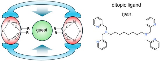
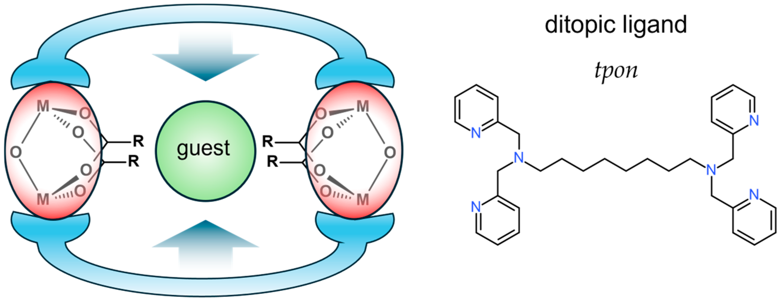
Herein, we report the synthesis and crystal structure of a cyclic tetranuclear Fe(III) complex that encapsulates an acetone molecule. The complex is constructed from μ-oxo-bis-μ-carboxylato dinuclear Fe(III) SBUs and tpon ligands that link the terminal coordination sites (Figure 1).
Figure 1.
Schematic representation of the present complexes. Red ellipsoids represent the dinuclear SBUs, blue linkers represent the ditopic ligands, and the green sphere represents the encapsulated guest molecule. tpon = N,N,N’,N’-tetrakis(2-pyridylmethyl)octane-1,8-diamine.
2. Materials and Methods
2.1. Preparation
All reagents and solvents were purchased from commercial sources and used as received, unless otherwise stated. Methanol was purified by distillation over magnesium turnings. The ligand tpon was synthesized according to the reported literature procedure [19].
2.1.1. [{Fe2(O)(OAc)2(tpon)}2](BPh4)4 (1)
A methanolic solution (5 mL) of iron(III) nitrate nonahydrate (0.202 g, 0.50 mmol) was mixed with an aqueous solution (3 mL) of sodium acetate trihydrate (0.206 g, 1.5 mmol) and a methanolic solution (5 mL) of tpon (0.124 g, 0.24 mmol) to give a dark brown solution. After stirring for 30 min, a methanolic solution (5 mL) of sodium tetraphenylborate (0.172 g, 0.50 mmol) was added, and the mixture was stirred for a further 30 min. The resulting brown precipitate was collected by suction filtration and dried in vacuo for 24 h. Recrystallization from an acetone/hexane (1:1, v/v) mixture afforded brown microcrystals. The products were collected at the microcrystalline stage prior to complete crystal growth. The crystals were ground to a powder and dried at 40 °C under reduced pressure for 8 h to give 1∙2H2O (0.158 g, 44.8% yield). Anal. Calcd for C168H176B4Fe4N12O12: C, 71.51; H, 6.29; and N, 5.96%. Found: C, 71.25; H, 6.16; and N, 6.06%. IR (ATR, cm−1): 3053 (vw), 2924 (vw), 2853 (vw), 1607 (m), 1545 (m), 1479 (m), 1438 (s), 1427 (s), 1024 (s), 766 (m), 733 (vs), 704 (vs), 658 (m), 611 (s), 534 (w), and 470 (w).
Single crystals suitable for X-ray crystallographic analysis were obtained as brown plates after four days by liquid–phase diffusion of hexane into an acetone solution of 1 (0.01 g) using an H-tube.
2.1.2. [{Fe2(O)(OBz)2(tpon)}2]2(BPh4)4 (2)
Complex 2 was prepared following a procedure similar to that described for 1, using sodium benzoate (0.215 g, 1.5 mmol) instead of sodium acetate trihydrate. Yield: 0.158 g (47.3%). Recrystallization of the crude product from an acetone/hexane mixture yielded a few single crystals directly suitable for single-crystal X-ray diffraction analysis. IR (ATR, cm−1): 3053 (vw), 2924 (vw), 2854 (vw), 1606 (m), 1536 (s), 1477 (m), 1396 (s), 1024 (s), 839 (w), 731 (vs), 706 (vs), 673 (m), 648 (m), 611 (s), 534 (m), and 463 (s).
2.2. Physical Measurements
Elemental analyses (C, H and N) were performed at the Service Center of the Elementary Analysis of Organic Compounds, Faculty of Science, Kyushu University. Infrared (IR) spectra were recorded on a Bruker VERTEX70-S FT-IR Spectrometer (Bruker Corp., Billerica, MA, USA) using the ATR (Attenuated Total Reflection) method. The spectra of 1 and 2 are provided in Figures S1 and S2. Electronic absorption and reflection spectra were recorded using an Ocean Optics USB2000+ Fiber Optic Spectrometer (Ocean Optics Inc., Dunedin, FL, USA). Magnetic susceptibilities were measured using a Sherwood Scientific MSB-MK 1 magnetic susceptibility balance (Sherwood Scientific Ltd., Cambridge, UK) at room temperature. The susceptibilities were corrected for diamagnetism using Pascal’s constants [20].
2.3. X-Ray Crystallography
Single crystal X-ray diffraction analyses (SCXRD) were performed on a Rigaku Vari-Max Saturn CCD 724 diffractometer (Rigaku Corp., Tokyo, Japan) using graphite monochromated Mo Ka radiation (λ = 0.71069 Å) at the Analytical Research Center for Experimental Sciences, Saga University. Data collections and processing were carried on using CrystalClear SM 2.0 r16 [21]. The crystals were maintained at 113 K, and a multi-scan absorption correction was applied. Crystallographic data and experimental parameters are summarized in Table 1. Structures were solved by direct methods (ShelXT-2014/5) [22] and refined using ShelXL-2016/6 [23]. Non-hydrogen atoms were refined anisotropically, and hydrogen atoms were placed in calculated positions using a riding model. Olex2-1.5 [24] was used as the interface for the ShelX program package. Molecular graphics were generated using Mercury-2025.3.3 [25]. Hirshfeld surface was generated using Crystal Explorer 3.1 [26].
Table 1.
Crystallographic data and refinement parameters.
3. Results and Discussion
3.1. Structural Studies
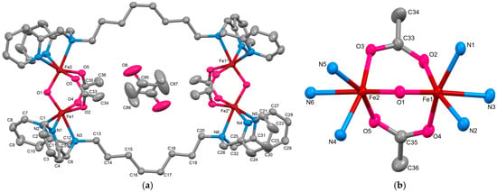
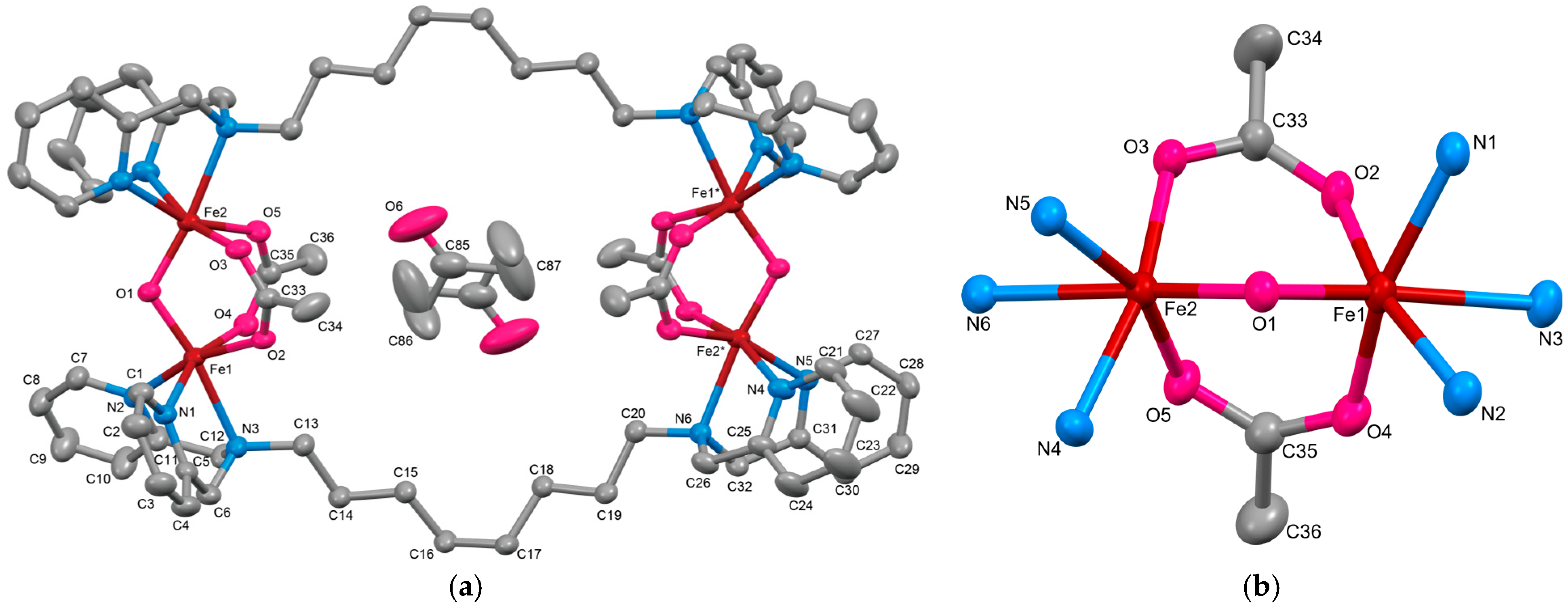
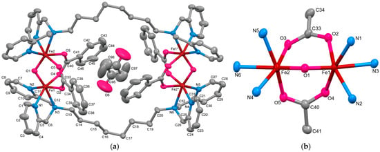
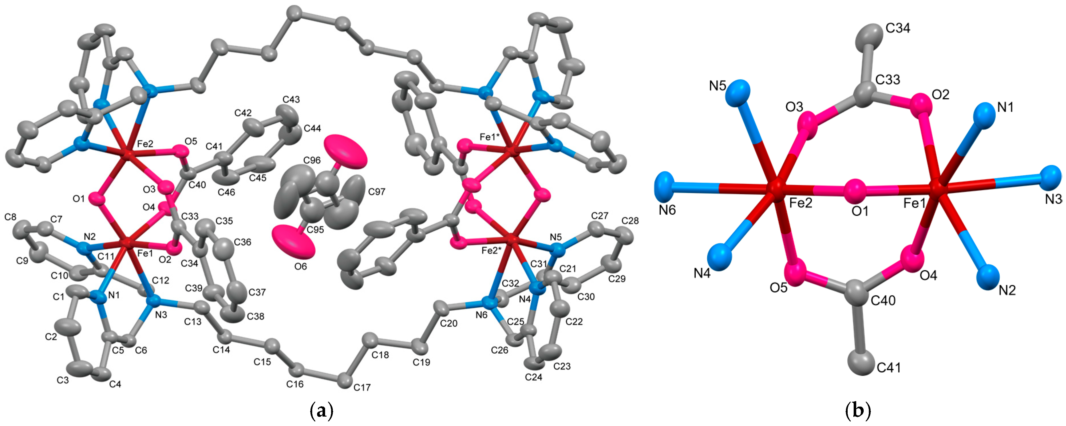
The cationic structure of 1∙5Me2CO and its dinuclear core are shown in Figure 2a and Figure 2b, respectively. Selected bond lengths and angles around the Fe(III) centers are summarized in Table 2.
Figure 2.
(a) X-ray crystal structure for the complex cation of 1 with the encapsulated acetone (symmetrically disordered), showing thermal ellipsoids at a 50% probability level. Hydrogen atoms have been omitted for clarity. (b) Metal core structure of 1. * denotes a symmetry operation.
Table 2.
Selected bond distances and angles of 1∙5Me2CO.
Complex 1∙5Me2CO was identified as a 26-membered metallacyclic tetranuclear Fe(III) complex, [{Fe2(μ-O)(μ-acetato-O,O′)2(tpon)}2](BPh4)4∙5Me2CO. In this structure, two [Fe2(μ-O)(μ-acetato-O,O′)2(tpon)]2+ units are linked by the terminal coordination of the tridentate moieties of two tpon ligands. This tetranuclear structure is fundamentally similar to that of the related complex [{Fe2O(OAc)2(tpbn)}2](NO3)4 (where tpbn contains a C4 methylene spacer) reported by Toftlund and co-workers [27]. The complex crystallizes in a centrosymmetric space group, with the asymmetric unit consisting of one [Fe2(O)(OAc)2(tpon)]2+ moiety. The unit cell contains five acetone molecules as crystallization solvent, one of which is encapsulated within the macrocyclic cavity of 1 and is modeled as symmetrically disordered.
Each Fe(III) ion adopts a six-coordinate distorted octahedral geometry, coordinated by one bridging oxo ligand, two oxygen atoms from bridging acetates, and three nitrogen atoms from the tpon ligand (two pyridyl and one tertiary amine nitrogen). The Fe–O(oxo) distances are characteristically short (Fe1–O1 = 1.7934(18) Å; Fe2–O1 = 1.806(2) Å), and notably, the amine nitrogen atom of tpon occupies the position trans to the oxo bridge. This arrangement is attributed to the strong trans influence of the highly electron-donating oxo ligand, which favors the coordination of the weaker-donating amine nitrogen over the more basic pyridyl nitrogen at the trans position. Consequently, the Fe–N(amine) bonds (Fe1–N3 = 2.276(2) Å; Fe2–N6 = 2.288(2) Å) are significantly longer than the Fe–N(pyridyl) distances. This dinuclear structural motif is consistent with related Fe(III) complexes bearing terminal N3-type ligands [27,28,29]. Continuous Shape Measure (CShM) analysis using SHAPE v2.1 [30] yielded octahedral distortion parameters of 0.850 for Fe1 and 0.475 for Fe2. These values indicate a degree of asymmetry within the dinuclear unit, with Fe1 exhibiting a more pronounced distortion compared to related dinuclear Fe(III) complexes (CShM = 0.479–0.628) [27,28,29].
The structural parameters of the bridging acetate ligands are summarized in Table 3. The torsion angles (Fe–O–C–C = 161.5(2)–172.2(2)° and Fe–O⋯O–Fe = 20.68(13)–24.94(14)°) are consistent with a syn-syn nonplanar bridging mode [31]. The Fe–O–Fe angle of 116.53(9)° is considerably more acute than those typically observed in μ-oxo dinuclear Fe(III) complexes lacking carboxylato bridges. Consequently, the Fe1⋯Fe2 distance is 3.0615(11) Å, significantly shorter than the values reported for complexes containing only a μ-oxo bridge (3.35–3.55 Å) [32,33]. The inter-core Fe⋯Fe separations are Fe1⋯Fe1* = 12.540(6) Å, Fe1⋯Fe2* = 12.145(6) Å, and Fe2⋯Fe2* = 12.510(6) Å (Symmetry code *: 1− x, 1−y, 1−z). The distance between the most distant atoms of the octyl chains (C16⋯C17*) is 12.090(6) Å, resulting in a nearly circular 26-membered macrocyclic framework. The crystal packing structure is shown in Figure S3. No significant intermolecular interactions, such as hydrogen bonding or π–π stacking, were observed.
Table 3.
Structural parameters and bridging modes of carboxylato bridges in Complex 1.
The cationic structure of 2∙8Me2CO is shown in Figure 3a and the dinuclear core structure is illustrated in Figure 3b. Selected bond lengths and bond angles around the Fe(III) ions are summarized in Table 4. Similar to 1∙5Me2CO, 2∙8Me2CO was identified as a tetranuclear Fe(III) complex, [{Fe2(μ-O)(μ-benzoato-O,O’)2(tpon)}2](BPh4)4∙8Me2CO, consisting of two μ-oxo-bis(μ-carboxylato) triple-bridged dinuclear Fe(III) units. Aside from the substitution of benzoate for acetate, the crystal structure of 2∙8Me2CO closely resembles that of 1∙5Me2CO. The unit cell contains eight acetone molecules, one of which is encapsulated within the cavity.
Figure 3.
(a) X-ray crystal structure for the complex cation of 2 with the encapsulated acetone (symmetrically disordered), showing thermal ellipsoids at a 50% probability level. Hydrogen atoms have been omitted for clarity. (b) Metal core structure of 2. * denotes a symmetry operation.
Table 4.
Selected bond distances and angles of 2∙8Me2CO.
The Fe–N bond lengths (Fe1: 2.131(3)–2.267(3) Å; Fe2: 2.137(3)–2.280(3) Å), Fe–O(carboxylato) distances (2.015(2)–2.088(2) Å), and Fe–O(oxo) distances (1.799(2)–1.802(2) Å) are comparable to those observed in 1. While the bond lengths around Fe centers are essentially identical to 1, CShM analysis indicates that both Fe centers in 2 are more significantly distorted (CShM = 0.793 for Fe1 and 0.997 for Fe2). This distortion also manifests in the benzoate bridges. The structural parameters of the bridging benzoate are summarized in Table 5. For instance, the Fe1–O4–C40–C41 torsion angle of 149.8(2)° is significantly smaller than the corresponding values in 1. In relation to this, the bridging mode of μ-benzoato ligand containing O4 and O5 is classified as syn-skew nonplanar mode. The inter-core Fe⋯Fe separations in 2 are all Fe1⋯Fe1* = 12.191(4) Å, Fe1⋯Fe2* = 11.792(5) Å, and Fe2⋯Fe2* = 12.190(5) Å, each ca. 0.35 Å shorter than those in 1, whereas the C16⋯C17* distance, 12.521(6) Å, is slightly (0.4 Å) longer. The crystal packing structure is shown in Figure S4. No significant intermolecular interactions, such as hydrogen bonding or π–π stacking, were observed.
Table 5.
Structural parameters and bridging modes of carboxylato bridges in Complex 2.
3.2. Electronic Spectra
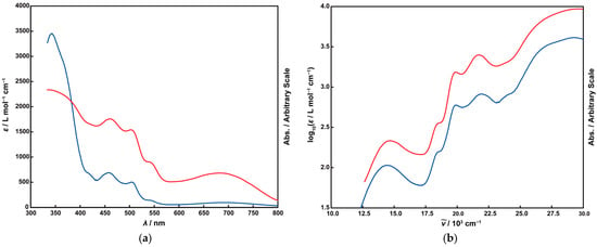
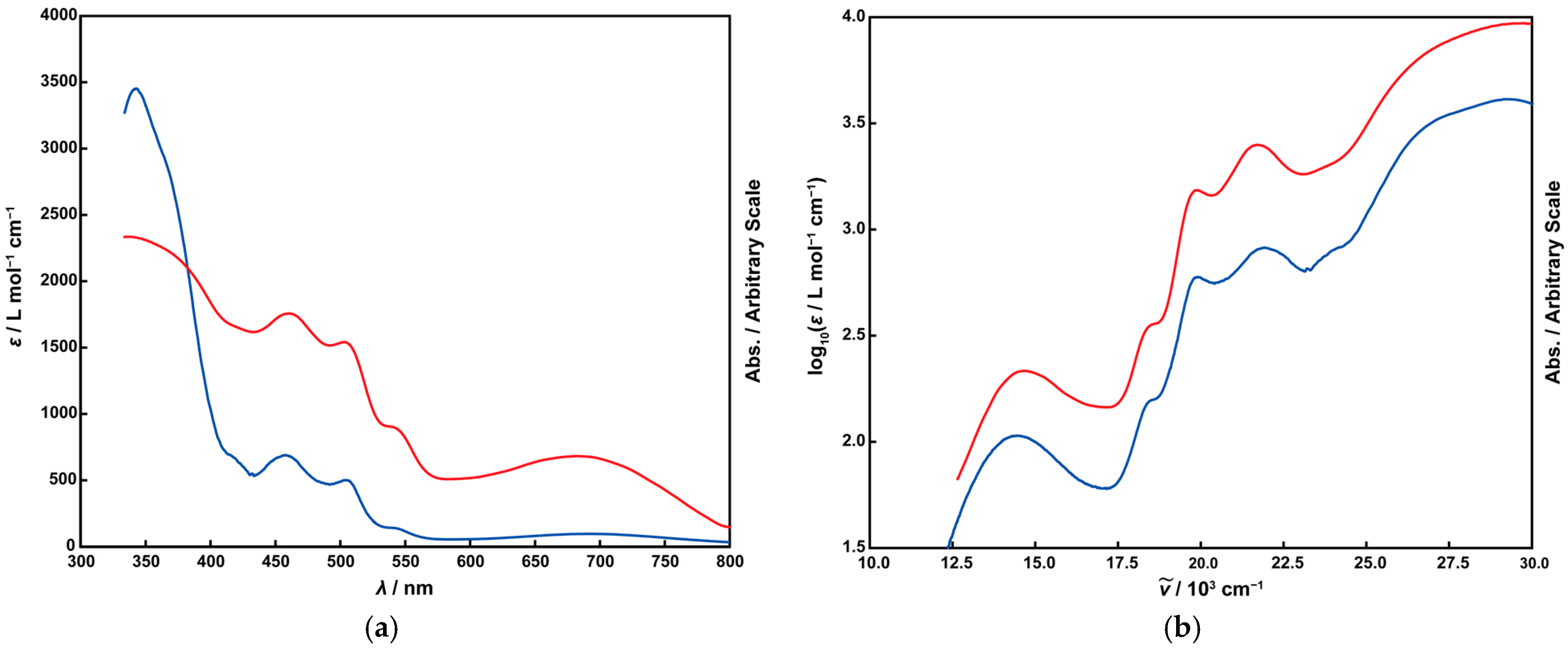
The UV-vis absorption spectrum of 1 in acetonitrile and the solid-state diffuse-reflectance spectrum are shown in Figure 4. In both spectra, four distinct absorption bands are observed in the 350–800 nm region. The striking similarity in peak positions and relative intensities indicates that the tetranuclear cationic structure of 1 remains intact in solution. The spectral data, including molar absorption coefficients (ε) normalized per Fe center, are summarized in Table 6.
Figure 4.
Electronic spectra of 1. (a) Absorption spectrum (ε vs. λ). (b) Plot of log ε vs. wavenumber (). The red and blue lines represent the diffuse-reflectance spectrum (solid) and the absorption spectrum of 1 in acetonitrile (1.0 mM), respectively.
Table 6.
Spectral data for absorption bands of 1.
Complexes featuring a μ-oxo-bis(μ-acetato) dinuclear Fe(III) core, such as 1, typically adopt high-spin configurations [15,16,17,27,28,29]. A characteristic feature of such complexes, particularly those with bent Fe–O–Fe angles, is the appearance of a weak-to-moderate absorption bands in the 600–800 nm region.
These features are attributed to exchange-coupled transitions arising from strong antiferromagnetic interactions between the Fe(III) centers [29,34]. The relatively high intensity of this band further supports this assignment, as it exceeds that expected for purely spin-forbidden d–d transitions. In these systems, magnetic coupling partially relaxes the spin selection rules, leading to enhanced intensity of otherwise spin-forbidden d–d transitions. Consistent with this interpretation, 1 exhibits an effective magnetic moment (μeff) of 1.51 μB at room temperature, indicative of strong antiferromagnetic coupling (J ≈ −140 cm−1). Accordingly, the absorption band at 691 nm (Figure 4a) is assigned to an exchange-coupled transition within the d5–d5 system [35]. Another characteristic feature of μ-oxo-bridged Fe(III) complexes is the presence of intense oxo-to-Fe(III) ligand-to-metal charge-transfer (LMCT) bands [27,29,35,36,37]. These transitions are typically observed as multiple bands in the 300–500 nm range; thus, the absorption bands at 343 and 504 nm are assigned to μ-oxo → Fe(III) LMCT transitions. Furthermore, the bands at 458 and 543 nm are assigned to charge-transfer-enhanced ligand-field transitions (6A1 → 4A1 and 6A1 → 4E), based on their energies (Figure 4b) and molar absorption coefficients, which are consistent with values reported for related dinuclear Fe(III) analogues [27,28,29,35,38].
4. Discussion
It is worth noting that structurally characterized metal complexes with tpon ligands remain relatively limited. In addition to the tetranuclear Mn(II) complexes reported by our group, a Cd(II) coordination polymer, {[Cd4(oxdz)4(tpon)2]·11H2O·4MeOH}n (oxdz2− = 4,4′-(1,3,4-oxadiazole-2,5-diyl)dibenzoate), has been structurally characterized [39]. In this complex, the tpon ligand adopts a fac-type tridentate coordination mode similar to that observed in Complex 1. However, because two types of ditopic ligands are present, the structure extends into a two-dimensional coordination network rather than forming a discrete macrocycle. More generally, for systems incorporating ditopic ligands such as tpon, variations in metal-to-ligand ratios and auxiliary ligands can lead to assemblies of different dimensionalities. These observations suggest that careful control of reaction conditions, particularly stoichiometry, is important for directing the formation of discrete metallacyclic architectures such as Complex 1 and the previously reported Mn(tpon) system.
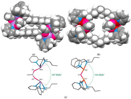
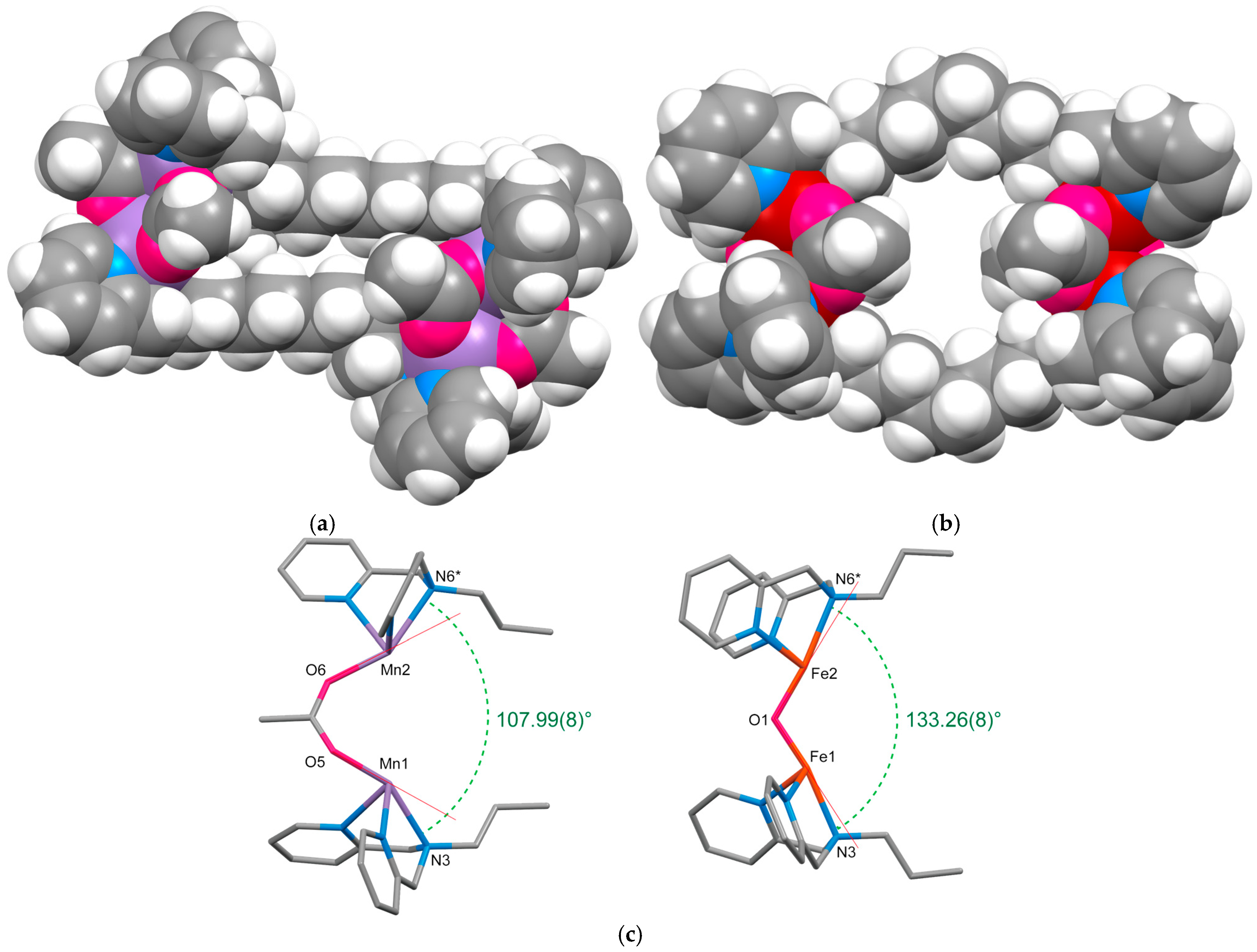
Space-filling models of the complex cations of the previously reported tetranuclear Mn(II) complex, [{Mn2(OAc)3(tpon)}2]2+ [13], and 1 are shown in Figure 5a and Figure 5b, respectively. In the Mn complex, the octyl chains of the tpon ligands connecting the dinuclear SBUs approach each other closely due to a “zipper effect” [40,41], resulting in a collapsed conformation lacking an internal cavity despite the macrocyclic framework. In contrast, Complex 1 clearly forms a macrocyclic cavity that encapsulates an acetone molecule. This guest inclusion is accompanied by an outward expansion of the octyl chains, which positions the two dinuclear Fe(III) units closer together. Consequently, the Fe1⋯Fe2* separation in 1 (12.145(6) Å) is approximately 1.2 Å shorter than the corresponding Mn1⋯Mn2* distance in the Mn analogue.
Figure 5.
Structural comparison of the complex cations of [{Mn2(OAc)3(tpon)}2](BPh4)2 and 1. (a) Space-filling model of [{Mn2(OAc)3(tpon)}2]2+. (b) Space-filling model of Complex 1 cation. (c) Comparison of the coordination geometries showing the angles subtended by the M1–N3 and M2–N6 (M = Mn or Fe) bonds. Selected bridging ligands are omitted for clarity. Red lines represent the vectors extending in the trans direction from the bridging atoms. * denotes a symmetry operation.
A key structural factor responsible for this difference is the presence of the μ-oxo bridge in the Fe complex. Figure 5c illustrates a simplified representation of the bridging moiety. When tpon coordinates as a terminal ligand while preserving the triply bridged dinuclear structure, the tertiary amine nitrogen atoms (N3 and N6) are expected to occupy the positions trans to the bridging atoms. The red lines in Figure 5c denote the ideal trans vectors extending from these bridging atoms. In the tris(μ-acetato)-bridged Mn complex, the alkyl chains of the tpon ligand would suffer from prohibitive steric congestion if the tertiary amines were positioned precisely along these red vectors. Consequently, the coordination geometry undergoes a significant angular distortion, as reflected by the bond angles of O5–Mn1–N3 = 154.00(9)° and O6–Mn2–N6 = 151.47(7)°. Although the angle between the Mn1–N3 and Mn2–N6 vectors (107.99(8)°) is wider than the ca. 54° angle defined by the trans vectors, the orientation of octyl chains remains closely aligned. This proximity induces a zipper effect between the opposing octyl chains, effectively “closing” the macrocyclic cavity. In contrast, the monoatomic oxo-bridged complex 1 maintains a geometry that more closely adheres to the trans vectors (O1–Fe1–N3 = 169.61(8)°, O1–Fe2–N6* = 172.65(7)°). Furthermore, the angle subtended by the Fe–N bonds (133.26(8)°) is considerably wider than that observed in the Mn analogue, favoring a more open structural arrangement. This widening of the coordination geometry dictates the outward orientation of the attached alkyl chains, preventing them from approaching one another. As a result, the zipper effect is substantially weakened, allowing the macrocyclic framework to adopt a more open conformation suitable for guest encapsulation.
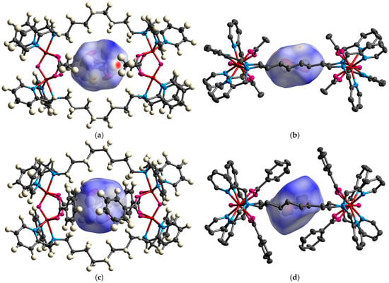
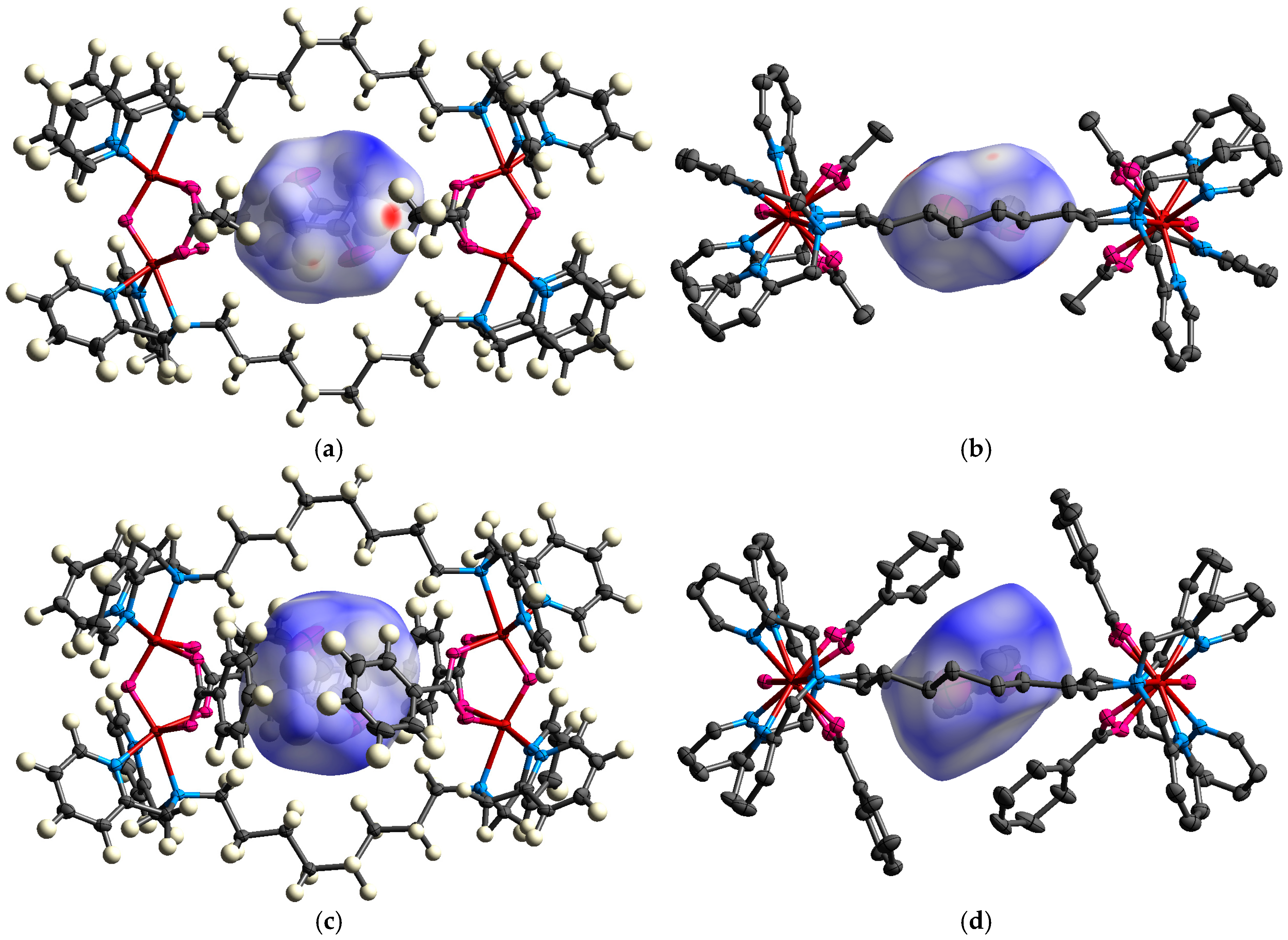
To evaluate the size and characteristics of the inclusion space in 1 and 2, Hirshfeld surface analysis was applied to the encapsulated acetone molecules. The surfaces obtained from the calculation are shown in Figure 6a–d. The calculated occupied volumes are 97 Å3 for 1 and 106 Å3 for 2. These values are slightly smaller than those observed for other acetone molecules present as crystallization solvents (ca. 110–120 Å3), indicating that the encapsulated acetone molecules experience significant spatial confinement within the macrocyclic cavity. This result supports the presence of an active molecular inclusion effect arising from the macrocyclic framework. A comparison of Figure 6b,d reveals a notable difference in the orientation of the guest molecules within the cavity. In 1, the acetone molecule is oriented nearly parallel to the macrocyclic plane, whereas in 2, the molecule is tilted by approximately 30°, aligning it roughly parallel to the phenyl rings of the bridging benzoate ligand. This arrangement suggests the presence of a weak π–π interaction (or CH–π interaction) between the benzoate phenyl rings and the carbonyl moiety of the acetone molecule.
Figure 6.
Molecular inclusion structures of the cations in complexes 1 and 2. The encapsulated guest molecules are represented by Hirshfeld surfaces. (a,c) Views along the normal to the macrocyclic plane for 1 and 2, respectively. (b,d) Side views illustrating the orientation of the guest molecule within the cavity. Hydrogen atoms are omitted for clarity.
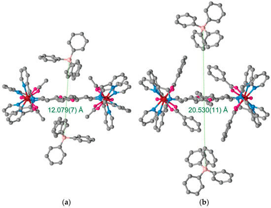
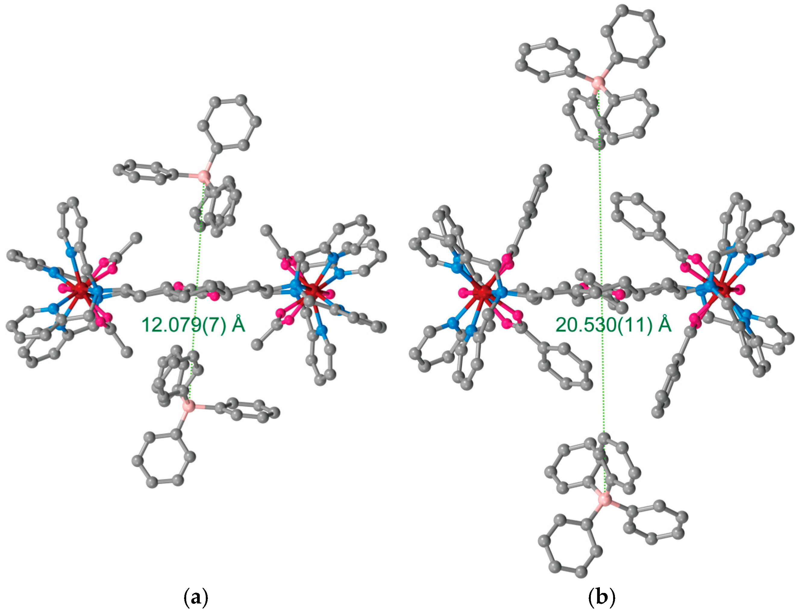
Although the bulky benzoate ligands in 2 might be expected to restrict the internal volume compared to the acetate ligands in 1, an analysis of the BPh4− counter anions reveals a striking contrast (Figure 7a,b). In 1, the B⋯B separation between the two BPh4− anions located above and below the macrocycle is approximately 12 Å (ca. 6 Å from the macrocyclic plane). In contrast, in 2, the acetone guest is partially shielded by the phenyl groups of the benzoate ligands, forcing the BPh4− anions to reside much further away (ca. 10 Å from the plane, B⋯B ≈ 20 Å). These structural differences indicate that while the inclusion space in 1 remains relatively accessible to external species, the cavity in 2 is effectively shielded by the surrounding aromatic ligands, creating a more isolated and confined internal environment. Such a sequestered cavity may provide a favorable platform for the selective encapsulation of guests matching the specific size and shape of the internal void. These findings highlight the critical role of local coordination geometry in dictating global supramolecular structure and function.
Figure 7.
Arrangement of tetraphenylborate anions with respect to the macrocyclic plane in complexes 1 and 2. The green dotted line indicates the B⋯B separation between the anions located above and below the macrocyclic ring. (a) Complex 1. (b) Complex 2.
5. Conclusions
In summary, we have successfully synthesized and structurally characterized a new class of tetranuclear Fe(III) metallacycles, [{Fe2(μ-O)(μ-RCOO)2(tpon)}2](BPh4)4 (R = Me, Ph), in which flexible ditopic ligands link μ-oxo-bis(μ-carboxylato) diiron units into a 26-membered macrocyclic framework. Incorporation of a monoatomic μ-oxo bridge induces a conformational switch that enforces outward orientation of the alkyl chains, suppresses the zipper effect, and enables guest encapsulation. These results demonstrate that modulation of local coordination geometry provides an effective strategy for controlling global structure and host–guest behavior in metallosupramolecular systems.
Supplementary Materials
The following supporting information can be downloaded at: https://www.mdpi.com/article/10.3390/cryst16050281/s1, Figure S1: IR spectra of 1; Figure S2: IR spectra of 2; Figure S3: Crystal packing diagrams of 1∙5Me2CO; Figure S4: Crystal packing diagrams of 2∙8Me2CO. The CIF files and checkCIF/PLATON reports are provided as separate files.
Author Contributions
Conceptualization, M.K.; Data Curation, M.K.; Formal Analysis, J.S., K.Y., and M.K.; Funding Acquisition, M.K.; Investigation, J.S. and M.K.; Project Administration, M.K.; Software, M.K.; Supervision, M.K.; Visualization, M.K.; Writing—Original Draft, J.S. and M.K.; Writing—Review and Editing, M.K. All authors have read and agreed to the published version of the manuscript.
Funding
This research was supported by Japan Society for the Promotion of Science (JSPS) KAKENHI, grant number 25K08679.
Data Availability Statement
The data that support the findings of this study are available from the author upon reasonable request.
Acknowledgments
This research was performed using the equipment (IR and SCXRD) of Analytical Research Center for Experimental Science, Saga University. The authors thank the Service Center of the Elementary Analysis of Organic Compounds, Faculty of Science, Kyushu University, for elementary analysis. During the preparation of this manuscript, the authors used ChatGPT 5.3 for English proofreading. The authors have reviewed and edited the generated output and take full responsibility for the content of this publication.
Conflicts of Interest
The authors declare no conflicts of interest. The funders had no role in the design of the study; in the collection, analyses, or interpretation of data; in the writing of the manuscript; or in the decision to publish the results.
Abbreviations
The following abbreviations are used in this manuscript:
| SBUs | Secondary Building Units |
| MOPs | Metal–Organic Polygons |
| tpon | N,N,N′,N′-tetrakis(2-pyridylmethyl)octane-1,8-diamine |
| tpbn | N,N,N′,N′-tetrakis(2-pyridylmethyl)butane-1,4-diamine |
| SCXRD | Single Crystal X-Ray Diffraction analysis |
| CShM | Continuous Shape Measure |
References
- Pedersen, C.J. Cyclic polyethers and their complexes with metal salts. J. Am. Chem. Soc. 1967, 89, 7017–7036. [Google Scholar] [CrossRef] [Scilit]
- Lehn, J.-M. Supramolecular chemistry—Scope and perspectives. Angew. Chem. Int. Ed. 1988, 27, 89–112. [Google Scholar] [CrossRef] [Scilit]
- Szejtli, J. Introduction and general overview of cyclodextrin chemistry. Chem. Rev. 1998, 98, 1743–1754. [Google Scholar] [CrossRef] [Scilit] [PubMed]
- Kim, K.; Selvapalam, N.; Ko, Y.H.; Park, K.M.; Kim, D.; Kim, J. Functionalized cucurbiturils and their applications. Chem. Soc. Rev. 2007, 36, 267–279. [Google Scholar] [CrossRef] [Scilit]
- Domoto, Y.; Fujita, M. Self-assembly of nanostructures with high complexity based on metal···unsaturated-bond coordination. Coord. Chem. Rev. 2022, 466, 214605. [Google Scholar] [CrossRef] [Scilit]
- Stang, P.J.; Olenyuk, B. Self-assembly, symmetry, and molecular architecture: Coordination as the motif in the rational design of supramolecular metallacyclic polygons and polyhedra. Acc. Chem. Res. 1997, 30, 502–518. [Google Scholar] [CrossRef] [Scilit]
- Cook, T.R.; Stang, P.J. Recent developments in the preparation and chemistry of metallacycles and metallacages via coordination. Chem. Rev. 2015, 115, 7001–7045. [Google Scholar] [CrossRef] [Scilit]
- Wu, D.-Y.; Sato, O.; Einaga, Y.; Duan, C.-Y. A spin-crossover cluster of iron(II) exhibiting a mixed-spin structure and synergy between spin transition and magnetic interaction. Angew. Chem. 2009, 121, 1503–1506. [Google Scholar] [CrossRef] [Scilit]
- Yin, C.; Du, J.; Olenyuk, B.; Stang, P.J.; Sun, Y. The applications of metallacycles and metallacages. Inorganics 2023, 11, 54. [Google Scholar] [CrossRef] [Scilit]
- Wu, J.; Yang, Q.; Wang, H.; Ge, Y.; Tang, J.; Qi, Z. Single-molecule magnets under dc field with an anion effect: Self-assembly of pure dysprosium(III) metallacycles. Dalton Trans. 2021, 50, 262–269. [Google Scholar] [CrossRef] [Scilit] [PubMed]
- Samanta, S.K. Metal organic polygons and polyhedra: Instabilities and remedies. Inorganics 2023, 11, 36. [Google Scholar] [CrossRef] [Scilit]
- Nath, B.D.; Takaishi, K.; Ema, T. Macrocyclic multinuclear metal complexes acting as catalysts for organic synthesis. Catal. Sci. Technol. 2020, 10, 12–34. [Google Scholar] [CrossRef] [Scilit]
- Sugiyama, J.; Umemoto, Y.; Sato, S.; Yoneda, K.; Koikawa, M. Crystal structures, magnetic properties, and redox behaviors of carboxylato-bridged Mn(II) complexes with ditopic ligands featuring N3-coordination sites. Chemistry 2024, 6, 601–617. [Google Scholar] [CrossRef] [Scilit]
- Coates, C.J.; Decker, H. Immunological properties of oxygen-transport proteins: Hemoglobin, hemocyanin and hemerythrin. Cell. Mol. Life Sci. 2017, 74, 293–317. [Google Scholar] [CrossRef] [Scilit]
- Wieghardt, K.; Pohl, K.; Gebert, W. [{(C6H15N3)Fe}2(μ-O)(μ-CH3CO2)2]2+ a dinuclear iron(III) complex with a metazidohemerythrin-type structure. Angew. Chem. Int. Ed. 1983, 22, 727. [Google Scholar] [CrossRef] [Scilit]
- Armstrong, W.H.; Lippard, S.J. (μ-Oxo)bis(μ-acetato)bis(tri-1-pyrazolylborato)diiron(III), [(HBpz3)FeO(CH3CO2)2Fe(HBpz3)]: Model for the binuclear iron center of hemerythrin. J. Am. Chem. Soc. 1983, 105, 4837–4838. [Google Scholar] [CrossRef] [Scilit]
- Yan, S.; Cox, D.D.; Pearce, D.D.; Juarez-Garcia, C.; Que, L., Jr.; Zhang, J.H.; O’Connor, C.J. A (μ-oxo)(μ-carboxylato)diiron(III) complex with distinct iron sites. Inorg. Chem. 1989, 28, 2507–2509. [Google Scholar] [CrossRef] [Scilit]
- Westerheide, L.; Pascaly, M.; Krebs, B. Methane monooxygenase and its related biomimetic models. Curr. Opin. Chem. Biol. 2000, 4, 235–241. [Google Scholar] [CrossRef] [Scilit] [PubMed]
- Lombardo, V.; Bonomi, R.; Sissi, C.; Mancin, F. Phosphate diesters and DNA hydrolysis by dinuclear Zn(II) complexes featuring a disulfide bridge and H-bond donors. Tetrahedron 2010, 66, 2189–2195. [Google Scholar] [CrossRef] [Scilit]
- Selwood, P.W. Magnetochemistry; Interscience Publishers: New York, NY, USA, 1956; pp. 78–91. [Google Scholar]
- Rigaku Corporation. Crystal Clear: Data Collection and Processing Software; Rigaku Corporation: Tokyo, Japan, 1998. [Google Scholar]
- Sheldrick, G.M. SHELXT—Integrated space-group and crystal-structure determination. Acta Crystallogr. Sect. A Found. Adv. 2015, 71, 3–8. [Google Scholar] [CrossRef] [Scilit] [PubMed]
- Sheldrick, G.M. Crystal structure refinement with SHELXL. Acta Crystallogr. Sect. C Struct. Chem. 2015, 71, 3–8. [Google Scholar] [CrossRef] [Scilit] [PubMed]
- Dolomanov, O.V.; Bourhis, L.J.; Gildea, R.J.; Howard, J.A.K.; Puschmann, H. OLEX2: A complete structure solution, refinement and analysis program. J. Appl. Cryst. 2009, 42, 339–341. [Google Scholar] [CrossRef] [Scilit]
- Macrae, C.F.; Sovago, I.; Cottrell, S.J.; Galek, P.T.A.; McCabe, P.; Pidcock, E.; Platings, M.; Shields, G.P.; Stevens, J.S.; Towler, M.; et al. Mercury 4.0: From visualization to analysis, design and prediction. J. Appl. Crystallogr. 2020, 53, 226–235. [Google Scholar] [CrossRef] [Scilit] [PubMed]
- Spackman, M.A.; Jayatilaka, D. Hirshfeld surface analysis. CrystEngComm 2009, 11, 19–32. [Google Scholar] [CrossRef] [Scilit]
- Jensen, K.B.; McKenzie, C.J.; Simonsen, O.; Toftlund, H.; Hazell, A. Dinuclear and tetranuclear iron(III) complexes containing the μ-oxo-di-μ-acetato motif. Inorganica Chim. Acta 1997, 257, 163–172. [Google Scholar] [CrossRef] [Scilit]
- Armstrong, W.H.; Spool, A.; Papaefthymiou, G.C.; Frankel, R.B.; Lippard, S.J. Assembly and characterization of an accurate model for the diiron center in hemerythrin. J. Am. Chem. Soc. 1984, 106, 3653–3667. [Google Scholar] [CrossRef] [Scilit]
- Kodera, M.; Shimakoshi, H.; Nishimura, M.; Okawa, H.; Iijima, S.; Kano, K. Synthesis, characterization, and crystal structure of a (μ-oxo)bis(μ-acetato)diiron(III) complex with a dinucleating hexapyridine ligand, 1,2-bis[2-(bis(2-pyridyl)methyl)-6-pyridyl]ethane. The first example of a discrete (μ-oxo)bis(μ-acetato)diiron(III) complex with a dinucleating ligand. Inorg. Chem. 1996, 35, 4967–4973. [Google Scholar] [CrossRef] [Scilit]
- Llunell, M.; Casanova, D.; Cirera, J.; Alemany, P.; Alvarez, S. SHAPE, Version 2.1; University of Barcelona: Barcelona, Spain, 2013.
- Wei, P.-R.; Li, Q.; Leung, W.-P.; Makt, T.C.W. A dinuclear manganese(ll) complex doubly bridged by skew-skew μ-carboxylato groups: Synthesis and X-ray structure of [{Mn(bpy)2(p-Me2N+C5H4NCH2CO2−)}2](C1O4)4. Polyhedron 1997, 16, 897–902. [Google Scholar] [CrossRef] [Scilit]
- Balamurugan, M.; Suresh, E.; Palaniandavar, M. μ-Oxo-bridged diiron(III) complexes of tripodal 4N ligands as catalysts for alkane hydroxylation reaction using m-CPBA as an oxidant: Substrate vs. self hydroxylation. RSC Adv. 2021, 11, 21514–21526. [Google Scholar] [CrossRef] [Scilit] [PubMed]
- Guchhait, T.; Sasmal, S.; Khan, F.S.T.; Rath, S.P. Oxo- and hydroxo-bridged diiron(III) porphyrin dimers: Inorganic and bio-inorganic perspectives and effects of intermacrocyclic interactions. Coord. Chem. Rev. 2017, 337, 112–144. [Google Scholar] [CrossRef] [Scilit]
- Qian, J.; Wang, Q.-L.; Gu, W.; Tian, J.-L.; Yan, S.-P.; Liao, D.-Z.; Cheng, P. Two novel dinuclear iron(III) complexes containing µ-oxo-di-µ-carboxylato motif. Transit. Met. Chem. 2007, 32, 847–850. [Google Scholar] [CrossRef] [Scilit]
- Que, L., Jr.; True, A.E. Dinuclear iron- and manganese-oxo sites in biology. Prog. Inorg. Chem. 1990, 38, 97–200. [Google Scholar] [CrossRef] [Scilit]
- Kao, W.-C.; Wang, V.C.-C.; Huang, Y.-C.; Yu, S.S.-F.; Chang, T.-C.; Chan, S.I. Isolation, purification and characterization of hemerythrin from Methylococcus capsulatus (Bath). J. Inorg. Biochem. 2008, 102, 1607–1614. [Google Scholar] [CrossRef] [Scilit] [PubMed]
- Agarwalla, U.S. Catalytic oxyfunctionalization of saturated hydrocarbons by non-heme oxo-bridged diiron(III) complexes: Role of acetic acid on oxidation reaction. Transit. Met. Chem. 2020, 45, 583–588. [Google Scholar] [CrossRef] [Scilit]
- Kurtz, D.M., Jr. Oxo- and hydroxo-bridged diiron complexes: A chemical perspective on a biological unit. Chem. Rev. 1990, 90, 585–606. [Google Scholar] [CrossRef] [Scilit]
- Gogia, A.; Bhambri, H.; Mandal, S.K. A paradigm shift in the room-temperature self-assembly of tunable metal-organic frameworks composed of flexible neutral linkers with six N-donor atoms and a curved dicarboxylate. J. Mater. Chem. A 2024, 12, 6476–6487. [Google Scholar] [CrossRef] [Scilit]
- Jeong, E.; Ito, T.; Takahashi, K.; Koganezawa, T.; Hayashi, H.; Aratani, N.; Suzuki, M.; Yamada, H. Exploration of alkyl group effects on the molecular packing of 5,15-disubstituted tetrabenzoporphyrins toward efficient charge-carrier transport. ACS Appl. Mater. Interfaces 2022, 14, 32319–32329. [Google Scholar] [CrossRef] [Scilit] [PubMed]
- Kang, M.J.; Doi, I.; Mori, H.; Miyazaki, E.; Takimiya, K.; Ikeda, M.; Kuwabara, H. Alkylated dinaphtho[2,3-b:2′,3′-f]thieno[3,2-b]thiophenes (Cn-DNTTs): Organic semiconductors for high-performance thin-film transistors. Adv. Mater. 2011, 23, 1222–1225. [Google Scholar] [CrossRef] [Scilit]

Disclaimer/Publisher’s Note: The statements, opinions and data contained in all publications are solely those of the individual author(s) and contributor(s) and not of MDPI and/or the editor(s). MDPI and/or the editor(s) disclaim responsibility for any injury to people or property resulting from any ideas, methods, instructions or products referred to in the content. |
© 2026 by the authors. Licensee MDPI, Basel, Switzerland. This article is an open access article distributed under the terms and conditions of the Creative Commons Attribution (CC BY) license.