


Simple Summary
Patients with advanced lung cancer who receive immunotherapy can experience very different outcomes, and simple tools are needed for clinicians to better predict prognosis in daily practice. This study investigates whether a blood-based score that combines routine laboratory values can help identify patients who are more likely to experience favorable clinical outcomes during treatment with nivolumab after previous therapy. By analyzing real-world data from multiple centers, we found that this score can meaningfully distinguish patients with better survival and disease control from those with poorer outcomes, even though it does not strongly predict early tumor shrinkage. Because the score relies on widely available and inexpensive tests, it may provide an easy way for clinicians to improve risk assessment and treatment planning, while also helping researchers design more tailored studies.
Abstract
Background: Immune checkpoint inhibitors (ICIs) have become a cornerstone in the treatment of advanced non-small cell lung cancer (NSCLC), yet substantial heterogeneity in clinical outcomes persists. Easily accessible biomarkers that can reliably stratify prognosis in real-world practice are still lacking. The Lung Immune Prognostic Index (LIPI), integrating lactate dehydrogenase (LDH) and the derived neutrophil-to-lymphocyte ratio (dNLR), has emerged as a promising candidate, yet its clinical relevance remains incompletely defined. Methods: We conducted a multicenter retrospective analysis of 211 patients with metastatic NSCLC treated with second-line nivolumab between 2017 and 2025. Patients were categorized into three groups (good, intermediate, and poor) according to their LIPI score, and the relationships between LIPI and objective response, disease control, progression-free survival (PFS), and overall survival (OS) were analyzed. Results: LIPI stratification effectively discriminated patients into prognostically distinct groups. Objective response rates were comparable across LIPI categories and did not differ significantly. In contrast, disease control declined progressively with worsening LIPI scores, reaching statistical significance (p < 0.001). Overall survival was significantly worse in patients with poor LIPI, demonstrating a clear stepwise reduction from good to poor LIPI groups (p = 0.007). Although progression-free survival showed a consistent numerical decrease across LIPI categories, this trend did not achieve statistical significance. Conclusions: In patients with metastatic NSCLC receiving second-line nivolumab in a real-world setting, LIPI reliably stratified overall survival and disease control outcomes, despite limited association with early response or progression-free survival. Its simplicity and reliance on routinely available laboratory parameters support its use as a clinically meaningful prognostic tool in everyday practice.
1. Introduction
Lung cancer continues to be the leading cause of cancer-related death worldwide and represents a substantial public health challenge. Data from the GLOBOCAN 2022 report estimate that nearly 2.48 million individuals were newly diagnosed with lung cancer globally in 2022 [1]. Non-small cell lung cancer (NSCLC) is the predominant histological subtype and accounts for the vast majority of cases [2].
Over the past decade, immunotherapy has significantly reshaped the therapeutic landscape of metastatic NSCLC. Immune checkpoint inhibitors (ICIs), particularly antibodies directed against programmed cell death protein-1 (PD-1) and programmed death-ligand 1 (PD-L1), are now widely used as standard treatment options, either alone or combined with chemotherapy [3]. Nevertheless, clinical outcomes remain highly variable, with some patients achieving durable benefit while others experience limited response. This variability highlights the ongoing need for practical and reliable biomarkers that can help identify patients most likely to benefit from ICI therapy [4].
Among the biomarkers investigated, lactate dehydrogenase (LDH), which reflects tumor burden and aggressive disease biology, and the derived neutrophil-to-lymphocyte ratio (dNLR), a marker of systemic inflammatory and immune status, have both been associated with prognosis in metastatic NSCLC. To improve risk stratification, these two parameters were combined to create the Lung Immune Prognostic Index (LIPI). This composite score has been proposed as a clinically useful tool to classify patients receiving immunotherapy into prognostic groups with distinct survival outcomes and treatment responses [5,6].
In this context, the present study aimed to assess the prognostic value of LIPI in patients with metastatic NSCLC treated with second-line nivolumab. We specifically examined the relationship between LIPI classification and treatment response, as well as progression and survival outcomes, to determine whether LIPI can serve as a practical biomarker for prognostic stratification in routine clinical practice.
2. Materials and Methods
2.1. Study Design and Patient Selection
This retrospective cohort study was conducted using data from patients diagnosed with metastatic non-small cell lung cancer (mNSCLC) who received nivolumab monotherapy as second-line treatment between January 2017 and November 2025. Patients were treated at the Medical Oncology departments of Istinye University Gaziosmanpaşa Hospital, Başakşehir Çam and Sakura City Hospital, and Istanbul University–Cerrahpaşa Faculty of Medicine Hospital.
2.2. Inclusion Criteria
Eligible patients fulfilled the following criteria:
- Histological or cytological confirmation of non-small cell lung cancer (NSCLC);
- Metastatic disease at the time of nivolumab initiation (stage IV according to the 8th edition of the TNM classification);
- Treatment with nivolumab administered as monotherapy in the second-line setting;
- Availability of baseline laboratory data prior to treatment initiation, including complete blood count and biochemical parameters, particularly neutrophil, lymphocyte, total white blood cell counts, and serum lactate dehydrogenase (LDH) levels;
- Age ≥ 18 years at the time of treatment.
2.3. Exclusion Criteria
Patients presenting with one or more of the following conditions were excluded from the study:
- A diagnosis of small cell lung cancer or mixed histology (NSCLC with a small cell component);
- Prior treatment with immune checkpoint inhibitors;
- Unavailability of pretreatment LDH or dNLR data;
- Presence of a concomitant malignancy;
- A follow-up duration of less than three months.
2.4. Follow-Up Requirement
A minimum follow-up of three months was applied to ensure standardized radiological response assessment, aligning with the institutional protocol of performing the first tumor evaluation at 12 weeks. Patients with shorter follow-up were excluded due to incomplete clinical records or loss to follow-up, ensuring a robust cohort for response and survival analysis.
2.5. Clinical and Pathological Assessment
Clinical and pathological data for all patients were retrospectively obtained from the electronic medical record system. Clinical variables included age, sex, smoking history, histological subtype, sites of metastasis, and treatment regimens. Histopathological diagnosis was established based on biopsy specimens obtained from either the primary tumor or metastatic lesions. Tumors were classified as adenocarcinoma, squamous cell carcinoma, or other rare subtypes according to the 2021 World Health Organization (WHO) Classification of Lung Tumors [7]. Disease staging was performed in accordance with the eighth edition of the TNM classification system proposed by the International Association for the Study of Lung Cancer (IASLC) [8]. The diagnosis of metastatic disease was confirmed using positron emission tomography/computed tomography (PET/CT), thoracic and abdominal CT scans, and brain magnetic resonance imaging (MRI). Molecular analyses performed prior to treatment were used to determine the presence of EGFR mutations, ALK rearrangements, ROS1 fusions, and KRAS mutations in tumor tissue.
PD-L1 expression was assessed on tumor tissue using immunohistochemistry (IHC) according to institutional practice at each participating center. PD-L1 tumor proportion score (TPS) was defined as the percentage of viable tumor cells showing membranous staining. Patients were categorized as TPS < 1%, TPS 1–49%, or TPS ≥ 50% for analysis.
Pretreatment laboratory parameters, including absolute neutrophil, lymphocyte, and total white blood cell counts, serum lactate dehydrogenase (LDH), C-reactive protein (CRP), and albumin levels, were recorded. For the purposes of analysis, CRP was categorized as ≤10 mg/L versus >10 mg/L, and albumin as ≥3.5 g/dL versus <3.5 g/dL.
2.6. Treatment and Response Assessment
Nivolumab was administered intravenously at either 3 mg/kg or as a flat dose of 240 mg every two weeks in accordance with institutional practice [9]. Treatment was maintained until radiological disease progression or unacceptable adverse events occurred. Radiologic evaluations were performed at 12-week intervals using computed tomography (CT) or positron emission tomography/computed tomography (PET/CT). Tumor response was assessed according to the Response Evaluation Criteria in Solid Tumors (RECIST) version 1.1 [10], the objective response rate (ORR) and the disease control rate (DCR) were calculated based on these criteria.
RECIST measurements were based on the anatomical CT component of imaging studies. When PET/CT was performed as part of routine clinical practice, metabolic response criteria were not used for response classification. Patients were considered evaluable for response if baseline imaging and at least one post-baseline radiological assessment were available after the initiation of nivolumab therapy.
2.7. Calculation of the Lung Immune Prognostic Index (LIPI)
The Lung Immune Prognostic Index (LIPI) was calculated using two baseline laboratory parameters obtained prior to the initiation of nivolumab therapy:
- Derived neutrophil-to-lymphocyte ratio (dNLR), calculated as the absolute neutrophil count divided by the difference between the total white blood cell count and the neutrophil count;
- Serum lactate dehydrogenase (LDH) level.
Patients were stratified into three prognostic groups according to the classification proposed by Mezquita et al. [6]:
- •
- Good LIPI: dNLR ≤ 3 and normal LDH levels;
- •
- Intermediate LIPI: impairment of one parameter (dNLR > 3 or elevated LDH);
- •
- Poor LIPI: impairment of both parameters (dNLR > 3 and elevated LDH).
Serum LDH levels were interpreted according to the upper limit of normal (ULN) defined by the local laboratory at each participating center, and LDH elevation was defined as a value above this threshold. Baseline laboratory parameters used for LIPI calculation were obtained prior to nivolumab initiation, and when multiple measurements were available, the value closest to the start of treatment was selected.
In addition to LIPI, other inflammation-based prognostic scores, including the Glasgow Prognostic Score (GPS), Prognostic Nutritional Index (PNI), and Neutrophil–Platelet Score (NPS), were calculated using baseline laboratory parameters. GPS was defined based on serum C-reactive protein and albumin levels, PNI was calculated as 10 × serum albumin (g/dL) + 0.005 × lymphocyte count (/mm3), and NPS was derived from predefined neutrophil and platelet thresholds.
For the purposes of statistical analysis, these indices were categorized as follows: GPS was grouped as 0 versus 1–2, NPS as 0 versus 1–2, and PNI was dichotomized as high versus low according to the predefined cutoff value used in the analysis. The cutoff value for PNI was set at 46.5, based on receiver operating characteristic (ROC) curve analysis.
2.8. Outcome Measures
The disease control rate (DCR) was defined as the percentage of patients who achieved complete response, partial response, or stable disease lasting at least six months after initiation of nivolumab therapy. The objective response rate (ORR) represented the percentage of patients with either complete or partial response. Overall survival (OS) was measured from the start of nivolumab treatment until death from any cause or the last follow-up visit. Progression-free survival (PFS) was defined as the time from the initiation of nivolumab therapy to the first occurrence of radiologically confirmed disease progression or death from any cause, whichever occurred first. Patients who were alive and without documented progression at the last follow-up were censored at the date of last contact.
2.9. Statistical Analysis
Patient demographics and clinical characteristics were summarized using descriptive statistics. Continuous variables were reported as mean ± standard deviation or median (range), depending on their distribution, while categorical variables were expressed as counts and percentages. Survival outcomes were analyzed using the Kaplan–Meier method, and differences between groups were evaluated with the log-rank test. Univariate and multivariate Cox proportional hazards regression analyses were conducted to identify independent prognostic factors associated with overall survival (OS) and progression-free survival (PFS). The proportional hazards assumption for the Cox regression models was evaluated using graphical inspection of log-minus-log survival plots. No missing data were present for the variables included in the analyses. Variables included in the analyses were LIPI, GPS, PNI, NPS, age, sex, histology, sites of metastasis (including visceral involvement), CRP, and albumin. Logistic regression was used to evaluate associations with the objective response rate (ORR) and disease control rate (DCR).
The association of these indices with survival outcomes was evaluated using univariate and multivariate Cox proportional hazards regression analyses.
To compare the discriminatory performance of these prognostic scores, receiver operating characteristic (ROC) curve analysis was performed at 12 months, and the area under the curve (AUC) was calculated for each score.
A two-sided p value of less than 0.05 was considered statistically significant. All analyses were performed using IBM SPSS Statistics software (version 26.0; IBM Corp., Armonk, NY, USA).
This study was conducted and reported in accordance with the Strengthening the Reporting of Observational Studies in Epidemiology (STROBE) guidelines and followed the general principles of the Reporting Recommendations for Tumor Marker Prognostic Studies (REMARK).
2.10. Ethics Approval
The study protocol was reviewed and approved by the Clinical Research Ethics Committee of Yeni Yüzyıl University Faculty of Medicine (Approval No: [2026/01-1814]; Date: [13 January 2026]). Given the retrospective nature of the study and the use of anonymized patient records, informed consent was waived. All procedures were conducted in accordance with the ethical standards set forth in the 2013 revision of the Declaration of Helsinki [11].
3. Results
3.1. Patient Characteristics
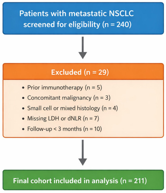
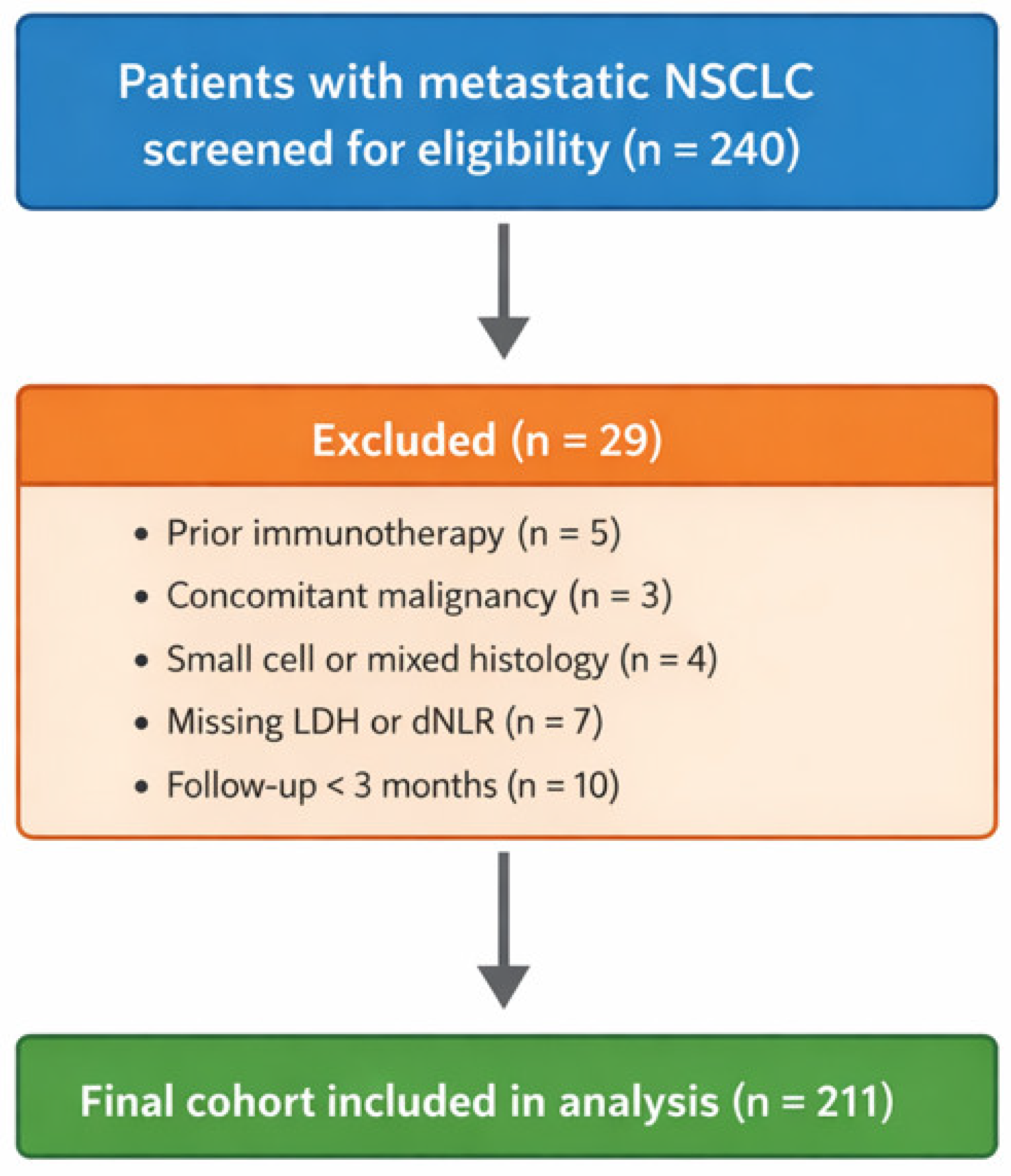
A total of 240 patients with metastatic NSCLC who received nivolumab were initially screened. After applying the predefined inclusion and exclusion criteria, 211 patients were included in the final analysis. The patient selection process and reasons for exclusion are summarized in Figure 1.
Figure 1.
Patient flow diagram. CONSORT-style diagram showing the screening, exclusion, and inclusion of patients for the final cohort. Exclusion reasons included prior immunotherapy, concomitant malignancy, small cell or mixed histology, missing LDH or dNLR data, and follow-up < 3 months.
The median age of the study population was 63 years (range, 23–84), with 94 patients (44.5%) aged 65 years or older. Of the patients, 176 (83.4%) were male, and 185 (87.7%) were smokers. ECOG performance status was 0–1 in 178 patients (84.4%), while 33 patients (15.6%) had an ECOG performance status of 2. Histopathological evaluation revealed non-squamous carcinoma in 138 cases (65.4%) and squamous cell carcinoma in 73 cases (34.6%). De novo metastatic disease was observed in 122 patients (58.2%), while recurrent disease was identified in 89 patients (41.8%). Regarding metastatic patterns, visceral metastases were detected in 87 patients (41.2%), including liver metastases in 29 patients (13.7%) and brain metastases in 39 patients (18.5%), whereas non-visceral metastases were present in 124 patients (58.8%), including bone metastases in 70 patients (33.2%). PD-L1 expression status was classified as high (tumor proportion score [TPS] ≥ 50) in 47 patients (22.3%), intermediate (TPS 1–49) in 71 patients (33.7%), and negative (TPS < 1) in 93 patients (44.1%). Serum albumin levels were ≥3.5 g/dL in 186 patients (88.2%), and CRP levels were >10 mg/L in 139 patients (65.9%).
According to LIPI stratification, 67 patients (31.75%) were classified as having good LIPI, 109 patients (51.65%) as having intermediate LIPI, and 35 patients (16.6%) as having poor LIPI. Patients in the poor LIPI group exhibited significantly higher rates of visceral metastasis (p = 0.046), liver metastasis (p = 0.021), elevated CRP (p = 0.042), and ECOG PS 2 (p = 0.040), while other clinicopathological variables showed no significant differences (Table 1).
Table 1.
Description of the study population according to the Lung Immune Prognostic Index (LIPI).
3.2. Response Rates
All patients received nivolumab as second-line therapy for advanced non-small cell lung cancer. Among the 211 patients evaluable for treatment response, the objective response rate (ORR) was 38.8% in the good LIPI group, 33.9% in the intermediate group, and 20% in the poor group, with no statistically significant differences observed between the groups (OR = 1.488; 95% CI: 0.964–2.297; p = 0.073). In contrast, the disease control rate (DCR) decreased progressively with worsening LIPI scores, reaching 76.1% in the good group, 60.6% in the intermediate group, and 37.1% in the poor group. This stepwise decline in DCR across LIPI categories was statistically significant (OR = 2.295; 95% CI: 1.479–3.561; p < 0.001). A summary of treatment response rates is provided in Table 2.
Table 2.
Treatment response rates in LIPI-good, LIPI-intermediate and LIPI-poor groups.
3.3. Progression-Free Survival (PFS)
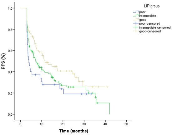
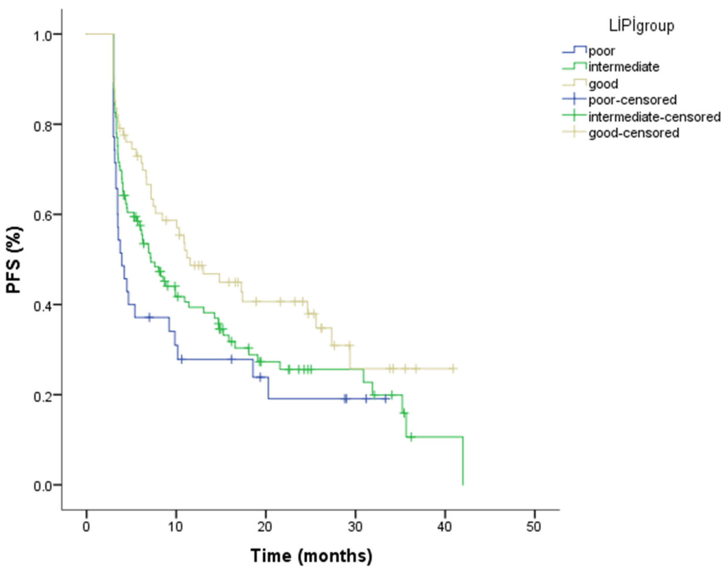
The median follow-up for the cohort was 12.05 months (range: 3.09–57.59). During follow-up, 146 PFS events occurred, including both radiologically documented disease progression and deaths without prior documented progression. For the entire population, the median progression-free survival (PFS) was 8.49 months (95% CI: 5.83–10.26). When stratified by LIPI score, the median PFS was 11.53 months (95% CI: 6.26–16.79) in the good LIPI group, 7.16 months (95% CI: 4.96–9.35) in the intermediate group, and 3.94 months (95% CI: 2.76–5.12) in the poor group. Kaplan–Meier analysis indicated statistically significant difference in PFS between LIPI categories (p = 0.037) (Figure 2; Table 3). The number of patients at risk for progression-free survival at selected time points for each LIPI group is presented in Table 4.
Figure 2.
Kaplan–Meier curves for progression-free survival according to Lung Immune Prognostic Index (LIPI) groups, demonstrating a statistically significant difference in PFS among the groups (log-rank p = 0.037). The number of patient at risk at selected time points is shown below the Kaplan–Meier curves.
Table 3.
Number of patients at risk for progression-free survival at selected time points according to LIPI group.
Table 4.
Prognostic variables for progression-free survival in univariate analysis and multivariate analysis.
In the univariate Cox regression analysis, female sex, liver metastasis, bone metastasis, low PD-L1 expression, hypoalbuminemia, elevated CRP levels, high GPS, low PNI, high NPS and poor LIPI group were associated with shorter progression-free survival. However, in the multivariate analysis, only female sex (HR = 2.41, 95% CI: 1.11–5.21, p = 0.025), lower PD-L1 expression (≥50% vs. <1%: HR = 3.17, 95% CI: 1.32–7.57, p = 0.009; ≥50% vs. 1–49%: HR = 2.48, 95% CI: 1.06–5.80, p = 0.036), and elevated CRP levels (HR = 2.14, 95% CI: 1.10–4.17, p = 0.023) remained independently associated with shorter progression-free survival, whereas the association between LIPI groups and PFS did not remain statistically significant after adjustment for confounding variables (Table 4).
3.4. Overall Survival (OS)
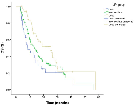
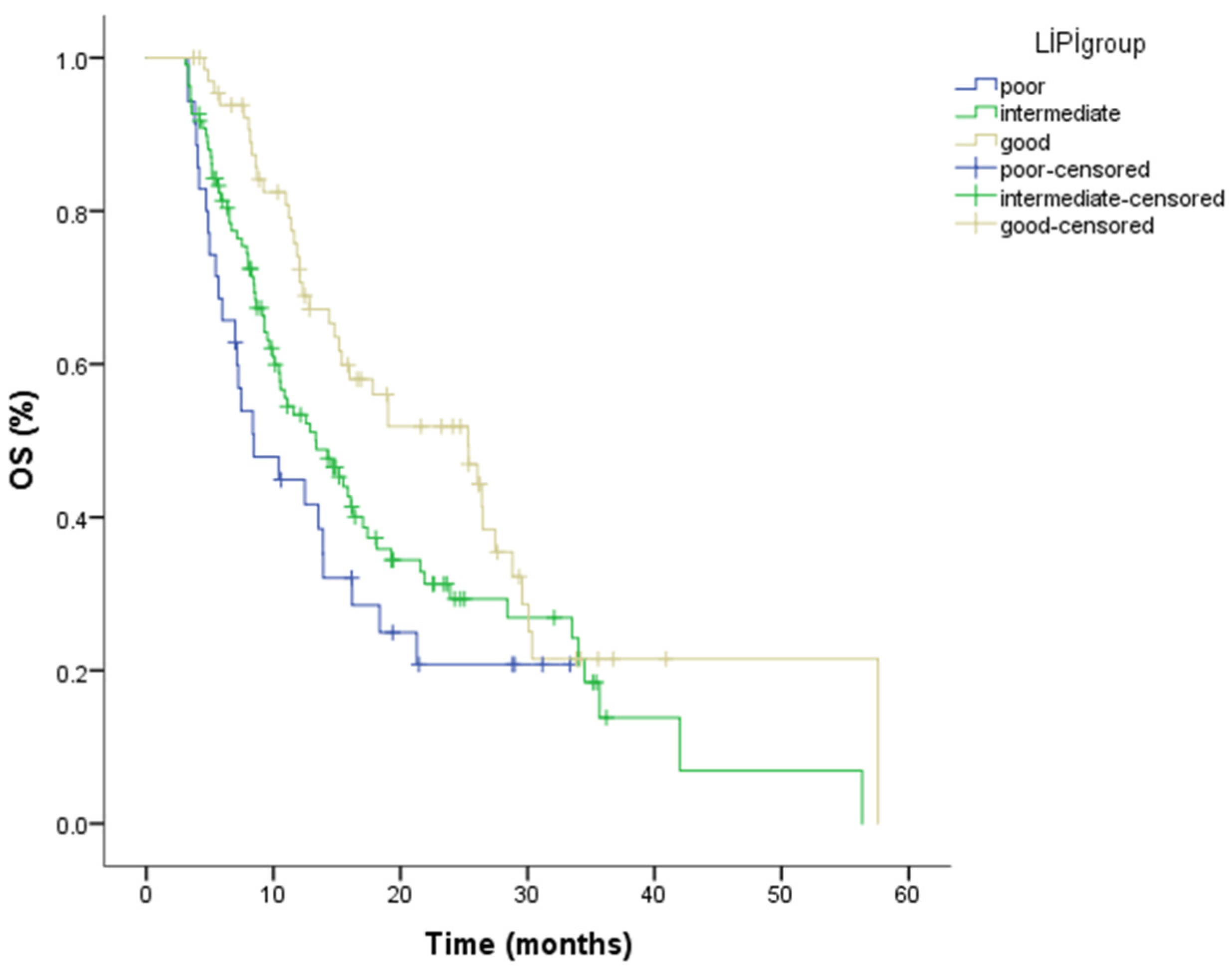
During follow-up, 137 patients (64.9%) died. The median overall survival (OS) for the entire cohort was 14.58 months (95% CI: 12.34–16.82). When analyzed by LIPI category, median OS was 25.33 months (95% CI: 15.96–34.69) in the good LIPI group, 13.33 months (95% CI: 9.35–17.31) in the intermediate group, and 8.44 months (95% CI: 3.99–12.89) in the poor group. Kaplan–Meier analysis revealed a statistically significant difference in OS across LIPI groups (p = 0.007) (Figure 3; Table 5). The numbers of patients at risk for overall survival at selected time points for each LIPI group are presented in Table 6.
Figure 3.
Kaplan–Meier curves for overall survival stratified by Lung Immune Prognostic Index (LIPI) groups, demonstrating a statistically significant difference in OS among the groups (log-rank p = 0.007). The number of patients at risk at selected time points is shown below the Kaplan–Meier curves.
Table 5.
Median PFS and OS in LIPI-good, LIPI-intermediate and LIPI-poor groups.
Table 6.
Number of patients at risk for overall survival at selected time points according to LIPI group.
In the univariate Cox regression analysis, albumin level, CRP level, PD-L1 expression, GPS, PNI, NPS and LIPI group were significantly associated with overall survival. In the multivariate analysis, lower PD-L1 expression (<1% vs. ≥50%: HR = 2.88, 95% CI: 1.25–6.65, p = 0.013), low albumin levels (HR = 3.16, 95% CI: 1.18–7.65, p = 0.016), and poor LIPI status (poor vs. good: HR = 2.53, 95% CI: 1.32–6.38, p = 0.034) remained independently associated with shorter overall survival, whereas CRP level and bone metastasis did not retain statistical significance after adjustment (Table 7).
Table 7.
Prognostic variables for overall survival in univariate and multivariate analyses.
Receiver operating characteristic (ROC) analysis was performed to evaluate the discriminatory performance of the inflammation-based prognostic scores. The AUC for LIPI was 0.605, indicating moderate discriminatory ability. In comparison, the AUC values were 0.576 for GPS, 0.560 for PNI, and 0.549 for NPS, suggesting overall modest performance for these indices. Among all evaluated scores, LIPI demonstrated the highest discriminatory accuracy (Table 8).
Table 8.
Comparison of the prognostic performance of inflammation-based scores.
4. Discussion
Despite the clinical benefits conferred by immune checkpoint inhibitors (ICIs) in the treatment of advanced non-small cell lung cancer (NSCLC), the accurate identification of patients who are most likely to benefit remains a critical clinical challenge. Although PD-L1 expression assessed by immunohistochemistry in tumor tissue initially emerged as a promising biomarker, its predictive utility in clinical practice is limited due to intratumoral heterogeneity, dynamic changes in expression, and methodological variability. In this context, there is growing interest in readily accessible biomarkers that reflect systemic tumor–host interactions. Considering the pivotal role of real-world data in biomarker validation, this study evaluated the association of the Lung Immune Prognostic Index (LIPI) with treatment response and survival outcomes in a cohort of patients with advanced NSCLC receiving second-line nivolumab, in order to assess its prognostic significance.
In our cohort, patients with poor LIPI scores exhibited a higher prevalence of visceral metastases, suggesting a link between adverse LIPI status and more aggressive disease biology. This finding is biologically plausible, as elevated lactate dehydrogenase reflects increased tumor burden and enhanced tumor metabolic activity, whereas a high derived neutrophil-to-lymphocyte ratio indicates a systemic inflammatory environment that may facilitate tumor progression and dissemination. Consistent with this interpretation, previous studies have shown that poor LIPI scores are associated with a greater number of metastatic sites at baseline and worse survival outcomes, including both overall survival (OS) and progression-free survival (PFS), supporting the notion that LIPI primarily reflects underlying tumor aggressiveness rather than solely predicting response to immunotherapy [12].
With respect to treatment response, objective response rates differed numerically across LIPI categories, with rates of 38.8%, 33.9%, and 20% observed in the good, intermediate, and poor LIPI groups, respectively; however, these differences did not reach statistical significance. Similar findings have been reported by Ruiz Bañobre and Zhi et al., who also did not observe a significant association between LIPI classification and objective response rates [13,14]. In contrast, a large cohort analysis by Sorich et al. reported a significant association between LIPI and ORR [15]. Differences across studies may partly reflect variations in study populations and treatment strategies, as previous analyses often included heterogeneous cohorts receiving immunotherapy in different treatment lines, various checkpoint inhibitors, or combination regimens, whereas our study focused exclusively on patients treated with second-line nivolumab.
In contrast to the ORR, the disease control rate (DCR) differed significantly according to LIPI classification, showing a clear stepwise decrease from the good to the poor LIPI groups (76.1%, 60.6%, and 37.1%, respectively). This pattern suggests that worsening LIPI scores are associated with reduced clinical benefit from immunotherapy. Similar observations have been reported in prior studies, including that by Ruiz Bañobre et al., where patients with poor LIPI scores demonstrated significantly lower disease control rates compared to other LIPI subgroups [13,14].
Further analyses of progression-free survival (PFS) revealed several clinical and biological factors associated with treatment outcomes. In our cohort, male sex was significantly associated with longer PFS. A similar observation was reported by Calleja-Chucla et al., who also found that male patients experienced longer PFS in the context of immunotherapy-treated NSCLC [16]. In contrast, a real-world study by Knetki-Wróblewska et al. did not identify a significant relationship between sex and PFS [17]. These discrepancies may largely reflect differences in patient populations and study designs. Notably, the study by Knetki-Wróblewska et al. included a heterogeneous cohort of patients receiving second-line nivolumab or atezolizumab, and reported that tumor burden, inflammatory parameters, and metastatic patterns had a stronger influence on PFS than demographic characteristics.
PD-L1 expression also demonstrated prognostic relevance in our analysis. Patients with higher PD-L1 expression exhibited significantly longer PFS. Similar findings were reported by Jingyaa Liu et al., who observed significantly prolonged PFS in patients with PD-L1 expression ≥ 1% [18]. However, other studies have reported inconsistent results. For example, Brahmer et al. and Lang et al. did not observe a significant association between PD-L1 expression and PFS [9,19]. Such discrepancies may arise from heterogeneity in patient populations, differences in treatment lines, variations in PD-L1 assessment thresholds, and the use of different immune checkpoint inhibitors across studies.
Systemic inflammatory markers significantly influenced treatment outcomes in our cohort, with elevated baseline CRP levels associated with both shorter PFS and poorer LIPI scores (p = 0.042). These findings are consistent with previous reports demonstrating that systemic inflammatory status, particularly baseline CRP levels, is a critical prognostic determinant of survival outcomes in NSCLC patients receiving immune checkpoint inhibitors [20,21,22]. Collectively, our data reinforce the notion that systemic inflammation plays a pivotal role in shaping clinical responses to immunotherapy.
Although a numerical trend toward shorter PFS was observed with worsening LIPI categories in our cohort, this difference did not reach statistical significance. Similar observations have been reported by Ruiz-Bañobre et al. and Huang et al., who described longer PFS in patients with favorable LIPI scores, although the differences did not achieve statistical significance [13,23]. In contrast, Zhi et al. and Sorich et al. demonstrated a significant association between poor LIPI scores and shorter PFS in patients with advanced NSCLC [14,15]. These inconsistencies may partly reflect differences in treatment regimens across studies. Many previous analyses included patients receiving both PD-1 and PD-L1 inhibitors and, in some cases, combination regimens with chemotherapy. In addition, variations in baseline disease burden and performance status may also contribute to the heterogeneity observed in the prognostic impact of LIPI.
With respect to overall survival (OS), higher PD-L1 expression was significantly associated with improved survival outcomes in our cohort. Similarly, Borghaei et al. reported that PD-L1 expression was associated with longer OS in patients with advanced NSCLC receiving second-line nivolumab [24]. However, other studies have reported conflicting results. For example, Brahmer et al. and Lang et al. did not observe a significant association between PD-L1 expression and OS [9,19]. These discrepancies likely reflect differences in patient populations, treatment lines, and clinical characteristics across studies. In particular, several reports have suggested that clinical factors such as performance status and treatment line may exert a stronger influence on survival outcomes than PD-L1 expression alone.
Serum albumin level also emerged as a significant prognostic factor for OS in our study, with higher baseline albumin levels associated with improved survival. This finding is consistent with prior evidence indicating that baseline serum albumin is associated with OS and may function as an independent prognostic marker in NSCLC patients, including those treated with immune checkpoint inhibitors [25,26,27]. Taken together, these data suggest that serum albumin, reflecting nutritional status, systemic inflammation, and overall physiological reserve, may serve as a readily available prognostic biomarker in patients undergoing immunotherapy.
Consistent with these observations, the LIPI score also demonstrated a clear prognostic impact on overall survival in our cohort. Patients with poor LIPI scores experienced significantly shorter OS compared to those with favorable scores. This finding is consistent with previous studies. Ruiz-Bañobre et al. reported that poor LIPI scores were significantly associated with shorter OS in patients with advanced NSCLC treated with nivolumab [13]. Similarly, Zhi et al. demonstrated significantly longer OS in patients with favorable LIPI scores and identified LIPI as an independent prognostic factor in multivariable analysis [14]. In addition, pooled analyses conducted by Sorich et al. across multiple clinical trials confirmed that LIPI classification was significantly associated with OS across different treatment groups [15]. Taken together, these findings support the clinical utility of LIPI as a practical and reproducible biomarker reflecting systemic inflammation and tumor biology, with potential value for prognostic stratification in NSCLC patients receiving immune checkpoint inhibitors.
In this comparative analysis, LIPI consistently demonstrated superior discriminatory performance compared to other commonly used inflammation-based prognostic scores, including GPS, PNI, and NPS. While all indices showed some degree of prognostic relevance, their overall ability to distinguish between risk groups was modest. Notably, NPS exhibited the lowest performance, whereas PNI and GPS provided intermediate discriminatory value. These findings suggest that LIPI, by integrating both inflammatory and tumor burden–related parameters, may offer a more robust tool for risk stratification in patients with advanced NSCLC receiving immune checkpoint inhibitors.
Strengths and Limitations
This study’s main strength is its multicenter real-world design, including a relatively large cohort of patients with metastatic NSCLC uniformly treated with second-line nivolumab, which enhances the generalizability of the findings. The use of routinely available laboratory parameters for LIPI calculation supports the clinical applicability of the results.
However, several limitations should be considered. Because this study was based on retrospectively collected data, the possibility of selection bias and residual confounding cannot be completely excluded. In addition, the duration of follow-up was relatively limited, which may have reduced the statistical power to detect potential differences in survival outcomes. In addition, patients with follow-up shorter than three months were excluded to ensure adequate radiological response assessment, as the first routine imaging is typically performed around 12 weeks after treatment initiation. While this criterion improves the reliability of response evaluation, it may have introduced a minor selection bias that should be considered when interpreting survival outcomes.
5. Conclusions
In summary, the findings of this multicenter real-world analysis indicate that the Lung Immune Prognostic Index (LIPI) has meaningful prognostic relevance in patients with metastatic non-small cell lung cancer receiving second-line nivolumab. While LIPI classification was not significantly related to progression-free survival or objective response rate, patients with unfavorable LIPI scores experienced poorer disease control and shorter overall survival. Because LIPI is derived from routinely available laboratory parameters and can be easily implemented in clinical practice, it may be a practical approach for prognostic risk stratification in patients undergoing immunotherapy.
Author Contributions
Conceptualization, Ş.B.,T.K., A.O., G.U.E. and N.B.; methodology, Ş.B., Y.B., E.Ç., N.B. and G.U.E.; formal analysis, Ş.B., A.O. and H.G.Y.D.; investigation, Ş.B. and H.A.; data curation, Ş.B., T.K., A.O., M.C.F., M.G., H.A. and N.S.D.; writing—original draft preparation, Ş.B.; writing—review and editing, Ş.B., T.K., Y.B., A.O., M.C.F., M.G., H.A., N.S.D., H.G.Y.D., E.Ç., N.B. and G.U.E.; visualization, Ş.B., Y.B., M.G. and G.U.E.; supervision, Ş.B. and G.U.E. All authors have read and agreed to the published version of the manuscript.
Funding
This research received no external funding.
Institutional Review Board Statement
The study was conducted in accordance with the Declaration of Helsinki and approved by the Institutional Review Board (Ethics Committee) of [Yeni Yüzyıl University Faculty of Medicine] (protocol code: 2026/01-1814; date: 13 January 2026).
Informed Consent Statement
As this was a retrospective study using anonymized data, the requirement for written informed consent was waived by the Ethics Committee.
Data Availability Statement
The original contributions presented in this study are included in the article. Further inquiries can be directed to the corresponding author.
Acknowledgments
The authors would like to thank all patients and healthcare professionals involved in this study.
Conflicts of Interest
The authors declare no conflicts of interest.
Correction Statement
This article has been republished with a minor correction to an Academic Editor’s name. This change does not affect the scientific content of the article.
References
- Zhou, J.; Xu, Y.; Liu, J.; Feng, L.; Yu, J.; Chen, D. Global burden of lung cancer in 2022 and projections to 2050: Incidence and mortality estimates from GLOBOCAN. Cancer Epidemiol. 2024, 93, 102693. [Google Scholar] [CrossRef] [Scilit]
- Siegel, R.L.; Miller, K.D.; Fuchs, H.E.; Jemal, A. Cancer statistics, 2022. CA Cancer J. Clin. 2022, 72, 7–33. [Google Scholar] [CrossRef] [Scilit] [PubMed]
- Garon, E.B.; Rizvi, N.A.; Hui, R.; Leighl, N.; Balmanoukian, A.S.; Eder, J.P.; Patnaik, A.; Aggarwal, C.; Gubens, M.; Horn, L.; et al. Pembrolizumab for the treatment of non-small-cell lung cancer. N. Engl. J. Med. 2015, 372, 2018–2028. [Google Scholar] [CrossRef] [Scilit] [PubMed]
- Guo, Y.; Xiang, D.; Wan, J.; Yang, L.; Zheng, C. Focus on the dynamics of Neutrophil to-lymphocyte ratio in Cancer patients treated with Immune Checkpoint inhibitors: A Meta-analysis and systematic review. Cancers 2022, 14, 5297. [Google Scholar] [CrossRef] [Scilit] [PubMed]
- Kazandjian, D.; Gong, Y.; Keegan, P.; Pazdur, R.; Blumenthal, G.M. Prognostic value of the lung immune prognostic index for patients treated for metastatic non–small cell lung cancer. JAMA Oncol. 2019, 5, 1481–1485. [Google Scholar] [CrossRef] [Scilit]
- Mezquita, L.; Auclin, E.; Ferrara, R.; Charrier, M.; Remon, J.; Planchard, D.; Ponce, S.; Ares, L.P.; Leroy, L.; Audigier-Valette, C.; et al. Association of the Lung Immune Prognostic Index with immune checkpoint inhibitor outcomes in patients with advanced non–small cell lung cancer. JAMA Oncol. 2018, 4, 351–357. [Google Scholar] [CrossRef] [Scilit]
- WHO Classification of Tumours Editorial Board. Thoracic Tumours, 5th ed.; IARC: Lyon, France, 2021. [Google Scholar]
- Goldstraw, P.; Chansky, K.; Crowley, J.; Rami-Porta, R.; Asamura, H.; Eberhardt, W.E.; Nicholson, A.G.; Groome, P.; Mitchell, A.; Bolejack, V. The IASLC Lung Cancer Staging Project: Proposals for Revision of the TNM Stage Groupings in the Forthcoming (Eighth) Edition of the TNM Classification for Lung Cancer. J. Thorac. Oncol. 2016, 11, 39–51. [Google Scholar] [CrossRef] [Scilit]
- Brahmer, J.; Reckamp, K.L.; Baas, P.; Crinò, L.; Eberhardt, W.E.; Poddubskaya, E.; Antonia, S.; Pluzanski, A.; Vokes, E.E.; Holgado, E.; et al. Nivolumab versus Docetaxel in Advanced Squamous-Cell Non-Small-Cell Lung Cancer. N. Engl. J. Med. 2015, 373, 123–135. [Google Scholar] [CrossRef] [Scilit] [PubMed] [PubMed Central]
- Eisenhauer, E.A.; Therasse, P.; Bogaerts, J.; Schwartz, L.H.; Sargent, D.; Ford, R.; Arbuck, S.; Gwyther, S.; Mooney, M.; Rubinstein, L.; et al. New response evaluation criteria in solid tumours: Revised RECIST guideline (version 1.1). Eur. J. Cancer 2009, 45, 228–247. [Google Scholar] [CrossRef] [Scilit]
- World Medical Association. Declaration of Helsinki: Ethical principles for medical research involving human subjects. JAMA 2013, 310, 2191–2194. [Google Scholar] [CrossRef] [Scilit]
- Auclin, E.; Roulleaux Dugage, M.; Gorria, T.; Vauchier, C.; Thibault, C.; Laguna, J.C.; Lupinacci, L.; Crous, C.; Naigeon, M.; Oudard, S.; et al. Validation of the Lung Immune Prognostic Index in patients with untreated advanced non-small cell lung cancer: Post hoc analysis of the IMpower 130, 131 and 150 trials. Lung Cancer 2025, 199, 108039. [Google Scholar] [CrossRef] [Scilit] [PubMed]
- Ruiz-Bañobre, J.; Areses-Manrique, M.C.; Mosquera-Martínez, J.; Cortegoso, A.; Afonso-Afonso, F.J.; de Dios-Álvarez, N.; Fernández-Núñez, N.; Azpitarte-Raposeiras, C.; Amenedo, M.; Santomé, L.; et al. Evaluation of the lung immune prognostic index in advanced non-small cell lung cancer patients under nivolumab monotherapy. Transl. Lung Cancer Res. 2019, 8, 1078–1085. [Google Scholar] [CrossRef] [Scilit] [PubMed] [PubMed Central]
- Zhi, X.; Zhang, Z.; Li, W.; Dong, Z.; Wu, X.; Ge, X.; Zhai, J.; Lu, D.; Yan, X.; Wang, J. Correlation of lung immune prognostic index with the efficacy of immune checkpoint inhibitors in Chinese patients with advanced non-small cell lung cancer. Holist. Integr. Oncol. 2024, 3, 2. [Google Scholar] [CrossRef] [Scilit]
- Sorich, M.J.; Rowland, A.; Karapetis, C.S.; Hopkins, A.M. Evaluation of the Lung Immune Prognostic Index for Prediction of Survival and Response in Patients Treated With Atezolizumab for NSCLC: Pooled Analysis of Clinical Trials. J. Thorac. Oncol. 2019, 14, 1440–1446. [Google Scholar] [CrossRef] [Scilit] [PubMed]
- Calleja-Chucla, T.; Cordeiro González, P.; Martínez Pradeda, A.; Pértega-Díaz, S.; Margusino-Framiñán, L.; Antolín Novoa, S. Effect of Gender on Patients with Non-Small-Cell Lung Cancer Treated with Immune Checkpoint Inhibitors: A Real-World Study. Biomedicines 2025, 13, 437. [Google Scholar] [CrossRef] [Scilit]
- Knetki-Wróblewska, M.; Tabor, S.; Piórek, A.; Płużański, A.; Winiarczyk, K.; Zaborowska-Szmit, M.; Zajda, K.; Kowalski, D.M.; Krzakowski, M. Nivolumab or Atezolizumab in the Second-Line Treatment of Advanced Non-Small Cell Lung Cancer? A Prognostic Index Based on Data from Daily Practice. J. Clin. Med. 2023, 12, 2409. [Google Scholar] [CrossRef] [Scilit] [PubMed] [PubMed Central]
- Liu, J.; Man, Y.; Gao, J.; Wang, X.; Zhang, L.; Li, M.; Yu, J. Correlation between PD-L1 expression status and efficacy of immunotherapy as second-line or later-line therapy in advanced non-small cell lung cancer patients. Eur. J. Cancer Prev. 2024, 33, 448–460. [Google Scholar] [CrossRef] [Scilit]
- Lang, D.; Huemer, F.; Rinnerthaler, G.; Horner, A.; Wass, R.; Brehm, E.; Akbari, K.; Granitz, M.; Hutarew, G.; Kaiser, B.; et al. Therapy Line and Associated Predictors of Response to PD-1/PD-L1-Inhibitor Monotherapy in Advanced Non-small-Cell Lung Cancer: A Retrospective Bi-centric Cohort Study. Target. Oncol. 2019, 14, 707–717. [Google Scholar] [CrossRef] [Scilit] [PubMed] [PubMed Central]
- Onodera, R.; Chiba, S.; Nihei, S.; Fujimura, I.; Akiyama, M.; Utsumi, Y.; Nagashima, H.; Kudo, K.; Maemondo, M. High level of C-reactive protein as a predictive factor for immune-related adverse events of immune checkpoint inhibitors in non-small cell lung cancer: A retrospective study. J. Thorac. Dis. 2023, 15, 4237–4247. [Google Scholar] [CrossRef] [Scilit] [PubMed] [PubMed Central]
- Riedl, J.M.; Barth, D.A.; Brueckl, W.M.; Zeitler, G.; Foris, V.; Mollnar, S.; Stotz, M.; Rossmann, C.H.; Terbuch, A.; Balic, M.; et al. C-Reactive Protein (CRP) Levels in Immune Checkpoint Inhibitor Response and Progression in Advanced Non-Small Cell Lung Cancer: A Bi-Center Study. Cancers 2020, 12, 2319. [Google Scholar] [CrossRef] [Scilit] [PubMed] [PubMed Central]
- Oya, Y.; Yoshida, T.; Kuroda, H.; Mikubo, M.; Kondo, C.; Shimizu, J.; Horio, Y.; Sakao, Y.; Hida, T.; Yatabe, Y. Predictive clinical parameters for the response of nivolumab in pretreated advanced non-small-cell lung cancer. Oncotarget 2017, 8, 103117–103128. [Google Scholar] [CrossRef] [Scilit] [PubMed] [PubMed Central]
- Huang, J.; Pu, H.; He, J.; Tang, X. Prognostic Value of the Lung Immune Prognostic Index for Metastatic Non-Small Cell Lung Cancer Patients: A Chinese Cohort Study. Int. J. Gen. Med. 2023, 16, 881–893. [Google Scholar] [CrossRef] [Scilit] [PubMed] [PubMed Central]
- Borghaei, H.; Paz-Ares, L.; Horn, L.; Spigel, D.R.; Steins, M.; Ready, N.E.; Chow, L.Q.; Vokes, E.E.; Felip, E.; Holgado, E.; et al. Nivolumab versus Docetaxel in Advanced Nonsquamous Non-Small-Cell Lung Cancer. N. Engl. J. Med. 2015, 373, 1627–1639. [Google Scholar] [CrossRef] [Scilit] [PubMed] [PubMed Central]
- Hashemi, S.; Fransen, M.F.; Niemeijer, A.; Ben Taleb, N.; Houda, I.; Veltman, J.; Becker-Commissaris, A.; Daniels, H.; Crombag, L.; Radonic, T.; et al. Surprising impact of stromal TIL’s on immunotherapy efficacy in a real-world lung cancer study. Lung Cancer 2021, 153, 81–89. [Google Scholar] [CrossRef] [Scilit] [PubMed]
- Guo, Y.; Wei, L.; Patel, S.H.; Lopez, G.; Grogan, M.; Li, M.; Haddad, T.; Johns, A.; Ganesan, L.P.; Yang, Y.; et al. Serum Albumin: Early Prognostic Marker of Benefit for Immune Checkpoint Inhibitor Monotherapy But Not Chemoimmunotherapy. Clin. Lung Cancer 2022, 23, 345–355. [Google Scholar] [CrossRef] [Scilit] [PubMed] [PubMed Central]
- Daher, S.; Lawrence, Y.R.; Dudnik, E.; Hanovich, E.; Urban, D.; Peled, N.; Navon, R.; Leibowitz, R.; Hammerman, A.; Battat, E.; et al. Nivolumab in Non-Small Cell Lung Cancer: Real World Long-Term Survival Results and Blood-Based Efficacy Biomarkers. Front. Oncol. 2021, 11, 625668. [Google Scholar] [CrossRef] [Scilit] [PubMed] [PubMed Central]

Disclaimer/Publisher’s Note: The statements, opinions and data contained in all publications are solely those of the individual author(s) and contributor(s) and not of MDPI and/or the editor(s). MDPI and/or the editor(s) disclaim responsibility for any injury to people or property resulting from any ideas, methods, instructions or products referred to in the content. |
© 2026 by the authors. Licensee MDPI, Basel, Switzerland. This article is an open access article distributed under the terms and conditions of the Creative Commons Attribution (CC BY) license.