


Simple Summary
Pleomorphic dermal sarcoma (PDS) is a rare skin cancer that predominantly affects elderly patients. Due to its rarity, information on treatment patterns and long-term outcomes remains limited. We analyzed 1069 patients from the Baden-Württemberg Cancer Registry, representing one of the largest PDS cohorts reported to date. The use of micrographically controlled surgery increased substantially over time, with no significant differences in complete tumor removal or overall survival compared with conventional surgery. Nearly half of the patients had a history of other cancers, particularly skin cancers. Our findings provide insights into the management and clinical characteristics of PDS in routine practice and highlight the need for further research on disease-specific outcomes.
Abstract
Background: Pleomorphic dermal sarcoma (PDS) is a rare, cutaneous sarcoma predominantly affecting sun-exposed skin in elderly patients. Surgical excision remains the mainstay of treatment, but evidence on optimal management and long-term outcomes is limited. Methods: We retrospectively analyzed 1069 patients diagnosed with PDS between 2009 and 2025 using data from the Baden-Württemberg Cancer Registry, Germany. Patient characteristics, surgical management, resection status, and preceding malignancies were evaluated. Temporal surgical trends were assessed using multivariable logistic regression. Overall survival (OS) was analyzed using Kaplan–Meier estimates and multivariable Cox regression. Results: Median age was 82 years, and 83.7% of patients were male. The hair-bearing scalp was the most frequent tumor site (55.0%). Among 449 patients with documented surgical procedures, 82.0% underwent micrographically controlled surgery (MGC), with increasing use over time (adjusted OR per year, 1.19; 95% CI, 1.11–1.29). R0-resections were comparable between MGC and conventional resection (75.7% vs. 71.6%). Among 679 patients evaluated for preceding malignancies, 39.7% had a documented cancer history, including 4.4% with hematologic malignancies. Among 723 patients with reliable follow-up, median OS was 72.4 months (95% CI, 61.3–82.1). Mortality was strongly associated with age, but not anatomical tumor site. Surgical approach was not significantly associated with OS after adjustment (MGC vs. conventional resection: HR, 1.49; 95% CI, 0.95–2.3; p = 0.083). Conclusions: This large registry-based study provides real-world evidence on PDS management and prognosis. The increasing adoption of MGC supports its growing role in clinical practice, while the high frequency of preceding malignancies highlights the complex oncological history of patients with PDS.
1. Introduction
Pleomorphic dermal sarcoma (PDS) is a rare malignant mesenchymal tumor of the skin that predominantly affects elderly patients and typically arises in chronically sun-exposed areas such as the scalp, face, and neck [1,2]. PDS is considered part of a disease spectrum with atypical fibroxanthoma (AFX), representing the more aggressive end due to the presence of high-risk histopathological features [3,4,5]. According to the current WHO classification, PDS is defined by criteria including invasion beyond the dermis into subcutaneous tissue, tumor necrosis, lymphovascular invasion, and/or perineural infiltration, which distinguish it from AFX and are associated with a worse clinical outcome [6,7,8,9]. Ultraviolet radiation-induced DNA damage is thought to play a central role in tumorigenesis, reflected by the frequent occurrence in elderly, fair-skinned individuals and the high mutational burden observed in molecular studies [10,11]. Over the last years the incidence of PDS in Germany has been rising and is expected to rise further in aging populations with substantial cumulative UV exposure [12].
Clinically, PDS often presents as a rapidly growing, ulcerated or nodular tumor, and diagnosis relies on histopathology supported by immunohistochemistry to exclude other spindle cell malignancies such as spindle cell squamous cell carcinoma, leiomyosarcoma, or melanoma. Compared with AFX, PDS exhibits higher rates of local recurrence (up to 15–30%) and a documented risk of regional and distant metastasis, especially to lymph nodes and lungs, reported in approximately 5–16% of cases [13,14,15,16]. Complete surgical excision with histologically negative margins is the cornerstone of treatment, with micrographically controlled surgery frequently recommended to achieve optimal local control [7,8,9,10,11,12,13,14,15,16,17,18,19]. The role of adjuvant radiotherapy or systemic therapy remains unclear and is generally reserved for advanced, recurrent, or metastatic disease [20]. Furthermore, the high tumor mutational burden and emerging clinical experience with immune checkpoint inhibition suggest a potential therapeutic option for advanced disease, although evidence is currently based primarily on small series and case reports [21].
Despite increasing recognition of PDS, evidence on its epidemiology, real-world management, associated malignancies, and long-term outcomes remains limited. In particular, large registry-based studies allowing characterization of contemporary surgical practice and longitudinal cancer histories are scarce.
We therefore conducted a large registry-based study of 1069 patients with PDS recorded in the Clinical Cancer Registry of Baden-Württemberg, Germany. We aimed to characterize patient and tumor features, evaluate patterns and temporal changes in surgical management, investigate malignancies preceding and following PDS, and assess long-term survival and factors associated with outcome.
2. Materials and Methods
2.1. Data Source and Study Population
Patient data were obtained from the Clinical Cancer Registry of Baden-Württemberg, Germany, which collects standardized information on cancer diagnosis, treatment, and follow-up based on mandatory reporting by healthcare providers. Data undergo routine completeness and plausibility checks as part of the registry’s quality assurance procedures.
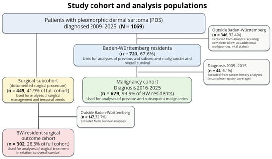
We conducted a retrospective population-based cohort study including patients aged ≥18 years diagnosed with PDS between 2009 and 2025. PDS cases were identified using the corresponding ICD-O morphology codes 8802/3 in combination with ICD-O topography codes C44.x, restricting the study population to cutaneous malignancies. Several analysis populations were defined according to data availability and completeness (Figure 1). The full cohort included all patients with PDS and was used to describe demographic and tumor characteristics, anatomical site, and documented treatment.
Figure 1.
Study cohort and analysis populations. Flow diagram illustrating the derivation of the full PDS cohort and the analysis populations according to residency, period of reliable registry coverage, and availability of surgical information. PDS, pleomorphic dermal sarcoma; BW, Baden-Württemberg.
Because complete ascertainment of additional malignancies and vital status could not be ensured for patients residing outside Baden-Württemberg, analyses requiring longitudinal follow-up were restricted to patients residing in Baden-Württemberg at PDS diagnosis. This resident cohort was used to assess malignancies preceding and following PDS and OS. Because cancer registration was incomplete during the early study period, analyses of cancer history and temporal trends were additionally restricted to the period with reliable registry coverage (2016–2025).
Detailed analyses of surgical management were conducted in a surgical subcohort comprising patients with documented surgical procedures. If both micrographically controlled surgery (MGC) and conventional resection were reported for the same patient, the patient was assigned to the MGC group. Analyses of surgical treatment in relation to survival were additionally restricted to Baden-Württemberg residents.
2.2. Variables and Outcomes
Patient characteristics included age, sex, year of diagnosis, and anatomical tumor site. Previous malignancies were categorized as none, non-hematologic, or hematologic. The type and timing of malignancies preceding and following PDS were additionally assessed.
Surgical treatment was classified as MGC or conventional resection, and resection status as R0, R1, or R2. Resection status was based on the first documented R classification, as subsequent re-excisions and their corresponding resection status were not consistently captured in the registry. Temporal changes in surgical management were assessed from 2016 to 2025.
Overall survival (OS) was defined from PDS diagnosis to death from any cause. Patients alive at the end of follow-up were censored at the last date of confirmed vital status.
2.3. Preceding Malignancies
Preceding malignancies were assessed among Baden-Württemberg residents diagnosed with PDS between 2016 and 2025 and classified as non-hematologic or hematologic (ICD-10 C81-C96). To avoid misclassification of the index PDS, corresponding diagnoses and C49 diagnoses recorded within 30 days before PDS diagnosis were excluded. Patients could contribute to more than one malignancy category. For patients with multiple preceding hematologic malignancies, the earliest diagnosis was used to calculate the interval to PDS.
2.4. Statistical Analysis
Continuous variables were summarized as medians with interquartile ranges and categorical variables as frequencies and percentages. Temporal trends in surgical management were evaluated using logistic regression, adjusting for age, sex, and anatomical site. OS was estimated using the Kaplan–Meier method, and associations with mortality were assessed using multivariable Cox regression. Effect estimates are reported with 95% confidence intervals. Analyses were performed using R version 4.2.1 (R Foundation for Statistical Computing, Vienna, Austria) [22], including the survival [23] and survminer [24] packages.
3. Results
3.1. Patient and Tumor Characteristics
A total of 1069 patients diagnosed with PDS between 2009 and 2025 were identified (Table 1). Median age at diagnosis was 82 years (range, 31–100), and 895 patients (83.7%) were male. The hair-bearing scalp was the most common tumor site (n = 588, 55.0%), followed by the face (n = 323, 30.2%) and trunk or extremities (n = 158, 14.8%). Patients with tumors of the hair-bearing scalp were more frequently male than those with tumors of the face or trunk/extremities (93.7% vs. 70.0% and 74.7%, respectively; p < 0.001). Age also differed modestly according to anatomical site (p = 0.007).
Table 1.
Baseline characteristics of patients with pleomorphic dermal sarcoma according to anatomical tumor site. Data are presented as n (%) unless otherwise indicated. Age is presented as median (range). p-values refer to comparisons across anatomical tumor sites.
Of the overall cohort, 723 patients (67.6%) were residents of Baden-Württemberg. Most patients were diagnosed during the period of reliable registry coverage, with 381 (35.6%) diagnosed in 2016–2020 and 639 (59.8%) in 2021–2025; only 49 (4.6%) were diagnosed before 2016. Accordingly, analyses of cancer history were restricted to patients diagnosed from 2016 onward, while analyses requiring complete longitudinal follow-up were additionally restricted to Baden-Württemberg residents.
3.2. Surgical Management and Temporal Trends
Detailed surgical information was available for 449 of 1069 patients (42.0%). Compared with patients without a documented surgical technique (n = 620), those with available surgical information more frequently had tumors of the hair-bearing scalp (63.3% vs. 49.0%; p < 0.001) and were more frequently diagnosed during 2021–2025 (66.1% vs. 55.2%; p = 0.001). Median age was 82 years in both groups, with no significant difference in sex distribution (p = 0.076). Completeness of surgical technique documentation was 36.7% in 2009–2015, 35.2% in 2016–2020, and 46.5% in 2021–2025.
Among patients with a documented surgical technique, MGC was the predominant surgical approach, performed in 368 patients (82%), whereas 81 patients (18.0%) underwent conventional resection. The use of MGC increased over time and this pattern was observed across anatomical tumor sites.
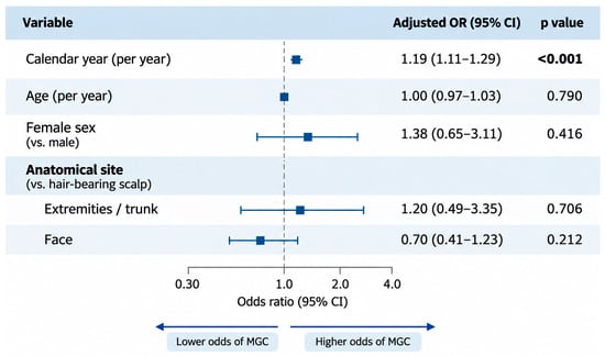
In multivariable logistic regression, each later calendar year was independently associated with higher odds of undergoing MGC (OR per year, 1.19; 95% CI, 1.11–1.29; p < 0.001), after adjustment for age, sex, and anatomical site (Figure 2). Age, sex, and anatomical site were not independently associated with surgical approach. There was no evidence that the temporal increase in MGC differed across anatomical sites (p for interaction = 0.98).
Figure 2.
Factors associated with the use of micrographically controlled surgery in patients with PDS. Forest plot showing adjusted odds ratios (ORs) and 95% confidence intervals (CIs) from multivariable logistic regression including calendar year, age, sex, and anatomical tumor site. Male sex and hair-bearing scalp served as reference categories. The interaction between calendar year and anatomical site was not significant (p for interaction = 0.98).
Resection status was available for 396 of 449 patients (88.2%). Overall, R0 resection was documented in 296 patients, R1 in 98, R2 in one, and Rx in one, while resection status was missing for 53 patients. Documentation completeness was similar after MGC and conventional resection (88.9% and 84.1%, respectively). R0 resection was documented in 75.7% of patients undergoing MGC and 71.6% undergoing conventional resection. Specifically, among patients undergoing MGC, 248 had R0, 79 R1, one R2, and 40 missing R status; among those undergoing conventional resection, 48 had R0, 19 R1, one Rx, and 13 missing R status. After adjustment for calendar year, age, sex, and anatomical site, MGC was not significantly associated with the likelihood of R0 resection (OR, 1.52; 95% CI, 0.83–2.73; p = 0.17).
3.3. Malignancies Preceding PDS
Among 679 patients diagnosed between 2016 and 2025 with reliable longitudinal registry information, 270 (39.7%) had a documented malignancy preceding PDS. Previous non-hematologic malignancies were recorded in 240 patients (35.3%), whereas 30 patients (4.4%) had a preceding hematologic malignancy.
Among patients with preceding non-hematologic malignancies, cutaneous malignancies were particularly common. Overall, 101 patients (14.9% of the entire PDS cohort) had a history of melanoma and/or non-melanoma skin cancer. Non-melanoma skin cancer was documented in 78 patients (11.5%) and melanoma in 29 (4.3%), with six patients having a history of both. Prostate cancer was the most frequent non-cutaneous preceding malignancy (n = 67, 9.9%). The distribution of preceding non-hematologic malignancies is shown in Table 2.
Table 2.
Spectrum of solid malignancies preceding pleomorphic dermal sarcoma. Distribution of preceding non-hematologic malignancies among 679 patients with PDS and reliable longitudinal registry information.
Patients with preceding malignancies differed modestly from those without a documented cancer history. Median age at PDS diagnosis was 82 years in patients without a preceding malignancy, 83 years in those with a preceding non-hematologic malignancy, and 79.5 years in those with a preceding hematologic malignancy (p = 0.077). Male patients were more frequent among those with preceding non-hematologic malignancies (89.4%) than among patients without a preceding malignancy (80.1%) or with a preceding hematologic malignancy (80.0%; p = 0.005). In contrast, the anatomical distribution of PDS did not differ significantly according to cancer history (p = 0.337).
Among the 30 patients with a preceding hematologic malignancy, non-Hodgkin lymphoma was the most frequent (n = 15, 50.0%), followed by chronic lymphocytic leukemia (n = 7, 23.3%) and myeloid leukemia (n = 5, 16.7%). Together, non-Hodgkin lymphoma and chronic lymphocytic leukemia accounted for 73.3% of preceding hematologic malignancies. These malignancies generally preceded PDS by several years: the median interval to PDS diagnosis was 4.9 years (IQR, 2.2–6.1) for non-Hodgkin lymphoma and 4.0 years (IQR, 3.5–7.8) for chronic lymphocytic leukemia. The complete spectrum and timing of preceding hematologic malignancies are presented in Table 3.
Table 3.
Preceding hematologic malignancies in patients with pleomorphic dermal sarcoma.
3.4. Overall Survival
Survival analyses were restricted to Baden-Württemberg residents (n = 723) with reliable vital-status ascertainment.
After a median follow-up of 50.2 months (reverse Kaplan–Meier), 277 deaths occurred. Median OS was 72.4 months (95% CI 61.3–82.1), with 1-, 3-, and 5-year OS rates of 88.3%, 71.3%, and 55.3%, respectively.
In multivariable Cox regression, increasing age was strongly associated with higher mortality (HR per year, 1.09; 95% CI, 1.07–1.1; p < 0.001). Female sex was associated with numerically lower mortality, although this did not reach statistical significance (HR, 0.71; 95% CI, 0.50–1.00; p = 0.055). Overall survival did not differ according to anatomical tumor site, with similar outcomes for tumors of the extremities/trunk (HR, 1.05; 95% CI, 0.72–1.5; p = 0.797) and face (HR, 0.94; 95% CI, 0.72–1.2; p = 0.68) compared with the hair-bearing scalp.
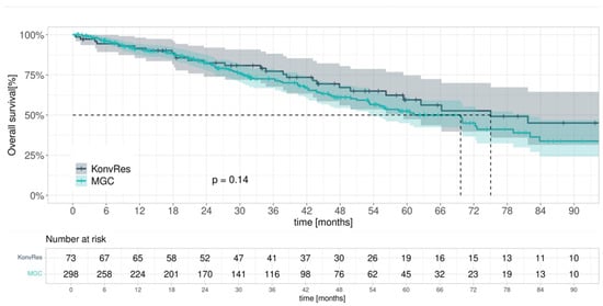
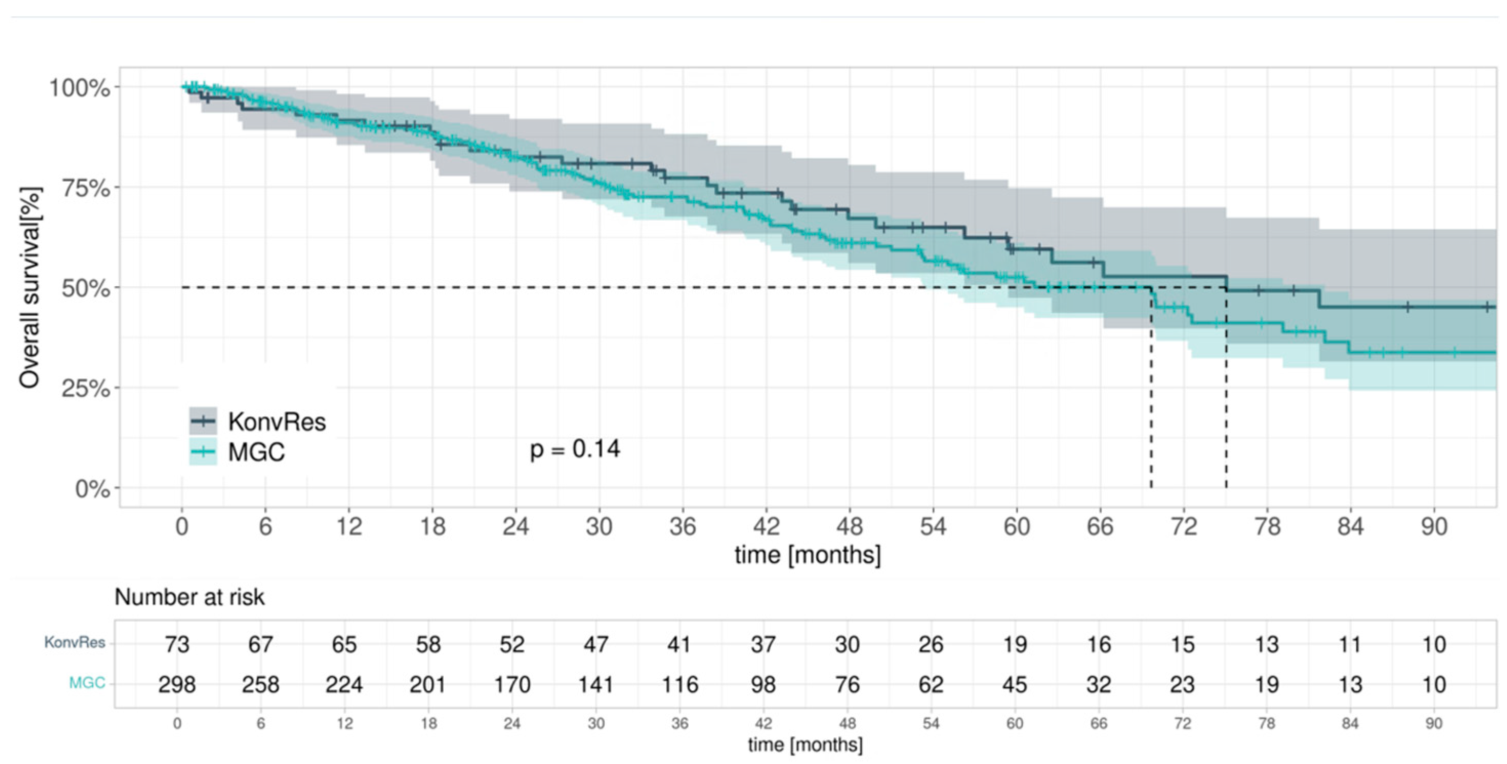
Among Baden-Württemberg residents with documented surgical treatment (n = 371), OS did not differ significantly between patients undergoing MGC and conventional resection (log-rank p = 0.14; Figure 3). After adjustment for age, sex, anatomical site, and year of diagnosis, surgical approach remained not significantly associated with OS (MGC vs. conventional resection: HR, 1.49; 95% CI, 0.95–2.3; p = 0.083). Age remained independently associated with mortality (HR per year, 1.09; 95% CI, 1.06–1.10; p < 0.001), while year of diagnosis was not associated with OS (HR per year, 1.00; 95% CI, 0.94–1.1; p = 0.902). In a sensitivity analysis restricted to patients aged <81 years (n = 145), there was likewise no significant association between surgical approach and OS (MGC vs. conventional resection: HR, 1.54; 95% CI, 0.7–3.4; p = 0.281).
Figure 3.
Overall survival according to surgical approach in patients with PDS. Kaplan–Meier estimates of overall survival among Baden-Württemberg residents with documented surgical treatment, stratified by MGC and conventional resection. Dashed lines indicate 95% confidence intervals.
4. Discussion
In this large registry-based cohort of 1069 patients with PDS, we characterized patient characteristics, surgical management, preceding malignancies, and long-term survival. Our findings highlight a substantial shift toward MGC, a high frequency of preceding malignancies, and the predominant influence of age on overall survival.
The demographic characteristics of our cohort were consistent with previous reports, with PDS predominantly affecting elderly men and arising in chronically sun-exposed areas, particularly the scalp and face [14,25]. The increasing number of registered cases over time may reflect improved diagnostic recognition and registry completeness rather than a true increase in incidence.
Surgical excision remains the cornerstone of PDS treatment [17,26,27,28,29]. In our cohort, the use of MGC increased significantly over time, independently of patient age, sex, and anatomical site. However, we observed no significant differences in R0 resection rates or overall survival between MGC and conventional resection. These findings are consistent with a SEER-based analysis of localized primary cutaneous pleomorphic sarcoma, which likewise found no difference in overall or disease-specific survival between Mohs surgery and wide local excision [30]. Nevertheless, our findings should not be interpreted as evidence of equivalent effectiveness, as important tumor characteristics, including size, depth of invasion, and histological risk factors, were not consistently available. These factors may influence both surgical treatment selection and outcomes, introducing potential confounding by indication. Furthermore, the principal advantages of margin-controlled surgery may relate to local tumor control and tissue preservation rather than overall survival. Achieving adequate surgical margins must also be balanced against the potential need for reconstruction, particularly in anatomically challenging locations. Wider excision may result in larger tissue defects requiring reconstructive procedures, as also described in the surgical management of soft tissue sarcomas [31]. Given the advanced age of this patient population and the substantial influence of competing mortality, overall survival alone may not adequately reflect differences in surgical effectiveness, particularly in the absence of reliable local recurrence data.
Nearly half of the patients with reliable longitudinal information had a documented preceding malignancy. Cutaneous malignancies were particularly common, consistent with shared risk factors such as cumulative ultraviolet exposure. Additionally, 4.4% had a preceding hematologic malignancy, predominantly non-Hodgkin lymphoma and chronic lymphocytic leukemia, generally diagnosed several years before PDS. Previous research has highlighted the potential relevance of hematologic malignancies to the clinical course of PDS [15]. Our findings extend these observations by providing information on the spectrum and timing of hematologic malignancies preceding PDS. Although a potential role of altered immune surveillance is biologically plausible, the underlying mechanisms remain unclear.
Median overall survival was 72.4 months, with a 5-year survival rate of 55.3%. Increasing age was strongly associated with mortality, whereas anatomical tumor site and surgical approach were not. Given the advanced age of the cohort, all-cause mortality likely reflects substantial competing mortality, limiting its value as a measure of PDS-specific prognosis. In contrast, the nationwide population-based study by Ørholt et al. (2023) [16] focused on local recurrence and metastatic progression, highlighting the importance of disease-specific outcomes in evaluating PDS prognosis.
The main strength of this study is its large registry-based cohort, providing insights into real-world management of a rare malignancy. Limitations include the retrospective design, incomplete surgical and histopathological information, and the lack of reliable data on local recurrence, metastatic progression, and cause-specific mortality. Although case identification was restricted to cutaneous tumors, histological diagnoses were based on registry-reported information without independent pathological review, and potential misclassification, particularly between PDS and atypical fibroxanthoma, cannot be excluded. Surgical technique was documented for only 42.0% of patients, with documentation completeness varying by anatomical site and diagnosis period. This may have introduced selection bias, particularly in the analysis of temporal trends in MGC use. In addition, resection status reflected the first documented surgical outcome rather than final margin clearance, as subsequent re-excisions were not consistently captured in the registry. Longitudinal analyses were restricted to Baden-Württemberg residents, and incomplete historical registration may have led to under-ascertainment of preceding malignancies.
Taken together, these findings reflect evolving surgical practices and the complex clinical characteristics of patients with PDS. However, the lack of detailed tumor characteristics and disease-specific outcomes limits conclusions regarding the comparative effectiveness of surgical approaches. Further studies incorporating local recurrence, metastatic progression, and disease-specific survival are needed to better define optimal treatment strategies.
5. Conclusions
This large registry-based study provides real-world evidence on the management and prognosis of pleomorphic dermal sarcoma. The use of micrographically controlled surgery increased substantially over time, without significant differences in documented resection status or overall survival compared with conventional surgery. The high frequency of preceding malignancies further highlights the complex oncological history of this predominantly elderly patient population.
Author Contributions
C.H.: Conceptualization, validation, visualization, writing—original draft, and writing—review and editing; J.S.U.: conceptualization, project administration, supervision, validation, and writing—review and editing; S.A.W.: conceptualization, validation, and writing—review and editing; I.S.: conceptualization, data curation, formal analysis, investigation, methodology, software, visualization, and writing—review and editing; C.O.: data curation, formal analysis, investigation, methodology, and writing—review and editing; C.B.-A.: conceptualization, data curation, formal analysis, investigation, methodology, software, visualization, and writing—review and editing; A.B.: data curation, formal analysis, software, visualization, and writing—review and editing; H.H.: data curation and writing—review and editing; P.M.: conceptualization, project administration, resources, supervision, validation, and writing—review and editing. All authors have read and agreed to the published version of the manuscript.
Funding
This research received no external funding.
Institutional Review Board Statement
Patient records were sourced from the clinical Cancer Registry Database (Klinisches Landeskrebsregister, KLR GmbH) of the German Federal State Baden-Württemberg (BW) with a population exceeding 11 million in 2019. The KLR BW collects standardized clinical, diagnostic, treatment, and follow-up information for all patients that are diagnosed with cancer in the BW state. All data used in this study were sourced from the BWCR. According to the Baden-Württemberg State Cancer Registry Act, patients must be informed of the intended or completed notification by their physician or dentist at the earliest opportunity, prior to data submission. The notification must confirm that the patient has been informed, and the patient has the right to object, in writing, to the further processing of their identity data by the trust center, clinical registry, or the epidemiological cancer registry. The data for this study were provided directly by the BWCR and were exclusively used for analysis in pseudonymized, aggregated form. No personal identifying information was accessible to the registry and was therefore not used. Therefore, ethical review and approval were not required for this study.
Informed Consent Statement
The study was conducted in accordance with the Declaration of Helsinki and applicable national data protection regulations. The use of anonymized registry data for research purposes was approved by the responsible ethics committee and the governing body of the Baden-Württemberg Cancer Registry. Informed consent was waived due to the retrospective and anonymized nature of the data.
Data Availability Statement
All data is provided in this manuscript. Further information is available from the corresponding author upon request.
Conflicts of Interest
J.S.U. is on the advisory board or has received honoraria and travel support from Amgen, Bristol Myers Squibb, GSK, Immunocore, LeoPharma, Merck Sharp and Dohme, Novartis, Pierre Fabre, Rheacell, Roche, and Sanofi outside the submitted work. All other authors declare no conflicts of interest. S.A.W. received honoraria from Bristol Myers Squibb, Novartis, Pierre Fabre and Sun Pharma outside the submitted work.
Abbreviations
The following abbreviations are used in this manuscript:
| PDS | Pleomorphic dermal sarcoma |
| AFX | Atypical fibroxanthoma |
| MGC | Micrographically controlled surgery |
| OS | Overall survival |
| BW | Baden-Württemberg |
References
- Brenn, T. Soft tissue special issue: Cutaneous pleomorphic spindle cell tumors. Head Neck Pathol. 2020, 14, 109–120. [Google Scholar] [CrossRef] [Scilit] [PubMed]
- Lo, A.C.Q.; McDonald, S.; Wong, K.Y. Case of pleomorphic dermal sarcoma with systematic review of disease characteristics, outcomes and management. BMJ Case Rep. 2021, 14, e244522. [Google Scholar] [CrossRef] [Scilit] [PubMed]
- Iglesias-Pena, N.; Martínez-Campayo, N.; López-Solache, L. Relation between atypical fibroxanthoma and pleomorphic dermal sarcoma: Histopathologic features and review of the literature. Actas Dermosifiliogr. 2021, 112, 392–405. [Google Scholar] [CrossRef] [Scilit] [PubMed]
- Carletti, M.; Nguyen, D.A.; Malouf, P.; Ingersoll, Z.; Hosler, G.; Weis, S.E. Pleomorphic dermal sarcoma: A clinical and histopathologic emulator of atypical fibroxanthoma, but different biologic behavior. HCA Healthc. J. Med. 2022, 3, 4. [Google Scholar] [CrossRef] [Scilit] [PubMed]
- Soleymani, T.; Aasi, S.Z.; Novoa, R.; Hollmig, S.T. Atypical fibroxanthoma and pleomorphic dermal sarcoma: Updates on classification and management. Dermatol. Clin. 2019, 37, 253–259. [Google Scholar] [CrossRef] [Scilit] [PubMed]
- WHO Classification of Tumours Editorial Board. Skin tumours. In WHO Classification of Tumours Series, 5th ed.; International Agency for Research on Cancer: Lyon, France; Volume 12, Available online: https://publications.iarc.fr (accessed on 6 November 2026).
- Saleh, J.S.; Whittington, C.P.; Bresler, S.C.; Patel, R.M. Pleomorphic dermal sarcoma. Surg. Pathol. Clin. 2024, 17, 153–158. [Google Scholar] [CrossRef] [Scilit] [PubMed]
- Mahmood, M.N. Histopathologic evaluation of atypical fibroxanthoma or pleomorphic dermal sarcoma debulk specimen from Mohs surgery. Dermatopathology 2024, 11, 184–191. [Google Scholar] [CrossRef] [Scilit] [PubMed]
- Miller, K.; Goodlad, J.R.; Brenn, T. Pleomorphic dermal sarcoma: Adverse histologic features predict aggressive behavior and allow distinction from atypical fibroxanthoma. Am. J. Surg. Pathol. 2012, 36, 1317–1326. [Google Scholar] [CrossRef] [Scilit] [PubMed]
- Ak, M.; Kahraman, A.; Arnold, F.M.; Turko, P.; Levesque, M.P.; Zoche, M.; Ramelyte, E.; Dummer, R. Clinicopathological and genomic profiles of atypical fibroxanthoma and pleomorphic dermal sarcoma identify overlapping signatures with a high mutational burden. Genes 2021, 12, 974. [Google Scholar] [CrossRef] [Scilit] [PubMed]
- Griewank, K.G.; Wiesner, T.; Murali, R.; Pischler, C.; Müller, H.; Koelsche, C.; Möller, I.; Franklin, C.; Cosgarea, I.; Sucker, A.; et al. Atypical fibroxanthoma and pleomorphic dermal sarcoma harbor frequent NOTCH1/2 and FAT1 mutations and similar DNA copy number alteration profiles. Mod. Pathol. 2018, 31, 418–428. [Google Scholar] [CrossRef] [Scilit] [PubMed]
- Kuntz, T.; Siebdrath, J.; Hofmann, S.C.; Baltaci, M.; Schaller, J.; Hellmich, M.; von Goltzheim, L.S.; Assaf, C.; Oellig, F.; Michalowitz, A.; et al. Increase of atypical fibroxanthoma and pleomorphic dermal sarcoma: A retrospective analysis of four German skin cancer centers. J. Dtsch. Dermatol. Ges. 2022, 20, 1581–1588. [Google Scholar] [CrossRef] [Scilit] [PubMed]
- Klein, J.C.; Wilky, B.A.; Ford, H.L. Molecular characterization of atypical fibroxanthoma and pleomorphic dermal sarcoma. Cancers 2025, 17, 1785. [Google Scholar] [CrossRef] [Scilit] [PubMed]
- Logan, I.T.; Vroobel, K.M.; le Grange, F.; Perrett, C.M. Pleomorphic dermal sarcoma: Clinicopathological features and outcomes from a 5-year tertiary referral centre experience. Cancer Rep. 2022, 5, e1583. [Google Scholar] [CrossRef] [Scilit] [PubMed]
- Helbig, D. Hemato-oncological diseases as risk factor for recurrence or metastasis of pleomorphic dermal sarcoma. Front Oncol. 2022, 12, 873771. [Google Scholar] [CrossRef] [Scilit] [PubMed]
- Ørholt, M.; Abebe, K.; Rasmussen, L.E.; Aaberg, F.L.; Lindskov, L.J.; Schmidt, G.; Wagenblast, A.L.; Petersen, M.M.; Loya, A.C.; Daugaard, S.; et al. Atypical fibroxanthoma and pleomorphic dermal sarcoma: Local recurrence and metastasis in a nationwide population-based cohort of 1118 patients. J. Am. Acad. Dermatol. 2023, 89, 1177–1184. [Google Scholar] [CrossRef] [Scilit] [PubMed]
- Helbig, D.; Ziemer, M.; Dippel, E.; Erdmann, M.; Hillen, U.; Leiter, U.; Mentzel, T.; Osterhoff, G.; Ugurel, S.; Utikal, J.; et al. S1-guideline atypical fibroxanthoma (AFX) and pleomorphic dermal sarcoma (PDS). J. Dtsch. Dermatol. Ges. 2022, 20, 235–243. [Google Scholar] [CrossRef] [Scilit] [PubMed]
- Ríos-Viñuela, E.; Serra-Guillén, C.; Llombart, B.; Requena, C.; Nagore, E.; Traves, V.; Guillén, C.; Vázquez, D.; Sanmartín, O. Pleomorphic dermal sarcoma: A retrospective study of 16 cases in a dermato-oncology centre and a review of the literature. Eur. J. Dermatol. 2020, 30, 545–553. [Google Scholar] [CrossRef] [Scilit] [PubMed]
- Sulciner, M.L.; Wang, J.; Lam, M.B.; Hornick, J.L.; Orgill, D.P.; Baldini, E.H.; Raut, C.P.; Fairweather, M. Multidisciplinary management of pleomorphic dermal sarcoma of the scalp: A single institution study. Surg. Oncol. Insight 2024, 1, 100036. [Google Scholar] [CrossRef] [Scilit]
- Lonie, S.; Yau, B.; Henderson, M.; Gyorki, D.; Angel, C.; Webb, A. Management of pleomorphic dermal sarcoma. ANZ J. Surg. 2020, 90, 2322–2324. [Google Scholar] [CrossRef] [Scilit] [PubMed]
- Helbig, D.; Klein, S. Immune checkpoint inhibitors for unresectable or metastatic pleomorphic dermal sarcomas. Front Oncol. 2022, 12, 975342. [Google Scholar] [CrossRef] [Scilit] [PubMed]
- Therneau, T.M. A Package for Survival Analysis in R. R Package Survival. Available online: https://CRAN.R-project.org/package=survival (accessed on 6 November 2026).
- Kassambara, A.; Kosinski, M.; Biecek, P. Survminer: Drawing Survival Curves Using ‘ggplot2′. R Package. Available online: https://rpkgs.datanovia.com/survminer/ (accessed on 6 November 2026).
- R Core Team. R: A Language and Environment for Statistical Computing. R Foundation for Statistical Computing. 2022. Available online: https://www.R-project.org/ (accessed on 6 November 2026).
- Janz, T.A.; Long, B.D.; Joshi, R.R.; Coblens, O.M. Survival differences of low-grade versus high-grade head and neck pleomorphic dermal sarcomas and a review of a scalp case. World J. Otorhinolaryngol. Head. Neck Surg. 2023, 9, 74–78. [Google Scholar] [CrossRef] [Scilit] [PubMed]
- Ibanez, M.A.; Rismiller, K.; Knackstedt, T. Prognostic factors, treatment, and survival in cutaneous pleomorphic sarcoma. J. Am. Acad. Dermatol. 2020, 83, 388–396. [Google Scholar] [CrossRef] [Scilit]
- Trøstrup, H.; Bigdeli, A.K.; Krogerus, C.; Kneser, U.; Schmidt, G.; Schmidt, V.J. A multidisciplinary approach to complex dermal sarcomas ensures an optimal clinical outcome. Cancers 2022, 14, 1693. [Google Scholar] [CrossRef] [Scilit] [PubMed]
- Persa, O.D.; Loquai, C.; Wobser, M.; Baltaci, M.; Dengler, S.; Kreuter, A.; Volz, A.; Laimer, M.; Emberger, M.; Doerler, M.; et al. Extended surgical safety margins and ulceration are associated with an improved prognosis in pleomorphic dermal sarcomas. J. Eur. Acad. Dermatol. Venereol. 2019, 33, 1577–1580. [Google Scholar] [CrossRef] [Scilit] [PubMed]
- Helbig, D.; Ziemer, M.; Crisan, D.; Dippel, E.; Erdmann, M.; Hillen, U.; Koeksal, M.; Leiter, U.; Mentzel, T.; Podleska, L.E.; et al. Update of the S1-guideline atypical fibroxanthoma (AFX) and pleomorphic dermal sarcoma (PDS). J. Dtsch. Dermatol. Ges. 2026, 1–11. [Google Scholar] [CrossRef] [Scilit] [PubMed]
- Moore, K.J.; Schmults, C.D.; Ruiz, E.S. No difference in survival for primary cutaneous pleomorphic sarcoma after Mohs surgery and wide local excision. J. Am. Acad. Dermatol. 2023, 88, 495–498. [Google Scholar] [CrossRef] [Scilit] [PubMed]
- Samà, L.; Binder, J.P.; Darrigues, L.; Couturaud, B.; Boura, B.; Helfre, S.; Chiche, L.; Nicolas, N.; Tzanis, D.; Bouhadiba, T.; et al. Safe-margin surgery by plastic reconstruction in extremities or parietal trunk soft tissue sarcoma: A tertiary single centre experience. Eur. J. Surg. Oncol. 2022, 48, 526–532. [Google Scholar] [CrossRef] [Scilit] [PubMed]

Disclaimer/Publisher’s Note: The statements, opinions and data contained in all publications are solely those of the individual author(s) and contributor(s) and not of MDPI and/or the editor(s). MDPI and/or the editor(s) disclaim responsibility for any injury to people or property resulting from any ideas, methods, instructions or products referred to in the content. |
© 2026 by the authors. Licensee MDPI, Basel, Switzerland. This article is an open access article distributed under the terms and conditions of the Creative Commons Attribution (CC BY) license.