


Simple Summary
Skin cancer surgery often requires reconstruction to restore the appearance and function of the affected area. While surgical procedures have been standardized, wound healing involves intricate processes involving a combination of immune cells, mediators of inflammation, blood vessels, and surrounding tissues. Different types of skin cancer, including basal cell carcinoma, squamous cell carcinoma, and melanoma, create distinct local immune environments. These differences may influence how tissues respond after tumor removal. This narrative review explores the relationship between skin cancer immunology and postoperative wound healing, with particular attention to surgical reconstruction. Understanding these biological processes may help researchers identify patients at greater risk of healing complications and develop more individualized reconstructive strategies. However, additional clinical studies are necessary to identify if immune factors associated with tumors affect the results of surgery.
Abstract
Cutaneous malignancies develop inside immune microenvironments composed of tumor cells, fibroblasts, vasculature, cytokines, and extracellular matrix components. These molecules control cell growth, escape from the immune system, development of blood vessels, and tissue invasion, and are also involved in biological reactions necessary for wound healing. After surgical excision, the local tissue environment needs to change from a chronically regulated tumor microenvironment to an acute response mechanism for repair of the wound. However, constant inflammatory, stromal, vascular, and immune alterations can disrupt this tumor-to-wound transition and contribute to delayed healing and reconstructive complications. This narrative review examines the immune microenvironments of basal cell carcinoma, cutaneous squamous cell carcinoma, and melanoma and evaluates how their distinct immunologic and stromal characteristics may influence postoperative wound healing and surgical reconstruction. This study also examines the interconnected functions of macrophages, neutrophils, natural killer cells, dendritic cells, T-lymphocytes, regulatory T-cells, cytokines, fibroblasts, and extracellular matrix remodeling in tumor biology and tissue regeneration. Patient- and treatment-related factors, including advanced age, diabetes mellitus, tobacco use, obesity, immunosuppression, prior radiation, and systemic anticancer therapy, may further modify the postoperative wound environment. While there is insufficient direct evidence to make any links between the biological transition of immune cells preoperatively and reconstruction results, gaining knowledge about this biological transition can allow identification of those patients who are at high risk for developing an infection or wound dehiscence. Future studies integrating tumor immune profiling with postoperative outcomes can support the development of immune-informed, individualized reconstructive strategies following skin cancer excision.
1. Introduction
Skin cancer is the most common malignancy worldwide, with basal cell carcinoma (BCC), cutaneous squamous cell carcinoma (cSCC), and melanoma accounting for the large majority of cases [1,2].
Surgical removal remains the main method for treatment for most localized cutaneous malignancies and is usually followed by reconstructive procedures in order to restore both function and aesthetic outcomes [3,4].
Even though reconstructive surgery post skin cancer removal is well established, postoperative healing is a complex biological and immunological process strongly impacted by the local immune microenvironment. Immune cells, cytokines and inflammatory mediators impact tissue repair and regeneration ability, angiogenesis, extracellular matrix remodeling, scar formation, as well as interaction with residual tumor cells environment [4]. In clinical practice, preoperative assessment of tumor depth, local invasion, and involvement of surrounding tissues is important for anticipating the extent of surgical excision and subsequent reconstructive requirements [5]. These factors form the basis for surgical planning, but the possible role of immune and stromal elements related to the tumor in reconstructive success needs to be explored further.
Understanding these immune signaling pathways and interactions can provide potential future surgical optimization and planning in order to improve patient outcomes [4]. Tumor biology and wound healing have usually been studied as two separate processes, creating an important gap in understanding what occurs when a tumor-conditioned tissue environment is transitioned into a postoperative wound environment after surgical excision. Due to the fact that different types of cutaneous malignancies create a unique conditioned immune microenvironment, these differences may influence how the surrounding tissue transitions towards a coordinated wound-healing response and could ultimately contribute to variation in postoperative healing and reconstructive outcomes [6,7].
This review summarizes the current understanding of the immune microenvironment of major cutaneous malignancies and explores how the transition from a tumor dominant conditioned microenvironment to a postoperative wound-healing tissue environment can impact future tissue repair, surgical reconstruction, wound healing, and postoperative outcomes after skin cancer removal.
2. Materials and Methods
This narrative review examines the relationship between the immune microenvironment of cutaneous malignancies, postoperative wound healing, and surgical reconstruction. The relevant literature was identified through searches of PubMed/MEDLINE and Google Scholar using combinations of keywords including “cutaneous malignancies,” “basal cell carcinoma,” “cutaneous squamous cell carcinoma,” “melanoma,” “tumor microenvironment,” “immune cells,” “cytokines,” “wound healing,” “surgical reconstruction,” and “postoperative complications.” Additional publications were identified through reference lists of relevant articles. Peer-reviewed original research, clinical studies, and review articles were considered based on their relevance to the objectives of this review, with particular emphasis on immune-cell activity, inflammatory signaling, angiogenesis, extracellular matrix remodeling, and postoperative tissue repair. The selected literature was synthesized narratively to explore overlapping biological pathways between tumor progression and wound healing and their potential implications for reconstructive outcomes following skin cancer excision. No formal meta-analysis or quantitative synthesis was performed.
The literature search was conducted between late July and August 2026, without predefined restrictions on publication year. Publications were initially evaluated based on their titles and abstracts, followed by examination of the full text of potentially relevant articles to assess their relevance to the review’s objectives. The selection of articles was made according to the presence of the topics concerning cutaneous cancers, the features of the immune microenvironment of tumors, inflammation and stromal signaling, the processes of wound healing, and the methods of reconstructive surgery. Both classical and recent sources were included in consideration when appropriate.
3. The Immune Microenvironment of Cutaneous Malignancies
The tumor microenvironment of cutaneous malignancies is composed of different types of cells such as stromal cells, extracellular matrix structures, blood vessels, and a mixed population of resident and recruited immune cells. These immune cells take part in tumor recognition, inflammation, angiogenesis, tissue remodeling, and immune suppression [4,8].
Skin tumors can change mechanisms that are normally involved in skin homeostasis and wound repair in order to support tumor survival. Consistent inflammatory signaling recruits immune cells which initially eliminate malignant cells but subsequently have an immunosuppressive effect [4,8,9].
3.1. Innate Immunity
Macrophages are highly plastic innate immune cells that regular phagocytosis, taking a part in antigen presentation as well as inflammatory signaling and extracellular matrix remodeling. When looking at the tumor microenvironment, macrophage behavior is influenced by cytokines, chemokines, hypoxia and tumor-derived metabolites [10].
Classically activated M1 macrophages promote inflammatory and antitumor activity, tumor-cell destruction, and lymphocyte responses [11]. Alternatively activated M2 macrophages are mainly associated with anti-inflammatory activity, tissue repair, angiogenesis, and extracellular remodeling. When present in the tumor microenvironment, these cells secrete immunosuppressive cytokines like IL-10 and transforming growth factor-β (TGF-β), and vascular endothelial growth factor (VEGF), promoting angiogenesis, suppressing antitumor immune responses, and facilitating tumor growth [11,12].
Within cutaneous malignancies, the infiltrating macrophages are referred to as tumor-associated macrophages (TAMs). While TAMs usually show M2-like features, they represent a heterogeneous population of macrophage with varying phenotypes depending on the tumor type, disease stage and the surrounding immune microenvironment. TAMs promote angiogenesis, extracellular matrix remodeling, immune suppression, and tumor invasion [12,13].
Lastly, macrophages also play a central role in regulation of wound healing. During the normal process of tissue repair, macrophages undergo a transition from the predominant pro-inflammatory signaling phenotype during early inflammation phase to a reparative promoting phenotype signaling for fibroblast activation, collagen deposition, and tissue remodeling. Disruption of this transition can impair wound healing and lead to excessive fibrosis and chronic inflammation [14,15].
Natural killer (NK) cells are important players of the innate immune system surveillance and are capable of recognizing and eliminating malignant cells without prior antigen sensitization. Their antitumor activity is regulated by the balance of activating and inhibitory receptors, and the immunosuppressive tumor microenvironment can impair NK-cell infiltration and cytotoxic activity, allowing malignant cells to escape immune surveillance [16]. In addition to their function in tumor immunity, NK cells can also be involved in the inflammatory process in cutaneous wound healing. Experimental studies demonstrate NK-cell accumulation within skin wounds, although prolonged NK-cell activity may contribute to excessive inflammation and impaired healing [17].
Neutrophils are actively recruited innate immune cells that show functional plasticity within the tumor microenvironment. Tumor-associated neutrophils can have an antitumor effect as well as a pro-tumor effect, depending on signals within the surrounding microenvironment. Under conditions which promote tumor signaling microenvironment, neutrophils can participate in immune suppression, angiogenesis, and extracellular matrix remodeling by releasing reactive oxygen species, proteases, cytokines, and matrix metalloproteinases [18,19]. Moreover, neutrophils are among the earliest immune cells to arrive in response to any type of tissue damage, therefore forming an important part of the inflammatory response in wound healing. However, excessive neutrophil signaling and migration can result in tissue damage contributing to persistent inflammation and delayed wound healing process [18].
Dendritic cells (DCs) serve as an important connection between innate and adaptive immunity through their ability to capture, process, and present tumor-associated antigens to T lymphocytes. In the tumor microenvironment, failure of dendritic cells to mature and function properly can result in poor antigen presentation and impaired T cell activation [20]. DCs also have roles in the repair of cutaneous wounds, where DCs help regulate the immune response to the trauma [21].
3.2. Adaptive Immunity
T lymphocytes are major components of the adaptive immune system and play important roles in immune surveillance within the tumor microenvironment. CD8+ T cells destroy malignant cells, whereas CD4+ helper T cells coordinate antitumor immune responses. Chronic antigen stimulation and immunosuppressive signaling can cause T-cell dysfunction and exhaustion, enabling immune evasion [22]. Regulation of inflammation and tissue repair following skin injury is also mediated by T lymphocytes. There are various types of T-cells that participate in the inflammation stage of healing wounds and provide signals that affect tissue repair [23].
Regulatory T cells (Tregs) can inhibit antitumor immunity by suppressing effector T cells and other immune-surveillance mechanisms [24]. Conversely, Tregs may support wound healing by suppressing excessive inflammation and promoting tissue repair [25].
3.3. Cytokines, Immune Checkpoints, and Extracellular Matrix
Both cytokines and growth factors act as critical regulatory mediators in the tumor microenvironment and also in normal wound healing. In skin tumors, inflammatory mediators such as IL-6, IL-10, TGF-β, TNF-α, and VEGF are involved in inflammation, immunosuppression, and angiogenesis [26]. Many of these same signaling molecules also signal for wound healing through regulated processes which include the mobilization of inflammatory cells, fibroblast activation, angiogenesis, collagen deposition, and the resolution of inflammation.
Immune checkpoint signaling pathways serve a pivotal role as regulators of immune responses in the tumor microenvironment. Increased activity of the inhibitory pathways, especially the PD-1/PD-L1 and CTLA-4 pathways, can inhibit the activation of T-cells and their cytotoxic activities [27]. However, how checkpoint signaling changes following tumor excision and whether these changes influence the subsequent wound-healing response remain poorly understood.
Fibroblasts and the extracellular matrix contribute to both tumor progression and wound healing. Tumor-associated fibroblasts promote matrix remodeling, growth-factor signaling, angiogenesis, and immune-cell interactions, whereas wound fibroblasts support collagen deposition, wound contraction, and matrix remodeling [28,29].
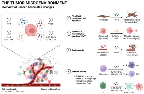
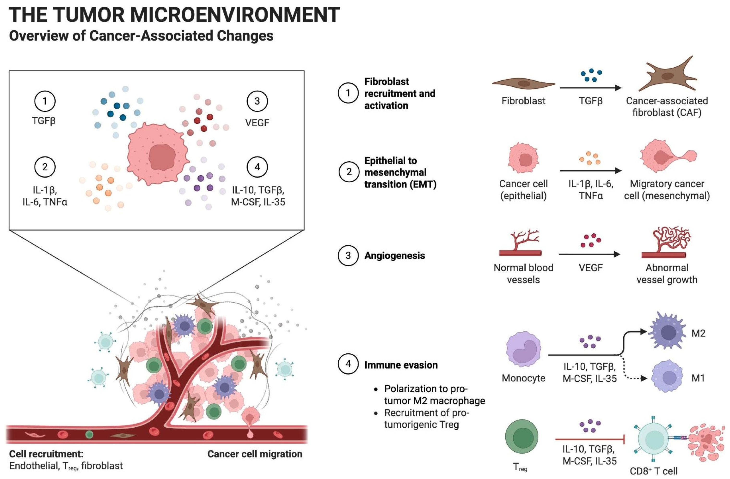
Altogether, interactions between tumor cells, immune cells, fibroblasts, cytokines, and the vasculature promote stromal activation, angiogenesis, tumor-cell migration, and immune evasion within the cutaneous tumor microenvironment (Figure 1).
Figure 1.
Cellular and molecular interactions within the cutaneous tumor microenvironment. Tumor-derived and immune-mediated signaling promotes fibroblast activation and migration, epithelial-to-mesenchymal transition, abnormal angiogenesis, and immune evasion. Cytokines and growth factors, including TGF-β, IL-1β, IL-6, TNF-α, VEGF, IL-10, M-CSF, and IL-35, contribute to cancer-associated fibroblast formation, tumor-cell migration, macrophage polarization, regulatory T-cell recruitment, and suppression of cytotoxic T-cell activity. Created with BioRender.com.
4. Immune Characteristic of Major Cutaneous Malignancies
4.1. Basal Cell Carcinoma
Basal cell carcinoma (BCC) is the most common type of skin cancer associated with slow growth, local tissue invasion, and low metastasis potential [22,30]. The immune infiltrate of BCC mainly consists of macrophages CD4+ and CD8+ T lymphocytes, dendritic cells, and natural killer cells. Despite this, BCC increases the production of immunosuppressive cytokines which weakens antitumor immune response [22,30]. Tumor-associated macrophages are usually observed within the BCC microenvironment and are thought to signal for angiogenesis, extracellular matrix remodeling, and local tissue infiltration. Moreover, modified cytokine signaling involving IL-10, TGF-β, and VEGF contributes to an immunosuppressive environment that supports persistent tumor growth while limiting cytotoxic T-cell activity [12,22].
Even though BCC rarely metastasizes, its locally invasive characteristics often require surgical intervention followed by reconstruction. In addition to histopathological evaluation, preoperative high-frequency ultrasound may provide complementary information regarding BCC depth and local tissue involvement. This imaging technique could assist in determining the size of the tumor as well as predicting the extent of surgical removal. Nonetheless, an ultrasound diagnosis should be made in conjunction with clinical and histopathology, since the diagnostic images cannot substitute for histopathological diagnosis [31].
4.2. Cutaneous Squamous Cell Carcinoma
Cutaneous squamous cell carcinoma (cSCC) is the second most common skin cancer and is characterized by a greater metastatic potential and more aggressive clinical behavior than basal cell carcinoma. Exposure to chronic ultraviolet (UV) rays, DNA damage accumulation, and constant inflammation contribute to cSCC development.
The immune infiltrate of cSCC includes macrophages, dendritic cells, neutrophils, natural killer (NK) cells, and CD4+ and CD8+ T lymphocytes. Compared with BCC, cSCC shows a higher inflammatory microenvironment characterized by the presence of increased inflammatory infiltrate as well as elevated expression of pro-inflammatory cytokines. However, despite the large immune infiltration, cSCC often escapes immune response due to upregulation of immunosuppressive cytokines, recruitment of regulatory T cells, as well as activation of immune checkpoint pathways which help with immune suppression and antitumor immunity [22,30].
Tumor-associated macrophages and neutrophils contribute to cSCC progression by advancing angiogenesis, extracellular matrix remodeling, and tumor invasion through the secretion of cytokines, growth factors, and matrix metalloproteinases. Increased expression of cytokines including IL-6, IL-10, TNF-α, and TGF-β further supports a pro-tumorigenic microenvironment while impairing cytotoxic T-cell function and facilitating local tumor progression [30,32].
4.3. Melanoma
Melanoma is an aggressive cutaneous malignancy known for its highly heterogeneous and immunologically active tumor microenvironment. Even with its immune-stimulating properties, there are ways by which melanoma is able to set up an immunosuppressive microenvironment [33].
Immunosuppressive cytokines, particularly IL-10 and TGF-β, further inhibit T-cell and NK-cell function, while immune checkpoint pathways including PD-1/PD-L1 contribute to immune escape [34]. Angiogenesis and stromal signaling, through the actions of VEGF and cancer-associated fibroblasts, also contribute to vascularization, ECM remodeling, and melanoma progression [35].
These characteristics help separate melanoma tumor microenvironment from those of BCC and cSCC and have the potential to provide a different immunologic and stromal environment at the time of surgical excision. Table 1 summarizes the distinguishing immune and stromal characteristics of BCC, cSCC, and melanoma based on existing literature. The proposed tumor-to-wound research questions are hypothesis-generating and should not be interpreted as established associations between tumor-specific immune microenvironments and postoperative wound-healing outcomes.
Table 1.
Comparison of Immune and Stromal Characteristics Across Major Cutaneous Malignancies and Proposed Implications for the Tumor-to-Wound Transition.
5. Tumor-to-Wound Transition
In this review, the term “tumor-to-wound transition” refers to the biological shift occurring after surgical excision of a cutaneous malignancy, during which the local tissue environment changes from a tumor-conditioned state toward the coordinated inflammatory, proliferative, and remodeling processes required for wound repair. Despite the well-known functions of the immune system cells, cytokines, fibroblasts, angiogenesis, and remodeling of extracellular matrix in the biology of tumors and wound healing, there is uncertainty regarding the persistence and effects of the alterations that occur within the tumors on healing after their surgical removal. Thus, the hypothesis of the transition of tumor into wound provides only a conceptual model of the processes. Surgical excision removes the primary source of tumor-derived signaling but may leave surrounding tissues with persistent inflammatory, stromal, vascular, and extracellular matrix alterations associated with the tumor microenvironment [36]. Surgical injury results in the release of damage-associated molecular patterns, activation of platelets and endothelial cells, and recruitment of neutrophils and monocytes/macrophages [37]. Tumor and healing wounds share several biological characteristics, including fibroblast recruitment and activation, angiogenesis, extracellular matrix deposition and remodeling, and inflammatory signaling [38]. After tissue injury, the early inflammatory phase transitions into the proliferative phase characterized by angiogenesis, fibroblast activation, extracellular matrix production, and re-epithelialization, followed by progressive matrix remodeling and resolution of inflammation [39].
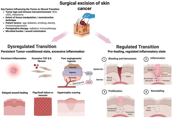
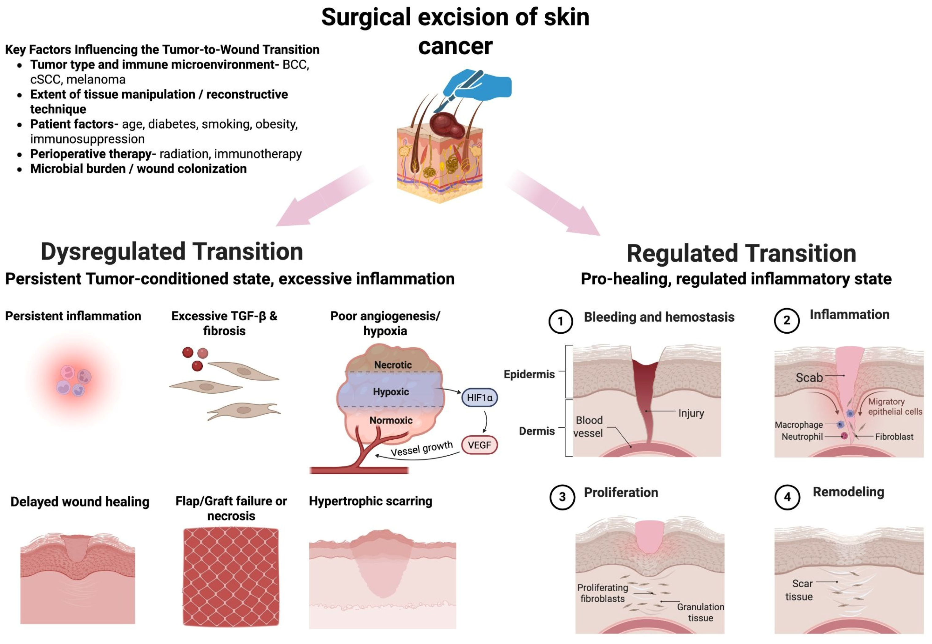
Specifically, it remains unclear how tumor-associated immune suppression, chronic inflammatory signaling, stromal activation, and extracellular matrix remodeling change following removal of the tumor and whether these pre-existing conditions influence subsequent inflammatory resolution, angiogenesis, fibroblast activity, and matrix remodeling during wound repair. Following surgical excision, a regulated transition supports the phases of hemostasis, inflammation, proliferation, and remodeling. Yet, whether such chronic changes in inflammation, stroma, and vasculature as conditioned by the tumor affect delayed healing or other reconstructive problems remains unknown and should be explored clinically (Figure 2).
Figure 2.
Regulated and dysregulated transitions from tumor microenvironment to the postoperative wound-healing environment. Following surgical excision of cutaneous malignancy, the local tissue environment can shift toward a regulated reparative response characterized by hemostasis, inflammation, proliferation, and remodeling. Alternatively, persistent tumor-conditioned inflammation, excessive profibrotic signaling, and impaired angiogenesis may result in delayed wound healing, flap or graft failure, tissue necrosis, or hypertrophic scarring. This transition may be modified by tumor type, reconstructive technique, patient characteristics, perioperative therapy, and microbial burden. Created with BioRender.com.
6. Surgical Reconstruction After Skin Cancer Excision
Surgical excision remains the primary treatment for most localized cutaneous malignancies and often results in defects which require reconstruction in order to restore both functional integrity as well as cosmetic appearance [40]. The reconstructive approach is individualized according to several factors, including tumor size, anatomical location, defect depth, surrounding tissue laxity, patient age, and medical comorbidities [5]. Reconstruction follows the principles of the reconstructive ladder, ranging from relatively simple approaches such as primary closure and healing by secondary intention to skin grafting, local and regional flaps, and free-tissue transfer for larger or more complex defects [41,42]. Healing by secondary intention allows specific surgical wounds to heal without immediate closure through granulation, contraction and re-epithelialization and can provide favorable cosmetic outcomes when appropriately selected [43]. Skin grafts can provide coverage for defects that cannot be closed primarily, whereas local flaps recruit adjacent tissue and have the ability to provide reconstruction with similar tissue characteristics while maintaining vascular supply [44]. Beyond surgical technique, successful reconstruction is also impacted by the biological environment of the wound. Local immune signaling, cytokine profiles, angiogenesis, and extracellular matrix remodeling regulate tissue repair, graft integration, flap survival, and scar formation.
Larger and more complex reconstructive procedures, specifically ones involving flap elevation or free-tissue transfer, also expose tissue to ischemia–reperfusion injury, which can promote reactive oxygen species production, neutrophil recruitment, endothelial dysfunction, and local inflammatory signaling [45]. These responses take place within the postoperative wound bed that may already show tumor-associated changes in fibroblast activity, extracellular matrix remodeling, angiogenesis, and immune-signaling [36]. An important unanswered question is therefore whether the magnitude of tissue manipulation and ischemic injury associated with different reconstructive approaches interacts with the pre-existing tumor-conditioned microenvironment to influence inflammatory resolution, graft integration, flap survival, and subsequent wound healing. Direct clinical studies evaluating this interaction remain limited.
Preliminary clinical evidence identifies several reconstructive outcomes that could serve as relevant endpoints for investigating this hypothesis. In a cohort of patients undergoing surgery for advanced cutaneous squamous cell carcinoma of the head and neck, postoperative complications including wound dehiscence, flap necrosis, infection, graft failure, and flap failure were observed, with complications significantly associated with free-flap reconstruction and radical resection [46]. Likewise, postoperative complications were found to be clinically significant among a national cohort of patients undergoing reconstruction after Mohs surgery as they depend on both patient and surgical parameters [3]. Although these studies do not establish a causal relationship between the tumor immune microenvironment and reconstructive failure, they identify clinically meaningful outcomes through which this relationship could be evaluated prospectively.
Future studies integrating preoperative tumor and peritumoral immune profiling with clinically relevant reconstructive outcomes- including graft take, flap survival, wound dehiscence, infection, and time to healing- may help determine whether tumor-associated biological characteristics influence postoperative reconstruction. Such studies could provide an important test of the proposed “tumor-to-wound” concept and ultimately determine whether biological features of the tumor microenvironment should be considered alongside traditional defect- and patient-specific factors when selecting reconstructive strategies.
7. Patient and Treatment Factors Modifying the Tumor-to-Wound Transition
The tumor-to-wound transition is influenced not only by tumor- and reconstruction-specific factors but also by patient characteristics and prior or ongoing therapies. Several factors including age, diabetes, tobacco exposure, obesity, immunosuppression, prior radiation, and systemic anticancer therapies can modify vascular function, immune signaling, angiogenesis, fibroblast activity, and extracellular matrix remodeling, potentially altering postoperative wound repair [39,47,48]. Although the effects of these factors on wound healing are supported by the existing literature, their specific influence on the proposed tumor-to-wound transition remains incompletely characterized.
Table 2 summarizes these factors and their potential effects on the local wound microenvironment, tumor-to-wound transition, and postoperative outcomes.
Table 2.
Patient and Treatment Factors Modifying the Tumor-to-Wound Transition.
These factors may interact with one another as well as with the pre-tumor conditioned microenvironment. Determining how these systemic and treatment-related factors interact with the local tumor immunobiology represents an important area for future research. More generally, findings from neuro-immune research suggest that self-sustaining interactions between ongoing inflammatory signaling and dysfunction in the innate immune response can be considered as an additional model to explore the involvement of inflammation in the process of transformation from tumor to wound following surgery [56].
8. Immune Checkpoint Inhibition and the Perioperative Immune Environment
Immune checkpoint inhibitors targeting PD-1/PD-L1 and CTLA-4 have become increasingly important in the treatment of advanced cutaneous malignancies by restoring antitumor T-cell activity [57]. Unlike surgery performed in an immunologically untreated tumor, neoadjuvant checkpoint inhibition may alter the composition and activation state of tumor-infiltrating lymphocytes, myeloid cells, stromal populations, and cytokine networks before the tumor is removed [58].
Even though current clinical research has not demonstrated a consistent adverse effect of checkpoint inhibition on surgical wound healing, postoperative outcomes remain incompletely characterized, particularly in patients undergoing complex reconstruction [53,54,59]. Given the context-dependent effects of immune checkpoint inhibition on inflammation and tissue repair, future studies should stratify reconstructive outcomes according to the timing of immunotherapy, the occurrence of immune-related adverse events, and the complexity of surgery to better identify patients who may require individualized perioperative monitoring.
Whether these changes accelerate inflammatory resolution, prolong inflammation, modify angiogenesis, or alter fibroblast and extracellular matrix activity remains poorly understood.
The timing of immunotherapy relative to surgery may represent an important determinant of the tumor-to-wound transition. Similarly, the effects of PD-1/PD-L1 and CTLA-4 blockade may differ depending on the baseline immune phenotype of the tumor, the extent of treatment response, and the complexity of reconstruction. Prospective studies integrating treatment timing with tumor immune profiling and postoperative outcomes are needed to determine whether perioperative immune modulation influences wound healing and reconstructive success.
Although the biological state of the tumor bed may eventually provide additional information for reconstructive planning, current evidence is insufficient to support its routine incorporation into clinical decision-making. Additional prospective studies would be required to assess whether tumor-related immune features offer any additional predictive value over existing anatomical, surgical, and individual risk factors.
9. Future Direction: Toward Immune-Informed Reconstruction
The identification of biomarkers capable of predicting postoperative wound healing and reconstructive success represents a pivotal area for future study. Candidate biomarkers include inflammatory cytokines, immune checkpoint molecules, circulating immune cells, gene-expression profiles, and extracellular matrix remodeling proteins. Moreover, inflammatory mediators like IL-1 and IL-6, as well as changes in matrix metalloproteinase (MMP) and tissue inhibitors of metalloproteinase (TIMP), have been linked to poor wound healing and may serve as reliable indicators of wound healing status [45]. These molecular profiles can then be correlated with clinically meaningful outcomes such as the duration required for re-epithelialization, which is an important marker of effective wound healing [60]. Biomarkers involved in fibroblast activation and extracellular matrix remodeling, specifically TGF-β signaling, may also provide insight into postoperative scar formation and the development of hypertrophic scarring [61].
For example, in patients undergoing reconstruction of large or anatomically complex defects, future immune profiling could be investigated alongside established factors such as defect size, anatomical location, vascular supply, and patient comorbidities to improve postoperative risk assessment [5]. Patients who suffer from excessive inflammatory responses or poor angiogenic profiles may serve as biomarkers for patients who are prone to suffer from delayed wound healing or graft complications. Alternatively, diabetic patients, those who have been exposed to radiation previously, or who have poor vascular perfusion locally, may be considered to see if biological risk assessment can offer any predictive value [47,48,51,52]. However, these applications remain hypothetical and require prospective clinical validation before they can inform reconstructive decisions.
An important next step will be determining whether these biomarkers are merely predictive or can be used to guide intervention. Rather than treating all postoperative wounds according to the same paradigm, immune-informed reconstruction could eventually allow surgical and postoperative strategies to be tailored according to the biological state of the tumor bed. For example, future studies could investigate whether patients demonstrating persistent inflammatory signaling, impaired angiogenic potential, or excessive fibrotic activity might benefit from individualized approaches to wound management or reconstruction. Such a framework would shift reconstruction from an anatomy-driven procedure toward a model that incorporates both structural requirements and biological characteristics of the wound. It is important to mention that these biomarkers and immunological-based treatment approaches are experimental with regard to reconstruction after oncological surgery. Future clinical research is required to determine the value and safety of these approaches to integrate them into clinical practice.
Stem cell therapy, use of extracellular vesicles, platelet-rich biological factors, and growth factor-based treatment modalities have been found to have the capability of accelerating wound repair via inducing angiogenesis, inflammation regulation, and tissue regeneration.
The recent advances in three-dimensional bioprinting, bioengineering of dermis, and vascularization of skin could prove beneficial in treating complicated skin cancer surgery [62,63]. However, translation into oncologic reconstruction will require more research evaluating long term safety.
10. Conclusions
Cutaneous malignancies and healing wounds rely on overlapping mutual biological machinery, including inflammatory and immune signaling, angiogenesis, fibroblast activity, and extracellular matrix remodeling. Within the tumor microenvironment, these processes can be dysregulated and exploited to promote tumor growth, immune evasion, and tissue invasion. Following surgical excision, however, many of these same processes must transition toward coordinated inflammation, tissue repair, vascularization, and extracellular matrix remodeling required for successful wound healing and reconstruction.
It is important to note that this transition may not occur from the same biological starting point across all the different cutaneous malignancies. BCC, cSCC, and melanoma demonstrate distinct patterns of immune activation, suppression, inflammatory signaling, and stromal remodeling, potentially creating different tumor-conditioned environments at the time of excision.
Despite extensive knowledge of tumor immunology and wound healing individually, the biological transition from a tumor-conditioned microenvironment to a postoperative healing wound remains incompletely characterized. Defining how tumor-associated immune and stromal pathways are redirected toward inflammatory resolution and tissue repair represents an important area for future investigation. Direct clinical studies correlating preoperative tumor immune profiles with graft take, flap survival, wound dehiscence, infection, delayed healing, or scar outcomes remain limited.
Ultimately, understanding the tumor-to-wound transition may provide a foundation for moving beyond anatomy- and patient-based reconstruction toward immune-informed reconstruction, in which tumor biology, local immune and stromal characteristics, and reconstructive strategy are considered together. However, the current evidence remains largely mechanistic and indirect, with limited prospective clinical studies directly evaluating the relationship between tumor-associated immune profiles and postoperative reconstructive outcomes. Therefore, immune-informed reconstructions should be seen as a potential research direction, not a proven methodology for clinical practice, and should be validated prospectively before its implementation.
Author Contributions
Conceptualization, D.G. and D.A.; investigation, D.G. and D.A.; writing—original draft preparation, D.G. and D.A.; writing—review and editing, M.E. and A.R.M.; visualization, D.G. and D.A.; supervision, M.E. and A.R.M. All authors have read and agreed to the published version of the manuscript.
Funding
This research received no external funding.
Institutional Review Board Statement
Not applicable.
Data Availability Statement
No new data were created or analyzed in this study. Data sharing is not applicable to this article.
Acknowledgments
ChatGPT (GPT-6, OpenAI) was used to assist with language editing, manuscript organization, and refinement of selected sections during manuscript preparation. All AI-assisted content was reviewed and revised by the authors, who take full responsibility for the accuracy, originality, and integrity of the manuscript.
Conflicts of Interest
The authors declare no conflict of interest.
References
- Radiation: Ultraviolet (UV) Radiation and Skin Cancer. Available online: https://www.who.int/news-room/questions-and-answers/item/radiation-ultraviolet-(uv)-radiation-and-skin-cancer (accessed on 12 October 2026).
- EPIC: The Skin Cancer Working Group. Available online: https://epic.iarc.who.int/research/skincancer/?utm_source=chatgpt.com (accessed on 12 October 2026).
- Parekh, H.N.; Manson, P.N.; Lewis, D.J.; Colakoglu, S.; Reddy, S.K. Risk Factors and Exploratory Clustering of Complications after Reconstruction Following Mohs Surgery: A National NIH All of Us Study. J. Plast. Reconstr. Aesthet. Surg. 2026, 117, 42–54. [Google Scholar] [CrossRef] [Scilit] [PubMed]
- Jakovija, A.; Chtanova, T. Skin Immunity in Wound Healing and Cancer. Front. Immunol. 2023, 14, 1060258. [Google Scholar] [CrossRef] [Scilit] [PubMed]
- Johnson, A.R.; Egeler, S.A.; Wu, W.W.; Bucknor, A.; Ibrahim, A.M.S.; Lin, S.J. Facial Reconstruction After Mohs Surgery: A Critical Review of Defects Involving the Cheek, Forehead, and Perioral Region. J. Craniofac. Surg. 2019, 30, 400–407. [Google Scholar] [CrossRef] [Scilit] [PubMed]
- Forsthuber, A.; Aschenbrenner, B.; Korosec, A.; Jacob, T.; Annusver, K.; Krajic, N.; Kholodniuk, D.; Frech, S.; Zhu, S.; Purkhauser, K.; et al. Cancer-Associated Fibroblast Subtypes Modulate the Tumor-Immune Microenvironment and Are Associated with Skin Cancer Malignancy. Nat. Commun. 2024, 15, 9678. [Google Scholar] [CrossRef] [Scilit] [PubMed]
- Lujano Olazaba, O.; Farrow, J.; Monkkonen, T. Fibroblast Heterogeneity and Functions: Insights from Single-Cell Sequencing in Wound Healing, Breast Cancer, Ovarian Cancer and Melanoma. Front. Genet. 2024, 15, 1304853. [Google Scholar] [CrossRef] [Scilit] [PubMed]
- Zhang, Y.; Lu, Q. Immune Cells in Skin Inflammation, Wound Healing, and Skin Cancer. J. Leukoc. Biol. 2024, 115, 852–865. [Google Scholar] [CrossRef] [Scilit] [PubMed]
- Lu, C.; Liu, Y.; Ali, N.M.; Zhang, B.; Cui, X. The Role of Innate Immune Cells in the Tumor Microenvironment and Research Progress in Anti-Tumor Therapy. Front. Immunol. 2023, 13, 1039260. [Google Scholar] [CrossRef] [Scilit] [PubMed]
- Habib, S.; Osborn, G.; Willsmore, Z.; Chew, M.W.; Jakubow, S.; Fitzpatrick, A.; Wu, Y.; Sinha, K.; Lloyd-Hughes, H.; Geh, J.L.C.; et al. Tumor Associated Macrophages as Key Contributors and Targets in Current and Future Therapies for Melanoma. Expert Rev. Clin. Immunol. 2014, 20, 895–911. [Google Scholar] [CrossRef] [Scilit] [PubMed]
- Li, C.; Yu, X.; Han, X.; Lian, C.; Wang, Z.; Shao, S.; Shao, F.; Wang, H.; Ma, S.; Liu, J. Innate Immune Cells in Tumor Microenvironment: A New Frontier in Cancer Immunotherapy. iScience 2024, 27, 110750. [Google Scholar] [CrossRef] [Scilit] [PubMed]
- Liu, J.; Lu, J.; Wu, L.; Zhang, T.; Wu, J.; Li, L.; Tai, Z.; Chen, Z.; Zhu, Q. Targeting Tumor-Associated Macrophages: Novel Insights into Immunotherapy of Skin Cancer. J. Adv. Res. 2024, 67, 231–252. [Google Scholar] [CrossRef] [Scilit] [PubMed]
- Pu, Y.; Ji, Q. Tumor-Associated Macrophages Regulate PD-1/PD-L1 Immunosuppression. Front. Immunol. 2022, 13, 874589. [Google Scholar] [CrossRef] [Scilit] [PubMed]
- Hassanshahi, A.; Moradzad, M.; Ghalamkari, S.; Fadaei, M.; Cowin, A.J.; Hassanshahi, M. Macrophage-Mediated Inflammation in Skin Wound Healing. Cells 2022, 11, 2953. [Google Scholar] [CrossRef] [Scilit] [PubMed]
- Gao, X.; Lu, C.; Miao, Y.; Ren, J.; Cai, X. Role of Macrophage Polarisation in Skin Wound Healing. Int. Wound J. 2023, 20, 2551–2562. [Google Scholar] [CrossRef] [Scilit] [PubMed]
- Saadh, M.J.; Rasulova, I.; Khalil, M.; Farahim, F.; Sârbu, I.; Ciongradi, C.I.; Omar, T.M.; Alhili, A.; Jawad, M.J.; Hani, T.; et al. Natural Killer Cell-Mediated Immune Surveillance in Cancer: Role of Tumor Microenvironment. Pathol. Res. Pract. 2024, 254, 155120. [Google Scholar] [CrossRef] [Scilit] [PubMed]
- Cavalcante-Silva, J.; Koh, T.J. Role of NK Cells in Skin Wound Healing of Mice. J. Immunol. 2023, 210, 981–990. [Google Scholar] [CrossRef] [Scilit] [PubMed]
- Koenderman, L.; Vrisekoop, N. Neutrophils in Cancer: From Biology to Therapy. Cell. Mol. Immunol. 2025, 22, 4–23. [Google Scholar] [CrossRef] [Scilit] [PubMed]
- Yu, X.; Li, C.; Wang, Z.; Xu, Y.; Shao, S.; Shao, F.; Wang, H.; Liu, J. Neutrophils in Cancer: Dual Roles through Intercellular Interactions. Oncogene 2024, 43, 1163–1177. [Google Scholar] [CrossRef] [Scilit] [PubMed]
- Mazzoccoli, L.; Liu, B. Dendritic Cells in Shaping Anti-Tumor T Cell Response. Cancers 2024, 16, 2211. [Google Scholar] [CrossRef] [Scilit] [PubMed]
- Gregorio, J.; Meller, S.; Conrad, C.; Di Nardo, A.; Homey, B.; Lauerma, A.; Arai, N.; Gallo, R.L.; Digiovanni, J.; Gilliet, M. Plasmacytoid Dendritic Cells Sense Skin Injury and Promote Wound Healing through Type I Interferons. J. Exp. Med. 2010, 207, 2921–2930. [Google Scholar] [CrossRef] [Scilit] [PubMed]
- Mousa, A.M.; Enk, A.H.; Hassel, J.C.; Reschke, R. Immune Checkpoints and Cellular Landscape of the Tumor Microenvironment in Non-Melanoma Skin Cancer (NMSC). Cells 2024, 13, 1615. [Google Scholar] [CrossRef] [Scilit] [PubMed]
- Meng, L.; Tang, Q.; Zhou, W.; Wei, D.; Zhao, J.; Shen, J.; Yang, M.; He, S.; Huang, S.; Qin, Y.; et al. Contributions of T Cell Signaling for Wound Healing. J. Burn Care Res. 2024, 45, 1513–1519. [Google Scholar] [CrossRef] [Scilit] [PubMed]
- Kumagai, S.; Itahashi, K.; Nishikawa, H. Regulatory T Cell-Mediated Immunosuppression Orchestrated by Cancer: Towards an Immuno-Genomic Paradigm for Precision Medicine. Nat. Rev. Clin. Oncol. 2024, 21, 337–353. [Google Scholar] [CrossRef] [Scilit] [PubMed]
- Knoedler, S.; Knoedler, L.; Kauke-Navarro, M.; Rinkevich, Y.; Hundeshagen, G.; Harhaus, L.; Kneser, U.; Pomahac, B.; Orgill, D.P.; Panayi, A.C. Regulatory T Cells in Skin Regeneration and Wound Healing. Mil. Med. Res. 2023, 10, 49. [Google Scholar] [CrossRef] [Scilit] [PubMed]
- Barrientos, S.; Stojadinovic, O.; Golinko, M.S.; Brem, H.; Tomic-Canic, M. Growth Factors and Cytokines in Wound Healing. Wound Repair. Regen. 2008, 16, 585–601. [Google Scholar] [CrossRef] [Scilit] [PubMed]
- He, X.; Xu, C. Immune Checkpoint Signaling and Cancer Immunotherapy. Cell Res. 2020, 30, 660–669. [Google Scholar] [CrossRef] [Scilit] [PubMed]
- Sahai, E.; Astsaturov, I.; Cukierman, E.; DeNardo, D.G.; Egeblad, M.; Evans, R.M.; Fearon, D.; Greten, F.R.; Hingorani, S.R.; Hunter, T.; et al. A Framework for Advancing Our Understanding of Cancer-Associated Fibroblasts. Nat. Rev. Cancer 2020, 20, 174–186. [Google Scholar] [CrossRef] [Scilit] [PubMed]
- Darby, I.A.; Laverdet, B.; Bonté, F.; Desmoulière, A. Fibroblasts and Myofibroblasts in Wound Healing. Clin. Cosmet. Investig. Dermatol. 2014, 7, 301–311. [Google Scholar] [CrossRef] [Scilit] [PubMed]
- Chiang, E.; Stafford, H.; Buell, J.; Ramesh, U.; Amit, M.; Nagarajan, P.; Migden, M.; Yaniv, D. Review of the Tumor Microenvironment in Basal and Squamous Cell Carcinoma. Cancers 2023, 15, 2453. [Google Scholar] [CrossRef] [Scilit] [PubMed]
- Popescu, C.; Iliescu, C.A.; Truica, A.M.; Popa, L.G.; Porosnicu, A.L.; Tudose, I.; Boeru, C.; Popescu, M.N. From Ultrasound to Histopathology: Agreement Analysis and Depth Correction Model for Basal Cell Carcinoma Using a Portable 20 MHz High-Frequency Ultrasound Device. Diagnostics 2026, 16, 978. [Google Scholar] [CrossRef] [Scilit] [PubMed]
- Abdul-Rahman, T.; Ghosh, S.; Badar, S.M.; Nazir, A.; Bamigbade, G.B.; Aji, N.; Roy, P.; Kachani, H.; Garg, N.; Lawal, L.; et al. The Paradoxical Role of Cytokines and Chemokines at the Tumor Microenvironment: A Comprehensive Review. Eur. J. Med. Res. 2024, 29, 124. [Google Scholar] [CrossRef] [Scilit] [PubMed]
- Zilberg, C.; Ferguson, A.L.; Lyons, J.G.; Gupta, R.; Damian, D.L. The Tumor Immune Microenvironment in Primary Cutaneous Melanoma. Arch. Dermatol. Res. 2025, 317, 273. [Google Scholar] [CrossRef] [Scilit] [PubMed]
- Bu, M.T.; Chandrasekhar, P.; Ding, L.; Hugo, W. The Roles of TGF-β and VEGF Pathways in the Suppression of Antitumor Immunity in Melanoma and Other Solid Tumors. Pharmacol. Ther. 2022, 240, 108211. [Google Scholar] [CrossRef] [Scilit] [PubMed]
- Kharouf, N.; Flanagan, T.W.; Hassan, S.-Y.; Shalaby, H.; Khabaz, M.; Hassan, S.-L.; Megahed, M.; Haikel, Y.; Santourlidis, S.; Hassan, M. Tumor Microenvironment as a Therapeutic Target in Melanoma Treatment. Cancers 2023, 15, 3147. [Google Scholar] [CrossRef] [Scilit] [PubMed]
- Tang, F.; Tie, Y.; Tu, C.; Wei, X. Surgical Trauma-induced Immunosuppression in Cancer: Recent Advances and the Potential Therapies. Clin. Transl. Med. 2020, 10, 199–223. [Google Scholar] [CrossRef] [Scilit] [PubMed]
- Cheng, X.; Zhang, H.; Hamad, A.; Huang, H.; Tsung, A. Surgery-Mediated Tumor-Promoting Effects on the Immune Microenvironment. Semin. Cancer Biol. 2022, 86, 408–419. [Google Scholar] [CrossRef] [Scilit] [PubMed]
- Foster, D.S.; Jones, R.E.; Ransom, R.C.; Longaker, M.T.; Norton, J.A. The Evolving Relationship of Wound Healing and Tumor Stroma. JCI Insight 2018, 3, e99911. [Google Scholar] [CrossRef] [Scilit] [PubMed]
- Peña, O.A.; Martin, P. Cellular and Molecular Mechanisms of Skin Wound Healing. Nat. Rev. Mol. Cell Biol. 2024, 25, 599–616. [Google Scholar] [CrossRef] [Scilit] [PubMed]
- Brandão, C.M.; Weimann, E.T.d.S.; Terzian, L.R.; Machado Filho, C.D.S.; Paschoal, F.M.; Criado, P.R. Keep It Simple. A Ten-Year Experience in Reconstructions after Mohs Micrographic Surgery. An. Bras. Dermatol. 2020, 95, 714–720. [Google Scholar] [CrossRef] [Scilit] [PubMed]
- De Francesco, F.; Zingaretti, N.; Parodi, P.C.; Riccio, M. The Evolution of Current Concept of the Reconstructive Ladder in Plastic Surgery: The Emerging Role of Translational Medicine. Cells 2023, 12, 2567. [Google Scholar] [CrossRef] [Scilit] [PubMed]
- Simman, R. Wound Closure and the Reconstructive Ladder in Plastic Surgery. J. Am. Col. Certif. Wound Spec. 2009, 1, 6–11. [Google Scholar] [CrossRef] [Scilit] [PubMed]
- Gil-Lianes, J.; Marti-Marti, I.; Morgado-Carrasco, D. Secondary Intention Healing After Mohs Micrographic Surgery: An Updated Review of Classic and Novel Applications, Benefits and Complications. Actas. Dermosifiliogr. 2025, 116, 511–520. [Google Scholar] [CrossRef] [Scilit] [PubMed]
- Desisto, N.G.; Ortiz, A.S.; Yang, S.F.; Stephan, S.J.; Patel, P.N. State of the Evidence for Facial Skin Cancer Reconstruction. Facial Plast. Surg. 2023, 39, 220–229. [Google Scholar] [CrossRef] [Scilit] [PubMed]
- van den Heuvel, M.G.W.; Buurman, W.A.; Bast, A.; van der Hulst, R.R.W.J. Review: Ischaemia-Reperfusion Injury in Flap Surgery. J. Plast. Reconstr. Aesthet. Surg. 2009, 62, 721–726. [Google Scholar] [CrossRef] [Scilit] [PubMed]
- Yung, A.E.; Crouch, G.; Varey, A.H.R.; Lo, S.; Elliott, M.S.; Lee, J.; Rawson, R.; Gupta, R.; Hong, A.M.; Clark, J.R.; et al. Benchmarking Survival Outcomes Following Surgical Management of pT3 and pT4 Cutaneous Squamous Cell Carcinoma of the Head and Neck. Ann. Surg. Oncol. 2022, 29, 5124–5138. [Google Scholar] [CrossRef] [Scilit] [PubMed]
- Guo, S.; Dipietro, L.A. Factors Affecting Wound Healing. J. Dent. Res. 2010, 89, 219–229. [Google Scholar] [CrossRef] [Scilit] [PubMed]
- Anderson, K.; Hamm, R.L. Factors That Impair Wound Healing. J. Am. Coll. Clin. Wound Spec. 2014, 4, 84–91. [Google Scholar] [CrossRef] [Scilit] [PubMed]
- Bootun, R. Effects of Immunosuppressive Therapy on Wound Healing. Int. Wound J. 2013, 10, 98–104. [Google Scholar] [CrossRef] [Scilit] [PubMed]
- Lopez, A.; Babadzhanov, M.; Cheraghlou, S.; Canavan, T.; Doudican, N.; Stevenson, M.; Carucci, J.A. Immunosuppressed Patients Are at Increased Risk of Local Recurrence, Metastasis, and Disease Specific Death from Cutaneous Squamous Cell Carcinoma. Arch. Dermatol. Res. 2023, 315, 1429–1433. [Google Scholar] [CrossRef] [Scilit] [PubMed]
- Dormand, E.-L.; Banwell, P.E.; Goodacre, T.E.E. Radiotherapy and Wound Healing. Int. Wound J. 2005, 2, 112–127. [Google Scholar] [CrossRef] [Scilit] [PubMed]
- Straub, J.M.; New, J.; Hamilton, C.D.; Lominska, C.; Shnayder, Y.; Thomas, S.M. Radiation-Induced Fibrosis: Mechanisms and Implications for Therapy. J. Cancer Res. Clin. Oncol. 2015, 141, 1985–1994. [Google Scholar] [CrossRef] [Scilit] [PubMed]
- Kuo, P.-J.; Lin, P.-C.; Hsieh, C.-H. Wound Healing in Cancer Patients under Immunotherapy. Int. J. Surg. 2025, 111, 7087–7098. [Google Scholar] [CrossRef] [Scilit] [PubMed]
- Mays, A.C.; Yarlagadda, B.; Achim, V.; Jackson, R.; Pipkorn, P.; Huang, A.T.; Rajasekaran, K.; Sridharan, S.; Rosko, A.J.; Orosco, R.K.; et al. Examining the Relationship of Immunotherapy and Wound Complications Following Flap Reconstruction in Patients with Head and Neck Cancer. Head Neck 2021, 43, 1509–1520. [Google Scholar] [CrossRef] [Scilit] [PubMed]
- Okay, E.; Connolly, J.J.; Gonzalez, M.R.; Lozano-Calderon, S.A. Wound-Healing Effects of Common Antineoplastic Agents and Perioperative Considerations for the Orthopaedic Surgeon. J. Am. Acad. Orthop. Surg. 2024, 32, e671–e682. [Google Scholar] [CrossRef] [Scilit] [PubMed]
- Haghighatian, Z. From Chronic Inflammation to Neurodegeneration: Mechanistic Convergence and Therapeutic Opportunities at the Neuroimmune Interface. Nexus Pathophysiol. Ther. 2026, 1, 007. [Google Scholar] [CrossRef]
- Barrios, D.M.; Do, M.H.; Phillips, G.S.; Postow, M.A.; Akaike, T.; Nghiem, P.; Lacouture, M.E. Immune Checkpoint Inhibitors to Treat Cutaneous Malignancies. J. Am. Acad. Dermatol. 2020, 83, 1239–1253. [Google Scholar] [CrossRef] [Scilit] [PubMed]
- Pavlick, A.C.; Ariyan, C.E.; Buchbinder, E.I.; Davar, D.; Gibney, G.T.; Hamid, O.; Hieken, T.J.; Izar, B.; Johnson, D.B.; Kulkarni, R.P.; et al. Society for Immunotherapy of Cancer (SITC) Clinical Practice Guideline on Immunotherapy for the Treatment of Melanoma, Version 3.0. J. Immunother. Cancer 2023, 11, e006947. [Google Scholar] [CrossRef] [Scilit] [PubMed]
- Philips, R.; Alnemri, A.; Amin, D.; Patel, J.; Topf, M.C.; Johnson, J.M.; BarAd, V.; Axelrod, R.; Argiris, A.; Fundakowski, C.; et al. Effect of Preoperative Programmed Death-1 or Programmed Death Ligand-1 Immune Check Point Inhibition on Complications after Surgery for Primary Head and Neck Cancer. Cancer 2024, 130, 863–875. [Google Scholar] [CrossRef] [Scilit] [PubMed]
- Lindley, L.E.; Stojadinovic, O.; Pastar, I.; Tomic-Canic, M. Biology and Biomarkers for Wound Healing. Plast. Reconstr. Surg. 2016, 138, 18S–28S. [Google Scholar] [CrossRef] [Scilit] [PubMed]
- Rousselle, P.; Braye, F.; Dayan, G. Re-Epithelialization of Adult Skin Wounds: Cellular Mechanisms and Therapeutic Strategies. Adv. Drug. Deliv. Rev. 2019, 146, 344–365. [Google Scholar] [CrossRef] [Scilit] [PubMed]
- Sierra-Sánchez, Á.; Kim, K.H.; Blasco-Morente, G.; Arias-Santiago, S. Cellular Human Tissue-Engineered Skin Substitutes Investigated for Deep and Difficult to Heal Injuries. npj Regen. Med. 2021, 6, 35. [Google Scholar] [CrossRef] [Scilit] [PubMed]
- Song, H.; Hao, D.; Zhou, J.; Farmer, D.; Wang, A. Development of Pro-Angiogenic Skin Substitutes for Wound Healing. Wound Repair Regen. 2024, 32, 208–216. [Google Scholar] [CrossRef] [Scilit] [PubMed]

Disclaimer/Publisher’s Note: The statements, opinions and data contained in all publications are solely those of the individual author(s) and contributor(s) and not of MDPI and/or the editor(s). MDPI and/or the editor(s) disclaim responsibility for any injury to people or property resulting from any ideas, methods, instructions or products referred to in the content. |
© 2026 by the authors. Licensee MDPI, Basel, Switzerland. This article is an open access article distributed under the terms and conditions of the Creative Commons Attribution (CC BY) license.