


Simple Summary
We studied a rare type of stomach cancer called gastric adenocarcinoma with enteroblastic differentiation to understand how often it spreads to nearby lymph nodes after growing into the layer beneath the stomach lining. We compared 84 patients with this tumor type and 345 with conventional stomach cancer. Cancer had spread to lymph nodes in 39.3% and 25.8% of patients, respectively. We also described which groups of lymph nodes were involved according to tumor location. These findings support careful assessment of nearby lymph nodes. However, the small patient groups limit conclusions about specific patterns of spread, and the prediction models had limited performance. Larger studies are needed to confirm these findings and improve prediction.
Abstract
Background: The risk of lymph node metastasis (LNM) in T1b gastric adenocarcinoma with enteroblastic differentiation (GAED) remains poorly defined. Objectives: This study compared nodal involvement between GAED and conventional gastric cancer and examined associated factors, metastatic nodal stations, and exploratory prediction models. Methods: This single-center retrospective study included 429 patients with pathologically confirmed T1b gastric cancer treated between 2013 and 2024, comprising 84 with GAED and 345 with conventional gastric cancer. Variables with an overall univariable p < 0.05 were entered simultaneously into one multivariable Firth logistic regression model. Within GAED, nodal station involvement and AFP, SALL4, and GPC3 expression were evaluated. Prediction models underwent 20 repetitions of fivefold cross-validation. Results: LNM occurred in 39.3% of patients with GAED and 25.8% with conventional gastric cancer. GAED remained associated with LNM after adjustment (odds ratio, 2.48; 95% confidence interval, 1.33–4.62; p = 0.004). Female sex, poor differentiation, and vascular tumor thrombus also remained associated with LNM. Signet-ring cell component, vascular invasion, and perineural invasion were not statistically significant after adjustment. Descriptively, stations 7 and 3 had the highest positive counts in proximal tumors, whereas stations 6, 3, and 5 had the highest counts in the pooled middle-plus-distal group; all positive station entries in the pooled group occurred in distal tumors. AFP, SALL4, and GPC3 staining grades showed no consistent increase in LNM. Prediction models showed limited discrimination, substantial miscalibration, and only small Brier-score improvements over an intercept-only comparator; no incremental improvement in LNM prediction from adding IHC markers was demonstrated in this dataset. Conclusions: T1b GAED was associated with higher odds of LNM than conventional gastric cancer. These findings support adequate regional lymphadenectomy in patients undergoing gastrectomy for T1b GAED. The exploratory models require further development and external validation before clinical use.
1. Introduction
Gastric cancer remains a major cause of cancer-related mortality worldwide and continues to impose a substantial disease burden in China [1,2]. For patients with early gastric cancer, accurate assessment of lymph node metastasis (LNM) is essential for determining the appropriate extent of treatment. This is particularly important for tumors with submucosal invasion, because the risk of nodal involvement increases once the tumor extends beyond the mucosa [3,4,5]. An international analysis reported LNM in 22.1% of Eastern and 27.3% of Western patients with pT1b gastric cancer [6]. Therefore, identifying pathological features associated with nodal spread is important for surgical decision-making and for determining the appropriate extent of lymphadenectomy.
Gastric adenocarcinoma with enteroblastic differentiation (GAED) is an uncommon histological subtype characterized by fetal intestinal-like morphology and cuboidal or columnar tumor cells with clear cytoplasm. AFP, SALL4, and GPC3 are commonly used to support the diagnosis of enteroblastic differentiation, although their expression varies among tumors [7,8]. Previous studies have shown that GAED is associated with frequent vascular invasion, LNM, and liver metastasis compared with conventional gastric adenocarcinoma [7,8,9]. More recent studies of gastric carcinomas with primitive enterocyte phenotypes, as well as multicenter analyses of hepatoid adenocarcinoma and GAED, have also reported aggressive clinicopathological features and unfavorable survival outcomes [10,11]. These findings suggest that GAED may have a greater tendency for early lymphatic and hematogenous spread than conventional gastric cancer.
The nodal behavior of T1b GAED, however, remains poorly defined. Previous studies of early gastric neoplasms have linked enteroblastic differentiation with submucosal invasion and lymphovascular invasion [12,13,14,15], but direct comparisons of LNM between T1b GAED and conventional gastric cancer are limited. In addition, little is known about the distribution of metastatic lymph node stations according to primary tumor location. Clarifying these patterns is clinically relevant because they may help determine whether patients with T1b GAED require more careful regional lymph node assessment and adequate lymphadenectomy at the time of gastrectomy.
In this study, we compared the clinicopathological characteristics and LNM rates of patients with T1b GAED and conventional gastric cancer. We also identified factors associated with LNM and examined the distribution of metastatic lymph node stations according to tumor location. Within the GAED cohort, we further explored the associations of immunohistochemical findings and a poorly cohesive carcinoma component with LNM. Finally, we evaluated exploratory prediction models for nodal metastasis. Our aim was to better define the nodal metastatic risk of T1b GAED and provide evidence for surgical planning and lymphadenectomy in this uncommon subtype.
2. Methods
2.1. Study Population and Data Source
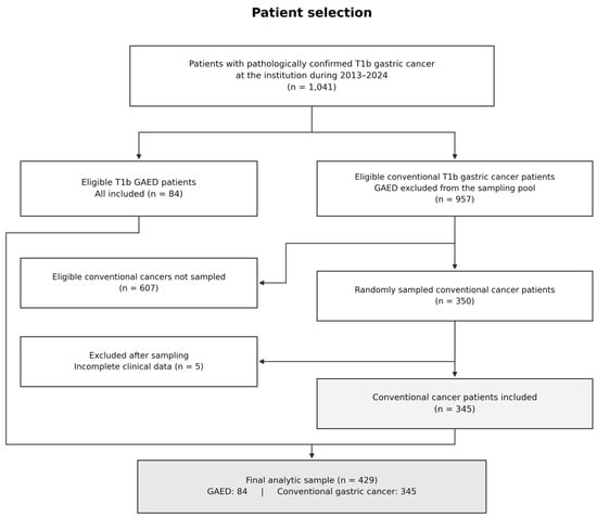
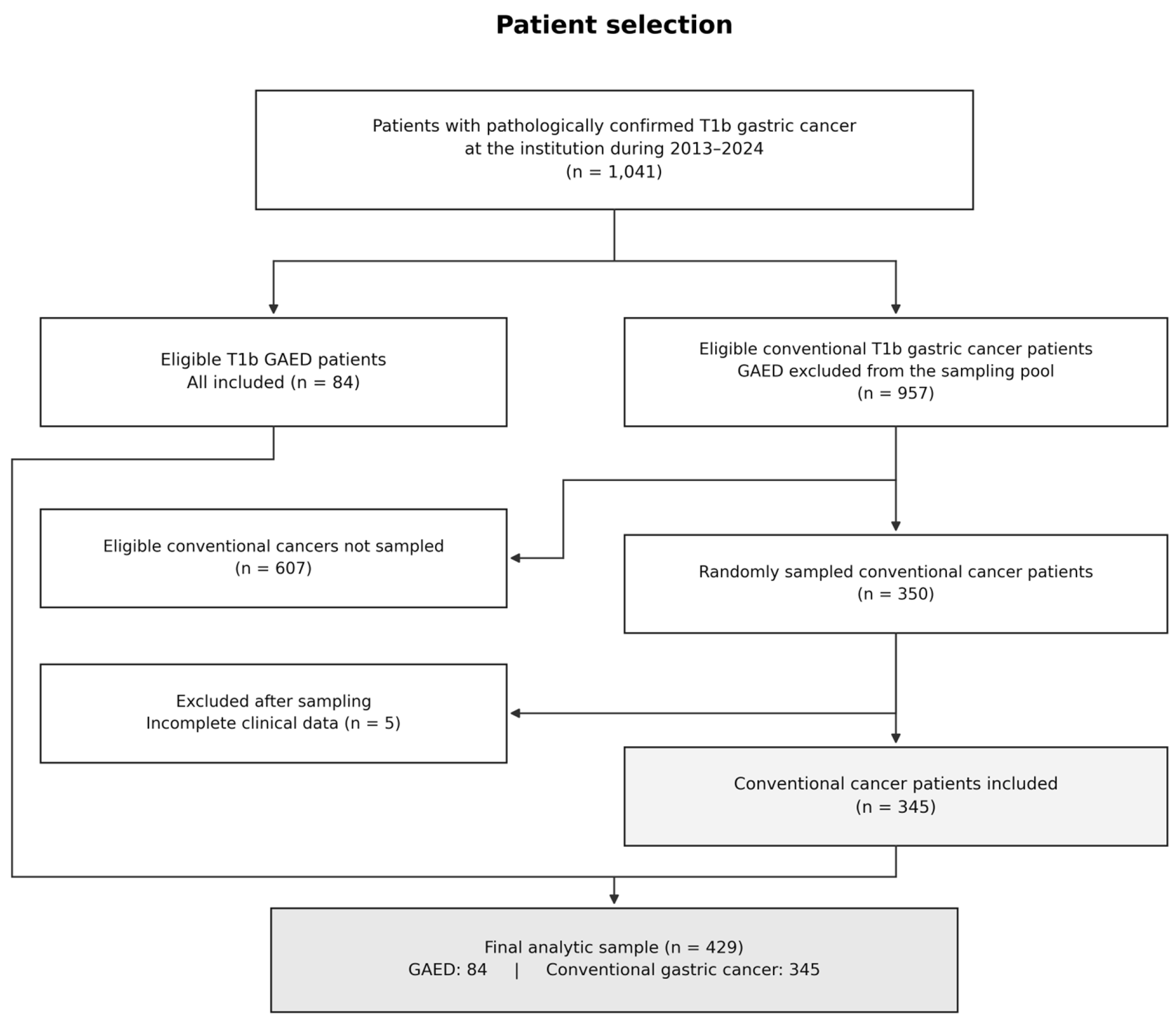
This single-center retrospective comparative study was derived from an institutional cohort of 1041 patients with pathologically confirmed T1b gastric cancer treated during 2013–2024. The source cohort included 84 patients with gastric adenocarcinoma with enteroblastic differentiation (GAED) and 957 with conventional gastric cancer. All 84 eligible GAED patients were included. A comparison group of 350 patients was randomly selected from the 957 conventional cancer patients, with GAED excluded from the sampling pool. Five sampled patients were excluded because of incomplete clinical data, leaving 345 conventional cancer patients. The final analytic sample therefore comprised 429 patients (Figure 1). T1b denotes invasion into the submucosa. All the patients underwent the corresponding surgeries, including distal subtotal gastrectomy, proximal subtotal gastrectomy, total gastrectomy, and D2 lymph node dissection. Clinicopathological data were obtained from the institutional database and pathology reports. Pathological staging was performed according to the eighth edition of the American Joint Committee on Cancer (AJCC) TNM staging system.
Figure 1.
Flowchart of patient selection.
The study was approved by the institutional ethics committee (approval no. 23/122-3864) and was conducted in accordance with the Declaration of Helsinki. This retrospective analysis used anonymized clinical data collected during routine care. All patients had provided written informed consent for treatment; no additional study-specific informed consent was obtained for this retrospective analysis.
2.2. Clinicopathological Variables and Outcome Definitions
GAED classification was based on final diagnoses issued by the Department of Pathology according to established histopathological criteria. Characteristic morphology includes enteroblastic cells with clear cytoplasm and a tubular or papillary growth pattern; AFP, SALL4, and GPC3 provide ancillary immunophenotypic information. Diagnoses for this analysis were taken from the pathology reports. Expression of each marker was graded as 0, 1+, 2+, or 3+ according to the extent and intensity of immunostaining.
The analyzed variables were histological group, age, sex, tumor size, differentiation, location, HER2 immunohistochemistry (IHC) score, signet-ring cell component, vascular invasion, perineural invasion, vascular tumor thrombus, and the number of retrieved lymph nodes. Tumor size was classified as <2 cm, 2–<4 cm, or ≥4 cm, and location as proximal, middle, or distal. Differentiation was presented in three categories and dichotomized as poor versus well/moderate for regression. Vascular invasion and vascular tumor thrombus were retained as separate recorded variables. The station-level analysis included stations 1, 2, 3, 4sa, 4sb, 4d, 5, 6, 7, 8a, 9, 11p, and 12a. Station involvement was defined as at least one metastatic node at that station. Middle and distal tumors were combined for descriptive graphical presentation; the separate middle and distal counts were also tabulated (Supplementary Table S3B).
2.3. Statistical Analysis
Continuous variables are presented as medians (IQRs) and categorical variables as counts (%). Between-group comparisons used Wilcoxon rank-sum, Pearson chi-square, Fisher exact, or Fisher–Freeman–Halton exact tests, as appropriate. Nodal-stage and IHC-stratified LNM proportions were summarized descriptively with exact binomial 95% CIs. For the full-cohort association analysis, each candidate variable was assessed using univariable Firth logistic regression [16], and variables with an overall two-sided penalized likelihood ratio p < 0.05 were entered simultaneously into a single multivariable Firth model, without further stepwise elimination. Multilevel categorical variables were screened using a joint test of all non-reference levels, and all levels were retained when the variable was selected. Age (per 10 years) and retrieved nodal yield (per node) were modeled linearly on the logit scale in univariable analysis. All association analyses used 429 complete records with 122 LNM events, without imputation; logistf version 1.26.1 provided profile penalized-likelihood 95% CIs and two-sided penalized likelihood ratio tests. Adjusted ORs describe conditional associations rather than causal effects. Within GAED (84 complete cases; 33 events), Firth and ridge models used a fixed six-slope clinicopathological predictor set, and a ridge model added three binary IHC predictors; no imputation or predictor screening was performed. All models shared 20 repetitions of stratified fivefold outer cross-validation; ridge standardization, fitting, and fivefold inner tuning by binomial deviance with the one-standard-error rule used training data only (Supplementary Table S4A). An intercept-only comparator predicted the event frequency in each outer training fold. Performance was assessed using AUC, Brier score, and calibration; IQRs summarize variation across repetitions and are not confidence intervals. Station-specific proportions and two-sided Clopper–Pearson exact 95% CIs were calculated using the number of patients assessed at each station. Station-level findings were descriptive. Analyses were performed using R version 4.3.3.
3. Results
3.1. Patient Characteristics
The cohort comprised 84 patients with GAED and 345 with GC (Table 1). Patients with GAED were older (median, 65 versus 58 years; p < 0.001) and had more retrieved lymph nodes (median, 34.5 versus 30.0; p = 0.012). Men accounted for 75.0% and 69.6% of the groups, respectively (p = 0.353). Tumor-size distributions were similar: <2 cm, 22.6% versus 23.2%; 2–<4 cm, 52.4% versus 58.8%; and ≥4 cm, 25.0% versus 18.0% (p = 0.329). Tumor location differed between groups (p < 0.001), with middle-third tumors less frequent in GAED (6.0% versus 36.2%). GAED had lower recorded frequencies of a signet-ring cell component (19.0% versus 36.2%; p = 0.003), vascular invasion (3.6% versus 18.3%; p < 0.001), and perineural invasion (7.1% versus 17.7%). HER2 score distributions differed (p = 0.026), whereas differentiation and vascular tumor thrombus did not show clear between-group differences (p = 0.185 and p = 0.140).
Table 1.
Clinicopathological characteristics of patients with T1b gastric cancer.
3.2. Lymph Node Metastasis and N-Stage Distribution
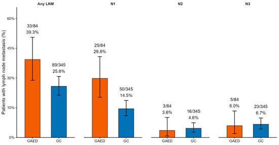
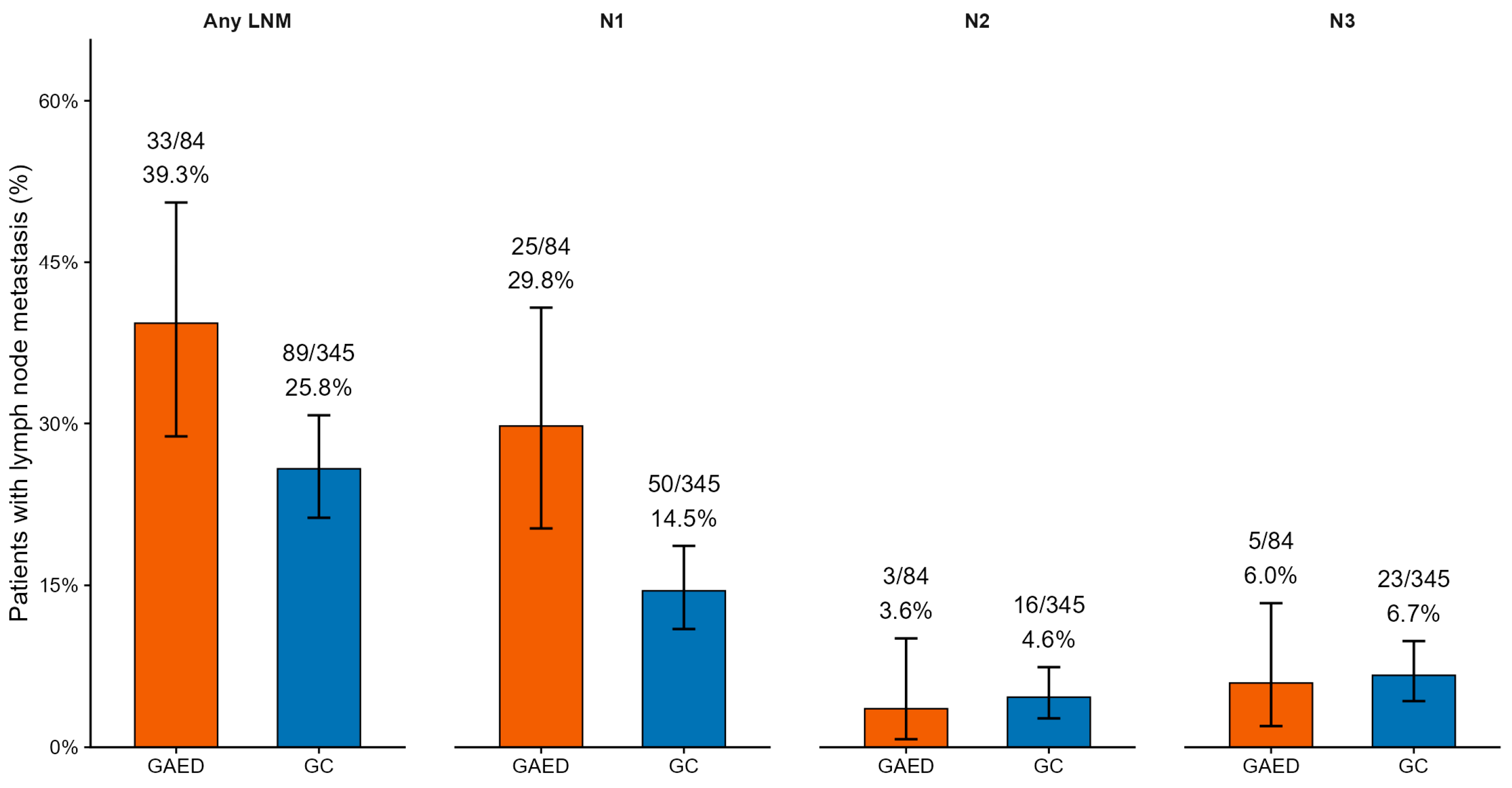
LNM was present in 33 of 84 GAED patients (39.3%) and 89 of 345 GC patients (25.8%). The observed LNM proportion was 13.5 percentage points higher in GAED than in conventional gastric cancer, yielding 122 events overall (Figure 2 and Supplementary Table S1). N1, N2, and N3 proportions were 29.8%, 3.6%, and 6.0% in GAED, compared with 14.5%, 4.6%, and 6.7% in GC.
Figure 2.
Lymph node metastasis and N-stage distribution in GAED and GC. Bars show observed proportions, and error bars indicate two-sided exact binomial (Clopper–Pearson) 95% confidence intervals. Note: GAED, gastric adenocarcinoma with enteroblastic differentiation; GC, conventional gastric cancer.
3.3. Factors Associated with Lymph Node Metastasis
Overall univariable tests identified histological group (p = 0.015), sex (p < 0.001), poor differentiation (p < 0.001), tumor location (p < 0.001), signet-ring cell component (p = 0.025), vascular invasion (p = 0.001), perineural invasion (p = 0.043), and vascular tumor thrombus (p < 0.001) as eligible for the multivariable model (Table 2 and Supplementary Table S2). Age (p = 0.684), tumor size (overall p = 0.055), HER2 score (overall p = 0.716), and retrieved nodal yield (p = 0.819) did not meet the selection criterion. Although the ≥4 versus <2 cm size contrast was significant (p = 0.017), the overall test for the three-category tumor-size variable was not significant.
Table 2.
Univariable and multivariable analyses of lymph node metastasis.
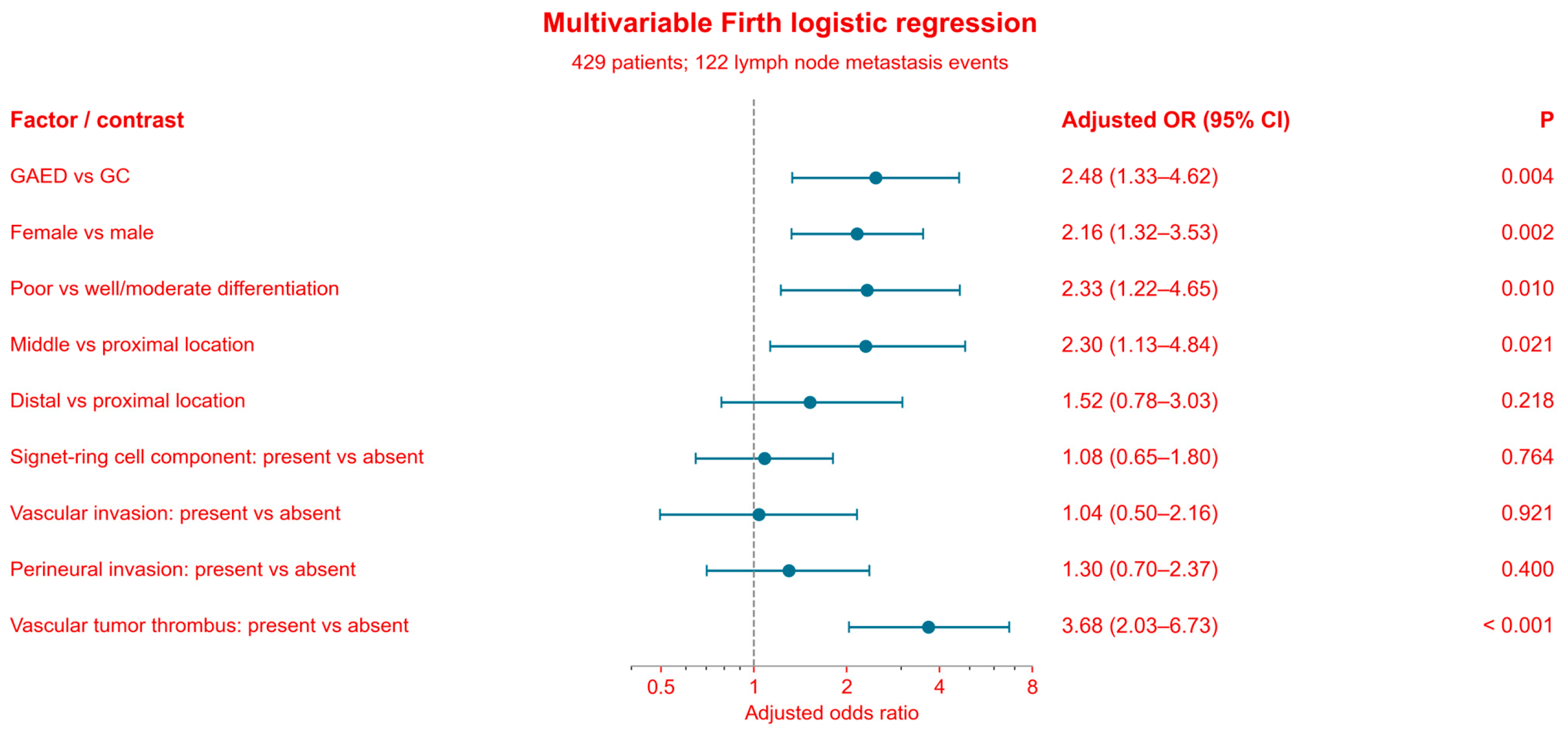
The single multivariable model included histological group, sex, poor differentiation, tumor location, signet-ring cell component, vascular invasion, perineural invasion, and vascular tumor thrombus. GAED remained associated with higher odds of LNM (adjusted OR, 2.48; 95% CI, 1.33–4.62; p = 0.004). Female sex (adjusted OR, 2.16; p = 0.002), poor differentiation (adjusted OR, 2.33; p = 0.010), and vascular tumor thrombus (adjusted OR, 3.68; p < 0.001) also remained associated with LNM. Signet-ring cell component (adjusted OR, 1.08; p = 0.764), vascular invasion (adjusted OR, 1.04; p = 0.921), and perineural invasion (adjusted OR, 1.30; p = 0.400) were not statistically significant after adjustment. The overall test for tumor location was not significant (p = 0.064), although the middle-versus-proximal contrast had p = 0.021 (Table 2 and Figure 3).
Figure 3.
Adjusted associations with lymph node metastasis in the single multivariable Firth logistic regression model (429 patients; 122 events). Points and horizontal bars represent adjusted odds ratios and profile penalized-likelihood 95% CIs; the dashed line indicates an odds ratio of 1. Displayed p values are two-sided coefficient-specific penalized likelihood ratio tests. The overall test for tumor location had p = 0.064. GAED, gastric adenocarcinoma with enteroblastic differentiation; GC, conventional gastric cancer.
Among patients with GAED, the observed proportion with lymph node metastasis was higher in those with a poorly cohesive carcinoma component than in those without (52.6% [10/19] vs. 35.4% [23/65]) (Supplementary Figure S1).
3.4. IHC Findings Within GAED
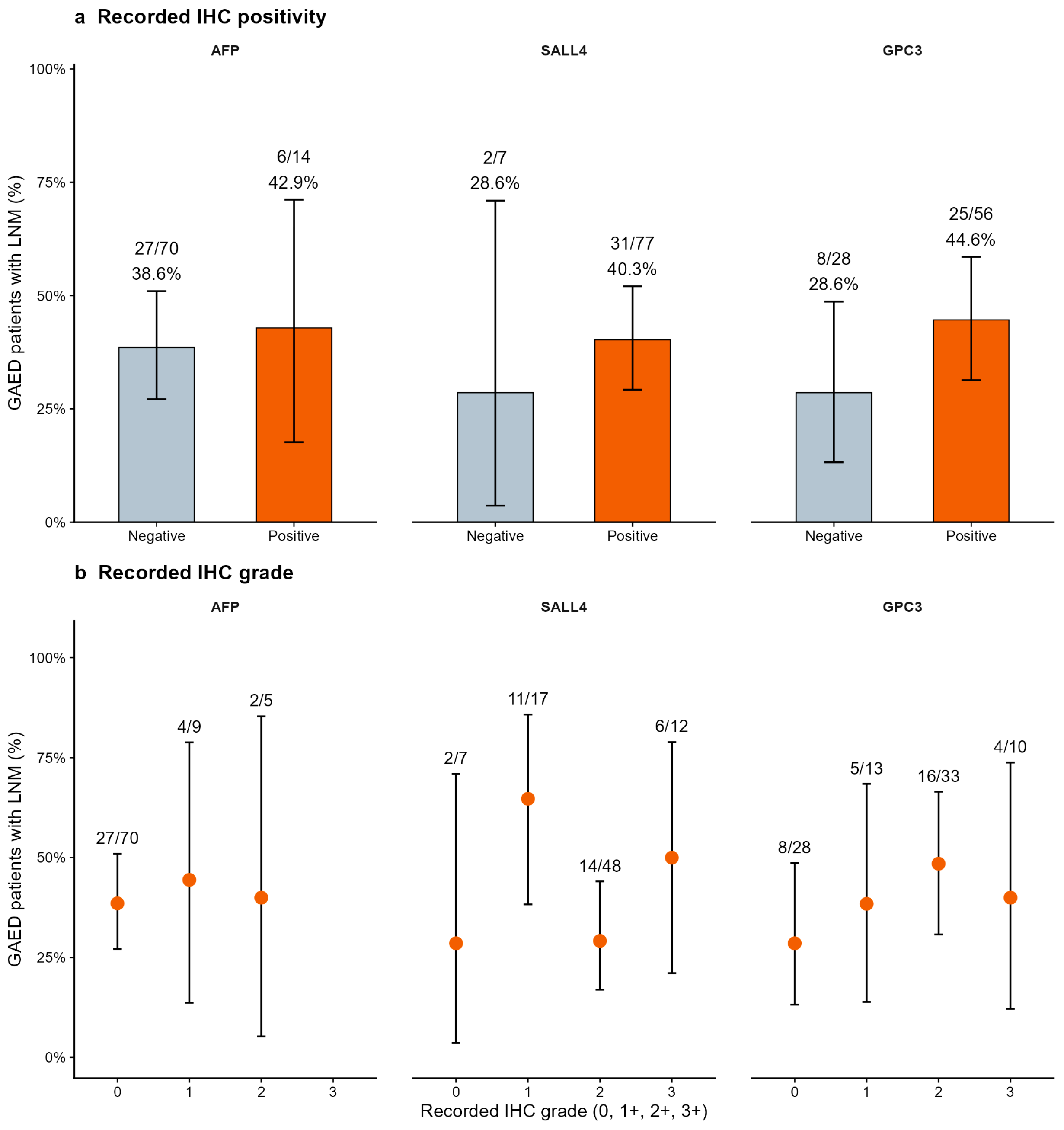
Among GAED patients, AFP, SALL4, and GPC3 were recorded as positive in 14 of 84 (16.7%), 77 of 84 (91.7%), and 56 of 84 (66.7%), respectively. LNM proportions in marker-positive versus marker-negative patients were 42.9% (6/14) versus 38.6% (27/70) for AFP, 40.3% (31/77) versus 28.6% (2/7) for SALL4, and 44.6% (25/56) versus 28.6% (8/28) for GPC3 (Figure 4a). Grade-stratified proportions showed no consistent monotonic increase across the three markers (Figure 4b). For SALL4, LNM proportions at grades 0, 1+, 2+, and 3+ were 28.6%, 64.7%, 29.2%, and 50.0%; corresponding GPC3 proportions were 28.6%, 38.5%, 48.5%, and 40.0%. For AFP, proportions at grades 0, 1+, and 2+ were 38.6%, 44.4%, and 40.0%.
Figure 4.
Lymph node metastasis according to AFP, SALL4, and GPC3 expression in GAED. Note: (a) Lymph node metastasis by AFP, SALL4, and GPC3 immunostaining status. (b) Lymph node metastasis by AFP, SALL4, and GPC3 immunostaining grade. Bars in panel (a) and points in panel (b) show observed LNM proportions; error bars indicate two-sided exact binomial (Clopper–Pearson) 95% confidence intervals.
3.5. Distribution of Involved Lymph Node Stations
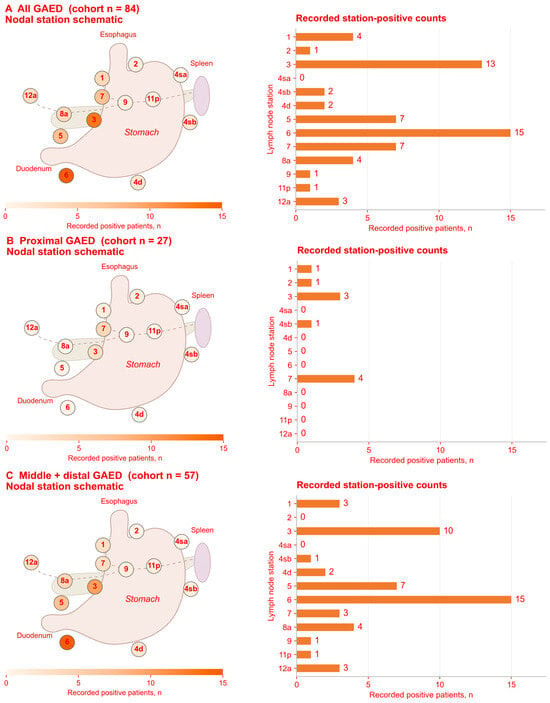
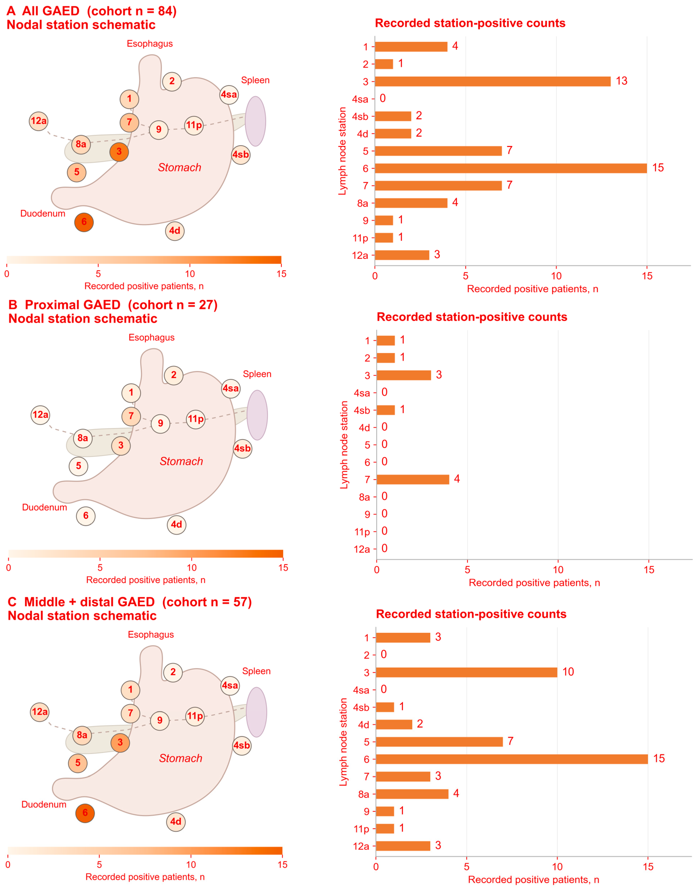
In the overall GAED cohort (n = 84), nodal metastasis was identified at station 6 in 15 patients, station 3 in 13 patients, and stations 5 and 7 in 7 patients each (Figure 5A; Supplementary Table S3A). Among patients with proximal tumors (n = 27), metastasis was identified at station 7 in 4 patients, station 3 in 3 patients, and stations 1, 2, and 4sb in 1 patient each (Figure 5B). The middle-plus-distal group included 57 patients (5 with middle and 52 with distal tumors). In this group, metastasis was identified at station 6 in 15 patients, station 3 in 10 patients, and station 5 in 7 patients (Figure 5C; Supplementary Table S3B). All station-level metastases in the combined group occurred in distal tumors; none of the five middle-location cases had metastasis at any of the examined stations.
Figure 5.
Distribution of lymph node metastasis by tumor location in GAED. (A) Overall cohort (n = 84); (B) proximal tumors (n = 27); and (C) pooled middle and distal tumors (n = 57). Bars show the number of patients with metastasis at each lymph node station. Note: GAED, gastric adenocarcinoma with enteroblastic differentiation.
3.6. Exploratory Prediction and Internal Validation
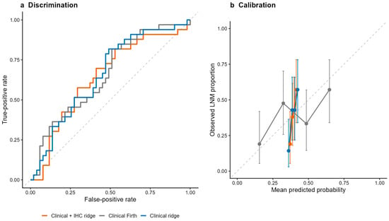
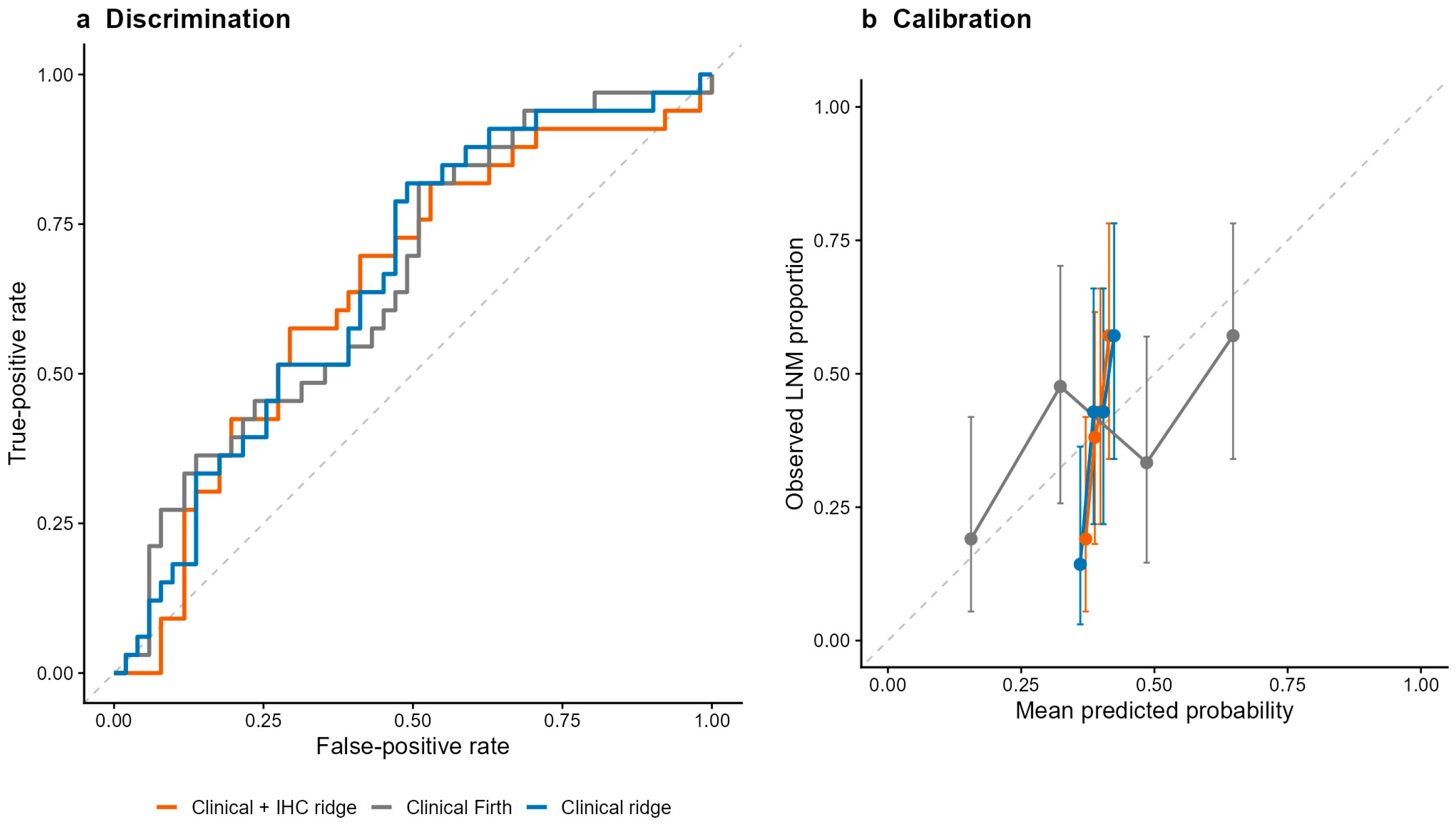
The cross-validated intercept-only comparator had a Brier score of 0.2387. Median Brier scores for the clinicopathological Firth, clinicopathological ridge, and clinicopathological-plus-IHC ridge models were 0.2329, 0.2341, and 0.2369, respectively; their median paired improvements over the comparator were 0.0058, 0.0046, and 0.0017. Median AUCs were 0.647, 0.619, and 0.580, and calibration slopes were 0.493, 2.787, and 1.846, respectively. No incremental improvement in LNM prediction from adding IHC markers to the clinicopathological ridge model was demonstrated in this dataset (Figure 6 and Supplementary Table S4B).
Figure 6.
Exploratory internal validation of LNM prediction models in GAED. (a) ROC curves; (b) calibration plots. Curves and calibration groups are based on each patient’s mean out-of-fold prediction across 20 repetitions; performance metrics in Supplementary Table S4B were calculated separately within each repetition. Calibration points summarize quartiles of mean predicted risk, and error bars show two-sided exact binomial 95% confidence intervals for observed LNM proportions. The diagonal lines indicate chance discrimination in panel (a) and ideal calibration in panel (b). The intercept-only Brier comparator is reported in Supplementary Table S4B. GAED, gastric adenocarcinoma with enteroblastic differentiation; LNM, lymph node metastasis; and ROC, receiver operating characteristic.
4. Discussion
In this study, T1b GAED was associated with a significantly higher risk of LNM than conventional gastric cancer (39.3% vs. 25.8%), and this association remained significant after multivariable adjustment (adjusted OR, 2.48; 95% CI, 1.33–4.62). Female sex, poor differentiation, and vascular tumor thrombus also remained associated with LNM after adjustment, whereas signet-ring cell component, vascular invasion, and perineural invasion did not. The overall adjusted association with tumor location was not statistically significant. Within the GAED group, the presence of a poorly cohesive carcinoma component was associated with a numerically higher LNM rate, although this finding should be interpreted cautiously. In contrast, the staining grades of AFP, SALL4, and GPC3 showed no consistent monotonic increase in observed LNM proportions. Descriptively, stations 7 and 3 had the highest numbers of positive cases in proximal tumors, whereas stations 6, 3, and 5 had the highest counts in the pooled middle-plus-distal group. The positive station entries in the pooled group arose entirely from distal tumors. Exploratory prediction models showed limited discrimination, substantial miscalibration, and only small Brier-score improvements over the intercept-only comparator; no incremental improvement in LNM prediction from adding IHC markers was demonstrated in this dataset. Taken together, these findings suggest that T1b GAED has a greater propensity for nodal spread than conventional gastric cancer and support careful assessment of regional lymph nodes when planning surgical treatment.
The higher rate of LNM observed in T1b GAED is consistent with the aggressive behavior reported for this histological subtype. Murakami et al. found lymph node metastasis in 69% of patients with GAED compared with 38% of those with conventional gastric adenocarcinoma, together with substantially higher rates of lymphatic and venous invasion [7]. Subsequent studies have also shown frequent vascular invasion, nodal involvement, liver metastasis, and unfavorable survival in GAED and other gastric carcinomas with a primitive enterocyte phenotype [8,9,10,11]. Even in early disease, Matsumoto et al. reported that enteroblastic components tended to be located in the deeper mucosa and submucosa and were frequently associated with lymphatic or venous invasion [12]. More recently, gastric carcinomas showing both primitive enterocyte and neuroendocrine differentiation were reported to have particularly frequent nodal metastasis even at the T1 stage [17]. These observations support the concept that the aggressive behavior of GAED develops early and is not confined to advanced tumors.
The biological basis for this behavior is not fully understood. GAED retains features of fetal intestinal differentiation and expresses oncofetal proteins such as SALL4, GPC3, and AFP [7,18]. Molecular studies have shown frequent TP53 alterations, HER2 expression or amplification, and distinct epigenetic changes in tumors with a primitive enterocyte phenotype [9,10]. Recent data also suggest that altered DNA methylation may contribute to the development and aggressive phenotype of these tumors [10]. SALL4 is of particular interest because experimental studies have linked it to tumor cell migration, stemness, epithelial–mesenchymal transition, and activation of TGF-β/SMAD signaling [19,20]. These findings provide possible biological explanations for the early invasive and metastatic potential of GAED, although they do not establish a direct mechanism for lymphatic spread in T1b disease.
From a surgical perspective, the magnitude of nodal risk in the present study is clinically relevant. The 39.3% LNM rate in T1b GAED was higher than the rates generally reported for unselected T1b gastric cancer. In the international Global Gastric Group analysis, LNM was present in 22.1% of Eastern and 27.3% of Western patients with T1b gastric cancer [6]. Current surgical strategies for early gastric cancer are largely based on depth of invasion, nodal status, tumor size, differentiation, and other clinicopathological features rather than on GAED histology itself [3,6]. Our results suggest that enteroblastic differentiation should also be regarded as a high-risk pathological feature when treatment is planned for T1b disease. In patients with confirmed GAED, particularly when additional adverse features are present, standard oncologic gastrectomy with adequate regional lymphadenectomy should be strongly considered. However, the present data do not justify extending lymphadenectomy beyond established anatomical fields solely because of GAED histology. Larger studies are needed before a GAED-specific extent of lymph node dissection can be recommended.
Several additional factors were independently associated with LNM in our cohort. Female sex, poor differentiation, and vascular tumor thrombus remained significant after multivariable adjustment. Signet-ring cell component, vascular invasion, and perineural invasion were associated with LNM in univariable analysis but not after their simultaneous inclusion in the multivariable model. These findings indicate limited evidence of an additional conditional association for these variables in this cohort, rather than absence of biological relevance. Although the middle-versus-proximal contrast had p = 0.021, the overall location test had p = 0.064; the location contrasts should therefore be interpreted cautiously. Similar associations have been reported in conventional early gastric cancer. The G3 Alliance identified female sex, tumor size, distal location, lymphovascular invasion, and moderate or poor differentiation as independent predictors of LNM in T1b disease [6]. Chinese series have also identified female sex, poor differentiation, lymphovascular invasion, and tumor location as relevant factors [21,22]. Poor differentiation probably reflects reduced cellular organization and a more invasive phenotype, whereas vascular tumor thrombus provides direct pathological evidence that tumor cells have acquired access to vascular spaces. The association with female sex has been reproduced in several cohorts, but its biological basis remains uncertain and should not be interpreted as a causal effect. Tumor location also deserves cautious interpretation because associations have varied among studies, probably reflecting differences in gastric lymphatic drainage, histological composition, and patient selection.
The presence of a poorly cohesive carcinoma component may provide additional information. In the GAED subgroup, LNM was observed in 52.6% of patients with a poorly cohesive component compared with 35.4% of those without one. Although this difference was descriptive and the number of patients was small, the direction of the association is consistent with previous studies of early gastric cancer. Fang et al. identified poorly cohesive carcinoma as an independent risk factor for LNM in a Chinese cohort, with a particularly strong association in distal early gastric cancer [21]. Histological heterogeneity is also increasingly recognized in tumors with a primitive enterocyte phenotype. Recent pathological studies have described mixtures of tubular, enteroblastic, hepatoid, and occasionally poorly cohesive patterns within the same tumor [10]. The biological behavior of GAED may therefore depend not only on whether enteroblastic differentiation is present, but also on the composition of the tumor as a whole.
The distribution of metastatic lymph nodes in our cohort provides further information for surgical planning. In proximal GAED, stations 7 and 3 were the most frequently involved, whereas stations 6, 3, and 5 predominated in middle and distal tumors. This pattern broadly follows the known lymphatic drainage of gastric cancer. A Korean series of 4929 patients identified tumor location, depth of invasion, differentiation, and lymphatic invasion as factors associated with both the presence and anatomical distribution of LNM [23]. In a Chinese cohort of 1041 patients with T1b gastric cancer, LNM occurred across almost all nodal stations without a consistent anatomical pattern [24]. Another Chinese cohort of 1140 patients with early gastric cancer reported location-related differences in station involvement, with stations 3, 4, and 6 most commonly involved [25]. Our findings extend these observations to GAED and suggest that tumor location remains useful when evaluating nodal basins in this subtype. Nevertheless, the station-specific numbers in our study were small. The absence of metastasis at a particular station, especially in the proximal subgroup, should therefore not be used as evidence to omit a station from a standard lymphadenectomy. At present, these results are more useful for understanding metastatic patterns and directing careful nodal assessment than for defining a reduced surgical field. Lymph node stations that were not dissected or pathologically examined should not be classified as metastasis-negative, as this could compromise the accuracy of nodal assessment.
The role of AFP, SALL4, and GPC3 requires a separate interpretation. These proteins are primarily markers of enteroblastic or oncofetal differentiation rather than established markers of nodal metastatic risk. SALL4 is highly sensitive for fetal gut differentiation, and GPC3 is frequently expressed in gastric carcinomas with enteroblastic, hepatoid, or AFP-producing phenotypes [7,18,26]. In broader gastric cancer populations, both SALL4 and GPC3 expression have been associated with aggressive clinicopathological features and nodal metastasis [19,26]. However, these associations do not necessarily mean that increasing staining intensity predicts LNM within an already selected GAED population. In our cohort, none of the three markers showed a consistent monotonic increase in observed LNM proportions across staining grades. The literature on GPC3 also illustrates this biological complexity: GPC3-positive gastric carcinomas have been associated with frequent nodal metastasis in some pathological series, whereas other experimental work has suggested a context-dependent metastasis-suppressive role [26,27]. No incremental improvement in LNM prediction from adding these markers to the clinicopathological ridge model was demonstrated in this dataset. This finding should not be interpreted as evidence that these markers lack prognostic value more generally.
The prediction models demonstrated little additional predictive value over the cross-validated intercept-only comparator. Calibration slopes of 0.493, 2.787, and 1.846 indicate overly extreme predictions for the Firth model and overly compressed predictions for the ridge models, despite near-zero calibration intercepts [28]. Thus, average calibration did not establish an adequate spread of individual risks. Repeated cross-validation cannot compensate for only 33 LNM events, and the reported IQRs describe resampling variation rather than confidence intervals. These exploratory models are not ready for clinical decision-making; further development and external validation in larger cohorts are required [29,30].
Several limitations of this study should be acknowledged. First, this was a retrospective study conducted at a single high-volume center. Despite the relatively long study period, GAED remains uncommon, particularly at the T1b stage. The GAED cohort included only 84 patients, with 33 LNM events, which limited the subgroup analyses and the development of prediction models. Small marker categories, particularly the presence of only seven SALL4-negative cases, reduce the precision of estimates and limit the ability to detect incremental predictive value. Second, information on the proportion of the enteroblastic component, the exact depth of submucosal invasion, lymphatic invasion and Borrmann’s classification was not available, which limited further assessment of their associations with nodal metastasis and left potential residual confounding. The multivariable adjustment included invasion-related features that may lie on pathways linking histology to nodal spread; its estimates are conditional associations and do not establish total causal effects. Univariable screening was data-driven and may yield unstable variable selection and overly optimistic inference. In addition, the observed characteristics of lymph node metastasis should be regarded as descriptive and should not be used alone to modify the standard extent of lymphadenectomy. Finally, the prediction models underwent internal cross-validation only. Given the rarity of T1b GAED and the limited number of available cases, reliable external validation could not be performed at this stage. Larger multicenter studies with more detailed pathological information and independent validation cohorts are needed to develop more reliable risk prediction models. Repeated cross-validation does not increase the independent information available from 33 GAED LNM events, and uncertainty in individual risk prediction remains substantial.
5. Conclusions
T1b GAED was associated with higher odds of lymph node metastasis than conventional gastric cancer after adjustment for the selected clinicopathological variables. Adequate lymphadenectomy should be strongly considered for patients with T1b GAED, particularly when additional high-risk features are present. Larger multicenter studies are needed to validate these findings and develop more reliable GAED-specific prediction models.
Supplementary Materials
The following supporting information can be downloaded at: https://www.mdpi.com/article/10.3390/cancers18223663/s1, Figure S1: Lymph node metastasis according to the presence of a poorly cohesive carcinoma component in GAED; Table S1: Descriptive distribution of lymph node metastasis; Table S2: Covariate coding and selection for the single multivariable Firth model; Table S3: Lymph node station assessment and metastasis in GAED; Table S4: Exploratory prediction model specifications and repeated internal validation in GAED (84 patients; 33 LNM events).
Author Contributions
Conceptualization, Z.-L.W., H.R., H.Z., D.A. and C.-G.G.; Methodology, Z.-L.W., H.R. and H.Z.; Software, Z.-L.W., H.R. and H.Z.; Formal analysis, Z.-L.W., H.F., P.-H.N. and H.-X.N.; Investigation, H.F. and P.-H.N.; Resources, H.F. and P.-H.N.; Data curation, Z.-L.W., H.R., H.Z., D.A. and H.-X.N.; Writing—original draft, Z.-L.W.; Writing—review & editing, Z.-L.W., H.R. and P.-H.N.; Visualization, C.-G.G. and D.-B.Z.; Supervision, C.-G.G. and D.-B.Z.; Project administration, D.-B.Z.; Funding acquisition, D.-B.Z. All authors contributed to data collection, data analysis, and writing the original draft. All authors have read and agreed to the published version of the manuscript.
Funding
This study was supported by National High Level Hospital Clinical Research Funding.
Institutional Review Board Statement
This retrospective study was approved by the institutional ethics committee (23/122-3864, approved on 18 April 2023) and conducted in accordance with the Declaration of Helsinki.
Informed Consent Statement
This retrospective analysis used anonymized clinical data collected during routine care. All patients had provided written informed consent for treatment; no additional study-specific informed consent was obtained for this retrospective analysis.
Data Availability Statement
The datasets used and analyzed during the current study are available from the corresponding author on reasonable request.
Acknowledgments
We acknowledge all the authors whose publications are referred to in our article.
Conflicts of Interest
The authors declare no conflict of interest.
References
- Bray, F.; Laversanne, M.; Sung, H.; Ferlay, J.; Siegel, R.L.; Soerjomataram, I.; Jemal, A. Global cancer statistics 2022: GLOBOCAN estimates of incidence and mortality worldwide for 36 cancers in 185 countries. CA Cancer J. Clin. 2024, 74, 229–263. [Google Scholar] [CrossRef] [Scilit] [PubMed]
- Han, B.; Zheng, R.; Zeng, H.; Wang, S.; Sun, K.; Chen, R.; Li, L.; Wei, W.; He, J. Cancer incidence and mortality in China, 2022. J. Natl. Cancer Cent. 2024, 4, 47–53. [Google Scholar] [CrossRef] [Scilit] [PubMed]
- Japanese Gastric Cancer Association. Japanese Gastric Cancer Treatment Guidelines 2021 (6th edition). Gastric Cancer 2023, 26, 1–25. [Google Scholar] [CrossRef] [Scilit] [PubMed]
- Pimentel-Nunes, P.; Libânio, D.; Bastiaansen, B.A.J.; Bhandari, P.; Bisschops, R.; Bourke, M.J.; Esposito, G.; Lemmers, A.; Maselli, R.; Messmann, H.; et al. Endoscopic submucosal dissection for superficial gastrointestinal lesions: European Society of Gastrointestinal Endoscopy (ESGE) Guideline—Update 2022. Endoscopy 2022, 54, 591–622. [Google Scholar] [CrossRef] [Scilit] [PubMed]
- Kim, I.H.; Kang, S.J.; Choi, W.; Seo, A.N.; Eom, B.W.; Kang, B.; Kim, B.J.; Min, B.H.; Tae, C.H.; Choi, C.I.; et al. Korean Practice Guidelines for Gastric Cancer 2024: An Evidence-based, Multidisciplinary Approach (Update of 2022 Guideline). J. Gastric Cancer 2025, 25, 5–114. [Google Scholar] [CrossRef] [Scilit] [PubMed]
- Vos, E.L.; Nakauchi, M.; Gönen, M.; Castellanos, J.A.; Biondi, A.; Coit, D.G.; Dikken, J.L.; D’ugo, D.; Hartgrink, H.; Li, P.; et al. Risk of Lymph Node Metastasis in T1b Gastric Cancer: An International Comprehensive Analysis from the Global Gastric Group (G3) Alliance. Ann. Surg. 2023, 277, e339–e345. [Google Scholar] [CrossRef] [Scilit] [PubMed]
- Murakami, T.; Yao, T.; Mitomi, H.; Morimoto, T.; Ueyama, H.; Matsumoto, K.; Saito, T.; Osada, T.; Nagahara, A.; Watanabe, S. Clinicopathologic and immunohistochemical characteristics of gastric adenocarcinoma with enteroblastic differentiation: A study of 29 cases. Gastric Cancer 2016, 19, 498–507. [Google Scholar] [CrossRef] [Scilit] [PubMed]
- Abe, D.; Akazawa, Y.; Yatagai, N.; Hayashi, T.; Ueyama, H.; Mine, S.; Fukunaga, T.; Nagahara, A.; Yao, T.; Saito, T. Clinicopathological characteristics of gastric adenocarcinoma with enteroblastic differentiation and gastric adenocarcinoma with enteroblastic marker expression. Virchows Arch. 2023, 483, 405–414. [Google Scholar] [CrossRef] [Scilit] [PubMed]
- Wang, Y.; Wei, X.; Ke, B.; Liu, J.; Guo, Y.; Liu, Y.; Chen, Y.; Ding, T.; Wang, Y.; Meng, B.; et al. Exploring the molecular characteristics of the malignant potential of gastric adenocarcinoma with enteroblastic differentiation. Histopathology 2023, 83, 631–646. [Google Scholar] [CrossRef] [Scilit] [PubMed]
- Li, H.Q.; Zheng, L.Q.; Huang, W.T.; Yu, X.B.; Zhang, X.; Lin, L.; Lv, S.S.; Yan, X.Y.; Chen, X.Y. Clinicopathological significance of histological diversity in gastric adenocarcinoma with primitive enterocyte phenotype: A methylation-driven aggressive entity. World J. Gastroenterol. 2025, 31, 108990. [Google Scholar] [CrossRef] [Scilit] [PubMed]
- Gu, L.; Zhang, X.; Ji, S.; Pan, Y.; Shen, Y.; Zhang, M.; Wu, Q.; Yu, W.; Wu, F.; Zheng, Q. Clinicopathological features and prognosis of patients with resectable HAS/GAED: A multicenter nested case-control study from China. Dig. Liver Dis. 2026, 58, 693–699. [Google Scholar] [CrossRef] [Scilit] [PubMed]
- Matsumoto, K.; Ueyama, H.; Matsumoto, K.; Akazawa, Y.; Komori, H.; Takeda, T.; Murakami, T.; Asaoka, D.; Hojo, M.; Tomita, N.; et al. Clinicopathological features of alpha-fetoprotein producing early gastric cancer with enteroblastic differentiation. World J. Gastroenterol. 2016, 22, 8203–8210. [Google Scholar] [CrossRef] [Scilit] [PubMed]
- Rokutan, H.; Ideyama, M.; Ono, S.; Kanazawa, N.; Nonaka, K.; Ishiwata, T.; Arai, T. Practical marker-based stratification of early gastric neoplasms in older adults. Am. J. Clin. Pathol. 2026, 165, aqag002. [Google Scholar] [CrossRef] [Scilit] [PubMed]
- Ishikawa, A.; Nakamura, K. Gastric Adenocarcinoma with Enteroblastic Differentiation Resected through Endoscopic Submucosal Dissection: A Case Report. Case Rep. Gastroenterol. 2024, 18, 68–73. [Google Scholar] [CrossRef] [Scilit] [PubMed]
- Lee, H.R.; Kim, G.H.; Joo, D.C.; Lee, M.W.; Lee, B.E.; Kim, K.B. Gastric adenocarcinoma with enteroblastic differentiation in a 67-year-old man in Korea: A case report. Ewha Med. J. 2024, 47, e28. [Google Scholar] [CrossRef] [Scilit] [PubMed]
- Heinze, G.; Schemper, M. A solution to the problem of separation in logistic regression. Stat. Med. 2002, 21, 2409–2419. [Google Scholar] [CrossRef] [Scilit] [PubMed]
- Wang, B.; Wang, Y.; Cheng, N.; Li, Z.; Wu, S.; Wang, L.; Xu, J.; Wang, Z.; Mu, J.; Wang, H.; et al. Dual-expression (primitive enterocyte phenotype and neuroendocrine differentiation) gastric adenocarcinoma: The unique high-aggressive subtype. Virchows Arch. 2025; Epub ahead of print. [CrossRef] [Scilit]
- Ushiku, T.; Shinozaki, A.; Shibahara, J.; Iwasaki, Y.; Tateishi, Y.; Funata, N.; Fukayama, M. SALL4 represents fetal gut differentiation of gastric cancer, and is diagnostically useful in distinguishing hepatoid gastric carcinoma from hepatocellular carcinoma. Am. J. Surg. Pathol. 2010, 34, 533–540. [Google Scholar] [CrossRef] [Scilit] [PubMed]
- Zhang, L.; Xu, Z.; Xu, X.; Zhang, B.; Wu, H.; Wang, M.; Zhang, X.; Yang, T.; Cai, J.; Yan, Y.; et al. SALL4, a novel marker for human gastric carcinogenesis and metastasis. Oncogene 2014, 33, 5491–5500. [Google Scholar] [CrossRef] [Scilit] [PubMed]
- Zhang, X.; Zhang, P.; Shao, M.; Zang, X.; Zhang, J.; Mao, F.; Qian, H.; Xu, W. SALL4 activates TGF-β/SMAD signaling pathway to induce EMT and promote gastric cancer metastasis. Cancer Manag. Res. 2018, 10, 4459–4470. [Google Scholar] [CrossRef] [Scilit] [PubMed]
- Fang, C.; Shi, J.; Sun, Q.; Gold, J.S.; Xu, G.F.; Liu, W.J.; Zou, X.P.; Huang, Q. Risk factors of lymph node metastasis in early gastric carcinomas diagnosed by WHO criteria in 379 Chinese patients. J. Dig. Dis. 2016, 17, 526–537. [Google Scholar] [CrossRef] [Scilit] [PubMed]
- Du, M.Z.; Gan, W.J.; Yu, J.; Liu, W.; Zhan, S.H.; Huang, S.; Huang, R.P.; Guo, L.C.; Huang, Q. Risk factors of lymph node metastasis in 734 early gastric carcinoma radical resections in a Chinese population. J. Dig. Dis. 2018, 19, 586–595. [Google Scholar] [CrossRef] [Scilit] [PubMed]
- Kim, S.M.; Lee, H.; Min, B.H.; Kim, J.J.; An, J.Y.; Choi, M.G.; Bae, J.M.; Kim, S.; Sohn, T.S.; Lee, J.H. A prediction model for lymph node metastasis in early-stage gastric cancer: Toward tailored lymphadenectomy. J. Surg. Oncol. 2019, 120, 670–675. [Google Scholar] [CrossRef] [Scilit] [PubMed]
- Fei, H.; Ren, H.; Wen, Z.; Sun, C.; Li, Z.; Niu, P.; Li, Z.; Guo, C.; Chen, Y.; Zhao, D. Lymph Node Metastasis Frequency and Development of Predictive Model in T1b Gastric Cancer: A Single-Center Study. Ann. Surg. Oncol. 2026, 33, 1391–1403. [Google Scholar] [CrossRef] [Scilit] [PubMed]
- He, F.J.; Zhang, W.H.; Jin, T.; Chen, X.L.; Zhu, Y.F.; Chen, Z.W.; Li, Z.D.; Chen, Z.H.; Liang, P.P.; Yang, K.; et al. Characteristics of Lymph Node Metastasis in Early Gastric Cancer: A Single-center Retrospective Study. Int. J. Surg. 2025, 112, 7530–7539. [Google Scholar] [CrossRef] [Scilit] [PubMed]
- Ushiku, T.; Uozaki, H.; Shinozaki, A.; Ota, S.; Matsuzaka, K.; Nomura, S.; Kaminishi, M.; Aburatani, H.; Kodama, T.; Fukayama, M. Glypican 3-expressing gastric carcinoma: Distinct subgroup unifying hepatoid, clear-cell, and alpha-fetoprotein-producing gastric carcinomas. Cancer Sci. 2009, 100, 626–632. [Google Scholar] [CrossRef] [Scilit] [PubMed]
- Han, S.; Ma, X.; Zhao, Y.; Zhao, H.; Batista, A.; Zhou, S.; Zhou, X.; Yang, Y.; Wang, T.; Bi, J.; et al. Identification of Glypican-3 as a potential metastasis suppressor gene in gastric cancer. Oncotarget 2016, 7, 44406–44416. [Google Scholar] [CrossRef] [Scilit] [PubMed]
- Van Calster, B.; McLernon, D.J.; van Smeden, M.; Wynants, L.; Steyerberg, E.W. Topic Group ‘Evaluating diagnostic tests and prediction models’ of the STRATOS initiative. Calibration: The Achilles heel of predictive analytics. BMC Med. 2019, 17, 230. [Google Scholar] [CrossRef] [Scilit] [PubMed]
- Zhu, H.; Wang, G.; Zheng, J.; Zhu, H.; Huang, J.; Luo, E.; Hu, X.; Wei, Y.; Wang, C.; Xu, A.; et al. Preoperative prediction for lymph node metastasis in early gastric cancer by interpretable machine learning models: A multicenter study. Surgery 2022, 171, 1543–1551. [Google Scholar] [CrossRef] [Scilit] [PubMed]
- Lee, H.D.; Nam, K.H.; Shin, C.M.; Lee, H.S.; Chang, Y.H.; Yoon, H.; Park, Y.S.; Kim, N.; Lee, D.H.; Ahn, S.H.; et al. Development and Validation of Models to Predict Lymph Node Metastasis in Early Gastric Cancer Using Logistic Regression and Gradient Boosting Machine Methods. Cancer Res. Treat. 2023, 55, 1240–1249. [Google Scholar] [CrossRef] [Scilit] [PubMed]

Disclaimer/Publisher’s Note: The statements, opinions and data contained in all publications are solely those of the individual author(s) and contributor(s) and not of MDPI and/or the editor(s). MDPI and/or the editor(s) disclaim responsibility for any injury to people or property resulting from any ideas, methods, instructions or products referred to in the content. |
© 2026 by the authors. Licensee MDPI, Basel, Switzerland. This article is an open access article distributed under the terms and conditions of the Creative Commons Attribution (CC BY) license.