


Simple Summary
Checkpoint inhibitors help some patients achieve durable tumor control, but they often fail when cancer-specific T cells are absent from tumors, unable to enter tumor tissue, or functionally suppressed. This Perspective evaluates nitric oxide, carbon monoxide, hydrogen sulfide, and molecular hydrogen as potential ways to modify these distinct barriers. It separates the available experimental evidence from preliminary human observations and examines how a gas reaches a tumor, how its local exposure can be measured, and which immune changes would justify combination with checkpoint blockade. The biological response depends on the gas species, concentration, tissue compartment, and duration. A small phase I conference report on intratumoral nitric oxide establishes early clinical feasibility, not anticancer efficacy. The proposed development pathway therefore requires tumor-specific selection, mechanistic controls, gas dosimetry, comparative immunological endpoints, and long-term safety evaluation.
Abstract
Immune checkpoint blockade (ICB) can restore antitumor T-cell activity, but immune-desert, immune-excluded, and T-cell-inflamed yet dysfunctional tumors present different forms of resistance. Nitric oxide (NO), carbon monoxide (CO), and hydrogen sulfide (H2S) are endogenous gasotransmitters that influence vascular function, redox signaling, metabolism, and immunity; molecular hydrogen (H2) is a distinct bioactive gas under investigation. This Perspective evaluates whether gas-based interventions can address specific barriers to ICB and differs from broad surveys of gas nanomedicines by linking tumor immune phenotypes to exposure verification, causal immune readouts, delivery design, and clinical decision gates. Effects depend on concentration, site, source, and duration and may suppress immunity. NO-releasing nanocarriers and multicomponent NO-generating nanoreactors have shown vascular and immune effects in preclinical systems, whereas a ten-patient phase I intratumoral NO report provides preliminary safety and feasibility information without controlled evidence of clinical efficacy. CO and H2S strategies remain predominantly preclinical, and human H2 findings are exploratory. We distinguish biomarkers of gas exposure and target engagement from immune remodeling and patient benefit, discuss molecular imaging and microbiome-related confounding, and propose mechanism-matched combination schedules and safety requirements. Gas-based treatment should therefore be tested against predefined resistance mechanisms rather than presumed to convert all cold tumors into ICB-responsive lesions.
1. Introduction
Immune checkpoint blockade (ICB) has established durable tumor control in subsets of patients across multiple malignancies, but primary and acquired resistance remain common [1]. Antibodies directed against PD-1 or PD-L1 interrupt inhibitory signaling between T cells and tumor or antigen-presenting cells, whereas CTLA-4 blockade primarily modifies T-cell priming and regulation; these interventions can amplify an existing immune response but do not reliably generate missing antitumor immunity de novo [1,2]. Immunologically “cold” tumors lack an effective intratumoral cytotoxic T-cell response and may be immune-desert (few effector T cells within or around the lesion) or immune-excluded (T cells retained at the invasive margin or stroma). “Hot” or T-cell-inflamed tumors contain infiltrating effector cells and inflammatory or interferon-associated programs, although such cells may still be dysfunctional or suppressed [3,4]. The categories form a continuum, not mutually exclusive diagnoses, and must be defined by tissue phenotype rather than PD-L1 expression alone. PD-1/PD-L1 blockade is clinically active in subsets of melanoma, non-small-cell lung cancer, renal-cell carcinoma, and mismatch-repair-deficient malignancies, among others; sensitivity varies substantially within each diagnosis and depends on antigenicity, immune-cell location, and additional resistance pathways [1,4,5]. The focus here is on cold or immune-excluded lesions because insufficient priming, recruitment, or entry limits the substrate on which ICB can act, although dysfunctional inflamed lesions offer a distinct gas-based hypothesis.
The cancer-immunity cycle provides a useful framework for interpreting these barriers. Effective antitumor immunity requires tumor-antigen release, antigen uptake and presentation, T-cell priming, trafficking, infiltration, recognition, and tumor-cell killing [2]. Each step can be disrupted by the tumor microenvironment (TME), where malignant, immune, vascular, stromal, and extracellular-matrix components interact dynamically [6]. Epigenetic suppression of T-cell-recruiting chemokines is one example of how tumors can limit effector-cell access even when potentially immunogenic antigens are present [7]. Consequently, the therapeutic problem is not simply to “activate T cells,” but to modify the ecological conditions that prevent immune activation from being initiated, maintained, or spatially delivered to the tumor bed [4,8].
ICB does not act as a general cytotoxic drug. PD-1 signaling restricts T-cell activation after antigen recognition; blockade can reinvigorate responsive T-cell populations, but effectiveness also depends on antigen presentation, local metabolic support, and access to malignant cells. A gas intervention can, in principle, affect an upstream bottleneck (e.g., perfusion or dendritic-cell priming) or a downstream constraint (e.g., myeloid suppression). The selected endpoint must correspond to that bottleneck: CD8+ cell counts alone do not establish antigen-specific killing or checkpoint-dependent benefit [1,2,5].
Gasotransmitters are relevant to this problem because they operate across several of these biological layers. Nitric oxide (NO), carbon monoxide (CO), and hydrogen sulfide (H2S) are endogenous, diffusible signaling molecules with concentration-dependent effects on vascular biology, mitochondrial function, redox homeostasis, cell death, inflammation, and immunity [9,10]. Recent work has extended this concept toward cancer immunotherapy by developing systems that generate or release gases within tumors rather than exposing the whole organism indiscriminately [11,12]. This is a critical transition. The therapeutic question is not whether a gas is intrinsically pro- or antitumor, but whether a defined exposure can be generated in the correct compartment for a sufficient period to produce a desired pharmacodynamic effect without unacceptable systemic toxicity.
NO is produced by NO synthase (NOS) isoforms and signals through soluble guanylate cyclase (sGC)–cyclic GMP (cGMP) and context-dependent protein S-nitrosylation; both endothelial and immune-cell sources can modify T-cell access or function [13,14,15]. CO arises from heme oxygenase (HO) activity, interacts with heme-containing targets, and can alter mitochondrial and myeloid responses [16,17,18]. H2S is generated through sulfur-metabolic pathways including cystathionine β-synthase (CBS), cystathionine γ-lyase (CSE), and 3-mercaptopyruvate sulfurtransferase (3-MST), with actions involving mitochondrial bioenergetics and protein persulfidation; local reactive sulfur species further complicate causal attribution [9,10,19]. These pathways are neither exclusive nor universally activating. Their effects must be tested at defined tissue concentrations and in the relevant producer and recipient cell populations. Potential NO–CO interactions at heme-containing targets and NO–H2S crosstalk through reactive nitrogen–sulfur chemistry further complicate predictions; these interactions should not be presumed to occur at therapeutically useful levels in every tumor [9,10].
Molecular hydrogen (H2) requires separate terminology. Unlike NO, CO, and H2S, H2 is not generally classified as a canonical endogenous gasotransmitter with a dedicated mammalian enzymatic signaling system. Nevertheless, H2 has been investigated as a bioactive medical gas with redox and immunometabolic effects and is therefore relevant to the broader discussion of gas-based modulation of cancer immunity. In this Perspective, we distinguish canonical gasotransmitter biology from the emerging therapeutic use of H2 and argue that this distinction improves mechanistic clarity.
The phrase “turn cold tumors hot” is used here only as a falsifiable immunological hypothesis, not as an established therapeutic outcome. For immune-desert lesions, such a transition would require new antigen-specific priming and tumor infiltration; for immune-excluded lesions, movement of CD8+ T cells into malignant-cell nests; and for inflamed but dysfunctional lesions, recovery of effector activity. Each would need to precede or accompany a demonstrable improvement in response to ICB, ideally compared with gas alone and ICB alone. Accordingly, a clinically useful framework must define the baseline resistance mechanism, target tissue exposure, delivery platform, mechanism-specific pharmacodynamics, treatment sequence, and safety margin. Gas exposure is not synonymous with immune activation because NO, CO, and H2S may also support tumor growth or suppress immunity in particular biological contexts [9,10,13].
2. Why Gas Biology Maps onto the Biology of ICB Resistance
Gas-based interventions are attractive because several determinants of ICB resistance are simultaneously sensitive to redox, metabolic, and vascular regulation. Abnormal tumor vessels restrict lymphocyte trafficking, create spatially heterogeneous perfusion, and sustain hypoxia. Myeloid-derived suppressor cells and tumor-associated macrophages further constrain T-cell priming and effector function, while chronic antigen exposure drives progressive CD8+ T-cell dysfunction. These processes are interdependent rather than independent barriers. For example, hypoxia can promote suppressive myeloid states and metabolic competition, while poor perfusion limits both immune-cell access and drug delivery [4,6].
The pharmacology of gases is therefore potentially well matched to the TME, but only if exposure is controlled. At low or physiologic concentrations, NO, CO, and H2S can participate in tissue homeostasis, vascular regulation, cytoprotection, and tumor-supportive signaling. At higher or more sustained local concentrations, the same molecules may induce mitochondrial disruption, oxidative or nitrosative stress, ferroptotic or other immunogenic forms of cell death, vascular changes, and inflammatory signaling [9,13]. The relevant dose–response relationship is unlikely to be captured by a single systemic concentration because intratumoral gradients, cellular sources, local scavenging, oxygen tension, and reaction partners determine biological availability.
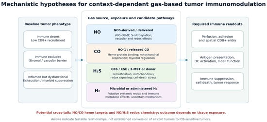
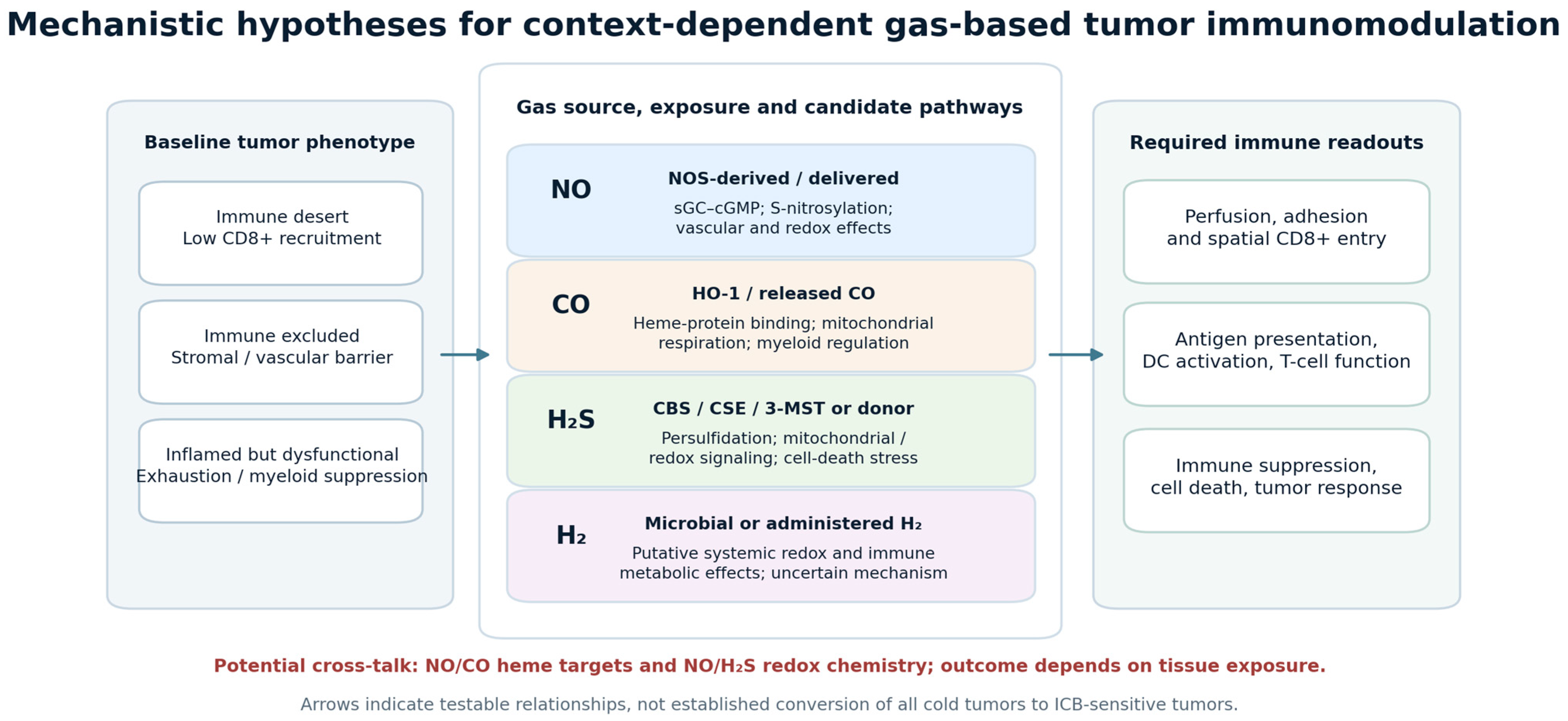
A given gas therefore cannot be assigned a stable “pro-immune” or “anti-immune” label from dose alone. Vascular endothelium, dendritic cells, tumor cells, macrophages, and lymphocytes may experience different local concentrations and react differently at the same time. An increase in circulating gas metabolites may coincide with insufficient tumor penetration, whereas local generation may cause a steep gradient with tissue stress at the release site and no effective immune remodeling elsewhere. Figure 1 distinguishes proposed signaling routes from the immune outcomes that would have to be measured experimentally.
Figure 1.
Conceptual relationship between baseline immune phenotypes, context-dependent NO, CO, H2S, and H2 biology, and testable immune endpoints. Solid arrows indicate hypotheses to test, not demonstrated therapeutic efficacy across tumor types. Abbreviations: CBS, cystathionine β-synthase; CSE, cystathionine γ-lyase; DC, dendritic cell; ICB, immune checkpoint blockade; NOS, nitric oxide synthase; sGC, soluble guanylate cyclase; 3-MST, 3-mercaptopyruvate sulfurtransferase.
This context dependence is a major reason why the historical literature appears contradictory. It is also the strongest argument for delivery systems that treat dose, duration, and location as part of the active therapeutic mechanism rather than as secondary formulation details. Recent nanomedicine reviews emphasize that immune effects depend on localized release and on coupling gas generation to tumor-specific or externally controlled triggers [11,12]. The practical implication is that two interventions described as “NO therapy,” for example, may be biologically dissimilar if one produces a brief systemic rise in NO metabolites and another maintains high intratumoral flux for hours or days. Table 1 compares the agents by their proposed tumor-immune effects, delivery strategies, and distinct levels of translational evidence; it does not imply equivalent clinical maturity.
Table 1.
Comparative immunological rationale and translational maturity of the bioactive gases discussed in this Perspective.
3. Nitric Oxide: The Strongest Translational Case
Among the agents considered, NO has the clearest, although still preliminary, translational sequence: a defined biochemical source and delivery chemistry; tumor-targeted preclinical platforms with mechanistic immune readouts; and the first published conference report of direct intratumoral exposure in patients [13,20,21,22]. “Translational development” here means progress from controlled intratumoral exposure and plausible mechanism through animal combination experiments to initial human dose-finding—not evidence of a clinically effective treatment or regulatory validation. NO can promote angiogenesis, invasion, and tumor-cell survival in some settings, while other exposure conditions generate mitochondrial dysfunction, nitrosative damage, or cell death [13]. The feasibility of safe, reproducible, therapeutically active tissue exposure remains unresolved.
Two classes of NO mechanism must be distinguished. An indirect vascular route involves endothelial NO signaling, changes in vessel maturation, perfusion and adhesion molecule expression, and subsequent T-cell access; low-dose nanocarrier NO has produced vascular normalization and altered immune-cell infiltration in preclinical hepatocellular-carcinoma models [21,27]. A second route involves local NO-dependent signaling in tumor, dendritic, myeloid, or T cells, including sGC–cGMP-mediated responses and protein S-nitrosylation, with potential effects on antigen presentation, T-cell activation, or suppression [13,15]. These routes can coexist and can oppose one another: NO generated by suppressive myeloid cells may impair T-cell responses. Observing more CD8+ cells after NO treatment cannot establish which pathway was causal. Discrimination requires serial perfusion imaging and intratumoral NO measurements together with spatial immune phenotyping, endothelial versus immune-cell perturbations where technically possible, and controls with inactive donors or NO scavenging. Pharmacological sGC inhibition could be informative but is not cell specific and therefore cannot independently prove a T-cell-intrinsic mechanism.
An independent preclinical example of NO-linked immune sensitization is the bioresponsive ALMn nanoreactor (hollow manganese dioxide nanoparticles encapsulating lactate oxidase and L-arginine) developed by Chen et al. [20]. This multicomponent formulation couples intratumoral lactate depletion with NO generation. In mouse tumor models, the treatment was associated with vascular normalization, increased effector-cell infiltration, and improved responses to anti-PD-L1 therapy. However, the formulation changes several aspects of tumor biology at once: lactate depletion and other carrier-dependent reactions could contribute independently to its immune effects. The findings support the activity of a defined NO-generating platform in experimental models, not the conclusion that NO alone accounts for the benefit or that comparable efficacy has been demonstrated clinically.
Multicomponent gas-generating platforms make causal attribution particularly difficult. The contribution of NO in the ALMn system cannot be inferred from combination efficacy alone because lactate metabolism, reactive intermediates, manganese-containing materials, and NO availability are altered concurrently [20]. Mechanistic experiments should compare matched formulations in which NO generation is selectively disabled while lactate depletion and carrier exposure remain as comparable as technically possible; parallel measurements of local NO exposure, perfusion, CD8+ cell localization, and antigen-specific T-cell function would then test the proposed pathway. Inactive-carrier controls and gas-only and ICB-only arms are necessary to separate platform activity from checkpoint sensitization.
Direct intratumoral NO has entered first-in-human investigation, but the evidence level must be specified. A 2026 American Association for Cancer Research (AACR) conference abstract described ten heavily pretreated patients with cutaneous or subcutaneous metastases who received a single intratumoral ultra-high-concentration NO administration at 25,000 or 50,000 ppm [22]. The abstract reported predominantly low-grade treatment-related adverse events and one serious treatment-related episode of hypoxia that resolved. The trial was an uncontrolled, heterogeneous dose-finding study; the published information is a meeting abstract, not a full peer-reviewed efficacy trial. Survival observations and isolated disease-free patients reported in that abstract cannot establish antitumor efficacy, immunological synergy, or causation. The appropriate conclusion is early procedural feasibility and a preliminary tolerability signal requiring independent, protocol-defined verification, not demonstrated human clinical benefit.
NO also illustrates why vascular biology should be incorporated into trial design. Tumor vessels are structurally and functionally abnormal, and interventions that improve perfusion can transiently increase drug delivery and immune-cell access [27]. A gas-based regimen intended to enhance ICB should therefore determine whether vascular changes are beneficial, neutral, or excessive in the relevant dose window. Dynamic contrast imaging, perfusion measurements, hypoxia markers, and paired biopsies could provide more information than systemic nitrite/nitrate measurements alone.
The main translational risk is assuming that all NO delivery systems are interchangeable [14]. Donor chemistry, release rate, oxygen tension, local pH, carrier degradation, and the cellular compartment in which NO is generated will alter downstream chemistry. Clinical development should therefore be platform-specific. The appropriate unit of comparison is not “NO versus control,” but a defined NO exposure profile with verified intratumoral pharmacology.
4. Carbon Monoxide: Immunomodulatory Potential with a More Complex Therapeutic Identity
CO biology is more difficult to translate because endogenous heme oxygenase-1 (HO-1) activity can be associated with cytoprotective and immunosuppressive programs, whereas exogenous CO delivered at higher or locally triggered exposures may produce antitumor stress. This duality is especially relevant in the myeloid compartment. HO-1 is expressed in tumor-associated macrophages and can contribute to transcriptional programs that suppress CD8+ T-cell activity; myeloid HO-1 deletion has enhanced antitumor vaccine responses in preclinical models [16]. This observation argues against a simplistic model in which increasing CO is necessarily immunostimulatory.
Conversely, CO-releasing molecules and nanomaterials can generate mitochondrial and oxidative stress in tumor cells, producing cytotoxicity that may become immunogenic under appropriate conditions [17,18]. A 2023 preclinical platform combined ultrasound-responsive CO generation with reactive-oxygen-species amplification, increased immunogenic cell death, alleviated hypoxia, and improved antitumor immune responses in tumor models [23]. Such systems support the concept that CO can participate in immune priming when it is generated in a spatially controlled therapeutic context.
The translational challenge is attribution. Metal-containing CO-releasing molecules can produce biological effects from released CO, the residual metal-containing fragment, or both. This complicates pharmacology, toxicology, and dose selection [18]. Systemic CO exposure also introduces the obvious risk of carboxyhemoglobin formation and impaired oxygen transport. Consequently, a credible CO immunotherapy program requires direct measurement of CO release kinetics, characterization of carrier metabolites, and evidence that the antitumor immune effect is causally linked to CO rather than to nonspecific carrier toxicity. At present, CO should be viewed as a mechanistically interesting but predominantly preclinical immunomodulatory strategy rather than a near-term clinical counterpart to intratumoral NO.
Longer exposure raises additional concerns beyond acute carboxyhemoglobin formation, including recurrent hypoxic stress, organ-specific accumulation of CO-releasing compounds or metal-containing residues, and delayed toxicities from repeated dosing. Carrier-only controls and extended recovery follow-up are particularly important because an apparent antitumor effect may be partly attributable to off-target carrier injury rather than CO-mediated immune activation.
5. Hydrogen Sulfide: Metabolic Reprogramming, Cell Death, and a Narrow Therapeutic Window
H2S shares the concentration-dependent pharmacology of NO but adds the complexity of sulfur metabolism. Endogenous H2S-generating pathways can support mitochondrial bioenergetics, redox adaptation, angiogenesis, and tumor-cell survival in several cancer contexts, whereas high or rapidly generated H2S can inhibit mitochondrial function, perturb redox balance, and promote cell death [9,10,19]. This produces a potentially exploitable therapeutic window, but also makes simple systemic exposure difficult to interpret.
Recent nanomedicine studies suggest that H2S may be particularly useful when integrated with defined forms of immunogenic tumor-cell death. Luo et al. developed a FeS/gasdermin D (GSDMD)nanoformulation designed to release H2S and Fe2+ while expressing GSDMD, thereby combining PANoptotic and ferroptotic stress. In murine models, the platform increased tumor immunogenicity, modulated indoleamine 2,3-dioxygenase 1 (IDO1)-associated immunosuppression, and enhanced innate and adaptive antitumor responses [24]. The study is important because H2S was not used as an isolated cytotoxin; it was incorporated into a mechanistically coupled system intended to increase immunogenicity and reduce a compensatory immunosuppressive pathway.
For clinical translation, however, H2S delivery must be characterized with greater chemical precision than is typical in proof-of-concept nanomedicine studies. Donor decomposition, sulfide speciation, local oxygen tension, pH, and thiol abundance can all influence effective exposure. Modern H2S-donor chemistry provides increasingly controllable release mechanisms [19], but oncology programs will need validated methods to connect material degradation to intratumoral H2S availability and biological response. Without this bridge, nominal donor dose is a poor surrogate for pharmacologically active gas exposure.
Because sulfur species interconvert rapidly, repeated or sustained release could disturb host sulfur metabolism or damage nonmalignant mitochondria even when the nominal administered donor dose appears low. Evaluations should include sulfur-species selectivity, tissue distribution, serial liver and kidney function, neurological observations where exposure warrants them, and recovery after dosing stops. These are prospective risk-assessment requirements rather than established long-term clinical complication rates for experimental H2S platforms.
6. Molecular Hydrogen: A Distinct Bioactive Gas with Exploratory Human Evidence
H2 is often grouped with NO, CO, and H2S in discussions of gas therapy, but mechanistic precision requires separating it from canonical gasotransmitters. Mammalian cells do not use a dedicated H2-producing enzymatic signaling pathway analogous to NOS, HO, or the principal H2S-generating enzymes. H2 is better considered a bioactive medical gas whose effects may arise through redox and metabolic modulation.
The most frequently cited human oncology study enrolled 55 patients with stage IV colorectal cancer who inhaled H2 for three hours per day while receiving conventional treatment. Changes in peripheral-blood CD8+ T-cell phenotypes were associated with clinical outcomes, including a reduction in terminal PD-1-positive CD8+ T cells and an increase in PD-1-negative CD8+ T cells [25]. The study is hypothesis-generating rather than definitive: it was not designed as a contemporary randomized ICB-combination trial, peripheral immune phenotypes are not equivalent to intratumoral immune reprogramming, and the contribution of concurrent therapy cannot be isolated.
A 2025 preclinical study provided a complementary mechanistic observation. H2 inhalation designed to reproduce exposure generated by gut microbial fermentation of inulin increased circulating CD4+ and CD8+ T cells and inhibited melanoma growth in mice, supporting a possible link between microbiota-derived H2 and immunosurveillance [26]. These findings justify further investigation but do not establish that systemic H2 can reproducibly convert immune-excluded human tumors into ICB-responsive lesions. Future studies should distinguish systemic immunometabolic effects from direct intratumoral gas pharmacology and should incorporate tumor tissue biomarkers whenever feasible.
Microorganisms modify this translational picture in at least three non-equivalent ways. First, engineered tumor-colonizing bacteria have been investigated as potential local delivery vehicles, but achieving reproducible gas production at the lesion and separating microbial immunostimulation from gas effects remain unresolved translational questions [11,12]. Second, gut microbial fermentation can produce H2 and other metabolites, making dietary substrate and microbiota composition potential confounders in studies of inhaled or microbiota-derived H2 [26]. Third, microbial signals can themselves alter myeloid priming, inflammatory tone, and checkpoint responsiveness independently of any gas. Therefore, bacterial-gas delivery studies need colonization measurements and biosafety controls, while systemic H2 studies should control diet, recent antibiotics, microbial activity when feasible, and concurrent anticancer therapy. Microbiome–gas associations alone cannot establish a causal gas-mediated immune mechanism.
7. Delivery and Dosimetry Are Part of the Mechanism
The field has often treated delivery as an engineering problem that follows biological discovery. For therapeutic gases, this order should be reversed. Delivery determines concentration, exposure time, spatial distribution, and often the chemical species that actually reaches the target; these variables determine whether the biological effect is cytoprotective, cytotoxic, immunosuppressive, or immunostimulatory. Therefore, a delivery platform is not merely a carrier. It is part of the pharmacological identity of the intervention.
Current strategies include donor-loaded nanoparticles, catalytic nanomaterials, injectable hydrogels, porous crystalline carriers, stimuli-responsive prodrugs, inhaled formulations, and engineered microorganisms [11,12,28,29]. Metal–organic frameworks are attractive because pore structure and coordination chemistry can be tuned to control loading and release, but biocompatibility, degradation products, and reproducible manufacturing remain central obstacles [29]. Engineered bacteria provide a different solution by converting the tumor into a bioreactor, but they introduce biosafety, containment, host-immunity, and interpatient-colonization questions that are distinct from those of synthetic carriers [11,12].
Tumor selectivity can be engineered through local chemistry or molecular recognition, but the supporting technologies must not be mistaken for validated gas-immunotherapy systems. Cao et al. reported pH-responsive polymer nanocapsules that exploit extracellular acidification associated with glycolysis to accumulate protein cargo at diseased sites, including experimental tumors [30]. An analogous pH-gated gas donor or gas-producing enzyme might improve localization, but its release profile, premature leakage, and activity in nonmalignant inflamed tissues would require separate verification. Antibody–nanoparticle conjugates provide another candidate route by recognizing tumor or stromal antigens [31]; adaptation to NO, CO, or H2S delivery would require retained antibody binding after donor loading, adequate penetration into heterogeneous tumors, and demonstration of active gas release in target cells. Neither paper establishes clinical gas-mediated immune activation. Multiple triggers (low pH, tumor enzymes, reactive oxygen species, ultrasound, light) may also be combined, but increased design complexity increases manufacturing, selectivity, and toxicology demands.
The second unmet need is measurement. Gasotransmitters diffuse rapidly, react with abundant cellular targets, and often exist at concentrations that are difficult to quantify directly in vivo. Advances in molecular probes and imaging now make spatiotemporal monitoring of NO, H2S, and CO increasingly feasible in experimental systems [32]. For clinical development, the ideal program would combine direct or surrogate measures of gas exposure with downstream pharmacodynamic markers. Examples include nitrosative-stress products for NO, carboxyhemoglobin plus local CO-responsive markers for CO, sulfur-metabolite signatures for H2S, and tissue-level immune readouts common to all platforms.
Systemic immunometabolic and intratumoral pharmacological effects can be partly disentangled by a compartment-resolved factorial design. Gas-producing donor, inactive matched carrier, carrier alone, systemic administration, and locally triggered administration should be compared at exposure-matched and, where feasible, route-matched doses. Intratumoral measurements should be paired with plasma and uninvolved-organ exposure, tumor oxygenation and perfusion, and sorted-cell immune-metabolic readouts before tumor size diverges. Isotopically labeled precursors or donor-derived metabolites may help identify chemical origin where analytically validated; cell-restricted NOS or heme-oxygenase perturbations and immune-cell depletion can test dependency, albeit with compensatory biology. Rapid diffusion, endogenous background production, probe consumption, nanocarrier immunogenicity, and discordance between blood and tissue sampling remain substantial technical barriers. Orthogonal chemical assays and spatial imaging are needed before attributing an immune phenotype to the intended gas.
A useful pharmacodynamic panel should reflect the proposed mechanism rather than maximize the number of biomarkers. If the therapeutic claim is improved immune-cell entry, the study should quantify perfusion, endothelial activation, chemokines, and spatial CD8+ T-cell distribution. If the claim is immunogenic cell death, calreticulin exposure, adenosine triphosphate (ATP) release, high-mobility group box 1 (HMGB1)-associated signaling, dendritic-cell activation, and antigen-specific T-cell responses are more informative than total necrosis alone [33]. If the claim is reversal of myeloid suppression, contemporary multidimensional phenotyping should replace the overly simplified assumption that all relevant macrophages can be classified as either M1 or M2; myeloid-derived suppressor-cell states and macrophage heterogeneity should be measured directly [34,35].
These readouts occupy distinct evidentiary levels. Detection of a released gas or characteristic reaction product addresses exposure; a change in a proximal biochemical pathway supports target engagement; tissue immune-cell redistribution or restored antigen-specific function indicates downstream immunomodulation; objective tumor control or survival improvement in a properly controlled study addresses therapeutic efficacy. No single layer substitutes for the next. Table 2 specifies the minimum evidence and principal failure modes for gas exposure, target engagement, immune responses, therapeutic efficacy, and subsequent clinical development.
Table 2.
Proposed development criteria for gas-based cancer immunotherapy.
8. Detection, Quantification, and In Vivo Imaging of Gasotransmitters
Direct measurement is central because administered dose does not identify active gas concentration in a tumor. Electrochemical microsensors, chemiluminescence, electron paramagnetic resonance and appropriately validated mass-spectrometric methods can provide complementary measurements of selected gases or reaction products, but many applications are invasive, spatially restricted, or susceptible to chemical interference. For NO, plasma nitrate/nitrite concentrations or nitrosated products are supportive systemic exposure markers rather than tumor-specific dosimeters. For CO, carboxyhemoglobin monitors systemic binding and safety but not local tumor-cell engagement; CO-responsive chemistry needs independent validation. For H2S, free sulfide, bound sulfane sulfur, and other reactive sulfur pools must be distinguished because sample processing may change speciation. Molecular H2 exposure can be investigated with validated gas analysis of breath, blood, or headspace samples, although none of these alone establishes intratumoral immunomodulation [19,32].
In vivo fluorescence, near-infrared, photoacoustic, magnetic resonance, and radionuclide approaches have been developed to image responses associated with NO, CO, or H2S in animal studies. They differ in penetration depth, temporal resolution, molecular selectivity, and quantitative calibration; a fluorescent increase caused by a reactive probe is not a direct absolute measurement of free gas [32]. For example, responsive magnetic-resonance probes for NO have been demonstrated in vivo, illustrating a route toward deeper-tissue readouts [36]. Nanomaterial-based agents, including ratiometric photoacoustic or activatable magnetic resonance imaging (MRI) systems, may combine anatomical localization and chemical response; prospective testing must establish the fraction of signal attributable to the gas rather than pH, oxygenation, reactive oxygen species, or uneven nanoparticle accumulation.
A translational imaging program should validate probe selectivity against related reactive species, determine detection limits in tumor-like matrices, compare signal with an orthogonal analytical reference, quantify signal changes over time and distance from the delivery site, and assess probe clearance and toxicity. Paired pretreatment and on-treatment biopsy specimens can relate imaging to a tissue-level mechanism, although biopsy sampling error and rapid gas turnover may prevent perfect agreement. Human application remains constrained by imaging-probe manufacturing, regulatory safety, availability of clinically compatible scanners and tracers, tumor accessibility, and lack of standardized absolute in vivo gas calibration. Imaging should therefore be described as an experimental pharmacology tool with potential clinical applications, not as a validated method for selecting patients for gas-based immunotherapy [32,36].
9. A Translational Roadmap for Gas-Based Immunotherapy
The development pathway must start with a defined ICB resistance mechanism. An immune-desert lesion with deficient antigen priming or CXCL9/CXCL10 recruitment requires a strategy capable of increasing antigen presentation and T-cell attraction, potentially following local immunogenic cell death; it should be evaluated by dendritic-cell activation, chemokine induction, and appearance of intratumoral antigen-specific CD8+ T cells. An immune-excluded lesion dominated by abnormal perfusion or stromal retention requires a plausible vascular or trafficking intervention, with paired perfusion imaging, endothelial adhesion markers, and spatial T-cell migration into tumor nests. A lesion with abundant dysfunctional T cells instead requires evidence that local gas exposure alleviates a measured suppressive pathway without further impairing T-cell fitness. No particular gas is established as the best intervention for each subtype; these are selection hypotheses that demand platform-specific testing [3,4,5,20,21,27].
Dose escalation should evaluate a biologically active local exposure range, not simply the maximum tolerated donor dose. Each step should record gas release kinetics or validated exposure surrogates, target pathway modulation, tissue injury, and systemic organ exposure. For NO, an exploratory window could be defined by an exposure-associated change in tumor perfusion or dendritic-cell and spatial CD8+ endpoints without unacceptable hypoxia or inflammatory injury; absence of this prespecified target engagement should interrupt escalation or trigger delivery redesign. The ten-patient intratumoral NO conference report supports only initial human feasibility and tolerability assessment and does not identify an effective immunotherapy dose [22].
ICB sequencing requires direct comparison rather than mechanistic storytelling after the event. For a putative immunogenic-cell-death platform, randomized experimental arms should compare gas alone, ICB alone, gas before ICB, concurrent administration, and, where meaningful, ICB before gas; antigen-presenting-cell and T-cell priming kinetics should be measured before response assessment. For a vascular-normalizing platform, the interval should be chosen from serial vessel/perfusion and T-cell-entry measurements, because any normalization window may be transient [21,27]. Multifunctional platforms require additional component controls to establish whether combined delivery produces an interaction beyond individual agents.
The field should separate systemic and local strategies. Inhaled H2 and intratumoral ultra-high -concentration NO should not be discussed as equivalent examples of gas therapy. They differ in target compartment, exposure magnitude, mechanism, safety profile, and likely regulatory pathway. The same distinction applies to endogenous pathway inhibition versus exogenous gas delivery. Inhibiting HO-1 in immunosuppressive myeloid cells, for example, is conceptually different from delivering a CO-releasing material to tumor cells [16,17,18].
Human immune measurements should be capable of refuting, not merely accommodating, the proposed mechanism. If the claim is improved lymphocyte entry, paired spatially registered biopsies showing no increase in CD8+ cells within tumor nests despite confirmed local exposure would weigh against it, even if peripheral CD8+ counts rise. If the claim is reversal of T-cell dysfunction, the study should assess antigen-specific effector responses, cellular states, and relevant clonal dynamics rather than equating PD-1 loss with restored function [37,38]. For presumed myeloid reprogramming, a prespecified suppressive-cell state and its functional association with T-cell inhibition should be measured. Negative mechanistic readouts may reflect sampling error or inadequate exposure, so both must be quantified before classifying the biological hypothesis as unsupported. Tumor response or survival must then be evaluated separately from molecular target engagement.
Preclinical evidence should not rely on a single rapidly growing subcutaneous, ICB-sensitive mouse tumor. At minimum, claims of checkpoint sensitization should be replicated in a second immunocompetent model with a different baseline CD8+ topology and documented resistance to the selected checkpoint agent. Orthotopic or genetically engineered models are particularly informative when delivery depends on vascular architecture, stromal density, hypoxia, or microbiota; dose–exposure relationships should be measured in each. Randomization, blinded outcome assessment where feasible, biological replicates, prespecified primary endpoints, and survival or durable-rechallenge measures improve interpretability. Studies must include gas-only, ICB-only, vehicle or carrier-only, and inactive-donor arms; multifunctional devices need component deconstruction. Cytokine changes or short-term tumor shrinkage alone are insufficient to establish immune-mediated synergy [12,20,39].
Adverse Effects, Delayed Complications, and Clinical Development Gates
Gas-specific risks must be distinguished from carrier and delivery risks. NO can cause systemic vasodilation or hypotension, methemoglobinemia, nitrosative tissue injury, and procedure-related complications; an episode of treatment-related hypoxia occurred in the small intratumoral phase I report [22]. CO may impair oxygen transport through carboxyhemoglobin and has potential hypoxic consequences even when a carrier is tumor-directed. H2S can inhibit mitochondrial respiration at excessive exposure, while reactive sulfur chemistry complicates prediction of organ toxicity. H2 has a different pharmacological profile, but clinical oncology exposure, long-term safety under repeated administration, and interactions with anticancer regimens remain insufficiently characterized. Local injection, bacterial vectors, and nanoparticles add independent hazards including infection, altered microbial dissemination, immunogenicity, inflammatory reactions, residual metal or polymer fragments, and batch-to-batch release variability [11,12,18,19,29].
Delayed risks should be assessed rather than assumed absent from short follow-up. These include recurrent exposure-related organ injury, persistent vascular or immune perturbation, changes in microbiota composition after live-vector treatment, carrier retention, and potential effects on healthy-tissue repair. Longitudinal preclinical toxicology should examine recovery after repeated dosing, reproductive and neurobehavioral endpoints when indicated by biodistribution, immune memory and autoimmunity signals, and histopathology of exposure-prone organs. Early human studies should prespecify acute and delayed adverse-event windows, gas-specific safety tests, microbiological surveillance for live vectors, and device or product stability criteria. Some risks are mechanistically plausible rather than observed long-term outcomes in gas-immunotherapy patients; that distinction is essential when assessing risk–benefit.
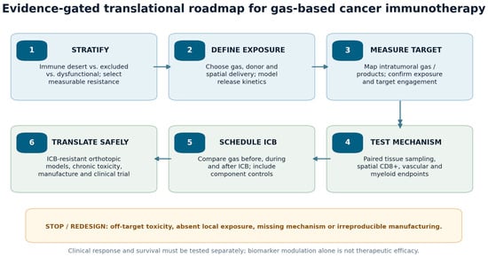
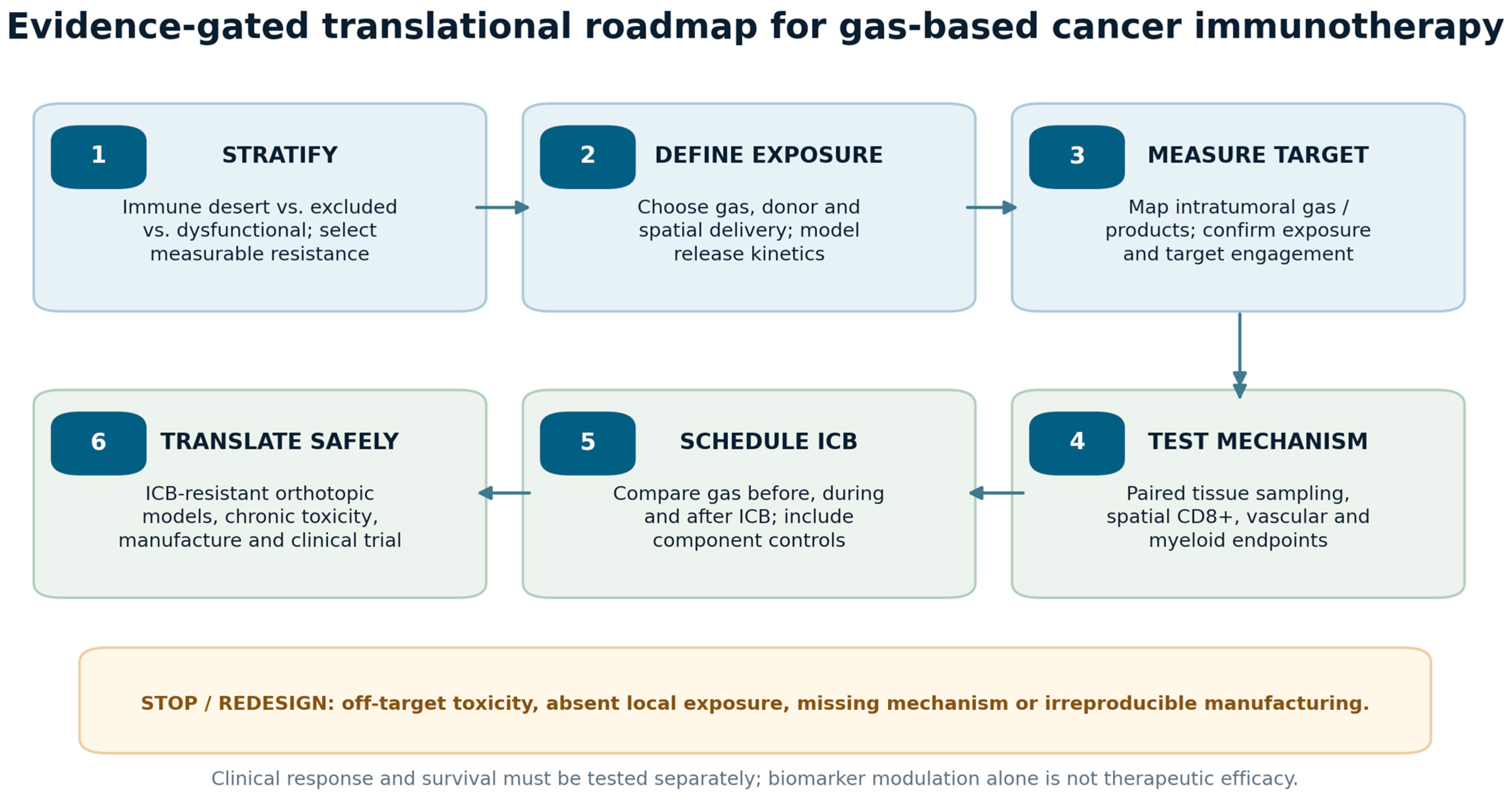
Figure 2 summarizes an evidence-gated progression from tumor stratification to exposure verification, mechanistic tests, combination scheduling, independent safety assessment, and early clinical evaluation. A platform should be redesigned if reproducible local exposure, proposed immune target engagement, acceptable toxicity, or manufacturing consistency cannot be demonstrated. Trials designed primarily for safety must not be cited as demonstrations of antitumor efficacy.
Figure 2.
Proposed translational decision pathway for gas-based immunotherapy. Each stage has a measurable pass/fail criterion; failure of exposure delivery, target engagement, or safety should trigger redesign rather than a presumption of checkpoint synergy. ICB, immune checkpoint blockade.
10. Conclusions
Gas-based approaches should be classified by their mechanism and exposure profile rather than grouped under the assumption that NO, CO, H2S, and H2 all activate antitumor immunity. Immune-desert, immune-excluded, and T-cell-inflamed but dysfunctional tumors represent different therapeutic problems, and a proposed gas intervention is interpretable only when matched to one of these barriers. Local concentration, cellular source, release kinetics, redox conditions, and duration can shift the same gas from cytoprotection or immunosuppression to stress responses and, in selected models, immune activation. A higher intratumoral CD8+ count or detectable gas metabolite by itself does not establish restored antigen-specific immunity or clinical benefit.
NO currently has the most developed preclinical mechanistic and delivery evidence among the gases reviewed, with preclinical studies connecting localized NO delivery to vascular normalization and immune-cell infiltration, and a multicomponent NO-generating platform to improved anti-PD-L1 responses [20,21]. Published human experience is confined here to preliminary phase I conference data reporting feasibility and adverse events in ten patients; controlled antitumor efficacy and ICB synergy have not been established [22]. CO and H2S remain mainly experimental oncology strategies, and H2 clinical immune associations do not identify a proven intratumoral mechanism [16,17,18,19,23,24,25,26]. Translation now hinges on tissue-resolved dosimetry, proof that the gas—not merely its carrier or associated metabolic changes—produces the proposed immune effect, imaging and pharmacodynamic assay validation, reproducible manufacturing, and gas- and platform-specific delayed toxicity assessment. Comparisons with ICB alone and component-controlled interventions in clinically informative resistant models are prerequisites for defensible claims of therapeutic synergy.
Author Contributions
Conceptualization, V.J.C.-S.; writing—original draft preparation, V.J.C.-S.; writing—review and editing, V.J.C.-S., J.F.L.-G. and R.Y.-S.; project administration, V.J.C.-S. All authors have read and agreed to the published version of the manuscript.
Funding
This research received no external funding.
Institutional Review Board Statement
Not applicable.
Informed Consent Statement
Not applicable.
Data Availability Statement
No new data were created or analyzed in this study. Data sharing is not applicable to this article.
Acknowledgments
During the preparation of this manuscript, the authors used ChatGPT, GPT-5.6 (OpenAI), for manuscript restructuring, drafting support, language refinement, and formatting. The authors reviewed and edited the output and take full responsibility for the content of this publication.
Conflicts of Interest
The authors declare no conflicts of interest.
References
- Ribas, A.; Wolchok, J.D. Cancer immunotherapy using checkpoint blockade. Science 2018, 359, 1350–1355. [Google Scholar] [CrossRef] [Scilit] [PubMed]
- Chen, D.S.; Mellman, I. Oncology Meets Immunology: The Cancer-Immunity Cycle. Immunity 2013, 39, 1–10. [Google Scholar] [CrossRef] [Scilit] [PubMed]
- Wu, B.; Zhang, B.; Li, B.; Wu, H.; Jiang, M. Cold and hot tumors: From molecular mechanisms to targeted therapy. Signal Transduct. Target. Ther. 2024, 9, 274. [Google Scholar] [CrossRef] [Scilit] [PubMed]
- Aliazis, K.; Christofides, A.; Shah, R.; Yeo, Y.Y.; Jiang, S.; Charest, A.; Boussiotis, V.A. The tumor microenvironment’s role in the response to immune checkpoint blockade. Nat. Cancer 2025, 6, 924–937. [Google Scholar] [CrossRef] [Scilit] [PubMed]
- Tumeh, P.C.; Harview, C.L.; Yearley, J.H.; Shintaku, I.P.; Taylor, E.J.M.; Robert, L.; Chmielowski, B.; Spasic, M.; Henry, G.; Ciobanu, V.; et al. PD-1 blockade induces responses by inhibiting adaptive immune resistance. Nature 2014, 515, 568–571. [Google Scholar] [CrossRef] [Scilit] [PubMed]
- Binnewies, M.; Roberts, E.W.; Kersten, K.; Chan, V.; Fearon, D.F.; Merad, M.; Coussens, L.M.; Gabrilovich, D.I.; Ostrand-Rosenberg, S.; Hedrick, C.C.; et al. Understanding the tumor immune microenvironment (TIME) for effective therapy. Nat. Med. 2018, 24, 541–550. [Google Scholar] [CrossRef] [Scilit] [PubMed]
- Peng, D.; Kryczek, I.; Nagarsheth, N.; Zhao, L.; Wei, S.; Wang, W.; Sun, Y.; Zhao, E.; Vatan, L.; Szeliga, W.; et al. Epigenetic silencing of TH1-type chemokines shapes tumour immunity and immunotherapy. Nature 2015, 527, 249–253. [Google Scholar] [CrossRef] [Scilit] [PubMed]
- Hegde, P.S.; Chen, D.S. Top 10 Challenges in Cancer Immunotherapy. Immunity 2020, 52, 17–35. [Google Scholar] [CrossRef] [Scilit] [PubMed]
- Szabo, C. Gasotransmitters in cancer: From pathophysiology to experimental therapy. Nat. Rev. Drug Discov. 2016, 15, 185–203. [Google Scholar] [CrossRef] [Scilit] [PubMed]
- Ballerini, G.; Balboni, A.; Garlatti, V.; Incerti, M.; Sica, A.; Consonni, F.M. Gasotransmitters bridging tumor biology and immunity: From pathophysiological insights to therapeutic potential. Front. Immunol. 2026, 17, 1671203. [Google Scholar] [CrossRef] [Scilit] [PubMed]
- Nguyen, V.-A.T.; Huang, C.-C.; Chen, Y. Gas-based therapeutics and delivery platforms in cancer immunotherapy. Adv. Drug Deliv. Rev. 2026, 229, 115746. [Google Scholar] [CrossRef] [Scilit] [PubMed]
- Sun, M.; Wang, T.; Zhu, Y.; Ling, F.; Bai, J.; Tang, C. Gas immnuo-nanomedicines fight cancers. Biomed. Pharmacother. 2024, 180, 117595. [Google Scholar] [CrossRef] [Scilit] [PubMed]
- Fukumura, D.; Kashiwagi, S.; Jain, R.K. The role of nitric oxide in tumour progression. Nat. Rev. Cancer 2006, 6, 521–534. [Google Scholar] [CrossRef] [Scilit] [PubMed]
- Sinha, B.K. Can Nitric Oxide-Based Therapy Be Improved for the Treatment of Cancers? A Perspective. Int. J. Mol. Sci. 2023, 24, 13611. [Google Scholar] [CrossRef] [Scilit] [PubMed]
- Navasardyan, I.; Bonavida, B. Regulation of T Cells in Cancer by Nitric Oxide. Cells 2021, 10, 2655. [Google Scholar] [CrossRef] [Scilit] [PubMed]
- Alaluf, E.; Vokaer, B.; Detavernier, A.; Azouz, A.; Splittgerber, M.; Carrette, A.; Boon, L.; Libert, F.; Soares, M.; Le Moine, A.; et al. Heme oxygenase-1 orchestrates the immunosuppressive program of tumor-associated macrophages. JCI Insight 2020, 5, e133929. [Google Scholar] [CrossRef] [Scilit] [PubMed]
- Vo, T.T.T.; Vo, Q.C.; Tuan, V.P.; Wee, Y.; Cheng, H.-C.; Lee, I.-T. The potentials of carbon monoxide-releasing molecules in cancer treatment: An outlook from ROS biology and medicine. Redox Biol. 2021, 46, 102124. [Google Scholar] [CrossRef] [Scilit] [PubMed]
- Mansour, A.M.; Khaled, R.M.; Ferraro, G.; Shehab, O.R.; Merlino, A. Metal-based carbon monoxide releasing molecules with promising cytotoxic properties. Dalton Trans. 2024, 53, 9612–9656. [Google Scholar] [CrossRef] [Scilit] [PubMed]
- Corvino, A.; Caliendo, G. Hydrogen Sulfide (H2S)-Donor Molecules: Chemical, Biological, and Therapeutical Tools. Int. J. Mol. Sci. 2024, 25, 7932. [Google Scholar] [CrossRef] [Scilit] [PubMed]
- Chen, J.; Bu, C.; Lu, Y.; Peng, X.; Yu, J.; Ding, X.; Yuan, P.; Hong, S. Bioresponsive nanoreactor initiates cascade reactions for tumor vascular normalization and lactate depletion to augment immunotherapy. Biomaterials 2025, 317, 123100. [Google Scholar] [CrossRef] [Scilit] [PubMed]
- Sung, Y.-C.; Jin, P.-R.; Chu, L.-A.; Hsu, F.-F.; Wang, M.-R.; Chang, C.-C.; Chiou, S.-J.; Qiu, J.T.; Gao, D.-Y.; Lin, C.-C.; et al. Delivery of nitric oxide with a nanocarrier promotes tumour vessel normalization and potentiates anti-cancer therapies. Nat. Nanotechnol. 2019, 14, 1160–1169. [Google Scholar] [CrossRef] [Scilit] [PubMed]
- Meirovitz, A.; Lisi, S.; Sheva, K.; Greenberg, D.; Jett, J. Abstract 304: A phase 1 study of intratumoral ultra-high concentration nitric oxide (UNO) gas injection in cutaneous and subcutaneous solid tumor metastases. Cancer Res. 2026, 86. [Google Scholar] [CrossRef] [Scilit]
- Wang, Q.; He, Z.; Zhang, R.; Du, J.; Zhu, L.; Li, X.; Yang, H.; Miao, Y.; Li, Y. Carbon monoxide-based immunogenic cell death amplifier remodels the hypoxic microenvironment for tumor sono-immunotherapy. Chem. Eng. J. 2023, 480, 148269. [Google Scholar] [CrossRef] [Scilit]
- Luo, Y.; Linghu, M.; Luo, X.; Li, D.; Wang, J.; Peng, S.; Ma, Y. Remodeling tumor immunosuppressive microenvironment through dual activation of immunogenic panoptosis and ferroptosis by H2S-amplified nanoformulation to enhance cancer immunotherapy. Acta Pharm. Sin. B 2025, 15, 1242–1254. [Google Scholar] [CrossRef] [Scilit] [PubMed]
- Akagi, J.; Baba, H. Hydrogen gas restores exhausted CD8+ T cells in patients with advanced colorectal cancer to improve prognosis. Oncol. Rep. 2019, 41, 301–311. [Google Scholar] [CrossRef] [Scilit] [PubMed]
- Pascal-Moussellard, V.; Alcaraz, J.-P.; Tanguy, S.; Salomez-Ihl, C.; Cinquin, P.; Boucher, F.; Boucher, E. Molecular hydrogen as a potential mediator of the antitumor effect of inulin consumption. Sci. Rep. 2025, 15, 11482. [Google Scholar] [CrossRef] [Scilit] [PubMed]
- Jain, R.K. Normalizing Tumor Microenvironment to Treat Cancer: Bench to Bedside to Biomarkers. J. Clin. Oncol. 2013, 31, 2205–2218. [Google Scholar] [CrossRef] [Scilit] [PubMed]
- Tang, Y.; Li, Q.; Zhou, Z.; Bai, H.; Xiao, N.; Xie, J.; Li, C. Nitric oxide-based multi-synergistic nanomedicine: An emerging therapeutic for anticancer. J. Nanobiotechnol. 2024, 22, 674. [Google Scholar] [CrossRef] [Scilit] [PubMed]
- Pinto, R.V.; Pinto, M.L.; Serre, C. Gasotransmitters in Modern Medicine: Promises and Challenges in the Use of Porous Crystalline Carriers. Adv. Healthc. Mater. 2025, 14, e2404553. [Google Scholar] [CrossRef] [Scilit] [PubMed]
- Cao, Z.; Cheng, X.; Lu, X.; He, Q.; Dai, Q.; Zhang, L.; Zhang, X.; Harding, J.; Xu, S.; van Dam, R.M.; et al. Universal diseased-site targeting via glycolysis-driven lactic acid gradient. Sci. Adv. 2026, 12, eaeb4570. [Google Scholar] [CrossRef] [Scilit] [PubMed]
- Fattahi, A.S.; Jafari, M.; Farahavar, G.; Haghighi, E.; Abolmaali, S.S.; Tamaddon, A.M. Antibody-nanoparticle conjugates for precision targeting of immunosuppressive tumor microenvironment. Crit. Rev. Oncol. Hematol. 2026, 221, 105219. [Google Scholar] [CrossRef] [Scilit] [PubMed]
- Lu, C.; Zhang, C.; Zhang, X.-B.; Song, G. In vivo imaging of gasotransmitters. Nat. Rev. Bioeng. 2026, 4, 653–674. [Google Scholar] [CrossRef] [Scilit]
- Galluzzi, L.; Buqué, A.; Kepp, O.; Zitvogel, L.; Kroemer, G. Immunogenic cell death in cancer and infectious disease. Nat. Rev. Immunol. 2017, 17, 97–111. [Google Scholar] [CrossRef] [Scilit] [PubMed]
- Veglia, F.; Sanseviero, E.; Gabrilovich, D.I. Myeloid-derived suppressor cells in the era of increasing myeloid cell diversity. Nat. Rev. Immunol. 2021, 21, 485–498. [Google Scholar] [CrossRef] [Scilit] [PubMed]
- Mantovani, A.; Marchesi, F.; Malesci, A.; Laghi, L.; Allavena, P. Tumour-associated macrophages as treatment targets in oncology. Nat. Rev. Clin. Oncol. 2017, 14, 399–416. [Google Scholar] [CrossRef] [Scilit] [PubMed]
- Lu, C.; Liao, S.; Chen, B.; Xu, L.; Wu, N.; Lu, D.; Kang, H.; Zhang, X.-B.; Song, G. Responsive probes for in vivo magnetic resonance imaging of nitric oxide. Nat. Mater. 2025, 24, 133–142. [Google Scholar] [CrossRef] [Scilit] [PubMed]
- Wherry, E.J.; Kurachi, M. Molecular and cellular insights into T cell exhaustion. Nat. Rev. Immunol. 2015, 15, 486–499. [Google Scholar] [CrossRef] [Scilit] [PubMed]
- Philip, M.; Schietinger, A. CD8+ T cell differentiation and dysfunction in cancer. Nat. Rev. Immunol. 2022, 22, 209–223. [Google Scholar] [CrossRef] [Scilit] [PubMed]
- Ji, P.; Yang, K.; Xu, Q.; Qin, G.; Zhu, Q.; Qian, Y.; Yao, W. Mechanisms and Application of Gas-Based Anticancer Therapies. Pharmaceuticals 2023, 16, 1394. [Google Scholar] [CrossRef] [Scilit] [PubMed]

Disclaimer/Publisher’s Note: The statements, opinions and data contained in all publications are solely those of the individual author(s) and contributor(s) and not of MDPI and/or the editor(s). MDPI and/or the editor(s) disclaim responsibility for any injury to people or property resulting from any ideas, methods, instructions or products referred to in the content. |
© 2026 by the authors. Licensee MDPI, Basel, Switzerland. This article is an open access article distributed under the terms and conditions of the Creative Commons Attribution (CC BY) license.