


Simple Summary
Artificial intelligence (AI) can classify kidney masses on scans, but high test accuracy does not necessarily improve patient care. We reviewed why promising models have so far produced little evidence of clinical benefit. Most were developed in patients whose masses were removed or biopsied, while small uncertain masses managed with surveillance are underrepresented. Independent and prospective testing in typical patients remains limited. A few studies suggest that AI helps radiologists read scans more accurately, but none measured whether patients were then managed better. Regulatory evidence is limited, European urology guidelines discuss AI only as an emerging technology, and hospitals face cost, information-technology, workflow, trust, and interpretability barriers. The next step is a prospective, multicenter study of one clearly defined use within the current pathway, including biopsy and surveillance. Until such evidence shows clinical benefit and oncologic safety, AI should support radiologists and urologists rather than determine treatment.
Abstract
Artificial intelligence (AI) models for solid renal mass characterization show promising discrimination in multicenter and some prospective test sets, yet little published evidence supports their routine clinical use. We examine four translation gaps. Independent, representative validation remains limited, and most studies use resected, pathologically confirmed masses, underrepresenting small indeterminate lesions managed by biopsy or surveillance. Recent multicenter models, including RenalCLIP and MPANet, broadened tasks and external testing but remain retrospective. Four retrospective reader experiments suggest that AI assistance can improve readers’ accuracy or consistency, but no study identified measured effects on management or patient outcomes. Regulatory evidence is limited: one renal tool reported by its manufacturer as certified under the European Union Medical Device Regulation rests on a single-center retrospective reader study of detection and reporting, and no characterization device was identified in the U.S. Food and Drug Administration AI-enabled device list. The 2026 European Association of Urology guidelines discuss AI as an emerging technology without a graded recommendation. Cost, information technology integration, trust, and interpretability further restrict adoption. We propose a roadmap built around one intended use, the small indeterminate solid mass considered for biopsy or surveillance, with prespecified thresholds, target-specific reference standards, and paired management and safety endpoints.
1. Introduction
Incidental detection of renal masses has risen with the growing use of cross-sectional imaging. The proportion of benign histology among masses resected on suspicion of malignancy is inversely related to size: pooled US surgical series report benign pathology in about 17–21% of masses between 1 and 4 cm and in about 40% of those smaller than 1 cm [1], and in a single-institution series of 2770 patients, 46% of tumors smaller than 1 cm were benign [2]. Benign histology does not by itself show that treatment was inappropriate, because symptoms, lesion behavior and the information available when the decision was made also count, but many of these patients are exposed to the morbidity of surgery or ablation for lesions that, in retrospect, did not require it. Computed tomography (CT) and magnetic resonance imaging (MRI) cannot reliably separate benign from malignant or indolent from aggressive disease at the point of decision [3]. Percutaneous biopsy performs well, with a pooled diagnostic yield of 90% and a change in management in 39% of patients in a 2026 meta-analysis of 103 studies [4], yet its use before surgery is limited: in a French national registry of 9283 surgically treated patients, only 17% of masses had been biopsied beforehand [5]. Because this denominator excludes patients whose biopsy result diverted them from surgery, the figure describes preoperative practice in that cohort rather than population-wide underuse. Surveillance, Epidemiology, and End Results (SEER) data from 2000 to 2021 show partial nephrectomy overtaking radical nephrectomy and a gradual rise in non-surgical management [6]. Practice has adapted to this diagnostic uncertainty; it has not resolved it.
Artificial intelligence (AI) has been studied extensively as a way to reduce this uncertainty [7,8]. The field has been surveyed repeatedly, in reviews of imaging-based deep learning in kidney disease [9] and of AI in renal imaging [10], in two 2026 reviews of applications across the renal mass pathway and the kidney cancer spectrum [11,12], in a review of multimodal AI in urological oncology [13], and in a registered protocol for a systematic review and meta-analysis of the diagnostic accuracy of AI for characterizing renal masses (PROSPERO CRD42024529929) [14]. These reviews catalog tasks and reported performance and list barriers to clinical use, such as scarce prospective validation, data heterogeneity, limited interpretability and the scarcity of studies in non-resected masses [9,10,12,13]. To our knowledge, none appraises the renal mass characterization evidence against the public regulatory record and the guideline text, separates standalone model performance from assisted reading and from effects on management, or specifies the study that would be needed to move a tool into practice. That is the object of this review: why many well-performing published models have so far generated little evidence that supports their routine clinical use. We examine four gaps, in validation, regulation, guidelines and implementation (Figure 1), argue that they are interdependent rather than strictly sequential, and propose a roadmap anchored in one concrete intended use, with evidence of clinical utility at its center.
Figure 1.
The translation gap in artificial intelligence-based renal mass characterization (conceptual schematic; positions, box sizes, and arrows are illustrative, not quantitative). Clinical utility, meaning evidence that using a tool improves management without compromising oncologic safety, is the central missing evidence; arrows indicate that evidence, or its absence, in one domain affects the others and the central question. Validation: cohorts are predominantly retrospective and pathology-selected. About 90% of 30 studies in a 2020 methodological review lacked independent external validation, a historical benchmark [15]; recent multicenter studies added external and prospective test sets, but surveillance-only populations remain unrepresented in the pivotal studies [16,17,18,19]. Regulation: one renal lesion detection and reporting tool has manufacturer-reported certification under the EU MDR, and its published evidence is limited to a single-center retrospective reader study [20,21]; no characterization device was identified in the FDA list searched in September 2026 [22,23]. Guidelines: the EAU guidelines discuss AI among emerging technologies without a graded recommendation [24]. Implementation: cost and IT integration are major adoption barriers where radiology AI is deployed [25]; assisted-reader evidence is limited [16,20,26,27], and effects on management and post-deployment performance remain unevaluated in the studies reviewed (Supplementary Material File S1). AI, artificial intelligence; EAU, European Association of Urology; FDA, U.S. Food and Drug Administration; IT, information technology; MDR, Medical Device Regulation.
2. Literature Search and Evidence Selection
This narrative review searched PubMed/MEDLINE for records published from 1 January 2015 through 24 September 2026, the review cutoff. The core search combined renal mass terms (kidney neoplasms [MeSH], renal mass, renal tumor, renal cell carcinoma), AI terms (artificial intelligence [MeSH and free text], deep learning, machine learning, radiomics) and characterization terms (characterization, differentiation, classification, benign). Targeted searches addressed external or prospective validation, AI-assisted reader studies, clinical-utility and management-impact studies, regulatory and commercial status, implementation and economic evaluation, prior reviews, and post-treatment recurrence. No language or publication-type filters were applied, except for the search for prior reviews. The exact search strings, the number of records retrieved by each string when the searches were re-run on 29 October 2026 with the same publication-date limits, the selection criteria, and the screening of the targeted searches that underpin statements about absent evidence are provided in Supplementary Material File S1. Consensus was used only as a supplementary discovery tool, reference lists of relevant reviews were screened, and key earlier studies were added when needed. The review focused on solid renal masses; cystic renal masses and AI applications centered on Bosniak classification were outside its scope. Studies whose populations included cystic lesions are identified as mixed populations wherever their results are reported. A.Z.-M. selected the evidence; M.A.A.-P. checked the selection and the data extracted from the pivotal studies (Table 1 and Table 2). The evidence matrix records population, task and model output, reference standard, validation design, reader component, calibration and decision-curve analyses, and outcome level (Table 1). Focused full-publication checks of confidence intervals, class and subgroup denominators, operating thresholds, calibration and decision curves covered Xiong et al., Dai et al., RenalCLIP and MPANet, including their supplementary information and, for RenalCLIP, linked Source Data (Table 2). For Zhou et al. and Han et al., the performance figures reproduced here were verified against the indexed abstracts; additional statistics and calibration or decision-curve analyses were not extracted from the complete publications. These items are labeled “not extracted”, without implying that they were not published. Statements that an item was not reported are limited to the materials actually checked, as specified in Table 2 and Supplementary Material File S1. Statements about the absence of evidence are bounded by the sources searched and the review cutoff. We prioritized systematic reviews, multicenter or prospective studies, regulatory analyses, and implementation studies; single-center development studies without external testing were cited only as examples. The 2026 European Association of Urology (EAU) Renal Cell Carcinoma Guidelines were read in full. Regulatory status was checked in the U.S. Food and Drug Administration (FDA) list of AI-enabled medical devices, a published review of urology-specific FDA-cleared devices, peer-reviewed studies, and manufacturer information. Because the FDA states that its AI list is not comprehensive, absence from that list was treated as no device identified, not proof that no authorization exists; no comparable public register exists for Conformité Européenne (CE)-marked AI products, so CE status was taken from manufacturer statements and published censuses. Claude Opus (Anthropic; exact model version not recorded; accessed September–October 2026), OpenAI Codex (OpenAI; exact model version not recorded; accessed September–October 2026), and Consensus (Consensus NLP; web-platform version not displayed; accessed September–October 2026) were used to identify candidate records, document the re-run searches and prepare a first-pass screening log, and support drafting, language editing, and table and figure formatting. These tools did not make inclusion decisions or independently verify sources. The authors verified all AI-assisted content against the original publications and public documents.
Table 1.
Evidence matrix for the pivotal studies: population, task, reference standard, validation design, reader component, and highest outcome level evaluated.
Table 2.
Reported performance of AI models for renal mass characterization, grouped by task.
3. Current Landscape of AI in Renal Mass Characterization
Before asking why AI has not translated, it is worth establishing that discrimination is no longer the only limitation. Xiong et al. developed multiphase CT deep learning models using nephrectomy cohorts from five hospitals and evaluated them in internal, external, public (The Cancer Imaging Archive, TCIA), and prospective test sets. Their 4557 patients included 388 from TCIA, and the cohorts were mixed: complex renal cysts formed part of the benign class, and cystic lesions accounted for 9.5–18.6% of the cohorts, so the whole-cohort estimates below do not refer to solid masses alone [16]. The malignancy model reached an area under the receiver operating characteristic curve (AUC) of 0.853 externally and 0.871 prospectively, exceeding the mean performance of seven experienced radiologists in the prospective set; the aggressiveness model, which separated benign or indolent masses from aggressive tumors defined by histological subtype, high nuclear grade, necrosis, sarcomatoid differentiation or invasion of major veins or perinephric tissue, reached 0.763 and 0.783, respectively [16]. In the solid T1 subgroups, the malignancy AUCs were 0.893 internally, 0.843 externally, and 0.884 prospectively, and the aggressiveness AUCs 0.780, 0.670, and 0.703; in solid T1a masses, aggressiveness discrimination fell to 0.619 in the external set [16]. Three of the four hospitals in the prospective set had contributed development data, so this component assessed temporal performance more directly than geographic transportability. Dai et al.’s multiphase CT algorithm for resected masses of 3 cm or less achieved AUCs of 0.80 versus 0.84 for urological radiologists in a five-hospital external set and 0.87 versus 0.92 in a single-hospital prospective set [17]. The differences were not statistically significant, but equivalence or non-inferiority was not prespecified and the model estimates were numerically lower. The two studies share a research program, authors, and recruitment sites and should not be read as fully independent confirmations; RenalCLIP, discussed below, also shares senior authors with them, and three of its five external test centers were development sites in Xiong et al. [16,18].
Two studies published in 2026 broadened the evidence base. RenalCLIP was developed with 27,866 CT scans from 8809 patients who underwent nephrectomy or biopsy (6867 of them for pretraining at four centers) and was evaluated on anatomical, diagnostic, and prognostic tasks. For malignancy, the AUC was 0.856 in the internal test cohort of 100 patients and 0.841 in a combined external cohort of 1017 patients from five centers not used for pretraining; it fell to 0.680 in TCIA, where benign lesions made up 3.3% of cases, and was 0.762 in external small renal masses of 4 cm or less [18]. The authors considered its accuracy in small renal masses insufficient to guide clinical decisions independently [18]. MPANet combined multiphase CT with age and sex in 1688 surgically treated patients to classify four tumor groups: clear cell renal cell carcinoma (RCC), papillary RCC, oncocytic neoplasms (chromophobe RCC and oncocytoma combined), and fat-poor angiomyolipoma; purely cystic masses were excluded [19]. Because chromophobe RCC and oncocytoma share one class, the model does not attempt to distinguish a malignant from a benign oncocytic tumor, which is the distinction that matters most for avoiding surgery on oncocytoma. Its output therefore does not translate directly into a benign-versus-malignant decision, and its macro-average AUCs of 0.850 internally and 0.811 and 0.813 in two external cohorts summarize multiclass discrimination rather than malignancy prediction. The combined oncocytic class had the lowest per-class AUCs (0.788, 0.737, and 0.709 in the three test sets), but these values measure how well the combined group was separated from the other three classes, not how well chromophobe RCC was separated from oncocytoma [19]. Overall accuracy was 73.2% internally in the published performance table and 62.5% and 71.0% externally; in the internal set, readers achieved 43.6–62.4% (four CT readers) and 49.5–52.5% (two MRI readers using the clear cell likelihood score) [19]. That internal set comprised 101 patients selected because they had both CT and MRI, so the split was not random and the reader comparison is imprecise. Both studies were retrospective. MPANet included pathologically confirmed tumors, and RenalCLIP’s authors acknowledged selection and spectrum bias; neither measured whether model use changed management or patient outcomes (Table 1 and Table 2).
Published work also covers detection, segmentation, radiomic subtyping, and prognosis [11,31]. Non-imaging approaches have also been explored: a plasma metabolomic signature reached AUCs above 0.9 in six validation cohorts, although it separated patients with RCC from healthy controls rather than characterizing an indeterminate renal mass [32]. Some imaging models now report results comparable to, or numerically better than, those of readers in selected cohorts [16,17,19], but no prespecified equivalence or non-inferiority analysis has established radiologist-level performance. Matching current reading performance is also not the clinical target: current diagnostic uncertainty still leads to benign resections. In Dai et al., benign lesions comprised only 12–14% of external and prospective test cases [17]. At that prevalence, an AUC does not show how many benign masses would avoid intervention or how many aggressive cancers would be missed. Sensitivity, specificity, and predictive values at a stated threshold are more informative. The four multicenter reports examined in full do not document evaluation of a management rule using a numerical probability cutoff fixed before external testing (Table 2). RenalCLIP maximized Youden’s index within each test cohort, including the external cohorts, so its operating-point estimates were optimized on the same cohort in which they were evaluated [18]. Its main-text sensitivity and specificity also differ from the linked Figure 3b Source Data, a discrepancy recorded in Table 2 and Supplementary Material File S1. Xiong et al. likewise selected cutoffs using Youden’s index, although their supplementary methods do not specify whether a single development-derived cutoff was carried across cohorts [16]. Dai et al. reported sensitivity for identifying benign masses at a target specificity of 90%, including for subcentimeter lesions; numerical score cutoffs were not given [17]. Zhou et al.’s abstract reports negative predictive values, but the underlying thresholds were not extracted from the complete publication [26]. Performance must therefore be reported at prespecified thresholds tied to the treatment decision. Choosing such thresholds also requires calibrated probabilities and an estimate of net benefit across clinically reasonable thresholds [33,34]. Some smaller studies do report calibration or decision-curve analysis [35,36], but we found neither a calibration assessment of the malignancy output nor a decision-curve analysis in the main reports or supplementary information of Xiong et al., Dai et al., or RenalCLIP; RenalCLIP reported time-dependent Brier scores only for its prognostic risk score [16,17,18]. For MPANet, whose output is a four-class probability vector, calibration would have to be assessed for each class against the model’s actual output, and none was reported in the main report or supplementary information [19].
The evidence on how models interact with readers falls into three levels that answer different questions. The first, the standalone comparison of a model with unaided readers, was reported by Xiong et al., Dai et al., and MPANet [16,17,19]; it shows whether a model can match readers on a test set, not whether readers using it decide better. The second, assisted reading, measures how readers perform when they are given the model output. We identified four such experiments relevant to renal mass characterization (Table 1). In Xiong et al., the seven radiologists re-read the prospective set after a washout of at least two months with access to the model predictions, and their diagnostic accuracy improved significantly without a change in reading time [16]. When the predictions were provided, the readers were told that the model had outperformed their average performance, which may have encouraged deference to the model, a recognized route to automation bias [37], and the sequential, non-randomized design cannot separate the effect of assistance from that of a second reading. In Zhou et al., the accuracy of three radiologists rose from 0.82 to 0.87, from 0.82 to 0.88, and from 0.76 to 0.87 when they referred to the output of a CT radiomics classifier for benign tumors versus RCC [26]. In Han et al., a classification-tree model built on clinical and CT features raised the AUCs of three radiologists for identifying clear cell RCC among small renal masses from 0.65 to 0.78 to 0.87–0.93 in an 81-patient independent test set [27], a subtyping rather than a benign-versus-malignant task. The fourth, the BMVision study, is discussed in Section 6 because it is the clinical evidence for a marketed device [20]. All four were retrospective, used pathology-selected or enriched samples, and measured reader accuracy, agreement or efficiency rather than the decisions that followed. The third level, studies of how AI use changes patient management or clinical outcomes, was absent: none of the 58 records retrieved by our targeted search for clinical-utility and management terms, nor any other study we identified, reported such an evaluation (Supplementary Material File S1).
4. Clinical Decision Points and Current Applicability
Renal mass AI addresses distinct clinical tasks that require different evidence. Automated detection and segmentation may support workflow, measurement, or structured reporting, but accuracy in these tasks does not establish safer treatment selection. Benign-versus-malignant classification has the clearest potential to reduce unnecessary biopsy or surgery, yet the available evidence is dominated by selected surgical cohorts. Tumor subtyping, aggressiveness assessment, and prognostic prediction may refine counseling or surveillance, but recent multicenter results remain retrospective and have not been tested as management interventions [16,18,19,31]. Multiclass outputs need particular care, because a class that groups benign and malignant entities, such as the oncocytic class of MPANet, leaves the distinction between those entities unaddressed [19]. AI-assisted anatomical scoring, such as automated R.E.N.A.L. (radius, exophytic/endophytic properties, nearness to the collecting system or sinus, anterior/posterior location, and location relative to the polar lines) nephrometry [18], may aid surgical planning, although we identified no study showing improved operative or functional outcomes from this use.
The defensible present role is therefore limited to research and supervised reader assistance. In the current pathway, an AI output could enter at three points: when the radiologist characterizes an indeterminate solid mass on CT or MRI, when the multidisciplinary team decides whether a biopsy is needed, and when a biopsied or unbiopsied mass is assigned to surveillance or treatment. At each point, the potential benefit, fewer benign resections or unnecessary biopsies and earlier treatment of aggressive tumors, has a matching risk, false reassurance that delays treatment or false alarms that add intervention, so the relevant endpoints differ. No published evidence identified in this review supports using an AI output alone to choose surveillance, biopsy, ablation, or surgery. Clinical deployment should keep the radiologist and urologist responsible for interpretation, define how discordant results are resolved, and prospectively monitor false reassurance, missed aggressive malignancy, and treatment delay. Section 9 develops the second and third decision points as a worked example (Box 1).
Box 1. Worked example: one intended use and the evaluation it would require.
Population. Adults with a newly detected, solid, enhancing renal mass of 4 cm or less (cT1a) without macroscopic fat, considered indeterminate on contrast-enhanced CT or MRI and discussed for biopsy or active surveillance. Cystic (Bosniak) lesions, hereditary syndromes, and metastatic disease are excluded.
Intended use. The AI output, a calibrated probability of malignant histology and, separately, of aggressive pathology, is provided to the radiologist and the multidisciplinary team as decision support when biopsy, surveillance, or active treatment is chosen. It does not replace imaging assessment or biopsy.
Thresholds. The probability of aggressive pathology governs the surveillance decision, because indolent cancer, unlike aggressive disease, is compatible with active surveillance in selected patients [24]; the malignancy probability informs counseling and biopsy decisions but does not override eligibility for surveillance based on the aggressive-pathology risk. A low-risk threshold of the aggressive-pathology probability, below which surveillance without biopsy may be offered, and a high-risk threshold, above which biopsy or treatment is recommended, are derived from an independent cohort and fixed in the protocol before evaluation; the model version is locked for the study. The low-risk threshold is set so that the residual risk of aggressive pathology among patients below it (1 − negative predictive value) stays under a prespecified limit. The proportion of all aggressive tumors that fall below it (1 − sensitivity) is reported alongside, because the two measures have different denominators and the first depends on prevalence. Both require verification of aggressive status in surveilled masses, as described in Section 9.
Comparator. Standard imaging assessment with access to renal mass biopsy, as in current practice [4,24].
Primary management endpoint. Potentially avoidable active treatment: surgery or ablation of a mass that proves benign, or low-risk by predefined pathological criteria, expressed per enrolled patient. Low-risk status is established on the resection specimen. When only pre-ablation core biopsy is available, a definitive benign diagnosis is accepted, but low-grade RCC on biopsy is recorded as biopsy-classified low risk and counted only in a prespecified sensitivity analysis, because biopsy grades less reliably than the surgical specimen and cannot reliably exclude invasion [4]; non-diagnostic or uncertain biopsies, including oncocytic neoplasms that cannot be classified on core tissue, are recorded as indeterminate rather than as low risk. Because benign or low-risk histology alone does not make an intervention unnecessary, each such case is adjudicated by a blinded panel as potentially avoidable or justified, using only the information available when the decision was made (symptoms, growth, comorbidity, and patient preference).
Safety endpoint. Serious oncologic outcomes that can be ascertained in every enrolled patient, whatever management follows: metastasis and cancer-specific death over a horizon fixed in the protocol, tested for non-inferiority. To keep ascertainment comparable between arms, all patients follow the same protocol-defined imaging schedule, and outcomes are adjudicated by assessors blinded to the AI output and to study arm. Because these events are rare in cT1a masses [38], the follow-up horizon and sample size are planned for this endpoint. Adverse pathology at delayed treatment (for example, upstaging to pT3a or higher) is reported separately as a secondary outcome, with patients providing evaluable resection specimens as the denominator. The proportion of treated patients providing such specimens and the timing of treatment are reported by arm; biopsy-only findings are reported separately and unavailable pathology is not counted as absence of adverse features. These secondary results require cautious interpretation because the pathway may change who is treated and when, and such findings may reflect occult disease present at baseline rather than progression during surveillance. Growth triggers used in surveillance (growth faster than 0.5 cm per year or beyond 4 cm [38]) prompt intervention under the protocol and are reported as secondary process measures, not as safety outcomes. The source of each pathological label (biopsy or surgical specimen) is recorded.
Why not other endpoints. Fewer biopsies are not inherently beneficial if tissue diagnosis is deferred in patients who need it, and a change in management does not show that management improved; appropriateness is judged against final histology or adjudicated outcome.
Handling uncertainty and disagreement. Outputs between the two thresholds, internally inconsistent outputs (a low malignancy probability with a high probability of aggressive pathology), and discordance between the AI output and the reader’s assessment lead to biopsy or multidisciplinary review according to the protocol; discordance is recorded and analyzed as a prespecified outcome. A high malignancy probability with a low probability of aggressive pathology is not treated as disagreement: it describes a probable indolent cancer, for which surveillance may be offered under the low-risk threshold.
Designs and the claims they support. Paired-reader studies, in which the same cases are read with and without AI, support diagnostic-assistance claims only. Uncontrolled before–after studies are vulnerable to secular trends and confounding and can at most generate hypotheses. Claims about management and safety require a concurrent comparison, ideally a cluster-randomized or stepped-wedge trial by center or multidisciplinary team, with patient-level follow-up [39,40].
Decisions do not end with treatment. After partial nephrectomy or ablation, follow-up imaging must distinguish local recurrence from post-treatment change, and kidney-sparing salvage options, including repeat partial nephrectomy and focal therapy, are feasible in selected patients [41]. Image-based AI models that predict recurrence and survival after surgery showed high pooled discrimination in a 2026 systematic review and meta-analysis, but the included studies were predominantly retrospective and of moderate methodological quality [42]. These models inform a different decision, the intensity of follow-up after treatment, and would need their own evidence of clinical utility; they lie outside the characterization focus of this review.
5. The Validation Gap
The strongest field-wide quantitative evidence for a translation gap concerns validation, but it is dated. Kocak et al.’s 2020 methodological review of 30 renal mass AI studies found that only 10% included independent or external validation, 89% were unsuccessful on clinical-utility items (87% did not compare the model with radiologists) and 97% did not share data [15]. A companion review of 41 radiomics papers found independent or external validation in only three (7%), and no paper considered slice-selection bias [7]. These percentages are historical benchmarks and should not be read as a description of the current field: the large studies published since 2024 discussed above all included external test sets, and two added prospective ones [16,17,18,19]. What has changed less is the population on which models are tested and the absence of decision-level evaluation (Table 3). Reporting standards have since been updated, notably TRIPOD + AI for prediction models [43], and newer studies can be audited against them.
Table 3.
Methodological quality gaps in AI for renal mass characterization: historical field-wide benchmarks and recent pivotal studies.
External validation does not produce a uniform pattern. In a meta-analysis of 64 studies (31 poolable), internally validated algorithms had pooled sensitivity and specificity of 85% and 76%, whereas externally validated algorithms had 80% and 90%; these estimates came from different study subsets and cannot be interpreted as a direct comparison [8]. Xiong et al.’s malignancy AUC changed from 0.898 internally to 0.853 externally and 0.881 in TCIA [16]. RenalCLIP changed from 0.856 internally to 0.841 in the combined external cohort but fell to 0.680 in TCIA [18], whereas MPANet’s macro-average AUCs were similar internally and in two external cohorts (0.850, 0.811, and 0.813) [19]. In radiology more broadly, 70 of 86 externally tested deep learning algorithms (81%) performed worse on external data, and 21 (24%) lost at least 0.10 on a unit scale [44]; scanner effects also remain important: Cui et al.’s segmentation model reached a median Dice similarity coefficient of 0.769–0.855 on GE scanners but 0.523–0.785 on non-GE scanners from two participating centers [30], and Uhlig et al.’s subtype classifier fell from an AUC of 0.81 at the training center to 0.75 at an independent center [29]. The renal literature therefore shows both transportable results and domain shift, but too few independent tests to define generalizability. “External” also means different things in these studies (Table 1). Xiong et al.’s prospective set was largely temporal; MPANet’s internal test set was a non-random subset defined by the availability of MRI; and the same public TCIA collections serve as external test sets in several studies [16,18,19] while also being used to train other models, including BMVision [20]. These public collections also differ demographically from population-based renal cancer data [45]. Patient overlap between cohorts drawn by related research groups from the same hospitals cannot be excluded from the published reports. A 2026 systematic review of 47 CT radiomics studies similarly found high median AUCs but cited protocol standardization, automated segmentation, and prospective multicenter validation as unresolved barriers [31].
A further limitation is specific to renal masses. Characterization models are usually developed and tested on resected masses because surgical pathology provides a definitive reference standard at scale. Xiong et al. and Dai et al. included surgically treated patients [16,17], and MPANet required pathological confirmation [19]; RenalCLIP also included biopsied masses, but not masses managed by surveillance alone [18]. Masses managed by active surveillance or diagnosed by biopsy are underrepresented, creating partial-verification and spectrum biases precisely where AI could influence care. Patients under surveillance tend to be older and more comorbid and to have smaller masses [38], which are more often benign or indolent; a model trained on resected masses has learned a different case mix and prevalence, so its sensitivity, specificity, and calibration cannot be assumed to carry over to the surveillance population, and the direction of any error cannot be predicted from surgical data alone. Future cohorts should therefore include surgical, biopsied, and surveilled masses, with a reference standard defined for each target condition (Section 9) rather than treating imaging stability alone as proof of benignity.
6. The Regulatory Gap
Regulatory pathways for AI imaging devices are maturing quickly. Muehlematter et al. identified 222 US- and 240 EU-approved AI and machine learning devices between 2015 and 2020, many of them for radiology, with most devices approved in both regions reaching the EU market first [46]. A census of CE-marked radiology AI products found 173 products by 2023, up from 100 in 2020, and the share backed by peer-reviewed evidence rose from 36% to 66%. However, at the study level, studies addressing clinical decision-making, patient outcomes or socioeconomic impact remained a minority (22% in 2020 and 24% in 2023), and prospective designs fell from 19% to 16% [47].
Current pathways include the US 510(k), De Novo and premarket approval routes; the EU Medical Device Regulation (MDR) and the risk-based AI Act; and China’s 2022 National Medical Products Administration classification guidance [48]. Under a 2026 amendment to the AI Act, obligations for high-risk AI embedded in regulated products such as medical devices will apply from 2 August 2028 [49]. Evidentiary depth for authorized devices remains limited. Of 903 FDA-authorized AI-enabled devices listed by August 2024 (76.6% in radiology), 55.9% reported a clinical performance study; only 8.1% of those studies were prospective and 2.4% randomized [50]. A systematic review of 723 radiology AI devices authorized by the FDA up to June 2024 found that 97% of all AI devices were cleared through the 510(k) pathway and that, among radiology devices with documentation, 5% had undergone prospective testing and 29% clinical testing [51].
Against this backdrop, renal mass characterization remains almost absent from the public regulatory record. In what follows we distinguish commercial availability; regulatory authorization or certification, such as FDA clearance or CE marking under the EU MDR, which concerns safety and performance for a stated intended use; independent clinical validation of diagnostic performance; and demonstrated clinical effectiveness, meaning better decisions or outcomes. A product can hold the first two without the others. Large surveys of US and EU authorizations do not report a device for renal mass characterization [46,50], and a recent comparison of EU, US, and Chinese frameworks names none [48]. Among 30 urology-specific AI devices cleared by the FDA through September 2025, 13 (43%) addressed prostate imaging, one addressed kidney tumor contouring, and none addressed renal mass characterization [22]. We also identified no characterization device in the FDA AI-enabled device list searched on 24 September 2026 using the terms renal, kidney, renal mass, tumor, and characterization [23]. The FDA notes that this list is not comprehensive, and no comparable public register exists for CE-marked AI products, so these results indicate that no device was identified in the sources reviewed, not that none exists; locally authorized or undocumented products may have been missed. In the sources reviewed, we identified one renal tool with manufacturer-reported certification: BMVision Kidney, which its manufacturer describes as MDR-certified and CE-marked for the detection, classification, and quantification of renal lesions [21]. We did not find a public intended-use statement specifying which clinical decision its classification output is meant to inform.
The associated peer-reviewed clinical evidence is a two-stage retrospective reader study at one hospital, led by the manufacturer [20]. Its design has real strengths: six radiologists read 200 scans with and without AI assistance in a fully crossed design with randomized case order, a washout of three to four weeks, a prespecified primary endpoint and an advance sample-size calculation, yielding 2400 reads. Assistance shortened reporting time by about one third, increased object-level sensitivity for benign lesions from 79.9% to 86.3%, did not change sensitivity for malignant lesions (95.6% versus 96.7%), and improved inter-reader agreement (kappa 0.68 to 0.88) [20]. Its applicability to renal mass characterization is narrower than these results suggest. The study was powered for reporting time rather than diagnostic accuracy; it compared 100 patients with histologically confirmed renal cancer with 100 controls with appendicitis, a case–control spectrum unlike that of indeterminate renal masses; readers were instructed to focus on the kidneys; and the reference standard was the consensus of at least two radiologists, with histology available only for the cancers. Its benign category comprised Bosniak I, II, and IIF cysts, and its malignant category combined Bosniak III–IV lesions with solid tumors [20], so the classification it evaluated is not equivalent to separating benign from malignant indeterminate solid masses. The study therefore supports claims about lesion detection, measurement, reporting efficiency, and reader consistency rather than about solid mass characterization. Manufacturer leadership does not by itself invalidate findings obtained with a rigorous reader-study design, but it strengthens the case for the independent multicenter evaluation that the authors themselves recommend [20].
The public evidence for this market entry resembles the pattern reported in breast-cancer screening, where FDA clearances relied on retrospectively collected data and four of nine products did not report whether external validation had been performed [52]. For the renal tool, manufacturer-reported certification under the EU MDR precedes any peer-reviewed prospective clinical validation identified in this review (Table 4). Certification and deployment should also be kept apart from evidence of benefit. According to an institutional report from Tartu University Hospital, which we include as gray literature, BMVision Kidney was run for six months alongside routine radiology workflows on abdominal CT scans from 4276 patients; it flagged 171 possible malignant findings and pointed to four kidney lesions that had not been described in the initial reports, while the full analysis of the test period was still pending, false-positive findings were not reported, and a scientific publication was planned [53]. This illustrates supervised pilot deployment of a detection tool, not routine use or demonstrated clinical effectiveness, and it concerns incidental detection rather than characterization. Publicly available evidence cannot establish the full content of a confidential regulatory dossier.
Table 4.
Regulatory footprint of AI-based imaging devices: general radiology versus renal mass characterization.
7. The Clinical Guideline Gap
The 2026 EAU Renal Cell Carcinoma Guidelines address AI in Section 5.2.3, “Other investigations and emerging technologies” (the guideline’s reference numbers are omitted): “In an SR and meta-analysis from 64 studies, mainly based on CT scan imaging, AI algorithms showed acceptable performance compared to clinicians in the detection and classification of RCC. However, heterogeneity and inconsistency between studies underscore the necessity for cautious interpretation and the need for further prospective studies. A retrospective observational study showed that deep learning models can predict the likelihood of malignant and aggressive pathology of a renal mass based on preoperative multiphase CT images.” The section adds that “these novel imaging tools may aid in patient management by providing a comprehensive virtual biopsy with information on tumor histology and thereby disease prognosis” [24]. This passage recognizes AI as an emerging technology; it is not a formal recommendation for, or against, clinical use.
The two studies cited in this passage (references 151 and 152 of the guideline) are Gouravani et al. [8] and Xiong et al. [16], both also central to our synthesis. The panel describes the latter as a retrospective observational study although it included a prospective test set. That description is defensible: prospective collection of a test set measures model performance in consecutive new patients, but it is neither a prospective evaluation of how the model performs when used in practice nor a prospective assessment of AI-assisted decision-making, and no renal mass study has yet performed either. AI is not singled out. In the preceding paragraph of the same section, the panel reviews prostate-specific membrane antigen (PSMA) positron emission tomography (PET)/CT, 99ᵐTc-sestamibi single-photon emission CT (SPECT)/CT and 89Zr-girentuximab PET/CT, citing among others the phase 3 ZIRCON trial [54], and concludes that “the level of published evidence is not sufficient in terms of external validation to allow any guideline recommendation to be made” [24]. None of these technologies, AI included, appear in a graded recommendation. The guideline gap is therefore not specific to AI: it is the evidentiary bar that every new renal mass characterization technology currently fails to clear, and the panel’s call for cautious interpretation and further prospective studies of AI mirrors the validation findings above [7,8,15].
The pattern predates 2026. A 2024 imaging update considered AI results encouraging but described radiomics as hampered by poor reproducibility and still in the research phase [55]. Nafie’s TRIAGE-AI framework, developed independently of the guideline process, classifies renal mass characterization tools as premature or high risk for adoption, whereas it considers prostate MRI AI and selected pathology biomarkers closer to readiness [56]. In the panel’s own words, what is missing for AI is consistency across studies and prospective evidence, and for molecular imaging, it is external validation [24]; for renal mass AI, both are still largely absent.
8. The Implementation Gap
Even a validated, authorized and guideline-endorsed tool would still have to be adopted into clinical workflows. Implementation evidence specific to renal masses is limited to the reader experiments summarized in Section 3 and Table 1, the BMVision study and pilot report [20,53], one patient survey, and two model-based economic evaluations (Section 9). In the 2020 methodological review, 87% of renal mass AI studies did not compare model performance with radiologists [15]; since then, standalone model-versus-reader comparisons have become common, and four assisted-reading experiments have been published [16,20,26,27]. These experiments suggest that readers given model outputs can become more accurate or more consistent, but they were retrospective, used pathology-selected or enriched samples, and did not observe the decisions that followed. One survey suggests that patients may be receptive: in a convenience sample of 84 Greek patients with renal cancer, 89.3% were comfortable with an AI system supporting physicians in treatment selection, although willingness to endorse such a system was lower among those aged 65 years or older [57]. The remaining evidence in this section comes from the general radiology AI literature and is extrapolated to renal masses; Table 5 indicates which findings are renal-specific.
Strohm et al.’s interview study in seven Dutch hospitals, based on the Non-adoption, Abandonment, Scale-up, Spread and Sustainability (NASSS) framework, identified inconsistent technical performance, unstructured implementation processes, uncertain clinical added value and variable acceptance and trust among radiologists and referring clinicians as the main hindering factors [58]. A prospective NASSS-based evaluation across the pre-, peri- and post-implementation phases of a radiology decision-support tool found that trust remained constrained by performance inconsistency, weak communication and medicolegal uncertainty [59]. Verwey et al.’s systematic review found that clinical accuracy is necessary but not sufficient for acceptance: AI literacy, regulation and ethics, user guidance, transparency and representative training data also shape adoption, and resistance is strongest against fully automated tasks central to radiologists’ core competencies [60]. This finding supports evaluating renal mass AI as reader assistance rather than autonomous classification, while directly studying user interaction. In the Netherlands, the number of radiology departments using AI grew from 14 to 23 between 2020 and 2022 yet cost and information technology (IT) integration remained the main obstacles [25]. A review endorsed by the French radiology societies grouped the challenges into seven domains: human and perceptual factors, technical and clinical limitations of the models, unclear return on investment, limits of current human–AI interaction models, IT integration, ethics and regulatory compliance, and environmental cost [61]. How these barriers weigh in renal mass care, and how they differ between health systems with different reimbursement, IT infrastructure, and access to biopsy, has not been studied.
Interpretability adds a further barrier. A recent review concluded that current explainable-AI methods, including the heatmaps offered by some commercial systems, are not yet ready for clinical use because their explanations are difficult for clinicians to interpret [62]. For a decision as consequential as forgoing biopsy or surgery on the basis of an AI output, this limitation matters. DECIDE-AI provides reporting guidance for early-stage clinical evaluation of AI decision-support systems [39], while TRIPOD + AI addresses prediction-model reporting [43]. Nafie’s TRIAGE-AI framework complements these developer- and trial-focused standards by supporting adoption decisions by clinicians and governance committees [56] (Table 5).
Post-deployment monitoring connects implementation to the roadmap below. A deployed renal mass model should be monitored for calibration drift, which has been documented over time for recurrence-prediction models in renal cancer [63]; for performance in clinically relevant subgroups such as lesion size, histology, age, sex, and contrast-phase availability; and for the effects of scanner, protocol, or software changes and of model-version updates. Clinician overrides and discordant cases should be logged and reviewed, and governance should specify in advance the performance thresholds that trigger recalibration, retraining, or suspension of the tool [12,64].
Table 5.
Barriers to clinical implementation of AI and their relevance to renal mass characterization, with the evidence base for each claim.
9. A Proposed Roadmap
We present the four gaps as a dependency chain rather than a strict sequence, because the chain leaks in both directions (Figure 1). Public authorization evidence may remain retrospective, as reported for breast-cancer screening [52], while the manufacturer-reported certified renal tool has only retrospective peer-reviewed clinical evaluation [20,21], and hospitals adopt CE-marked radiology AI products [25] although, at the product level, only 31% of 173 such products had evidence of clinical or socioeconomic impact [47]. Prospective multicenter validation is not sufficient either: 89Zr-girentuximab PET/CT was evaluated in a 36-center, nine-country phase 3 trial [54] and remains outside the EAU graded recommendations [24]. What the AI evidence reviewed here has not yet shown is decision impact, that is, evidence that using the test changes management for the better, for example by reducing the resection of benign masses or by safely extending active surveillance. This spans levels 3 and 4 of the Fryback–Thornbury hierarchy (effects on diagnostic thinking and management); patient outcomes are evaluated at level 5 and societal costs and benefits at level 6 [28]. The relevant comparator is the current diagnostic pathway, including biopsy, which is associated with a change in management in about four in ten patients who undergo it [4]. A change in management is not the same as benefit, however: for molecular imaging, which our searches did not cover systematically, a single-center observational study of 43 patients found that a 99ᵐTc-sestamibi SPECT/CT result consistent with oncocytoma was associated with more biopsies, and a negative result with more surgery without biopsy [67], but such associations do not establish improved outcomes or comparative clinical benefit.
We therefore propose five milestones (Table 6): First, validation: prospective, multicenter, multivendor external validation that includes surgically treated, biopsied, and surveilled masses, uses reference standards matched to each target condition, reports calibration and net benefit at decision-relevant thresholds [33,34], and follows TRIPOD + AI or STARD-AI, as applicable [43,68]. Second, clinical utility: evaluation of a defined intended use with one primary management endpoint and one safety endpoint (Box 1); early-stage clinical evaluations should follow DECIDE-AI [39], and randomized evaluations CONSORT-AI [40]. Third, regulation: evidence requirements proportionate to clinical risk, including prospective evaluation where the output may alter treatment [48,52]. Fourth, guidelines: formal evidence-to-recommendation review once clinical-utility evidence exists; TRIAGE-AI may support local adoption assessment [56]. Fifth, implementation: local governance and health technology assessment (HTA) informed by NASSS-based implementation research [58,59] and multidomain barrier models [61], with post-deployment monitoring (Section 8). External validation should be embedded throughout, not treated as a completed step before utility testing.
Table 6.
Proposed roadmap: current state and milestones by domain.
Including non-resected masses requires reference standards that match the claim being tested. Three target conditions should be kept apart: malignant histology; aggressive pathology, defined as in the source studies by histological subtype, high nuclear grade, necrosis, sarcomatoid or rhabdoid features, or invasion [16,18]; and serious oncologic outcomes, such as metastasis or cancer-specific death, within a prespecified horizon. Adverse pathology found at delayed treatment is not a comparable safety outcome, because it can be observed only in patients who undergo intervention and may reflect disease present at baseline; it should be reported separately (Box 1). Growth faster than 0.5 cm per year or beyond 4 cm [38] is a surveillance trigger that prompts intervention, not an oncologic outcome in itself, and should be analyzed as such. The verifying sources also differ. The surgical specimen can verify malignancy and the full set of aggressive features; biopsy verifies malignancy and histological subtype reliably but grade only moderately, with 64% concordance with surgical pathology for four-tier and 78% for two-tier grading [4], and core biopsy cannot reliably exclude or comprehensively assess perinephric or sinus fat invasion, so aggressiveness labels derived from biopsy carry more uncertainty; longitudinal observation can verify outcomes but not histology. These labels are therefore not interchangeable. A diagnostic-accuracy claim for histology should be estimated in lesions with histological verification, with differential verification handled explicitly: by reporting the verification pattern, by sensitivity analyses that assign unverified lesions under best-case and worst-case assumptions, or by correction methods for partial verification. Lesions without a definitive label should be reported as indeterminate rather than reassigned [69]. A safety claim for surveilled lesions should be tested against serious oncologic outcomes over a horizon fixed in the protocol, with loss to follow-up reported and handled as missing data rather than counted as absence of progression. Outcomes should be adjudicated by assessors blinded to the AI output. Separating resected from non-resected lesions in sensitivity analyses remains useful, but it does not by itself resolve the mismatch between histological and clinical endpoints.
Economic evidence is at an early stage and entirely model-based. A model-based HTA found MRI plus AI to be dominant over MRI alone for incidental renal lesions, but only under a base-case assumption of zero cost for the AI; the strategy ceased to be cost-saving at USD 115 per use and exceeded a willingness-to-pay threshold of USD 100,000 per quality-adjusted life year at USD 667 [65]. A 2026 Markov microsimulation of small renal masses found CT-based AI workflows cost-effective compared with standard care [66], but its conclusions rest on assumed diagnostic performance and downstream management rather than on observed effects. No peer-reviewed real-world effectiveness or cost evaluation specific to renal mass AI was identified in our searches (Supplementary Material File S1).
10. Conclusions and Future Directions
Renal mass AI has produced promising discrimination in multicenter and some prospective test sets, and early reader experiments suggest that assistance can make readers more accurate or more consistent. Formal equivalence with expert readers and, above all, clinical benefit remain unproven. These are statements about missing evidence, not negative findings: the absence of demonstrated benefit is not evidence that AI cannot provide it, and it reflects studies that have not yet been done. What is established is that discrimination in retrospective, pathology-selected cohorts transfers only partly across centers; what is preliminary are the reader-assistance effects; and what is missing is evidence at the level of decisions and outcomes. The evidence needed for practice is broader external validation that includes non-resected masses, prespecified decision thresholds, and proof that AI improves management without increasing serious oncologic outcomes, such as metastasis or cancer-specific death. Recent large models expand the range of tasks and cohorts [18,19], but they do not close this clinical-utility gap.
The pivotal next study is a prospective multicenter evaluation of a defined intended use against the current pathway, including biopsy and surveillance, such as the design outlined in Box 1. Until such evidence demonstrates benefit and oncologic safety, AI output should not independently determine surveillance or intervention.
This review has limitations. It is a narrative rather than a systematic review: although the search strings and retrieval counts are reported (Supplementary Material File S1), selection was based on the authors’ judgment, was not performed in duplicate for every search, and may be incomplete, and risk of bias was not formally assessed. Publication bias is likely, because negative external validations and unsuccessful implementations are less often published. Several conclusions rest on a small number of pivotal studies, three of them from overlapping research groups and one led by a manufacturer. Full-publication checks of missing statistics, calibration and decision curves covered Xiong et al., Dai et al., RenalCLIP and MPANet; additional full-publication statistics for Zhou et al. and Han et al. were not extracted, and their absence is not inferred from the indexed abstracts. These checks do not extend to unpublished material, code repositories or correspondence with the authors. Internal discrepancies between the main text and linked Source Data of RenalCLIP remain unresolved and are identified in Table 2 and Supplementary Material File S1. The regulatory census relied on the FDA AI-enabled device list, published reviews and manufacturer information, and may miss devices authorized locally or without public documentation, particularly CE-marked products, for which no comprehensive public register exists. The field changes quickly; the evidence is summarized up to 24 September 2026, and gray literature on deployment was considered up to the revision of this manuscript.
The research gaps are specific: representative validation cohorts, transparent operating thresholds, prospective management and safety endpoints, and real-world effectiveness and cost evaluation. Regulatory authorization or guideline discussion alone cannot justify routine use of AI to decide whether a renal mass requires treatment.
Supplementary Materials
The following supporting information can be downloaded at: https://www.mdpi.com/article/10.3390/cancers18223637/s1, Supplementary Material File S1: Search strategy and evidence selection (Table S1: search strings and records retrieved; Table S2: selection criteria; Table S3: screening of the targeted searches for assisted-reader and management-impact studies).
Author Contributions
Conceptualization, A.Z.-M. and M.Á.A.-P.; methodology, A.Z.-M. and M.Á.A.-P.; investigation, A.Z.-M., F.G.T., P.R.-P., I.M.-R. and M.Á.A.-P.; validation, F.G.T., P.R.-P., I.M.-R. and M.Á.A.-P.; formal analysis, A.Z.-M. and M.Á.A.-P.; data curation, A.Z.-M.; writing—original draft preparation, A.Z.-M.; writing—review and editing, all authors; visualization, A.Z.-M.; supervision, M.Á.A.-P. All authors have read and agreed to the published version of the manuscript.
Funding
This research received no external funding.
Institutional Review Board Statement
Not applicable. This narrative review used only published literature and public documents and did not involve human participants or animals.
Informed Consent Statement
Not applicable.
Data Availability Statement
No new data were created or analyzed in this study. Data sharing is not applicable to this article.
Acknowledgments
During the preparation and revision of this manuscript, the authors used Claude Opus 5.5 (Anthropic; exact model version not recorded; accessed September–October 2026), OpenAI Codex (OpenAI; exact model version not recorded; accessed September–October 2026), and Consensus (Consensus NLP; web-platform version not displayed; accessed September–October 2026) to identify candidate records, document the re-run searches and prepare a first-pass screening log, and support drafting, language editing, and table and figure formatting. A.Z.-M. made all inclusion decisions, and M.Á.A.-P. checked the selection and the extracted data. The authors reviewed and edited the outputs, verified factual and bibliographic content against the original sources, and take full responsibility for the content of the publication.
Conflicts of Interest
The authors declare no conflicts of interest.
References
- Johnson, D.C.; Vukina, J.; Smith, A.B.; Meyer, A.-M.; Wheeler, S.B.; Kuo, T.-M.; Tan, H.-J.; Woods, M.E.; Raynor, M.C.; Wallen, E.M.; et al. Preoperatively Misclassified, Surgically Removed Benign Renal Masses: A Systematic Review of Surgical Series and United States Population Level Burden Estimate. J. Urol. 2015, 193, 30–35. [Google Scholar] [CrossRef] [Scilit] [PubMed]
- Frank, I.; Blute, M.L.; Cheville, J.C.; Lohse, C.M.; Weaver, A.L.; Zincke, H. Solid Renal Tumors: An Analysis of Pathological Features Related to Tumor Size. J. Urol. 2003, 170, 2217–2220. [Google Scholar] [CrossRef] [Scilit] [PubMed]
- Roussel, E.; Capitanio, U.; Kutikov, A.; Oosterwijk, E.; Pedrosa, I.; Rowe, S.P.; Gorin, M.A. Novel Imaging Methods for Renal Mass Characterization: A Collaborative Review. Eur. Urol. 2022, 81, 476–488. [Google Scholar] [CrossRef] [Scilit] [PubMed]
- Mjaess, G.; Nicolazzini, M.; Amparore, D.; Bex, A.; Campi, R.; Cei, F.; Noyes, S.L.; Re, C.; Rosiello, G.; Stewart, G.D.; et al. Systematic Review and Meta-analysis on Renal Tumor Biopsy: Accuracy, Safety, and Impact on Clinical Decision-making. Eur. Urol. 2026. online ahead of print. [Google Scholar] [CrossRef] [Scilit]
- Proye, P.; Gondran-Tellier, B.; Michel, F.; Bensalah, K.; Bigot, P.; Audenet, F.; Champy, C.; Merlin, P.; Bruyere, F.; Roupret, M.; et al. Renal Mass Biopsy Prior to Surgical Excision: Practice, Diagnostic Performance, and Impact on Management in the UroCCR Registry (Ancillary Study No. 118). Eur. Urol. Open Sci. 2025, 73, 60–67. [Google Scholar] [CrossRef] [Scilit] [PubMed]
- Abdelaziz, A.; Omari, S.A.; Urkmez, A.; Hayes, C.E.; Khazaali, M.; Orabi, H.; Joshi, G.; Hammad, M.A.; Ingham, M.D.; Eswara, J.R. Small renal masses: Where do we stand? An updated analysis of trends and outcomes from the SEER database (2000–2021). Urol. Oncol. 2025, 43, 707.e9–707.e20. [Google Scholar] [CrossRef] [Scilit] [PubMed]
- Kocak, B.; Durmaz, E.S.; Erdim, C.; Ates, E.; Kaya, O.K.; Kilickesmez, O. Radiomics of Renal Masses: Systematic Review of Reproducibility and Validation Strategies. AJR Am. J. Roentgenol. 2020, 214, 129–136. [Google Scholar] [CrossRef] [Scilit] [PubMed]
- Gouravani, M.; Shahrabi Farahani, M.; Salehi, M.A.; Shojaei, S.; Mirakhori, S.; Harandi, H.; Mohammadi, S.; Saleh, R.R. Diagnostic performance of artificial intelligence in detection of renal cell carcinoma: A systematic review and meta-analysis. BMC Cancer 2025, 25, 155. [Google Scholar] [CrossRef] [Scilit] [PubMed]
- Zhang, M.; Ye, Z.; Yuan, E.; Lv, X.; Zhang, Y.; Tan, Y.; Xia, C.; Tang, J.; Huang, J.; Li, Z. Imaging-based deep learning in kidney diseases: Recent progress and future prospects. Insights Imaging 2024, 15, 50. [Google Scholar] [CrossRef] [Scilit] [PubMed]
- Sheikhy, A.; Dehghani Firouzabadi, F.; Lay, N.; Jarrah, N.; Yazdian Anari, P.; Malayeri, A. State of the art review of AI in renal imaging. Abdom. Radiol. 2025, 50, 5305–5323. [Google Scholar] [CrossRef] [Scilit] [PubMed]
- Buali, H.F.; Abushloa, T.; Al Shaibani, A.; Bastawisy, A.; Farouqi, U.S.; Rafie, M. Artificial Intelligence in the Detection, Characterization, and Management of Renal Masses: A Narrative Review. Cureus 2026, 18, e112349. [Google Scholar] [CrossRef] [Scilit] [PubMed]
- Abusafieh, H.; Jevnikar, D.; Heller, N.; Weight, C.J. Artificial Intelligence in Kidney Cancer: A Review of Clinical Applications across the Disease Spectrum. Curr. Opin. Urol. 2026, 36, 592–598. [Google Scholar] [CrossRef] [Scilit] [PubMed]
- Rajaee Rizi, F.; Jamadi, M.S.; Rahimi, S.; Bighamian, M.; Paidar, P.; Taki, M.J.; Bahmaie, N. Multimodal artificial intelligence in urologic precision oncology: From algorithm to translational medicine (a systemized narrative review). Front. Oncol. 2026, 16, 1763359. [Google Scholar] [CrossRef] [Scilit] [PubMed]
- Ofagbor, O.; Bhardwaj, G.; Zhao, Y.; Baana, M.; Arkwazi, M.; Lami, M.; Bolton, E.; Heer, R. Utilisation of artificial intelligence to enhance the detection rates of renal cancer on cross-sectional imaging: Protocol for a systematic review and meta-analysis. BMJ Open 2025, 15, e090422. [Google Scholar] [CrossRef] [Scilit] [PubMed]
- Kocak, B.; Kaya, O.K.; Erdim, C.; Kus, E.A.; Kilickesmez, O. Artificial Intelligence in Renal Mass Characterization: A Systematic Review of Methodologic Items Related to Modeling, Performance Evaluation, Clinical Utility, and Transparency. AJR Am. J. Roentgenol. 2020, 215, 1113–1122. [Google Scholar] [CrossRef] [Scilit] [PubMed]
- Xiong, Y.; Yao, L.; Lin, J.; Yao, J.; Bai, Q.; Huang, Y.; Zhang, X.; Huang, R.; Wang, R.; Wang, K.; et al. Artificial intelligence links CT images to pathologic features and survival outcomes of renal masses. Nat. Commun. 2025, 16, 1425. [Google Scholar] [CrossRef] [Scilit] [PubMed]
- Dai, C.; Xiong, Y.; Zhu, P.; Yao, L.; Lin, J.; Yao, J.; Zhang, X.; Huang, R.; Wang, R.; Hou, J.; et al. Deep Learning Assessment of Small Renal Masses at Contrast-enhanced Multiphase CT. Radiology 2024, 311, e232178. [Google Scholar] [CrossRef] [Scilit] [PubMed]
- Tao, Y.; Zhao, Z.; Wang, Z.; Luo, X.; Chen, F.; Wang, K.; Wu, C.; Zhang, X.; Zhang, S.; Yao, J.; et al. A disease-centric vision-language foundation model for precision oncology in kidney cancer. Nat. Commun. 2026, 17, 7313. [Google Scholar] [CrossRef] [Scilit] [PubMed]
- Luo, S.; Quan, Q.; Yang, R.; Li, C.; Yao, J.; Tang, F.; Wang, Y.; Zhou, K.S.; Liu, J. Multimodal deep learning model for multiclass classification of renal tumors. npj Digit. Med. 2026, 9, 524. [Google Scholar] [CrossRef] [Scilit] [PubMed]
- Petryshak, B.; Iljin, M.; Denissova, A.; Ariva, J.; Häide, T.; Lasn, V.; Reim, M.; Ivaniv, I.; Lillsaar, T.; Vilt, H.; et al. A clinically validated AI framework for kidney cancer detection and characterization. Commun. Med. 2025, 5, 541. [Google Scholar] [CrossRef] [Scilit] [PubMed]
- Better Medicine. BMVision Kidney: AI Kidney Cancer Detection Software. Available online: https://bettermedicine.ai/bmvision/kidney/ (accessed on 24 September 2026).
- Qian, Z.; Brin, P.; Korn, S.; Cosenza, A.; Zurl, H.; Pohl, K.; Pakpoor, J.; Moore, C.M.; Giganti, F.; Trinh, Q.; et al. Artificial intelligence in urology: A review of United States Food and Drug Administration-cleared devices. BJU Int. 2026, 138, 424–429. [Google Scholar] [CrossRef] [Scilit] [PubMed]
- U.S. Food and Drug Administration. List of Artificial Intelligence-Enabled Medical Devices. Available online: https://www.fda.gov/medical-devices/artificial-intelligence-enabled-medical-devices/list-artificial-intelligence-enabled-medical-devices (accessed on 24 September 2026).
- European Association of Urology. EAU Guidelines on Renal Cell Carcinoma; EAU Guidelines Office: Arnhem, The Netherlands, 2026; Available online: https://uroweb.org/guidelines/renal-cell-carcinoma (accessed on 24 September 2026).
- van Leeuwen, K.G.; de Rooij, M.; Schalekamp, S.; van Ginneken, B.; Rutten, M.J.C.M. Clinical use of artificial intelligence products for radiology in the Netherlands between 2020 and 2022. Eur. Radiol. 2024, 34, 348–354. [Google Scholar] [CrossRef] [Scilit] [PubMed]
- Zhou, T.; Guan, J.; Feng, B.; Xue, H.; Cui, J.; Kuang, Q.; Chen, Y.; Xu, K.; Lin, F.; Cui, E.; et al. Distinguishing Common Renal Cell Carcinomas from Benign Renal Tumors Based on Machine Learning: Comparing Various CT Imaging Phases, Slices, Tumor Sizes, and ROI Segmentation Strategies. Eur. Radiol. 2023, 33, 4323–4332. [Google Scholar] [CrossRef] [Scilit] [PubMed]
- Han, J.; Chen, B.; Cheng, C.; Liu, T.; Tao, Y.; Lin, J.; Yin, S.; He, Y.; Chen, H.; Lu, Y.; et al. Development and Validation of a Diagnostic Model for Identifying Clear Cell Renal Cell Carcinoma in Small Renal Masses Based on CT Radiological Features: A Multicenter Study. Acad. Radiol. 2024, 31, 4085–4095. [Google Scholar] [CrossRef] [Scilit] [PubMed]
- Fryback, D.G.; Thornbury, J.R. The Efficacy of Diagnostic Imaging. Med. Decis. Mak. 1991, 11, 88–94. [Google Scholar] [CrossRef] [Scilit] [PubMed]
- Uhlig, A.; Uhlig, J.; Leha, A.; Biggemann, L.; Bachanek, S.; Stöckle, M.; Reichert, M.; Lotz, J.; Zeuschner, P.; Maßmann, A. Radiomics and machine learning for renal tumor subtype assessment using multiphase computed tomography in a multicenter setting. Eur. Radiol. 2024, 34, 6254–6263. [Google Scholar] [CrossRef] [Scilit] [PubMed]
- Cui, M.; Zeng, Z.; Chen, S.; Wang, H.; Jiang, J.; Cao, Y.; Ding, X.; Xu, W.; Zhao, T.; Zhao, J.; et al. Artificial intelligence-based segmentation of small renal masses: A multi-center, multi-scanner, multi-sequence study. Abdom. Radiol. 2026, 51, 2642–2653. [Google Scholar] [CrossRef] [Scilit] [PubMed]
- Koumpis, P.; Vartholomatos, E.; Romeo, E.; Alexiou, G.A.; Argyropoulou, M.I.; Tsili, A.C. CT-Based Radiomics in the Characterization of Solid Renal Tumors: A Systematic Review. Cancers 2026, 18, 1758. [Google Scholar] [CrossRef] [Scilit] [PubMed]
- Huang, C.; Wang, G.; Yuan, Y.; Zou, Y.; Tang, X.; Guo, H.; Song, H.; Gao, W.; Wang, A.; Yu, Y.; et al. Development and Validation of a Novel Plasma Metabolomic Signature for the Detection of Renal Cell Carcinoma. Eur. Urol. 2026, 89, 521–531. [Google Scholar] [CrossRef] [Scilit] [PubMed]
- Van Calster, B.; McLernon, D.J.; van Smeden, M.; Wynants, L.; Steyerberg, E.W.; Topic Group ‘Evaluating Diagnostic Tests and Prediction Models’ of the STRATOS Initiative. Calibration: The Achilles heel of predictive analytics. BMC Med. 2019, 17, 230. [Google Scholar] [CrossRef] [Scilit] [PubMed]
- Vickers, A.J.; Elkin, E.B. Decision curve analysis: A novel method for evaluating prediction models. Med. Decis. Mak. 2006, 26, 565–574. [Google Scholar] [CrossRef] [Scilit] [PubMed]
- Yu, S.; Yang, Y.; Wang, Z.; Zheng, H.; Cui, J.; Zhan, Y.; Liu, J.; Li, P.; Fan, Y.; Jia, W.; et al. CT-Based Conventional Radiomics and Quantification of Intratumoral Heterogeneity for Predicting Benign and Malignant Renal Lesions. Cancer Imaging 2024, 24, 130. [Google Scholar] [CrossRef] [Scilit] [PubMed]
- Lv, D.; Liu, R.; Wang, X.; Liang, T.; Zhang, L.; Zeng, W.; He, Z.; Deng, L.; Zhang, Z.; Qin, G.; et al. A Comprehensive Multi-Task Deep Learning Model for Kidney Cancer: Histological Subtyping, Clinical Staging, and Anatomical Complexity Grading. Eur. Radiol. 2026, 36, 4513–4526. [Google Scholar] [CrossRef] [Scilit] [PubMed]
- Dratsch, T.; Chen, X.; Rezazade Mehrizi, M.; Kloeckner, R.; Mähringer-Kunz, A.; Püsken, M.; Baeßler, B.; Sauer, S.; Maintz, D.; Pinto dos Santos, D. Automation Bias in Mammography: The Impact of Artificial Intelligence BI-RADS Suggestions on Reader Performance. Radiology 2023, 307, e222176. [Google Scholar] [CrossRef] [Scilit] [PubMed]
- Pierorazio, P.M.; Johnson, M.H.; Ball, M.W.; Gorin, M.A.; Trock, B.J.; Chang, P.; Wagner, A.A.; McKiernan, J.M.; Allaf, M.E. Five-Year Analysis of a Multi-Institutional Prospective Clinical Trial of Delayed Intervention and Surveillance for Small Renal Masses: The DISSRM Registry. Eur. Urol. 2015, 68, 408–415. [Google Scholar] [CrossRef] [Scilit] [PubMed]
- Vasey, B.; Nagendran, M.; Campbell, B.; Clifton, D.A.; Collins, G.S.; Denaxas, S.; Denniston, A.K.; Faes, L.; Geerts, B.; Ibrahim, M.; et al. Reporting guideline for the early-stage clinical evaluation of decision support systems driven by artificial intelligence: DECIDE-AI. Nat. Med. 2022, 28, 924–933. [Google Scholar] [CrossRef] [PubMed]
- Liu, X.; Cruz Rivera, S.; Moher, D.; Calvert, M.J.; Denniston, A.K.; SPIRIT-AI and CONSORT-AI Working Group. Reporting guidelines for clinical trial reports for interventions involving artificial intelligence: The CONSORT-AI extension. Nat. Med. 2020, 26, 1364–1374. [Google Scholar] [CrossRef] [Scilit] [PubMed]
- Di Bello, F.; Gallioli, A.; Uleri, A.; Ortner, G.; Somani, B.K.; Ribeiro de Oliveira, T.; Barret, E.; Kallidonis, P.; Avesani, G.; Longo, N.; et al. Kidney-Sparing Treatments for Local Recurrence in Renal Cell Tumours After Partial Nephrectomy or Focal Therapy: A Systematic Review. Eur. Urol. Focus 2026, 12, 796–804. [Google Scholar] [CrossRef] [Scilit] [PubMed]
- Mjaess, G.; Diamand, R.; Dikete, N.; Kwong, J.C.C.; Pezzullo, M.; Margue, G.; Pasoglou, V.; Michoux, N.; Campi, R.; Amparore, D.; et al. Radiomics and Image-Based Artificial Intelligence for Predicting Recurrence and Survival After Surgery in Localized Renal Cell Carcinoma: An APPRAISE-AI Systematic Review and Meta-Analysis. Eur. Urol. Focus 2026, 12, 510–526. [Google Scholar] [CrossRef] [Scilit] [PubMed]
- Collins, G.S.; Moons, K.G.M.; Dhiman, P.; Riley, R.D.; Beam, A.L.; Van Calster, B.; Ghassemi, M.; Liu, X.; Reitsma, J.B.; van Smeden, M.; et al. TRIPOD+AI statement: Updated guidance for reporting clinical prediction models that use regression or machine learning methods. BMJ 2024, 385, e078378. [Google Scholar] [CrossRef] [Scilit] [PubMed]
- Yu, A.C.; Mohajer, B.; Eng, J. External Validation of Deep Learning Algorithms for Radiologic Diagnosis: A Systematic Review. Radiol. Artif. Intell. 2022, 4, e210064. [Google Scholar] [CrossRef] [Scilit] [PubMed]
- Cen, H.S.; Dandamudi, S.; Lei, X.; Weight, C.; Desai, M.; Gill, I.; Duddalwar, V. Diversity in Renal Mass Data Cohorts: Implications for Urology AI Researchers. Oncology 2024, 102, 574–584. [Google Scholar] [CrossRef] [Scilit] [PubMed]
- Muehlematter, U.J.; Daniore, P.; Vokinger, K.N. Approval of artificial intelligence and machine learning-based medical devices in the USA and Europe (2015–20): A comparative analysis. Lancet Digit. Health 2021, 3, e195–e203. [Google Scholar] [CrossRef] [Scilit] [PubMed]
- Antonissen, N.; Tryfonos, O.; Houben, I.B.; Jacobs, C.; de Rooij, M.; van Leeuwen, K.G. Artificial intelligence in radiology: 173 commercially available products and their scientific evidence. Eur. Radiol. 2026, 36, 526–536. [Google Scholar] [CrossRef] [Scilit] [PubMed]
- Pesapane, F.; De Cecco, C.; Wang, H.; Hauglid, M.K.; Sardanelli, F. Artificial intelligence as medical device in radiology in 2025: The regulatory scenario in the EU, USA, and China. Eur. Radiol. 2026, 36, 6246–6257. [Google Scholar] [CrossRef] [Scilit] [PubMed]
- European Parliament; Council of the European Union. Regulation (EU) 2026/1744 of the European Parliament and of the Council of 8 July 2026 Amending Regulations (EU) 2024/1689, (EU) 2018/1139 and (EU) 2023/1230 as Regards the Simplification of the Implementation of Harmonised rules on Artificial Intelligence (Digital Omnibus on AI). OJ L 2026/1744. 24 July 2026. Available online: https://eur-lex.europa.eu/eli/reg/2026/1744/oj/eng (accessed on 24 September 2026).
- Windecker, D.; Baj, G.; Shiri, I.; Kazaj, P.M.; Kaesmacher, J.; Gräni, C.; Siontis, G.C.M. Generalizability of FDA-Approved AI-Enabled Medical Devices for Clinical Use. JAMA Netw. Open 2025, 8, e258052. [Google Scholar] [CrossRef] [Scilit] [PubMed]
- Sivakumar, R.; Lue, B.; Kundu, S. FDA Approval of Artificial Intelligence and Machine Learning Devices in Radiology: A Systematic Review. JAMA Netw. Open 2025, 8, e2542338. [Google Scholar] [CrossRef] [Scilit] [PubMed]
- Potnis, K.C.; Ross, J.S.; Aneja, S.; Gross, C.P.; Richman, I.B. Artificial Intelligence in Breast Cancer Screening: Evaluation of FDA Device Regulation and Future Recommendations. JAMA Intern. Med. 2022, 182, 1306–1312. [Google Scholar] [CrossRef] [Scilit] [PubMed]
- Tartu University Hospital. Tehisintellekt aitas Tartu Ülikooli Kliinikumis avastada neljal patsiendil märkamata jäänud neerukasvaja [Artificial Intelligence Helped Detect Previously Unreported Kidney Tumours in Four Patients at Tartu University Hospital]. 26 August 2026. Available online: https://www.kliinikum.ee/tehisintellekt-aitas-tartu-ulikooli-kliinikumis-avastada-neljal-patsiendil-markamata-jaanud-neerukasvaja/ (accessed on 29 October 2026).
- Shuch, B.; Pantuck, A.J.; Bernhard, J.-C.; Morris, M.A.; Master, V.; Scott, A.M.; van Praet, C.; Bailly, C.; Önal, B.; Aksoy, T.; et al. [89Zr]Zr-girentuximab for PET–CT imaging of clear-cell renal cell carcinoma: A prospective, open-label, multicentre, phase 3 trial. Lancet Oncol. 2024, 25, 1277–1287. [Google Scholar] [CrossRef] [Scilit] [PubMed]
- Bellin, M.-F.; Valente, C.; Bekdache, O.; Maxwell, F.; Balasa, C.; Savignac, A.; Meyrignac, O. Update on Renal Cell Carcinoma Diagnosis with Novel Imaging Approaches. Cancers 2024, 16, 1926. [Google Scholar] [CrossRef] [Scilit] [PubMed]
- Nafie, S. TRIAGE-AI: A clinician-facing implementation-readiness framework for evaluating artificial intelligence in cancer pathways. Int. J. Med. Inform. 2026, 220, 106623. [Google Scholar] [CrossRef] [Scilit] [PubMed]
- Golna, C.; Tzavara, C.; Golnas, P.; Nikitara, A.; Nomikou, M.; Kapetanakis, G.; Daghir-Wojtkowiak, E.; Barud, K.; Shah, S.Z.H.; Laird, A.; et al. Patient perspectives on the use of artificial intelligence to support treatment decision making in renal cancer: Findings from the KATY project. BMC Med. Inform. Decis. Mak. 2026, 26, 161. [Google Scholar] [CrossRef] [Scilit] [PubMed]
- Strohm, L.; Hehakaya, C.; Ranschaert, E.R.; Boon, W.P.C.; Moors, E.H.M. Implementation of artificial intelligence (AI) applications in radiology: Hindering and facilitating factors. Eur. Radiol. 2020, 30, 5525–5532. [Google Scholar] [CrossRef] [Scilit] [PubMed]
- Naicker, S.; Schmidt, P.; Shar, B.; Tariq, A.; Earnshaw, A.; McPhail, S. Implementing an Artificial Intelligence Decision Support System in Radiology: Prospective Qualitative Evaluation Study Using the Nonadoption Abandonment Scale-Up, Spread, and Sustainability (NASSS) Framework. J. Med. Internet Res. 2026, 28, e80342. [Google Scholar] [CrossRef] [Scilit] [PubMed]
- Verwey, J.; Zwart, B.; IJzerman, M.; Visser, J.J.; Sülz, S. Factors influencing AI acceptance in radiology: A systematic review across the radiology workflow. Eur. Radiol. 2026, 36, 3687–3696. [Google Scholar] [CrossRef] [Scilit] [PubMed]
- Crombé, A.; Bourillon, C.; Chaillou, S.; Bechet, C.; Dupont, J.; Bône, A.; Louvet-de Verchère, F.; Luciani, A.; Bellin, M.-F.; Aubé, C.; et al. Understanding the challenges to implementing AI solutions in radiology departments and how to overcome them: A comprehensive review endorsed by the French College of Radiologists (CERF) and the French Society of Radiology (SFR). Diagn. Interv. Imaging 2026, 107, 133–142. [Google Scholar] [CrossRef] [Scilit] [PubMed]
- Saw, S.N.; Yan, Y.Y.; Ng, K.H. Current status and future directions of explainable artificial intelligence in medical imaging. Eur. J. Radiol. 2025, 183, 111884. [Google Scholar] [CrossRef] [Scilit] [PubMed]
- Khene, Z.-E.; Bigot, P.; Doumerc, N.; Ouzaid, I.; Boissier, R.; Nouhaud, F.-X.; Albiges, L.; Bernhard, J.-C.; Ingels, A.; Borchiellini, D.; et al. Application of Machine Learning Models to Predict Recurrence After Surgical Resection of Nonmetastatic Renal Cell Carcinoma. Eur. Urol. Oncol. 2023, 6, 323–330. [Google Scholar] [CrossRef] [Scilit] [PubMed]
- Feng, J.; Phillips, R.V.; Malenica, I.; Bishara, A.; Hubbard, A.E.; Celi, L.A.; Pirracchio, R. Clinical Artificial Intelligence Quality Improvement: Towards Continual Monitoring and Updating of AI Algorithms in Healthcare. npj Digit. Med. 2022, 5, 66. [Google Scholar] [CrossRef] [Scilit] [PubMed]
- Marka, A.W.; Luitjens, J.; Gassert, F.T.; Steinhelfer, L.; Burian, E.; Rübenthaler, J.; Schwarze, V.; Froelich, M.F.; Makowski, M.R.; Gassert, F.G. Artificial intelligence support in MR imaging of incidental renal masses: An early health technology assessment. Eur. Radiol. 2024, 34, 5856–5865. [Google Scholar] [CrossRef] [Scilit] [PubMed]
- Rich, J.M.; Bajakian, T.; Gala, T.; Aminy, M.; Forsyth, E.; Raff, E.; Brunner, J.; Gill, I.; Oberai, A.; Duddalwar, V.; et al. Comparative Effectiveness and Cost-Effectiveness of an Artificial Intelligence Workflow for Small Renal Mass Diagnosis on Computed Tomography. Urol. Pract. 2026, 13, 483–492. [Google Scholar] [CrossRef] [Scilit] [PubMed]
- Yong, C.; Tong, Y.; Tann, M.; Sundaram, C.P. The Impact of Sestamibi Scan on Clinical Decision-Making for Renal Masses: An Observational Single-Center Study. Indian J. Urol. 2024, 40, 151–155. [Google Scholar] [CrossRef] [Scilit] [PubMed]
- Sounderajah, V.; Guni, A.; Liu, X.; Collins, G.S.; Karthikesalingam, A.; Markar, S.R.; Golub, R.M.; Denniston, A.K.; Shetty, S.; Moher, D.; et al. The STARD-AI reporting guideline for diagnostic accuracy studies using artificial intelligence. Nat. Med. 2025, 31, 3283–3289. [Google Scholar] [CrossRef] [Scilit] [PubMed]
- Naaktgeboren, C.A.; Bertens, L.C.M.; van Smeden, M.; de Groot, J.A.H.; Moons, K.G.M.; Reitsma, J.B. Value of Composite Reference Standards in Diagnostic Research. BMJ 2013, 347, f5605. [Google Scholar] [CrossRef] [Scilit] [PubMed]
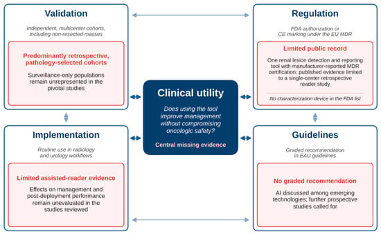
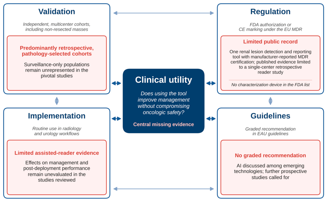

Disclaimer/Publisher’s Note: The statements, opinions and data contained in all publications are solely those of the individual author(s) and contributor(s) and not of MDPI and/or the editor(s). MDPI and/or the editor(s) disclaim responsibility for any injury to people or property resulting from any ideas, methods, instructions or products referred to in the content. |
© 2026 by the authors. Licensee MDPI, Basel, Switzerland. This article is an open access article distributed under the terms and conditions of the Creative Commons Attribution (CC BY) license.