


Simple Summary
Concurrent chemoradiotherapy (CCRT) is the standard of care for locally advanced cervical cancer (LACC), yet its efficacy remains suboptimal, underscoring the need for novel predictive biomarkers to address these clinical challenges. In this study, we found that pan-immune-inflammation value (PIV) demonstrates strong predictive capability and may serve as a practical tool for risk stratification and personalized treatment planning in clinical practice.
Abstract
Background: Cervical cancer remains a leading cause of cancer-related mortality in women. CCRT is standard for LACC, but its efficacy varies. This study was designed to evaluate the prognostic value of PIV in predicting short-term efficacy and long-term survival in LACC patients receiving CCRT. Methods: We retrospectively analyzed 205 patients with stage IB3–IVA cervical cancer treated with CCRT at the Affiliated Hospital of Jiangnan University (2018–2024). Variables included age, histopathological subtype, FIGO stage, lymph node status, pre-treatment serum SCCA, platelet-to-lymphocyte ratio [PLR], neutrophil-to-lymphocyte ratio [NLR], albumin-to-fibrinogen ratio [AFR], and PIV (calculated as neutrophil × platelet × monocyte counts/lymphocyte count). Short-term efficacy was assessed by the complete response rate (CRR) at 6 months post-treatment. Long-term survival was assessed by overall survival (OS) and progression-free survival (PFS). Results: Multivariate logistic regression identified tumor size ≥ 4 cm, NLR ≥ 3.32, and PIV ≥ 350.15 as independent risk factors for poor short-term response. Multivariate Cox analysis revealed that tumor size ≥ 4 cm, FIGO stage IVA, SCCA ≥ 1.84 ng/mL, and PIV ≥ 350.15 were independent adverse prognostic factors for OS. For PFS, independent predictors included moderate/poor differentiation, FIGO stage IVA, SCCA ≥ 1.84 ng/mL, and PIV ≥ 350.15. Adding PIV to a clinical model (FIGO stage + SCCA) improved the prediction of OS. Conclusions: PIV provides independent prognostic information and may serve as a practical tool for risk stratification and personalized treatment in LACC.
1. Introduction
Globally, cervical cancer ranks 8th in incidence and 9th in mortality among all female malignancies, and is the 4th leading cause of cancer-related death in women. China accounts for nearly 20% of the global annual new cases, reflecting the substantial disease burden and the urgent need for improved prevention and control strategies [1].
Current treatment modalities for cervical cancer encompass surgery, chemotherapy, radiotherapy, targeted therapy, and immunotherapy. Surgery and radiotherapy represent the primary curative options. Surgery is generally preferred for early-stage disease, whereas radiotherapy is stage-independent and can be applied across all stages [2]. According to the 2018 International Federation of Gynecology and Obstetrics (FIGO) staging system, stages IB3 to IVA are classified as locally advanced cervical cancer (LACC) [3,4,5]. Given that early-stage cervical cancer is often asymptomatic, approximately 70% of patients are already diagnosed with LACC at their initial clinical presentation [6]. Platinum-based concurrent chemoradiotherapy (CCRT) is recommended as the standard treatment by both the National Comprehensive Cancer Network (NCCN) and the European Society for Medical Oncology (ESMO) for patients with LACC [7]. CCRT is associated with a 30–50% reduction in the risk of death compared with radiotherapy alone [8]. Despite being the standard of care, the efficacy of CCRT for LACC remains suboptimal. Factors such as tumor heterogeneity, drug resistance, and treatment-related toxicity may contribute to poor treatment response and limited long-term survival in these patients. Thus, there is an urgent need to identify novel therapeutic strategies and predictive biomarkers to overcome these clinical challenges and improve outcomes in LACC.
Established prognostic factors for cervical cancer include pathological subtype, clinical stage, lymph node status, tumor size, hemoglobin levels, and the use of concurrent chemotherapy [9]. The tumor microenvironment (TME) has emerged as a major area in cancer research over recent years. The TME encompasses the cellular and non-cellular components surrounding tumor cells, which collectively influence key malignant behaviors, including proliferation and metastasis. The TME exerts dual effects on tumor cells, capable of both promoting and suppressing their proliferation, differentiation, and metastatic capacity. Moreover, it modulates the differentiation, functional polarization, and effector activity of immune cells within the tumor milieu [10]. Tumor-associated inflammation constitutes a key component of the TME. Inflammatory cells within the TME can damage biological macromolecules (e.g., DNA and proteins), elevate inflammatory cytokines and reactive oxygen species (ROS), and exacerbate local hypoxia, collectively driving epigenetic reprogramming [11]. Inflammatory mediators—including interleukins and tumor necrosis factors—released by tumor cells can recruit leukocytes, thereby promoting tumor initiation and metastasis. Myeloid-derived suppressor cells (MDSCs) can exert immunosuppressive effects that facilitate immune evasion and metastasis, and also promote tumor angiogenesis [12]. Peripheral blood-based inflammatory indices, including neutrophil-to-lymphocyte ratio (NLR), lymphocyte-to-monocyte ratio (LMR), and platelet-to-lymphocyte ratio (PLR), have been shown to predict prognosis in cervical cancer [13,14,15]. For instance, NLR has been found to be correlated with PFS and OS and may serve as an independent prognostic indicator in LACC patients receiving definitive chemoradiotherapy (dCRT) [16]. A retrospective cohort study identified PLR as an independent prognostic factor for both PFS and OS, whereas NLR was an independent predictor for PFS in LACC [17]. The pan-immune-inflammatory value (PIV)—calculated as neutrophil × platelet × monocyte counts/lymphocyte count—has been associated with median and long-term PFS and OS in pancreatic cancer [18], suggesting that PIV may represent a novel independent prognostic biomarker.
This study retrospectively enrolled LACC patients receiving CCRT at the Affiliated Hospital of Jiangnan University between September 2018 and September 2024. Treatment efficacy was systematically evaluated using objective response rate (ORR), progression-free survival (PFS), and overall survival (OS). Univariate and multivariate analyses were performed to identify independent prognostic factors. This study aimed to establish an individualized prognostic model and provide evidence to support precision treatment strategies for LACC.
2. Materials and Methods
2.1. Study Population
This study retrospectively enrolled patients with LACC who received CCRT at the Affiliated Hospital of Jiangnan University between September 2018 and September 2024. This study was conducted according to the 1964 Helsinki Declaration and its later amendments and approved by the Medical Ethics Committee of the Affiliated Hospital of Jiangnan University (Approval Number: LS2025513). All patient data were fully anonymized prior to analysis.
2.2. Inclusion Criteria
Patients were included if they met the following criteria: (1) clinically confirmed stage IB3–IVA cervical cancer and treated with CCRT; (2) histologically diagnosed as squamous cell carcinoma or adenocarcinoma; (3) Karnofsky Performance Status (KPS) score ≥ 70; and (4) complete clinical and follow-up data with a minimum follow-up duration of 5 months.
2.3. External Beam Radiotherapy (EBRT)
Pelvic EBRT was delivered at a conventional prescription dose of 45–50 Gy in 25 fractions, with a single dose of 1.8–2.0 Gy per fraction, five fractions per week. For patients with lymph node metastasis, a boost dose of up to 55–60 Gy was administered to the involved areas. Following EBRT, all patients received three-dimensional conformal brachytherapy.
2.4. Brachytherapy
All patients underwent three-dimensional conformal brachytherapy after EBRT. Positioning scans were performed after inserting the applicators. The target volume and organs at risk were delineated based on diagnostic MRI, ultrasonography, and gynecological examination findings. The prescribed dose to the high-risk clinical target volume (HR-CTV), which included residual cervical lesions and high-risk perimetrial areas, was dynamically adjusted according to its volume and the cumulative external irradiation dose (typically 45–50 Gy) to meet total dose requirements. A total brachytherapy dose of 30 Gy was delivered to the HR-CTV in 6 fractions, with the D90 of the HR-CTV reaching an EQD2 of no less than 80–85 Gy. Treatment planning was evaluated using volume-dose parameters, with the rectum, sigmoid colon, and bladder as key organs at risk. Dose constraints were set as follows: D2cm3 ≤ 75 Gy for the rectum and sigmoid colon, and D2cm3 ≤ 90 Gy for the bladder.
2.5. Concurrent Chemotherapy
Concurrent chemotherapy was administered weekly using cisplatin (40 mg/m2) during radiotherapy, with a planned duration of at least 4 cycles. The actual number of cycles and dosages were individually adjusted based on the patient’s physical condition and treatment tolerance.
2.6. Data Collection
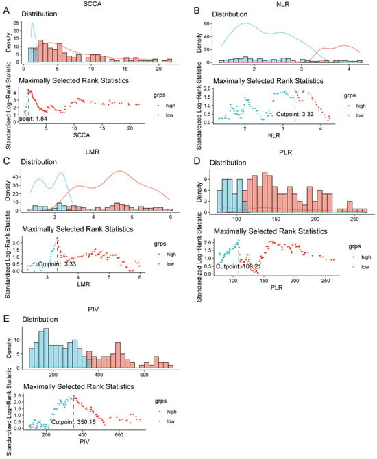
Enrolled patients were confirmed by pathological and imaging staging, including pelvic MRI, chest-abdominal CT, bone scan, or PET-CT. Pre-treatment laboratory results and all imaging and pathological data were integrated for subsequent clinical statistical analysis. The following patient subgroups were defined based on pre-treatment variables: (1) clinical stage IB3, II, III, or IVA; (2) serum SCCA < 1.84 ng/mL vs. ≥1.84 ng/mL; (3) NLR < 3.32 vs. ≥3.32; (4) LMR < 3.33 vs. ≥3.33; (5) PLR < 109.21 vs. ≥109.21; and (6) PIV < 350.15 vs. ≥350.15. Optimal cut-off values for SCCA, NLR, LMR, PLR, and PIV were determined using the surv_cutpoint function in R (version 4.4.1).
2.7. Follow-Up
Patients were followed up every 3 months in the first year post-treatment, every 3–6 months during the second year, and every 6–12 months thereafter until September 2025. Follow-up records primarily included general health status and the timing of disease progression.
2.8. Short-Term Efficacy Evaluation
Short-term efficacy was assessed based on re-examination results at 6 months after treatment completion, according to the RECIST 1.1 criteria [19]. Responses were classified as complete response (CR; both imaging and clinical complete response) or incomplete response (stable disease, partial response, or progressive disease).
2.9. Long-Term Efficacy Evaluation
OS and PFS were defined as the primary endpoints for long-term efficacy evaluation. OS was calculated from the date of definitive diagnosis to death from any cause or the last follow-up (September 2025). Patients lost to follow-up were censored at the last known follow-up date. PFS was calculated from the date of definitive diagnosis to the first radiological or clinical evidence of disease progression, death from any cause, or the last follow-up. Patients without endpoint events or lost to follow-up were censored at the last confirmed non-progression date.
2.10. Statistical Analysis
Statistical analyses were performed using SPSS 29.0, and figures were generated using R (version 4.4.1). Categorical variables are presented as frequencies (n, %). Continuous variables with a normal distribution are expressed as mean ± standard deviation (x̄ ± s), whereas non-normally distributed variables are presented as median with interquartile range [P25, P75]. Optimal cut-off values for SCCA, NLR, LMR, PLR, and PIV were determined using the surv_cutpoint function in the R package survminer, based on maximally selected rank statistics. OS was selected as the reference outcome for cut-off determination. The optimal thresholds were identified by maximizing the standardized survival separation between the two resulting groups. The OS-derived cut-off values were 1.84 ng/mL for SCCA, 3.32 for NLR, 3.33 for LMR, 109.21 for PLR, and 350.15 for PIV. These thresholds were subsequently applied consistently to analyses of short-term treatment response, OS, and PFS, without further outcome-specific optimization. To evaluate the stability of the selected cut-off values and minimize potential overfitting, 10-fold cross-validation was performed. The study cohort was randomly partitioned into 10 approximately equal subsets. In each fold, nine subsets were used for model development, while the remaining subset was reserved for validation. Optimal cut-off values were independently re-estimated using OS data from the training subsets only. The prognostic models were subsequently fitted using the training data and evaluated in the corresponding held-out validation subset. Predictive discrimination was assessed using the concordance index (C-index). The variability of the selected cut-off values across folds was also examined to assess their stability. This procedure ensured that outcome information from the validation subsets was not used during cut-off selection or model development.
Short-term efficacy was compared using the chi-square test (odds ratio [OR] and 95% confidence interval [CI]). Long-term survival was estimated using the Kaplan–Meier method, and survival curves were compared using the log-rank test. Independent prognostic factors for OS and PFS were identified using Cox proportional hazards regression (hazard ratio [HR] and 95% CI). A nomogram predicting OS and PFS was constructed based on the multivariate Cox analysis results, and model performance was evaluated using calibration curves and the concordance index (C-index). To assess the stability of the cut-off determination and multivariate Cox model, internal cross-validation was performed using a 10-fold approach. The dataset was randomly divided into 10 subsets; 9 subsets served as the training set and the remaining 1 as the validation set, with the process repeated 10 times. The C-index was calculated for each validation set, and the mean value of the 10 iterations was reported as the internal validation performance metric. The stability of regression coefficients across folds was also examined to confirm model robustness.
Given that the inflammatory indices (NLR, LMR, PLR, and PIV) share common components (neutrophil, lymphocyte, platelet, and monocyte counts), potential multicollinearity was assessed. Spearman correlation analysis was used to evaluate correlations among these indices, and a correlation matrix was generated. Variance inflation factor (VIF) and tolerance were subsequently calculated to quantify collinearity levels (VIF > 5 indicating moderate and VIF > 10 indicating severe collinearity). In the presence of significant collinearity, clinically meaningful or better-performing indicators (e.g., PIV) were preferentially retained, and highly correlated variables were not included in the same multivariate model. Stepwise regression, penalized regression (e.g., LASSO), or dimensionality reduction methods were considered to further optimize model stability if needed.
To evaluate the incremental predictive value of incorporating PIV, three prediction models were constructed: (1) a FIGO staging model, (2) a clinical model (FIGO + SCCA), and (3) the nomogram model (FIGO + SCCA + PIV). The integrated discrimination improvement (IDI) and net reclassification improvement (NRI) were calculated to assess performance improvement of the nomogram model over the clinical model. The 95% CIs for NRI and IDI were estimated using the bootstrap resampling method (1000 repetitions). All statistical tests were two-sided, and a p value < 0.05 was considered statistically significant.
3. Results
3.1. Patient Baseline Characteristics
A total of 205 LACC patients were enrolled in this study, with a mean age of 59.6 ± 11.0 years. Most patients had squamous cell carcinoma (191, 93.2%), tumors ≥ 4 cm (124, 60.5%), FIGO stage III–IVA disease (149, 72.7%), and lymph node metastasis (120, 58.5%). In total, 80 patients (39.0%) had a high PIV (≥350.15). Detailed baseline characteristics are summarized in Table 1.
Table 1.
Basic clinical data of 205 patients with LACC.
3.2. Clinical Characteristics of Short-Time Therapeutic Efficacy
Optimal cut-off values for SCCA, NLR, LMR, PLR, and PIV were determined using the surv_cutpoint function in R based on OS. Internal cross-validation was performed to assess the robustness of the OS-derived cut-off values and prognostic models. The cross-validated C-index values were 0.692 for OS and 0.651 for PFS, compared with the apparent C-index values of 0.713 and 0.679, respectively. These results suggest a modest reduction in discrimination after internal validation and provide an internal assessment of model performance. The identified cut-off values were as follows: SCCA, 1.84 ng/mL; NLR, 3.32; PLR, 109.21; LMR, 3.33; and PIV, 350.15. Based on these thresholds, patients were stratified into high- and low-level groups for each indicator (Figure 1).
Figure 1.
Truncated values of (A) SCCA, (B) NLR, (C) LMR, (D) PLR and (E) PIV. The vertical dashed line indicates the optimal cut-off values for survival stratification. The blue and red density curves represent patients with low and high biomarker levels stratified by the optimal cut-off values, respectively.
In terms of evaluation of short-term therapeutic efficacy, 188 patients (91.7%) achieved CR and 17 (8.3%) had a non-complete response (NCR) 6 months after treatment completion. Kaplan–Meier survival analysis demonstrated that patients achieving CR had significantly better survival outcomes than those with NCR (Figure S1). The 1-, 3-, and 5-year OS rates were 97.9%, 82.0%, and 49.0% in the CR group, compared with 67.5%, 56.7%, and 31.8% in the NCR group (log-rank χ2 = 370.635, p < 0.0001, Table S1). Corresponding PFS rates were 94.1%, 70.7%, and 29.4% versus 47.1%, 11.8%, and 0% (log-rank χ2 = 206.767, p < 0.0001, Table S2).
The complete response rate (CRR) showed no significant differences across subgroups of age (<60 years: 47.9% vs. ≥60 years: 52.1%; χ2 = 0.748, p = 0.387), pathological type (squamous cell carcinoma: 93.1% vs. adenocarcinoma: 6.9%; χ2 = 0.001, p = 0.997), tumor differentiation (moderate/poor: 71.8% vs. well-differentiated: 28.2%; χ2 = 0.422, p = 0.515), FIGO stage (I–II: 28.7%, III: 56.9%, IVA: 14.4%; χ2 = 2.672, p = 0.262), lymph node metastasis (without: 42.6% vs. with: 57.4%; χ2 = 1.109, p = 0.292), pre-treatment SCCA (<1.84 ng/mL: 21.3% vs. ≥1.84 ng/mL: 78.7%; χ2 = 3.241, p = 0.071), and pre-treatment PLR (<109.21: 29.3% vs. ≥109.21: 70.7%; χ2 = 0.272, p = 0.601) 6 months after treatment completion.
However, pre-treatment NLR ≥ 3.32 was associated with a significantly lower CRR compared with NLR < 3.32 (23.4% vs. 76.6%; χ2 = 8.337, p = 0.003). Similarly, patients with LMR ≥ 3.33 and PIV ≥ 350.15 had significantly lower CRR than those with lower levels (χ2 = 7.698, p = 0.005; χ2 = 7.761, p = 0.005, respectively). Tumor size ≥ 4 cm also correlated with a lower CRR compared with <4 cm (58% vs. 42%; χ2 = 5.971, p = 0.014). In conclusion, the pre-treatment NLR, LMR and PIV were closely related to CRR. Detailed results of the univariate and multivariate analyses of short-term efficacy are presented in Table S3 and Table 2, respectively.
Table 2.
Multivariate logistic regression analysis of short-term therapeutic efficacy.
3.3. Clinical Characteristics of Long-Term Therapeutic Efficacy
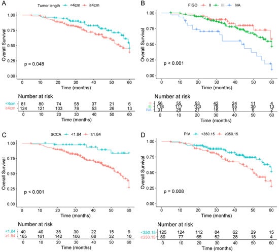
In terms of OS, univariate Cox regression analysis identified the following factors as significantly associated with worse survival outcomes: tumor length ≥ 4 cm (HR = 1.614, 95% CI 1.003–2.736, p = 0.048), FIGO stage IVA (HR = 3.983, 95% CI 1.893–8.382, p < 0.001, with stage II as reference), SCCA ≥ 1.84 ng/mL (HR = 5.556, 95% CI 2.007–15.381, p < 0.001), NLR ≥ 3.32 (HR = 2.116, 95% CI 1.294–3.461, p = 0.003), LMR < 3.33 (HR = 0.564, 95% CI 0.345–0.923, p = 0.023), and PIV ≥ 350.15 (HR = 1.918, 95% CI 1.176–3.128, p = 0.009). No statistically significant associations were observed for age ≥ 60 years, adenocarcinoma histology, moderate/poor differentiation, lymph node metastasis, PLR ≥ 109.21, or FIGO stage III (all p > 0.05). While multivariate Cox regression analysis confirmed that tumor size ≥ 4 cm (HR = 2.375, 95% CI 1.270–4.442, p = 0.007), FIGO stage IVA (HR = 2.102, 95% CI 1.452–2.688, p = 0.003), SCCA ≥ 1.84 ng/mL (HR = 3.762, 95% CI 1.245–11.368, p = 0.019), and PIV ≥ 350.15 (HR = 1.924, 95% CI 1.175–3.442, p = 0.008) remained independent adverse prognostic factors for survival (Table S4).
Collectively, these findings identify tumor size ≥ 4 cm, FIGO stage IVA, elevated SCCA, and elevated PIV as core determinants of long-term survival in LACC patients, providing valuable references for risk stratification and individualized treatment planning. The Kaplan–Meier curves for each significant factor are presented in Figure 2.
Figure 2.
Kaplan–Meier curves for factors influencing overall survival in LACC patients. (A). Tumor length. (B). FIGO stage. (C). SCCA. (D). PIV.
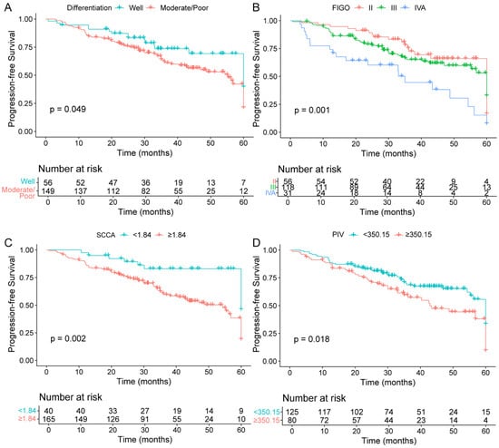
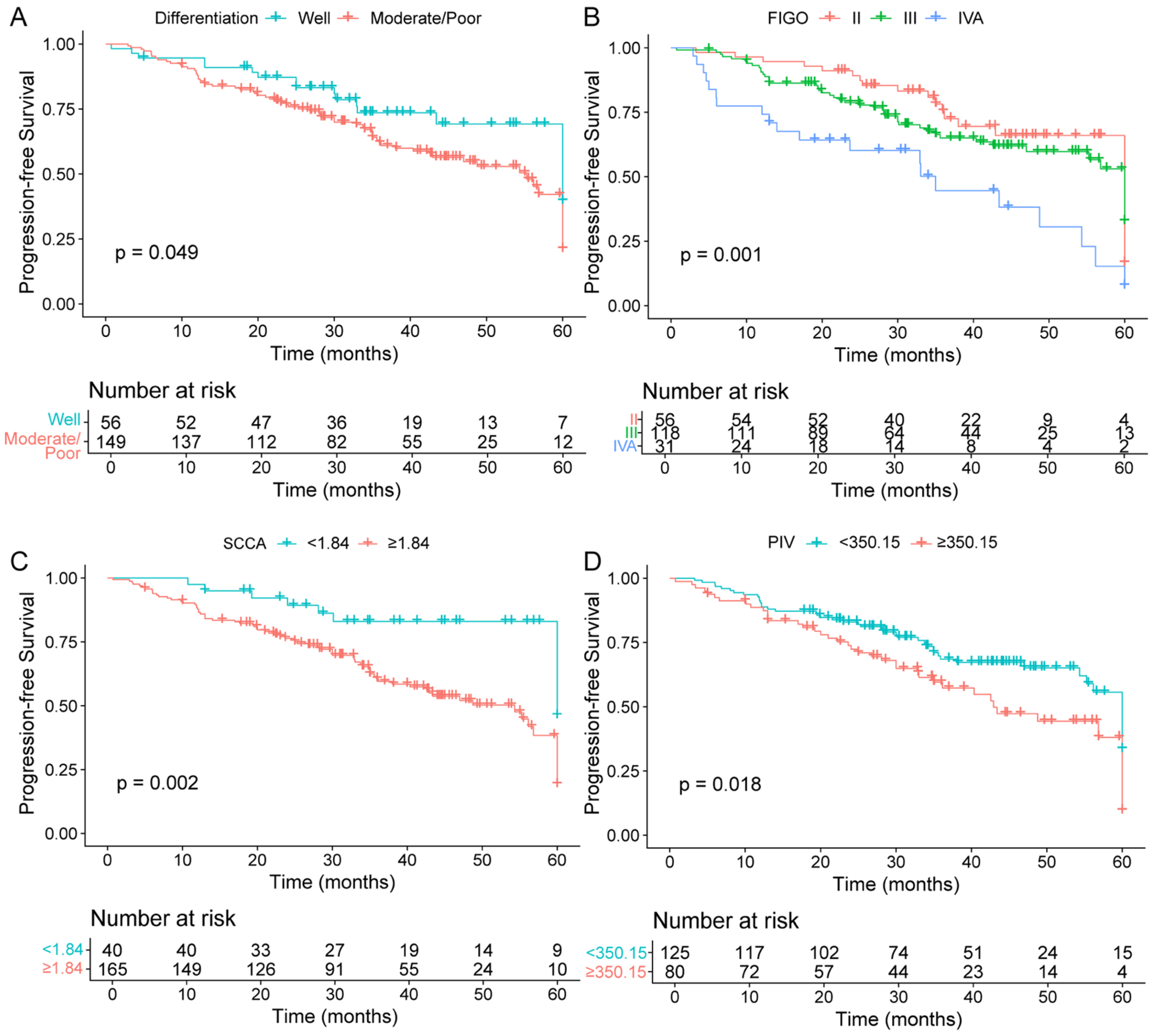
In terms of PFS, univariate analysis identified several factors significantly associated with worse PFS: moderate/poor differentiation (HR = 1.670, p = 0.049), FIGO stage IVA (HR = 2.785, p = 0.002), SCCA ≥ 1.84 ng/mL (HR = 2.731, p = 0.003), NLR ≥ 3.32 (HR = 1.868, p = 0.005), LMR < 3.33 (HR = 1.724, p = 0.015), and PIV ≥ 350.15 (HR = 1.701, p = 0.016). No significant associations were observed for age, pathological type, tumor size (≥4 cm, p = 0.358), FIGO stage III, lymph node metastasis, ALB, or PLR (all p > 0.05). Notably, tumor size was not significantly associated with PFS in the unadjusted model, differing from its prognostic value for OS. Multivariate Cox regression model identified four independent risk factors for PFS: moderate/poor differentiation (HR = 1.641, 95% CI 1.022–2.921, p = 0.042), FIGO stage IVA (HR = 2.612, 95% CI 1.341–5.088, p = 0.005), SCCA ≥ 1.84 ng/mL (HR = 2.159, 95% CI 1.068–4.363, p = 0.031), and PIV ≥ 350.15 (HR = 1.283, 95% CI 1.087–2.138, p = 0.029). Age, pathological type, lymph node metastasis, NLR, PLR, and ALB were not independent prognostic factors for PFS.
Collectively, these findings identify moderate/poor differentiation, FIGO stage IVA, elevated SCCA, and elevated PIV as core determinants of long-term survival in LACC patients. Kaplan–Meier curves for the significant prognostic factors are presented in Figure 3, and the complete results of the univariate and multivariate analyses are summarized in Table S5.
Figure 3.
Kaplan–Meier curves for factors influencing progression-free survival in LACC patients. (A). Differentiation grade. (B). FIGO stage. (C). SCCA. (D). PIV.
3.4. The Prognostic Role of PIV in Each Subgroup
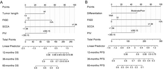
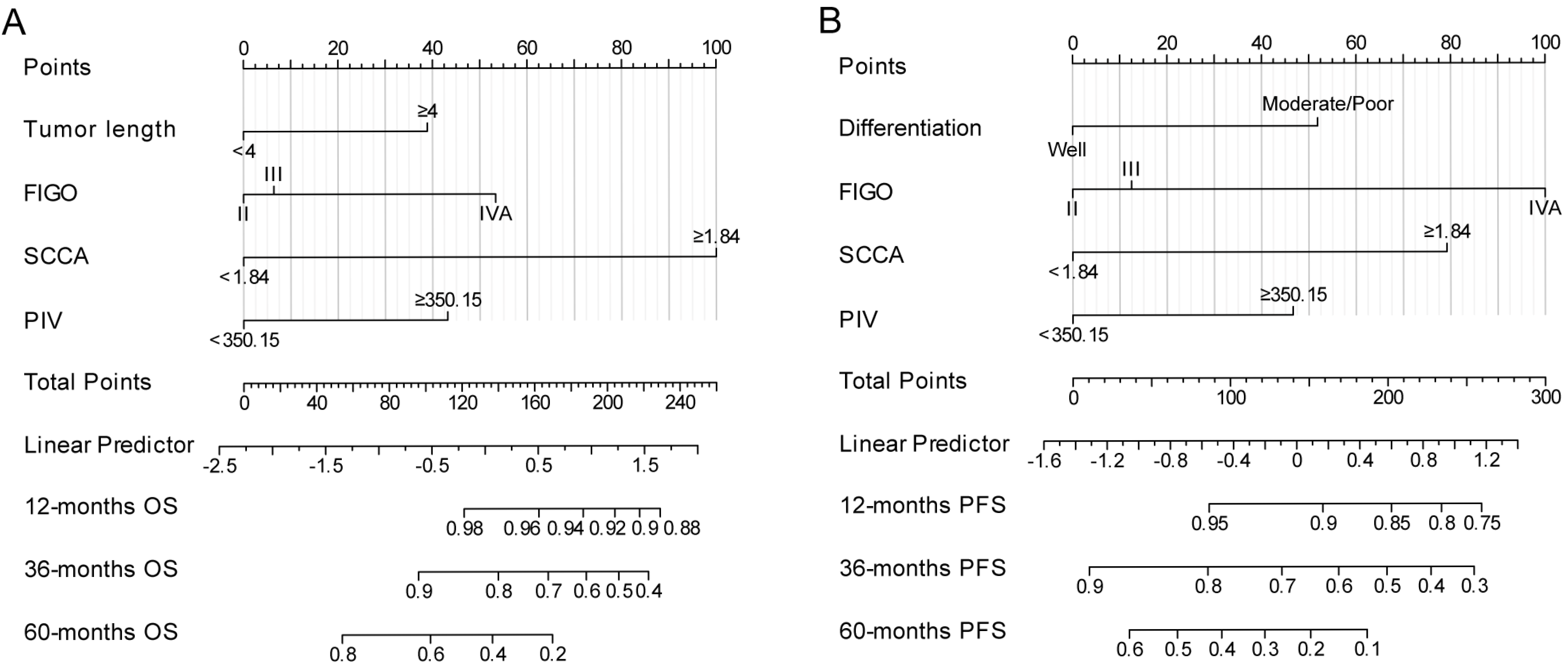
Based on the multivariate analysis results, several independent prognostic factors—tumor length, moderate/poor differentiation, FIGO stage IVA, elevated SCCA (≥1.84 ng/mL), and elevated PIV (≥350.15)—were incorporated into the nomogram (Figure 4) to predict OS and PFS. Model validation yielded concordance indices (C-index) of 0.713 for the OS model and 0.679 for the PFS model. Calibration curves (Figure S2) demonstrated good agreement between predicted and observed probabilities at 1-, 3-, and 5-year time points for both OS and PFS.
Figure 4.
Nomogram prediction model for 12-, 36-, and 60-month survival in LACC patients. (A). OS. (B). PFS.
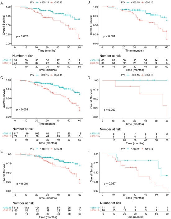
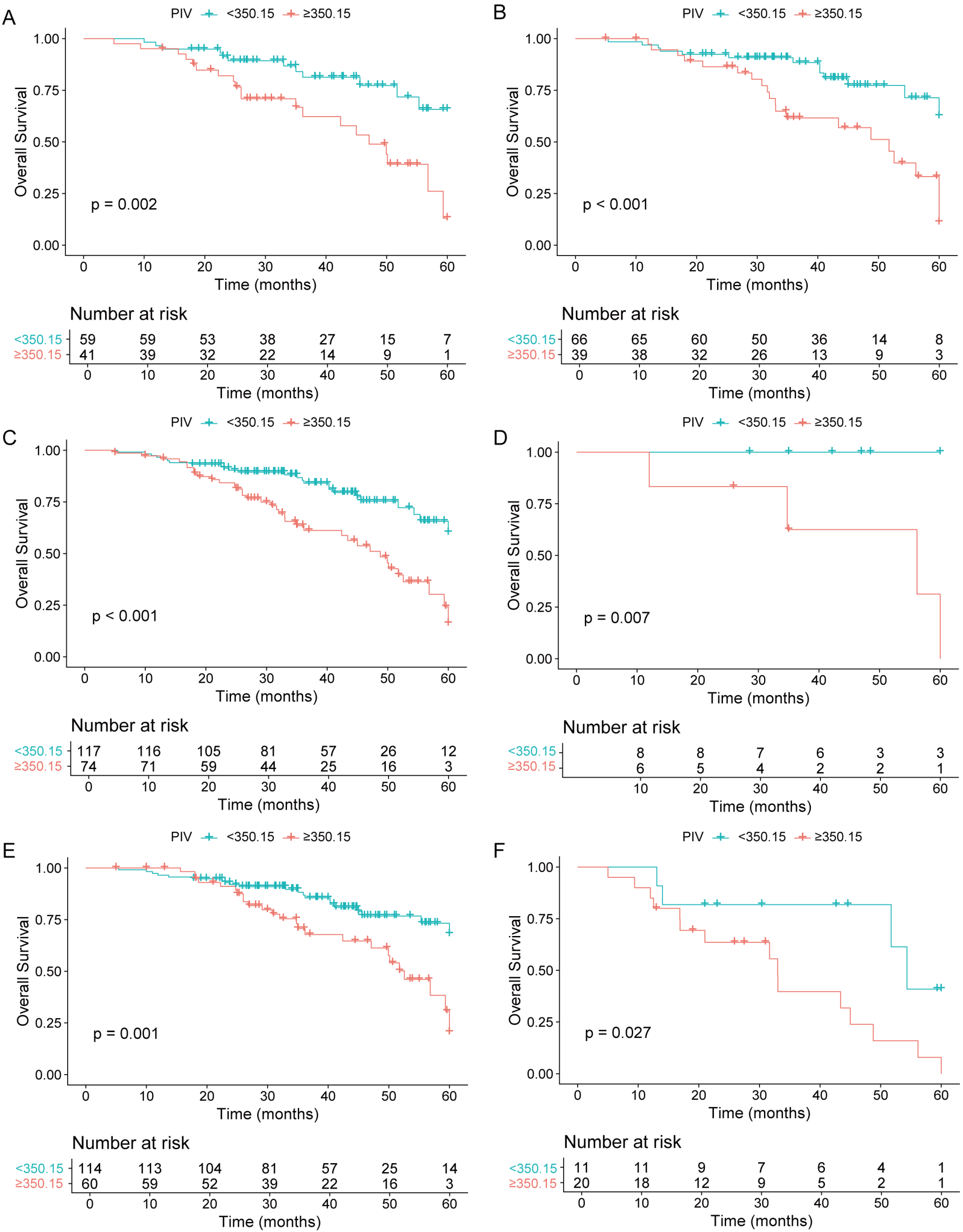
Given that the prognostic value of PIV in LACC patients receiving CCRT has not been fully characterized, we further performed subgroup analyses stratified by age, pathological type, and FIGO stage to further evaluate its predictive utility across diverse clinical subsets (Figure 5). Across all subgroups, elevated pre-treatment PIV (≥350.15) remained a significant adverse predictor for both OS and PFS.
Figure 5.
The effect of pre-treatment PIV levels on overall survival (OS) of patients in different subgroups. (A). Age < 60 years. (B). Age ≥ 60 years. (C). Squamous cell carcinoma. (D). Adenocarcinoma. (E). Stage II/III. (F). Stage IVA.
3.5. Construction of the Clinical Survival Prediction Model
Prior to constructing the multivariate Cox regression model, we assessed multicollinearity among the candidate independent variables (Table S6). The VIF for all variables was <5, with corresponding tolerance values > 0.2, indicating no severe multicollinearity. Specifically, VIF values for tumor length, FIGO stage, SCCA, and differentiation grade were 1.23, 1.32, 1.56, and 1.20, respectively, reflecting low correlations with other covariates. For the inflammatory indices, VIF values were 2.94 for NLR, 2.43 for LMR, 3.45 for PLR, and 3.87 for PIV. Although moderate correlations were observed among these inflammatory markers, none exceeded the conventional threshold (VIF < 5), indicating acceptable model stability.
Given that FIGO stage, SCCA, and PIV are all associated with long-term survival in LACC patients in terms of OS and PFS, we further constructed corresponding predictive models. Compared with the clinical model (FIGO + SCCA), the model incorporating PIV yielded an IDI of 0.067 (p = 0.004) and an NRI of 0.192 (p < 0.001) for OS prediction, indicating a significant improvement in discriminatory performance. For PFS prediction, the corresponding IDI and NRI were 0.058 (p = 0.009) and 0.173 (p < 0.001), respectively (Table 3). These findings indicate that adding PIV to the model significantly improves both discrimination and risk reclassification performance. Before the clinical application of LACC treatment, this model can be used to evaluate the efficacy of CCRT, enabling the formulation of individualized treatment plans based on each patient’s specific conditions.
Table 3.
Model performance comparison for survival prediction.
4. Discussion
The prognosis of cervical cancer is closely associated with a range of clinical factors, including clinical stage [20], pathological subtype, and lymph node status [21]. Recent advances in imaging have enabled the development of deep learning-based automatic models using pre-treatment T2-weighted MRI to predict prognosis in LACC patients receiving CCRT [22]. A previous retrospective study of 245 LACC patients showed that independent predictors of para-aortic lymph node metastasis were tumor size assessed by MRI and para-aortic lymph node status determined by PET/CT [23]. Accumulating evidence has demonstrated that circulating inflammatory cells and factors—including neutrophils, lymphocytes, platelets, and fibrinogen—are actively involved in tumor progression and metastasis. These cells and factors contribute critically to the formation and remodeling of the TME through the release of inflammatory mediators [24]. The inflammatory milieu within TME is shaped by multiple factors, including infection, hypoxia, and mechanical stress. During this process, reticular trap structures formed by either host or tumor cells facilitate tumor cell dissemination and metastasis.
NLR, LMR, and PLR have recently garnered considerable attention as readily accessible biomarkers reflecting systemic inflammation and immune status in LACC. NLR is a well-established biomarker of systemic inflammation and immune status. Elevated NLR has been associated with adverse clinical outcomes, unfavorable pathological features, and poor prognosis across multiple malignancies, including breast, gastric, ovarian, esophageal, and colorectal cancers [25]. Pre-treatment NLR has been shown to correlate with tumor invasiveness and clinical stage in cervical cancer [26]. A comprehensive study demonstrated that elevated pre-treatment NLR was significantly associated with poorer OS (HR = 1.58, 95% CI: 1.44–1.74; p < 0.00001) and PFS (HR = 1.48, 95% CI: 1.34–1.63; p < 0.00001), indicating that elevated pre-treatment NLR serves as an independent prognostic factor for both OS and PFS in cervical cancer patients [27]. In a large retrospective study, elevated pre-treatment PLR was identified as an independent adverse prognostic factor for both PFS and OS, while elevated NLR was independently associated with worse PFS in LACC [17]. Similarly, a retrospective study of 1051 patients with FIGO stage IB2–IVA cervical cancer treated with definitive radiotherapy (with or without chemotherapy) demonstrated that PLR remained an independent risk factor for DFS and OS after adjustment for clinical covariates, including FIGO stage [28]. A further study focusing on patients with stage IIB–III cervical cancer receiving radiotherapy reported that the optimal cut-off values for NLR, PLR, and monocyte-to-lymphocyte ratio (MLR) were determined as 2.8, 186.67, and 0.41, respectively. Elevated levels of all three indices were significantly associated with poorer 5-year OS and PFS [29]. A prognostic nomogram based on systemic inflammatory markers was proven to provide reliable risk stratification at treatment initiation and facilitate individualized clinical decision-making [30]. In this study, NLR and LMR were significantly associated with treatment response and long-term survival in univariate analysis. However, they did not retain independent prognostic significance in the multivariate model. PLR, on the other hand, showed no significant predictive value in this cohort.
Therefore, we introduced PIV, which integrates multiple inflammatory cells, including neutrophils, platelets, monocytes, and lymphocytes, into a single composite measure. Compared with single-pair ratios such as NLR, LMR, and PLR, PIV may more comprehensively reflect the inflammatory and immune responses. Elevated PIV typically reflects an increase in neutrophils, platelets, and monocytes, accompanied by a relative decrease in lymphocytes. This composite pattern may attenuate sensitivity to radiotherapy and chemotherapy and promote tumor recurrence and metastasis through multiple interrelated pathways. First, neutrophils secrete various inflammatory and pro-angiogenic factors (e.g., IL-6, VEGF) and form the immunosuppressive TME. Second, monocytes can polarize into immunosuppressive tumor-associated macrophages, which further suppress T-cell function via pathways involving TGF-β and IL-10, collectively establishing a pro-tumorigenic inflammatory environment [31]. Platelets can encapsulate circulating tumor cells and release various growth factors and chemokines, thereby promoting tumor cell survival and metastasis [32]. Concurrently, the reduction in lymphocytes, particularly the deficiency or exhaustion of CD8+ cytotoxic T cells, may lead to insufficient immune activation during chemoradiotherapy, thereby increasing the persistence of minimal residual disease and the risk of recurrence. PIV, derived from neutrophils, monocytes, platelets, and lymphocytes, may more accurately reflect the immune-inflammatory TME. In this study, PIV retained independent prognostic significance in both the OS and PFS multivariate models, showing the potential advantage of composite inflammatory indicators in prognostic assessment of LACC.
However, this study has several limitations. First, as a single-center retrospective analysis, it is subject to selection and information biases, and certain clinical variables may not have been completely documented. Second, the modest sample size (n = 205) limits statistical power in subgroup analyses and may increase type II error risk. Third, only baseline inflammatory markers were used, without dynamic monitoring during treatment. Fourth, the nomogram lacks external validation. Overfitting remains a concern despite acceptable C-index values. Future multicenter, prospective studies with dynamic inflammatory monitoring and mechanistic explorations are warranted to enhance model generalizability and clinical utility.
5. Conclusions
In conclusion, this study demonstrates that systemic inflammatory status is a key determinant of treatment response and survival in LACC patients receiving CCRT. Inflammatory markers, particularly PIV, provided independent prognostic information and improved the predictive ability of the clinical model in our cohort. Pre-treatment PIV may serve as a potential prognostic indicator for response and survival in LACC patients receiving CCRT. Future studies should expand the research center, increase the sample size, dynamically monitor inflammatory changes, and stratify the patients’ prognosis to enhance the generalizability and clinical practicability of the model.
Supplementary Materials
The following supporting information can be downloaded at https://www.mdpi.com/article/10.3390/cancers18223605/s1, Figure S1: Kaplan–Meier curves of patients in the CR group and NCR group; Figure S2: Calibration curves of 12-, 36-, and 60-month survival in the Nomogram prediction model; Table S1: Comparison of OS between the CR group and the NCR group; Table S2: Comparison of PFS been the CR group and the NCR group; Table S3: Comparison of clinical characteristics of short-time therapeutic efficacy; Table S4: Cox regression analysis of OS; Table S5: Cox regression analysis of PFS; Table S6: Evaluation of multicollinearity among prognostic factors.
Author Contributions
Conceptualization, L.G., H.C., W.H. and J.C.; methodology, L.G., H.C., Y.F., W.H. and J.C.; validation, L.G., H.C., W.H. and J.C.; formal analysis, L.G., H.C. and Y.F.; visualization, L.G., H.C. and Y.F.; project administration, J.C.; resources, J.C.; supervision, W.H. and J.C.; writing—original draft, L.G.; writing—review & editing, J.C. All authors have read and agreed to the published version of the manuscript.
Funding
The study has been supported by the National Natural Science Foundation of China (No.82574016, 82103772, and 82404192), Wuxi Municipal Health Commission (Q202520 and Q202433), Wuxi Bureau of Science and Technology (K2025002 and 20241002).
Institutional Review Board Statement
This retrospective study was conducted according to the 1964 Helsinki Declaration and its later amendments and approved by the Medical Ethics Committee of the Affiliated Hospital of Jiangnan University (Approval Number: LS2025513, approved on 26 December 2025). All patient data were fully anonymized prior to analysis.
Informed Consent Statement
This retrospective study was approved by the Medical Ethics Committee of the Affiliated Hospital of Jiangnan University (Approval Number: LS2025513). The requirement for informed consent was waived by the Ethics Committee because the study involved only the analysis of existing medical records, posed minimal risk to participants, and obtaining consent was impracticable due to the large sample size and long follow-up period. All patient data were anonymized to protect privacy.
Data Availability Statement
All data which support the findings of the study are within the manuscript or in Supplemental Materials.
Conflicts of Interest
The authors declare no conflicts of interest.
References
- Global cancer burden growing, amidst mounting need for services. Saudi Med. J. 2024, 45, 326–327. [Google Scholar] [CrossRef] [Scilit]
- Manso, L.; Ramchandani-Vaswani, A.; Romero, I.; Sánchez-Lorenzo, L.; Bermejo-Pérez, M.J.; Estévez-García, P.; Fariña-Madrid, L.; García García, Y.; Gil-Martin, M.; Quindós, M. SEOM-GEICO Clinical Guidelines on cervical cancer (2023). Clin. Transl. Oncol. 2024, 26, 2771–2782. [Google Scholar] [CrossRef] [Scilit]
- Grigsby, P.W.; Massad, L.S.; Mutch, D.G.; Powell, M.A.; Thaker, P.H.; McCourt, C.; Hagemann, A.; Fuh, K.; Kuroki, L.; Schwarz, J.K.; et al. FIGO 2018 staging criteria for cervical cancer: Impact on stage migration and survival. Gynecol. Oncol. 2020, 157, 639–643. [Google Scholar] [CrossRef] [Scilit]
- Pécout, M.; Phalippou, J.; Azaïs, H.; Ouldamer, L.; Bolze, P.A.; Ballester, M.; Huchon, C.; Mimoun, C.; Akladios, C.; Lecointre, L.; et al. Comparison of retroperitoneal and transperitoneal surgical routes in laparoscopic nodal staging for locally advanced cervical cancers (FIGO IB3-IVA). Eur. J. Surg. Oncol. 2022, 48, 2061–2067. [Google Scholar] [CrossRef] [Scilit]
- Olawaiye, A.B.; Baker, T.P.; Washington, M.K.; Mutch, D.G. The new (Version 9) American Joint Committee on Cancer tumor, node, metastasis staging for cervical cancer. CA Cancer J. Clin. 2021, 71, 287–298. [Google Scholar] [CrossRef] [Scilit]
- Espías-Alonso, M.; Gorostidi, M.; Zapardiel, I.; Gracia, M. Assessment of ESGO Quality Indicators and Factors Associated with Recurrence Following Surgery for Early-Stage Cervical Cancer: A Retrospective Cohort Study. J. Clin. Med. 2025, 14, 7041. [Google Scholar] [CrossRef] [Scilit]
- Marth, C.; Landoni, F.; Mahner, S.; McCormack, M.; Gonzalez-Martin, A.; Colombo, N. Cervical cancer: ESMO Clinical Practice Guidelines for diagnosis, treatment and follow-up. Ann. Oncol. 2017, 28, iv72–iv83. [Google Scholar] [CrossRef] [Scilit]
- Abu-Rustum, N.R.; Yashar, C.M.; Arend, R.; Barber, E.; Bradley, K.; Brooks, R.; Campos, S.M.; Chino, J.; Chon, H.S.; Crispens, M.A.; et al. NCCN Guidelines® Insights: Cervical Cancer, Version 1.2024. J. Natl. Compr. Cancer Netw. 2023, 21, 1224–1233. [Google Scholar] [CrossRef] [Scilit]
- Dueňas-González, A.; Orlando, M.; Zhou, Y.; Quinlivan, M.; Barraclough, H. Efficacy in high burden locally advanced cervical cancer with concurrent gemcitabine and cisplatin chemoradiotherapy plus adjuvant gemcitabine and cisplatin: Prognostic and predictive factors and the impact of disease stage on outcomes from a prospective randomized phase III trial. Gynecol. Oncol. 2012, 126, 334–340. [Google Scholar] [CrossRef] [Scilit]
- Liu, Y.; Cao, X. Characteristics and Significance of the Pre-metastatic Niche. Cancer Cell 2016, 30, 668–681. [Google Scholar] [CrossRef] [Scilit]
- Denk, D.; Greten, F.R. Inflammation: The incubator of the tumor microenvironment. Trends Cancer 2022, 8, 901–914. [Google Scholar] [CrossRef] [Scilit]
- De Palma, M.; Biziato, D.; Petrova, T.V. Microenvironmental regulation of tumour angiogenesis. Nat. Rev. Cancer 2017, 17, 457–474. [Google Scholar] [CrossRef] [Scilit]
- Ferioli, M.; Benini, A.; Malizia, C.; Forlani, L.; Medici, F.; Laghi, V.; Ma, J.; Galuppi, A.; Cilla, S.; Buwenge, M.; et al. Classical Prognostic Factors Predict Prognosis Better than Inflammatory Indices in Locally Advanced Cervical Cancer: Results of a Comprehensive Observational Study including Tumor-, Patient-, and Treatment-Related Data (ESTHER Study). J. Pers. Med. 2023, 13, 1229. [Google Scholar] [CrossRef] [Scilit]
- Huang, H.; Liu, Q.; Zhu, L.; Zhang, Y.; Lu, X.; Wu, Y.; Liu, L. Prognostic Value of Preoperative Systemic Immune-Inflammation Index in Patients with Cervical Cancer. Sci. Rep. 2019, 9, 3284. [Google Scholar] [CrossRef] [Scilit]
- Han, X.; Liu, S.; Yang, G.; Hosseinifard, H.; Imani, S.; Yang, L.; Maghsoudloo, M.; Fu, S.; Wen, Q.; Liu, Q. Prognostic value of systemic hemato-immunological indices in uterine cervical cancer: A systemic review, meta-analysis, and meta-regression of observational studies. Gynecol. Oncol. 2021, 160, 351–360. [Google Scholar] [CrossRef] [Scilit]
- Trinh, H.; Dzul, S.P.; Hyder, J.; Jang, H.; Kim, S.; Flowers, J.; Vaishampayan, N.; Chen, J.; Winer, I.; Miller, S. Prognostic value of changes in neutrophil-to-lymphocyte ratio (NLR), platelet-to-lymphocyte ratio (PLR) and lymphocyte-to-monocyte ratio (LMR) for patients with cervical cancer undergoing definitive chemoradiotherapy (dCRT). Clin. Chim. Acta 2020, 510, 711–716. [Google Scholar] [CrossRef] [Scilit]
- Fullerton, R.; Martell, K.; Khanolkar, R.; Phan, T.; Banerjee, R.; Meyer, T.; Traptow, L.; Köbel, M.; Ghatage, P.; Doll, C.M. Impact of immune, inflammatory and nutritional indices on outcome in patients with locally advanced cervical cancer treated with definitive (chemo)radiotherapy. Gynecol. Oncol. 2024, 190, 291–297. [Google Scholar] [CrossRef] [Scilit]
- Topkan, E.; Selek, U.; Kucuk, A.; Pehlivan, B. Low Pre-ChemoradiotherapyPan-Immune-Inflammation Value (PIV) Measures Predict Better Survival Outcomes in Locally Advanced Pancreatic Adenocarcinomas. J. Inflamm. Res. 2022, 15, 5413–5423. [Google Scholar] [CrossRef] [Scilit]
- Petrelli, F.; Riboldi, V.; Bruschieri, L.; Villa, A.; Cribiu, F.M.; Borgonovo, K.; Ghilardi, M.; Ghidini, A.; Seghezzi, S.; Stefani, A.; et al. First-line treatment of locally advanced cervical carcinoma: An updated systematic review and Bayesian network meta-analysis. Cancer Treat. Rev. 2025, 135, 102921. [Google Scholar] [CrossRef] [Scilit]
- Gao, X.; Kong, Y.; Ning, Y.; Tian, T.; Gai, X.; Lei, K.; Cui, Z. The prognosis of patients with locally advanced cervical cancer undergoing surgical versus non-surgical treatment: A retrospective cohort study based on SEER database and a single-center data. Int. J. Surg. 2025, 111, 1619–1623. [Google Scholar] [CrossRef] [Scilit]
- Guo, M.; He, M.; Dang, Y.; Lei, L.; Li, Q.; Huang, Y.; Du, L.; Lei, H.; Zheng, Q.; Wang, J.; et al. Predictors of para-aortic lymph node metastasis based on pathological diagnosis via surgical staging in patients with locally advanced cervical cancer: A multicenter study. Cancer Lett. 2025, 616, 217545. [Google Scholar] [CrossRef] [Scilit]
- Feng, Y.; Sun, Z.; Li, Y.; Wang, F.; Li, Q.; Ma, J.; Zhang, X.; Ye, H.; Lv, X.; Wang, Z.; et al. A machine learning-based framework for prognostic prediction and tumor microenvironment characterization of locally advanced cervical cancer with concurrent chemoradiotherapy. npj Precis. Oncol. 2026, 10, 31. [Google Scholar] [CrossRef] [Scilit]
- Shim, S.H.; Kim, D.Y.; Lee, S.J.; Kim, S.N.; Kang, S.B.; Lee, S.W.; Park, J.Y.; Suh, D.S.; Kim, J.H.; Kim, Y.M.; et al. Prediction model for para-aortic lymph node metastasis in patients with locally advanced cervical cancer. Gynecol. Oncol. 2017, 144, 40–45. [Google Scholar] [CrossRef] [Scilit]
- Elinav, E.; Nowarski, R.; Thaiss, C.A.; Hu, B.; Jin, C.; Flavell, R.A. Inflammation-induced cancer: Crosstalk between tumours, immune cells and microorganisms. Nat. Rev. Cancer 2013, 13, 759–771. [Google Scholar] [CrossRef] [Scilit]
- Vidal, A.C.; Howard, L.E.; de Hoedt, A.; Cooperberg, M.R.; Kane, C.J.; Aronson, W.J.; Terris, M.K.; Amling, C.L.; Taioli, E.; Fowke, J.H.; et al. Neutrophil, lymphocyte and platelet counts, and risk of prostate cancer outcomes in white and black men: Results from the SEARCH database. Cancer Causes Control 2018, 29, 581–588. [Google Scholar] [CrossRef] [Scilit]
- Prabawa, I.P.Y.; Bhargah, A.; Liwang, F.; Tandio, D.A.; Tandio, A.L.; Lestari, A.A.W.; Budiana, I.N.G.; Manuaba, I. Pretreatment Neutrophil-to-Lymphocyte ratio (NLR) and Platelet-to-Lymphocyte Ratio (PLR) as a Predictive Value of Hematological Markers in Cervical Cancer. Asian Pac. J. Cancer Prev. 2019, 20, 863–868. [Google Scholar] [CrossRef] [Scilit]
- Zhuang, X.; Li, Y.; Zheng, H.; Fu, L. Evaluating the prognostic relevance of neutrophil-to-lymphocyte ratio in cervical cancer: A systematic review and meta-analysis. Front. Oncol. 2024, 14, 1461175. [Google Scholar] [CrossRef] [Scilit]
- Kumar, A.; Gurram, L.; Naga Ch, P.; Nayak, P.; Mulye, G.; Chopra, S.; Engineer, R.; Shrivastava, S.K.; Gupta, S.; Ghosh, J.; et al. Correlation of Hematological Parameters with Clinical Outcomes in Cervical Cancer Patients Treated with Radical Radio(chemo)therapy: A Retrospective Study. Int. J. Radiat. Oncol. Biol. Phys. 2024, 118, 182–191. [Google Scholar] [CrossRef] [Scilit]
- Wang, H.B.; Xu, X.T.; Tian, M.X.; Ding, C.C.; Tang, J.; Qian, Y.; Jin, X. Prognostic values of the prognostic nutritional index, geriatric nutritional risk index, and systemic inflammatory indexes in patients with stage IIB-III cervical cancer receiving radiotherapy. Front. Nutr. 2023, 10, 1000326. [Google Scholar] [CrossRef] [Scilit]
- Guo, H.; Feng, S.; Li, Z.; Yin, Y.; Lin, X.; Yuan, L.; Sheng, X.; Li, D. Prognostic Value of Body Composition and Systemic Inflammatory Markers in Patients with Locally Advanced Cervical Cancer Following Chemoradiotherapy. J. Inflamm. Res. 2023, 16, 5145–5156. [Google Scholar] [CrossRef] [Scilit]
- Si, Y.; Merz, S.F.; Jansen, P.; Wang, B.; Bruderek, K.; Altenhoff, P.; Mattheis, S.; Lang, S.; Gunzer, M.; Klode, J.; et al. Multidimensional imaging provides evidence for down-regulation of T cell effector function by MDSC in human cancer tissue. Sci. Immunol. 2019, 4, aaw9159. [Google Scholar] [CrossRef] [Scilit]
- Lu, M.; Gong, X.; Zhang, Y.M.; Guo, Y.W.; Zhu, Y.; Zeng, X.B.; Gao, J.H.; Liu, L.M.; Shu, D.; Ma, R.; et al. Platelets promote primary hepatocellular carcinoma metastasis through TGF-β1-mediated cancer cell autophagy. Cancer Lett. 2024, 600, 217161. [Google Scholar] [CrossRef] [Scilit]

Disclaimer/Publisher’s Note: The statements, opinions and data contained in all publications are solely those of the individual author(s) and contributor(s) and not of MDPI and/or the editor(s). MDPI and/or the editor(s) disclaim responsibility for any injury to people or property resulting from any ideas, methods, instructions or products referred to in the content. |
© 2026 by the authors. Licensee MDPI, Basel, Switzerland. This article is an open access article distributed under the terms and conditions of the Creative Commons Attribution (CC BY) license.