


Simple Summary
Sequestosome 1 (SQSTM1/p62) is involved in cellular waste handling and stress signaling. It is also measurable in breast cancer tissue, but it is not specific for mitochondrial removal. Human triple-negative breast cancer studies report inconsistent associations between p62 staining and outcome. This narrative review examines what those studies establish and proposes a small set of tissue measurements for future testing. The proposed combinations describe observable patterns of damage, mitochondrial content and stress signaling, but they do not represent validated biological subtypes. Studies should distinguish response prediction based on initial biopsy from adaptation in residual tumors after treatment. Experiments in representative models are then needed to determine whether a tissue pattern corresponds to a mitochondrial dependency.
Abstract
Sequestosome 1 (SQSTM1/p62) links autophagy to cellular stress responses and can be assessed by immunohistochemistry. In triple-negative breast cancer (TNBC), however, its staining has no single biological interpretation. This selective narrative review examines human tissue studies alongside experimental evidence, focusing on the pathological assessment of mitochondrial stress. Human evidence remains limited—retrospective cohorts report inconsistent prognostic associations, and treatment-related changes are poorly characterized. Increased p62 can reflect enhanced synthesis or impaired degradation; mitochondrial clearance can also proceed without it. These observations explain why p62 staining alone cannot measure mitophagic flux. We propose assessing p62 distribution and abundance alongside mitochondrial content, oxidative damage and autophagy–lysosomal markers to describe exploratory tissue profiles. Each profile is linked to a proposed mechanism and to findings that would support or challenge that explanation. We consider baseline response associations separately from changes in residual disease, because complete responders lack residual invasive tumor for paired analysis. Mitochondrial turnover, respiratory function and genetic perturbation with rescue offer complementary ways to test the proposed dependencies. This framework provides a basis for investigating the relationship between mitochondrial damage, clearance, recovery and treatment persistence; its clinical relevance remains to be established.
1. Introduction
Triple-negative breast cancer (TNBC) brings together tumors with markedly different morphology, molecular features and treatment responses [1,2]. Estrogen receptor (ER) and progesterone receptor (PR) are each considered negative when fewer than 1% of tumor nuclei stain for the corresponding receptor [3]. Human epidermal growth factor receptor 2 (HER2) negativity is established using the American Society of Clinical Oncology/College of American Pathologists (ASCO/CAP) testing algorithm [4]. ER-low-positive disease should be recorded separately. HER2-low and HER2-ultralow designate levels of HER2 expression and should not replace the receptor criteria used to define TNBC [5].
Understanding how tumor cells manage mitochondrial injury is relevant to the study of their survival during treatment. Here, mitochondrial stress refers to disturbances of mitochondrial homeostasis, including loss of membrane potential, increased mitochondrial oxidant burden, impaired respiration, damage to deoxyribonucleic acid (DNA) or proteins within mitochondria, and changes in organelle architecture. These alterations need not occur together, and oxidative damage staining in tissue does not by itself identify mitochondrial injury. Cells maintain and restore mitochondrial function through protein quality control, biogenesis, fission and fusion, and organelle disposal. Mitophagy, the selective autophagic degradation of mitochondria, is one component of this broader quality control system [6,7,8].
Recent reviews have examined TNBC metabolism, mitochondrial quality control and mitochondrial targeting in detail [6,7,8,9,10]. The present review focuses on how this biology can be investigated through human tissue observations and appropriate experimental models. Pathological complete response (pCR) and residual cancer burden (RCB) provide established response outcomes for such studies [11,12,13]. Interpreting the biology of residual disease also requires a distinction between treatment persistence and resistance. Throughout this review, persistence describes survival during or after a defined exposure and may be reversible; resistance denotes reduced sensitivity demonstrated by response testing or clinical failure. Residual invasive carcinoma alone does not establish stable cell-intrinsic resistance.
Sequestosome 1 (SQSTM1/p62) illustrates the difficulty of moving from a tissue measurement to a mechanistic interpretation. It is detectable in fixed tissue and links cargo organization to redox and nutrient signaling [14,15,16]. Human evidence in TNBC remains limited, with differences in scoring methods, populations and treatment settings that complicate comparison across studies [17,18,19]. Mitophagy can also proceed without p62 in experimental systems [20,21]. We therefore consider p62 alongside measurements of mitochondrial content and damage, defining candidate tissue profiles and the experimental findings needed to support or refute their proposed mechanisms. The resulting profiles are research hypotheses whose reproducibility, functional basis and clinical relevance require independent evaluation.
Selective Literature Approach and Evidence Appraisal
This is a selective critical narrative review, not a systematic review. Sources were identified through targeted web searches for PubMed-indexed records, examination of primary publications and citation tracing, covering literature available through 1 November 2026. The searches emphasized p62 immunohistochemistry in human TNBC, mitochondrial quality control mechanisms and tissue assay methodology. Part A of Supplementary Table S1 records the search formulations and selection boundaries. These formulations document the discovery route and are not presented as an exhaustive PubMed database search. English-language reports were used; systematic translation and language-independent retrieval were not undertaken.
Original human TNBC studies were prioritized for tissue claims and original perturbation studies for mechanistic interpretation. Mixed breast cancer cohorts, non-TNBC experiments and methodological studies were included when they addressed a specific question relevant to the review, with their limited roles stated explicitly. Reviews provided background and helped identify primary sources but were not treated as independent validation of their findings. Reports unrelated to p62, mitochondrial quality control, TNBC pathology or a defined measurement problem were outside the scope. Selection was non-exhaustive. Complete logs of retrieved records, duplicate handling and study exclusions were not maintained, and dual independent screening was not performed. The bibliography therefore records cited publications rather than a retrieval or inclusion denominator.
Evidence was appraised qualitatively according to population and specimen directness, treatment context, analytical reproducibility, dynamic measurements, causal perturbation and rescue, statistical support, and independent validation (Supplementary Table S1, Part B). These dimensions guided source-specific interpretation; no complete matrix of categorical ratings or numerical appraisal score was generated. A human association may be directly relevant to disease yet provide limited mechanistic insight; a genetic rescue experiment may provide strong mechanistic evidence in a model while remaining clinically indirect. No patient-level reanalysis or meta-analysis was performed.
2. p62 Within the Mitochondrial Quality Control Network
2.1. p62 Functions Relevant to Tissue Interpretation
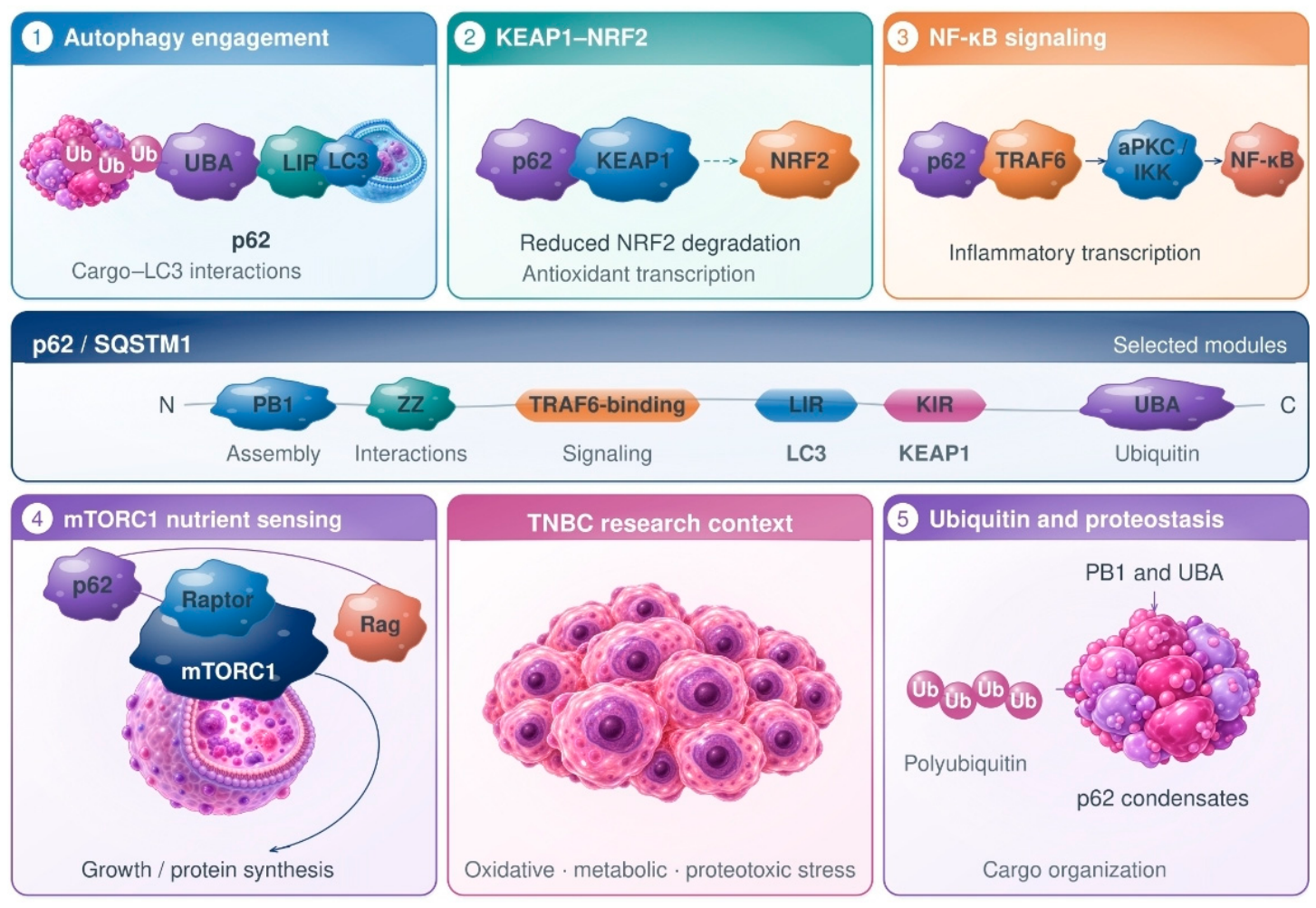
p62 uses distinct structural regions to organize cargo and engage its binding partners. Its Phox and Bem1 (PB1) domain mediates oligomerization, and the ubiquitin-associated domain binds ubiquitinated cargo. Binding to microtubule-associated protein 1 light chain 3 (LC3) and gamma-aminobutyric acid type A receptor-associated protein (GABARAP) family proteins is mediated by the LC3-interacting region (LIR). The region that binds Kelch-like ECH-associated protein 1 (KEAP1) connects cargo organization with nuclear factor erythroid 2-related factor 2 (NRF2) signaling [22,23,24]. Other interactions link p62 to nutrient sensing by mechanistic target of rapamycin complex 1 (mTORC1) [25]. In experimental systems, p62 also couples tumor necrosis factor receptor-associated factor 6 (TRAF6) signaling to the activation of the nuclear factor kappa-light-chain-enhancer of activated B cells (NF-κB) through atypical protein kinase C (aPKC) and the IκB (inhibitor of NF-κB) kinase (IKK) complex [26]. Polyubiquitin promotes p62 condensation, while phosphorylation alters cargo binding and condensate processing [23,24,27]. Figure 1 summarizes this experimental evidence, which should not be read as a TNBC-specific validation of every interaction.
At steady state, p62 abundance and localization reflect its synthesis, degradation, redistribution and organization into condensates. Phosphorylation at serine residue 403 (Ser403) alters the handling of ubiquitinated cargo without identifying that cargo as mitochondrial [22,23,24,27,28]. Similar staining patterns can therefore arise from different biological processes. Pathological assessment should distinguish cellular and subcellular compartments and interpret p62 alongside independent measurements of damage and mitochondrial content. Staining categories alone cannot resolve turnover; the experiments needed to assess it are described in Section 6.
Figure 1.
Selected p62 interactions and signaling pathways. Blue marks autophagy engagement, teal marks KEAP1–NRF2 signaling and orange marks NF-κB signaling. Purple identifies the nutrient-sensing and proteostasis panels, and magenta identifies the TNBC context. The strip arranges domains and binding regions in sequence order without representing their relative lengths or residue coordinates [14,15,16,23,24]. The numbered scenes illustrate (1) engagement of ubiquitin-linked cargo with p62 and LC3 [23,24]; (2) KIR–KEAP1 binding and indirect stabilization of NRF2 [22]; (3) TRAF6-associated aPKC/IKK signaling to NF-κB [26]; (4) separate interactions of p62 with Raptor and Rag in amino-acid-dependent mTORC1 signaling at the lysosomal surface [25]; and (5) PB1/UBA-associated cargo condensation and processing [23,24,27]. Rag regulates mTORC1 recruitment but is not a core subunit of the complex. The TNBC illustration provides the disease context and does not indicate tumor-specific validation of these interactions. Clustering mediated by p62 does not establish that p62 is required for mitochondrial degradation [20]. Alternative receptor mechanisms include OPTN/NDP52, BNIP3/NIX and FUNDC1 [21,29,30,31]. Supplementary Table S2 identifies the sources and evidence limits for individual connectors. Abbreviations: p62, sequestosome 1 protein; SQSTM1, the gene encoding p62; Ub, ubiquitin; PB1, Phox and Bem1 protein-interaction domain; ZZ, ZZ-type zinc-finger domain (conventional domain designation); UBA, ubiquitin-associated domain; LIR, LC3-interacting region; KIR, KEAP1-interacting region; LC3, microtubule-associated protein 1 light chain 3; KEAP1, Kelch-like ECH-associated protein 1; NRF2, nuclear factor erythroid 2-related factor 2; TRAF6, tumor necrosis factor receptor-associated factor 6; aPKC, atypical protein kinase C; IKK, IκB kinase (IκB, inhibitor of nuclear factor κB); NF-κB, nuclear factor kappa-light-chain-enhancer of activated B cells; mTORC1, mechanistic target of rapamycin complex 1; Raptor, regulatory-associated protein of mechanistic target of rapamycin complex 1; Rag, Ras-related guanosine triphosphate-binding proteins; TNBC, triple-negative breast cancer; OPTN, optineurin; NDP52, nuclear dot protein 52; BNIP3, B-cell lymphoma 2-interacting protein 3; NIX, B-cell lymphoma 2-interacting protein 3-like protein; FUNDC1, FUN14 domain-containing protein 1. N and C indicate the amino and carboxyl termini, respectively.
2.2. Ubiquitin-Dependent Recruitment and Mitochondrial Receptor Pathways
When mitophagy is triggered after mitochondrial depolarization through phosphatase and tensin homolog (PTEN)-induced kinase 1 (PINK1) and the ubiquitin ligase Parkin, ubiquitin signals recruit receptors such as optineurin (OPTN) and nuclear dot protein 52 (NDP52), also known as calcium-binding and coiled-coil domain-containing protein 2 (CALCOCO2). Knockout and reconstitution experiments identify these proteins as important recruiters of the autophagy machinery [21]. p62 can cluster damaged mitochondria without being required for their degradation [20], a distinction between organization and disposal that is also addressed by recent condensation studies [32]. Mechanistic validation should therefore include OPTN and NDP52 even when practical considerations favor p62 as an exploratory tissue stain.
B-cell lymphoma 2-interacting protein 3 (BNIP3) and B-cell lymphoma 2-interacting protein 3-like protein (BNIP3L, also known as NIX) engage the autophagy machinery at mitochondria without requiring the same Parkin-dependent ubiquitin cascade. NIX receptor function was established in experimental systems outside TNBC [30]. In a mammary tumor model, Bnip3 loss increased abnormal mitochondrial accumulation and tumor progression, with re-expression providing support for a causal relationship [29]. The human analyses in that study examined copy number and expression, including 113 TNBC cases in the copy number comparison and a 101-case TNBC expression–survival series. These observations do not validate BNIP3 immunohistochemistry (IHC) in human TNBC. FUN14 domain-containing protein 1 (FUNDC1) mediates another hypoxia-regulated receptor route described in non-TNBC experimental systems [31]. These alternative pathways explain why a negative PINK1/Parkin or p62 result does not exclude mitochondrial disposal.
Mitochondrial clearance also includes routes that export material from cells without lysosomal destruction. Vykoukal et al. demonstrated vesicle-associated clearance in TNBC models and linked its disruption to changes in sphingolipids, respiration and tumor growth [33]. Distinguishing intracellular lysosomal delivery from extracellular export and transfer to neighboring cells is therefore essential when assigning a clearance mechanism.
3. Human Evidence in TNBC
3.1. p62 Immunohistochemistry in Human TNBC
Luo et al. assessed p62 in 163 TNBCs using tissue microarrays with triplicate cores from each tumor and classified expression as high in 51 cases (31.3%). Ten-year disease-free survival was 54.6% in the high-expression group and 78.2% in the low-expression group; the corresponding distant metastasis-free survival estimates were 58.9% and 92.5% [17]. These findings associate p62 staining with outcome in a retrospective cohort, but mitochondrial turnover was not measured. The study used clone D-3 and an intensity–proportion score, although the Methods and Results describe cutoff selection differently. Assessment by a single reader, tissue-microarray sampling and historical receptor definitions limit the application of these categories to other cohorts.
Kim et al. examined 119 TNBCs and found higher nuclear p62 in the molecular-apocrine and claudin-low categories. Their multivariable analysis associated p62 negativity with shorter disease-free and overall survival [18], a direction that differs from the findings of Luo et al. The studies assessed different compartments and used different scoring systems and case definitions, so they do not support a common high-versus-low threshold. A mixed series of 489 breast cancers also showed that autophagy marker expression varied by subtype and compartment [34]. That study provides context for interpreting staining patterns, rather than an independent TNBC-specific validation of mitophagy.
Wang et al. examined 107 breast cancers, including 31 TNBCs, before neoadjuvant chemotherapy and in the residual tumors available after treatment [19]. In total, 12 TNBC patients achieved study-defined pCR (Grade 3), while 19 were classified as non-pCR. Baseline p62 categories were available for 30 TNBCs (20 high and 10 low), compared with 19 after treatment (9 high and 10 low). Because these denominators differ, the category counts cannot be interpreted as a within-patient decrease in p62. The report provided neither a TNBC-specific paired effect size nor a paired-change test. Its recurrence model combined breast cancer subtypes, and the adjusted association with p62 did not reach statistical significance. The study therefore supports the feasibility of examining p62 before and after treatment, while leaving the direction and magnitude of TNBC-specific adaptation unresolved.
3.2. Complementary Human Studies and Limits of Interpretation
Lee H. et al. assessed nuclear and cytoplasmic p62 in 439 TNBC resections from patients who had not received neoadjuvant therapy. The material comprised temporal cohorts of 218 and 221 cases [35], and cytoplasmic p62 correlated with cyclooxygenase-2 (COX-2) in both. These are static associations between markers of cellular stress and autophagy. The same publication also examined 37 patient-derived xenograft (PDX) tumors; those findings come from models and should be considered separately from the clinical resection findings.
Other human studies provide information about redox responses. Nuclear NRF2 and cytoplasmic KEAP1 were examined in operable TNBC [36], while another series related pretreatment phosphorylated NRF2 to response after neoadjuvant therapy [37]. These observations do not by themselves show that p62 activates NRF2 or that mitochondria are the source of the oxidants. A further study assessed hypoxia- and autophagy-related stains in pretreatment biopsies from 57 TNBC patients receiving dose-dense doxorubicin/cyclophosphamide plus paclitaxel, with or without carboplatin [38]. The exploratory response associations in the pooled cohort were not significant within the individual regimen strata and do not establish mitophagy. Complementary evidence comes from the human genomic and transcriptomic BNIP3 analyses reported by Chourasia et al. [29].
Taddei et al. examined nuclear NRF2 staining and MET receptor tyrosine kinase expression by IHC in 15 TNBC biopsies [39]. In a separate publication, a reanalysis of the PROMIX dataset of HER2-negative breast cancers found oxidative phosphorylation (OXPHOS) enrichment in a subset of residual tumors with suppression of MYC, a proto-oncogene encoding a transcription factor [40]. The same publication reported similar B-cell lymphoma-extra-large protein (BCL-XL) staining in paired baseline and residual tumors from 10 patients treated with anthracycline-based neoadjuvant therapy.
In 79 TNBCs treated with doxorubicin/cyclophosphamide followed by paclitaxel, Liu et al. associated higher tumor necrosis factor receptor superfamily member 19 (TNFRSF19) IHC with pCR and Miller–Payne grades 4–5 [41]. The study defined pCR as ypT0 ypN0: no residual invasive or in situ breast carcinoma, and no regional nodal metastasis after neoadjuvant treatment. Promoter methylation was measured by quantitative methylation-specific polymerase chain reaction (qMSP) in the same institutional cohort. Survival associations in public gene-expression datasets were analyzed separately. These associations do not establish differential treatment benefit.
Further studies associated SQSTM1 expression with epithelial–mesenchymal transition (EMT)/stemness [42] and developed a mitophagy-related prognostic signature [43]. These expression-based findings address different questions from protein localization or mitochondrial turnover. Their independence as evidence depends on checking for overlap between the public datasets. Supplementary Table S3 summarizes the selected evidence by type. Table 1 presents the findings from human tissue and molecular studies.
Table 1.
Selected human observations relevant to p62 and companion measurements.
4. Exploratory Tissue Measurements and Candidate Profiles
4.1. Morphology and Compartment-Specific p62 Assessment
Morphological assessment begins with hematoxylin and eosin (H&E) examination to identify viable invasive carcinoma, necrosis, stroma, inflammatory cells and treatment effects. High grade, geographic necrosis and a lymphocyte-rich border can be seen in basal-like and triple-negative breast cancers [46,47], but these features do not define a mitochondrial state. Apocrine differentiation also warrants attention because granular eosinophilic cytoplasm may already be a feature of the tumor at baseline; its distinctive clinicopathological context is illustrated by a series of 42 cases [48]. Histological type and diagnostic mimics also guide the choice of further immunohistochemical and molecular tests [49].
Cytoplasmic eosinophilia is a morphological observation; attributing it to mitochondrial enrichment requires organelle-specific evidence [50]. Three-dimensional (3D) electron microscopy has also documented mitochondrial remodeling in TNBC PDXs [51], although these remain model observations rather than serial measurements in patients.
A useful p62 assessment distinguishes staining in tumor epithelium, stroma and immune cells. Within tumor cells, nuclear staining, diffuse cytoplasmic staining, puncta and larger inclusions represent separate patterns. Possible exploratory measurements include a continuous cytoplasmic H-score (0–300), the percentage of nuclear-positive cells and the density of puncta per viable tumor area. Defining the cell population, numerator and denominator makes these measurements interpretable and distinguishes cell-based proportions from area-normalized measurements [52]. Nuclear positivity and puncta density therefore remain separate variables. These p62 scoring systems have not been clinically validated. Nuclear and cytoplasmic p62 patterns have been recorded in meningioma and chordoma [53,54], but their cutoffs and prognostic associations cannot be transferred to TNBC. Figure 2 brings together the possible explanations for high and low p62 measurements and the independent evidence needed to distinguish them.
Figure 2.
Competing explanations for steady-state p62 abundance. Orange and blue identify possible explanations for high and low p62, respectively. High p62 can reflect increased synthesis, impaired degradation or changes in localization and condensation; low p62 can reflect increased p62 degradation, reduced synthesis or compartment and sampling effects [22,23,24,27,28,55]. Neither high nor low p62 alone establishes mitochondrial turnover or a treatment dependency. The lower panels outline the complementary evidence needed to distinguish these explanations: dynamic measurements of mitochondrial delivery and cargo turnover in living models, pathway-specific receptor context, mitochondrial content and function, and tumor cell, spatial and treatment context [20,21,28,29,30,31,56,57,58]. Arrows organize the competing explanations and the tests used to investigate them; they do not assign a mechanism to an individual tumor. Abbreviations: p62, sequestosome 1 protein; SQSTM1, the gene encoding p62; ROS, reactive oxygen species; OPTN, optineurin; NDP52, nuclear dot protein 52; BNIP3, B-cell lymphoma 2-interacting protein 3; NIX, B-cell lymphoma 2-interacting protein 3-like protein; FUNDC1, FUN14 domain-containing protein 1.
A reproducibility study would use at least two blinded observers to score prespecified regions after calibration with reference images. Absolute-agreement intraclass correlation coefficients (ICCs), reported with confidence intervals, quantify agreement for continuous measurements; weighted kappa applies to ordered categories. Repeat scoring and changes in region selection help identify sources of variability. Digital segmentation requires an independent tumor mask and manual review of errors, particularly in necrotic or lymphocyte-rich areas. The proportion of interpretable measurements is reported separately from agreement among evaluable specimens. These performance estimates require testing directly in TNBC material.
In metastatic specimens, mammary origin should be confirmed through clinicopathological assessment before stress markers are interpreted. Combined staining for transcriptional repressor GATA binding 1 (TRPS1) and GATA-binding protein 3 (GATA3) can support this lineage assessment in ER-negative or ER-low breast carcinoma [59].
4.2. Candidate Panel and Analytical Qualification
An initial exploratory panel could combine compartment-specific p62 with microtubule-associated protein 1 light chain 3 beta (LC3B) assessment. Mitochondrial content could be assessed using translocase of outer mitochondrial membrane 20 (TOMM20/TOM20), with cytochrome c oxidase subunit IV (COX IV) as an orthogonal marker. An oxidative-damage measure such as 4-hydroxy-2-nonenal (4-HNE) or 8-hydroxy-2′-deoxyguanosine (8-OHdG) could be added alongside nuclear NRF2. Lysosome-associated membrane protein 1 (LAMP1) would provide the corresponding compartment context. Additional measurements could include phospho-p62 Ser403 and BNIP3/NIX once antibody performance has been established, with verification of phosphoepitope preservation for phospho-p62. The inclusion of OPTN, NDP52, FUNDC1, PINK1, Parkin, dynamin-related protein 1 (DRP1) or optic atrophy 1 mitochondrial dynamin-like protein (OPA1) should depend on the mechanism under investigation; these markers need not be applied to every biopsy.
The LC3 isoform and staining pattern determine what is being scored. The “stone-like” structures associated with the alpha isoform of LC3 (LC3A) in endometrial pathology [60,61] differ from the LC3B puncta proposed here; combining them would obscure that distinction.
Commercial availability or use in a previous publication is insufficient to establish an antibody’s analytical validity in formalin-fixed, paraffin-embedded (FFPE) TNBC. Qualification examines localization, agreement with immunoblotting or another independent assay, positive and negative controls, variation between lots and platforms, and sensitivity to ischemia and fixation. Genetically depleted and reconstituted FFPE cell blocks provide useful controls where feasible, with phosphatase-sensitive controls for phosphorylation-specific staining. The comparison of TOMM20 and COX IV with independent measures of mitochondrial content is particularly important: altered protein expression or selective membrane protein degradation can change either stain without a proportional loss of organelles. Neither stain establishes respiratory competence.
Documented cold ischemia, fixation and block storage conditions help assess pre-analytical variation. Staining paired core-biopsy and resection specimens in balanced batches with shared controls addresses an additional source of variability. Interpretation also depends on what each assay measures. Total DRP1 staining does not measure fission activity, while total OPA1 staining does not resolve protein processing or establish fusion competence. PINK1 stabilization and Parkin recruitment to mitochondria are difficult to establish from diffuse FFPE immunoreactivity. NRF2 is interpreted through its nuclear staining, whereas KEAP1 abundance alone cannot demonstrate functional sequestration by p62. No antibody clone or diagnostic cutoff is endorsed for the exploratory panel; published reagents are provided as source identifiers to support reproducibility. Table 2 summarizes the exploratory status of each candidate measurement.
Table 2.
Exploratory measurements and published reagents for FFPE TNBC.
4.3. Minimum Profile Definitions and Falsification Criteria
A candidate tissue profile combines measurements of mitochondrial content, damage and compartment-specific stress or autophagy in the same registered region of viable tumor. Profiles A and C require concordant TOMM20 and COX IV results. Profile B uses a qualified TOMM20 or COX IV assay selected before analysis; when both are measured, content is indeterminate if one is reduced and the other is preserved or increased. Increased 4-HNE or 8-OHdG provides the oxidative damage criterion. The attribution of that damage to mitochondria requires validated subcellular colocalization; the registration of serial sections establishes correspondence between regions, not between individual organelles. The profile names describe the observed tissue pattern, while functional experiments test its proposed mechanism.
The three research profiles in Table 3 describe damage accompanied by preserved or enriched mitochondrial content and redox signaling, damage accompanied by cargo or lysosome-associated accumulation, and damage accompanied by reduced mitochondrial content. Their expected marker directions refer to a predefined reference distribution or a matched baseline. Continuous measurements are preferable during assay development. Where research categories are needed, their reference percentiles are fixed in a training set before outcome testing; a paired change qualifies only if it exceeds the assay-specific 95% repeatability limit established from replicate measurements. Any resulting thresholds require independent validation, and no universal numerical cutoff for p62 or mitochondrial stress is currently supported. Figure 3 shows how the profiles can be investigated in separate baseline and residual-disease study designs.
Table 3.
Minimum definitions of candidate tissue profiles for research.
Figure 3.
Separate study designs for baseline response and residual disease. (A) Pretreatment features are examined for associations with pCR and RCB under a defined neoadjuvant regimen. Here, pCR is ypT0/Tis ypN0, corresponding to RCB 0 [11,12,13,65]. (B) Paired analysis requires adequate viable carcinoma from the same anatomical compartment at both time points. Residual disease confined to lymph nodes does not provide a paired-change score for the breast. Observed differences can reflect adaptation, selection or sampling; bias from comparing core biopsies with resections requires independent calibration. (C) Three exploratory profiles, which may overlap, specify expected marker directions. Table 3 defines marker requirements, proposed dependencies and falsification criteria. Marker directions are assessed in registered viable-tumor regions against a predefined reference or matched baseline, with normalization to viable cells. Mitochondrial attribution of damage requires colocalization. No validated cutoffs are available. Active delivery and impaired disposal remain alternative explanations for the middle profile. Arrows in (A,B) indicate the study sequence. In (C), ↑, ↓ and ↔ indicate increased, decreased and preserved marker levels, respectively. The blue, purple and teal headers identify panels (A–C), respectively. Abbreviations: Chemo, chemotherapy; ICI, immune checkpoint inhibitor; BRCA, breast cancer susceptibility genes 1 and 2; HRD, homologous recombination deficiency; pCR, pathological complete response; RCB, residual cancer burden; TOMM20, translocase of outer mitochondrial membrane 20; COX IV, cytochrome c oxidase subunit IV; 4-HNE, 4-hydroxy-2-nonenal; 8-OHdG, 8-hydroxy-2′-deoxyguanosine; NRF2, nuclear factor erythroid 2-related factor 2; p62, sequestosome 1 protein; LC3B, microtubule-associated protein 1 light chain 3 beta; LAMP1, lysosome-associated membrane protein 1. ypT0/Tis ypN0 denotes post-neoadjuvant pathological stage with no residual invasive breast carcinoma (in situ disease may remain) and no regional nodal metastasis.
A tumor can contain more than one profile, so classification applies to registered regions of viable carcinoma.
5. Regimen-Specific Experimental Evidence
5.1. Chemotherapy and Mitochondrial Adaptation
Studies in cell lines and patient-derived models show distinct responses to cytotoxic treatment. Baek et al. linked survival after DNA-damaging chemotherapy to mitochondrial elongation associated with OPA1, increased oxidative metabolism and regrowth [63]. After anthracycline exposure, Andrzejewski et al. identified an OXPHOS-high state dependent on BCL-XL. Responses varied across cell lines and differed from those in cells persisting after taxanes [40]. Respiratory vulnerability has been demonstrated in selected TNBC models [45,66], but greater mitochondrial content alone does not establish respiratory dependence.
The PDX study using 3D electron microscopy reconstructed 3750 mitochondria and 800 lipid droplets across models and treatment conditions [51]. These are nested organelle observations, not independent patients or animals. Another PDX study linked a basal population enriched in chromosome 15 open reading frame 48 (C15ORF48) to mitochondrial reactive oxygen species (ROS) regulation after doxorubicin/cyclophosphamide [67]. Studies of giant-cell states induced by paclitaxel [68], cells tolerant to doxorubicin or paclitaxel [64], and MDA-MB-231 cells depleted of transient receptor potential mucolipin 1 (TRPML1) [69] addressed different exposures and selection histories. Their autophagy and lysosome findings should not be pooled as a single validated TNBC phenotype.
In breast cancer models, Parkin suppression or microRNA-218-5p (miR-218-5p) enhanced the effects of doxorubicin [70]. Experiments involving TNFRSF19 suggest a separate mitophagy hypothesis specific to doxorubicin [41]. Polyphyllin I induced mitochondrial injury and changes associated with mitophagy, yet PINK1 depletion increased apoptosis [71], consistent with a protective contribution from clearance. Isorhamnetin combined with chloroquine linked mitochondrial fission and apoptosis through signaling involving ROS, calcium/calmodulin-dependent protein kinase II (CaMKII) and DRP1 [72]. This association does not establish excessive mitochondrial clearance as the cause of death.
5.2. Non-Mitochondrial p62 Functions and Resistance to Poly(adenosine Diphosphate-Ribose) Polymerase Inhibitors
Post-transcriptional regulation can raise p62 without impaired degradation. An SQSTM1 transcript with a short 3′ untranslated region (UTR) promoted chemoresistance in breast cancer [55]. Autophagy inhibition can also generate condensates containing the NBR1 autophagy cargo receptor and p62, affecting itchy E3 ubiquitin-protein ligase (ITCH), tumor protein p63 and metastatic programs [73].
In cells lacking secretory carrier membrane protein 3 (SCAMP3), the inhibition of extracellular signal-regulated kinases 1 and 2 (ERK1/2) increased p62 and lipidated LC3B [74]. The peptide WK-13-3D impaired bulk autophagic flux and p62 degradation [75], while experiments with compound 7h associated nuclear p62 accumulation with impaired DNA repair [76]. These studies show how changes in p62 can arise from processes outside mitochondrial quality control.
Steady-state p62 was reduced in olaparib-resistant SUM159 and MDA-MB-468 models. Both had intact breast cancer susceptibility genes 1 and 2 (BRCA1/2), although homologous recombination deficiency (HRD) was reported for MDA-MB-468 [77]. Nitro-oleic acid experiments included resistant TNBC models with mutant BRCA1 and implicated p62 modification at cysteine residues 105 and 113 (Cys105/Cys113) and disruption of the autophagy–lysosome system [78]. These studies address different molecular contexts. Neither establishes a tissue p62 cutoff that predicts benefit from poly(adenosine diphosphate-ribose) polymerase (PARP) inhibition in patients.
5.3. Immunotherapy and Radiotherapy: Distinct Treatment Contexts
In TNBC models, the p62-dependent degradation of tenascin-C supports T-cell-mediated killing [44]. The cargo in this mechanism is an extracellular matrix protein, so the finding does not identify a mitophagic mechanism of checkpoint-inhibitor response. Human spatial analyses such as NeoTRIP examine response to specific neoadjuvant chemotherapy and immunotherapy combinations [79]. Evidence on survival with immunochemotherapy in advanced or metastatic disease addresses a different clinical question [80]. Programmed death-ligand 1 (PD-L1) assessment in metastatic TNBC also depends on the assay and the cell compartments included in scoring [81].
Radiotherapy studies implicate the HECT-domain E3 ubiquitin-protein ligase 3 (HECTD3; HECT, homologous to the E6-associated protein carboxyl terminus)–p62 axis and a program involving p62 and vascular cell adhesion molecule 1 (VCAM-1) [82,83]. These model studies connect p62-associated signaling with DNA repair and general autophagy during radiation responses. Comparisons between biomarker studies also depend on clearly defined treatment and sampling times [84].
5.4. Therapeutic Hypotheses and the Standard for Lethal Clearance
Studies in experimental models have tested respiratory inhibition, lysosomal disruption, signaling involving MET, the non-receptor tyrosine kinase SRC and NRF2, and drug delivery targeting mitochondria as treatment strategies [39,45,85,86].
In a study of carboplatin, the depletion of autophagy-related protein 5 (ATG5) did not change sensitivity to carboplatin or BMS-754807 [85]. Autophagy inhibition alone therefore did not explain the effects of hydroxychloroquine.
Bi-magnolignan studies combined measurements of mitochondrial delivery using the mitochondria-targeted Keima (mt-Keima) reporter with ultrastructural analysis, the assessment of depolarization and apoptosis, and the perturbation of TANK-binding kinase 1 (TBK1; TANK, tumor necrosis factor receptor-associated factor family member-associated NF-κB activator) [62]. Studies of redox-responsive conjugates also linked mitochondrial injury and mitophagy-associated measurements to antitumor effects [87]. Whether clearance contributes to cell death remains a hypothesis, because TBK1 pleiotropy and other drug effects offer alternative explanations. A causal interpretation requires selective clearance blockade to preserve mitochondrial mass, respiration and long-term viability under comparable initial exposure and injury. Restoring the targeted pathway should then reinstate mitochondrial clearance and cell death (Section 6). Table 4 compares selected experiments by model, exposure, endpoint and strength of causal evidence.
Table 4.
Selected preclinical experiments relevant to mitochondrial stress in TNBC.
6. Functional Testing of Tissue-Based Hypotheses
6.1. Measuring Mitochondrial Delivery and Turnover
Functional validation asks whether a tissue pattern corresponds to its proposed mechanism (Figure 4), using models matched to the source tumor compartment, molecular context and exact treatment exposure. Time-resolved mt-Keima or the mitochondrial quality control reporter (mito-QC) can track mitochondrial delivery into acidic compartments, with signals normalized to viable cells and reporter expression [56,57]. Interpretation requires confirmed mitochondrial targeting, lysosomal colocalization and an established positive/negative dynamic range. These reporters capture delivery and accumulated signal, so a single endpoint cannot provide a complete degradation-rate estimate. Sensitivity also depends on the reporter: a direct comparison found mito-QC less sensitive to events dependent on PINK1 and the Parkin ubiquitin ligase, encoded by PRKN [58]. A negative result requires an adequate pathway-specific dynamic range for interpretation. Tandem LC3 reporters assess general autophagy and cannot replace a mitochondrial reporter [28].
Figure 4.
Functional testing of a candidate tissue profile. (A) Models matched to the tumor and treatment context combine time-resolved measurements of mitochondrial delivery and cargo turnover with independent assays of content, membrane potential, respiration, injury and survival [28,56,57,58]. Reporter interpretation requires pH and proteolytic controls and independent cargo-turnover measurements, because lysosomal alkalinization changes reporter signals. (B) Pathway-specific loss-of-function experiments, rescue with a functional wild-type construct and informative mutants test the proposed dependency [20,21,29]. (C) Dashed arrows lead to the adaptive-clearance hypothesis (teal) and the death-associated-clearance hypothesis (orange). Upward and downward arrows denote increases and decreases, respectively. Selective clearance blockade is expected to impair survival when clearance is adaptive, or to preserve mitochondrial content, respiration and survival when clearance contributes to death. These comparisons require comparable initial drug injury; a discordant response challenges the proposed interpretation. Abbreviations: mt-Keima, mitochondria-targeted Keima fluorescent reporter; mito-QC, mitochondrial quality control fluorescent reporter; pH, measure of acidity defined by the negative base-10 logarithm of hydrogen-ion activity; TMRM, tetramethylrhodamine methyl ester; TMRE, tetramethylrhodamine ethyl ester; OCR, oxygen-consumption rate; ROS, reactive oxygen species; TNBC, triple-negative breast cancer.
Short, titrated lysosomal-blockade controls help interpret mitochondrial cargo turnover and LC3 processing, alongside measurements of lysosomal acidity (pH) and cell toxicity. Bafilomycin and chloroquine alter the acidic environment on which mt-Keima and mito-QC depend, so reporter changes after alkalinization cannot be interpreted by simple inhibitor subtraction. Calibrated reporter kinetics should be considered with independent measurements of matrix and membrane cargo loss/turnover, lysosomal protease controls where appropriate, and the genetic interruption of autophagosome delivery [28,56,57]. Electron microscopy adds structural evidence of sequestration without establishing a rate.
6.2. Measuring Mitochondrial Content, Membrane Potential and Respiration
The functional assessment of mitochondrial content combines at least two orthogonal measurements, such as TOMM20/COX IV imaging, the mitochondrial DNA (mtDNA)-to-nuclear DNA ratio and citrate synthase activity. Cell size, ploidy and cell death influence these readouts and their interpretation. Membrane-potential measurements with tetramethylrhodamine methyl ester (TMRM) or tetramethylrhodamine ethyl ester (TMRE) are interpreted against a depolarization control and normalized to mitochondrial content; dye fluorescence is not an independent measure of mass. Respiratory assessment includes appropriately normalized basal, adenosine triphosphate (ATP)-linked and maximal oxygen consumption, spare respiratory capacity and non-mitochondrial oxygen consumption. Compartment-appropriate probes and interference controls distinguish ROS signals from nonspecific oxidation or assay effects [19,28,29,63].
A respiratory dependency is supported when orthogonal respiratory perturbations reduce clonogenic survival or regrowth; high oxygen consumption alone is insufficient. For redox dependence, genetic or pharmacological pathway manipulation is paired with suitable rescue and controls for direct effects of antioxidants on the compound. Replication in more than one biologically relevant model helps establish whether the relationship extends beyond a single experimental system. Animals or independent cultures provide the biological replicates; individual cells and organelles are nested measurements.
6.3. Genetic Perturbation, Rescue and Tests of the Proposed Mechanism
The choice of perturbation should follow the pathway observed in the model: PINK1/Parkin, OPTN/NDP52, BNIP3/NIX or FUNDC1, with SQSTM1 targeted only when p62 dependence is being claimed. Testing pathway redundancy helps clarify the basis of the response. Further support comes from using at least two independent targeting reagents and performing rescue with an appropriately expressed wild-type construct resistant to the targeting reagent. Cargo-binding or LIR-defective mutants can help separate signaling and cargo organization from delivery. The inhibition of ATG5, autophagy-related protein 7 (ATG7) or lysosomal function alone cannot establish that an effect is specific to mitochondria [20,21,28,29,30,31].
An adaptive role for clearance is supported when its selective interruption reduces mitochondrial delivery, increases damaged organelle burden and impairs survival under matched exposure. Restoring the functional pathway should recover delivery and turnover and reverse the survival defect. A lethal-clearance mechanism predicts the opposite survival effect—interruption preserves mitochondrial content, respiration and clonogenic survival despite comparable initial injury, while pathway restoration reinstates clearance and cell death. Failure to observe the predicted survival change after verified blockade challenges the proposed dependency under the conditions tested. Death preceding organelle loss specifically challenges lethal clearance. Rescue through downstream or compensatory signaling alone neither establishes nor excludes clearance-specific causality. Together, these experiments test the damage–clearance–recovery hypothesis under defined experimental conditions.
7. Clinical Study Design, Statistical Analysis and Feasibility
7.1. Baseline Markers and Treatment Response
Baseline response studies encompass consecutive eligible patients with pretreatment TNBC, including those who subsequently achieve pCR. Here, pCR denotes no residual invasive breast carcinoma (in situ carcinoma may remain) and no regional nodal metastasis after neoadjuvant treatment (ypT0/Tis ypN0), with RCB recorded according to an established protocol [11,12,13]. Logistic regression can relate a prespecified continuous marker or small panel to pCR, with stage, regimen and relevant molecular features chosen in advance. A model for RCB accounts for its distribution, including the concentration of observations at zero in complete responders; one option is a two-part model separating pCR from positive RCB. These analyses estimate associations with response under the regimen studied. Evidence that a marker predicts benefit from one treatment over another instead comes from a comparative design with a prespecified marker-by-treatment interaction.
7.2. Sampling and Change in Residual Disease
Paired analyses examine how measured profiles differ after treatment among patients with residual disease. They require adequate viable carcinoma from the same anatomical compartment at both time points. A breast analysis therefore compares the pretreatment breast core with residual invasive breast carcinoma. Patients with residual nodal disease but no invasive breast tumor remain eligible for the baseline outcome analysis, but cannot contribute a breast paired-change measurement. Nodal adaptation requires its own pretreatment nodal specimen and a separately specified post-treatment analysis. The absence of residual tumor has no corresponding marker score and cannot be coded as zero expression. Reporting includes the selection of pairs, comparisons between included and excluded non-pCR patients, and tissue adequacy separately from specimen availability. Weighting can address some measured sources of selection, but cannot recover a post-treatment tumor profile where no tumor remains.
Core biopsies and resections sample different volumes and regions and often undergo different fixation conditions. Multiregion baseline sampling, where feasible, together with consistent cell compartments, viable-tumor masks and replicate regions can help characterize this variability. The original tumor bed, residual cellularity and nodal disease should be documented according to neoadjuvant reporting recommendations [65]. Spatial studies show how tissue composition influences interpretation [79,88,89,90,91,92,93], although exact correspondence between regions before and after treatment remains unavailable. Specimen and patient counts should therefore be reported separately. Even with careful sampling, a paired difference alone cannot distinguish treatment-induced adaptation from the selection of a pre-existing cell population.
Mixed-effects models can account for repeated continuous measurements, regions nested within patients and time points, and identifiable batch or technical effects. A fundamental limitation remains when all baseline samples are cores and all post-treatment samples are resections: specimen type is inseparable from time and treatment, and regression adjustment cannot resolve that confounding. An independent core–resection calibration set or matched processing experiment is needed to estimate the technical contribution. Changes would be reported with confidence intervals and the proportion exceeding analytical repeatability limits. Associations between residual-disease profiles and subsequent outcomes would be evaluated from a prespecified post-surgery landmark, accounting for RCB and other justified covariates. Use of a Cox model requires assessment of the proportional-hazards assumption.
7.3. Recording Treatment and Molecular Context
Treatment records need to identify the exact agents, doses and schedules, clinical setting, interval from the last dose to sampling, prior exposures and subsequent therapy. Anthracycline/cyclophosphamide, taxane and platinum components are recorded separately, together with any concurrent checkpoint inhibitor. For PARP inhibitors, the relevant context includes neoadjuvant, adjuvant or metastatic use; germline and somatic BRCA1/2 status; the HRD assay and cutoff; and previous platinum or PARP-inhibitor exposure. An HRD-positive research category is not equivalent to established BRCA-based clinical selection. Radiotherapy records also include dose, fractionation and concurrent drugs. Analyses that combine these exposures need explicit interactions or separate strata to preserve the distinctions between their selective pressures.
7.4. Statistical Analysis and Sample-Size Planning
This review involved no new statistical testing; all numerical findings are attributed to the original studies. For future investigations, continuous measurements preserve more information than outcome-optimized cutoffs, and a limited set of candidate parameters helps contain overfitting. Prespecifying the primary profile and endpoint separates confirmatory comparisons from exploratory analyses. Secondary panels call for the control of false discovery, with confidence intervals reported alongside effect estimates. Patient-level bootstrap resampling supports internal validation and correction for optimism; a separate temporal or external cohort assesses performance independently. Both discrimination and calibration are relevant to prediction models. A prespecified missing-data plan distinguishes missing covariates, failed staining and exclusions for insufficient tumor. Multiple imputation is appropriate only when its assumptions are defensible and cannot substitute for a residual tumor that is absent.
A pilot study establishes tissue adequacy and staining success, sensitivity to region selection and repeatability between observers. The practical losses can be substantial; in a pretreatment TNBC study, 24 of 31 BRCA1/BRCA2-tested women met the specimen, panel and exposure eligibility criteria [94]. That immune/stromal study illustrates eligibility attrition in a selected clinical population, rather than validation of mitochondrial profiles. Qualifying a small core panel before adding receptor or phosphoprotein measurements in informative subsets could conserve biopsy material. Multiplex assays need checks for spectral overlap and antibody interference, while serial sections depend on accurate registration. Once analytical feasibility is established, formal sample-size planning considers outcome frequency, candidate parameter count and anticipated performance, rather than a fixed events-per-variable rule [95]. For external validation, a separate sample size calculation aims to estimate calibration and discrimination with adequate precision.
The number of usable pairs can be far smaller than the number of patients enrolled. For illustration, if 200 patients enter a study, 60% achieve pCR and paired material is inadequate in 15% of the remainder, only 200 × 0.40 × 0.85 = 68 pairs would be available. These are assumed planning values, not observed results or a recommended sample size. Further division by regimen or BRCA status, together with a limited number of survival events, would restrict model complexity. Multicenter recruitment and a small panel, developed first analytically and then biologically, are likely to be more feasible than deriving a large multiplex classifier from a small single-center series.
7.5. Reporting and Independent Validation
Reproducible reporting allows another laboratory to reconstruct the assay and its interpretation. This requires the antibody clone or catalog number, dilution, retrieval, controls, platform and batch, together with cold ischemia, fixation and block age. Cellular and subcellular compartments, scoring and segmentation rules, region selection, blinding, observer agreement and the origin of each cutoff also need explicit definitions. Clinical annotation includes standard receptor status, stage, morphology, tumor-infiltrating lymphocytes (TILs), treatment, BRCA/HRD context and pCR/RCB. Independent clinical validation uses the assay, threshold and analysis plan fixed during development. Comparing the panel with its individual markers under a prespecified decision rule and against the same independent endpoint tests whether combining them adds useful information.
8. Limitations and Conclusions
The selected human p62 studies are retrospective, use heterogeneous scoring methods and lack simultaneous measurements of mitochondrial flux. Redox, hypoxia and genomic studies add complementary observations, but do not validate the proposed composite profiles. Experimental findings also depend on the model, culture medium, oxygen tension, treatment exposure and selection history. Broad mitochondrial or lysosomal drugs affect normal tissues and multiple cellular pathways, further complicating translation [96,97]. Representative histological images would help readers compare the proposed patterns with actual tissue appearances; their absence is a limitation of this review.
Pathology can identify reproducible spatial patterns that guide mechanistic testing. Whether mitochondrial quality control changes with treatment in human TNBC remains an open question requiring suitable sampling, clearly defined regimens and independent clinical validation. The damage–clearance–recovery model is a testable hypothesis; its value depends on whether experiments support the predicted dependencies and whether those findings explain clinically relevant differences in residual disease. The proposed profiles have no established role in treatment selection.
Supplementary Materials
The following supporting information can be downloaded at: https://www.mdpi.com/article/10.3390/cancers18213589/s1, Table S1: Review scope and evidence appraisal; Table S2: Evidence supporting the figures and limits of interpretation; Table S3: Evidence counts by type for selected claims in this review. References [1,2,6,11,12,13,14,15,16,17,18,19,20,21,22,23,24,25,26,27,28,29,30,31,34,35,36,37,38,39,40,41,44,45,51,55,56,57,58,63,65,67] are cited in the Supplementary Materials.
Author Contributions
Conceptualization, V.Z., T.M.M., L.P. and V.F.; methodology, V.F., L.P., V.Z. and A.I.; investigation, V.F., L.P. and V.Z.; data curation, I.R., G.C., C.S. and M.A.M.; validation, V.F., A.I., R.C. and M.B.; resources, M.A., R.C., M.B. and G.T.; visualization, V.Z., T.M.M., L.P., G.T., V.F. and A.I.; writing—original draft preparation, V.Z., T.M.M., V.F., L.P. and A.I.; writing—review and editing, all authors; supervision, M.B., G.T., V.F. and A.I. All authors have read and agreed to the published version of the manuscript.
Funding
This research received no external funding.
Institutional Review Board Statement
Not applicable.
Informed Consent Statement
Not applicable.
Data Availability Statement
No new patient-level datasets were generated or analyzed. Source publications and the evidence-appraisal framework are identified in the manuscript and Supplementary Materials.
Acknowledgments
During preparation of this manuscript, the authors used OpenAI ChatGPT and ChatGPT Images (OpenAI; accessed September–October 2026) to assist with language editing, schematic figure preparation, layout and labeling. The authors used GPT-5.6 Sol for the text and ChatGPT Images 2.5 for the figures. The authors reviewed and edited all artificial intelligence (AI)-assisted outputs, checked the final figures for scientific accuracy, and take full responsibility for the content of this publication.
Conflicts of Interest
The authors declare no conflicts of interest.
Abbreviations
The following abbreviations are used in this manuscript:
| 2D/3D | two-dimensional/three-dimensional |
| 4-HNE | 4-hydroxynonenal |
| 8-OHdG | 8-hydroxy-2′-deoxyguanosine |
| AC | doxorubicin and cyclophosphamide |
| aPKC | atypical protein kinase C |
| ASCO/CAP | American Society of Clinical Oncology/College of American Pathologists |
| ATG | autophagy-related |
| ATG5 and ATG7 | autophagy-related proteins 5 and 7 |
| ATP | adenosine triphosphate |
| Bcl-2 | B-cell lymphoma 2 protein |
| BCL-XL | B-cell lymphoma-extra large |
| BNIP3 | BCL2 interacting protein 3 |
| BRCA1/2 | breast cancer susceptibility genes 1 and 2 |
| C15ORF48 | chromosome 15 open reading frame 48 |
| CALCOCO2 | calcium binding and coiled-coil domain 2 |
| CaMKII | calcium/calmodulin-dependent protein kinase II |
| CCCP | carbonyl cyanide m-chlorophenyl hydrazone |
| CD8 | cluster of differentiation 8 |
| CNV | copy number variation |
| COX-2 | cyclooxygenase-2 |
| COX IV | cytochrome c oxidase subunit IV |
| CRISPR | clustered regularly interspaced short palindromic repeats |
| Cys | cysteine |
| ddAC | dose-dense doxorubicin and cyclophosphamide |
| DFS | disease-free survival |
| DNA | deoxyribonucleic acid |
| DRP1 | dynamin-related protein 1 |
| EGFP | enhanced green fluorescent protein |
| eIF2α | eukaryotic translation initiation factor 2 subunit alpha |
| EMT | epithelial–mesenchymal transition |
| ER | estrogen receptor |
| ERK1/2 | extracellular signal-regulated kinases 1 and 2 |
| FEC | Fluorouracil, epirubicin and cyclophosphamide |
| FFPE | formalin-fixed paraffin-embedded |
| FUNDC1 | FUN14 domain-containing protein 1 |
| GABARAP | gamma-aminobutyric acid type A receptor-associated protein |
| GATA3 | GATA-binding protein 3 |
| GEO | Gene Expression Omnibus |
| H&E | hematoxylin and eosin |
| HCQ | hydroxychloroquine |
| HECTD3 | HECT-domain E3 ubiquitin-protein ligase 3 (HECT, homologous to the E6-associated protein carboxyl terminus) |
| HER2 | human epidermal growth factor receptor 2 |
| HIF1A/HIF1α | hypoxia-inducible factor 1 alpha gene/protein |
| HR | homologous recombination |
| HRD | homologous recombination deficiency |
| ICC | intraclass correlation coefficient |
| ICI | immune checkpoint inhibitor |
| IHC | immunohistochemistry |
| IKK | IκB kinase (IκB, inhibitor of nuclear factor κB) |
| ITCH | itchy E3 ubiquitin protein ligase |
| KEAP1 | Kelch-like ECH-associated protein 1 |
| KIR | KEAP1-interacting region |
| LAMP1 | lysosome-associated membrane protein 1 |
| LC3 | microtubule-associated protein 1 light chain 3; LC3A and LC3B, microtubule-associated protein 1 light chain 3 alpha and beta, respectively |
| LIR | LC3-interacting region |
| MET | MET receptor tyrosine kinase |
| METABRIC | Molecular Taxonomy of Breast Cancer International Consortium |
| mito-QC | mitochondrial quality-control fluorescent reporter |
| mtDNA | mitochondrial DNA |
| mt-Keima | mitochondria-targeted Keima fluorescent reporter |
| mTORC1 | mechanistic target of rapamycin complex 1 |
| NBR1 | NBR1 autophagy cargo receptor |
| NDP52 | nuclear dot protein 52/CALCOCO2 |
| NF-κB | nuclear factor κB |
| NIX/BNIP3L | B-cell lymphoma 2-interacting protein 3-like protein |
| NRF2 | nuclear factor erythroid 2-related factor 2; p-NRF2/phospho-NRF2, phosphorylated NRF2 |
| OCR | oxygen-consumption rate |
| OPA1 | optic atrophy 1 mitochondrial dynamin-like protein |
| OPTN | optineurin |
| OS | overall survival |
| OXPHOS | oxidative phosphorylation |
| PARP | poly(adenosine diphosphate-ribose) polymerase |
| PB1 | Phox and Bem1p domain |
| pCR | pathological complete response |
| PCR | polymerase chain reaction |
| PD-1 | programmed cell death protein 1 |
| PD-L1 | programmed death-ligand 1 |
| PDO | patient-derived organoid |
| PDX | patient-derived xenograft |
| pH | measure of acidity defined by the negative base-10 logarithm of hydrogen-ion activity |
| PINK1 | PTEN-induced kinase 1 (PTEN, phosphatase and tensin homolog) |
| PR | progesterone receptor |
| PRKN | Parkin E3 ubiquitin-protein ligase gene |
| qMSP | quantitative methylation-specific polymerase chain reaction |
| Rag GTPases | Ras-related guanosine triphosphate (GTP)-binding proteins |
| Raptor | regulatory-associated protein of mTOR |
| RCB | residual cancer burden |
| RNA | ribonucleic acid |
| RNA-seq | RNA sequencing |
| ROS | reactive oxygen species |
| SCAMP3 | secretory carrier membrane protein 3 |
| Ser403 | serine residue 403 of p62 |
| siRNA | small interfering ribonucleic acid |
| SQSTM1/p62 | sequestosome 1 |
| SRC | SRC non-receptor tyrosine kinase |
| TAC | Docetaxel, doxorubicin and cyclophosphamide |
| TBK1 | TANK-binding kinase 1 (TANK, tumor necrosis factor receptor-associated factor family member-associated nuclear factor kappa-B activator) |
| TCGA | The Cancer Genome Atlas |
| TEM | transmission electron microscopy |
| TIL | tumor-infiltrating lymphocyte |
| TMA | tissue microarray |
| TMRE | tetramethylrhodamine ethyl ester |
| TMRM | tetramethylrhodamine methyl ester |
| TNBC | triple-negative breast cancer |
| TNC | tenascin-C |
| TNFRSF19 | tumor necrosis factor receptor superfamily member 19 |
| TOMM20/TOM20 | translocase of outer mitochondrial membrane 20 |
| TRAF6 | tumor necrosis factor receptor-associated factor 6 |
| TRPML1 | transient receptor potential mucolipin 1 |
| TRPS1 | transcriptional repressor GATA binding 1 |
| Ub | ubiquitin |
| UBA | ubiquitin-associated domain |
| UTR | untranslated region |
| VCAM-1 | vascular cell adhesion molecule 1 |
| ZZ | ZZ-type zinc-finger domain |
References
- Bianchini, G.; Balko, J.M.; Mayer, I.A.; Sanders, M.E.; Gianni, L. Triple-negative breast cancer: Challenges and opportunities of a heterogeneous disease. Nat. Rev. Clin. Oncol. 2016, 13, 674–690. [Google Scholar] [CrossRef] [Scilit] [PubMed]
- Martorana, T.M.; Ricciardi, G.; Tralongo, P.; Fiorentino, V.; Pizzimenti, C.; Franchina, M.; Marino, M.A.; Santarpia, M.; Tuccari, G.; Ieni, A.; et al. Targeting the tumor microenvironment in triple-negative breast cancer: Biological insights and therapeutic opportunities. Pathol. Res. Pract. 2025, 275, 156226. [Google Scholar] [CrossRef] [Scilit] [PubMed]
- Allison, K.H.; Hammond, M.E.H.; Dowsett, M.; McKernin, S.E.; Carey, L.A.; Fitzgibbons, P.L.; Hayes, D.F.; Lakhani, S.R.; Chavez-MacGregor, M.; Perlmutter, J.; et al. Estrogen and Progesterone Receptor Testing in Breast Cancer: ASCO/CAP Guideline Update. J. Clin. Oncol. 2020, 38, 1346–1366. [Google Scholar] [CrossRef] [Scilit] [PubMed]
- Wolff, A.C.; Somerfield, M.R.; Dowsett, M.; Hammond, M.E.H.; Hayes, D.F.; McShane, L.M.; Saphner, T.J.; Spears, P.A.; Allison, K.H. Human Epidermal Growth Factor Receptor 2 Testing in Breast Cancer: ASCO-College of American Pathologists Guideline Update. J. Clin. Oncol. 2023, 41, 3867–3872. [Google Scholar] [CrossRef] [Scilit] [PubMed]
- Franchina, M.; Pizzimenti, C.; Fiorentino, V.; Martini, M.; Ricciardi, G.R.R.; Silvestris, N.; Ieni, A.; Tuccari, G. Low and Ultra-Low HER2 in Human Breast Cancer: An Effort to Define New Neoplastic Subtypes. Int. J. Mol. Sci. 2023, 24, 12795. [Google Scholar] [CrossRef] [Scilit] [PubMed]
- Pei, W.; Dai, L.; Li, M.; Cao, S.; Xiao, Y.; Yang, Y.; Ma, M.; Deng, M.; Mo, Y.; Liu, M. Targeting Mitochondrial Quality Control for the Treatment of Triple-Negative Breast Cancer: From Molecular Mechanisms to Precision Therapy. Biomolecules 2025, 15, 970. [Google Scholar] [CrossRef] [Scilit] [PubMed]
- Chang, Y.; Zhou, Y.; Yang, L.; Gao, L.; Zhang, Y.; Feng, M.; Shen, M.; Wei, B.; Bu, H.; Zhang, Z. Mitophagy in breast cancer: Regulatory mechanisms and therapeutic potential. Chin. Med. J. (Engl.) 2026, 139, 478–501. [Google Scholar] [CrossRef] [Scilit] [PubMed]
- Chen, C.; Xiang, A.; Lin, X.; Guo, J.; Liu, J.; Hu, S.; Rui, T.; Ye, Q. Mitophagy: Insights into its signaling molecules, biological functions, and therapeutic potential in breast cancer. Cell Death Discov. 2024, 10, 457. [Google Scholar] [CrossRef] [Scilit] [PubMed]
- Yuan, Z.; Mu, D.; Chen, X.; Yang, Q.; Zhang, J.; Zhang, L. Targeting mitochondria in triple-negative breast cancer: Emerging therapeutic and drug development strategies. Biochim. Biophys. Acta Rev. Cancer 2026, 1881, 189591. [Google Scholar] [CrossRef] [Scilit] [PubMed]
- Morabito, F.; Martino, E.A.; Bruzzese, A.; Amodio, N.; Vigna, E.; Gentile, M. Mitochondrial Fitness as a Functional Immune Checkpoint in Cancer: Metabolic Plasticity, Tumor Evolution, and Immunotherapy. Cancers 2026, 18, 2818. [Google Scholar] [CrossRef] [Scilit] [PubMed]
- Litton, J.K.; Regan, M.M.; Pusztai, L.; Rugo, H.S.; Tolaney, S.M.; Garrett-Mayer, E.; Amiri-Kordestani, L.; Basho, R.K.; Best, A.F.; Boileau, J.F.; et al. Standardized Definitions for Efficacy End Points in Neoadjuvant Breast Cancer Clinical Trials: NeoSTEEP. J. Clin. Oncol. 2023, 41, 4433–4442. [Google Scholar] [CrossRef] [Scilit] [PubMed]
- Symmans, W.F.; Wei, C.; Gould, R.; Yu, X.; Zhang, Y.; Liu, M.; Walls, A.; Bousamra, A.; Ramineni, M.; Sinn, B.; et al. Long-Term Prognostic Risk After Neoadjuvant Chemotherapy Associated with Residual Cancer Burden and Breast Cancer Subtype. J. Clin. Oncol. 2017, 35, 1049–1060. [Google Scholar] [CrossRef] [Scilit] [PubMed]
- Yau, C.; Osdoit, M.; van der Noordaa, M.; Shad, S.; Wei, J.; de Croze, D.; Hamy, A.S.; Laé, M.; Reyal, F.; Sonke, G.S.; et al. Residual cancer burden after neoadjuvant chemotherapy and long-term survival outcomes in breast cancer: A multicentre pooled analysis of 5161 patients. Lancet Oncol. 2022, 23, 149–160. [Google Scholar] [CrossRef] [Scilit] [PubMed]
- Kim, Y.J.; Lee, H.H.; Jung, T.Y.; Jeong, Y.H.; Lim, K.H.; Han, J.M. The Roles of SQSTM1/p62 in Selective Autophagy and Oncogenic Signaling. Int. J. Mol. Sci. 2026, 27, 2342. [Google Scholar] [CrossRef] [Scilit] [PubMed]
- Choi, E.J.; Jeon, S.M. The oncogenic signalosome: SQSTM1/p62 as a master integrator of signaling, metabolism, and autophagy in cancer. Toxicol. Res. 2026, 42, 325–343. [Google Scholar] [CrossRef] [Scilit] [PubMed]
- Sakamaki, J.I.; Komatsu, M. P62/SQSTM1: From an autophagy receptor to a condensate organizer of selective autophagy and stress signaling. FEBS J. 2026, Early View. [Google Scholar] [CrossRef] [Scilit] [PubMed]
- Luo, R.Z.; Yuan, Z.Y.; Li, M.; Xi, S.Y.; Fu, J.; He, J. Accumulation of p62 is associated with poor prognosis in patients with triple-negative breast cancer. Onco Targets Ther. 2013, 6, 883–888. [Google Scholar] [CrossRef] [Scilit] [PubMed]
- Kim, S.; Jung, W.H.; Koo, J.S. Differences in autophagy-related activity by molecular subtype in triple-negative breast cancer. Tumour Biol. 2012, 33, 1681–1694. [Google Scholar] [CrossRef] [Scilit] [PubMed]
- Wang, Y.; Harada-Shoji, N.; Kitamura, N.; Yamazaki, Y.; Ebata, A.; Amari, M.; Watanabe, M.; Miyashita, M.; Tada, H.; Abe, T.; et al. Mitochondrial dynamics as a novel treatment strategy for triple-negative breast cancer. Cancer Med. 2024, 13, e6987. [Google Scholar] [CrossRef] [Scilit] [PubMed]
- Narendra, D.; Kane, L.A.; Hauser, D.N.; Fearnley, I.M.; Youle, R.J. P62/SQSTM1 is required for Parkin-induced mitochondrial clustering but not mitophagy; VDAC1 is dispensable for both. Autophagy 2010, 6, 1090–1106. [Google Scholar] [CrossRef] [Scilit] [PubMed]
- Lazarou, M.; Sliter, D.A.; Kane, L.A.; Sarraf, S.A.; Wang, C.; Burman, J.L.; Sideris, D.P.; Fogel, A.I.; Youle, R.J. The ubiquitin kinase PINK1 recruits autophagy receptors to induce mitophagy. Nature 2015, 524, 309–314. [Google Scholar] [CrossRef] [Scilit] [PubMed]
- Ichimura, Y.; Waguri, S.; Sou, Y.S.; Kageyama, S.; Hasegawa, J.; Ishimura, R.; Saito, T.; Yang, Y.; Kouno, T.; Fukutomi, T.; et al. Phosphorylation of p62 activates the Keap1-Nrf2 pathway during selective autophagy. Mol. Cell 2013, 51, 618–631. [Google Scholar] [CrossRef] [Scilit] [PubMed]
- Sun, D.; Wu, R.; Zheng, J.; Li, P.; Yu, L. Polyubiquitin chain-induced p62 phase separation drives autophagic cargo segregation. Cell Res. 2018, 28, 405–415. [Google Scholar] [CrossRef] [Scilit] [PubMed]
- Matsumoto, G.; Wada, K.; Okuno, M.; Kurosawa, M.; Nukina, N. Serine 403 phosphorylation of p62/SQSTM1 regulates selective autophagic clearance of ubiquitinated proteins. Mol. Cell 2011, 44, 279–289. [Google Scholar] [CrossRef] [Scilit] [PubMed]
- Duran, A.; Amanchy, R.; Linares, J.F.; Joshi, J.; Abu-Baker, S.; Porollo, A.; Hansen, M.; Moscat, J.; Diaz-Meco, M.T. P62 is a key regulator of nutrient sensing in the mTORC1 pathway. Mol. Cell 2011, 44, 134–146. [Google Scholar] [CrossRef] [Scilit] [PubMed]
- Sanz, L.; Diaz-Meco, M.T.; Nakano, H.; Moscat, J. The atypical PKC-interacting protein p62 channels NF-κB activation by the IL-1–TRAF6 pathway. EMBO J. 2000, 19, 1576–1586. [Google Scholar] [CrossRef] [Scilit] [PubMed]
- Komatsu-Hirota, S.; Tabata, K.; Sou, Y.S.; Kakuta, S.; Sakamaki, J.I.; Tsuchiya, H.; Li, J.; Kumeta, H.; Sakai, Y.; Fujioka, Y.; et al. Phosphorylation tunes p62 condensates to drive autophagic degradation of ubiquitinated proteins. EMBO J. 2026, 45, 4061–4093. [Google Scholar] [CrossRef] [Scilit] [PubMed]
- Klionsky, D.J.; Abdel-Aziz, A.K.; Abdelfatah, S.; Abdellatif, M.; Abdoli, A.; Abel, S.; Abeliovich, H.; Abildgaard, M.H.; Abudu, Y.P.; Acevedo-Arozena, A.; et al. Guidelines for the use and interpretation of assays for monitoring autophagy (4th edition). Autophagy 2021, 17, 1–382. [Google Scholar] [CrossRef] [Scilit] [PubMed]
- Chourasia, A.H.; Tracy, K.; Frankenberger, C.; Boland, M.L.; Sharifi, M.N.; Drake, L.E.; Sachleben, J.R.; Asara, J.M.; Locasale, J.W.; Karczmar, G.S.; et al. Mitophagy defects arising from BNip3 loss promote mammary tumor progression to metastasis. EMBO Rep. 2015, 16, 1145–1163. [Google Scholar] [CrossRef] [Scilit] [PubMed]
- Novak, I.; Kirkin, V.; McEwan, D.G.; Zhang, J.; Wild, P.; Rozenknop, A.; Rogov, V.; Löhr, F.; Popovic, D.; Occhipinti, A.; et al. Nix is a selective autophagy receptor for mitochondrial clearance. EMBO Rep. 2010, 11, 45–51. [Google Scholar] [CrossRef] [Scilit] [PubMed]
- Liu, L.; Feng, D.; Chen, G.; Chen, M.; Zheng, Q.; Song, P.; Ma, Q.; Zhu, C.; Wang, R.; Qi, W.; et al. Mitochondrial outer-membrane protein FUNDC1 mediates hypoxia-induced mitophagy in mammalian cells. Nat. Cell Biol. 2012, 14, 177–185. [Google Scholar] [CrossRef] [Scilit] [PubMed]
- Sun, S.; Xin, J.; Zhang, Y.; Yang, B.; Su, D.; Ni, R.; Ma, Q.; Li, N.; Ma, G.; Peng, Q.; et al. P62/SQSTM1 Condensation Modulates Mitochondrial Clustering to Participate in Mitochondrial Quality Control. Aging Cell 2026, 25, e70402. [Google Scholar] [CrossRef] [Scilit] [PubMed]
- Vykoukal, J.; Chen, Y.; Zuo, M.; Ballarò, R.; Hong, M.J.; Krishna, H.; Rodriquez-Perera, D.B.; Katayama, H.; Irajizad, E.; Wu, R.; et al. Vesicle-mediated mitochondrial clearance presents an actionable metabolic vulnerability in triple-negative breast cancer. Cell Rep. Med. 2025, 6, 102478. [Google Scholar] [CrossRef] [Scilit] [PubMed]
- Choi, J.; Jung, W.; Koo, J.S. Expression of autophagy-related markers beclin-1, light chain 3A, light chain 3B and p62 according to the molecular subtype of breast cancer. Histopathology 2013, 62, 275–286. [Google Scholar] [CrossRef] [Scilit] [PubMed]
- Lee, H.; Jung, S.; Gong, G.; Lim, B.; Lee, H.J. Association of cyclooxygenase-2 expression with endoplasmic reticulum stress and autophagy in triple-negative breast cancer. PLoS ONE 2023, 18, e0289627. [Google Scholar] [CrossRef] [Scilit] [PubMed]
- Lee, Y.S.; Kang, J.; Jung, E.S.; Lee, A. High Expression of NRF2 and Low Expression of KEAP1 Predict Worse Survival in Patients with Operable Triple-Negative Breast Cancer. J. Breast Cancer 2023, 26, 461–478. [Google Scholar] [CrossRef] [Scilit] [PubMed]
- Yu, J.; Yao, T.; Zhang, M.; Li, B.; Xie, J.; Wan, Y.; Zhang, Z.; Yang, S.; Ge, Y.; Sun, G.; et al. Correlations of phosphorylated Nrf2 with responses to neoadjuvant chemotherapy in patients with triple-negative breast cancer. BMC Womens Health 2025, 25, 347. [Google Scholar] [CrossRef] [Scilit] [PubMed]
- Mavili, H.S.; Atmış, Ö.; Pehlivan, F.S.; Ekinci, F.; Bektaş, B.G.; Eniseler, E.B.; Tan, A. Stromal hypoxia and autophagy signatures as indicators of pathologic response in triple-negative breast cancer. BMC Cancer 2026, 26, 552. [Google Scholar] [CrossRef] [Scilit] [PubMed]
- Taddei, I.; Cirotti, C.; Lamballe, F.; Castellanet, O.; Maina, F.; Medici, V.; Fierro, F.; Corleone, G.; De Nicola, F.; Fanciulli, M.; et al. Targeting of a novel interplay between MET tyrosine kinase and NRF2 enhances sensitivity to Paclitaxel in triple negative breast cancer. J. Exp. Clin. Cancer Res. 2026, 45, 52. [Google Scholar] [CrossRef] [Scilit] [PubMed]
- Andrzejewski, S.; Winter, M.; Encarnacao Garcia, L.; Akinrinmade, O.; Madeira Marques, F.; Zacharioudakis, E.; Skwarska, A.; Aguirre-Ghiso, J.; Konopleva, M.; Zheng, G.; et al. Quiescent OXPHOS-High Triple-Negative Breast Cancer Cells That Persist After Chemotherapy Depend on BCL-XL for Survival. Cells 2025, 14, 1557. [Google Scholar] [CrossRef] [Scilit] [PubMed]
- Liu, S.; Liu, C.; Zheng, W.; Tong, X.; Gui, Z.; Lu, Z.; Ieong, S.; Zhang, L. Epigenetic Reactivation of TNFRSF19 Suppresses Mitophagy and Sensitizes Triple-Negative Breast Cancer to Doxorubicin. Adv. Sci. 2026, e77529. [Google Scholar] [CrossRef] [Scilit] [PubMed]
- Shome, R.; Sen, P.; Sarkar, S.; Ghosh, S.S. Single-cell transcriptomics reveals the intra-tumoral heterogeneity and SQSTM1/P62 and Wnt/β-catenin mediated epithelial to mesenchymal transition and stemness of triple-negative breast cancer. Exp. Cell Res. 2024, 438, 114032. [Google Scholar] [CrossRef] [Scilit] [PubMed]
- Liu, G.; Yu, G.; Yin, D.; Ma, J. Discovery of a new mitophagy-related gene signature for predicting the outlook and immunotherapy in triple-negative breast cancer. Sci. Rep. 2025, 15, 6794. [Google Scholar] [CrossRef] [Scilit] [PubMed]
- Li, Z.L.; Zhang, H.L.; Huang, Y.; Huang, J.H.; Sun, P.; Zhou, N.N.; Chen, Y.H.; Mai, J.; Wang, Y.; Yu, Y.; et al. Autophagy deficiency promotes triple-negative breast cancer resistance to T cell-mediated cytotoxicity by blocking tenascin-C degradation. Nat. Commun. 2020, 11, 3806. [Google Scholar] [CrossRef] [Scilit] [PubMed]
- Evans, K.W.; Yuca, E.; Scott, S.S.; Zhao, M.; Arango, N.P.; Cruz Pico, C.X.; Saridogan, T.; Shariati, M.; Class, C.A.; Bristow, C.A.; et al. Oxidative Phosphorylation Is a Metabolic Vulnerability in Chemotherapy-Resistant Triple-Negative Breast Cancer. Cancer Res. 2021, 81, 5572–5581. [Google Scholar] [CrossRef] [Scilit] [PubMed]
- Thike, A.A.; Cheok, P.Y.; Jara-Lazaro, A.R.; Tan, B.; Tan, P.; Tan, P.H. Triple-negative breast cancer: Clinicopathological characteristics and relationship with basal-like breast cancer. Mod. Pathol. 2010, 23, 123–133. [Google Scholar] [CrossRef] [Scilit] [PubMed]
- Livasy, C.A.; Karaca, G.; Nanda, R.; Tretiakova, M.S.; Olopade, O.I.; Moore, D.T.; Perou, C.M. Phenotypic evaluation of the basal-like subtype of invasive breast carcinoma. Mod. Pathol. 2006, 19, 264–271. [Google Scholar] [CrossRef] [Scilit] [PubMed]
- Schwartz, C.J.; Ruiz, J.; Bean, G.R.; Sirohi, D.; Joseph, N.M.; Hosfield, E.M.; Jacobs, T.W.; Mukhtar, R.A.; Chen, Y.Y.; Krings, G. Triple-Negative Apocrine Carcinomas: Toward a Unified Group with Shared Molecular Features and Clinical Behavior. Mod. Pathol. 2023, 36, 100125. [Google Scholar] [CrossRef] [Scilit] [PubMed]
- Zuccalà, V.; Rizzuto, I.; Pepe, L.; Martorana, T.M.; Fiorentino, V.; Tralongo, P.; Ricciardi, G.; Ciappina, G.; Aricò, F.M.; Marino, M.A.; et al. Morphology-Guided Diagnosis of Triple-Negative Breast Carcinoma: From Histopathological Evaluation to Molecular Testing. J. Clin. Med. 2026, 15, 8197. [Google Scholar] [CrossRef] [Scilit] [PubMed]
- Caruso, R.A.; Napoli, P.; Nania, A.; Parisi, A.; Fedele, F.; Zuccalà, V. Mitochondrion-rich differentiated adenocarcinomas of the stomach: Clinicopathological, immunohistochemical and electron microscopy study of nine cases. Virchows Arch. 2010, 456, 499–505. [Google Scholar] [CrossRef] [Scilit] [PubMed]
- Berner, M.J.; Beasley, H.K.; Rodriguez, B.; Lane, A.; Wall, S.W.; Marshall, A.G.; Vue, Z.; Vang, L.; Baek, M.L.; Killion, M.; et al. 3D EM uncovers mitochondrial network remodeling in residual triple negative breast cancer after conventional chemotherapy treatments. iScience 2026, 29, 115865. [Google Scholar] [CrossRef] [Scilit] [PubMed]
- Germanà, E.; Pepe, L.; Pizzimenti, C.; Ballato, M.; Pierconti, F.; Tuccari, G.; Ieni, A.; Giuffrè, G.; Fadda, G.; Fiorentino, V.; et al. Programmed Cell Death Ligand 1 (PD-L1) Immunohistochemical Expression in Advanced Urothelial Bladder Carcinoma: An Updated Review with Clinical and Pathological Implications. Int. J. Mol. Sci. 2024, 25, 6750. [Google Scholar] [CrossRef] [Scilit] [PubMed]
- Ieni, A.; Pizzimenti, C.; Fiorentino, V.; Franchina, M.; Germanò, A.; Raffa, G.; Martini, M.; Fadda, G.; Tuccari, G. Immunohistochemical Profile of p62/SQSTM1/Sequestosome-1 in Human Low- and High-Grade Intracranial Meningiomas. Anal. Cell. Pathol. (Amst.) 2024, 2024, 5573892. [Google Scholar] [CrossRef] [Scilit] [PubMed]
- Pizzimenti, C.; Curcio, A.; Fiorentino, V.; Germanò, A.; Martini, M.; Ieni, A.; Tuccari, G. Immunoexpression of autophagy-related proteins in a single-center series of sporadic adult conventional clival chordomas. Oncol. Lett. 2025, 29, 32. [Google Scholar] [CrossRef] [Scilit] [PubMed]
- Guo, Q.; Wang, H.; Duan, J.; Luo, W.; Zhao, R.; Shen, Y.; Wang, B.; Tao, S.; Sun, Y.; Ye, Q.; et al. An Alternatively Spliced p62 Isoform Confers Resistance to Chemotherapy in Breast Cancer. Cancer Res. 2022, 82, 4001–4015. [Google Scholar] [CrossRef] [Scilit] [PubMed]
- Sun, N.; Yun, J.; Liu, J.; Malide, D.; Liu, C.; Rovira, I.I.; Holmström, K.M.; Fergusson, M.M.; Yoo, Y.H.; Combs, C.A.; et al. Measuring in vivo mitophagy. Mol. Cell 2015, 60, 685–696. [Google Scholar] [CrossRef] [Scilit] [PubMed]
- McWilliams, T.G.; Prescott, A.R.; Allen, G.F.G.; Tamjar, J.; Munson, M.J.; Thomson, C.; Muqit, M.M.K.; Ganley, I.G. Mito-QC illuminates mitophagy and mitochondrial architecture in vivo. J. Cell Biol. 2016, 214, 333–345. [Google Scholar] [CrossRef] [Scilit] [PubMed]
- Liu, Y.T.; Sliter, D.A.; Shammas, M.K.; Huang, X.; Wang, C.; Calvelli, H.; Maric, D.S.; Narendra, D.P. Mt-Keima detects PINK1-PRKN mitophagy in vivo with greater sensitivity than mito-QC. Autophagy 2021, 17, 3753–3762. [Google Scholar] [CrossRef] [Scilit] [PubMed]
- Zhang, R.; Liu, J.; Jiang, L.; Lang, Z. Application of TRPS1 in ER-negative or low expression distant metastatic breast carcinoma. Pathol. Oncol. Res. 2025, 31, 1612138. [Google Scholar] [CrossRef] [Scilit] [PubMed]
- Pizzimenti, C.; Fiorentino, V.; Ruggeri, C.; Franchina, M.; Ercoli, A.; Tuccari, G.; Ieni, A. Autophagy Involvement in Non-Neoplastic and Neoplastic Endometrial Pathology: The State of the Art with a Focus on Carcinoma. Int. J. Mol. Sci. 2024, 25, 12118. [Google Scholar] [CrossRef] [Scilit] [PubMed]
- Sivridis, E.; Giatromanolaki, A.; Liberis, V.; Koukourakis, M.I. Autophagy in endometrial carcinomas and prognostic relevance of ‘stone-like’ structures (SLS): What is destined for the atypical endometrial hyperplasia? Autophagy 2011, 7, 74–82. [Google Scholar] [CrossRef] [Scilit] [PubMed]
- Chu, T.; Yang, D.; Zheng, D.; Liang, S.; Yang, L.; Zhang, K.; Yang, Y.; Ma, W. Bi-magnolignan suppresses triple-negative breast cancer by triggering TBK1-dependent mitophagy. Tissue Cell 2026, 102, 103550. [Google Scholar] [CrossRef] [Scilit] [PubMed]
- Baek, M.L.; Lee, J.; Pendleton, K.E.; Berner, M.J.; Goff, E.B.; Tan, L.; Martinez, S.A.; Mahmud, I.; Wang, T.; Meyer, M.D.; et al. Mitochondrial structure and function adaptation in residual triple negative breast cancer cells surviving chemotherapy treatment. Oncogene 2023, 42, 1117–1131. [Google Scholar] [CrossRef] [Scilit] [PubMed]
- Chatterjee, E.; Dey, B.; Sharma, A.; Dharavath, A.; Naik, A.; Singh, H.; Basak, S.; Pratyusha, B.; Ashireddygari, V.R.; Tammineni, P.; et al. Autophagy-lysosomal dependency defines a vulnerable physiological state in drug-tolerant persister cells of triple-negative breast cancer. Apoptosis 2026, 31, 136. [Google Scholar] [CrossRef] [Scilit] [PubMed]
- Bossuyt, V.; Provenzano, E.; Symmans, W.F.; Webster, F.; Allison, K.H.; Dang, C.; Gobbi, H.; Kulka, J.; Lakhani, S.R.; Moriya, T.; et al. A dedicated structured data set for reporting of invasive carcinoma of the breast in the setting of neoadjuvant therapy: Recommendations from the International Collaboration on Cancer Reporting (ICCR). Histopathology 2024, 84, 1111–1129. [Google Scholar] [CrossRef] [Scilit] [PubMed]
- Uslu, C.; Kapan, E.; Lyakhovich, A. OXPHOS inhibition overcomes chemoresistance in triple negative breast cancer. Redox Biol. 2025, 83, 103637. [Google Scholar] [CrossRef] [Scilit] [PubMed]
- Jiang, Y.; Abdulkareem, N.M.; Rinkenbaugh, A.L.; Qi, Y.; Wall, S.W.; Zhang, X.; Shao, J.; Jeter-Jones, S.; Cai, S.; Hancock, F.B.; et al. Regulation of mitochondrial ROS by C15ORF48 in a basal cell subpopulation contributes to chemotherapy resistance in TNBC. Sci. Adv. 2026, 12, eaec8684. [Google Scholar] [CrossRef] [Scilit] [PubMed]
- Ghosh, D.; White, B.; Lu, X.; Perricone, M.; Zhou, M.; Xuan, B.; Li, Y.; Wang, Y.; Dawson, M.R. Autophagy is essential for survival and function of polyploid giant cancer cells under therapeutic stress. Cancer Lett. 2026, 646, 218431. [Google Scholar] [CrossRef] [Scilit] [PubMed]
- Syeda, A.K.R.; Almasi, S.; Benson, C.J.; Kennedy, B.E.; Yoast, R.M.; Emrich, S.M.; Delima-Baril, A.; Slade, L.; Pakkiriswami, S.; Vijayan, V.V.; et al. TRPML1 loss disrupts lysosome-mitochondria homeostasis and sensitizes MDA-MB-231 triple-negative breast cancer cells to chemotherapy. J. Biol. Chem. 2026, 302, 113596. [Google Scholar] [CrossRef] [Scilit] [PubMed]
- Naso, F.D.; Bruqi, K.; Manzini, V.; Chiurchiù, V.; D’Onofrio, M.; Arisi, I.; Strappazzon, F. miR-218-5p and doxorubicin combination enhances anticancer activity in breast cancer cells through Parkin-dependent mitophagy inhibition. Cell Death Discov. 2024, 10, 149. [Google Scholar] [CrossRef] [Scilit] [PubMed]
- Li, G.B.; Fu, R.Q.; Shen, H.M.; Zhou, J.; Hu, X.Y.; Liu, Y.X.; Li, Y.N.; Zhang, H.W.; Liu, X.; Zhang, Y.H.; et al. Polyphyllin I induces mitophagic and apoptotic cell death in human breast cancer cells by increasing mitochondrial PINK1 levels. Oncotarget 2017, 8, 10359–10374. [Google Scholar] [CrossRef] [Scilit] [PubMed]
- Hu, J.; Zhang, Y.; Jiang, X.; Zhang, H.; Gao, Z.; Li, Y.; Fu, R.; Li, L.; Li, J.; Cui, H.; et al. ROS-mediated activation and mitochondrial translocation of CaMKII contributes to Drp1-dependent mitochondrial fission and apoptosis in triple-negative breast cancer cells by isorhamnetin and chloroquine. J. Exp. Clin. Cancer Res. 2019, 38, 225. [Google Scholar] [CrossRef] [Scilit] [PubMed]
- Mondal, G.; Gonzalez, H.; Marsh, T.; Leidal, A.M.; Vlahakis, A.; Phadatare, P.R.; Bustamante Eguiguren, S.; Bruck, M.; Naik, A.; Magbanua, M.J.M.; et al. Autophagy-targeted NBR1-p62/SQSTM1 complexes promote breast cancer metastasis by sequestering ITCH. Nat. Cell Biol. 2025, 27, 1098–1113. [Google Scholar] [CrossRef] [Scilit] [PubMed]
- Morales-Cabán, B.M.; Cantres-Rosario, Y.M.; Tosado-Rodríguez, E.L.; Roche-Lima, A.; Meléndez, L.M.; Boukli, N.M.; Suarez-Arroyo, I.J. SCAMP3-Driven Regulation of ERK1/2 and Autophagy Phosphoproteomics Signatures in Triple-Negative Breast Cancer. Int. J. Mol. Sci. 2025, 26, 9577. [Google Scholar] [CrossRef] [Scilit] [PubMed]
- Su, X.; Zhang, W.; Ma, J.; Bai, Y.; Xu, G.; Zhu, M.; Wang, X. Antimicrobial peptide WK-13-3D inhibits triple-negative breast cancer by blocking autophagic flux through inhibiting the AKT/mTOR pathway and targeting binding immunoglobulin protein (BiP). Bioorg. Chem. 2026, 179, 110015. [Google Scholar] [CrossRef] [Scilit] [PubMed]
- Yang, D.L.; Zhang, Y.J.; Lei, J.; Li, S.Q.; He, L.J.; Tang, D.Y.; Xu, C.; Zhang, L.T.; Wen, J.; Lin, H.K.; et al. Discovery of fused benzimidazole-imidazole autophagic flux inhibitors for treatment of triple-negative breast cancer. Eur. J. Med. Chem. 2022, 240, 114565. [Google Scholar] [CrossRef] [Scilit] [PubMed]
- Uddin, M.H.; Zhou, J.Y.; Pimentel, J.; Patrick, S.M.; Kim, S.; Shekhar, M.P.; Wu, G.S. Proteomic Analysis Identifies p62/SQSTM1 as a Critical Player in PARP Inhibitor Resistance. Front. Oncol. 2022, 12, 908603. [Google Scholar] [CrossRef] [Scilit] [PubMed]
- Hong, L.; Lee, S.; Frisbie, L.; Zhao, Y.; Skoko, J.J.; Merkel, C.; Kataura, T.; Korolchuk, V.I.; Coffman, L.G.; Lee, A.V.; et al. Nitroalkenes exploit dependence on autophagy-lysosome pathway in PARPi-resistant triple negative breast cancer. Cancer Lett. 2026, 651, 218504. [Google Scholar] [CrossRef] [Scilit] [PubMed]
- Wang, X.Q.; Danenberg, E.; Huang, C.S.; Egle, D.; Callari, M.; Bermejo, B.; Dugo, M.; Zamagni, C.; Thill, M.; Anton, A.; et al. Spatial predictors of immunotherapy response in triple-negative breast cancer. Nature 2023, 621, 868–876. [Google Scholar] [CrossRef] [Scilit] [PubMed]
- Chen, H.; Ling, M.; Zhang, Y.; Sun, L.; Lv, X. Immunochemotherapy in advanced or metastatic triple-negative breast cancer: A systematic review and meta-analysis of prospective randomized trials. Clin. Exp. Obstet. Gynecol. 2025, 52, 37642. [Google Scholar] [CrossRef] [Scilit]
- Fusco, N.; Ivanova, M.; Frascarelli, C.; Criscitiello, C.; Cerbelli, B.; Pignataro, M.G.; Pernazza, A.; Sajjadi, E.; Venetis, K.; Cursano, G.; et al. Advancing the PD-L1 CPS test in metastatic TNBC: Insights from pathologists and findings from a nationwide survey. Crit. Rev. Oncol. Hematol. 2023, 190, 104103. [Google Scholar] [CrossRef] [Scilit] [PubMed]
- Huang, M.; Liu, W.; Cheng, Z.; Li, F.; Kong, Y.; Yang, C.; Tang, Y.; Jiang, D.; Li, W.; Hu, Y.; et al. Targeting the HECTD3-p62 axis increases the radiosensitivity of triple negative breast cancer cells. Cell Death Discov. 2024, 10, 462. [Google Scholar] [CrossRef] [Scilit] [PubMed]
- Yadav, N.; Banerjee, A.; Tanzeela; Saha, P.; Singh, N.C.; Bhardwaj, A.; Yadav, M.; Narayan, S.; Singh, A.; Jain, N.; et al. Concurrent upregulation of autophagic flux and induction of the SQSTM1/p62-VCAM-1 axis deter radiosensitivity of triple-negative breast cancer cells. Cell Death Discov. 2026. [Google Scholar] [CrossRef] [Scilit]
- Girolami, I.; Marletta, S.; Fiorentino, V.; Battocchio, S.; Cerbelli, B.; Fiamengo, B.; Gerosa, C.; Gianatti, A.; Morelli, L.; Riva, G.; et al. Effect of Radio-Chemotherapy on PD-L1 Immunohistochemical Expression in Head and Neck Squamous Cell Carcinoma. J. Pers. Med. 2023, 13, 363. [Google Scholar] [CrossRef] [Scilit] [PubMed]
- Ho, A.N.; Kiesel, V.A.; Gates, C.E.; Brosnan, B.H.; Connelly, S.P.; Glenny, E.M.; Cozzo, A.J.; Hursting, S.D.; Coleman, M.F. Exogenous Metabolic Modulators Improve Response to Carboplatin in Triple-Negative Breast Cancer. Cells 2024, 13, 806. [Google Scholar] [CrossRef] [Scilit] [PubMed]
- Shen, X.; Cai, H.; Wang, Y.; Xie, M.; Li, Y.; Pan, D.; Jing, J.; Gong, Q.; Luo, K. Metabolic Targeting of Oxidative Phosphorylation Enhances Chemosensitivity in Triple-Negative Breast Cancer via a Synergistic Nanomedicine. Theranostics 2025, 15, 7607–7626. [Google Scholar] [CrossRef] [Scilit] [PubMed]
- Liu, G.; Wang, B.; Chen, P.; Li, Z.; Cheng, X.; Gong, Q.; Luo, K. Redox-responsive dendritic copolymer-drug conjugates enhance therapeutic mitophagy through coordinated microtubule destabilization for synergistic triple-negative breast cancer therapy. Acta Biomater. 2025, 202, 248–261. [Google Scholar] [CrossRef] [Scilit] [PubMed]
- Wang, X.; Venet, D.; Lifrange, F.; Larsimont, D.; Rediti, M.; Stenbeck, L.; Dupont, F.; Rouas, G.; Garcia, A.J.; Craciun, L.; et al. Spatial transcriptomics reveals substantial heterogeneity in triple-negative breast cancer with potential clinical implications. Nat. Commun. 2024, 15, 10232. [Google Scholar] [CrossRef] [Scilit] [PubMed]
- Seo, E.S.; Park, S.; Cho, E.Y.; Lee, J.E.; Jung, H.H.; Hyeon, J.; An, S.; Kim, S.W.; Shin, J.; Ahn, J.S.; et al. Spatial and genomic profiling of residual breast cancer after neoadjuvant chemotherapy unveil divergent fates for each breast cancer subtype. Cell Rep. Med. 2025, 6, 102164. [Google Scholar] [CrossRef] [Scilit] [PubMed]
- Yan, Y.; Lin, Y.; Kumar, T.; Bai, S.; Thennavan, A.; Li, J.; Sei, E.; Tran, T.; Hu, M.; Rao, M.; et al. Ecotypes of triple-negative breast cancer in response to chemotherapy. Nature 2026, 654, 1088–1097. [Google Scholar] [CrossRef] [Scilit] [PubMed]
- Zhang, Y.; Moughari, F.A.; Mavrommati, I.; Doleschall, N.J.; Moore, K.; Muirhead, G.; Srinivasan, R.R.; Gong, P.; Lei, J.T.; Fleming, A.; et al. Co-occurrence of transcriptionally distinct persister cell states underpins neoadjuvant therapy resistance in triple-negative breast cancer. Genome Med. 2026, 18, 65. [Google Scholar] [CrossRef] [Scilit] [PubMed]
- Túrós, D.; Decollogny, M.; Moyseos, A.; Chanfon, A.; Siffert, M.; Bousmar, J.; Romanens, L.; Tille, J.C.; Tredan, O.; Labidi-Galy, I.; et al. Spatiotemporal organisation of residual disease in mouse and human BRCA1-deficient mammary tumours and breast cancer. Nat. Commun. 2026, 17, 7456. [Google Scholar] [CrossRef] [Scilit] [PubMed]
- Meyer, L.; Jackson, H.W.; Eling, N.; Zhao, S.; Usui, G.; Dakhli, H.; Schraml, P.; Dettwiler, S.; Elfgen, C.; Varga, Z.; et al. A stratification system for breast cancer based on basoluminal tumor cells and spatial tumor architecture. Cancer Cell 2025, 43, 1637–1655.e9. [Google Scholar] [CrossRef] [Scilit] [PubMed]
- Zuccalà, V.; Staropoli, N.; Labanca, C.; Ricciardi, G.; Tralongo, P.; Fiorentino, V.; Procopio, E.; Renne, M.; Ieni, A.; Martini, M.; et al. Immune- and Stromal-Associated Tissue-Context Profiles in Pretreatment Triple-Negative Breast Cancer: An Exploratory Institutional Cohort Study. Diseases 2026, 14, 367. [Google Scholar] [CrossRef] [Scilit] [PubMed]
- Riley, R.D.; Ensor, J.; Snell, K.I.E.; Harrell, F.E., Jr.; Martin, G.P.; Reitsma, J.B.; Moons, K.G.M.; Collins, G.; van Smeden, M. Calculating the sample size required for developing a clinical prediction model. BMJ 2020, 368, m441. [Google Scholar] [CrossRef] [Scilit] [PubMed]
- Wang, A.; Ye, X.; Zhao, Y.; Zhang, C. Mitochondrial dynamics and cancer: Mechanisms and targeted therapy. Cell Biol. Toxicol. 2026, 42, 66. [Google Scholar] [CrossRef] [Scilit] [PubMed]
- Mishra, H.; Yadav, A.; Kushwaha, V.B.; Singh, M.P. Mitochondrial dynamics: A promising tool for personalized TNBC management. Genes Dis. 2026, 13, 102157. [Google Scholar] [CrossRef] [Scilit] [PubMed]
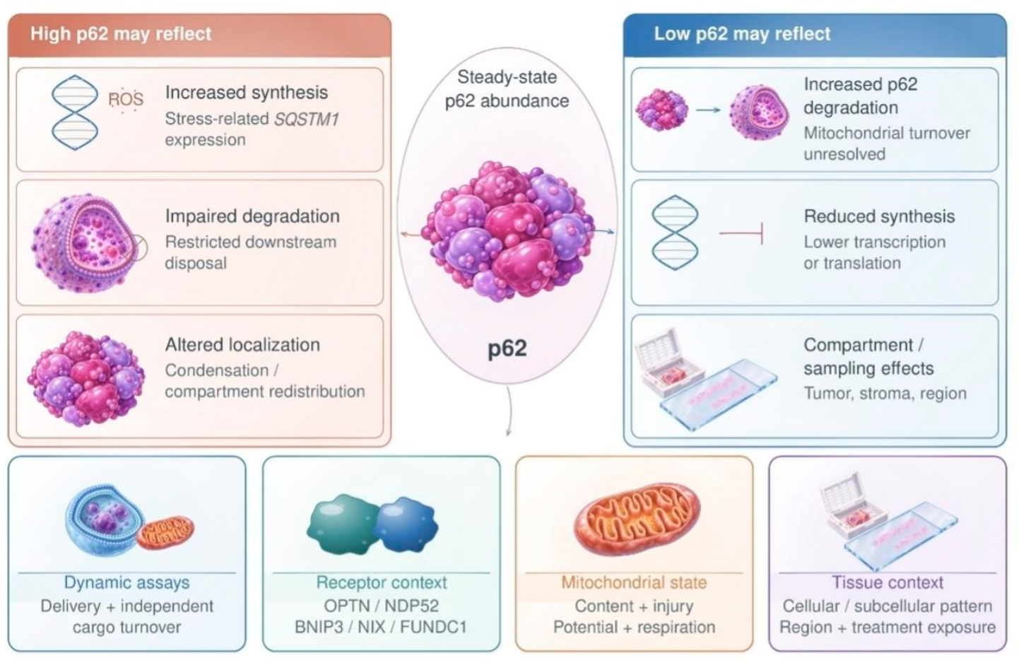
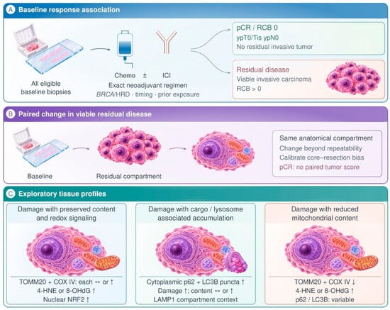
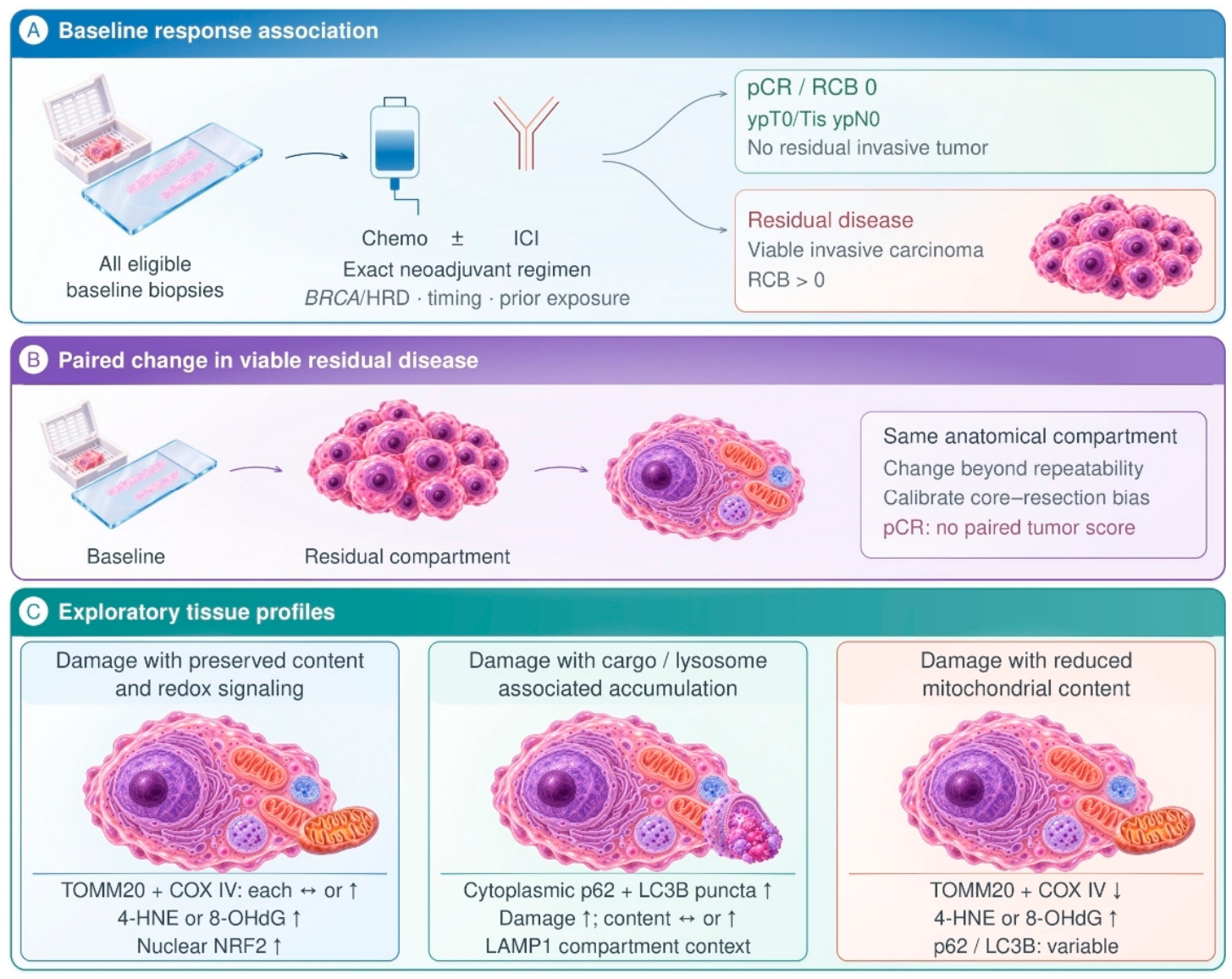
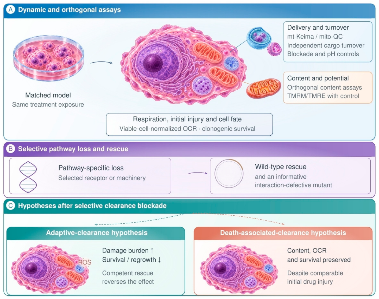

Disclaimer/Publisher’s Note: The statements, opinions and data contained in all publications are solely those of the individual author(s) and contributor(s) and not of MDPI and/or the editor(s). MDPI and/or the editor(s) disclaim responsibility for any injury to people or property resulting from any ideas, methods, instructions or products referred to in the content. |
© 2026 by the authors. Licensee MDPI, Basel, Switzerland. This article is an open access article distributed under the terms and conditions of the Creative Commons Attribution (CC BY) license.