


Simple Summary
Standard surgery for early gastric cancer involves removing a large part of the stomach and nearby lymph nodes. This extensive removal occurs even though most early tumors have not yet spread to these nodes. Since nodal status cannot be confirmed before surgery, many patients undergo unnecessary tissue removal, often leading to long-term difficulties with eating, weight management, and nutrition. Sentinel node navigation surgery offers an alternative. A tracer is injected around the tumor to identify the first draining lymph nodes. If these “sentinel” nodes are found to be clear of cancer during the operation, the rest of the stomach and its associated nodes are left intact. This review re-evaluated the accuracy reported in published studies. The mapping technique appears accurate enough for consideration in newly diagnosed, untreated patients. However, it is not recommended for patients who have already undergone endoscopic tumor removal. The accuracy of this technique also depends on the thoroughness with which the removed nodes are examined under a microscope, not solely on how they are identified during surgery.
Abstract
Background/Objectives: Gastric cancer remains a leading cause of cancer mortality. However, earlier detection has increased the number of cases suitable for organ-preserving treatment. Sentinel node navigation surgery has been proposed as an alternative to standard gastrectomy for early gastric cancer. The safety of this procedure hinges on how accurately sentinel node mapping predicts nodal status. This systematic review synthesizes its diagnostic accuracy and outcome data. Methods: We searched PubMed/MEDLINE, Embase, CENTRAL, the Web of Science Core Collection, and Scopus from inception without date limits, along with two trial registries; screening was restricted to English-language reports. Estimates and exact binomial (Clopper–Pearson) 95% confidence intervals were recalculated from each study’s reported contingency counts using a consistent statistical method. Each included cohort was appraised using QUADAS-2. Results: Eight cohorts contributed per-patient accuracy data, and three contributed per-node molecular comparisons. Detection rates were 97.1% or higher in every cohort entry, with the exception of the naked-eye dye arm of a within-cohort comparison and the smallest feasibility series. Sensitivity was 93.0% (53/57; 95% CI, 83.0–98.1) in the single adequately powered treatment-naive cohort, compared to 69.6% (16/23; 47.1–86.8) in the single prospective trial conducted after non-curative endoscopic resection. Sensitivity also depended on the thoroughness of sentinel node examination: within one cohort, the same patients yielded a sensitivity of 53.6% with single-plane frozen section versus 85.7% with multiple paraffin sections. Therefore, estimates were not pooled. No appraised cohort demonstrated a low risk of bias across all four QUADAS-2 domains. Conclusions: Stomach preservation guided by sentinel node mapping may be a reasonable approach for treatment-naive disease in experienced centers. However, it is not supported after non-curative endoscopic resection.
1. Introduction
Gastric cancer is a leading cause of cancer-related death worldwide, with the stage at diagnosis varying widely by region [1]. In Japan and Korea, national screening programs have led to early detection in over half of cases, compared to roughly one in five in most Western countries [1]. Consequently, surgeons in screening regions frequently encounter patients with tumors confined to the mucosa or submucosa, which offer a high probability of cure [2]. For these patients, long-term well-being is more influenced by the functional impact of surgery than by oncological control [3,4].
Currently, gastrectomy with D1+ or D2 lymphadenectomy remains the standard treatment, partly because nodal status cannot be determined preoperatively [5]. While resection offers high survival rates (the Japanese nationwide registry reports a five-year survival of 91.5% for pathological stage IA disease [2]), the functional costs are substantial. Patients typically lose about 6% of their body weight after distal gastrectomy and 15% after total gastrectomy within a year. Micronutrient deficiency, dumping syndrome, bile reflux, and reduced bone density are common, necessitating dedicated follow-up [3,4].
The extent of nodal dissection further exacerbates these burdens. Clearing lymph nodes beyond the perigastric region requires the division of the celiac and hepatic branches of the vagus nerve. A randomized trial in early gastric cancer demonstrated that preserving these branches reduced diarrhea and appetite loss at 12 months [6], a finding supported by a subsequent pooled analysis [7].
Despite these considerations, the prior probability of nodal disease is low, affecting approximately 3% of mucosal and 20% of submucosal tumors [8]. This means that most patients undergoing radical gastrectomy for early gastric cancer do not benefit from extensive lymphadenectomy or the associated resection [5,8].
Endoscopic submucosal dissection (ESD) offers a solution for some cases. Lesions meeting the eCura A or B criteria carry a very low risk of nodal involvement and allow the stomach to remain intact [5]. However, tumors outside these curative criteria, or those found to have been non-curatively resected on final pathology, present a dilemma. Their nodal risk is low but not negligible [5,8,9]. Current guidelines recommend gastrectomy for these cases, yet most resected specimens ultimately prove node-negative [5,9].
Sentinel node navigation surgery (SNNS) was developed to identify which patients with early gastric cancer can be spared regional lymphadenectomy [10,11]. Whether it extends to the post-endoscopic-resection group above, where submucosal scarring may have redirected lymphatic drainage, is a separate question that this review addresses. This technique maps lymphatic drainage during surgery, examines the sentinel nodes, and, if these are clear, preserves the remaining regional nodes and most of the stomach [12,13]. Due to the multidirectional nature of gastric lymphatic drainage, the technique has evolved to dissect and assess the entire lymphatic basin containing the mapped nodes as a single unit [14]. The safety of SNNS relies on successful mapping in nearly every patient and on a tumor-free basin accurately predicting a tumor-free regional field [12,14].
This systematic review synthesizes the evidence for sentinel node navigation surgery in gastric cancer, with a particular focus on its diagnostic performance. Estimates were recalculated from reported contingency counts and are presented with exact binomial confidence intervals to allow direct comparison of precision across studies of varying sizes. Each included cohort was appraised using QUADAS-2 [15].
2. Materials and Methods
2.1. Protocol and Registration
This systematic review of diagnostic test accuracy adhered to the PRISMA 2020 statement [16] and its PRISMA-DTA extension [17]; the completed PRISMA 2020 and PRISMA-DTA checklists are provided as Table S4 and Table S5, respectively. The protocol was retrospectively registered with OSF Registries on 22 August 2026 (https://osf.io/ca7p2; PRISMA item 24a). All eligibility criteria, primary outcomes, and planned analyses were followed. The eligibility criteria and primary outcomes were finalized a priori, before the searches were run; each database was last searched on the date given in Supplementary Table S1 (between 10 and 19 August 2026), after which title/abstract and full-text screening of the retrieved records were completed, and the review was registered retrospectively on 22 August 2026. During data synthesis, a qualitative synthesis was chosen over pooled estimates (see Section 2.8).
2.2. Eligibility Criteria
Eligibility criteria were defined based on two components: diagnostic accuracy and outcomes. These criteria considered the population, index test, reference standard, target condition, and study design.
For the diagnostic accuracy component, eligible studies included adults with histologically confirmed gastric adenocarcinoma who underwent sentinel node mapping using any tracer strategy. This included patients who were treatment-naive, those with clinically early disease or those who had undergone non-curative endoscopic resection. Sentinel node status had to be determined histopathologically or molecularly. The reference standard was histopathology of regional nodes resected at lymphadenectomy for per-patient analyses, or of the paired node half for per-node analyses, with regional nodal metastasis as the target condition. We included randomized trials, cohort studies, and diagnostic accuracy designs if they reported, or allowed unambiguous reconstruction of, primary 2 × 2 contingency counts.
For the outcome component, we included randomized trials that compared stomach-preserving sentinel node navigation surgery with standard gastrectomy for oncological, surgical, or patient-reported outcomes, regardless of diagnostic accuracy reporting. These studies evaluate surgical strategy rather than test performance, so they are reported separately (Section 3.9) and were excluded from QUADAS-2 appraisal.
We excluded case reports, editorials, narrative reviews, animal and ex vivo studies, non-English publications, and conference abstracts lacking reconstructible outcome data. When multiple publications described the same patient cohort, all were retrieved, but the cohort was evaluated as a single study.
2.3. Information Sources and Search Strategy
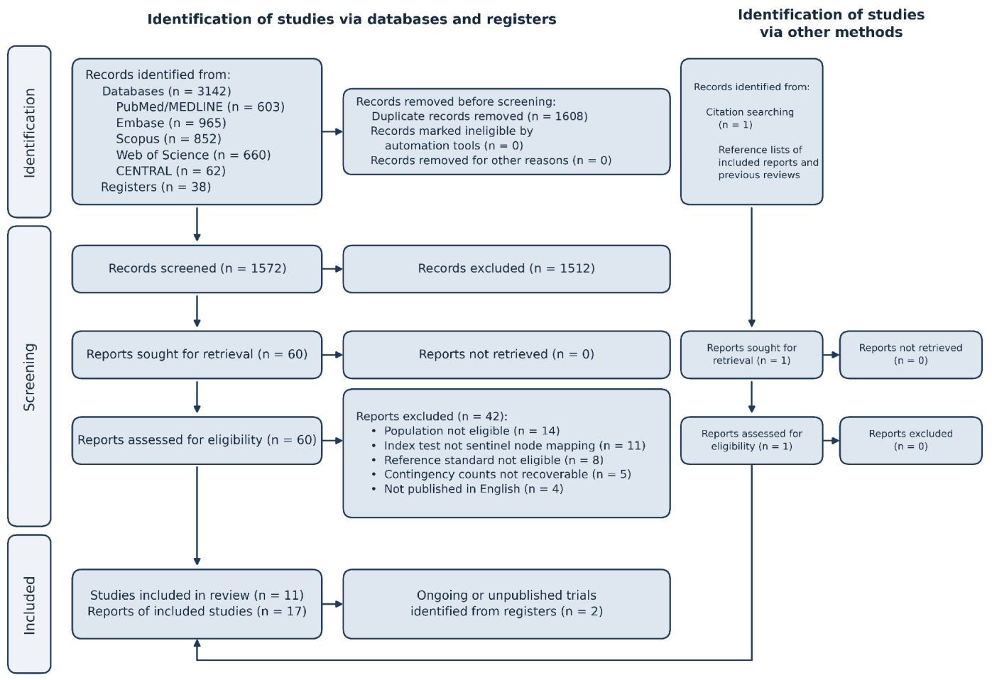
We searched PubMed/MEDLINE, Embase, the Cochrane Central Register of Controlled Trials (CENTRAL), the Web of Science Core Collection, and Scopus from inception through the dates specified for each database in Supplementary Table S1. There were no language or publication-date restrictions during the initial search; however, non-English records were excluded during full-text screening (Figure 1). We hand-searched reference lists of included studies and reviews and screened trial registries (ClinicalTrials.gov, UMIN-CTR) for ongoing or unpublished trials. One additional report was identified through citation searching.
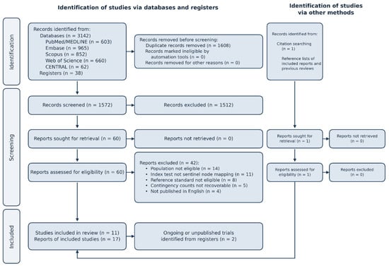
Figure 1.
PRISMA flow diagram of study selection. Seventeen reports corresponding to 11 unique studies were included, along with 2 ongoing trial records. Multiple reports describing the same cohort were linked and counted as a single study. Thirteen reports contributed to the diagnostic accuracy stream and 4 to the outcome component; no report was counted twice, although the SENORITA phase III trial contributed to both. The full legend is given in the Supplementary Note S1.
2.4. Study Selection
All retrieved records were exported to Zotero (https://www.zotero.org, accessed on 11 October 2026) for deduplication, with software-identified duplicates manually verified before removal. Two of three reviewers (J.W.K., H.D.K., and E.L.) independently screened titles, abstracts, and full texts in duplicate. Discrepancies were resolved by consensus or senior adjudication (S.K.). Records disputed at the title and abstract stage advanced to full-text review. Exclusion criteria were applied in a fixed sequence, stopping at the first unmet criterion, and a single primary reason was recorded for each excluded report (Figure 1).
2.5. Data Extraction
Data were extracted into a piloted spreadsheet, capturing study design, setting, population, sample size, tracer strategy, index test, reference standard, node-specific pathology protocols, detection rate, and primary 2 × 2 contingency counts. We recorded whether counts were directly reported or reconstructed. True-negative counts were derived from the number of patients with detected sentinel nodes and total node-positive cases. Values requiring reconstruction beyond direct subtractions are flagged in table footnotes.
Extraction was performed independently and in duplicate by J.W.K. and H.D.K. Discrepancies were resolved by checking source documents, performing arithmetic checks, or through adjudication by S.K.
2.6. Statistical Analysis
Detection rate, sensitivity, negative predictive value, and per-node molecular estimates were recalculated directly from reported counts. Exact binomial (Clopper–Pearson) 95% confidence intervals were computed in R (version 4.3.1; R Foundation for Statistical Computing, Vienna, Austria) using the stats package. Outcome and comparative statistics from the SENORITA phase III trial are reproduced as published. Minor numerical differences from source reports reflect the use of the exact binomial method rather than differences in the underlying data.
Specificity and overall accuracy are not reported for the per-patient analyses because full regional lymphadenectomy served as the reference standard. This precluded false positives and fixed specificity at 100%. The JCOG0302 study is the sole exception, as its index test was intraoperative frozen section and its reference standard was final paraffin. Specificity is reported for the per-node molecular comparisons (Section 3.8), where the paired node half provides an independent reference standard.
Data were evaluated at two levels: per-patient, testing whether sentinel node status predicts regional nodal status, and per-node, comparing molecular assays against paired histopathology. Sensitivity denominators follow each source report. When a cohort was evaluated under multiple index tests or pathology protocols, its results appear as linked rows under the study name to prevent double-counting. The two SENORITA trials are distinguished throughout: the phase III randomized trial in treatment-naive disease and the phase II SENORITA 2 trial after non-curative endoscopic resection.
2.7. Risk of Bias and Applicability
Risk of bias and applicability were evaluated across four domains using QUADAS-2 [15], with signaling questions tailored to the review question beforehand. Because sentinel node status in most studies was determined by the same paraffin examination serving as the reference standard, independent interpretation was judged partial incorporation rather than blinding and recorded as a concern in both the index test and reference standard domains. Reference standards were judged prone to misclassification where node examination was confined to a single plane or where non-uniform lymphadenectomy left metastatic nodes outside the resected field unexamined. QUADAS-2 and RoB 2 appraisals were performed independently by two reviewers (J.W.K. and H.D.K.); disagreements were resolved by discussion, with adjudication by a third reviewer (S.K.) where consensus could not be reached. Each domain was rated as having low or some concerns or a high risk of bias, with a low rating requiring affirmative answers across all signaling questions. We use the label “some concerns” (as in RoB 2) rather than the standard QUADAS-2 “unclear” category for the intermediate rating, because these ratings denote identified but non-critical limitations—chiefly partial incorporation of the index test into the reference standard—rather than an absence of information.
Risk of bias was assessed separately after extraction to keep the record of what each study did distinct from the judgment of how well it was done. Linked rows representing single cohorts under altered protocols were appraised separately to directly evaluate the protocol comparison. The outcome-stream randomized trial was evaluated using the Cochrane RoB 2 tool [18] (Section 3.9).
2.8. Synthesis
Diagnostic estimates were synthesized qualitatively rather than pooled via meta-analysis. Pooling was precluded by structural heterogeneity in how index tests and reference standards were paired across studies, not by study volume. For example, a single cohort evaluated under two pathology protocols yielded sensitivity estimates differing by 32 percentage points; pooling them would average across incompatible diagnostic scales. Studies are grouped by clinical setting to reflect the distinct diagnostic anatomy of intact versus previously operated stomachs. As a post hoc, exploratory analysis that was not pre-specified in the protocol, we additionally pooled sensitivity across the treatment-naive cohorts examined by paraffin section of all regional nodes, using a DerSimonian–Laird random-effects model applied to the logit-transformed proportions and back-transformed to the sensitivity scale (meta package, https://CRAN.R-project.org/package=meta, accessed on 11 October 2026; R version 4.3.1). This analysis is reported only to bound the plausible range of sensitivity in that subgroup and does not supersede the qualitative synthesis.
3. Results
3.1. Study Selection
The searches yielded 3142 records from five bibliographic databases and 38 from two trial registries. After removing 1608 duplicates, 1572 unique records were screened by title and abstract, leading to the exclusion of 1512. Sixty-one full-text reports were assessed: 60 retrieved from databases and registries, and one identified via citation searching. Seventeen reports corresponding to 11 unique studies met all eligibility criteria and were included, along with two ongoing trial records. Eight cohorts provided per-patient diagnostic accuracy data, three studies contributed four per-node molecular comparisons, and one randomized trial (along with its secondary and post hoc reports) provided the outcome stream.
3.2. Study Characteristics
The included cohorts span two decades of practice and encompass the four technical eras summarized in Table 1: dye read by eye, radioisotope combined with dye, lymphatic basin dissection, and near-infrared fluorescence. Their study-level characteristics, including design, setting, population, analyzed sample size, tracer strategy, index test, and reference standard, are detailed in Table 2. Six cohorts enrolled treatment-naive patients, while two enrolled patients after non-curative endoscopic resection.
Table 1.
Technical evolution of sentinel node mapping in gastric cancer.
Table 2.
Study characteristics, per-patient diagnostic accuracy cohorts.
Eight cohorts reported per-patient accuracy in a format that allowed contingency counts to be recovered; their characteristics are presented in Table 2, their recalculated performance in Table 3 (complete 2 × 2 counts in Table 4), and risk-of-bias judgments for all eight in Section 3.10. The patient serves as the unit of analysis, with histopathology of the regional nodes as the reference standard. A patient with clear sentinel nodes but metastasis elsewhere counts as a false negative.
Table 3.
Per-patient diagnostic performance.
Table 4.
Complete 2 × 2 contingency counts and mapping failures for each per-patient analysis. FP = 0, where the sentinel node and reference standard derive from the same paraffin examination; JCOG0302 multiple-paraffin TN was not reported (NR); Shoji is not estimable (NE; 1 pN+). Counts are on the detected-node basis except Takahashi (intention-to-diagnose).
The two settings (unoperated vs. previously operated stomachs) are treated separately below, as mapping an unoperated stomach presents different challenges than mapping one where submucosal scarring may have redirected lymphatic flow.
3.3. Detection Rates
Detection rates only loosely correlate with the mapping technique. Six of the eight entries in Table 3 range from 97.1% to 100%, spanning cohorts across two decades, 44 to 397 patients, and both single-center and multicenter consortia. The two rates below 95% are from the naked-eye dye arm of Takahashi’s trial [21] and Shoji’s 20-patient series [28]. Tracer strategy does not explain this spread. For instance, JCOG0302 achieved 97.7% with ICG read by eye, whereas Shoji achieved 85.0% with fluorescence [26,28]. Takahashi also stratified seven surgeons by experience; basin detection was 100% in every stratum with no false negatives under infrared, while the same surgeons found sentinel nodes in only 79.5% of the same patients under ordinary light. This suggests that the imaging technique, rather than operator experience, was the larger factor, although the trial was too small to definitively exclude a learning effect.
3.4. Diagnostic Accuracy in Treatment-Naive Disease
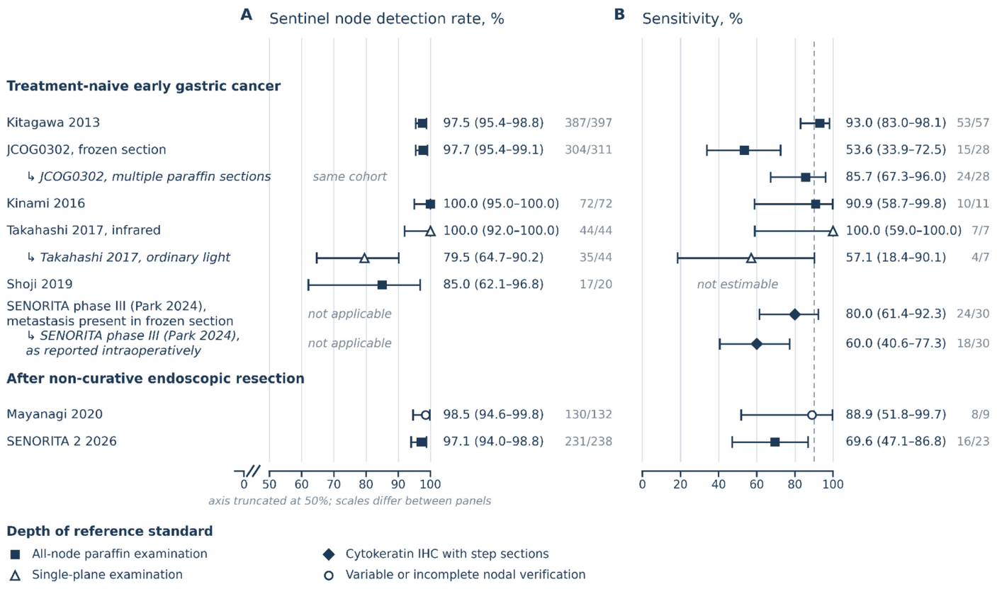
In treatment-naive disease, the precision of accuracy estimates is limited by the number of node-positive patients available for analysis and by the depth of the reference standard against which they are counted, rather than by the mapping technique. Sentinel node detection is consistently high, exceeding 97% across institutions, but only Kitagawa’s cohort yields a usably narrow sensitivity interval. That phase II trial identified 53 out of 57 node-positive patients (93.0%; 95% CI, 83.0–98.1) [13]. JCOG0302 and SENORITA phase III accrued 28 and 30 node-positive patients, but were measured against different reference standards and yield intervals no narrower than 29 percentage points (Section 3.7). The remaining ICG series had fewer than twelve node-positive patients each, leaving confidence intervals too wide to confirm or refute mapping accuracy (Table 3 and Figure 2) [21,27,28].
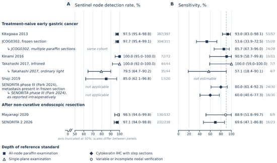
Figure 2.
Per-patient detection rate and sensitivity, with exact binomial 95% confidence intervals. (A) Sentinel node detection rate; (B) sensitivity. Markers are point estimates and horizontal lines are Clopper–Pearson exact 95% confidence intervals, recalculated from the counts in each source report. Marker shape and fill denote the depth of the reference standard; marker size is uniform and encodes no weight, as no pooled estimate is computed. Indented italic rows (prefixed ↳) are the same cohort reanalyzed under a second index test, reading basis or pathology protocol. Source reports: Kitagawa 2013 [13]; JCOG0302 [26]; Kinami 2016 [27]; Takahashi 2017 [21]; Shoji 2019 [28]; SENORITA phase III (Park 2024) [19,29]; Mayanagi 2020 [9]; SENORITA 2 2026 [22,30]. The two panels use different horizontal scales, and panel A is truncated at 50%, so interval widths should not be compared by eye between panels. The full legend is given in the Supplementary Note S2.
The available estimates are also difficult to compare directly because they are measured on different scales. Sensitivity denominators vary, as most series only count patients with a detected node, while Takahashi counts all enrolled patients. Reference standards also differ in depth, meaning the figures reflect index test–reference standard pairings as much as the mapping itself (Section 3.7). JCOG0302 illustrates both effects within a single cohort: single-plane frozen section identified 15 out of 28 node-positive patients (53.6%), rising to 24 out of 28 (85.7%) when the same nodes were examined with multiple paraffin sections [26]. As a post hoc, exploratory analysis (Section 2.8), restricting synthesis to the treatment-naive cohorts examined by paraffin section of all regional nodes (Kitagawa, 53/57; the multiple-paraffin re-analysis of JCOG0302, 24/28; and the feasibility phase of Kinami, 7/7—a subset of the combined Kinami cohort of 10/11 reported in Table 3, whose single false negative arose in the frozen-section clinical phase), a random-effects model gives a pooled sensitivity of 89.4% (95% CI, 81.2–94.3; I2 = 0%); the lower confidence bound still permits roughly one node-positive patient in five to be missed, and this subgroup continues to combine single- and multiple-plane paraffin examination, so narrative synthesis remains the primary approach (Section 2.8).
3.5. Diagnostic Accuracy After Non-Curative Endoscopic Resection
Mapping is markedly less reliable after non-curative endoscopic resection, where submucosal scarring can redirect lymphatic flow. In the only prospective trial in this setting, SENORITA 2 (n = 238), mapping missed seven out of 23 node-positive patients (sensitivity 69.6%; 95% CI, 47.1–86.8), despite a 97.1% detection rate [22,30]. A smaller retrospective cohort reported higher sensitivity (Mayanagi, 88.9%; 8/9) [9]. However, its reference standard was incomplete and varied by patient, with approximately a quarter of patients receiving less than full regional clearance. Unexamined patients were counted as true negatives, inflating sensitivity by an unquantifiable amount. This cohort is the only one at high risk of bias across all four QUADAS-2 domains (Section 3.10). Sentinel-node mapping, sentinel-basin assessment, and per-node pathology are distinct endpoints that can diverge within one patient: in Mayanagi’s cohort, the single false negative was not a mapping failure but a metastasis in a non-sentinel node (station 5) lying within a correctly mapped basin, so a basin-level and a node-level analysis of the same case reach different conclusions.
In this setting, false negatives resulted from mapping rather than pathological examination. SENORITA 2 did not use frozen sections, and deeper sectioning of all seven false-negative nodes found no micrometastases. The investigators attributed these misses to post-resection fibrosis and skip metastasis, where the missed disease lay outside the mapped basin [22]. Due to small patient numbers and varying tracer choices across participating centers, tracer subgroups could not be compared, making it impossible to separate the effect of the tracer from that of the center. The evidence for mapping after endoscopic resection relies on a single feasibility-powered trial that missed approximately three out of every ten node-positive patients.
3.6. Mapping Compared with Anatomically Defined Regional Dissection
A post hoc analysis of 237 SENORITA navigation-arm patients investigated whether mapping offers advantages over anatomically defined dissection [19]. This analysis reconstructed the yield of regional lymphadenectomy performed without mapping from the same specimens. Sensitivity was 96.8% with mapping versus 80.6% without (p = 0.016), and the negative predictive value was 99.5% versus 97.2% (p = 0.021). Metastasis outside a tumor-free sentinel basin occurred in only one out of 237 patients (0.4%). Its node-positive denominator differs by one from the pathology re-review of the same 237 patients (Section 3.7), reflecting the different reference protocols of the two reports [19,29]. Because the comparator was reconstructed rather than randomized, this analysis is hypothesis-generating and applies only to treatment-naive disease.
3.7. Pathology Protocol Depth and Measured Sensitivity
Two within-cohort comparisons demonstrate that measured sensitivity is highly dependent on the thoroughness of node examination. In JCOG0302 [26], the same 311 patients and sentinel nodes yielded 53.6% sensitivity with single-plane frozen section versus 85.7% with multiple paraffin sections. Since the surgical procedure remained unchanged, the 32-percentage-point difference is solely attributable to the pathology protocol.
The SENORITA phase III cohort [29] identifies two ways a protocol can fail. Re-examination of all 237 navigation-arm patients revealed that 30 were node-positive on enhanced permanent pathology. Frozen section identified all 13 macrometastases but missed six out of 17 micrometastases. This allows for the calculation of two different sensitivities for the same 30 patients.
When measured against metastases identifiable on the frozen slide upon later review (permanent pathology), sensitivity was 80.0% (24/30; 95% CI, 61.4–92.3). However, when measured against the result of the frozen section reported to the surgeon during the operation, sensitivity was 60.0% (18/30; 40.6–77.3). The 20-percentage-point difference (six patients) represents cases where deposits were present on the slide examined by the pathologist but were not identified at the time, leading to a negative intraoperative report. The six-patient gap therefore reflects deposits present on the same frozen slide the pathologist examined but not reported during the operation (an interpretation difference), not additional tissue sampling, because no further sections were introduced between the intraoperative reading and the review.
This discrepancy highlights the difference between sampling failure and interpretation failure. Sampling failure occurs when a deposit lies outside the plane that was cut and can be recovered by deeper sectioning. Interpretation failure, conversely, is when a deposit present on the slide is not recognized. Deposit size influences both types of failure; a protocol adequate for macrometastasis may still miss micrometastatic disease. Notably, none of the six patients with missed micrometastases developed nodal recurrence, leading the authors to conclude that single-section examination is adequate in practice [29]. Therefore, an argument for a deep intraoperative protocol has been made based on avoiding under-staging rather than on a demonstrated reduction in recurrence. A separate prospective cohort in treatment-naive early gastric cancer reinforces this: its measured false-negative rate fell from 18.2% with intraoperative hematoxylin–eosin alone to 0% once rapid cytokeratin immunohistochemistry was added, confirming that sentinel-node sensitivity reflects the depth of the pathology protocol as much as the mapping itself [31].
However, the depth of pathological examination does not fully account for all instances of low sensitivity. For example, SENORITA 2 did not utilize frozen sections and found no micrometastases even after deeper sectioning of all seven false-negative nodes. This suggests that its failures were related to the mapping technique itself [22]. Kinami’s single false-negative case was attributed to frozen-section failure [27]. In contrast, Takahashi’s 100% sensitivity [21] was measured against a shallow, single-plane standard. This method is less likely to reveal a false negative compared to the all-node paraffin examination employed by Kitagawa, JCOG0302, and Kinami [13,26,27].
Thus, reported sensitivity reflects the specific combination of the index test and the reference standard, rather than the mapping technique in isolation. This, more than the limited number of studies, is the primary reason why no pooled estimate is offered. Any center adopting SNNS must address both the pathology protocol and the operative technique. These within-cohort comparisons establish a strong pathology effect within the cohorts studied; they do not, however, establish that pathology contributes more than mapping to the discordance seen across the literature as a whole.
3.8. Molecular Nodal Assessment
Intraoperative pathology is the principal limitation because the extensive depth of examination required for high sensitivity cannot be achieved within the operative window. One-step nucleic acid amplification (OSNA) offers a solution by quantifying cytokeratin 19 messenger RNA in homogenized nodal tissue within this timeframe [23]. By analyzing the entire node rather than sampling planes, OSNA avoids the sampling failure observed in JCOG0302 [25,26].
However, current validation does not yet support OSNA for this role. Three studies contributed four node-level comparisons against paired histopathology, revealing a consistent pattern (Table S2) [23,24,25]. OSNA rarely incorrectly identifies a negative node as positive, demonstrating specificity ranging from 95.9% to 99.5%. Its sensitivity, however, is less consistent, varying from 63.6% to 88.9%. The lowest sensitivity value comes from the only cohort restricted to early gastric cancer [25]; in those 43 patients, sensitivity was 63.6% against permanent sections and 80.0% against intraoperative sections.
Applicability to early disease is further limited: many tumors in the Yaguchi and Kumagai studies were advanced (17 out of 32 subserosal or serosa-exposed; 29 out of 61 T3 or T4), which tend to have larger deposits [23,24]. Discordance is concentrated in poorly differentiated and signet-ring histology [24,25], and homogenized tissue cannot be subsequently examined histologically [23]. These per-node figures establish agreement between the assay and the pathologist for a single node, but they do not confirm whether a tumor-free basin predicts a tumor-free field. OSNA is therefore reasonable as an adjunct to, rather than a replacement for, the standard pathology protocol. Patient-level validation in early disease is still lacking. These per-node binomial intervals also treat nodes as independent and therefore ignore clustering within patients, which can overstate precision; OSNA and histology are moreover performed on different physical portions of the same node (tissue-allocation bias), and paired histopathology is itself an imperfect reference rather than an error-free standard.
3.9. Clinical and Functional Outcomes
Randomized evidence from the SENORITA trial (580 patients across seven Korean centers) suggests a trade-off rather than a decisive advantage for either approach. Stomach-preserving navigation surgery did not establish non-inferiority for three-year disease-free survival (a 3.7% difference favoring standard gastrectomy; 95% CI, −0.6 to 8.1, spanning both the 5% margin and zero). This renders the trial inconclusive rather than negative [12]. While overall and disease-specific survival showed no statistically significant difference at five years (OS p = 0.7403; DSS p = 0.9586), effective endoscopic and surgical salvage of metachronous lesions may have contributed to this convergence rather than demonstrably superior nodal clearance, although the trial design cannot establish that mechanism. Additionally, later estimates for the standard-gastrectomy arm are based on short follow-up [32]. Serious complications were similar in both arms, indicating that adding mapping did not increase operative risk (Table 5) [33].
Table 5.
SENORITA phase III trial outcomes.
In exchange for intensive surveillance, patients who retained their stomach experienced functional benefits: quality of life was better on all eight scales at three months, and nutritional parameters were higher at one year (Table 5) [34]. These represent the program’s most consistent benefits, although the endpoints were patient-reported in an open-label trial. This carries a high risk of measurement bias under RoB 2 [18] and applies only to the 81.4% of the navigation arm who retained their stomach; the remaining patients converted to gastrectomy due to failed mapping, positive nodes, or inadequate margins [33]. Because this entire clinical and functional evidence base derives from a single phase III program conducted at seven Korean centers, these findings should be regarded as trial- and setting-specific rather than generalizable without confirmation elsewhere.
3.10. Risk of Bias and Applicability
Table 6 summarizes the strength of the evidence in each domain along with its primary limitations. Study-level QUADAS-2 judgments appear in Table 7 for per-patient cohorts and Table S3 for per-node comparisons. Eight cohorts contributed per-patient accuracy data, and all eight are appraised in Table 7: the seven independent diagnostic cohorts together with the SENORITA phase III diagnostic cohort, which is appraised in the final row. None of the appraised cohorts was at low risk of bias across all four domains. Mayanagi’s retrospective cohort was the only one rated high in every domain, mainly because the extent of lymphadenectomy—and thus the reference standard—varied between patients. SENORITA 2 carried the lowest risk among the post-endoscopic-resection cohorts, and the JCOG0302 frozen-section comparison was low in three of the four domains.
Table 6.
Critical appraisal by evidence domain.
Table 7.
Risk of bias and applicability, per-patient diagnostic accuracy cohorts (QUADAS-2).
4. Discussion
4.1. Evolution and Technique of Sentinel Node Mapping
Adapted from breast cancer and melanoma protocols in the 1990s, sentinel node mapping encountered specific challenges in the stomach: multidirectional drainage, perigastric fat that obscures targeted nodes, and marked operator dependence [10,11,14]. Successive technical advancements, summarized in Table 1, have largely overcome these obstacles.
Early dye-only mapping, while inexpensive and requiring no equipment, was limited by a narrow timing window and poor visibility in fat [11]. Radioisotope mapping with technetium-99m tin colloid, injected submucosally the day before surgery, is retained in first-echelon nodes and can be localized by gamma probe regardless of overlying tissue. This technique, combined with dye, became the reference in the 2000s and underpinned Kitagawa’s trial [13]. However, its adoption remains constrained by the need for nuclear-medicine infrastructure and radiation-safety licensing [13,20].
Lymphatic basin dissection removes the primary vascular basin en bloc, ensuring that any metastasis missed by mapping still lies within the resected tissue [14,19]. The SENORITA post hoc analysis supports this, with metastasis lying outside a tumor-free basin in only one out of 237 patients [19]. In contrast, in JCOG0302, seven out of 13 false negatives had metastases outside the basin, a difference explained by that trial’s dye-only mapping, node-by-node retrieval, and single-plane examination [26].
Near-infrared fluorescence with indocyanine green (ICG) overcomes the tissue-penetration limits of visual dye without requiring radioisotopes. Takahashi’s within-patient comparison achieved 100% detection with infrared versus 79.5% under white light (Table 3) [21]. However, it has not yet displaced dual-tracer mapping due to the confounded, staggered adoption in SENORITA 2 and the small size of single-agent feasibility cohorts. Therefore, a well-powered randomized comparison of single-agent fluorescence and dual tracers is still needed [20,22,28].
4.2. Interpretation of the Randomized Evidence
At five years, disease-free survival did not differ significantly between the arms (88.9% versus 80.1%, p = 0.0561); this borderline result is fragile: follow-up in the standard-gastrectomy arm reached only 62.5 months, and with only about 24 patients per arm still at risk at 60 months (versus 254 and 229 at 54 months in the full analysis set) the standard-arm estimate is driven by few events and carries a wide confidence interval of 47.2 to 94.1 (Table 5) [32]. Overall and disease-specific survival showed no statistically significant difference (p = 0.7403 and 0.9586), and three-year disease-free survival favored standard gastrectomy [12]. The mechanism is more informative than any of these figures. Disease-specific survival may have been supported by effective surgical salvage (the early detection and resection of recurrences and metachronous lesions in the remnant stomach) rather than by superior mapping accuracy. Long-term success therefore depends on rigorous surveillance and timely salvage. This reliance on surveillance is itself a burden borne by the patient: it presumes sustained, potentially indefinite endoscopic follow-up, with cumulative procedural and financial cost, the possibility of declining adherence over time, and a psychological impact of ongoing cancer surveillance that the trial did not measure.
4.3. Current Role and Patient Selection
Current evidence does not establish SNNS as the standard of care but supports its selective use at high-volume centers. For treatment-naive early gastric cancer meeting SENORITA criteria, SNNS offers acceptable diagnostic accuracy and superior functional preservation, with no statistically significant difference in five-year overall or disease-specific survival and a five-year disease-free survival difference that did not reach significance, despite the unmet non-inferiority endpoint [13,32,34]. Until a trial succeeds, SNNS should be confined to clinical trials and adequately resourced centers, and offered only to patients who are counseled that gastric preservation is likely but not guaranteed. No universally accepted false-negative threshold exists for gastric sentinel node surgery; the benchmarks commonly adapted from breast cancer, melanoma, and the Japanese trials are a false-negative rate of 10% or less (sensitivity of 90% or more) together with a detection rate of at least 95%. Even the largest treatment-naive cohort meets this only at its point estimate: its lower 95% confidence bound of 83% still permits a clinically important rate of missed nodal disease. Selective use should therefore be confined to the criteria under which it has been studied—clinical T1, cN0, solitary tumors of 4 cm or less, located away from sites that would otherwise require total or proximal gastrectomy.
There is currently insufficient evidence to support omitting standard lymphadenectomy after non-curative endoscopic resection. A prospective multicenter trial reported 69.6% sensitivity, missing seven out of 23 node-positive patients [22]. The higher accuracy suggested by retrospective data [9] was subject to verification bias from an incomplete reference standard, which inflates sensitivity by an amount the published data cannot quantify. Omitting lymphadenectomy in this setting cannot be recommended [22].
Benefit also depends on the planned resection: patients facing total or proximal gastrectomy gain more from preservation than those undergoing distal resection [3,34]. Not every preserving operation maintains function equally, as segmental resection was linked to diarrhea and eating restriction at three years [34].
4.4. Technical Requirements
The clinical adoption of SNNS hinges on adherence to three core technical standards:
- Tracer selection. Available evidence favors dual-tracer or near-infrared fluorescence mapping for reliable sentinel node detection, whereas single-agent visual dye has shown lower detection when compared within a trial [20,21]; its effect on sensitivity, however, remains unestablished (Table 6). Dual-tracer or fluorescence mapping is therefore preferred, although it has not been shown to improve sensitivity.
- Resection unit. Intraoperative removal must encompass the entire lymphatic basin as a single unit, rather than involving selective node-by-node excision [14,19].
- Standardized pathology protocols. Institutions must establish a sectioning and staining protocol prior to implementation, as measured sensitivity is highly dependent on pathological depth (Section 3.7). While deeper sectioning corrects for sampling error, it does not address interpretation error. Standardized processing is a prerequisite for adoption.
4.5. Limitations
The existing literature presents both geographic and methodological limitations. Most diagnostic cohorts originate from Japanese and Korean centers, which identify a high proportion of early-stage disease through screening. Consequently, the generalizability of these findings to Western populations, and to other Asian populations such as Chinese patients, remains unproven; these groups differ in screening exposure, tumor biology, baseline stage distribution, and accumulated mapping experience [1]. Node-positive events are rare in most cohorts, which restricts the precision of sensitivity estimates. In SENORITA 2, 73.5% of patients were recruited at the lead center [22]. The depth of reference standards also varied across cohorts.
Review-level limitations include retrospective protocol registration, restriction to English-language publications, a major limitation that may bias the evidence base (this excluded four full-text articles [35,36,37,38]; Figure 1), and the inability to contact authors directly for missing data. A quantitative assessment of publication bias was not possible, as Deeks’ test [39] requires at least ten studies [40]. Because fewer than ten cohorts contributed, we cannot statistically exclude the selective non-publication of small feasibility studies with poor mapping results; any such bias would inflate reported detection rates more than sensitivity estimates.
One appraisal finding pertains to the field as a whole rather than to individual studies: none of the eight appraised cohorts achieved a low risk of bias across all four QUADAS-2 domains [15]. This is primarily because the index test and reference standard are not independent in surgical mapping (Section 2.7), meaning that reported accuracy addresses a narrower question than it might appear to.
4.6. Future Directions
Establishing the oncological safety of mapping after non-curative endoscopic resection necessitates a trial powered for diagnostic accuracy rather than mere feasibility. Such a trial would need to enroll a sufficient number of node-positive patients to meaningfully define sensitivity, under a pathology protocol prespecified for both arms rather than left to individual centers. No trial has yet met either requirement.
Two registered trials aim to address these gaps. A Japanese multicenter non-randomized phase III trial [41] plans to enroll 225 patients across 13 centers. This trial will compare stomach preservation with standard gastrectomy using dual-tracer mapping, intraoperative pathology, and basin dissection in sentinel-negative patients, with five-year relapse-free survival as the primary endpoint. Its design differs sufficiently from SENORITA that a concordant result would carry considerable weight. A Korean feasibility study of fluorescence-guided mapping is also registered [42].
Reporting standards should improve in one key area: studies should present complete two-by-two contingency tables alongside the sectioning and staining protocol for both sentinel and non-sentinel nodes. This would allow readers to directly verify true-positive and false-negative rates, rather than relying on opaque denominators. Adhering to STARD reporting guidelines [43] would achieve this. Since detection and sensitivity are diagnostic accuracy endpoints, STARD naturally complements the surgical reporting standards, such as STROCSS [44], that studies in this field commonly follow [22].
5. Conclusions
Sentinel node navigation surgery reliably identifies sentinel nodes but is less consistent in ruling out nodal disease. Within the cohorts that examined it, this variability was linked at least as much to the depth of pathological examination as to the mapping technique, highlighting that the pathology protocol warrants as much scrutiny as the tracer.
For treatment-naive early gastric cancer, the accuracy evidence, although largely based on a single adequately powered cohort, supports stomach preservation within clinical trials and at adequately resourced centers. This approach offers improved function and quality of life in exchange for intensive surveillance, and patients should be counseled that gastric preservation is likely but not guaranteed.
In contrast, after non-curative endoscopic resection, the current evidence does not support omitting lymphadenectomy. Broader adoption should await trials powered for diagnostic accuracy and reported with complete contingency data.
Supplementary Materials
The following supporting information can be downloaded at https://www.mdpi.com/article/10.3390/cancers18203332/s1: Table S1: Search strategy, by database and register; Table S2: OSNA against paired histopathology, per lymph node; Table S3: Risk of bias and applicability, per-node molecular comparisons (QUADAS-2); Table S4: PRISMA 2020 checklist; Table S5: PRISMA-DTA checklist. Note S1: Extended legend to Figure 1, Study selection; Note S2: Extended legend to Figure 2, Per-patient detection rate and sensitivity.
Author Contributions
Conceptualization, S.K., J.W.K., and H.D.K.; methodology, J.W.K.; investigation (screening), J.W.K., H.D.K., and E.L.; data curation, J.W.K. and H.D.K.; formal analysis, J.W.K.; validation, H.D.K. and E.L.; writing (original draft preparation), J.W.K.; writing (review and editing), J.W.K., E.L., H.D.K., and S.K.; supervision, S.K. All authors have read and agreed to the published version of the manuscript.
Funding
This research received no external funding.
Institutional Review Board Statement
Not applicable.
Informed Consent Statement
Not applicable.
Data Availability Statement
This review analyzed data extracted from published studies. The complete extracted 2 × 2 contingency data are provided within the article (Table 3 and the accompanying Table 4 block) and in the Supplementary Materials; all estimates are exact binomial (Clopper–Pearson) 95% confidence intervals computed in R (stats package) from those counts and are therefore reproducible directly from the published tables. The analysis scripts are available from the corresponding author upon reasonable request. The search strings, eligibility criteria and study selection process are reported in full in Section 2, Section 3.1, Figure 1 and Supplementary Table S1.
Conflicts of Interest
The authors declare no conflicts of interest.
Abbreviations
The following abbreviations are used in this manuscript:
| CI | Confidence interval |
| DFS | Disease-free survival |
| DSS | Disease-specific survival |
| ESD | Endoscopic submucosal dissection |
| FFPE | Formalin-fixed, paraffin-embedded |
| FNR | False-negative rate |
| HE | Hematoxylin and eosin |
| ICG | Indocyanine green |
| LSG | Laparoscopic standard gastrectomy |
| LSNNS | Laparoscopic sentinel node navigation surgery |
| NPV | Negative predictive value |
| OS | Overall survival |
| OSNA | One-step nucleic acid amplification |
| QoL | Quality of life |
| RI | Radioisotope |
| SNNS | Sentinel node navigation surgery |
References
- Huang, Y.; Shao, Y.; Yu, X.; Chen, C.; Guo, J.; Ye, G. Global progress and future prospects of early gastric cancer screening. J. Cancer 2024, 15, 3045–3064. [Google Scholar] [CrossRef] [Scilit] [PubMed]
- Katai, H.; Ishikawa, T.; Akazawa, K.; Isobe, Y.; Miyashiro, I.; Oda, I.; Tsujitani, S.; Ono, H.; Tanabe, S.; Fukagawa, T.; et al. Five-year survival analysis of surgically resected gastric cancer cases in Japan: A retrospective analysis of more than 100,000 patients from the nationwide registry of the Japanese Gastric Cancer Association (2001–2007). Gastric Cancer 2018, 21, 144–154. [Google Scholar] [CrossRef] [Scilit] [PubMed]
- Davis, J.L.; Selby, L.V.; Chou, J.F.; Schattner, M.; Ilson, D.H.; Capanu, M.; Brennan, M.F.; Coit, D.G.; Strong, V.E. Patterns and predictors of weight loss after gastrectomy for cancer. Ann. Surg. Oncol. 2016, 23, 1639–1645. [Google Scholar] [CrossRef] [Scilit] [PubMed]
- Teixeira Farinha, H.; Bouriez, D.; Grimaud, T.; Rotariu, A.-M.; Collet, D.; Mantziari, S.; Gronnier, C. Gastro-intestinal disorders and micronutrient deficiencies following oncologic esophagectomy and gastrectomy. Cancers 2023, 15, 3554. [Google Scholar] [CrossRef] [Scilit] [PubMed]
- Japanese Gastric Cancer Association. Japanese Gastric Cancer Treatment Guidelines 2025 (7th edition). Gastric Cancer 2026, 29, 271–299. [Google Scholar] [CrossRef] [Scilit] [PubMed]
- Kim, S.M.; Cho, J.; Kang, D.; Oh, S.J.; Kim, A.R.; Sohn, T.S.; Noh, J.H.; Kim, S. A randomized controlled trial of vagus nerve-preserving distal gastrectomy versus conventional distal gastrectomy for postoperative quality of life in early stage gastric cancer patients. Ann. Surg. 2016, 263, 1079–1084. [Google Scholar] [CrossRef] [Scilit] [PubMed]
- Jin, T.; Chen, Z.-H.; Liang, P.-P.; Li, Z.-D.; He, F.-J.; Chen, Z.-W.; Hu, J.-K.; Yang, K. A gastrectomy for early-stage gastric cancer patients with or without preserving celiac branches of vagus nerves: A meta-analysis. Surgery 2023, 173, 375–382. [Google Scholar] [CrossRef] [Scilit] [PubMed]
- Gotoda, T.; Yanagisawa, A.; Sasako, M.; Ono, H.; Nakanishi, Y.; Shimoda, T.; Kato, Y. Incidence of lymph node metastasis from early gastric cancer: Estimation with a large number of cases at two large centers. Gastric Cancer 2000, 3, 219–225. [Google Scholar] [CrossRef] [Scilit] [PubMed]
- Mayanagi, S.; Takahashi, N.; Mitsumori, N.; Arigami, T.; Natsugoe, S.; Yaguchi, Y.; Suda, T.; Kinami, S.; Ohi, M.; Kawakubo, H.; et al. Sentinel node mapping for post-endoscopic resection gastric cancer: Multicenter retrospective cohort study in Japan. Gastric Cancer 2020, 23, 716–724. [Google Scholar] [CrossRef] [Scilit] [PubMed]
- Siewert, J.R.; Sendler, A. Potential and futility of sentinel node detection for gastric cancer. Recent Results Cancer Res. 2000, 157, 259–269. [Google Scholar] [CrossRef] [Scilit] [PubMed]
- Hiratsuka, M.; Miyashiro, I.; Ishikawa, O.; Furukawa, H.; Motomura, K.; Ohigashi, H.; Kameyama, M.; Sasaki, Y.; Kabuto, T.; Ishiguro, S.; et al. Application of sentinel node biopsy to gastric cancer surgery. Surgery 2001, 129, 335–340. [Google Scholar] [CrossRef] [Scilit] [PubMed]
- Kim, Y.W.; Min, J.S.; Yoon, H.M.; An, J.Y.; Eom, B.W.; Hur, H.; Lee, Y.J.; Cho, G.S.; Park, Y.K.; Jung, M.R.; et al. Laparoscopic sentinel node navigation surgery for stomach preservation in patients with early gastric cancer: A randomized clinical trial. J. Clin. Oncol. 2022, 40, 2342–2351. [Google Scholar] [CrossRef] [Scilit] [PubMed]
- Kitagawa, Y.; Takeuchi, H.; Takagi, Y.; Natsugoe, S.; Terashima, M.; Murakami, N.; Fujimura, T.; Tsujimoto, H.; Hayashi, H.; Yoshimizu, N.; et al. Sentinel node mapping for gastric cancer: A prospective multicenter trial in Japan. J. Clin. Oncol. 2013, 31, 3704–3710. [Google Scholar] [CrossRef] [Scilit] [PubMed]
- Kinami, S.; Fujimura, T.; Ojima, E.; Fushida, S.; Ojima, T.; Funaki, H.; Fujita, H.; Takamura, H.; Ninomiya, I.; Nishimura, G.; et al. PTD classification: Proposal for a new classification of gastric cancer location based on physiological lymphatic flow. Int. J. Clin. Oncol. 2008, 13, 320–329. [Google Scholar] [CrossRef] [Scilit] [PubMed]
- Whiting, P.F.; Rutjes, A.W.S.; Westwood, M.E.; Mallett, S.; Deeks, J.J.; Reitsma, J.B.; Leeflang, M.M.G.; Sterne, J.A.C.; Bossuyt, P.M.M. QUADAS-2: A revised tool for the quality assessment of diagnostic accuracy studies. Ann. Intern. Med. 2011, 155, 529–536. [Google Scholar] [CrossRef] [Scilit] [PubMed]
- Page, M.J.; McKenzie, J.E.; Bossuyt, P.M.; Boutron, I.; Hoffmann, T.C.; Mulrow, C.D.; Shamseer, L.; Tetzlaff, J.M.; Akl, E.A.; Brennan, S.E.; et al. The PRISMA 2020 statement: An updated guideline for reporting systematic reviews. BMJ 2021, 372, n71. [Google Scholar] [CrossRef] [Scilit] [PubMed]
- McInnes, M.D.F.; Moher, D.; Thombs, B.D.; McGrath, T.A.; Bossuyt, P.M.; The PRISMA-DTA Group. Preferred Reporting Items for a Systematic Review and Meta-analysis of Diagnostic Test Accuracy Studies: The PRISMA-DTA statement. JAMA 2018, 319, 388–396. [Google Scholar] [CrossRef] [Scilit] [PubMed]
- Sterne, J.A.C.; Savovic, J.; Page, M.J.; Elbers, R.G.; Blencowe, N.S.; Boutron, I.; Cates, C.J.; Cheng, H.Y.; Corbett, M.S.; Eldridge, S.M.; et al. RoB 2: A revised tool for assessing risk of bias in randomized trials. BMJ 2019, 366, l4898. [Google Scholar] [CrossRef] [Scilit] [PubMed]
- Park, S.H.; Kim, Y.W.; Min, J.S.; Yoon, H.M.; An, J.Y.; Eom, B.W.; Hur, H.; Lee, Y.J.; Cho, G.S.; Park, Y.K.; et al. Feasibility of regional lymphadenectomy for stomach-preserving surgery in early gastric cancer omitting sentinel node navigation: A post hoc analysis of the SENORITA trial. Ann. Surg. Oncol. 2024, 31, 6939–6946. [Google Scholar] [CrossRef] [Scilit] [PubMed]
- Sherwinter, D.A.; Boni, L.; Bouvet, M.; Ferri, L.; Hyung, W.J.; Ishizawa, T.; Kaleya, R.N.; Kelly, K.; Kokudo, N.; Lanzarini, E.; et al. Use of fluorescence imaging and indocyanine green for sentinel node mapping during gastric cancer surgery: Results of an intercontinental Delphi survey. Surgery 2022, 172, S29–S37. [Google Scholar] [CrossRef] [Scilit] [PubMed]
- Takahashi, N.; Nimura, H.; Fujita, T.; Mitsumori, N.; Shiraishi, N.; Kitano, S.; Satodate, H.; Yanaga, K. Laparoscopic sentinel node navigation surgery for early gastric cancer: A prospective multicenter trial. Langenbecks Arch. Surg. 2017, 402, 27–32. [Google Scholar] [CrossRef] [Scilit] [PubMed]
- Park, J.H.; Cho, I.; Ryu, K.W.; Kim, Y.W.; Yoon, H.M.; Park, J.Y.; Min, J.S.; Cho, G.S.; Lee, Y.J.; Jung, M.R.; et al. Feasibility of sentinel basin mapping after non-curative endoscopic resection for early gastric cancer: A prospective multicenter cohort study (SENORITA 2 trial). Int. J. Surg. 2026, 112, 9692–9699. [Google Scholar] [CrossRef] [Scilit] [PubMed]
- Yaguchi, Y.; Sugasawa, H.; Tsujimoto, H.; Takata, H.; Nakabayashi, K.; Ichikura, T.; Ono, S.; Hiraki, S.; Sakamoto, N.; Horio, T.; et al. One-step nucleic acid amplification (OSNA) for the application of sentinel node concept in gastric cancer. Ann. Surg. Oncol. 2011, 18, 2289–2296. [Google Scholar] [CrossRef] [Scilit] [PubMed]
- Kumagai, K.; Yamamoto, N.; Miyashiro, I.; Tomita, Y.; Katai, H.; Kushima, R.; Tsuda, H.; Kitagawa, Y.; Takeuchi, H.; Mukai, M.; et al. Multicenter study evaluating the clinical performance of the OSNA assay for the molecular detection of lymph node metastases in gastric cancer patients. Gastric Cancer 2014, 17, 273–280. [Google Scholar] [CrossRef] [Scilit] [PubMed]
- Shimada, A.; Takeuchi, H.; Nishi, T.; Mayanagi, S.; Fukuda, K.; Suda, K.; Nakamura, R.; Wada, N.; Kawakubo, H.; Nakahara, T.; et al. Utility of the one-step nucleic acid amplification assay in sentinel node mapping for early gastric cancer patients. Gastric Cancer 2020, 23, 418–425. [Google Scholar] [CrossRef] [Scilit] [PubMed]
- Miyashiro, I.; Hiratsuka, M.; Sasako, M.; Sano, T.; Mizusawa, J.; Nakamura, K.; Nashimoto, A.; Tsuburaya, A.; Fukushima, N. High false-negative proportion of intraoperative histological examination as a serious problem for clinical application of sentinel node biopsy for early gastric cancer: Final results of the Japan Clinical Oncology Group multicenter trial JCOG0302. Gastric Cancer 2014, 17, 316–323. [Google Scholar] [CrossRef] [Scilit] [PubMed]
- Kinami, S.; Oonishi, T.; Fujita, J.; Tomita, Y.; Funaki, H.; Fujita, H.; Nakano, Y.; Ueda, N.; Kosaka, T. Optimal settings and accuracy of indocyanine green fluorescence imaging for sentinel node biopsy in early gastric cancer. Oncol. Lett. 2016, 11, 4055–4062. [Google Scholar] [CrossRef] [Scilit] [PubMed][Green Version]
- Shoji, Y.; Kumagai, K.; Kamiya, S.; Ida, S.; Nunobe, S.; Ohashi, M.; Yoshimizu, S.; Horiuchi, Y.; Yoshio, T.; Ishiyama, A.; et al. Prospective feasibility study for single-tracer sentinel node mapping by ICG fluorescence and OSNA assay in laparoscopic gastric cancer surgery. Gastric Cancer 2019, 22, 873–880. [Google Scholar] [CrossRef] [Scilit] [PubMed]
- Park, S.H.; Chung, S.Y.; Lee, J.H.; Kim, H.K.; Lee, D.; Kim, H.; Kim, J.H.; Kim, M.S.; Lee, J.H.; Park, J.Y.; et al. Feasibility of intraoperative pathologic examination for sentinel lymph nodes during sentinel node navigation surgery in early gastric cancer: Results of pathologic protocol for SENORITA trial. Gastric Cancer 2024, 27, 858–868. [Google Scholar] [CrossRef] [Scilit] [PubMed]
- Eom, B.W.; Yoon, H.M.; Min, J.S.; Cho, I.; Park, J.H.; Jung, M.R.; Hur, H.; Kim, Y.W.; Park, Y.K.; Nam, B.H.; et al. Prospective multicenter feasibility study of laparoscopic sentinel basin dissection after endoscopic submucosal dissection for early gastric cancer: SENORITA 2 trial protocol. J. Gastric Cancer 2019, 19, 157–164. [Google Scholar] [CrossRef] [Scilit] [PubMed]
- Lee, H.S.; Lee, H.E.; Park, D.J.; Park, Y.S.; Kim, H.-H. Precise pathologic examination decreases the false-negative rate of sentinel lymph node biopsy in gastric cancer. Ann. Surg. Oncol. 2012, 19, 772–778. [Google Scholar] [CrossRef] [Scilit] [PubMed]
- Hur, H.; Lee, Y.J.; Kim, Y.W.; Min, J.S.; Yoon, H.M.; An, J.Y.; Eom, B.W.; Cho, G.S.; Park, Y.K.; Jung, M.R.; et al. Clinical efficacy of laparoscopic sentinel node navigation surgery for stomach preservation in patients with early gastric cancer: 5-year results of the SENORITA trial. Ann. Surg. 2025, 281, 296–303. [Google Scholar] [CrossRef] [Scilit] [PubMed]
- An, J.Y.; Min, J.S.; Hur, H.; Lee, Y.J.; Cho, G.S.; Park, Y.K.; Jung, M.R.; Park, J.H.; Hyung, W.J.; Jeong, S.H.; et al. Laparoscopic sentinel node navigation surgery versus laparoscopic gastrectomy with lymph node dissection for early gastric cancer: Short-term outcomes of a multicentre randomized controlled trial (SENORITA). Br. J. Surg. 2020, 107, 1429–1439. [Google Scholar] [CrossRef] [Scilit] [PubMed]
- Eom, B.W.; Yoon, H.M.; Kim, Y.W.; Min, J.S.; An, J.Y.; Hur, H.; Lee, Y.J.; Cho, G.S.; Park, Y.K.; Jung, M.R.; et al. Quality of life and nutritional outcomes of stomach-preserving surgery for early gastric cancer: A secondary analysis of the SENORITA randomized clinical trial. JAMA Surg. 2024, 159, 900–908. [Google Scholar] [CrossRef] [Scilit] [PubMed]
- Miwa, K. Endoscopic lymphatic mapping (ELM). Igaku no Ayumi 1994, 170, 940–941. (In Japanese) [Google Scholar]
- Kitagawa, Y.; Fujii, H.; Mukai, M.; Kubota, T.; Ando, N.; Ozawa, S.; Kitajima, M. The validity of the sentinel node concept in gastrointestinal cancers. Nippon Geka Gakkai Zasshi 2000, 101, 315–319. (In Japanese) [Google Scholar] [PubMed]
- Surgical Society of the Chinese Medical Association; Chinese Medical Doctor Association. Chinese expert consensus on function-preserving surgery for gastric cancer (2021 edition). Chin. J. Gastrointest. Surg. 2021, 24, 377–382. (In Chinese) [Google Scholar]
- Shin, D.J.; Son, S.-Y.; Kim, H.-H. Sentinel node navigation surgery in gastric cancer. Korean J. Clin. Oncol. 2014, 10, 6–11. (In Korean) [Google Scholar] [CrossRef] [Scilit]
- Deeks, J.J.; Macaskill, P.; Irwig, L. The performance of tests of publication bias and other sample size effects in systematic reviews of diagnostic test accuracy was assessed. J. Clin. Epidemiol. 2005, 58, 882–893. [Google Scholar] [CrossRef] [Scilit] [PubMed]
- Higgins, J.P.T.; Thomas, J.; Chandler, J.; Cumpston, M.; Li, T.; Page, M.J.; Welch, V.A. (Eds.) Cochrane Handbook for Systematic Reviews of Interventions, version 6.5; Cochrane: London, UK, 2024; Available online: https://training.cochrane.org/handbook (accessed on 11 October 2026).
- Kamiya, S.; Takeuchi, H.; Fukuda, K.; Kawakubo, H.; Takahashi, N.; Mitsumori, N.; Terashima, M.; Tsujimoto, H.; Kinami, S.; Natsugoe, S.; et al. A multicenter non-randomized phase III study of sentinel node navigation surgery for early gastric cancer. Jpn. J. Clin. Oncol. 2021, 51, 305–309. [Google Scholar] [CrossRef] [Scilit] [PubMed]
- National Cancer Center, Korea. Feasibility Study of Sentinel Navigation Surgery in Early Gastric Cancer Using Fluorescence (SENORITA4 Trial). ClinicalTrials.gov Identifier NCT05978882. Available online: https://clinicaltrials.gov/study/NCT05978882 (accessed on 23 August 2026).
- Bossuyt, P.M.; Reitsma, J.B.; Bruns, D.E.; Gatsonis, C.A.; Glasziou, P.P.; Irwig, L.; Lijmer, J.G.; Moher, D.; Rennie, D.; de Vet, H.C.W.; et al. STARD 2015: An updated list of essential items for reporting diagnostic accuracy studies. BMJ 2015, 351, h5527. [Google Scholar] [CrossRef] [Scilit] [PubMed]
- Rashid, R.; Sohrabi, C.; Kerwan, A.; Franchi, T.; Mathew, G.; Nicola, M.; Agha, R.A. The STROCSS 2024 guideline: Strengthening the reporting of cohort, cross-sectional, and case-control studies in surgery. Int. J. Surg. 2024, 110, 3151–3165. [Google Scholar] [CrossRef] [Scilit] [PubMed]

Disclaimer/Publisher’s Note: The statements, opinions and data contained in all publications are solely those of the individual author(s) and contributor(s) and not of MDPI and/or the editor(s). MDPI and/or the editor(s) disclaim responsibility for any injury to people or property resulting from any ideas, methods, instructions or products referred to in the content. |
© 2026 by the authors. Licensee MDPI, Basel, Switzerland. This article is an open access article distributed under the terms and conditions of the Creative Commons Attribution (CC BY) license.