


Simple Summary
Basophils are rare immune cells best known for their role in allergic reactions, but they can also release web-like structures made mainly of mitochondrial DNA and cell proteins. These structures, called basophil extracellular traps, may help capture and kill bacteria, yet they may also contribute to inflammation when they accumulate or persist in tissues. Because this field is still new, many basic questions remain unanswered, including how these traps are formed, how their components leave the cell, and whether they are protective or harmful in different diseases. This review brings together current evidence on their formation, composition, antimicrobial effects, and possible involvement in inflammatory skin disorders, parasite-related inflammation, and cancer. By clearly separating findings demonstrated in basophils from mechanisms inferred from other immune cells, the review identifies major knowledge gaps and provides a framework for future laboratory and clinical studies.
Abstract
Basophil extracellular traps (BETs) represent a recently recognized extracellular effector mechanism whose biology remains far less defined than that of neutrophil extracellular traps. This review critically examines the terminology, molecular composition, signaling requirements, and disease relevance of BET formation. Current evidence supports a rapid, predominantly non-lytic process in which mitochondrial reactive oxygen species promote the externalization of mitochondrial DNA, particularly after IL-3 priming followed by FcεRI or C5a receptor activation. The resulting networks contain mtDNA associated with basophil granule proteins, including basogranulin in humans and mMCP-8 in mice, although their complete proteomic composition and route of cellular export remain unresolved. Functional studies indicate that BETs can immobilize Escherichia coli and Staphylococcus aureus and contribute to extracellular bacterial killing, while in vivo observations place BET release in helminth-associated inflammation and several inflammatory dermatoses. A small cervical cancer study reported circulating BET-like structures during anticancer treatment, although their basophil origin and molecular composition were not comprehensively confirmed and their biological significance remains unknown. By separating direct basophil evidence from mechanisms inferred from other trap-forming cells, this review defines the current experimental boundaries of the field and highlights priorities for mechanistic and translational investigation.
1. Introduction
Basophils are rare circulating granulocytes best known for their involvement in immunoglobulin E (IgE)-mediated allergy and type 2 immune responses, although their functions also include antimicrobial defense, tissue inflammation, regulation of other immune cells, and tumor-associated immunity [1]. Beyond releasing soluble mediators and granule contents, activated basophils can form basophil extracellular traps (BETs), extracellular networks composed mainly of mitochondrial DNA (mtDNA) and basophil-derived proteins [2]. Current evidence suggests that BETs can be released rapidly without immediate cell lysis and may be triggered by allergic, microbial, complement-derived, cytokine-mediated, and sterile inflammatory stimuli [3,4]. BETs have been associated with extracellular bacterial control and have been observed in experimental helminth infection, inflammatory skin lesions, and, on the basis of still limited evidence, in patients with cancer [3,5,6]. However, the intracellular mechanisms governing mtDNA mobilization, the complete molecular composition of these structures, and their protective or pathological effects remain incompletely defined. This review therefore integrates current knowledge on BET formation, regulation, structure, and biological functions, while clearly distinguishing experimentally established findings from mechanisms inferred from other extracellular-trap systems and identifying the principal questions that should guide future research.
2. Basophils: Development, Activation, and Immune Functions
Basophils were first identified as a distinct blood granulocyte more than 140 years ago, when Paul Ehrlich used basic aniline dyes to demonstrate their characteristic metachromatic staining [7]. They are now recognized as bone marrow-derived granulocytes that normally represent less than 1% of circulating leukocytes [1]. Their cytoplasm contains numerous intensely staining granules rich in histamine, basogranulin, and various preformed inflammatory mediators [8]. Because basophils have a relatively short lifespan in the circulation, they are continuously replaced by cells arising from myeloid progenitors in the bone marrow [7]. Most basophils circulate in the bloodstream, although distinct basophil populations have also been identified in tissues. Allergic reactions, infections, helminth exposure, and other inflammatory stimuli can promote their migration into affected tissues [8].
Human and mouse basophils express the high-affinity IgE receptor, Fc epsilon receptor I (FcεRI). Cross-linking of FcεRI rapidly activates these cells, triggers granule release, and stimulates the production of additional mediators, which explains their well-established role in immediate hypersensitivity and anaphylaxis [9]. Basophils can also respond independently of IgE [9]. Microbial products can activate them through pattern-recognition receptors such as Toll-like receptor 2 (TLR2) and Toll-like receptor 4 (TLR4), whereas proteases derived from allergens or parasites and complement component 5a (C5a) provide additional activating signals [1,9]. IL-3 influences mature basophils by extending their survival and increasing their responsiveness to subsequent receptor-dependent and innate stimuli, although IL-3 alone does not always induce a complete effector response [10,11]. Basophil characteristics are also shaped during development. In mice, basophils that develop in the presence of IL-3 differ transcriptionally and functionally from those influenced by thymic stromal lymphopoietin (TSLP), suggesting that basophils do not constitute a single uniform population [12].
Activated basophils release stored granule contents together with newly synthesized cytokines, chemokines, lipid mediators, and proteolytic enzymes [13]. These mediators include histamine, cysteinyl leukotrienes (CysLTs), platelet-activating factor (PAF), granzyme B (GzmB), interleukin-4 (IL-4), and interleukin-13 (IL-13) [13]. Collectively, they increase vascular permeability, contribute to acute allergic symptoms and type 2 inflammation, and influence the recruitment and differentiation of other immune cells [14]. Experimental studies also suggest that basophils can promote T helper 2 (Th2) cell polarization and function as antigen-presenting cells, although the physiological importance of these activities appears to depend on the experimental model [8,15,16]. Basophils may also contribute to longer-term tissue responses by producing vascular endothelial growth factor A (VEGF-A), angiopoietin-1 (Ang-1), hepatocyte growth factor (HGF), and amphiregulin (AREG), which may support angiogenesis, tissue repair, and remodeling [8,17]. Their migration into inflamed tissues is partly regulated by C-C chemokine receptor 2 (CCR2) and C-C chemokine receptor 3 (CCR3), together with chemokines such as C-C motif chemokine ligand 2 (CCL2), C-C motif chemokine ligand 5 (CCL5), C-C motif chemokine ligand 7 (CCL7), C-C motif chemokine ligand 8 (CCL8), C-C motif chemokine ligand 11 (CCL11), and C-C motif chemokine ligand 13 (CCL13) [18]. Basophil characteristics vary between species and are influenced by developmental conditions, tissue localization, and activation state [7]. Basophils that migrate into tissues may therefore acquire features distinct from those of circulating cells, which themselves comprise phenotypically diverse subsets [7,19]. These differences should be considered when interpreting experimental findings. In complex inflammatory infiltrates, reliable identification generally requires basophil-specific molecular markers because cellular morphology alone is often insufficient [20]. Taken together, basophil biology can be viewed as a continuum extending from bone marrow differentiation and circulation to stimulus-dependent activation, chemokine-guided tissue recruitment, and context-specific effector functions within inflamed tissues (Figure 1).
Figure 1.
Basophil development, activation, and transition from circulating to tissue effector cells. Basophils arise from bone marrow myeloid progenitors under cytokine-dependent differentiation programs involving IL-3 and TSLP and subsequently enter the circulation as a low-abundance granulocyte population. Once activated through FcεRI or innate receptors by allergens, microbial products, complement-derived signals, cytokines, chemokines, or other inflammatory stimuli, basophils release preformed and newly synthesized mediators, including histamine, lipid mediators, cytokines, chemokines, and proteolytic enzymes. These products contribute to immediate vascular and allergic responses, promote type 2 inflammation, and influence the recruitment and activity of neighboring immune cells. During sustained inflammation, chemokine-receptor signaling, particularly through CCR2 and CCR3, promotes basophil recruitment from the circulation into affected tissues, where their functional profile is shaped by the local microenvironment. Tissue-associated basophils may then participate in allergic and infectious inflammation and contribute to angiogenesis, tissue repair, and remodeling. The figure therefore summarizes basophil biology as a continuous sequence from bone marrow development and systemic circulation to stimulus-dependent activation and context-specific functions within inflamed tissues.
3. Extracellular Trap Formation and ETosis
The term extracellular trap-associated cell death (ETosis) was initially introduced to describe a distinct form of cell death in which chromatin undergoes extensive decondensation, the nuclear envelope disintegrates, and nuclear material becomes associated with granular and cytoplasmic proteins before rupture of the plasma membrane releases extracellular traps (ETs) [21]. Subsequent studies demonstrated, however, that DNA can also be released without cell lysis or an immediate loss of viability [22]. ET formation is therefore a broader concept that encompasses both lytic, or suicidal, ETosis and vital pathways in which the cell remains temporarily viable [22,23]. During lytic ETosis, the nucleus progressively disintegrates, the plasma membrane ruptures, and cellular viability is irreversibly lost [24]. During vital ET formation, DNA is released in a more controlled manner without immediate disruption of the plasma membrane, allowing at least some cellular functions to be preserved [22].
ETs were first described in neutrophils as antimicrobial structures that immobilize microorganisms and retain toxic mediators at sites of infection [25,26]. Similar DNA-based structures were subsequently identified in eosinophils, basophils, mast cells, macrophages, plasmacytoid dendritic cells (pDCs), and certain lymphocyte populations, giving rise to terminology based on the cell type from which they originate [2,27]. These findings also demonstrated that ETs do not represent a single uniform entity. Their composition and mode of release vary according to the cell of origin, activating stimulus, degree of cellular priming, metabolic conditions, and surrounding tissue environment [28]. ETs can also be classified according to whether their DNA scaffold is derived from nuclear DNA (nDNA) or mtDNA [2,29]. nDNA is most commonly released during lytic chromatin extrusion, although vesicular export without immediate cell death has been demonstrated in some neutrophil models [29]. By contrast, mtDNA can be released rapidly through pathways associated with mitochondrial reactive oxygen species (ROS), while nuclear architecture and early cellular viability remain preserved [30]. However, the origin of the DNA does not itself determine whether ET formation is lytic or non-lytic, because both nDNA and mtDNA may be externalized under different cellular conditions [2]. DNA origin and cell viability should therefore be evaluated as separate characteristics of ET formation [28].
3.1. Classification of Extracellular Trap-Forming Pathways and Their Relevance to BET Formation
Because most mechanistic models of ET formation were established in neutrophils, they provide a useful framework for BET biology; however, it remains important to distinguish mechanisms demonstrated directly in basophils from those inferred from other extracellular trap systems. Suicidal, or lytic, ETosis is a form of ET release that culminates in irreversible loss of cell viability and rupture of the plasma membrane [28]. It has been studied most extensively in neutrophils, in which classical experimental models usually develop over one to four hours, although similar lytic responses have also been reported in other immune cells under certain experimental or pathological conditions [26,29]. Depending on the cell type and activating stimulus, lytic ET formation may be induced by microorganisms, immune complexes, autoantibodies, inflammatory cytokines, oxidants, crystals, or potent pharmacological activators [28]. In the classical neutrophil model induced by phorbol 12-myristate 13-acetate (PMA), activation of protein kinase C (PKC) and the rapidly accelerated fibrosarcoma–mitogen-activated protein kinase kinase–extracellular signal-regulated kinase (Raf–MEK–ERK) signaling pathway stimulates nicotinamide adenine dinucleotide phosphate (NADPH) oxidase and increases ROS production [29,31]. Neutrophil elastase (NE) and myeloperoxidase (MPO) are subsequently released from granules and translocate to the nucleus, where they contribute to histone degradation and chromatin relaxation [32]. In calcium-dependent pathways, peptidylarginine deiminase 4 (PAD4) may also promote chromatin decondensation by citrullinating histones, thereby reducing their positive charge and weakening their interaction with DNA [24,28,29]. As the process progresses, the nucleus loses its lobulated morphology, and the nuclear envelope disintegrates, allowing decondensed chromatin to mix with cytoplasmic and granule proteins [29]. The resulting DNA–protein material is eventually released following rupture of the plasma membrane. In some lytic pathways, gasdermin D (GSDMD) may facilitate this process by forming pores in granule and plasma membranes, thereby promoting the redistribution of granule enzymes and the terminal loss of membrane integrity [24,29]. However, this sequence does not apply to every form of lytic ET release. The involvement of NADPH oxidase, PAD4, granule proteases, gasdermins, and nuclear chromatin extrusion varies among stimuli and cell types [33]. Suicidal ETosis is therefore best identified by its final morphological and functional characteristics, including irreversible nuclear breakdown, loss of plasma membrane integrity, and extracellular release of DNA associated with cellular proteins, rather than by the presence of any single signaling pathway [34].
Vital ET formation, more cautiously described as non-lytic ET release, involves the externalization of DNA without immediate rupture of the plasma membrane or clear evidence of irreversible cell death [35,36]. This process has been studied most extensively in neutrophils and generally occurs more rapidly than classical suicidal ETosis [37]. In some experimental models, cells temporarily retain functions such as migration, chemotaxis, and phagocytosis [37]. nDNA may be externalized through budding of the nuclear envelope, vesicular transport across the cytoplasm, and subsequent fusion of DNA-containing vesicles with the plasma membrane [38]. In other non-lytic pathways, the released DNA originates from mitochondria. The principal feature distinguishing these responses from lytic ETosis is therefore the preservation of cellular integrity rather than the nuclear or mitochondrial origin of the extracellular DNA [39]. Rapid nDNA release has been observed during severe infections, including platelet-dependent responses in sepsis. In these models, TLR4-activated platelets stimulate neutrophils to form ETs within minutes, whereas membrane-impermeable dyes show limited entry into the cells [40]. Staphylococcus aureus can also induce ET release within approximately 5–60 min through a TLR2-dependent pathway that appears to occur independently of conventional NADPH oxidase-derived ROS production [38]. These responses differ from the slower lytic ET formation induced by PMA [29]. However, rapid release and temporary preservation of plasma membrane integrity do not by themselves demonstrate that the cell remains viable or fully functional over time [39]. Cellular viability and retained functions should therefore be evaluated using several complementary methods. The underlying mechanisms also differ among immune cell types. Vesicular release of nDNA has been described mainly in neutrophils, whereas rapid non-lytic release of mtDNA has been reported in eosinophils and basophils [3,41]. The term vital should therefore be used cautiously when the available evidence demonstrates only the absence of immediate lysis. DNA origin, route of release, signaling pathways, retained cellular functions, and subsequent cell fate should be evaluated separately for each cell type and stimulus. Among the pathways described above, this rapid mtDNA-associated response most closely resembles the BET phenotype reported in primary human basophils. However, preserved membrane integrity at the time of DNA release only supports the absence of immediate lysis; it does not show that the basophils remain viable and functionally active thereafter. For this reason, the available evidence supports describing BET release as non-lytic rather than as a definitively vital process.
Mitochondrial DNA-based extracellular trap formation represents a rapid trap-releasing pathway in which mitochondrial DNA, rather than nuclear chromatin, provides the principal extracellular scaffold [30]. It has been described in several granulocyte populations and commonly occurs without extensive nuclear disintegration, early plasma membrane rupture, or immediate loss of cellular viability [42]. Nevertheless, mitochondrial origin of the released DNA should not be taken as sufficient evidence of a vital process, because cellular survival and functional preservation must be demonstrated independently [39]. This pathway was first described in neutrophils, in which priming with granulocyte–macrophage colony-stimulating factor (GM-CSF) followed by stimulation with lipopolysaccharide (LPS) or C5a induced mtDNA release within approximately 15–20 min [30]. Staphylococcus aureus can also induce mitochondrial ET formation, with oxidative signals generated by mitochondrial respiratory chain complex III contributing to the response [43]. Mitochondrial reactive oxygen species (mtROS) therefore appear to regulate mtDNA mobilization and release, although the precise respiratory-chain source and redox-sensitive molecular targets involved may vary depending on the stimulus [30]. In neutrophils, small-conductance calcium-activated potassium (SK) channels have also been associated with mtDNA release, pointing to a possible interplay between calcium signaling, ion-channel activity, and mitochondrial ROS production [44]. Other mechanisms have thus far been identified only in specific disease models. In aged tumor-associated neutrophils (TANs), sirtuin 1 (SIRT1)-related signaling has been associated with opening of the mitochondrial permeability transition pore (mPTP), whereas optic atrophy 1 (OPA1) appears to support the mitochondrial and microtubular changes required for mtDNA transport [45]. These findings should not yet be extrapolated beyond the experimental systems in which they were observed. These neutrophil studies provide examples of how mitochondrial activation may result in extracellular mtDNA release, but the same mechanisms have not been established for BET formation. In particular, SK-channel signaling, SIRT1-dependent mPTP opening, OPA1-mediated mitochondrial remodeling, and related processes have not yet been demonstrated in basophils. Similar mtDNA-based ETs have been reported in eosinophils and in primed and activated basophils, in which the released DNA subsequently associates with granule-derived proteins [3,41]. In basophils, mtDNA release depends on mtROS production but not on the conventional NADPH oxidase 2 (NOX2)-dependent respiratory burst [3,30]. Mitochondria may therefore provide both the oxidative signals that initiate ET formation and the DNA that forms the extracellular network [30]. However, it remains unclear how mtDNA crosses the mitochondrial membranes, moves through the cytoplasm, and ultimately exits the cell, and these steps are likely to differ among cell types and activating stimuli. Taken together, BETs share the basic structural features of other extracellular traps, consisting of extracellular DNA associated with proteins released from the producing cell. However, the mechanism most clearly described in basophils differs in several respects from classical NET formation. The extracellular DNA is predominantly mitochondrial in origin, its release occurs rapidly and without obvious disruption of the nucleus, oxidant production is linked mainly to mitochondrial activity rather than to conventional NOX2-dependent pathways, and the released DNA is associated with basophil granule proteins. None of these features is completely restricted to basophils, since rapid extracellular release of mtDNA has also been reported in eosinophils and in some neutrophil models. BET formation is therefore better regarded as a basophil-specific variation in extracellular trap release, with features shared with other ET-forming cells, rather than as a wholly distinct process or a direct counterpart of NET formation.
3.2. Protective and Pathological Effects of Extracellular Traps
The biological effects of extracellular DNA are influenced by the amount released, its local accumulation, the proteins carried within the extracellular material, and the efficiency of its removal [34]. Most of what is currently known about the protective and pathological consequences of extracellular trap formation comes from studies of NETs and, to a lesser extent, traps released by other immune cells. In extracellular traps, DNA forms a fibrous network that captures microorganisms, restricts their spread, and retains antimicrobial proteins and enzymes near the trapped pathogens, thereby facilitating their phagocytic clearance [46]. These traps may also contain crystals, cellular debris, and necrotic material, reducing their immediate contact with viable cells [47,48]. However, these protective effects may be lost when traps are produced in excess or remain in tissues for prolonged periods [34]. Dense trap deposits can block glandular ducts, promote microbial biofilm formation, and expose surrounding tissues to histones, proteases, and other harmful components [48,49,50]. Persistent DNA and protein complexes may also reveal autoantigens, maintain inflammatory signaling, and contribute to microvascular obstruction, thrombosis, fibrosis, and chronic tissue damage [51]. The effect of extracellular DNA therefore depends on the balance between its ability to contain harmful material locally and the inflammatory, immunogenic, and physical damage that may result when traps accumulate. For BETs specifically, direct evidence currently supports microbial entrapment and antibacterial activity more strongly than the broader inflammatory, vascular, or tissue-damaging effects described for other ET systems.
3.2.1. Immune Activation by Extracellular DNA
Beyond serving as the structural scaffold of ETs, extracellular DNA can also act as a proinflammatory stimulus, particularly when it remains associated with histones, antimicrobial peptides (AMPs), or other proteins released by the producing cell [52]. These molecular interactions may protect DNA from nuclease-mediated degradation, prolong its persistence within tissues, and facilitate its uptake by immune cells, where it can be recognized by intracellular nucleic acid sensors [52,53]. pDCs are particularly responsive to DNA-containing complexes, which can activate endosomal Toll-like receptor 9 (TLR9) and stimulate the production of type I interferons (IFN-I) [53]. ET-derived material may also activate monocytes, macrophages, and the complement system, thereby extending the inflammatory response beyond the cell population that originally released the traps [54]. When exposure persists, extracellular DNA together with structurally altered or oxidized proteins may weaken immune tolerance to self-antigens and provide a sustained source of antigenic material for autoreactive B cells, promoting autoantibody production and the formation of immune complexes (ICs) [34,52]. These ICs can subsequently induce additional ET release, establishing a self-reinforcing cycle of extracellular DNA accumulation, innate immune activation, autoantibody responses, and tissue injury [55,56]. Oxidized mtDNA appears to be particularly immunogenic and may further amplify IFN-I-driven inflammation [57].
3.2.2. Extracellular Trap-Mediated Endothelial and Epithelial Injury
ETs can damage endothelial and epithelial barriers through the combined effects of their molecular components [50,58]. Histones bound to extracellular DNA can interact with cell membranes and directly compromise their integrity, whereas signaling through TLR2, TLR4, and myeloid differentiation primary response 88 (MyD88) can increase the production of inflammatory mediators [59]. Proteases carried by ETs may further damage barrier tissues by degrading extracellular matrix proteins, intercellular junctions, and protective molecules on epithelial and endothelial surfaces [59,60]. Within blood vessels, ETs can adhere to endothelial cells, promote a proinflammatory state, and recruit leukocytes and platelets [59]. Endothelial injury may then induce additional ET release, further aggravating vascular inflammation and microcirculatory disturbances [61,62]. Similar effects have been described in renal tubular cells and other epithelial tissues, where they may contribute to local inflammation, loss of junctional integrity, impaired barrier function, cellular dysfunction, and, in more severe cases, cell death [58,61]. Tissue injury therefore results not from extracellular DNA alone but from prolonged exposure to histones, proteases, and other DNA-associated proteins, together with the inflammatory responses sustained by these components.
3.2.3. Antimicrobial Functions and Microbial Evasion
ETs support innate defense by confining microorganisms within DNA-based networks and restricting their movement through infected tissues [34,63]. This physical containment may remain beneficial even when immediate microbial killing is incomplete because it delays dissemination and provides time for the recruitment and activation of additional immune cells [46,64]. The chromatin network also brings trapped pathogens into close contact with proteases, peroxidases, histones, and other antimicrobial proteins, thereby increasing their exposure to extracellular antimicrobial activity [25]. However, microorganisms can weaken this protective effect through several escape mechanisms. Pathogen-derived deoxyribonucleases (DNases) can degrade the DNA network, release captured microorganisms, and reduce their exposure to ET-associated proteins [65]. Changes in microbial surface composition or electrical charge may also weaken interactions with extracellular DNA and facilitate escape [66]. ET formation should therefore be regarded as one component of host defense rather than an invariably effective antimicrobial response. Its effectiveness depends on the stability of the DNA network, the activity of the proteins associated with it, and the ability of the pathogen to resist entrapment or dismantle the trap.
4. Basophil Extracellular Traps
In addition to releasing soluble mediators, activated basophils can form extracellular DNA–protein networks known as basophil extracellular traps [3]. These structures are composed mainly of mitochondrial DNA and can capture microorganisms while keeping basophil granule proteins close to their surface [5]. This suggests that BET formation is an active part of innate defense rather than simply a by-product of nonspecific cell damage [5]. The effects of BETs probably depend on how extensively they are formed and how long they remain in tissues. Brief and controlled release may help contain microorganisms, whereas prolonged accumulation of extracellular mtDNA and basophil-derived proteins may sustain local immune activation and contribute to tissue inflammation. Current evidence supports an antimicrobial role for BETs more clearly than a direct contribution to disease [5]. It is still unclear which receptors recognize BET-derived material, which signaling pathways are subsequently activated, and how these responses affect surrounding immune cells and tissues.
4.1. Inducers of Basophil Extracellular Trap Formation
BETs can form following activation through FcεRI, as well as in response to cytokines, chemokines, complement components, lipid mediators, and microbial products recognized by receptors such as Toll-like receptors (TLRs) [2,3]. In several studies, extracellular DNA appeared within minutes of stimulation, indicating that BET release can occur rapidly after basophil activation [3]. The broad range of reported triggers suggests that BET formation is not restricted to a single allergic, infectious, or inflammatory context but may be initiated through several receptor-dependent pathways. However, it remains unclear to what extent this response is shaped by stimulus intensity, prior cellular priming, or the combined effects of multiple signals.
The requirement for IL-3 priming depends on the stimulus that induces BET formation [3]. In human peripheral blood basophils, FcεRI cross-linking, C5a, CCL11, and TSLP induced extracellular DNA release only after prior exposure to IL-3 [3,23]. Consistent with this finding, combined stimulation with IL-3 and C5a produced stronger BET-associated antibacterial activity than bacterial stimulation alone, suggesting that priming and activating signals can act cooperatively to enhance the response [5]. In contrast, N-formyl-methionyl-leucyl-phenylalanine (fMLF), PAF, LPS, and lipoteichoic acid (LTA) triggered extracellular DNA release without prior IL-3 exposure [3]. These findings indicate that some signaling pathways require cellular priming, whereas others can initiate BET formation directly. Previous exposure to cytokines may therefore shape the magnitude and characteristics of basophil responses to subsequent activating signals [10].
Monosodium urate (MSU) crystals can induce extracellular DNA release from isolated human basophils, leading to the formation of dense DNA-rich aggregates around the crystals within approximately two hours [67]. Basophils exposed to MSU crystals also showed increased expression of the activation marker cluster of differentiation 203c (CD203c), whereas no clear evidence of crystal internalization was observed [67]. This finding suggests that particle uptake was not required for the response [68]. These observations broaden the range of known BET inducers to include crystal-associated sterile inflammation [67]. However, the biological significance of these DNA aggregates remains unclear. Their presence in tissues or synovial fluid from patients with gout has not yet been convincingly demonstrated, and it therefore remains uncertain whether BETs contribute to the development or progression of human gout [69]. Together, these findings indicate that BET formation reflects the integration of priming-dependent and priming-independent signals arising in allergic, infectious, and sterile inflammatory settings (Figure 2).
Figure 2.
Stimulus-dependent induction and antibacterial activity of basophil extracellular traps. FcεRI cross-linking, C5a, eotaxin/CCL11, and TSLP induce BET formation after IL-3 priming, whereas fMLF, PAF, LPS, and LTA can act without prior IL-3 exposure. MSU crystals provide an additional stimulus associated with sterile inflammation, while cooperative activation by IL-3 and C5a may enhance BET-related antibacterial activity.
4.2. NOX-Independent Oxidative Regulation of BET Formation
Unlike neutrophils, human basophils have only a limited capacity to generate ROS through the conventional phagocyte NADPH oxidase system [3]. Molecular analyses have failed to detect p47phox and p67phox, two cytosolic components required for the assembly of a functional NOX2 complex, in examined human basophils [3,70]. This molecular deficiency is consistent with their weak oxidative intracellular killing capacity and distinguishes them from professional phagocytes that depend on a robust respiratory burst for antimicrobial activity [3,5,70]. The ability of basophils to release ETs despite this limitation indicates that BET formation does not require canonical NOX2 activity [3]. Findings in other granulocytes similarly suggest that ET release can instead be supported by alternative sources of ROS, including mitochondria [3].
Although NOX2 activity is not required for BET formation, ROS still appear to be important for extracellular DNA release [3]. Diphenyleneiodonium (DPI) markedly reduced DNA externalization from activated human basophils, whereas stimulation through FcεRI or with C5a increased oxidant production in IL-3-primed cells [3,70]. However, because DPI inhibits several flavin-dependent enzymes, this finding does not identify the enzymatic source of the oxidant signal required for the response [3]. The increase in mtROS after basophil activation, together with the absence of essential NOX2 components, suggests that mitochondria are the more likely source [3]. The rapid externalization of mtDNA without immediate cell lysis also distinguishes BET formation from the slower, PMA-induced, NOX-dependent lytic pathway described in neutrophils [3,30]. Taken together, these findings suggest that BET formation is driven predominantly by mitochondrial oxidant production rather than NOX2 activity. However, the mitochondrial enzymes involved and the mechanisms linking oxidant generation to mtDNA release remain unclear.
4.3. Mitochondrial Regulation of BET Formation
Mitochondria appear to have a dual role in BET formation: they generate the oxidative signal required for the response and provide the mtDNA that forms the extracellular network [3,70]. In the most detailed study of human basophils to date, IL-3 alone did not induce trap release but primed the cells for mtDNA externalization following FcεRI-mediated activation, without an immediate loss of viability [3,23]. Inhibition of mtROS with mitochondria-targeted ubiquinone (MitoQ) markedly reduced extracellular mtDNA release, indicating that mtROS act upstream of DNA externalization rather than arising merely as a parallel consequence of basophil activation [3]. These findings support a sequence in which IL-3 increases basophil responsiveness, FcεRI activation stimulates mitochondrial oxidant production, and the resulting redox signal promotes mtDNA release [3,70]. However, the mechanisms through which mtROS initiate mtDNA mobilization across the mitochondrial membranes, through the cytoplasm, and ultimately into the extracellular space remain unclear.
Electron microscopy (EM) provides further evidence that BET release is centered on mitochondria and can occur without overt cell lysis [3]. Following FcεRI stimulation, mitochondria and cytoplasmic granules accumulated toward the leading region of activated human basophils, whereas nuclear architecture remained preserved [3,23]. The simultaneous detection of extracellular DNA and an intact nucleus argues against extensive decondensation of nuclear chromatin as the principal source of the released DNA and instead supports selective mtDNA externalization [2,3,5]. This response may allow basophils to contain microorganisms extracellularly without relying on phagocytosis or immediate destruction of the producing cell [5]. Preservation of cellular integrity also raises the possibility that basophils continue to release mediators or perform other immune functions after BET formation, although this has not been directly demonstrated. Because these conclusions derive largely from a single study and depend partly on experiments using pharmacological inhibitors, further investigation is needed to determine how mtDNA crosses mitochondrial membranes, moves through the cytoplasm, and exits the cell. The longer-term survival and functional activity of basophils following BET release also remain uncertain.
4.4. Established and Proposed Cellular Steps of Mitochondrial BET Release
The individual steps proposed to underlie BET formation are supported by different levels of experimental evidence. Direct studies in human basophils support IL-3-dependent priming, induction of extracellular DNA release through FcεRI or C5aR1, increased mitochondrial oxidant production, and mtROS-dependent mtDNA externalization [3]. They also show preservation of nuclear architecture during early BET release and redistribution of mitochondria and granules toward peripheral regions of activated cells [3]. By contrast, mitochondrial Ca2+ uptake, MCU-, mPTP-, and VDAC1-dependent membrane passage, nucleoid remodeling, intracellular mtDNA transport, and the final mechanism of plasma membrane crossing have not been demonstrated directly in basophils. These latter steps are discussed as testable mechanistic models derived from related immune-cell systems rather than as established components of BET formation.
4.4.1. IL-3-Dependent Priming of Basophils
IL-3 acts as an early priming cytokine that increases the responsiveness of mature basophils to subsequent receptor stimulation, although it does not necessarily induce a complete effector response on its own [10]. It signals through a heterodimeric receptor composed of the IL-3-specific α-chain, cluster of differentiation 123 (CD123), and the common β-chain, cluster of differentiation 131 (CD131), which is also shared by the receptors for IL-5 and GM-CSF [10,71]. IL-3 binding primarily activates the Janus kinase 2–signal transducer and activator of transcription 5 (JAK2–STAT5) pathway, but also stimulates phosphoinositide 3-kinase–protein kinase B (PI3K–AKT) and rat sarcoma–RAF–MEK–ERK (RAS–RAF–MEK–ERK) signaling, induces proviral integration site for Moloney murine leukemia virus 1 (Pim1), and supports cell survival [10]. In primary human basophils, IL-3 induces rapid and concentration-dependent STAT5 phosphorylation, which reaches a peak approximately 10–20 min after stimulation, while producing little or no increase in the degranulation marker cluster of differentiation 63 (CD63) [72,73]. FcεRI activation produces a distinct response, characterized by a marked increase in CD63 expression but little detectable STAT5 phosphorylation [74]. When IL-3 and FcεRI stimulation are combined, degranulation remains intact, whereas STAT5 phosphorylation increases above the level induced by IL-3 alone [72]. This enhanced STAT5 response is reduced only when JAK2 and spleen tyrosine kinase (Syk) are inhibited simultaneously, suggesting that signals from the IL-3 receptor and FcεRI converge at the level of STAT5 regulation [72]. C5a-mediated activation provides another example of this priming effect. Brief exposure to IL-3 enhances C5a-induced histamine release and enables rapid leukotriene C4 (LTC4) production, whereas neither signal alone efficiently induces LTC4 synthesis [14,75]. Priming develops within minutes, varies according to IL-3 concentration and the order in which the stimuli are applied, and can remain effective for several hours [76]. In this setting, IL-3 induces phosphorylation of cytosolic phospholipase A2 (cPLA2) before C5a receptor activation, allowing the subsequent transient increase in intracellular Ca2+ to promote arachidonic acid release and leukotriene synthesis [14]. These findings suggest that IL-3 prepares selected signaling pathways and effector mechanisms before receptor activation rather than merely amplifying a response that is already underway. This may account for the requirement for IL-3 priming before certain stimuli can trigger BET formation. However, because these experiments did not directly measure extracellular mtDNA release, they do not demonstrate that STAT5, Pim1, or cPLA2 is directly involved in BET formation.
4.4.2. FcεRI Activation and Downstream Signaling as the Second Signal for BET Formation
In primary human basophils, Morshed et al. demonstrated that FcεRI cross-linking with an anti-FcεRIα antibody induced concentration-dependent extracellular DNA release, but only in cells previously primed with IL-3 [3]. Neither IL-3 priming nor FcεRI stimulation alone elicited this response [3]. Under these conditions, approximately 60–80% of basophils formed extracellular DNA structures [3]. Furthermore, cross-linking of IgE-bound FcεRI activates an early Lyn–Syk tyrosine kinase pathway that has been directly demonstrated in primary human basophils [77]. Kepley et al. showed that receptor aggregation initially activates the Src-family kinase Lyn, followed by Syk and, to a lesser extent, zeta-chain-associated protein kinase 70 (ZAP-70), together with widespread phosphorylation of intracellular proteins [78]. Inhibition of Syk-family kinases reduced Syk and ZAP-70 activation, overall protein tyrosine phosphorylation, and IgE-dependent secretion without affecting Lyn activation, placing Lyn upstream of these kinases [78,79]. Other studies showed that disruption of Lyn activity delayed Syk phosphorylation, intracellular Ca2+ mobilization, and histamine release [80]. The importance of Syk is also evident in nonreleaser basophils, which lack detectable Syk and do not respond to FcεRI cross-linking [81]. When Syk expression is restored during culture with IL-3, IgE-dependent secretion also recovers [82]. These findings provide strong evidence that Lyn and Syk are central early components of FcεRI signaling in human basophils, although their involvement in BET formation has not been examined directly.
The steps connecting Syk activation to Ca2+ mobilization are less well defined in human basophils, and a complete signaling complex involving Syk, linker for activation of T cells (LAT), Src homology 2 domain-containing leukocyte protein of 76 kDa (SLP-76), and phospholipase C gamma (PLCγ) has not been directly demonstrated in these cells. Studies in mouse bone marrow-derived basophils (BMBs) show that SLP-76 is required for efficient FcεRI-dependent activation of phospholipase C gamma 2 (PLCγ2), degranulation, and IL-4 production. SLP-76 also contributes to PLCγ2 activation downstream of the IL-3 receptor, raising the possibility that priming and activating signals converge at this point [83,84]. Findings from mast cells suggest how the pathway may continue [79]. FcεRI aggregation induces phospholipase C gamma 1 (PLCγ1) phosphorylation and the hydrolysis of phosphatidylinositol 4,5-bisphosphate (PIP2) into inositol 1,4,5-trisphosphate (IP3) and diacylglycerol (DAG) [79]. IP3 releases Ca2+ from the endoplasmic reticulum (ER), whereas DAG acts together with Ca2+ to activate PKC [79]. This provides a plausible model involving adaptor proteins, PLCγ, IP3, and DAG, although the complete signaling sequence has not yet been confirmed in primary human basophils.
Primary human basophils nevertheless exhibit the principal Ca2+ response expected from this pathway [80,85]. FcεRI stimulation initially releases Ca2+ from intracellular stores, followed by sustained entry of extracellular Ca2+ [85]. The initial transient increase remains detectable when extracellular Ca2+ is chelated, whereas the sustained phase and full IgE-dependent degranulation are lost, demonstrating that release from intracellular stores alone is insufficient [80,85]. PI3K appears to strengthen the downstream response, as its inhibition reduces RAS–MEK–ERK activation, Ca2+ mobilization, histamine release, and LTC4 production without preventing Syk phosphorylation [86]. Collectively, the evidence supports a directly demonstrated Lyn–Syk pathway in human basophils, followed by a probable adaptor–PLCγ–Ca2+ sequence supported partly by findings from mouse basophils and mast cells. This pathway could connect FcεRI activation with changes in mitochondrial activity. However, it has not yet been demonstrated that PLCγ-dependent IP3 production, opening of IP3 receptors, or Ca2+ mobilization is required for mtROS generation or BET release.
4.4.3. C5aR1-Mediated Activation of Primed Basophils
C5a provides an alternative activating signal for IL-3-primed basophils through C5aR1, also designated CD88, a seven-transmembrane G protein-coupled receptor expressed on human basophils [87]. Structural analyses have confirmed that activated C5aR1 can directly engage heterotrimeric Gi proteins, although these studies were not performed in primary basophils [88]. Its functional coupling to pertussis toxin-sensitive Gi/o proteins has, however, been demonstrated directly in human basophils [88,89]. Pretreatment with pertussis toxin almost completely suppressed mediator release induced by C5a, while responses to FcεRI cross-linking or direct calcium mobilization remained preserved [90]. These findings establish that C5aR1 and FcεRI initiate activation through different receptor-proximal mechanisms: C5aR1 signals through Gi/o proteins rather than through ITAM-dependent Lyn and Syk activation. Despite this distinction, both receptors ultimately influence calcium-dependent signaling, lipid mediator production, MAPK activation, and cellular metabolism, providing potential points of convergence downstream of their respective proximal pathways.
Following C5aR1 engagement, Gi/o activation is expected to release Gβγ subunits that stimulate PLCβ, most likely PLCβ2 or PLCβ3, and possibly PI3Kγ [91]. This arrangement is well established for chemoattractant receptors in related myeloid cells, but the individual PLCβ isoforms have not been directly examined in primary human basophils [91]. Available basophil studies nevertheless show that C5a induces a rapid and predominantly transient increase in cytosolic Ca2+, arising mainly from intracellular stores [92]. In unprimed cells, this short-lived signal is sufficient for substantial degranulation but does not efficiently support LTC4 synthesis [75]. IL-3 does not markedly increase the initial calcium peak; instead, it prepares downstream effector machinery so that the same transient C5a-induced signal can promote arachidonic acid release, leukotriene production, and more sustained functional activation [92]. The most plausible sequence therefore involves Gi/o-dependent activation of PLCβ, generation of IP3 and diacylglycerol, release of Ca2+ from intracellular stores, and subsequent engagement of PKC and MAPK pathways [93]. However, direct evidence linking a specific PLCβ isoform, IP3 receptor activation, or calcium mobilization to mtROS production and BET release is currently lacking. C5aR1 should therefore be regarded as an experimentally established BET-inducing receptor in primed basophils, whereas the intermediate molecular steps connecting receptor activation to mitochondrial responses remain inferred from conventional basophil signaling and related myeloid-cell models.
4.4.4. Proposed Mitochondrial Ca2+–Metabolic Model Upstream of mtROS Generation
Receptor-induced cytosolic Ca2+ mobilization could provide a link between basophil activation and the mitochondrial oxidant response required for BET formation. One plausible model is that part of this cytosolic Ca2+ signal enters mitochondria through the mitochondrial calcium uniporter (MCU), alters mitochondrial metabolism, and increases electron flow through the respiratory chain, thereby favoring mtROS generation [3,94,95]. This sequence has not been demonstrated during BET formation. Basophil studies have neither measured mitochondrial matrix Ca2+ nor shown that MCU or its regulatory components are required for extracellular mtDNA release.
Support for such a connection comes from other granulocyte populations. FcεRI activation can increase mitochondrial Ca2+ uptake in mast cells, whereas altered regulation of the MCU complex in neutrophils has been linked to increased mitochondrial Ca2+ accumulation, superoxide production, and NET formation [94,95]. These findings show that receptor-driven Ca2+ signaling can influence mitochondrial redox activity in related immune cells, but they cannot establish the same pathway in basophils, particularly because the neutrophil model involved predominantly lytic NET formation.
What has been demonstrated directly in human basophils is narrower but important: FcεRI or C5aR1 activation of IL-3-primed cells increases mitochondrial oxidant production, and MitoQ reduces both this oxidant signal and extracellular mtDNA release [3]. Thus, mitochondrial oxidative activity lies upstream of BET-associated mtDNA externalization, whereas mitochondrial Ca2+ uptake, MCU activity, TCA-cycle stimulation, the responsible electron-transport-chain site, and mPTP opening remain proposed intermediate steps [96,97,98,99,100].
4.4.5. mtROS as a Signaling Intermediate in BET Formation
Among the intracellular steps proposed for BET formation, the requirement for mitochondrial oxidative activity is supported most directly by basophil experiments. In IL-3-primed human basophils, receptor activation increases mitochondrial oxidant production, whereas MitoQ suppresses both the oxidant signal and extracellular mtDNA release [3]. In activated basophils, mtROS may oxidatively modify mitochondrial proteins and membrane lipids, alter membrane properties, reorganize mitochondrial nucleoids, and facilitate the mobilization and subsequent release of mtDNA [101,102]. They may also contribute to mitochondrial remodeling and redistribution toward the cell periphery [57]. However, none of these events has been directly demonstrated in BET-forming basophils, and the relevant redox-sensitive molecular targets remain unidentified. At controlled intracellular concentrations, ROS can function as signaling molecules in pathways involving PI3K–AKT–mechanistic target of rapamycin (mTOR) and mitogen-activated protein kinase (MAPK)–ERK signaling, although whether these pathways contribute to mtDNA release remains unknown [103,104]. Oxidants are also known to influence chromatin remodeling, granule enzyme redistribution, and membrane disruption during conventional lytic NETosis, but these predominantly nuclear processes should not be assumed to occur during the non-lytic release of mtDNA by basophils [105]. The available evidence therefore supports the conclusion that mitochondrial oxidative activity precedes and is required for extracellular mtDNA release. Its proposed effects on mitochondrial membranes, nucleoids, mitochondrial redistribution, and mtDNA transport remain to be tested directly.
4.4.6. Proposed Nucleoid Remodeling and mtDNA Mobilization
Before mtDNA can become incorporated into the extracellular BET network, it must first be mobilized from mitochondrial nucleoids, compact DNA–protein structures in which the mitochondrial genome is organized primarily by mitochondrial transcription factor A (TFAM) and other nucleoid-associated proteins [106]. Because mtDNA is tightly packaged rather than freely dispersed within the mitochondrial matrix, its release is likely to require changes in nucleoid organization, such as reduced DNA compaction or partial disruption of nucleoid structure [106]. Mitochondrial oxidative signals may contribute to this process by modifying mtDNA, TFAM, or other nucleoid proteins, thereby altering DNA packaging and facilitating its mobilization [107,108]. However, such changes have not been directly visualized or biochemically characterized in activated basophils. It therefore remains unclear whether BETs contain intact circular mitochondrial genomes, shorter DNA fragments, TFAM-associated DNA, or a combination of these forms. It is also unknown whether mtDNA release is preceded by enlargement, relaxation, or oxidative modification of mitochondrial nucleoids. Findings from neutrophils support the possibility of an oxidant-dependent process. Following exposure to ribonucleoprotein (RNP)-containing ICs, neutrophils generated mitochondrial superoxide, partially lost their mitochondrial membrane potential, redistributed mitochondrial structures toward the cell periphery, and released extracellular material enriched in oxidized mtDNA [57]. Reducing mtROS decreased both DNA release and the associated interferon response, whereas oxidized DNA induced stronger signaling through stimulator of interferon genes (STING) than non-oxidized DNA [57]. These findings suggest that mtROS may influence not only mtDNA mobilization and release but also its molecular properties and immunostimulatory potential. Their relevance to BET formation remains uncertain, however, because it has not been established whether BET-derived mtDNA is oxidized, remains associated with TFAM, or activates TLR9, cyclic GMP–AMP synthase (cGAS)–STING, or other DNA-sensing pathways. Changes in mitochondrial nucleoid organization therefore remain a plausible but still unconfirmed step linking mtROS production to mtDNA release in basophils.
4.4.7. Inferred Routes of mtDNA Passage Across Mitochondrial Membranes
How mtDNA crosses the inner and outer mitochondrial membranes during BET formation is unknown. No pore, transporter, or membrane-remodeling mechanism responsible for this step has been identified in basophils. A mechanistically relevant model comes from activated macrophages, in which MCU-dependent Ca2+ uptake, mPTP opening, and VDAC1 oligomerization have been linked to the movement of oxidatively processed mtDNA from the mitochondrial matrix into the cytosol [109,110]. Oxidatively damaged mtDNA was subsequently processed by DNA repair enzymes, including 8-oxoguanine DNA glycosylase 1 (OGG1) and flap endonuclease 1 (FEN1), generating fragments approximately 500–650 base pairs (bp) in length that were later detected in the cytosol [109]. However, the involvement of the mPTP does not necessarily indicate that large DNA fragments pass directly through a defined channel [109,111]. Instead, mPTP opening may destabilize the inner mitochondrial membrane, induce mitochondrial swelling, or alter contact sites between the inner and outer membranes, thereby facilitating the movement of mtDNA fragments toward oligomerized VDAC1 structures that may form larger openings in the outer membrane [109]. An alternative pathway involving BCL-2-associated X protein (BAX) and BCL-2 antagonist/killer 1 (BAK) macropores, together with herniation of the inner mitochondrial membrane, has been described during apoptosis [112]. However, its close association with apoptotic remodeling makes it a less likely explanation for rapid, non-lytic BET release. None of these potential routes has been directly examined in activated basophils, and the size and molecular form of mtDNA released during BET formation remain unknown. The proposed MCU–mPTP–VDAC1 sequence should therefore be regarded as a model derived from findings in another myeloid cell type rather than as an established pathway of mtDNA passage across mitochondrial membranes in basophils.
4.4.8. Peripheral Mitochondrial Redistribution: Direct Observation and Proposed Transport Mechanisms
After crossing the mitochondrial membranes, mtDNA must be directed toward specific release sites at the cell surface rather than simply accumulating within the cytosol [3]. Observations in activated basophils suggest that this redistribution is spatially organized, as mitochondria and cytoplasmic granules accumulate at the leading edge of the cell, close to the sites from which thin extracellular DNA fibers emerge [3]. Studies of GM-CSF-primed, C5a-activated neutrophils provide one possible explanation for this process. In these cells, OPA1 preserved mitochondrial complex I activity and the availability of oxidized nicotinamide adenine dinucleotide (NAD+), thereby supporting the glycolytic production of adenosine triphosphate (ATP) required for continuous microtubule (MT) reorganization [113,114]. Loss of OPA1 activity prevented mitochondrial redistribution toward the plasma membrane and was accompanied by reduced MT formation, degranulation, and extracellular DNA release [113]. DNA externalization was also inhibited when MTs were either disrupted with nocodazole or excessively stabilized with taxol, indicating that dynamic MT reorganization, rather than their mere presence, is required for the response [113,115]. These findings support active, energy-dependent mitochondrial repositioning as part of ET formation, but they do not demonstrate that individual mitochondrial nucleoids are transported directly along MTs or establish the temporal sequence of mitochondrial movement, degranulation, and DNA release. In basophils, direct evidence is limited to the peripheral redistribution of mitochondria and cytoplasmic granules observed after activation [3]. The potential roles of OPA1, cellular metabolism, MTs, and motor proteins such as kinesins and dyneins have not yet been examined. It also remains unclear whether mtROS induce changes in the MT network or whether mitochondrial movement is regulated through a separate metabolic pathway. The proposed relationship among OPA1 activity, NAD+ availability, glycolytic ATP production, and MT reorganization therefore provides a useful model derived from neutrophil studies but should not yet be regarded as an established pathway of BET formation.
4.4.9. Proposed Routes of mtDNA Passage Across the Plasma Membrane
The final membrane-crossing event remains unresolved. Basophil studies support rapid mtDNA externalization without evidence of widespread early plasma membrane rupture, but they do not reveal how DNA physically exits the cell [3]. Selective mtDNA release with preserved nuclear architecture supports a mitochondrial origin, whereas the maintenance of early cellular viability makes widespread plasma membrane rupture unlikely [3,41]. However, brief and highly localized membrane opening at the site of DNA extrusion cannot be excluded. In viable eosinophils, individual mtDNA release events have been described as catapult-like and occurring in less than one second, but these observations define only the rapid kinetics and spatial localization of DNA release rather than the mechanism through which mtDNA crosses the membrane [41,116]. One possibility is that mtDNA becomes enclosed within an intracellular membrane-bound compartment that transiently fuses with the plasma membrane and releases its contents at a defined site [117]. Direct fusion of an intact mitochondrion with the cell surface would be insufficient because mtDNA is located within the mitochondrial matrix and must first cross or become separated from the inner mitochondrial membrane [118]. Alternatively, mtDNA may leave the cell within extracellular vesicles (EVs) or larger mitochondria-derived membrane-bound structures and become exposed only after extracellular rupture or loss of their membrane coating [119]. The released DNA could then unfold and associate with basophil granule proteins. Although membrane-enclosed mitochondrial material has been reported in monocytes and other cell types, mtDNA-containing EVs and the cellular pathways required for their release have not been demonstrated in basophils [119]. Transient passage through a regulated protein pore represents another possibility, but GSDMD and related pore-forming proteins have not been examined during BET formation and are not required in every ET pathway [120]. Current evidence therefore supports a rapid and spatially restricted release process without clear evidence of widespread plasma membrane rupture [3]. Transient exocytosis, release within EVs, and passage through regulated pores remain plausible mechanisms, but each requires direct imaging and experimental validation in basophils.
4.4.10. Extracellular Assembly and Functional Maturation of BETs
Once mtDNA has crossed the plasma membrane, the released DNA and basophil granule products must assemble into an organized extracellular BET network [3]. Findings from vital eosinophil extracellular trap (EET) formation suggest that DNA and granule proteins are not necessarily released as a preassembled complex [116]. In cytokine-primed eosinophils, degranulation and extracellular EPX release reach near-maximal levels within approximately one minute, whereas extracellular mtDNA continues to accumulate and peaks later [116]. This difference in timing suggests that the two components may be released separately and associate only after entering the extracellular space [116]. Comparable kinetic studies have not yet been performed in basophils. Human BETs contain mtDNA together with basogranulin, whereas murine BETs contain mtDNA and mouse mast cell protease 8 (mMCP-8), but these observations demonstrate only that the components colocalize within the same extracellular regions [3]. They do not establish which component is released first or whether these proteins bind directly to DNA [3]. Granule proteins may associate with negatively charged DNA through electrostatic and other non-covalent interactions, although the binding strength, relative proportions, and molecular basis of basogranulin or mMCP-8 association with mtDNA remain unknown [116]. The physical form of the released mtDNA is also unclear. After externalization, the DNA and granule products appear to spread and assemble into an extended fibrous network.
The resulting mtDNA network helps organize basophil antimicrobial activity by trapping microorganisms and retaining BET-associated proteins close to their surfaces [5]. Human basophils can reduce the viability of Escherichia coli and Staphylococcus aureus despite their limited phagocytic activity [5]. Deoxyribonuclease I (DNase I) only partially reduces this antibacterial effect, suggesting that extracellular DNA contributes to microbial control but acts together with soluble mediators or other DNA-independent processes [5]. Although basogranulin and mMCP-8 have been detected in human and murine BETs, respectively, their individual contributions to bacterial killing have not been determined [3]. The broader protein composition of BETs also remains poorly characterized. Their antibacterial activity therefore most likely reflects the combined effects of the extracellular mtDNA network and its associated basophil-derived products rather than the activity of mtDNA or any single granule protein alone. Overall, current evidence supports a core BET pathway in which IL-3 priming and receptor-dependent activation are followed by increased mitochondrial oxidant production and extracellular mtDNA release. The intervening events that may connect receptor signaling to mitochondrial activation, mtDNA mobilization, membrane passage, intracellular transport, and extracellular assembly remain unevenly defined. The mitochondrial Ca2+/MCU, nucleoid-remodeling, MCU–mPTP–VDAC1, microtubule-transport, and plasma membrane export models discussed above should therefore be viewed as experimentally testable hypotheses rather than consecutive steps of an established BET pathway (Figure 3).
Figure 3.
Evidence-supported and proposed cellular events involved in mitochondrial DNA release and BET formation. Direct studies in basophils support IL-3-dependent priming, activation through FcεRI or C5aR1, cytosolic Ca2+ mobilization, increased mitochondrial oxidant production, mtROS-dependent extracellular mtDNA release, peripheral redistribution of mitochondria and granules, association of extracellular mtDNA with basophil granule proteins, and BET-mediated microbial entrapment and antibacterial activity. The molecular events connecting these experimentally supported observations remain incompletely defined. Mitochondrial Ca2+ uptake and MCU involvement are proposed on the basis of findings obtained mainly in mast cells and neutrophils; nucleoid remodeling and oxidant-dependent mtDNA mobilization are inferred primarily from neutrophil and other mitochondrial DNA-release models; the MCU–mPTP–VDAC1 mechanism of mtDNA passage across mitochondrial membranes derives predominantly from macrophage studies; and OPA1- and microtubule-dependent mitochondrial transport is based largely on neutrophil experiments. Likewise, the mechanism by which mtDNA crosses the basophil plasma membrane remains unknown, with rapid release, vesicular transport, transient membrane fusion, and regulated pore formation representing possibilities derived from eosinophils and other cell systems rather than mechanisms demonstrated in basophils.
4.5. DNA and Protein Components of BETs
In the experimental models, the extracellular DNA within BETs is derived predominantly from mitochondria rather than the nucleus [3]. This conclusion is supported by the selective reduction in intracellular mitochondrial cytochrome c oxidase subunit 1 (COX1) DNA sequences following basophil activation, whereas nuclear 18S ribosomal DNA (18S rDNA) remains unchanged [3]. Histones are also absent from the extracellular fibers and remain confined within the intact nucleus, arguing against direct externalization of nuclear chromatin [3]. Because most intracellular mtDNA remains detectable after activation, BET formation appears to involve the selective release of only a fraction of the mitochondrial DNA pool rather than the complete mtDNA content of the cell [3]. BETs therefore differ from lytic nuclear ETs in both the origin of their DNA and the mechanism through which the extracellular network is generated [3]. Nevertheless, the mitochondrial origin of the released DNA should be verified in each experimental setting rather than inferred solely from the presence of extracellular DNA near basophils.
The extracellular mtDNA network also contains basophil granule-derived proteins, indicating that BETs are multicomponent structures. Confocal microscopy has demonstrated colocalization of extracellular DNA with basogranulin in human basophils and with mMCP-8 in murine basophils [3]. These proteins may help confirm the basophil origin of the structures, but colocalization alone does not demonstrate direct binding to mtDNA or define the strength, stoichiometry, or molecular nature of these interactions. The complete protein composition of BETs has not yet been established, and the mature network may also contain mitochondrial, cytoplasmic, membrane-derived, or extracellular proteins. BETs are therefore most appropriately described as extracellular mtDNA networks associated with basophil granule products, whose composition may differ between species and vary according to the activating stimulus and surrounding tissue environment.
Inflammatory Signaling Induced by Mitochondrial DNA
Once released into the extracellular space, mtDNA can act as a damage-associated molecular pattern (DAMP) because it retains several features of bacterial DNA, including a relatively high abundance of poorly methylated cytosine–phosphate–guanine (CpG) motifs [121,122]. Following cellular uptake, one of the best-established routes of mtDNA recognition involves TLR9, which detects DNA within acidified endolysosomal compartments rather than at the cell surface [123,124]. DNase I and deoxyribonuclease 1-like 3 (DNASE1L3) can process extracellular DNA before its cellular uptake, whereas deoxyribonuclease II (DNase II) continues DNA degradation within endolysosomes [125]. This processing may generate shorter mtDNA fragments that are more readily recognized by TLR9 [126]. Receptor activation induces dimerization and MyD88 recruitment, followed by nuclear factor kappa B (NF-κB)-dependent expression of inflammatory genes [123]. In responsive cells, trafficking of TLR9 to specialized lysosome-related compartments can also activate interferon regulatory factor 7 (IRF7) and promote IFN-I production [123]. When mtDNA is enclosed within EVs, endocytosis may deliver it directly to endosomal TLR9 or allow it to reach the cytosol after release from the vesicle [127]. Its intracellular destination is therefore an important determinant of the resulting response. DNA retained within endosomes can activate TLR9, whereas cytosolic mtDNA can be recognized by cGAS–STING or absent in melanoma 2 (AIM2) and may also contribute indirectly to activation of the NLR family pyrin domain containing 3 (NLRP3) inflammasome [121]. Oxidative damage may enhance the immunostimulatory activity of mtDNA, but no specific sensing pathway should be assumed unless DNA uptake and delivery to the relevant intracellular compartment have been demonstrated [128].
The effects of mtDNA recognition depend strongly on its mode of release, site of accumulation, and access to specific sensing pathways. Studies of intracellular mtDNA release during infection show that cytosolic mtDNA can activate cGAS–STING-associated autophagy and IFN-I signaling, as well as inflammasome responses that may contribute to microbial restriction [122,129]. However, mtDNA itself has not been shown to exert direct bactericidal activity [130]. Depending on the pathogen and cellular environment, mtDNA-induced immune responses may support microbial control or instead promote excessive inflammation and tissue injury [122,129]. Outside the context of infection, extracellular and circulating mtDNA may contribute to local or systemic sterile inflammation, particularly when it is oxidized or inefficiently cleared [131]. Experimental administration of mtDNA has induced arthritis-like inflammation, whereas elevated levels of cell-free mtDNA have been reported in rheumatoid arthritis and in conditions associated with aging, cellular senescence, defective mitophagy, and systemic IFN-I activity [132,133]. These findings may be relevant to BETs because their extracellular DNA is predominantly mitochondrial and may retain its immunostimulatory potential after release. However, it remains unknown whether BET-associated mtDNA is oxidized, internalized by neighboring cells, or capable of activating TLR9, cGAS–STING, AIM2, or inflammasome-related pathways. BET-derived mtDNA should therefore be considered potentially immunostimulatory, although the receptors involved and the resulting inflammatory effects require direct experimental investigation.
5. Biological Roles of BETs in Host Defense and Inflammation
Experimental studies indicate that BETs can contribute directly to extracellular bacterial control, whereas their detection in helminth-associated inflammation and human inflammatory skin diseases demonstrates that these structures can also form within complex tissue environments in vivo [3,5]. These two lines of evidence should be interpreted separately. Antibacterial activity has been evaluated directly, whereas the presence of BETs within tissues does not establish whether they are protective, harmful, or merely associated with local basophil activation [5]. Their biological effects are likely to vary according to the initiating stimulus, the composition of immune and stromal cells within the tissue, and the persistence and clearance of extracellular mtDNA and its associated proteins [134]. In some settings, BET formation may help restrict microbial dissemination, whereas in others it may simply reflect basophil activation or contribute to the amplification of ongoing inflammation [3,5]. Because the strength and nature of the available evidence differ substantially among disease settings, the detection of BETs within a lesion should not by itself be considered evidence of either host protection or tissue injury.
The low abundance of basophils places an important limit on how the biological contribution of BETs should be interpreted. In inflammatory settings dominated by neutrophils, particularly acute bacterial infection, basophils are unlikely to generate an extracellular-trap burden comparable to that produced by the much larger neutrophil population. No study has yet quantified BET and NET formation side by side within the same lesion or infection model, however, and their relative contribution cannot currently be assigned on a quantitative basis. Cell number may also be less informative in tissues in which basophils are selectively recruited. Repeated helminth challenge, for example, produced marked local basophil accumulation, with extracellular traps detected in more than 30% of the basophils present in the affected skin [3]. Similarly, basogranulin-associated extracellular DNA has been detected in a substantial fraction of tissue basophils in several inflammatory skin diseases [3]. These observations suggest that BETs are better viewed as a spatially restricted and context-dependent effector mechanism than as a major source of extracellular traps across the inflammatory compartment. During bacterial inflammation dominated by neutrophils, BETs are unlikely to represent a major source of extracellular traps compared with NETs. By contrast, in allergic or type 2 inflammatory settings where basophils preferentially accumulate, the local release of mtDNA and granule-derived proteins may exert biologically meaningful effects despite the overall scarcity of basophils in the circulation and tissues. Whether this localized activity measurably alters pathogen control, inflammatory amplification, or tissue injury remains unknown.
5.1. Antibacterial Functions of Basophil Extracellular Traps
BET formation provides basophils with a direct extracellular mechanism for controlling microorganisms, complementing their better-established role in regulating immune responses [1,5]. The DNA network can trap microorganisms, restrict their movement through surrounding tissues, and retain them in close proximity to effector molecules released by basophils [5,134]. This mechanism may be particularly important because basophils have limited phagocytic capacity compared with professional phagocytes and therefore cannot rely to the same extent on intracellular microbial uptake and killing [1]. By releasing BETs, basophils can participate in innate defense without internalizing bacteria, although the importance of this response during naturally occurring infections remains unclear [5].
Antibacterial activity against Escherichia coli and Staphylococcus aureus has been demonstrated in primary human basophils and homeobox B8 (Hoxb8)-derived murine basophils [5]. Confocal microscopy showed fluorescently labeled bacteria trapped within extracellular DNA networks, whereas live-cell imaging revealed nonviable bacteria associated with these structures [2,5]. Treatment with DNase I released the trapped microorganisms and partially reduced bacterial killing, indicating that the DNA network contributes to both bacterial retention and loss of viability but does not fully account for either effect [5]. Soluble mediators and basophil-derived proteins associated with the DNA are therefore also likely to contribute. The extracellular fibers were clearly separated from the Hoechst-positive nucleus, supporting a non-nuclear origin of the released DNA [5]. The cells also excluded propidium iodide (PI), consistent with DNA release occurring without immediate loss of plasma membrane integrity [5].
Direct bacterial exposure produced measurable antibacterial activity, and this response was enhanced when basophils were first primed with IL-3 and subsequently stimulated with C5a [3,5]. These findings suggest that bacteria can initiate the extracellular response directly, whereas cytokine and complement signals increase basophil responsiveness and strengthen bacterial control [3,5]. The individual proteins responsible for damaging bacteria have not yet been identified, and it remains uncertain whether microbial killing or physical containment represents the more important contribution of BETs in vivo. Only a small number of bacterial species have been examined, and the range of microorganisms susceptible to BET-associated containment or killing remains poorly defined. More importantly, no study has directly compared the relative contribution of BETs and NETs under the same infectious conditions. The relative contribution of BET formation to neutrophil-rich antimicrobial responses therefore remains unclear. Current evidence suggests that BETs may have a more limited, locally relevant role, rather than a contribution comparable to the much better established NET response. [2].
5.2. BET Formation During Helminth Infection
Basophils are recruited into tissues during helminth infection, where they participate in type 2 immune responses and local antiparasitic defense [135]. BET formation in vivo has been investigated in mice using an intradermal model of Nippostrongylus brasiliensis infection that induces pronounced basophil accumulation within the skin [3]. Repeated challenge resulted in substantially greater basophil recruitment than a single exposure, and ETs were detected in more than 30% of the basophils present within repeatedly challenged lesions [3]. The colocalization of extracellular DNA with the murine basophil granule marker mMCP-8 supported the basophil origin of these structures [3]. These findings demonstrate that BETs can form within parasite-induced inflammatory tissues and are not restricted to experiments performed ex vivo or in vitro [3]. Their presence suggests that extracellular DNA release forms part of the basophil response to helminth-associated inflammation. However, it remains unclear whether BETs directly trap or damage the parasite, influence the recruitment or activity of other immune cells, or merely reflect prolonged basophil activation within the affected tissue.
5.3. Basophil Extracellular Traps in Inflammatory Skin Diseases
Bullous pemphigoid (BP), Wells syndrome (WS), eosinophilic folliculitis (EF), atopic dermatitis (AD), and urticaria differ substantially in their clinical manifestations and underlying pathogenic mechanisms [136]. Nevertheless, all of these disorders may involve inflammatory environments in which type 2 mediators and recruited granulocytes contribute to the development or persistence of cutaneous lesions [137]. Basophils may participate in these responses by infiltrating affected skin and releasing histamine, lipid mediators, cytokines, and granule proteins that influence vascular permeability, leukocyte recruitment, and local immune activation [13,137]. In BP, basophils accumulate within an inflammatory environment rich in autoantibodies and granulocytes and associated with separation of the epidermis from the dermis [138]. WS and EF are characterized by prominent eosinophilic infiltrates, within which basophils may provide an additional source of type 2 mediators [136]. Basophils are generally less abundant in AD and urticaria, but their number may not fully reflect their biological influence because even a small population can remain highly responsive within tissues rich in cytokines, chemokines, or IgE [139]. Their detection across these disorders suggests recurrent involvement in cutaneous inflammation, although their contribution is likely to differ among diseases and may include initiating the response, sustaining established inflammation, or being recruited after the inflammatory process has already developed [139].
BET formation has also been directly observed within these inflammatory skin lesions [3]. The structures were identified by the colocalization of extracellular DNA with basogranulin and appeared as long, thin fibers extending from individual basophils into areas containing neighboring inflammatory cells [3]. BETs were detected in BP, WS, EF, AD, and urticaria, with basogranulin-associated extracellular DNA present in approximately 15–40% of the tissue basophils examined [3]. The finding that only a subset of basophils formed BETs suggests that the response depends on local activating signals and is restricted to particular cells within the inflammatory infiltrate [3,137]. Comparable structures were not detected in normal skin, in which basophils were only occasionally observed, indicating that tissue recruitment alone is probably insufficient and that additional inflammatory stimulation is required [3,139]. The extracellular fibers may help retain basophil-derived material within densely inflamed regions, but their biological effects in these lesions remain unclear. Their presence does not establish whether BETs contribute to epithelial or vascular injury, prolong eosinophil-rich inflammation, or promote chronic tissue damage. It also remains uncertain whether they actively participate in disease pathogenesis or merely indicate intense local basophil activation. A balanced synthesis of the available evidence, its principal limitations, and the potential clinical relevance of BETs across infectious and inflammatory settings is provided in Table 1.
Table 1.
Context-dependent biological roles and potential clinical relevance of basophil extracellular traps in host defense and inflammation.
6. Basophil Extracellular Traps in Cancer
Basophils are increasingly recognized as context-dependent participants in tumor immunity whose effects can vary substantially with the surrounding cellular and molecular environment [13]. In some settings, they may support angiogenesis, tissue remodeling, and immunosuppressive macrophage activity, whereas in others they may contribute to the recruitment and activation of cytotoxic lymphocytes [140,141]. BET formation could represent an additional, still largely unexplored mechanism through which basophils influence the tumor microenvironment (TME) [3,142]. At present, this possibility is supported mainly by parallels with ETs released by other granulocytes and by preliminary reports of circulating basophil-associated traps in patients with cervical cancer [6]. However, neither line of evidence demonstrates that BETs directly promote tumor progression or contribute to antitumor immunity. For this reason, the cancer-related evidence is best considered at three distinct levels: effects demonstrated for basophils within tumors, observations directly involving BET-like structures in patients with cancer, and mechanisms inferred from other extracellular-trap systems that remain untested in basophils.
6.1. Context-Dependent Roles of Basophils in Cancer
Basophils can influence tumor development indirectly by modifying the behavior of other cells within the TME [13]. In the lung, resident basophils contribute to the regulation of macrophage development and can promote an M2-like phenotype [19]. This provides one possible explanation for how basophils might influence macrophage states associated with tissue remodeling and immune suppression in tumors [143]. Basophil activity has also been associated with increased tumor growth in experimental models of inflammation-driven skin carcinogenesis [13,144]. Although the underlying mechanisms were not fully defined, they may involve the release of cytokines, histamine, lipid mediators, and angiogenic factors, together with changes in neighboring immune and stromal cells [143,144]. These findings should not be generalized to all cancers because the effects of basophil activation are likely to depend on the tumor type and the surrounding inflammatory and cellular environment.
In other experimental models, basophils appear to support antitumor responses rather than tumor growth [145]. In melanoma, their activity has been associated with stronger chemotactic signaling, increased infiltration of cluster of differentiation 8-positive (CD8+) T cells, and more effective cytotoxic responses during tumor rejection [141]. These findings demonstrate that basophils can exert markedly different and sometimes opposing effects depending on the tumor context. However, most available studies have focused on mediator release and interactions with other cell populations [141]. There is currently no direct evidence that BET formation drives macrophage polarization, angiogenesis, tumor-associated inflammation, or CD8+ T-cell recruitment in these models. The protumor and antitumor effects described here therefore represent relevant biological settings in which the specific contribution of BETs remains to be investigated.
These opposing observations are particularly relevant when considering where BET formation might fit within the TME. Basophils do not act as isolated effector cells; their influence depends on the immune populations that surround them and on the cytokine environment that determines their activation state. Associations between peripheral basophils and M2 macrophage infiltration in advanced gastric cancer, together with experimental evidence linking IL-4-dependent signaling to protumorigenic myelopoiesis, point toward settings in which basophil-derived mediators could reinforce an immunoregulatory myeloid compartment. Conversely, basophil-dependent chemotactic recruitment of CD8+ T cells and IL-3-driven T cell–basophil crosstalk demonstrate that the same cell population can participate in immune circuits that favor tumor control. BET release could theoretically modify either type of interaction by retaining mtDNA and granule-derived products within a restricted extracellular space, but there is currently no evidence that BET formation is required for macrophage polarization, T-cell recruitment, or either of these opposing tumor outcomes. This distinction is important: the established biology of tumor-associated basophils defines the cellular setting in which BETs may operate, but it does not yet establish BETs as mediators of that biology.
6.2. Tumor-Specific Roles of Basophils
Basophil activity in cancer varies considerably between tumor types and appears to be shaped by the local immune context in which these cells are recruited and activated [145]. Their effects may depend on the composition of the immune infiltrate, the cytokine milieu, the anatomical site, and the stage of disease [143,146]. In some tumors, basophil recruitment or activation has been associated with stronger cytotoxic immune responses and more favorable clinical features, whereas in others it has been linked to Th2-dominant inflammation, accumulation of immunoregulatory myeloid cells, or more aggressive disease [141,145]. Differences between circulating and tumor-infiltrating basophils further suggest that peripheral blood counts do not necessarily reflect their activity within the tumor microenvironment. Studies in melanoma, lung cancer, colorectal cancer, and other malignancies have begun to define the specific settings in which basophils may favor antitumor immunity or, conversely, contribute to tumor progression.
- Melanoma
Melanoma provides one of the clearest examples of the functional plasticity of tumor-associated basophils [145]. Earlier experimental work showed that basophils can support tumor rejection rather than tumor growth. In murine HCmel12 and B16 melanoma models, profound regulatory T-cell depletion was followed by basophil accumulation that preceded CD8+ T-cell infiltration [141]. Tumor-associated basophils produced CCL3 and CCL4, and basophil depletion impaired CD8+ T-cell recruitment and prevented efficient tumor rejection [141]. Increasing basophil numbers with IL-3/anti-IL-3 complexes likewise enhanced the recruitment of tumor-specific CD8+ T cells and improved tumor control [141]. More recent studies have identified an additional IL-3-dependent antitumor mechanism. In B16 melanoma, progressive CTL exhaustion was accompanied by declining IL-3 production; restoration of IL-3 increased intratumoral basophils, while IL-3-activated basophils improved CTL survival and IFN-γ production through basophil-derived IL-4 [145]. Basophil depletion weakened the antitumor effect of IL-3, whereas adoptively transferred activated basophils slowed tumor growth and prolonged survival. IL-3 supplementation also reduced experimental melanoma metastasis [145]. These findings establish two mechanistically distinct settings in which basophils can reinforce cytotoxic antitumor immunity.
Human melanoma, however, reveals the opposite side of this biology. Tajima et al. recently identified BB1-positive basophils in primary cutaneous melanoma and sentinel lymph nodes (SLNs) [147]. Higher basophil infiltration in primary lesions was associated with greater Breslow depth and shorter progression-free survival [147]. Basophils were also present in metastasis-negative SLNs at early disease stages, where their abundance increased from stage I to stage II and correlated with IL-4 expression; in situ hybridization confirmed IL4 production by the infiltrating basophils [147]. The authors proposed that basophil-derived IL-4 may help establish a Th2-polarized environment before overt nodal metastasis [147]. Thus, even within melanoma, basophil-derived IL-4 cannot be assigned a fixed biological meaning: in an experimentally generated CTL-dominant environment it can sustain cytotoxic T-cell function, whereas in human sentinel nodes it may contribute to a Th2-biased milieu associated with progression.
- Lung cancer
Lung cancer provides another setting in which basophils may influence tumor biology through mechanisms that extend beyond the immediate tumor compartment. In human and murine non-small-cell lung cancer, IL-4 derived from bone marrow basophils and eosinophils was shown to act on granulocyte–monocyte progenitors and promote the development of immunosuppressive, tumor-supporting myeloid cells [143]. Basophil depletion in the corresponding experimental models reduced tumor burden and partially normalized myelopoiesis, supporting a functional contribution of these cells rather than a simple association [143].
A more direct interaction between human basophils and lung adenocarcinoma cells has been demonstrated in vitro. Schroeder et al. showed that coculture of basophils with A549 cells produced a contact-dependent response involving IL-3 priming, basophil-bound IgE, and A549-associated galectin-3 [148]. Basophils contributed IL-4 and IL-13, whereas the coculture induced A549-derived IL-6 and VEGF-A; notably, IL-3-primed basophils retained the ability to induce IL-6 and VEGF-A from A549 cells even after fixation, supporting an important contribution of cell-surface interactions [148]. These findings identify a bidirectional basophil–tumor-cell interaction capable of generating inflammatory and angiogenic mediators. However, the experiments were performed in vitro and did not assess extracellular DNA release. They therefore provide evidence for basophil–lung cancer crosstalk, but not for BET formation or a BET-dependent tumor-promoting mechanism.
- Colorectal cancer
Recent colorectal cancer data make the importance of anatomical compartment even clearer. Rahkola et al. examined tumor tissue from two independent CRC cohorts comprising 1830 patients and quantified basophils by double immunohistochemistry using proMBP1 together with cytokeratin. Higher intratumoral basophil density was associated with lower tumor stage, less frequent lymphovascular invasion, and lower cancer-specific mortality. After adjustment for major prognostic variables, including stage and mismatch-repair status, the hazard ratio for cancer-specific mortality comparing high with low basophil density was 0.67 in the first cohort and 0.54 in the second, with the latter association clearly significant. Circulating basophil counts correlated only weakly with tumor basophil density and were not themselves associated with prognosis in that study [149]. These findings provide some of the strongest current human evidence that locally recruited basophils can mark a favorable immune context within a solid tumor.
At the same time, a separate analysis of pretreatment peripheral immune composition in stage I–III CRC reached a different conclusion for circulating cells. Richards et al. found that higher circulating basophil counts and proportions were associated with worse overall survival, with stronger associations in patients with rectal tumors and earlier-stage disease [150]. These findings do not necessarily conflict with the results of the larger tissue-based study. Basophils detected within tumors represent cells that have already migrated into and interacted with the local microenvironment, whereas circulating basophil counts are influenced by recruitment from the blood as well as by systemic inflammatory responses. Peripheral counts may therefore differ substantially from both the abundance and the functional state of basophils within the tumor. Earlier clinical studies have likewise produced variable associations between peripheral basophil levels and CRC outcome [151]. The recent CRC studies therefore highlight the importance of distinguishing between circulating basophils and those present within the tumor tissue [149]. Future BET studies should follow the same principle, distinguishing circulating BET-like material from extracellular traps demonstrated spatially within tumor tissue rather than treating the two as interchangeable biomarkers. Neither of the recent CRC studies assessed extracellular trap formation.
- Other tumor settings
Comparable context dependence is evident in other malignancies. In advanced gastric cancer, higher baseline peripheral basophil counts were associated with a lower response rate and shorter progression-free and overall survival in patients receiving anti-PD-1 therapy combined with chemotherapy [140]. Tumor-infiltrating basophils were spatially associated with M2 macrophages, and the abundance of basophils and M2 macrophages was greater in nonresponders, supporting a possible link between basophils and an immunoregulatory myeloid environment [140]. Pancreatic ductal adenocarcinoma provides an established mechanistic example of a tumor-promoting basophil circuit: IL-4-producing basophils accumulate in tumor-draining lymph nodes, correlate with the intratumoral Th2/Th1 balance and poorer postoperative survival, and are recruited through a pathway involving CCL7-producing alternatively activated monocytes and activated by T-cell-derived IL-3. Basophil-deficient mice also showed impaired long-term pancreatic tumor establishment, supporting a functional rather than purely correlative role [146]. By contrast, ovarian cancer studies have associated higher circulating basophil numbers, greater ex vivo basophil responsiveness, and transcriptional evidence of activated intratumoral basophils with better patient survival [152]. More recently, a study of clear-cell renal cell carcinoma found that higher preoperative circulating basophil counts were independently associated with high WHO/ISUP grade and perirenal or renal-sinus fat invasion, again illustrating that the prognostic direction of basophil-associated signals differs among tumor types [153].
6.3. Potential Role of BETs in the Tumor Microenvironment
ETs are increasingly being investigated as active components of the TME rather than merely as by-products of inflammation [154,155,156]. Most available evidence concerns NETs, which have been associated with tumor progression, metastatic dissemination, reactivation of dormant cancer cells, vascular obstruction, and cancer-associated thrombosis [157,158]. These findings raise the possibility that ETs released by other leukocyte populations may also influence tumor behavior. Cancer cells may induce not only lytic release of nDNA but also rapid, non-lytic release of mtDNA from viable immune cells [159,160]. In one experimental model, anaplastic thyroid cancer cells induced mtDNA-based ET formation in neutrophils, demonstrating that signals derived from malignant cells can activate this type of response [159]. This observation provides a relevant comparison for BETs. However, there is currently no direct evidence that tumor cells induce non-lytic mtDNA release from basophils or that the same signaling and membrane-transport mechanisms are involved.
- Potential Tumor-Promoting Consequences of BET Formation
Several mechanisms could potentially link BET formation to tumor-promoting inflammation, although direct evidence in cancer is currently lacking. Persistent extracellular mtDNA may contribute to the damage-associated signals generated within chronically inflamed tumors [161,162]. In contrast to many NETs, which predominantly contain nuclear chromatin, the best-characterized BETs contain mainly mtDNA. This difference suggests that, if BETs influence the tumor microenvironment, their inflammatory activity may be more closely related to recognition of extracellular or internalized DNA than to the toxic effects of extracellular histones. It remains unknown whether BET-derived mtDNA is taken up by tumor, stromal, or myeloid cells and gains access to TLR9, cGAS–STING, AIM2, or other inflammasome-associated sensing pathways. Evidence obtained with cell-free mtDNA does not establish that DNA released within BETs behaves in the same way.
Second, the extracellular network may increase the local persistence of basophil-derived mediators [3]. Basophils can release histamine, lipid mediators, cytokines, proteases, and angiogenic factors, and their association with extracellular DNA could alter the spatial distribution or duration of these signals within the TME [13]. Such retention could, in principle, influence vascular permeability, macrophage state, stromal remodeling, or tumor-cell behavior without requiring BETs to exert direct cytotoxic effects. This possibility is particularly relevant in tumors in which basophil activity is already associated with angiogenic or immunoregulatory programs.
Third, parallels with NET biology raise the possibility that extracellular DNA networks might affect tumor-cell adhesion, migration, metastatic seeding, or vascular complications [163]. NETs can contribute to pre-metastatic niche formation, suppression of CD8+ T-cell activity, metastatic behavior, angiogenesis, and cancer-associated thrombosis [154,164]. These findings demonstrate what extracellular DNA–protein networks are capable of doing within cancer-associated inflammation, but they do not establish that BETs reproduce these effects. The lower abundance of basophils, the mitochondrial origin of BET DNA, and their distinct protein cargo make direct extrapolation particularly uncertain. To establish a role for BETs in cancer, it will not be sufficient simply to detect them morphologically in tumor tissue. Their localization within the tumor microenvironment should be demonstrated, and experimental studies should show that altering BET formation changes a relevant tumor-associated response.
- Potential Contribution of BETs to Antitumor Immunity
The possibility that BETs contribute to antitumor immunity deserves equal consideration. Basophils can support tumor rejection through mechanisms that include chemotactic recruitment of CD8+ T cells, and more recent experimental work has identified IL-3-dependent crosstalk between T cells and basophils as a pathway that can strengthen antitumor responses [145]. These observations show that basophil activation within cancer is not inherently tumor promoting. Whether extracellular trap release participates in these responses, however, remains unknown.
One possibility is that localized BET formation could retain inflammatory or granule-derived mediators close to activated immune cells and thereby reinforce short-range communication within the TME. Extracellular DNA networks might also restrict the dispersal of cellular material released during tumor injury or treatment and influence its uptake by antigen-presenting cells. Conversely, prolonged exposure to extracellular mtDNA could drive chronic innate signaling that ultimately favors immune dysfunction rather than tumor rejection. The direction of the response may therefore depend on the amount and persistence of BET material, the cells exposed to it, and the inflammatory state of the tumor. At present, there is no evidence that BET formation enhances antigen presentation, CD8+ T-cell priming, cytotoxicity, or tumor rejection directly. These possibilities should therefore be tested independently from the established antitumor effects of basophils themselves.
6.4. Circulating BET-like Structures in Cervical Cancer
Cervical cancer (CC) develops within tissues affected by persistent virus-driven epithelial transformation, local inflammation, vascular remodeling, and, during treatment, additional tissue injury [165]. Neutrophils and other myeloid populations have been studied more extensively in this setting, whereas the contribution of basophils remains poorly understood, and direct evidence linking them to CC progression is limited [166,167]. Because basophils release vasoactive and immunoregulatory mediators, they could influence systemic inflammation or local immune responses within the tumor [142]. A small clinical study reported extracellular trap-like structures in peripheral blood smears from some patients receiving combined anticancer therapy [6]. The structures were considered to originate from basophils because of their morphology, but their composition was not examined in sufficient detail to confirm that they were true BETs. In particular, mitochondrial DNA content, colocalization with basophil-specific granule proteins, preservation of basophil viability, and rigorous exclusion of extracellular traps released by other granulocyte populations were not comprehensively demonstrated. The structures were also detected in peripheral blood rather than within cervical tumor tissue. They are therefore more appropriately referred to as BET-like structures. Their presence suggests that systemic granulocyte activation may occur during combined anticancer treatment, but it does not establish intratumoral BET formation or a direct influence on malignant cells or the cervical tumor microenvironment.
The reported frequency of ETs varied among the small treatment groups. Following radiotherapy (RT) combined with ftorafur, NETs were detected in approximately half of the examined patients, generally as only one or two structures per smear, whereas individual samples contained between one and seven structures interpreted as BET-like [6]. Among patients treated with RT and cisplatin, NETs were detected in 37.5% of cases, with two to fifteen structures per smear, whereas BET-like structures were reported in 12.5%, with one to nine structures per smear [6]. Because the treatment subgroups were small and the structures were identified in peripheral blood smears, these findings should be regarded as preliminary observations rather than reliable evidence of differences between therapeutic regimens. The concurrent presence of NETs and BET-like structures in some patients suggests that treatment may generate a systemic inflammatory environment capable of activating multiple granulocyte populations, but it does not demonstrate that the same pathway induces both responses. Tissue injury and inflammatory signals generated during treatment may contribute to their formation, although the responsible triggers were not examined directly [6]. The available evidence therefore associates combined therapy with the appearance of circulating BET-like structures but does not establish the mechanisms or stimuli responsible for their formation. It also remains unknown whether comparable structures are present within cervical tumors or whether circulating or intratumoral BETs influence vascular inflammation, tumor-cell behavior, metastatic dissemination, or antitumor immunity.
6.5. Clinical and Translational Relevance of BETs in Cancer
The clinical relevance of BETs in cancer remains undefined, but several questions can now be formulated in testable terms. The first is whether circulating BETs reflect tumor-associated basophil activation, treatment-induced tissue injury, or a broader systemic inflammatory response. The limited cervical cancer observations cannot distinguish among these possibilities because BET-like structures were evaluated in peripheral blood rather than tumor tissue and were not correlated with treatment response, toxicity, progression, or survival [6]. Consequently, circulating BETs should not currently be regarded as biomarkers of tumor burden or therapeutic efficacy.
A second question is whether BET formation within tumors reflects local basophil activity more accurately than peripheral blood basophil counts. This distinction may be important because basophils are numerically rare and their peripheral abundance may not reflect selective recruitment or activation within individual tumors. Studies of tumor tissue should confirm that extracellular DNA is associated with basophil-specific proteins and is predominantly mitochondrial in origin. At the same time, the spatial relationship of these structures to tumor cells, other immune cells, endothelial cells, and stromal cells should be documented. Such approaches would make it possible to determine whether BETs preferentially occur in necrotic areas, vascular niches, invasive fronts, immune aggregates, or treatment-damaged regions.
Therapeutic targeting of BETs requires even stronger evidence. Strategies developed against NETs, including nuclease-mediated degradation of extracellular DNA or inhibition of upstream trap-forming pathways, cannot simply be transferred to BETs. BET formation may coexist with beneficial basophil functions, including support of cytotoxic immune-cell recruitment, and extracellular DNA degradation could also remove potentially protective microbial or inflammatory containment mechanisms. A BET-directed therapeutic strategy would therefore require evidence that these structures actively contribute to tumor progression or treatment-related toxicity, together with an intervention that can modify BET formation without broadly suppressing basophil function.
Before BETs can be considered as therapeutic targets, their presence, biological activity, and contribution to tumor behavior need to be established in relevant cancer models and human tissues. Prospective studies should determine whether BETs are present within tumor tissue, whether their abundance changes during systemic therapy or radiotherapy, and whether they correlate with defined immune states, vascular complications, metastatic behavior, or clinical outcome. Until such data are available, BETs are best considered a candidate marker and mechanistic component of tumor-associated basophil activation rather than an established prognostic factor or therapeutic target.
Taken together, the current evidence positions BETs as a plausible but still unproven link between basophil activation, tumor-associated inflammation, and treatment-related systemic responses, as schematically illustrated in Figure 4 and summarized in Table 2.
Figure 4.
Context-dependent roles of basophils and the hypothetical contribution of basophil extracellular traps (BETs) in cancer. Tumor-derived and inflammatory signals may recruit and activate basophils within the tumor microenvironment, where their conventional functions can exert either tumor-promoting or tumor-restricting effects depending on the cellular and cytokine context. Experimentally supported basophil activities include the release of mediators capable of promoting M2-like macrophage polarization, angiogenesis, tissue remodeling, and immunosuppression, as well as the production of chemotactic signals associated with CD8-positive T-cell recruitment and antitumor responses, particularly in experimental melanoma models. Experimentally supported basophil-mediated pathways contributing to these tumor-promoting or tumor-restricting effects are indicated by solid arrows. In contrast, the formation and functional consequences of tumor-associated BETs remain largely unresolved. A potential mtDNA-based BET phenotype is illustrated as a conceptual model, but neither direct induction of BET formation by tumor cells nor BET-mediated tumor-promoting or tumor-restricting activity has been established. Hypothetical or currently unconfirmed relationships involving tumor-associated BET formation and potential BET-mediated tumor-supporting or tumor-restricting effects are indicated by dashed arrows.
Table 2.
Current evidence and clinical relevance of basophil extracellular traps in cancer.
7. Conclusions
Basophil extracellular traps represent a distinct addition to the effector repertoire of basophils, with the best-characterized response involving rapid release of mitochondrial DNA, preservation of nuclear architecture and early plasma membrane integrity, dependence on mitochondrial oxidant activity, and association of the extracellular DNA network with basophil-derived granule proteins. Although these features distinguish BET formation from classical lytic NETosis, they are not entirely unique to basophils and overlap in part with mitochondrial trap formation described in other granulocytes. The strongest functional evidence for BETs remains their ability to entrap bacteria and contribute to extracellular antimicrobial activity, while their detection in helminth-associated inflammation and human inflammatory skin lesions confirms that BET formation also occurs in vivo. At the same time, the low abundance of basophils suggests that BETs are more likely to act as a localized and context-dependent effector mechanism than as a quantitatively dominant source of extracellular traps. Their broader inflammatory, tissue-damaging, vascular, and tumor-related effects remain insufficiently defined, and current evidence does not justify considering BETs an established biomarker or therapeutic target. The key unresolved question is therefore no longer whether basophils can form extracellular traps, but how much this response contributes to host defense and disease within complex inflammatory environments where other, more abundant trap-forming cells are simultaneously active.
Author Contributions
Conceptualization, B.S., M.D.S., B.M., D.L. and V.C.; methodology, B.S., M.D.S., I.M.B. and G.M.; validation, A.M., J.N., S.E., N.Z. and I.M.B.; investigation, V.V. (Verica Vukicevic), D.B.S., S.P., B.M., V.V. (Vesna Vulovic) and G.M.; resources, B.S., M.D.S., D.L. and B.M.; data curation, B.S., I.M.B., V.V. (Verica Vukocevic), S.E. and S.K.; writing—original draft preparation, B.S., I.M.B., V.V. (Vesna Vulovic), S.K., S.E., N.Z., V.C., G.M., S.P. D.B.S. and M.D.S.; writing—review and editing, A.M., N.Z., J.N., V.V. (Verica Vukicevic) and G.M.; visualization, B.S. and M.D.S.; supervision, A.M., G.M., V.V. (Vesna Vulovic) and B.S. All authors have read and agreed to the published version of the manuscript.
Funding
This research received no external funding.
Institutional Review Board Statement
Not applicable.
Informed Consent Statement
Not applicable.
Data Availability Statement
As this is a review article, no new data were created or analyzed. All data supporting the findings of this study are available in the cited literature. Therefore, a data availability statement is not applicable.
Acknowledgments
The authors sincerely thank the Faculty of Medical Sciences, University of Kragujevac, Serbia, for their continuous support.
Conflicts of Interest
The authors declare no conflicts of interest.
Abbreviations
The following abbreviations are used in this manuscript:
| AD | atopic dermatitis |
| AIM2 | absent in melanoma 2 |
| AKT | protein kinase B |
| AMPs | antimicrobial peptides |
| Ang-1 | angiopoietin-1 |
| AREG | amphiregulin |
| ATP | adenosine triphosphate |
| BAK | B-cell lymphoma 2 antagonist/killer 1 |
| BAX | B-cell lymphoma 2-associated X protein |
| BETs | basophil extracellular traps |
| BMBs | bone marrow-derived basophils |
| BP | bullous pemphigoid |
| bp | base pairs |
| C5a | complement component 5a |
| C5aR1 | complement C5a receptor 1 |
| CC | cervical cancer |
| CCL2 | C-C motif chemokine ligand 2 |
| CCL5 | C-C motif chemokine ligand 5 |
| CCL7 | C-C motif chemokine ligand 7 |
| CCL8 | C-C motif chemokine ligand 8 |
| CCL11 | C-C motif chemokine ligand 11 |
| CCL13 | C-C motif chemokine ligand 13 |
| CCR2 | C-C chemokine receptor 2 |
| CCR3 | C-C chemokine receptor 3 |
| CD8+ | cluster of differentiation 8-positive |
| CD63 | cluster of differentiation 63 |
| CD123 | cluster of differentiation 123 |
| CD131 | cluster of differentiation 131 |
| CD203c | cluster of differentiation 203c |
| cGAS | cyclic guanosine monophosphate–adenosine monophosphate synthase |
| COX1 | cytochrome c oxidase subunit 1 |
| CpG | cytosine–phosphate–guanine |
| cPLA2 | cytosolic phospholipase A2 |
| CysLTs | cysteinyl leukotrienes |
| DAG | diacylglycerol |
| DAMP | damage-associated molecular pattern |
| DNA | deoxyribonucleic acid |
| DNASE1L3 | deoxyribonuclease 1-like 3 |
| DNase I | deoxyribonuclease I |
| DNase II | deoxyribonuclease II |
| DNases | deoxyribonucleases |
| DPI | diphenyleneiodonium |
| ECP | eosinophil cationic protein |
| EDN | eosinophil-derived neurotoxin |
| EETs | eosinophil extracellular traps |
| EF | eosinophilic folliculitis |
| EM | electron microscopy |
| EoP | eosinophil progenitor |
| EPX | eosinophil peroxidase |
| ER | endoplasmic reticulum |
| ERK | extracellular signal-regulated kinase |
| ETC | electron transport chain |
| ETosis | extracellular trap-associated cell death |
| ETs | extracellular traps |
| EVs | extracellular vesicles |
| FADH2 | reduced flavin adenine dinucleotide |
| FcεRI | high-affinity receptor for immunoglobulin E |
| FEN1 | flap endonuclease 1 |
| fMLF | N-formyl-methionyl-leucyl-phenylalanine |
| GM-CSF | granulocyte–macrophage colony-stimulating factor |
| GSDMD | gasdermin D |
| GTPase | guanosine triphosphatase |
| GzmB | granzyme B |
| HGF | hepatocyte growth factor |
| Hoxb8 | homeobox B8 |
| ICs | immune complexes |
| IFN-I | type I interferons |
| IgE | immunoglobulin E |
| IL-3 | interleukin-3 |
| IL-4 | interleukin-4 |
| IL-5 | interleukin-5 |
| IL-13 | interleukin-13 |
| IP3 | inositol 1,4,5-trisphosphate |
| IRF7 | interferon regulatory factor 7 |
| ITAM | immunoreceptor tyrosine-based activation motif |
| JAK2 | Janus kinase 2 |
| LAT | linker for activation of T cells |
| LPS | lipopolysaccharide |
| LTA | lipoteichoic acid |
| LTC4 | leukotriene C4 |
| Lyn | Lck/Yes-related novel tyrosine kinase |
| MAPK | mitogen-activated protein kinase |
| MBP | major basic protein |
| MCU | mitochondrial calcium uniporter |
| MEK | mitogen-activated protein kinase kinase |
| MICU1 | mitochondrial calcium uptake 1 |
| MitoQ | mitochondria-targeted ubiquinone |
| mMCP-8 | mouse mast cell protease 8 |
| MPO | myeloperoxidase |
| mPTP | mitochondrial permeability transition pore |
| MSU | monosodium urate |
| MT | microtubule |
| mtDNA | mitochondrial DNA |
| mTOR | mechanistic target of rapamycin |
| mtROS | mitochondrial reactive oxygen species |
| MyD88 | myeloid differentiation primary response 88 |
| NAD+ | oxidized nicotinamide adenine dinucleotide |
| NADH | reduced nicotinamide adenine dinucleotide |
| NADPH | nicotinamide adenine dinucleotide phosphate |
| nDNA | nuclear DNA |
| NE | neutrophil elastase |
| NETosis | neutrophil extracellular trap formation |
| NETs | neutrophil extracellular traps |
| NF-κB | nuclear factor kappa B |
| NLRP3 | NLR family pyrin domain containing 3 |
| NOX | NADPH oxidase |
| NOX2 | NADPH oxidase 2 |
| OGG1 | 8-oxoguanine DNA glycosylase 1 |
| OPA1 | optic atrophy 1 |
| p47phox | 47 kDa phagocyte oxidase subunit |
| p67phox | 67 kDa phagocyte oxidase subunit |
| PAD4 | peptidylarginine deiminase 4 |
| PAF | platelet-activating factor |
| pDCs | plasmacytoid dendritic cells |
| PI | propidium iodide |
| PI3K | phosphoinositide 3-kinase |
| Pim1 | proviral integration site for Moloney murine leukemia virus 1 |
| PIP2 | phosphatidylinositol 4,5-bisphosphate |
| PKC | protein kinase C |
| PLCγ | phospholipase C gamma |
| PLCγ1 | phospholipase C gamma 1 |
| PLCγ2 | phospholipase C gamma 2 |
| PMA | phorbol 12-myristate 13-acetate |
| RAF | rapidly accelerated fibrosarcoma kinase |
| RAS | rat sarcoma GTPase |
| rDNA | ribosomal DNA |
| RNP | ribonucleoprotein |
| ROS | reactive oxygen species |
| RT | radiotherapy |
| SIRT1 | sirtuin 1 |
| SK | small-conductance calcium-activated potassium channels |
| SLP-76 | Src homology 2 domain-containing leukocyte protein of 76 kDa |
| SOD2 | superoxide dismutase 2 |
| Src | proto-oncogene tyrosine-protein kinase Src |
| STAT5 | signal transducer and activator of transcription 5 |
| STING | stimulator of interferon genes |
| Syk | spleen tyrosine kinase |
| TANs | tumor-associated neutrophils |
| TCA | tricarboxylic acid |
| TFAM | mitochondrial transcription factor A |
| Th2 | T helper 2 |
| TLR2 | Toll-like receptor 2 |
| TLR4 | Toll-like receptor 4 |
| TLR9 | Toll-like receptor 9 |
| TLRs | Toll-like receptors |
| TME | tumor microenvironment |
| TSLP | thymic stromal lymphopoietin |
| VDAC1 | voltage-dependent anion channel 1 |
| VEGF-A | vascular endothelial growth factor A |
| WS | Wells syndrome |
| ZAP-70 | zeta-chain-associated protein kinase 70 |
References
- Miyake, K.; Ito, J.; Karasuyama, H. Role of Basophils in a Broad Spectrum of Disorders. Front. Immunol. 2022, 13, 902494. [Google Scholar] [CrossRef] [Scilit] [PubMed]
- Conceição-Silva, F.; Reis, C.S.M.; De Luca, P.M.; Leite-Silva, J.; Santiago, M.A.; Morrot, A.; Morgado, F.N. The Immune System Throws Its Traps: Cells and Their Extracellular Traps in Disease and Protection. Cells 2021, 10, 1891. [Google Scholar] [CrossRef] [Scilit] [PubMed]
- Morshed, M.; Hlushchuk, R.; Simon, D.; Walls, A.F.; Obata-Ninomiya, K.; Karasuyama, H.; Djonov, V.; Eggel, A.; Kaufmann, T.; Simon, H.U.; et al. NADPH oxidase-independent formation of extracellular DNA traps by basophils. J. Immunol. 2014, 192, 5314–5323. [Google Scholar] [CrossRef] [Scilit] [PubMed]
- Ramos-Martínez, E.; Hernández-González, L.; Ramos-Martínez, I.; Pérez-Campos Mayoral, L.; López-Cortés, G.I.; Pérez-Campos, E.; Mayoral Andrade, G.; Hernández-Huerta, M.T.; José, M.V. Multiple Origins of Extracellular DNA Traps. Front. Immunol. 2021, 12, 621311. [Google Scholar] [CrossRef] [Scilit] [PubMed]
- Yousefi, S.; Morshed, M.; Amini, P.; Stojkov, D.; Simon, D.; von Gunten, S.; Kaufmann, T.; Simon, H.U. Basophils exhibit antibacterial activity through extracellular trap formation. Allergy 2015, 70, 1184–1188. [Google Scholar] [CrossRef] [Scilit] [PubMed]
- Fomenko, Y.; Kolesnikova, Y.; Beynikova, I.; Muravlyova, L.; Sirota, V.; Bakirova, R. Influence of Combined Therapy on Generation of Neutrophil Extracellular Traps in Patients with Cervical Cancer. Open Access Maced. J. Med. Sci. 2018, 6, 2097–2100. [Google Scholar] [CrossRef] [Scilit] [PubMed]
- Park, J.; Kang, S.J. The ontogenesis and heterogeneity of basophils. Discov. Immunol. 2024, 3, kyae003. [Google Scholar] [CrossRef] [Scilit] [PubMed]
- Poto, R.; Loffredo, S.; Marone, G.; Di Salvatore, A.; de Paulis, A.; Schroeder, J.T.; Varricchi, G. Basophils beyond allergic and parasitic diseases. Front. Immunol. 2023, 14, 1190034. [Google Scholar] [CrossRef] [Scilit] [PubMed]
- Pellefigues, C.; Mehta, P.; Chappell, S.; Yumnam, B.; Old, S.; Camberis, M.; Le Gros, G. Diverse innate stimuli activate basophils through pathways involving Syk and IκB kinases. Proc. Natl. Acad. Sci. USA 2021, 118, e2019524118. [Google Scholar] [CrossRef] [Scilit] [PubMed]
- Varricchi, G.; Poto, R.; Marone, G.; Schroeder, J.T. IL-3 in the development and function of basophils. Semin. Immunol. 2021, 54, 101510. [Google Scholar] [CrossRef] [Scilit] [PubMed]
- Li, Y.; Qi, X.; Zhao, D.; Urban, J.F.; Huang, H. IL-3 Expands Pre-Basophil and Mast Cell Progenitors by Upregulating the IL-3 Receptor Expression. Cell. Immunol. 2022, 374, 104498. [Google Scholar] [CrossRef] [Scilit] [PubMed]
- Siracusa, M.C.; Saenz, S.A.; Hill, D.A.; Kim, B.S.; Headley, M.B.; Doering, T.A.; Wherry, E.J.; Jessup, H.K.; Siegel, L.A.; Kambayashi, T.; et al. TSLP promotes interleukin-3-independent basophil haematopoiesis and type 2 inflammation. Nature 2011, 477, 229–233. [Google Scholar] [CrossRef] [Scilit] [PubMed]
- Poto, R.; Gambardella, A.R.; Marone, G.; Schroeder, J.T.; Mattei, F.; Schiavoni, G.; Varricchi, G. Basophils from allergy to cancer. Front. Immunol. 2022, 13, 1056838. [Google Scholar] [CrossRef] [Scilit] [PubMed]
- Pałgan, K. Mast Cells and Basophils in IgE-Independent Anaphylaxis. Int. J. Mol. Sci. 2023, 24, 12802. [Google Scholar] [CrossRef] [Scilit] [PubMed]
- Möbs, C.; Salheiser, M.; Bleise, F.; Witt, M.; Mayer, J.U. Basophils control T cell priming through soluble mediators rather than antigen presentation. Front. Immunol. 2022, 13, 1032379. [Google Scholar] [CrossRef] [Scilit] [PubMed]
- Sokol, C.L.; Chu, N.Q.; Yu, S.; Nish, S.A.; Laufer, T.M.; Medzhitov, R. Basophils function as antigen-presenting cells for an allergen-induced T helper type 2 response. Nat. Immunol. 2009, 10, 713–720. [Google Scholar] [CrossRef] [Scilit] [PubMed]
- Marone, G.; Schroeder, J.T.; Mattei, F.; Loffredo, S.; Gambardella, A.R.; Poto, R.; de Paulis, A.; Schiavoni, G.; Varricchi, G. Is There a Role for Basophils in Cancer? Front. Immunol. 2020, 11, 2103. [Google Scholar] [CrossRef] [Scilit] [PubMed]
- Heinemann, A.; Hartnell, A.; Stubbs, V.E.; Murakami, K.; Soler, D.; LaRosa, G.; Askenase, P.W.; Williams, T.J.; Sabroe, I. Basophil responses to chemokines are regulated by both sequential and cooperative receptor signaling. J. Immunol. 2000, 165, 7224–7233. [Google Scholar] [CrossRef] [Scilit] [PubMed]
- Cohen, M.; Giladi, A.; Gorki, A.D.; Solodkin, D.G.; Zada, M.; Hladik, A.; Miklosi, A.; Salame, T.M.; Halpern, K.B.; David, E.; et al. Lung Single-Cell Signaling Interaction Map Reveals Basophil Role in Macrophage Imprinting. Cell 2018, 175, 1031–1044.e1018. [Google Scholar] [CrossRef] [Scilit] [PubMed]
- Shah, H.; Eisenbarth, S.; Tormey, C.A.; Siddon, A.J. Behind the scenes with basophils: An emerging therapeutic target. Immunother. Adv. 2021, 1, ltab008. [Google Scholar] [CrossRef] [Scilit] [PubMed]
- Wartha, F.; Henriques-Normark, B. ETosis: A novel cell death pathway. Sci. Signal. 2008, 1, pe25. [Google Scholar] [CrossRef] [Scilit] [PubMed]
- Rosazza, T.; Warner, J.; Sollberger, G. NET formation—Mechanisms and how they relate to other cell death pathways. FEBS J. 2021, 288, 3334–3350. [Google Scholar] [CrossRef] [Scilit] [PubMed]
- Huang, S.U.; O’Sullivan, K.M. The Expanding Role of Extracellular Traps in Inflammation and Autoimmunity: The New Players in Casting Dark Webs. Int. J. Mol. Sci. 2022, 23, 3793. [Google Scholar] [CrossRef] [Scilit] [PubMed]
- Thiam, H.R.; Wong, S.L.; Qiu, R.; Kittisopikul, M.; Vahabikashi, A.; Goldman, A.E.; Goldman, R.D.; Wagner, D.D.; Waterman, C.M. NETosis proceeds by cytoskeleton and endomembrane disassembly and PAD4-mediated chromatin decondensation and nuclear envelope rupture. Proc. Natl. Acad. Sci. USA 2020, 117, 7326–7337. [Google Scholar] [CrossRef] [Scilit] [PubMed]
- Brinkmann, V.; Reichard, U.; Goosmann, C.; Fauler, B.; Uhlemann, Y.; Weiss, D.S.; Weinrauch, Y.; Zychlinsky, A. Neutrophil extracellular traps kill bacteria. Science 2004, 303, 1532–1535. [Google Scholar] [CrossRef] [Scilit] [PubMed]
- Vulovic, V.; Stojanovic, B.; Jovanovic, I.; Dimitrijevic Stojanovic, M.; Stojanovic, B.S.; Gacic, J.; Petricevic, S.; Kostic, J.; Nikolic, N.; Lukic, S.; et al. Neutrophil Extracellular Traps in Exocrine Pancreatic Disease: A Comprehensive Review of Pathogenesis, Severity Stratification, and Therapeutic Targeting. Cells 2026, 15, 440. [Google Scholar] [CrossRef] [Scilit] [PubMed]
- Stojanovic, B.; Milivojcevic Bevc, I.; Stojanovic, B.S.; Dimitrijevic Stojanovic, M.; Zornic, N.; Lukovic, A.; Mirkovic, N.; Krsmanovic, S.; Nesic, J.; Tasic-Uros, D.; et al. Macrophage Extracellular Traps in Health and Disease: Current Concepts, Pathogenic Mechanisms and Clinical Implications. Cells 2026, 15, 1242. [Google Scholar] [CrossRef] [Scilit] [PubMed]
- Tonello, S.; Vercellino, N.; D’Onghia, D.; Fracchia, A.; Caria, G.; Sola, D.; Tillio, P.A.; Sainaghi, P.P.; Colangelo, D. Extracellular Traps in Inflammation: Pathways and Therapeutic Targets. Life 2025, 15, 627. [Google Scholar] [CrossRef] [Scilit] [PubMed]
- Fuchs, T.A.; Abed, U.; Goosmann, C.; Hurwitz, R.; Schulze, I.; Wahn, V.; Weinrauch, Y.; Brinkmann, V.; Zychlinsky, A. Novel cell death program leads to neutrophil extracellular traps. J. Cell. Biol. 2007, 176, 231–241. [Google Scholar] [CrossRef] [Scilit] [PubMed]
- Yousefi, S.; Mihalache, C.; Kozlowski, E.; Schmid, I.; Simon, H.U. Viable neutrophils release mitochondrial DNA to form neutrophil extracellular traps. Cell Death Differ. 2009, 16, 1438–1444. [Google Scholar] [CrossRef] [Scilit] [PubMed]
- Hakkim, A.; Fuchs, T.A.; Martinez, N.E.; Hess, S.; Prinz, H.; Zychlinsky, A.; Waldmann, H. Activation of the Raf-MEK-ERK pathway is required for neutrophil extracellular trap formation. Nat. Chem. Biol. 2011, 7, 75–77. [Google Scholar] [CrossRef] [Scilit] [PubMed]
- Papayannopoulos, V.; Metzler, K.D.; Hakkim, A.; Zychlinsky, A. Neutrophil elastase and myeloperoxidase regulate the formation of neutrophil extracellular traps. J. Cell. Biol. 2010, 191, 677–691. [Google Scholar] [CrossRef] [Scilit] [PubMed]
- Kenny, E.F.; Herzig, A.; Krüger, R.; Muth, A.; Mondal, S.; Thompson, P.R.; Brinkmann, V.; Bernuth, H.V.; Zychlinsky, A. Diverse stimuli engage different neutrophil extracellular trap pathways. eLife 2017, 6, e24437. [Google Scholar] [CrossRef] [Scilit] [PubMed]
- Wang, H.; Kim, S.J.; Lei, Y.; Wang, S.; Wang, H.; Huang, H.; Zhang, H.; Tsung, A. Neutrophil extracellular traps in homeostasis and disease. Signal Transduct. Target. Ther. 2024, 9, 235. [Google Scholar] [CrossRef] [Scilit] [PubMed]
- Bamps, D.; Vriens, J.; de Hoon, J.; Voets, T. TRP Channel Cooperation for Nociception: Therapeutic Opportunities. Annu. Rev. Pharmacol. Toxicol. 2021, 61, 655–677. [Google Scholar] [CrossRef] [Scilit] [PubMed]
- Schoen, J.; Euler, M.; Schauer, C.; Schett, G.; Herrmann, M.; Knopf, J.; Yaykasli, K.O. Neutrophils’ Extracellular Trap Mechanisms: From Physiology to Pathology. Int. J. Mol. Sci. 2022, 23, 12855. [Google Scholar] [CrossRef] [Scilit] [PubMed]
- Thiam, H.R.; Wong, S.L.; Wagner, D.D.; Waterman, C.M. Cellular Mechanisms of NETosis. Annu. Rev. Cell Dev. Biol. 2020, 36, 191–218. [Google Scholar] [CrossRef] [Scilit] [PubMed]
- Pilsczek, F.H.; Salina, D.; Poon, K.K.; Fahey, C.; Yipp, B.G.; Sibley, C.D.; Robbins, S.M.; Green, F.H.; Surette, M.G.; Sugai, M.; et al. A novel mechanism of rapid nuclear neutrophil extracellular trap formation in response to Staphylococcus aureus. J. Immunol. 2010, 185, 7413–7425. [Google Scholar] [CrossRef] [Scilit] [PubMed]
- Boeltz, S.; Amini, P.; Anders, H.J.; Andrade, F.; Bilyy, R.; Chatfield, S.; Cichon, I.; Clancy, D.M.; Desai, J.; Dumych, T.; et al. To NET or not to NET:current opinions and state of the science regarding the formation of neutrophil extracellular traps. Cell Death Differ. 2019, 26, 395–408. [Google Scholar] [CrossRef] [Scilit] [PubMed]
- Clark, S.R.; Ma, A.C.; Tavener, S.A.; McDonald, B.; Goodarzi, Z.; Kelly, M.M.; Patel, K.D.; Chakrabarti, S.; McAvoy, E.; Sinclair, G.D.; et al. Platelet TLR4 activates neutrophil extracellular traps to ensnare bacteria in septic blood. Nat. Med. 2007, 13, 463–469. [Google Scholar] [CrossRef] [Scilit] [PubMed]
- Yousefi, S.; Gold, J.A.; Andina, N.; Lee, J.J.; Kelly, A.M.; Kozlowski, E.; Schmid, I.; Straumann, A.; Reichenbach, J.; Gleich, G.J.; et al. Catapult-like release of mitochondrial DNA by eosinophils contributes to antibacterial defense. Nat. Med. 2008, 14, 949–953. [Google Scholar] [CrossRef] [Scilit] [PubMed]
- Wang, Y.; Du, C.; Zhang, Y.; Zhu, L. Composition and Function of Neutrophil Extracellular Traps. Biomolecules 2024, 14, 416. [Google Scholar] [CrossRef] [Scilit] [PubMed]
- Dunham-Snary, K.J.; Surewaard, B.G.; Mewburn, J.D.; Bentley, R.E.; Martin, A.Y.; Jones, O.; Al-Qazazi, R.; Lima, P.A.; Kubes, P.; Archer, S.L. Mitochondria in human neutrophils mediate killing of Staphylococcus aureus. Redox Biol. 2022, 49, 102225. [Google Scholar] [CrossRef] [Scilit] [PubMed]
- Douda, D.N.; Khan, M.A.; Grasemann, H.; Palaniyar, N. SK3 channel and mitochondrial ROS mediate NADPH oxidase-independent NETosis induced by calcium influx. Proc. Natl. Acad. Sci. USA 2015, 112, 2817–2822. [Google Scholar] [CrossRef] [Scilit] [PubMed]
- Yang, C.; Wang, Z.; Li, L.; Zhang, Z.; Jin, X.; Wu, P.; Sun, S.; Pan, J.; Su, K.; Jia, F.; et al. Aged neutrophils form mitochondria-dependent vital NETs to promote breast cancer lung metastasis. J. Immunother. Cancer 2021, 9, e002875. [Google Scholar] [CrossRef] [Scilit] [PubMed]
- Monteith, A.J.; Miller, J.M.; Maxwell, C.N.; Chazin, W.J.; Skaar, E.P. Neutrophil extracellular traps enhance macrophage killing of bacterial pathogens. Sci. Adv. 2021, 7, eabj2101. [Google Scholar] [CrossRef] [Scilit] [PubMed]
- Bilyy, R.; Fedorov, V.; Vovk, V.; Leppkes, M.; Dumych, T.; Chopyak, V.; Schett, G.; Herrmann, M. Neutrophil Extracellular Traps Form a Barrier between Necrotic and Viable Areas in Acute Abdominal Inflammation. Front. Immunol. 2016, 7, 424. [Google Scholar] [CrossRef] [Scilit] [PubMed]
- Leppkes, M.; Maueröder, C.; Hirth, S.; Nowecki, S.; Günther, C.; Billmeier, U.; Paulus, S.; Biermann, M.; Munoz, L.E.; Hoffmann, M.; et al. Externalized decondensed neutrophil chromatin occludes pancreatic ducts and drives pancreatitis. Nat. Commun. 2016, 7, 10973. [Google Scholar] [CrossRef] [Scilit] [PubMed]
- Slobodianyk-Kolomoiets, M.; Khlebas, S.; Mazur, I.; Rudnieva, K.; Potochilova, V.; Iungin, O.; Kamyshnyi, O.; Kamyshna, I.; Potters, G.; Spiers, A.J.; et al. Extracellular host DNA contributes to pathogenic biofilm formation during periodontitis. Front. Cell. Infect. Microbiol. 2024, 14, 1374817. [Google Scholar] [CrossRef] [Scilit] [PubMed]
- Saffarzadeh, M.; Juenemann, C.; Queisser, M.A.; Lochnit, G.; Barreto, G.; Galuska, S.P.; Lohmeyer, J.; Preissner, K.T. Neutrophil extracellular traps directly induce epithelial and endothelial cell death: A predominant role of histones. PLoS ONE 2012, 7, e32366. [Google Scholar] [CrossRef] [Scilit] [PubMed]
- Demkow, U. Molecular Mechanisms of Neutrophil Extracellular Trap (NETs) Degradation. Int. J. Mol. Sci. 2023, 24, 4896. [Google Scholar] [CrossRef] [Scilit] [PubMed]
- Wigerblad, G.; Kaplan, M.J. Neutrophil extracellular traps in systemic autoimmune and autoinflammatory diseases. Nat. Rev. Immunol. 2023, 23, 274–288. [Google Scholar] [CrossRef] [Scilit] [PubMed]
- Lande, R.; Ganguly, D.; Facchinetti, V.; Frasca, L.; Conrad, C.; Gregorio, J.; Meller, S.; Chamilos, G.; Sebasigari, R.; Riccieri, V.; et al. Neutrophils activate plasmacytoid dendritic cells by releasing self-DNA-peptide complexes in systemic lupus erythematosus. Sci. Transl. Med. 2011, 3, 73ra19. [Google Scholar] [CrossRef] [Scilit] [PubMed]
- Warnatsch, A.; Ioannou, M.; Wang, Q.; Papayannopoulos, V. Inflammation. Neutrophil extracellular traps license macrophages for cytokine production in atherosclerosis. Science 2015, 349, 316–320. [Google Scholar] [CrossRef] [Scilit] [PubMed]
- Garcia-Romo, G.S.; Caielli, S.; Vega, B.; Connolly, J.; Allantaz, F.; Xu, Z.; Punaro, M.; Baisch, J.; Guiducci, C.; Coffman, R.L.; et al. Netting neutrophils are major inducers of type I IFN production in pediatric systemic lupus erythematosus. Sci. Transl. Med. 2011, 3, 73ra20. [Google Scholar] [CrossRef] [Scilit] [PubMed]
- Khandpur, R.; Carmona-Rivera, C.; Vivekanandan-Giri, A.; Gizinski, A.; Yalavarthi, S.; Knight, J.S.; Friday, S.; Li, S.; Patel, R.M.; Subramanian, V.; et al. NETs are a source of citrullinated autoantigens and stimulate inflammatory responses in rheumatoid arthritis. Sci. Transl. Med. 2013, 5, 178ra140. [Google Scholar] [CrossRef] [Scilit] [PubMed]
- Lood, C.; Blanco, L.P.; Purmalek, M.M.; Carmona-Rivera, C.; De Ravin, S.S.; Smith, C.K.; Malech, H.L.; Ledbetter, J.A.; Elkon, K.B.; Kaplan, M.J. Neutrophil extracellular traps enriched in oxidized mitochondrial DNA are interferogenic and contribute to lupus-like disease. Nat. Med. 2016, 22, 146–153. [Google Scholar] [CrossRef] [Scilit] [PubMed]
- Floyd, J.L.; Prasad, R.; Dupont, M.D.; Adu-Rutledge, Y.; Anshumali, S.; Paul, S.; Li Calzi, S.; Qi, X.; Malepati, A.; Johnson, E.; et al. Intestinal neutrophil extracellular traps promote gut barrier damage exacerbating endotoxaemia, systemic inflammation and progression of diabetic retinopathy in type 2 diabetes. Diabetologia 2025, 68, 866–889. [Google Scholar] [CrossRef] [Scilit] [PubMed]
- Pérez-Cremades, D.; Bueno-Betí, C.; García-Giménez, J.L.; Ibañez-Cabellos, J.S.; Pallardó, F.V.; Hermenegildo, C.; Novella, S. Extracellular histones trigger oxidative stress-dependent induction of the NF-kB/CAM pathway via TLR4 in endothelial cells. J. Physiol. Biochem. 2023, 79, 251–260. [Google Scholar] [CrossRef] [Scilit] [PubMed]
- Boxio, R.; Wartelle, J.; Nawrocki-Raby, B.; Lagrange, B.; Malleret, L.; Hirche, T.; Taggart, C.; Pacheco, Y.; Devouassoux, G.; Bentaher, A. Neutrophil elastase cleaves epithelial cadherin in acutely injured lung epithelium. Respir. Res. 2016, 17, 129. [Google Scholar] [CrossRef] [Scilit] [PubMed]
- Wang, H.; Gao, T.; Zhang, R.; Hu, J.; Gao, S.; Wang, Y.; Qi, X.; Zhou, Y.; Zheng, G.; Dong, H. Neutrophil Extracellular Traps Aggravate Contrast-Induced Acute Kidney Injury by Damaging Glomeruli and Peritubular Capillaries. J. Inflamm. Res. 2023, 16, 5629–5646. [Google Scholar] [CrossRef] [Scilit] [PubMed]
- Gupta, A.K.; Joshi, M.B.; Philippova, M.; Erne, P.; Hasler, P.; Hahn, S.; Resink, T.J. Activated endothelial cells induce neutrophil extracellular traps and are susceptible to NETosis-mediated cell death. FEBS Lett. 2010, 584, 3193–3197. [Google Scholar] [CrossRef] [Scilit] [PubMed]
- Baz, A.A.; Hao, H.; Lan, S.; Li, Z.; Liu, S.; Chen, S.; Chu, Y. Neutrophil extracellular traps in bacterial infections and evasion strategies. Front. Immunol. 2024, 15, 1357967. [Google Scholar] [CrossRef] [Scilit] [PubMed]
- McDonald, B.; Urrutia, R.; Yipp, B.G.; Jenne, C.N.; Kubes, P. Intravascular neutrophil extracellular traps capture bacteria from the bloodstream during sepsis. Cell Host Microbe 2012, 12, 324–333. [Google Scholar] [CrossRef] [Scilit] [PubMed]
- Liao, C.; Mao, F.; Qian, M.; Wang, X. Pathogen-Derived Nucleases: An Effective Weapon for Escaping Extracellular Traps. Front. Immunol. 2022, 13, 899890. [Google Scholar] [CrossRef] [Scilit] [PubMed]
- Wartha, F.; Beiter, K.; Albiger, B.; Fernebro, J.; Zychlinsky, A.; Normark, S.; Henriques-Normark, B. Capsule and D-alanylated lipoteichoic acids protect Streptococcus pneumoniae against neutrophil extracellular traps. Cell. Microbiol. 2007, 9, 1162–1171. [Google Scholar] [CrossRef] [Scilit] [PubMed]
- Schorn, C.; Janko, C.; Latzko, M.; Chaurio, R.; Schett, G.; Herrmann, M. Monosodium urate crystals induce extracellular DNA traps in neutrophils, eosinophils, and basophils but not in mononuclear cells. Front. Immunol. 2012, 3, 277. [Google Scholar] [CrossRef] [Scilit] [PubMed]
- Ma, Q.; Steiger, S. Neutrophils and extracellular traps in crystal-associated diseases. Trends Mol. Med. 2024, 30, 809–823. [Google Scholar] [CrossRef] [Scilit] [PubMed]
- Tan, H.; Li, Z.; Zhang, S.; Zhang, J.; Jia, E. Novel perception of neutrophil extracellular traps in gouty inflammation. Int. Immunopharmacol. 2023, 115, 109642. [Google Scholar] [CrossRef] [Scilit] [PubMed]
- Vorobjeva, N.V.; Chelombitko, M.A.; Sud’ina, G.F.; Zinovkin, R.A.; Chernyak, B.V. Role of Mitochondria in the Regulation of Effector Functions of Granulocytes. Cells 2023, 12, 2210. [Google Scholar] [CrossRef] [Scilit] [PubMed]
- Pant, H.; Hercus, T.R.; Tumes, D.J.; Yip, K.H.; Parker, M.W.; Owczarek, C.M.; Lopez, A.F.; Huston, D.P. Translating the biology of β common receptor-engaging cytokines into clinical medicine. J. Allergy Clin. Immunol. 2023, 151, 324–344. [Google Scholar] [CrossRef] [Scilit] [PubMed]
- Verweij, M.M.; Sabato, V.; Nullens, S.; Bridts, C.H.; De Clerck, L.S.; Stevens, W.J.; Ebo, D.G. STAT5 in human basophils: IL-3 is required for its FcεRI-mediated phosphorylation. Cytom. B Clin. Cytom. 2012, 82, 101–106. [Google Scholar] [CrossRef] [Scilit] [PubMed]
- Salabert-Le Guen, N.; Hémont, C.; Delbove, A.; Poli, C.; Braudeau, C.; Fantou, A.; Amouriaux, K.; Bériou, G.; Martin, J.C.; Colas, L.; et al. Thymic stromal lymphopoietin does not activate human basophils. J. Allergy Clin. Immunol. 2018, 141, 1476–1479.e1476. [Google Scholar] [CrossRef] [Scilit] [PubMed]
- Jiang, Q.; Fu, C.; Wang, Z.Y. A Unified Agrobacterium-Mediated Transformation Protocol for Alfalfa (Medicago sativa L.) and Medicago truncatula. Methods Mol. Biol. 2019, 1864, 153–163. [Google Scholar] [CrossRef] [Scilit] [PubMed]
- Kurimoto, Y.; de Weck, A.L.; Dahinden, C.A. Interleukin 3-dependent mediator release in basophils triggered by C5a. J. Exp. Med. 1989, 170, 467–479. [Google Scholar] [CrossRef] [Scilit] [PubMed]
- Miura, K.; MacGlashan, D.W., Jr. Dual phase priming by IL-3 for leukotriene C4 generation in human basophils: Difference in characteristics between acute and late priming effects. J. Immunol. 2000, 164, 3026–3034. [Google Scholar] [CrossRef] [Scilit] [PubMed]
- Ando, T.; Kitaura, J. Tuning IgE: IgE-Associating Molecules and Their Effects on IgE-Dependent Mast Cell Reactions. Cells 2021, 10, 1697. [Google Scholar] [CrossRef] [Scilit] [PubMed]
- Kepley, C.L.; Wilson, B.S.; Oliver, J.M. Identification of the Fc epsilonRI-activated tyrosine kinases Lyn, Syk, and Zap-70 in human basophils. J. Allergy Clin. Immunol. 1998, 102, 304–315. [Google Scholar] [CrossRef] [Scilit] [PubMed]
- Nagata, Y.; Suzuki, R. FcεRI: A Master Regulator of Mast Cell Functions. Cells 2022, 11, 622. [Google Scholar] [CrossRef] [Scilit] [PubMed]
- MacGlashan, D., Jr.; Lavens-Phillips, S. Characteristics of the free cytosolic calcium timelag following IgE-mediated stimulation of human basophils: Significance for the nonreleasing basophil phenotype. J. Leukoc. Biol. 2001, 69, 224–232. [Google Scholar] [CrossRef] [Scilit]
- Kepley, C.L.; Youssef, L.; Andrews, R.P.; Wilson, B.S.; Oliver, J.M. Syk deficiency in nonreleaser basophils. J. Allergy Clin. Immunol. 1999, 104, 279–284. [Google Scholar] [CrossRef] [Scilit] [PubMed]
- Kepley, C.L.; Youssef, L.; Andrews, R.P.; Wilson, B.S.; Oliver, J.M. Multiple defects in Fc epsilon RI signaling in Syk-deficient nonreleaser basophils and IL-3-induced recovery of Syk expression and secretion. J. Immunol. 2000, 165, 5913–5920. [Google Scholar] [CrossRef] [Scilit] [PubMed]
- Hidano, S.; Kitamura, D.; Kumar, L.; Geha, R.S.; Goitsuka, R. SLP-76 is required for high-affinity IgE receptor- and IL-3 receptor-mediated activation of basophils. Int. Immunol. 2012, 24, 719–727. [Google Scholar] [CrossRef] [Scilit] [PubMed]
- Arthur, G.K.; Cruse, G. Regulation of Trafficking and Signaling of the High Affinity IgE Receptor by FcεRIβ and the Potential Impact of FcεRIβ Splicing in Allergic Inflammation. Int. J. Mol. Sci. 2022, 23, 788. [Google Scholar] [CrossRef] [Scilit] [PubMed]
- MacGlashan, D., Jr.; Botana, L.M. Biphasic Ca2+ responses in human basophils. Evidence that the initial transient elevation associated with the mobilization of intracellular calcium is an insufficient signal for degranulation. J. Immunol. 1993, 150, 980–991. [Google Scholar] [CrossRef] [Scilit]
- Miura, K.; MacGlashan, D.W. Phosphatidylinositol-3 kinase regulates p21ras activation during IgE-mediated stimulation of human basophils. Blood 2000, 96, 2199–2205. [Google Scholar] [CrossRef] [Scilit]
- Eglite, S.; Plüss, K.; Dahinden, C.A. Requirements for C5a receptor-mediated IL-4 and IL-13 production and leukotriene C4 generation in human basophils. J. Immunol. 2000, 165, 2183–2189. [Google Scholar] [CrossRef] [Scilit] [PubMed]
- Feng, Y.; Zhao, C.; Deng, Y.; Wang, H.; Ma, L.; Liu, S.; Tian, X.; Wang, B.; Bin, Y.; Chen, P.; et al. Mechanism of activation and biased signaling in complement receptor C5aR1. Cell Res. 2023, 33, 312–324. [Google Scholar] [CrossRef] [Scilit] [PubMed]
- Wang, Y.; Liu, W.; Xu, Y.; He, X.; Yuan, Q.; Luo, P.; Fan, W.; Zhu, J.; Zhang, X.; Cheng, X.; et al. Revealing the signaling of complement receptors C3aR and C5aR1 by anaphylatoxins. Nat. Chem. Biol. 2023, 19, 1351–1360. [Google Scholar] [CrossRef] [Scilit] [PubMed]
- Warner, J.A.; Yancey, K.B.; MacGlashan, D.W., Jr. The effect of pertussis toxin on mediator release from human basophils. J. Immunol. 1987, 139, 161–165. [Google Scholar] [CrossRef] [Scilit]
- Li, Z.; Jiang, H.; Xie, W.; Zhang, Z.; Smrcka, A.V.; Wu, D. Roles of PLC-beta2 and -beta3 and PI3Kgamma in chemoattractant-mediated signal transduction. Science 2000, 287, 1046–1049. [Google Scholar] [CrossRef] [Scilit] [PubMed]
- MacGlashan, D.W., Jr.; Hubbard, W.C. IL-3 alters free arachidonic acid generation in C5a-stimulated human basophils. J. Immunol. 1993, 151, 6358–6369. [Google Scholar] [CrossRef] [Scilit]
- Trambas, I.A.; Coughlan, M.T.; Tan, S.M. Therapeutic Potential of Targeting Complement C5a Receptors in Diabetic Kidney Disease. Int. J. Mol. Sci. 2023, 24, 8758. [Google Scholar] [CrossRef] [Scilit] [PubMed]
- Suzuki, Y.; Yoshimaru, T.; Inoue, T.; Nunomura, S.; Ra, C. The high-affinity immunoglobulin E receptor (FcepsilonRI) regulates mitochondrial calcium uptake and a dihydropyridine receptor-mediated calcium influx in mast cells: Role of the FcepsilonRIbeta chain immunoreceptor tyrosine-based activation motif. Biochem. Pharmacol. 2008, 75, 1492–1503. [Google Scholar] [CrossRef] [Scilit] [PubMed]
- Monteith, A.J.; Miller, J.M.; Beavers, W.N.; Maloney, K.N.; Seifert, E.L.; Hajnoczky, G.; Skaar, E.P. Mitochondrial Calcium Uniporter Affects Neutrophil Bactericidal Activity during Staphylococcus aureus Infection. Infect. Immun. 2022, 90, e0055121. [Google Scholar] [CrossRef] [Scilit] [PubMed]
- De Nicolo, B.; Cataldi-Stagetti, E.; Diquigiovanni, C.; Bonora, E. Calcium and Reactive Oxygen Species Signaling Interplays in Cardiac Physiology and Pathologies. Antioxidants 2023, 12, 353. [Google Scholar] [CrossRef] [Scilit] [PubMed]
- Baev, A.Y.; Vinokurov, A.Y.; Novikova, I.N.; Dremin, V.V.; Potapova, E.V.; Abramov, A.Y. Interaction of Mitochondrial Calcium and ROS in Neurodegeneration. Cells 2022, 11, 706. [Google Scholar] [CrossRef] [Scilit] [PubMed]
- Hernansanz-Agustín, P.; Enríquez, J.A. Generation of Reactive Oxygen Species by Mitochondria. Antioxidants 2021, 10, 415. [Google Scholar] [CrossRef] [Scilit] [PubMed]
- Rodríguez, L.R.; Lapeña-Luzón, T.; Benetó, N.; Beltran-Beltran, V.; Pallardó, F.V.; Gonzalez-Cabo, P.; Navarro, J.A. Therapeutic Strategies Targeting Mitochondrial Calcium Signaling: A New Hope for Neurological Diseases? Antioxidants 2022, 11, 165. [Google Scholar] [CrossRef] [Scilit] [PubMed]
- Endlicher, R.; Drahota, Z.; Štefková, K.; Červinková, Z.; Kučera, O. The Mitochondrial Permeability Transition Pore-Current Knowledge of Its Structure, Function, and Regulation, and Optimized Methods for Evaluating Its Functional State. Cells 2023, 12, 1273. [Google Scholar] [CrossRef] [Scilit] [PubMed]
- Zhao, M.; Wang, Y.; Li, L.; Liu, S.; Wang, C.; Yuan, Y.; Yang, G.; Chen, Y.; Cheng, J.; Lu, Y.; et al. Mitochondrial ROS promote mitochondrial dysfunction and inflammation in ischemic acute kidney injury by disrupting TFAM-mediated mtDNA maintenance. Theranostics 2021, 11, 1845–1863. [Google Scholar] [CrossRef] [Scilit] [PubMed]
- Zhao, Y.; Liu, B.; Xu, L.; Yu, S.; Fu, J.; Wang, J.; Yan, X.; Su, J. ROS-Induced mtDNA Release: The Emerging Messenger for Communication between Neurons and Innate Immune Cells during Neurodegenerative Disorder Progression. Antioxidants 2021, 10, 1917. [Google Scholar] [CrossRef] [Scilit] [PubMed]
- Kma, L.; Baruah, T.J. The interplay of ROS and the PI3K/Akt pathway in autophagy regulation. Biotechnol. Appl. Biochem. 2022, 69, 248–264. [Google Scholar] [CrossRef] [Scilit] [PubMed]
- Sies, H.; Jones, D.P. Reactive oxygen species (ROS) as pleiotropic physiological signalling agents. Nat. Rev. Mol. Cell. Biol. 2020, 21, 363–383. [Google Scholar] [CrossRef] [Scilit] [PubMed]
- Azzouz, D.; Palaniyar, N. How Do ROS Induce NETosis? Oxidative DNA Damage, DNA Repair, and Chromatin Decondensation. Biomolecules 2024, 14, 1307. [Google Scholar] [CrossRef] [Scilit] [PubMed]
- Isaac, R.S.; Tullius, T.W.; Hansen, K.G.; Dubocanin, D.; Couvillion, M.; Stergachis, A.B.; Churchman, L.S. Single-nucleoid architecture reveals heterogeneous packaging of mitochondrial DNA. Nat. Struct. Mol. Biol. 2024, 31, 568–577. [Google Scholar] [CrossRef] [Scilit] [PubMed]
- Chew, K.; Zhao, L. Interactions of Mitochondrial Transcription Factor A with DNA Damage: Mechanistic Insights and Functional Implications. Genes 2021, 12, 1246. [Google Scholar] [CrossRef] [Scilit] [PubMed]
- Song, Y.; Wang, W.; Wang, B.; Shi, Q. The Protective Mechanism of TFAM on Mitochondrial DNA and its Role in Neurodegenerative Diseases. Mol. Neurobiol. 2024, 61, 4381–4390. [Google Scholar] [CrossRef] [Scilit] [PubMed]
- Xian, H.; Watari, K.; Sanchez-Lopez, E.; Offenberger, J.; Onyuru, J.; Sampath, H.; Ying, W.; Hoffman, H.M.; Shadel, G.S.; Karin, M. Oxidized DNA fragments exit mitochondria via mPTP- and VDAC-dependent channels to activate NLRP3 inflammasome and interferon signaling. Immunity 2022, 55, 1370–1385.e1378. [Google Scholar] [CrossRef] [Scilit] [PubMed]
- Kim, D.; Jin, J.; Lee, Y.R.; Kim, D.H.; Park, S.Y.; Byun, J.K.; Choi, Y.K.; Park, K.G. SLC25A33-mediated mitochondrial DNA synthesis plays a critical role in the inflammatory response of M1 macrophages by contributing to mitochondrial ROS and VDAC oligomerization. Int. J. Biol. Sci. 2025, 21, 2935–2953. [Google Scholar] [CrossRef] [Scilit] [PubMed]
- Yu, C.H.; Davidson, S.; Harapas, C.R.; Hilton, J.B.; Mlodzianoski, M.J.; Laohamonthonkul, P.; Louis, C.; Low, R.R.J.; Moecking, J.; De Nardo, D.; et al. TDP-43 Triggers Mitochondrial DNA Release via mPTP to Activate cGAS/STING in ALS. Cell 2020, 183, 636–649.e618. [Google Scholar] [CrossRef] [Scilit] [PubMed]
- McArthur, K.; Whitehead, L.W.; Heddleston, J.M.; Li, L.; Padman, B.S.; Oorschot, V.; Geoghegan, N.D.; Chappaz, S.; Davidson, S.; San Chin, H.; et al. BAK/BAX macropores facilitate mitochondrial herniation and mtDNA efflux during apoptosis. Science 2018, 359, eaao6047. [Google Scholar] [CrossRef] [Scilit] [PubMed]
- Amini, P.; Stojkov, D.; Felser, A.; Jackson, C.B.; Courage, C.; Schaller, A.; Gelman, L.; Soriano, M.E.; Nuoffer, J.M.; Scorrano, L.; et al. Neutrophil extracellular trap formation requires OPA1-dependent glycolytic ATP production. Nat. Commun. 2018, 9, 2958. [Google Scholar] [CrossRef] [Scilit] [PubMed]
- Rodríguez-Espinosa, O.; Rojas-Espinosa, O.; Moreno-Altamirano, M.M.; López-Villegas, E.O.; Sánchez-García, F.J. Metabolic requirements for neutrophil extracellular traps formation. Immunology 2015, 145, 213–224. [Google Scholar] [CrossRef] [Scilit] [PubMed]
- Stojkov, D.; Amini, P.; Oberson, K.; Sokollik, C.; Duppenthaler, A.; Simon, H.U.; Yousefi, S. ROS and glutathionylation balance cytoskeletal dynamics in neutrophil extracellular trap formation. J. Cell. Biol. 2017, 216, 4073–4090. [Google Scholar] [CrossRef] [Scilit] [PubMed]
- Germic, N.; Fettrelet, T.; Stojkov, D.; Hosseini, A.; Horn, M.P.; Karaulov, A.; Simon, D.; Yousefi, S.; Simon, H.U. The Release Kinetics of Eosinophil Peroxidase and Mitochondrial DNA Is Different in Association with Eosinophil Extracellular Trap Formation. Cells 2021, 10, 306. [Google Scholar] [CrossRef] [Scilit] [PubMed]
- Gigon, L.; Fettrelet, T.; Miholic, M.; McLeish, K.R.; Yousefi, S.; Stojkov, D.; Simon, H.U. Syntaxin-4 and SNAP23 are involved in neutrophil degranulation, but not in the release of mitochondrial DNA during NET formation. Front. Immunol. 2023, 14, 1272699. [Google Scholar] [CrossRef] [Scilit] [PubMed]
- Riley, J.S.; Quarato, G.; Cloix, C.; Lopez, J.; O’Prey, J.; Pearson, M.; Chapman, J.; Sesaki, H.; Carlin, L.M.; Passos, J.F.; et al. Mitochondrial inner membrane permeabilisation enables mtDNA release during apoptosis. EMBO J. 2018, 37, e99238. [Google Scholar] [CrossRef] [Scilit] [PubMed]
- Puhm, F.; Afonyushkin, T.; Resch, U.; Obermayer, G.; Rohde, M.; Penz, T.; Schuster, M.; Wagner, G.; Rendeiro, A.F.; Melki, I.; et al. Mitochondria Are a Subset of Extracellular Vesicles Released by Activated Monocytes and Induce Type I IFN and TNF Responses in Endothelial Cells. Circ. Res. 2019, 125, 43–52. [Google Scholar] [CrossRef] [Scilit] [PubMed]
- Stojkov, D.; Claus, M.J.; Kozlowski, E.; Oberson, K.; Schären, O.P.; Benarafa, C.; Yousefi, S.; Simon, H.U. NET formation is independent of gasdermin D and pyroptotic cell death. Sci. Signal. 2023, 16, eabm0517. [Google Scholar] [CrossRef] [Scilit] [PubMed]
- Newman, L.E.; Shadel, G.S. Mitochondrial DNA Release in Innate Immune Signaling. Annu. Rev. Biochem. 2023, 92, 299–332. [Google Scholar] [CrossRef] [Scilit] [PubMed]
- Hu, M.M.; Shu, H.B. Mitochondrial DNA-triggered innate immune response: Mechanisms and diseases. Cell. Mol. Immunol. 2023, 20, 1403–1412. [Google Scholar] [CrossRef] [Scilit] [PubMed]
- Miyake, K.; Shibata, T.; Fukui, R.; Sato, R.; Saitoh, S.I.; Murakami, Y. Nucleic Acid Sensing by Toll-Like Receptors in the Endosomal Compartment. Front. Immunol. 2022, 13, 941931. [Google Scholar] [CrossRef] [Scilit] [PubMed]
- Lind, N.A.; Rael, V.E.; Pestal, K.; Liu, B.; Barton, G.M. Regulation of the nucleic acid-sensing Toll-like receptors. Nat. Rev. Immunol. 2022, 22, 224–235. [Google Scholar] [CrossRef] [Scilit] [PubMed]
- Kocic, G.; Hadzi-Djokic, J.; Colic, M.; Veljkovic, A.; Tomovic, K.; Roumeliotis, S.; Smelcerovic, A.; Liakopoulos, V. The Role of Nucleases Cleaving TLR3, TLR7/8 and TLR9 Ligands, Dicer RNase and miRNA/piRNA Proteins in Functional Adaptation to the Immune Escape and Xenophagy of Prostate Cancer Tissue. Int. J. Mol. Sci. 2022, 24, 509. [Google Scholar] [CrossRef] [Scilit] [PubMed]
- Chan, M.P.; Onji, M.; Fukui, R.; Kawane, K.; Shibata, T.; Saitoh, S.; Ohto, U.; Shimizu, T.; Barber, G.N.; Miyake, K. DNase II-dependent DNA digestion is required for DNA sensing by TLR9. Nat. Commun. 2015, 6, 5853. [Google Scholar] [CrossRef] [Scilit] [PubMed]
- Rabas, N.; Palmer, S.; Mitchell, L.; Ismail, S.; Gohlke, A.; Riley, J.S.; Tait, S.W.G.; Gammage, P.; Soares, L.L.; Macpherson, I.R.; et al. PINK1 drives production of mtDNA-containing extracellular vesicles to promote invasiveness. J. Cell. Biol. 2021, 220, e202006049. [Google Scholar] [CrossRef] [Scilit] [PubMed]
- Xian, H.; Karin, M. Oxidized mitochondrial DNA: A protective signal gone awry. Trends Immunol. 2023, 44, 188–200. [Google Scholar] [CrossRef] [Scilit] [PubMed]
- Sun, Z.; Wang, Y.; Jin, X.; Li, S.; Qiu, H.J. Crosstalk between Dysfunctional Mitochondria and Proinflammatory Responses during Viral Infections. Int. J. Mol. Sci. 2024, 25, 9206. [Google Scholar] [CrossRef] [Scilit] [PubMed]
- Kausar, S.; Yang, L.; Abbas, M.N.; Hu, X.; Zhao, Y.; Zhu, Y.; Cui, H. Mitochondrial DNA: A Key Regulator of Anti-Microbial Innate Immunity. Genes 2020, 11, 86. [Google Scholar] [CrossRef] [Scilit] [PubMed]
- Zanini, G.; Selleri, V.; Lopez Domenech, S.; Malerba, M.; Nasi, M.; Mattioli, A.V.; Pinti, M. Mitochondrial DNA as inflammatory DAMP: A warning of an aging immune system? Biochem. Soc. Trans. 2023, 51, 735–745. [Google Scholar] [CrossRef] [Scilit] [PubMed]
- Collins, L.V.; Hajizadeh, S.; Holme, E.; Jonsson, I.M.; Tarkowski, A. Endogenously oxidized mitochondrial DNA induces in vivo and in vitro inflammatory responses. J. Leukoc. Biol. 2004, 75, 995–1000. [Google Scholar] [CrossRef] [Scilit] [PubMed]
- Willemsen, J.; Neuhoff, M.T.; Hoyler, T.; Noir, E.; Tessier, C.; Sarret, S.; Thorsen, T.N.; Littlewood-Evans, A.; Zhang, J.; Hasan, M.; et al. TNF leads to mtDNA release and cGAS/STING-dependent interferon responses that support inflammatory arthritis. Cell Rep. 2021, 37, 109977. [Google Scholar] [CrossRef] [Scilit] [PubMed]
- Schultz, B.M.; Acevedo, O.A.; Kalergis, A.M.; Bueno, S.M. Role of Extracellular Trap Release During Bacterial and Viral Infection. Front. Microbiol. 2022, 13, 798853. [Google Scholar] [CrossRef] [Scilit] [PubMed]
- Peng, J.; Siracusa, M.C. Basophils in antihelminth immunity. Semin. Immunol. 2021, 53, 101529. [Google Scholar] [CrossRef] [Scilit] [PubMed]
- Marzano, A.V.; Genovese, G. Eosinophilic Dermatoses: Recognition and Management. Am. J. Clin. Dermatol. 2020, 21, 525–539. [Google Scholar] [CrossRef] [Scilit] [PubMed]
- Wiebe, D.; Limberg, M.M.; Gray, N.; Raap, U. Basophils in pruritic skin diseases. Front. Immunol. 2023, 14, 1213138. [Google Scholar] [CrossRef] [Scilit] [PubMed]
- Kimura, R.; Sugita, K.; Horie, T.; Yamamoto, O. Dual role of basophils in the pathogenesis of bullous pemphigoid elucidated by pathological and ultrastructural studies. Eur. J. Dermatol. 2022, 32, 322–333. [Google Scholar] [CrossRef] [Scilit] [PubMed]
- Ito, Y.; Satoh, T.; Takayama, K.; Miyagishi, C.; Walls, A.F.; Yokozeki, H. Basophil recruitment and activation in inflammatory skin diseases. Allergy 2011, 66, 1107–1113. [Google Scholar] [CrossRef] [Scilit] [PubMed]
- Wu, C.; Qiu, Y.; Zhang, R.; Li, X.; Liang, H.; Wang, M.; Li, F.; Zhu, M.; Ye, G.; Liu, H.; et al. Association of peripheral basophils with tumor M2 macrophage infiltration and outcomes of the anti-PD-1 inhibitor plus chemotherapy combination in advanced gastric cancer. J. Transl. Med. 2022, 20, 386. [Google Scholar] [CrossRef] [Scilit] [PubMed]
- Sektioglu, I.M.; Carretero, R.; Bulbuc, N.; Bald, T.; Tüting, T.; Rudensky, A.Y.; Hämmerling, G.J. Basophils Promote Tumor Rejection via Chemotaxis and Infiltration of CD8+ T Cells. Cancer Res. 2017, 77, 291–302. [Google Scholar] [CrossRef] [Scilit] [PubMed]
- Chauhan, J.; Stavraka, C.; Grandits, M.; Palhares, L.; Josephs, D.H.; Lacy, K.E.; Spicer, J.; Bax, H.J.; Karagiannis, S.N. Clinical and Translational Significance of Basophils in Patients with Cancer. Cells 2022, 11, 438. [Google Scholar] [CrossRef] [Scilit] [PubMed]
- LaMarche, N.M.; Hegde, S.; Park, M.D.; Maier, B.B.; Troncoso, L.; Le Berichel, J.; Hamon, P.; Belabed, M.; Mattiuz, R.; Hennequin, C.; et al. An IL-4 signalling axis in bone marrow drives pro-tumorigenic myelopoiesis. Nature 2024, 625, 166–174. [Google Scholar] [CrossRef] [Scilit] [PubMed]
- Hayes, M.D.; Ward, S.; Crawford, G.; Seoane, R.C.; Jackson, W.D.; Kipling, D.; Voehringer, D.; Dunn-Walters, D.; Strid, J. Inflammation-induced IgE promotes epithelial hyperplasia and tumour growth. eLife 2020, 9, e51862. [Google Scholar] [CrossRef] [Scilit] [PubMed]
- Wei, J.; Mayberry, C.L.; Lv, X.; Hu, F.; Khan, T.; Logan, N.A.; Wilson, J.J.; Sears, J.D.; Chaussabel, D.; Chang, C.H. IL3-Driven T Cell-Basophil Crosstalk Enhances Antitumor Immunity. Cancer Immunol. Res. 2024, 12, 822–839. [Google Scholar] [CrossRef] [Scilit] [PubMed]
- De Monte, L.; Wörmann, S.; Brunetto, E.; Heltai, S.; Magliacane, G.; Reni, M.; Paganoni, A.M.; Recalde, H.; Mondino, A.; Falconi, M.; et al. Basophil Recruitment into Tumor-Draining Lymph Nodes Correlates with Th2 Inflammation and Reduced Survival in Pancreatic Cancer Patients. Cancer Res. 2016, 76, 1792–1803. [Google Scholar] [CrossRef] [Scilit] [PubMed]
- Tajima, A.; Yamazaki, F.; Kishimoto, I.; Ma, N.; Kume, N.; Walls, A.F.; Kambe, N.; Tanizaki, H. Basophil-Derived IL-4 Production and Its Potential Pro-Tumoural Role in Th2-Polarisation Within Sentinel Lymph Nodes of Primary Cutaneous Melanoma. Exp. Dermatol. 2025, 34, e70028. [Google Scholar] [CrossRef] [Scilit] [PubMed]
- Schroeder, J.T.; Ehrlich, L.; Bieneman, A.P. Basophils induce protumorigenic cytokines from A549 lung adenocarcinoma via mechanisms requiring IgE, galectin-3, and IL-3 priming. J. Leukoc. Biol. 2025, 117, qiae233. [Google Scholar] [CrossRef] [Scilit] [PubMed]
- Rahkola, O.; Karjalainen, H.; Äijälä, V.K.; Kastinen, M.; Tapiainen, V.V.; Elomaa, H.; Sirkiä, O.; Pohjanen, V.M.; Ahtiainen, M.; Helminen, O.; et al. Tumor-Infiltrating Basophils Are Associated With Improved Prognosis in Colorectal Cancer. Lab. Investig. 2026, 106, 106152. [Google Scholar] [CrossRef] [Scilit] [PubMed]
- Richards, A.R.; Gomez, M.F.; Dowling, B.I.; Bulka, C.M.; Gigic, B.; Figueiredo, J.C.; Li, C.I.; Shibata, D.; Toriola, A.T.; Byrd, D.A.; et al. Abundance and balance of circulating leukocyte subsets and colorectal cancer survival. Br. J. Cancer 2026, 135, 756–762. [Google Scholar] [CrossRef] [Scilit] [PubMed]
- Wei, Y.; Zhang, X.; Wang, G.; Zhou, Y.; Luo, M.; Wang, S.; Hong, C. The impacts of pretreatment circulating eosinophils and basophils on prognosis of stage I–III colorectal cancer. Asia Pac. J. Clin. Oncol. 2018, 14, e243–e251. [Google Scholar] [CrossRef] [Scilit] [PubMed]
- Bax, H.J.; Chauhan, J.; Stavraka, C.; Khiabany, A.; Nakamura, M.; Pellizzari, G.; Ilieva, K.M.; Lombardi, S.; Gould, H.J.; Corrigan, C.J.; et al. Basophils from Cancer Patients Respond to Immune Stimuli and Predict Clinical Outcome. Cells 2020, 9, 1631. [Google Scholar] [CrossRef] [Scilit] [PubMed]
- Arslan, B.; Ceylan, H.; Cura, O.; Kahraman, M.; Bektas, S.; Kardas, S.; Köse, M.G.; Gelmis, M.; Ayten, A.; Güler, A. The Significance of Circulating Basophils as Predictors of Tumor Aggressiveness in Clear-Cell Renal Cell Carcinoma. Asia Pac. J. Clin. Oncol. 2026, 22, 122–128. [Google Scholar] [CrossRef] [Scilit] [PubMed]
- Lee, W.; Ko, S.Y.; Akasaka, H.; Weigert, M.; Lengyel, E.; Naora, H. Neutrophil extracellular traps promote pre-metastatic niche formation in the omentum by expanding innate-like B cells that express IL-10. Cancer Cell 2025, 43, 69–85.e11. [Google Scholar] [CrossRef] [Scilit] [PubMed]
- Song, M.; Zhang, C.; Cheng, S.; Ouyang, D.; Ping, Y.; Yang, J.; Zhang, Y.; Tang, Y.; Chen, H.; Wang, Q.J.; et al. DNA of Neutrophil Extracellular Traps Binds TMCO6 to Impair CD8+ T-cell Immunity in Hepatocellular Carcinoma. Cancer Res. 2024, 84, 1613–1629. [Google Scholar] [CrossRef] [Scilit] [PubMed]
- Stojanovic, B.; Stojanovic, B.S.; Dimitrijevic Stojanovic, M.; Cvetkovic, A.; Milosevic, B.; Vulovic, V.; Milivojcevic Bevc, I.; Jevtovic, A.; Tasic-Uros, D.; Knezevic, S.; et al. ChemoNETosis in Cancer: A Comprehensive Review of Treatment-Induced NET Formation and Therapeutic Consequences. Cells 2026, 15, 536. [Google Scholar] [CrossRef] [Scilit] [PubMed]
- Poto, R.; Cristinziano, L.; Modestino, L.; de Paulis, A.; Marone, G.; Loffredo, S.; Galdiero, M.R.; Varricchi, G. Neutrophil Extracellular Traps, Angiogenesis and Cancer. Biomedicines 2022, 10, 431. [Google Scholar] [CrossRef] [Scilit] [PubMed]
- Demers, M.; Krause, D.S.; Schatzberg, D.; Martinod, K.; Voorhees, J.R.; Fuchs, T.A.; Scadden, D.T.; Wagner, D.D. Cancers predispose neutrophils to release extracellular DNA traps that contribute to cancer-associated thrombosis. Proc. Natl. Acad. Sci. USA 2012, 109, 13076–13081. [Google Scholar] [CrossRef] [Scilit] [PubMed]
- Cristinziano, L.; Modestino, L.; Loffredo, S.; Varricchi, G.; Braile, M.; Ferrara, A.L.; de Paulis, A.; Antonelli, A.; Marone, G.; Galdiero, M.R. Anaplastic Thyroid Cancer Cells Induce the Release of Mitochondrial Extracellular DNA Traps by Viable Neutrophils. J. Immunol. 2020, 204, 1362–1372. [Google Scholar] [CrossRef] [Scilit] [PubMed]
- Park, J.; Wysocki, R.W.; Amoozgar, Z.; Maiorino, L.; Fein, M.R.; Jorns, J.; Schott, A.F.; Kinugasa-Katayama, Y.; Lee, Y.; Won, N.H.; et al. Cancer cells induce metastasis-supporting neutrophil extracellular DNA traps. Sci. Transl. Med. 2016, 8, 361ra138. [Google Scholar] [CrossRef] [Scilit] [PubMed]
- Martins-Cardoso, K.; Almeida, V.H.; Bagri, K.M.; Rossi, M.I.D.; Mermelstein, C.S.; König, S.; Monteiro, R.Q. Neutrophil Extracellular Traps (NETs) Promote Pro-Metastatic Phenotype in Human Breast Cancer Cells through Epithelial-Mesenchymal Transition. Cancers 2020, 12, 1542. [Google Scholar] [CrossRef] [Scilit] [PubMed]
- Yang, Q.; Cui, M.; Wang, J.; Zhao, Y.; Yin, W.; Liao, Z.; Liang, Y.; Jiang, Z.; Li, Y.; Guo, J.; et al. Circulating mitochondrial DNA promotes M2 polarization of tumor associated macrophages and HCC resistance to sorafenib. Cell Death Dis. 2025, 16, 153. [Google Scholar] [CrossRef] [Scilit] [PubMed]
- Yang, L.; Liu, Q.; Zhang, X.; Liu, X.; Zhou, B.; Chen, J.; Huang, D.; Li, J.; Li, H.; Chen, F.; et al. DNA of neutrophil extracellular traps promotes cancer metastasis via CCDC25. Nature 2020, 583, 133–138. [Google Scholar] [CrossRef] [Scilit] [PubMed]
- Teijeira, Á.; Garasa, S.; Gato, M.; Alfaro, C.; Migueliz, I.; Cirella, A.; de Andrea, C.; Ochoa, M.C.; Otano, I.; Etxeberria, I.; et al. CXCR1 and CXCR2 Chemokine Receptor Agonists Produced by Tumors Induce Neutrophil Extracellular Traps that Interfere with Immune Cytotoxicity. Immunity 2020, 52, 856–871.e858. [Google Scholar] [CrossRef] [Scilit] [PubMed]
- Li, Y.; Deng, J.; Liu, Y.; Yu, S. HPV infection and the immune microenvironment in cervical cancer. Front. Immunol. 2025, 16, 1645019. [Google Scholar] [CrossRef] [Scilit] [PubMed]
- Gjyshi, O.; Grippin, A.; Andring, L.; Jhingran, A.; Lin, L.L.; Bronk, J.; Eifel, P.J.; Joyner, M.M.; Sastry, J.K.; Yoshida-Court, K.; et al. Circulating neutrophils and tumor-associated myeloid cells function as a powerful biomarker for response to chemoradiation in locally advanced cervical cancer. Clin. Transl. Radiat. Oncol. 2023, 39, 100578. [Google Scholar] [CrossRef] [Scilit] [PubMed]
- Morawiec, M.L.; Kubina, R.; Stępień, S.; Smycz-Kubańska, M.; Królewska-Daszczyńska, P.; Ratajczak-Wrona, W.; Mielczarek-Palacz, A. Neutrophil extracellular traps in diseases of the female reproductive organs. Front. Immunol. 2025, 16, 1589329. [Google Scholar] [CrossRef] [Scilit] [PubMed]
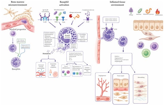
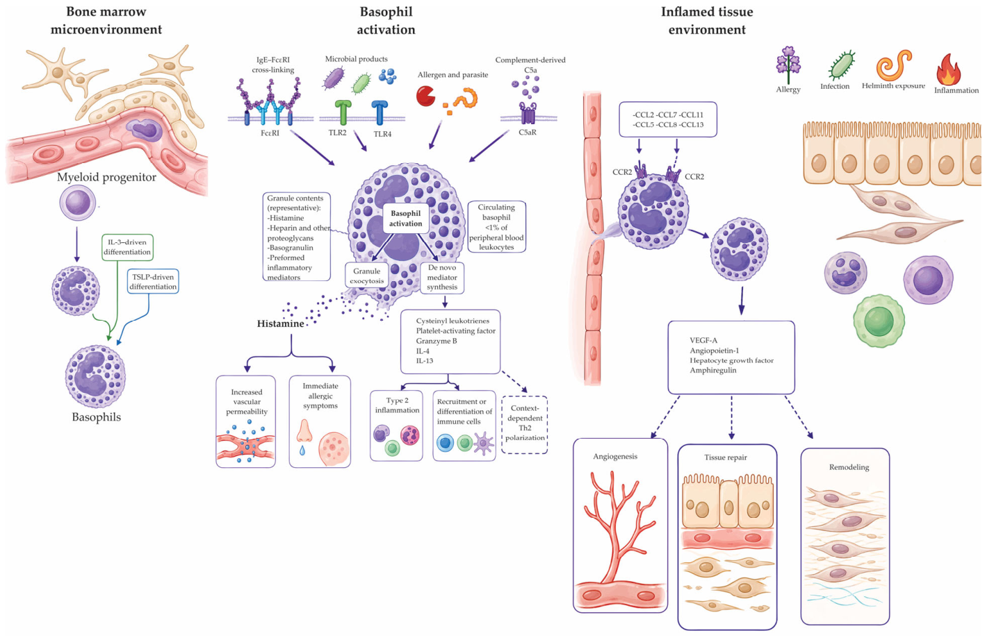
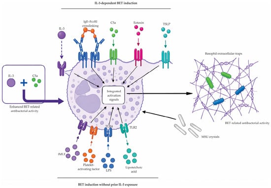
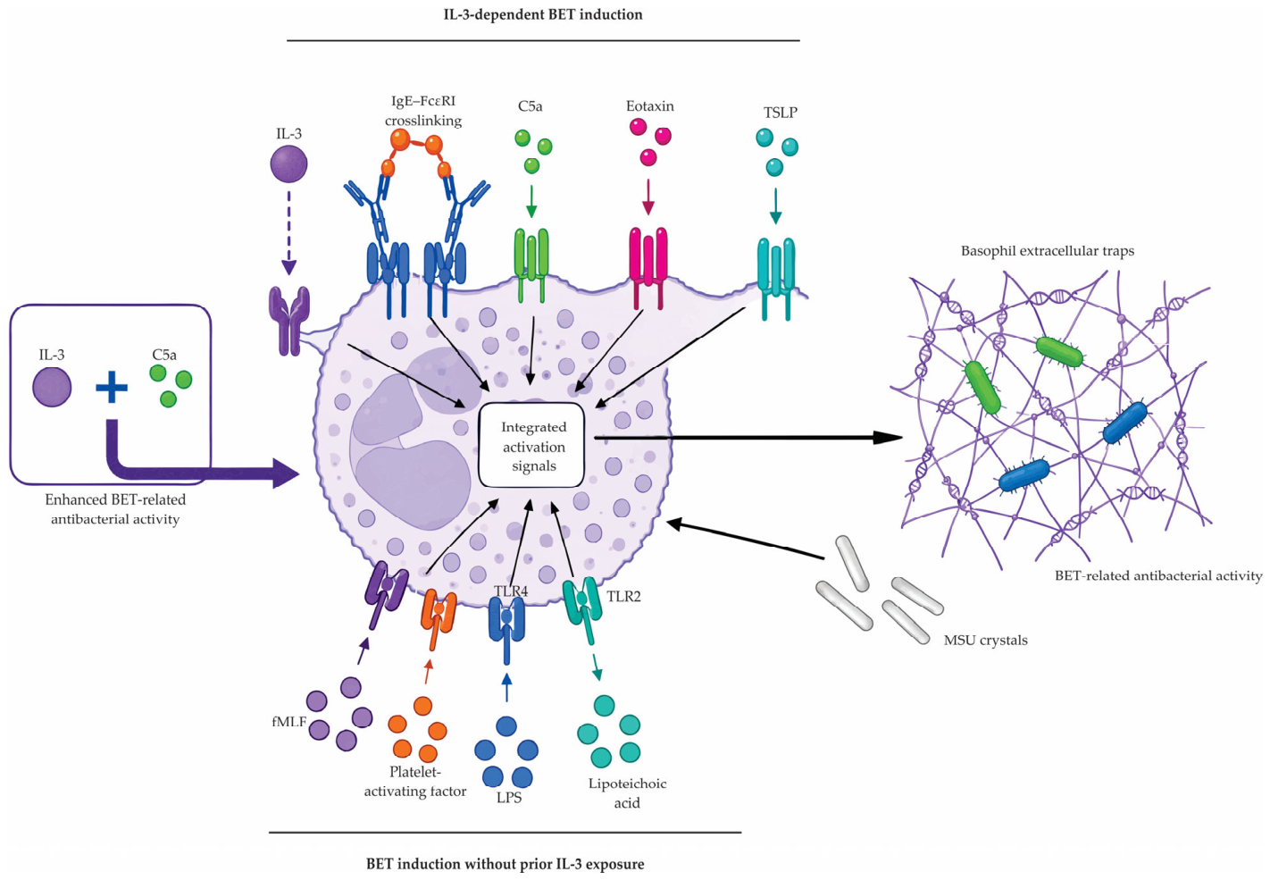
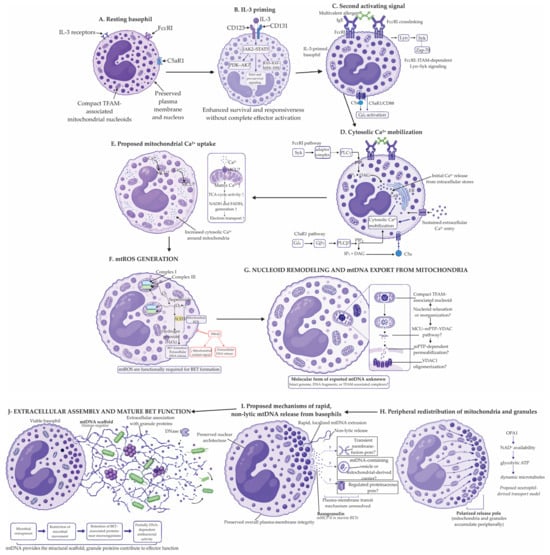
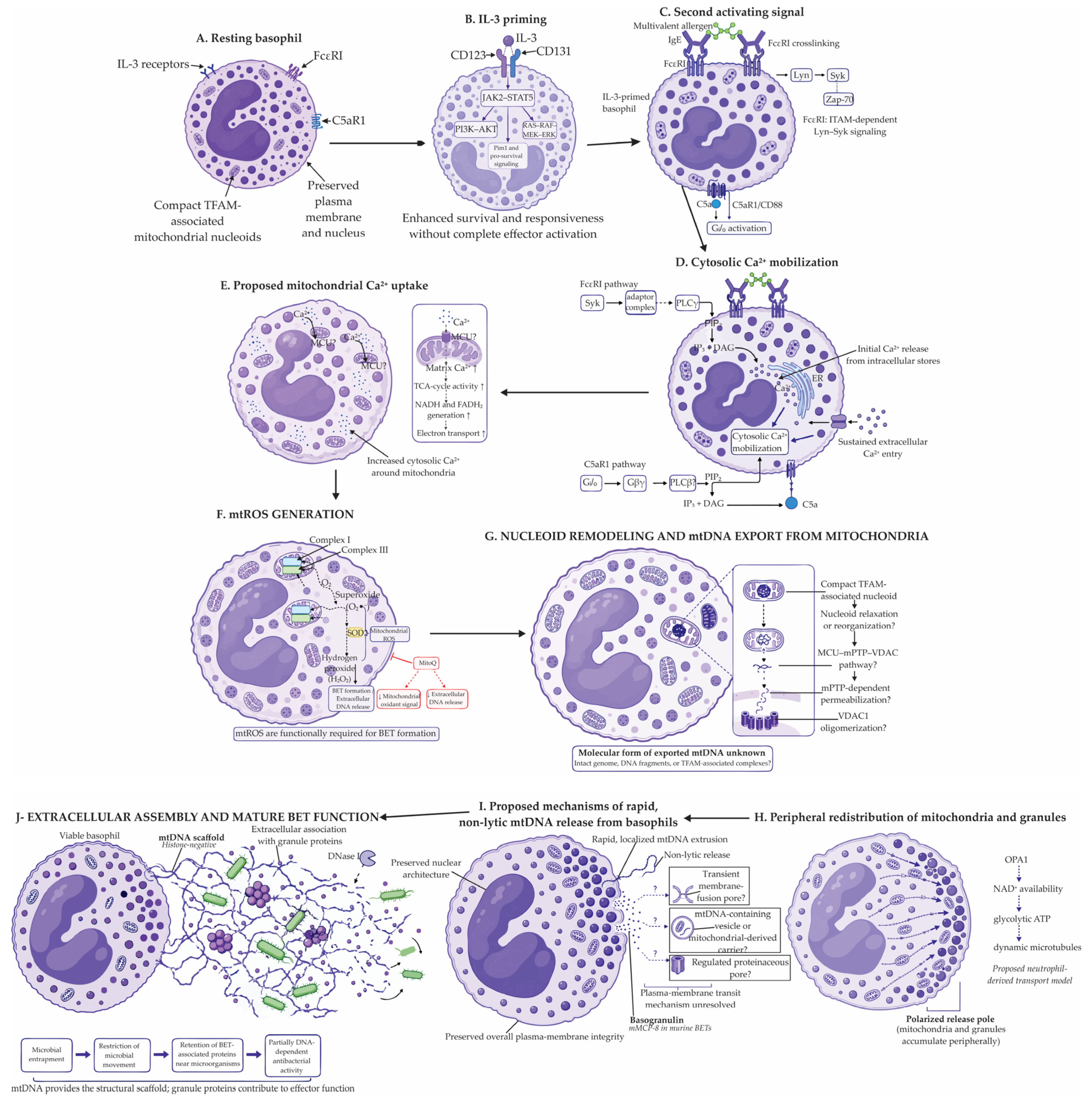
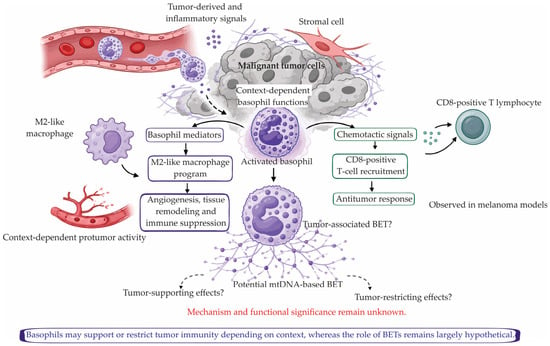
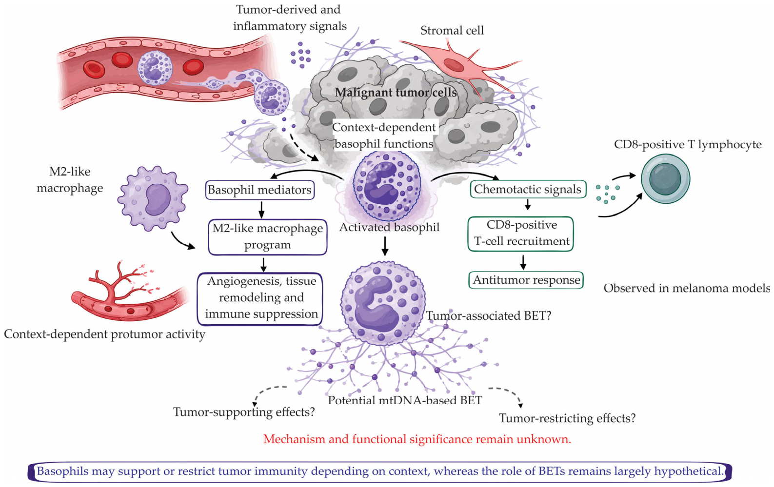

Disclaimer/Publisher’s Note: The statements, opinions and data contained in all publications are solely those of the individual author(s) and contributor(s) and not of MDPI and/or the editor(s). MDPI and/or the editor(s) disclaim responsibility for any injury to people or property resulting from any ideas, methods, instructions or products referred to in the content. |
© 2026 by the authors. Licensee MDPI, Basel, Switzerland. This article is an open access article distributed under the terms and conditions of the Creative Commons Attribution (CC BY) license.