


Simple Summary
Colorectal cancer (CRC) is not a single disease but a complex condition shaped by genetic changes, altered cell signaling, metabolism, and interactions with intestinal microbes. Traditional approaches that examine only one biological layer at a time often miss how these factors work together. Multi-omics research combines information from genes, RNA, proteins, metabolites, and the microbiome to provide a more complete picture of tumor behavior. This review explains how such integrated approaches are improving disease classification, biomarker discovery, and treatment selection, including for immunotherapy and early-onset colorectal cancer. It also highlights the growing role of artificial intelligence in analyzing these complex datasets. At the same time, important barriers remain, such as limited standardization, high cost, and difficulties in translating research findings into routine clinical care. Understanding these advances and challenges may help guide more precise and practical colorectal cancer management in the future.
Abstract
Colorectal cancer (CRC) is a biologically heterogeneous disease in which single-omics analyses incompletely capture the cross-layer mechanisms underlying tumor progression, immune evasion, and therapeutic resistance. This review critically examines how the integration of genomics, transcriptomics, proteomics, metabolomics, and microbiome profiling is redefining CRC biology and precision oncology. Landmark integrative efforts, including TCGA analyses of 276 colorectal cancer samples, CPTAC proteogenomic profiling of 95 tumors, and recent whole-genome sequencing studies of 2023 CRC cases, have refined molecular subtyping, expanded the driver landscape, and revealed clinically relevant discordance between mRNA abundance and protein activity. Integrative studies further show that oncogenic signaling may be driven by post-transcriptional and post-translational regulation, while spatially resolved profiling and microbiome–metabolite analyses are uncovering previously obscured tumor–microenvironment interactions. We also discuss how artificial intelligence-based approaches, including factor analysis, deep learning, graph-based models, and explainable AI, are improving subtype classification, biomarker discovery, and treatment-response prediction, with particular relevance to microsatellite instability-high and early-onset CRC. Finally, we critically evaluate the principal barriers to clinical translation, including batch effects, cross-platform variability, limited external validation, regulatory constraints, and cost, and outline priorities for building reproducible, clinically deployable multi-omics pipelines for CRC management.
1. Introduction
Colorectal cancer (CRC) remains one of the most frequently diagnosed malignancies and a major cause of cancer-related death worldwide. In 2022, there were an estimated 1,926,425 new cases and 904,019 deaths, underscoring the continuing need for better strategies in detection, risk stratification, and treatment [1]. Colonoscopy, fecal immunochemical testing, and computed tomography colonography remain central to CRC screening [2]. However, the main limitations of precision oncology in CRC extend beyond screening alone. In routine practice, treatment decisions still rely on a relatively narrow set of clinicopathological parameters and selected molecular markers. However, patients with apparently similar profiles often show markedly different responses, patterns of resistance, and clinical outcomes. Current approaches therefore do not fully account for the biological diversity of CRC or its evolution over time.
This problem is especially relevant in the context of early-onset colorectal cancer (EOCRC), usually defined as CRC diagnosed before the age of 50 [3]. Although hereditary syndromes remain important in a subset of patients, they explain only a minority of EOCRC cases [4]. This suggests that broader environmental and lifestyle-related influences are contributing to the recent rise in incidence. Factors such as Westernized dietary patterns, obesity, physical inactivity, antibiotic exposure, and other early-life perturbations have all been proposed as possible contributors [5]. Increasing attention has also been directed toward the gut microbiome. Younger patients may harbor altered microbial communities and microbe-derived metabolites that influence mucosal inflammation, epithelial barrier integrity, and carcinogenic signaling. Emerging metabolomic evidence further suggests that EOCRC may involve changes in bile acid metabolism, short-chain fatty acid balance, and amino acid utilization [6]. Taken together, these observations suggest that EOCRC is not simply CRC occurring at a younger age. Instead, it may reflect a distinct biological setting in which environmental, microbial, and metabolic factors play a greater role [7]. CRC itself is shaped by interactions across multiple biological layers, including genomic alterations, transcriptional programs, proteomic signaling, metabolic rewiring, and host–microbiome dynamics [8]. Single-omics studies have provided an important foundation. They identify recurrent driver mutations such as APC, KRAS, and TP53 and supporting classification systems such as the Consensus Molecular Subtypes (CMS). Even so, these approaches capture only part of the disease process and often do not explain how different molecular layers converge to influence progression, immune escape, or treatment resistance. Landmark integrative studies helped shift this view. Work from The Cancer Genome Atlas (TCGA) established a foundational molecular framework for CRC by integrating genomic and transcriptomic features [9]. Studies from the Clinical Proteomic Tumor Analysis Consortium (CPTAC) have shown that clinically relevant protein activity and phosphorylation-based signaling states are not always directly reflected by mRNA abundance. These findings made it increasingly clear that multi-omics integration is not merely an expansion of profiling depth. It is a more appropriate framework for studying a disease as heterogeneous as CRC [10]. Several major questions still remain unresolved. How tumor-intrinsic alterations interact with the immune and stromal microenvironment cannot be fully understood without integrating genomic, transcriptomic, proteomic, and spatial information. Likewise, the variability observed within established CRC subtypes suggests that transcriptional classifications alone are insufficient. Additional proteomic, metabolic, and microbiome layers may help explain clinically divergent behavior. Therapeutic resistance also tends to emerge through coordinated changes across multiple biological levels rather than through a single molecular event. In addition, robust biomarkers for early detection, prognosis, and longitudinal monitoring will likely require composite, multi-layered signatures rather than isolated markers. Viewed in this way, multi-omics integration is not simply a broader profiling strategy. Rather, it is a necessary framework for addressing key unresolved questions in CRC biology and precision oncology.
In this review, we examine how integrated genomic, transcriptomic, proteomic, metabolomic, and microbiome analyses are reshaping current understanding of CRC biology, molecular classification, and clinical stratification. We also highlight translational challenges and the growing relevance of EOCRC. Despite substantial progress, formidable obstacles remain. Data standardization, cohort heterogeneity, computational integration frameworks, and rigorous clinical validation continue to hinder routine clinical implementation. In addition, large-scale, demographically diverse, and longitudinally characterized patient cohorts, including adequate EOCRC representation, are urgently needed for robust biomarker validation and mechanistic investigation (Figure 1).
Figure 1.
Integrative multi-omics framework for CRC research. Multi-omics profiling (genomics, transcriptomics, proteomics, metabolomics, metagenomics) enables unified molecular models informing biomarker discovery, therapeutic target identification, and precision medicine applications.
2. Genomics: The Landscape of Driver Alterations and Molecular Subtypes
2.1. Key Genomic Drivers and Pathways
Driver mutations confer selective growth advantages propelling tumor initiation and progression. They are distinct from passenger mutations that accumulate passively. In CRC, the mutational landscape is dominated by alterations in the WNT signaling pathway, particularly APC (~80%) [11]. Frequent mutations are also found in RAS–MAPK pathway (KRAS mutations in ~40–45%, BRAF V600E in ~10–15%) [12,13], PI3K–AKT pathway (PIK3CA mutations in ~15–20%) [14], and TP53 tumor suppressor pathway (mutations in ~50–60% of cases) [15] (Table 1). These mutations drive the sequential adenoma-to-carcinoma progression (Figure 2).
Table 1.
Key signaling pathways and their genetic alterations in CRC. The table summarizes major dysregulated pathways, their frequently mutated genes, core biological functions, and supporting references.
Figure 2.
Classical adenoma–carcinoma sequence in colorectal cancer. Sequential accumulation of canonical driver alterations, including APC, KRAS, and TP53, and the associated histopathological progression from normal epithelium to invasive carcinoma. Although this model remains a useful framework, alternative routes of CRC development, including serrated and mismatch repair-deficient pathways, are also recognized.
Although the adenoma–carcinoma sequence remains a useful conceptual model for CRC development, it does not capture the full diversity of tumorigenic routes. A substantial subset of CRCs arises through alternative pathways, most notably the serrated neoplasia pathway. This pathway is commonly associated with sessile serrated lesions, BRAF mutation, and CpG island methylator phenotype (CIMP). In some cases, it is also linked to subsequent mismatch repair deficiency and microsatellite instability [21]. In parallel, hypermutated CRCs may develop through mismatch repair-deficient mechanisms that do not conform to the classical stepwise APC–KRAS–TP53 progression [22]. More recent genomic studies also suggest that CRC evolution is often non-linear, with branching trajectories shaped by chromosomal instability, epigenetic remodeling, and structural variation [23]. Taken together, these observations indicate that Figure 2 should be viewed as a representative model rather than a universal pathway for all CRCs.
APC mutations disrupt the β-catenin destruction complex. This leads to constitutive WNT activation. Mutation location also affects phenotype. Truncating mutations in the mutation cluster region are associated with intermediate WNT activity and a more favorable prognosis. By contrast, mutations outside this region are linked to higher WNT activity and more aggressive disease [24]. KRAS mutations (codons 12, 13, 61) constitutively activate MAPK and PI3K signaling. However, different KRAS variants are not biologically equivalent. Functional studies have shown that specific substitutions such as G12D and G12V can differ in signaling output. They also differ in therapeutic vulnerability, including differential sensitivity to MEK pathway inhibition [25]. By contrast, G13D shows distinct biochemical behavior. It may also retain altered interactions with regulatory proteins such as NF1. This may partly explain its atypical response to EGFR inhibition [26]. KRAS mutations predict anti-EGFR therapy resistance (cetuximab, panitumumab), mandating testing before treatment [12]. BRAF V600E mutations (~10–15%) confer poor prognosis in microsatellite-stable tumors. They are also associate with right-sided location and CMS1 subtype, and may show modest benefit from encorafenib plus cetuximab combination therapy [27]. TP53 mutations (~50–60%) arise during late adenoma-to-carcinoma transition. Missense mutations in the DNA-binding domain exhibit gain-of-function properties, promoting invasion, metastasis, and chemoresistance. Emerging evidence also links TP53 mutation status to chemotherapy response [28]. PIK3CA mutations (~15–20%) activate PI3K–AKT–mTOR signaling and frequently co-occur with KRAS mutations. Although PIK3CA inhibitors have shown limited CRC efficacy, these mutations may predict aspirin chemoprevention benefit (Table 2) [29].
Beyond these canonical driver events, recent whole-genome sequencing studies have substantially expanded the genomic framework of colorectal cancer [30]. In particular, large-scale analysis of 2023 CRCs from the UK 100,000 Genomes Project provided a high-resolution map of somatic alterations [23] and identified more than 250 putative driver genes. Many of these genes had not previously been implicated in CRC or, in some cases, in cancer more broadly. These data also refined the mutational processes operating in CRC and extended the set of molecular pathways involved in tumor development. Importantly, the study detected recurrent alterations outside the coding genome, highlighting the likely contribution of non-coding regulatory mutations that may influence transcriptional control and tumor behavior. Whole-genome sequencing also captures structural variation more effectively than targeted or exome-based approaches, including large-scale rearrangements, copy-number changes, and other chromosomal alterations that contribute to genomic instability and inter-tumoral heterogeneity. Together, these findings indicate that CRC genomics cannot be fully understood through a coding-centric view alone. More recent integrative genome–transcriptome analyses of over 1000 primary CRCs have reinforced this shift by linking newly identified driver events to functional and prognostic consequences [30]. International whole-genome studies further suggest that mutational processes vary according to geography and age. These observations are especially relevant to current efforts to refine molecular subtypes and explain biological diversity within clinically similar tumors. They are also important for linking genomic architecture with downstream transcriptomic and proteomic states [31].
Table 2.
Prevalence, roles and references of key genetic alterations in CRC. The table summarizes frequently mutated genes in CRC, their occurrence rates, functional impact on tumor biology, and associated literature.
Recent whole-genome sequencing of 2023 CRC specimens identified over 250 putative driver genes (Figure 3), including subtype-specific candidates [23]. Integrated multi-omics analyses have resolved temporal driver events and established molecular subtypes defined by Wnt, EGFR, and TGF-β signaling engagement [33]. Epigenetic regulators, including ATRX (~7% of CRCs), also contribute to CRC progression [34]. They may promote metastasis through epithelial-to-mesenchymal transition and chromatin remodeling, revealing potentially actionable therapeutic targets for precision intervention.
Figure 3.
Classification and mechanisms of driver genes in CRC. Key driver genes categorized as oncogenes (orange) and tumor suppressor genes (blue), showing representative examples and their molecular mechanisms in tumor initiation and progression.
2.2. Microsatellite Instability (MSI) as a Biomarker Paradigm
Microsatellite instability (MSI) results from defective DNA mismatch repair (MMR). It is typically caused by biallelic inactivation of MLH1, MSH2, MSH6, or PMS2 through germline mutations (Lynch syndrome, ~3% of CRCs) or somatic MLH1 promoter hypermethylation (sporadic cases, ~12%) [35]. MSI-high (MSI-H) tumors exhibit a hypermutator phenotype. They often show frameshift mutations in TGFBR2 and BAX, right-sided location, mucinous histology, and prominent immune infiltration. High neoantigen burden drives robust antitumor immunity, conferring favorable stage-adjusted survival. However, BRAF V600E co-mutations may worsen outcomes.
Beyond its biological significance, MSI has direct clinical utility across several stages of colorectal cancer management. Universal MMR/MSI testing is now recommended at diagnosis in NCCN guideline for colon cancer. This helps identify patients with possible Lynch syndrome and guides treatment planning. In localized disease, dMMR/MSI-H status is particularly relevant in stage II colon cancer. These tumors generally have a more favorable stage-adjusted prognosis and do not derive meaningful benefit from fluoropyrimidine monotherapy [36]. The NCCN guideline also now includes dedicated biomarker-stratified pathways for dMMR/MSI-H rectal cancer, including checkpoint inhibitor-based strategies in selected locally advanced and metastatic settings. In advanced disease, MSI-H/dMMR has become one of the most established predictive biomarkers for immune checkpoint blockade [37]. In the 5-year follow-up of the phase III KEYNOTE-177 trial, 307 patients with previously untreated MSI-H/dMMR metastatic colorectal cancer were randomized to pembrolizumab (n = 153) or chemotherapy (n = 154). Median overall survival was 77.5 versus 36.7 months, 5-year overall survival was 54.8% versus 44.2%. Median progression-free survival was 16.5 versus 8.2 months and median duration of response was 75.4 versus 10.6 months [38]. More recent phase III data from CheckMate 8HW further support first-line nivolumab plus ipilimumab in this setting. Among 303 randomized patients, including 255 with centrally confirmed MSI-H/dMMR tumors, 24-month progression-free survival was 72% with nivolumab plus ipilimumab compared with 14% with chemotherapy. Grade 3 or 4 treatment-related adverse events occurred in 23% versus 48% of patients, respectively [39]. Together, these applications make MSI one of the clearest examples of how molecular classification has already entered routine clinical decision-making in CRC.
2.3. Consensus Molecular Subtypes (CMS)
Integration of genomic and transcriptomic data has classified CRC into four biologically distinct subtypes with therapeutic implications [40]. CMS1 (MSI-Immune, ~14%) exhibits hypermutation, immune activation, frequent BRAF mutations, and responsiveness to immunotherapy. CMS2 (Canonical, ~37%) shows epithelial differentiation, WNT/MYC activation, and chromosomal instability. CMS3 (Metabolic, ~13%) displays metabolic dysregulation with frequent KRAS mutations. CMS4 (Mesenchymal, ~23%) demonstrates TGF-β activation, stromal invasion, epithelial–mesenchymal transition, and poor prognosis. Recent multi-omics and spatial analyses indicate that CMS4 is not solely a tumor-intrinsic transcriptional state. Instead, it reflects a stromal-rich microenvironment characterized by activated cancer-associated fibroblasts, TGF-β signaling, and immune exclusion [41]. This framework provides a foundation for understanding CRC heterogeneity. Ongoing multi-omics studies are further refining subtype boundaries and clinical utility (Table 3).
Table 3.
Biological and clinical features of the consensus molecular subtypes in CRC. References: Summarized from [42,43], with additional CMS4-related stromal and immune microenvironment features adapted from [44].
3. Transcriptomics: From Bulk Profiling to Spatial Resolution
Transcriptomics profiles RNA transcripts to link genomic alterations with functional outcomes in CRC. By quantifying mRNA and non-coding RNAs, it delineates gene-expression dynamics, regulatory networks, and disease-specific signatures, enabling biomarker discovery for diagnosis, prognosis, and therapy. This section highlights key advances in transcriptomic profiling, the roles of non-coding RNAs, and their translational applications in CRC [42].
3.1. Gene Expression Signatures and Biomarkers
Transcriptome analysis, led by RNA-seq, has redefined the gene-expression landscape in CRC. Comparison of tumor and normal tissues reveals differentially expressed genes (DEGs) involved in cancer hallmarks, including cell-cycle dysregulation, immune evasion, and EMT [43,44]. Overexpression of genes like MMP7, S100A4, and CEACAM5 is consistently linked to invasion, metastasis, and poor prognosis [45]. Expression signatures further stratify patients into molecular subgroups and complement genomic classifications such as CMS (Table 4).
Table 4.
Representative transcriptomic biomarkers and gene-expression signatures in CRC. Key coding and non-coding RNA biomarkers and gene-expression signatures, highlighting their functional roles, clinical relevance, and validation status in CRC.
Interpretation of RNA-seq data in CRC also depends heavily on the computational pipeline used. In most bulk RNA-seq studies, raw reads first undergo quality control and filtering, followed by adapter trimming and alignment to the reference genome or pseudoalignment to the transcriptome. Gene- or transcript-level quantification is then generated and subjected to normalization before differential expression analysis, clustering, and downstream functional interpretation. Depending on the aims of the study, these data may also be combined with pathway enrichment, co-expression network analysis, immune deconvolution, or integrative multi-omics modeling. In single-cell and spatial transcriptomic datasets, further computational steps are usually required, including cell filtering, batch correction, dimensionality reduction, clustering, trajectory inference, and ligand–receptor or cell–cell communication analysis. These analytical choices are not merely technical details, as they can substantially influence subtype assignment, biomarker discovery, and biological interpretation in CRC [58,59].
In CRC, transcriptomic studies have identified gene expression signatures associated with proliferation, epithelial–mesenchymal transition, immune exclusion, stromal activation, and metastatic potential. Such signatures have helped refine molecular stratification beyond mutational status alone and have highlighted why tumors with similar genomic profiles may still behave differently in clinical practice. Transcriptomic biomarkers are therefore increasingly relevant not only for mechanistic understanding, but also for improving prognostic models and identifying patient subsets that may benefit from specific therapeutic strategies [30,60].
3.2. The Non-Coding RNA Regulome
Non-coding RNAs (ncRNAs), including microRNAs (miRNAs), long non-coding RNAs (lncRNAs), and circular RNAs (circRNAs), regulate gene expression in CRC [61,62]. MiRNAs such as miR-21, miR-135b, and miR-200c modulate post-transcriptional silencing and affect proliferation, apoptosis, and metastasis. Serum miR-21 has also shown diagnostic potential [63]. Oncogenic lncRNAs such as HOTAIR, MALAT1, and CCAT1 promote metastasis through mechanisms including PRC2-mediated epigenetic silencing. CircRNAs such as circHIPK3 and circRNA_100290 function as miRNA sponges or protein scaffolds, contributing to chemoresistance [64].
Beyond their individual expression changes, non-coding RNAs in CRC increasingly appear to function within broader regulatory networks rather than as isolated molecules. In particular, competing endogenous RNA (ceRNA) mechanisms provide one framework for understanding how lncRNAs and circRNAs modulate gene expression by sequestering shared miRNAs. This may relieve repression of downstream mRNA targets. Through these interactions, ncRNA-mediated networks may influence key CRC-associated processes, including epithelial–mesenchymal transition, proliferation, invasion, stemness, immune signaling, and drug resistance. This is especially relevant in CRC, where transcript abundance alone may not fully capture effective regulatory output. A network-based view also helps explain why dysregulated miRNAs, lncRNAs, and circRNAs are increasingly being investigated not only as individual biomarkers, but also as functionally connected regulators of tumor behavior and therapeutic response [65,66]. For example, circRNA- and lncRNA-centered ceRNA circuits have been reported to regulate CRC growth, migration, and treatment resistance through miRNA-mediated control of oncogenic pathways [67].
3.3. Single-Cell and Spatial Transcriptomics
Single-cell RNA-seq (scRNA-seq) resolves tumor microenvironment heterogeneity. It can identify functionally distinct tumor-associated fibroblast subsets that promote progression and reveal therapeutic targets [68]. Spatial transcriptomics preserves tissue architecture while profiling gene expression. It can map expression patterns to four anatomical regions: tumor core, stromal interface, immune infiltration zones, and normal epithelium [69]. These analyses show TMSB4X enrichment in tumor regions, suggesting a role in tumorigenesis. They also reveal active stromal-tumor interface communication via ligand-receptor pairs such as C5AR1-RPS19. In addition, they identify region-specific immune infiltration patterns, with immune-excluded phenotypes exhibiting T cell separation by stromal barriers [70].
4. Proteomics: The Functional Effector Layer
Proteomics provides essential functional insights in CRC by directly linking genotype to phenotype. As proteins execute cellular functions and serve as therapeutic targets, proteomic profiling elucidates disease mechanisms, reveals biomarkers, and identifies actionable targets [71]. Recent mass spectrometry advances have greatly improved the depth and coverage of proteomic analysis. These advances enable comprehensive characterization of protein expression, post-translational modifications, and protein–protein interactions in CRC tissues and biofluids [72]. Spatial transcriptomics has particular value in colorectal cancer because it allows transcriptional programs to be interpreted within the architectural context of the tumor microenvironment rather than in dissociated cell populations alone. This is especially important in CRC, where stromal activation, immune-cell localization, and epithelial–mesenchymal interactions can vary substantially across tumor regions [73]. By preserving spatial information, these approaches can distinguish tumor core, invasive front, stromal-rich niches, and immune-excluded versus immune-infiltrated areas, thereby providing a more precise view of local tumor ecology [74]. Spatial transcriptomic analyses have begun to uncover ligand–receptor interactions and region-specific communication among malignant, stromal, endothelial, and immune cells. This spatial information adds important biological context that is not captured by bulk profiling alone. It may help explain differences in progression, metastasis, and treatment response among tumors with otherwise similar molecular features [75]. In this sense, spatial transcriptomics is not only a high-resolution mapping tool. It also serves as an important bridge between molecular subtype classification and the biological organization of the CRC tumor microenvironment. Accordingly, spatial transcriptomics provides a way to link molecular heterogeneity with clinically relevant microenvironmental states. These include immune exclusion, stromal remodeling, and region-specific signaling programs that may influence prognosis and therapeutic sensitivity.
4.1. Technological Advances
Mass spectrometry-based proteomics has revolutionized CRC molecular characterization. Liquid chromatography-tandem mass spectrometry (LC-MS/MS) achieves proteome coverage exceeding 10,000 proteins from single biopsies with high sensitivity and specificity. Quantitative approaches include Tandem Mass Tag (TMT) isobaric labeling and Data-Independent Acquisition (DIA). TMT enables multiplexed analysis of up to 18 samples in a single run. By contrast, DIA supports cost-effective and reproducible workflows in large cohorts with coefficient of variation below 20% [76]. Targeted phosphoproteomics reveals dysregulated signaling including β-catenin (WNT) and AKT (PI3K/AKT) phosphorylation, identifying therapeutic vulnerabilities [77]. Glycoproteomics characterizes altered glycosylation patterns affecting adhesion, invasion, and immune recognition [78].
Emerging spatial proteomics technologies, including imaging mass cytometry (IMC) and multiplexed ion beam imaging (MIBI), have expanded the scope of proteomic analysis. These approaches enable spatially resolved, single-cell protein quantification within intact tissues. They also allow mapping expression across tumor, stromal, and immune compartments [79]. These approaches reveal spatial organization and cell–cell interactions, identifying distinct proteomic signatures at tumor invasion fronts and immune exclusion zones.
Different proteomic platforms offer complementary rather than interchangeable advantages in CRC research. Conventional LC-MS/MS provides broad proteome coverage and is well suited for discovery-oriented studies, but its performance can be influenced by sample complexity, instrument time, and batch effects [80]. TMT-based workflows enable high multiplexing and are advantageous for comparative analyses across multiple samples. However, ratio compression and dependence on careful batch design may affect quantitative accuracy [81]. DIA approaches generally offer better reproducibility and scalability for large cohorts. This makes them attractive for biomarker verification studies. Nevertheless, depending on the spectral library and acquisition settings, they may provide lower depth for some low-abundance proteins. Targeted phosphoproteomics is particularly useful for interrogating pathway activation and drug-response signaling [82]. By contrast, spatial proteomics platforms such as IMC and MIBI provide critical contextual information by resolving protein expression within tumor, stromal, and immune niches. However, these approaches are associated with higher cost, lower throughput, and greater analytical complexity. Accordingly, platform selection should be guided by the specific clinical or biological question. This may include biomarker discovery, pathway interrogation, cohort-scale validation, or spatial characterization of the tumor microenvironment [83]. The main applications are summarized in Table 5.
Table 5.
Comparison of major proteomic platforms in CRC research. Different proteomic platforms provide complementary rather than interchangeable advantages in CRC research, and platform selection should be guided by the primary study objective, including discovery, cohort-scale validation, signaling analysis, or spatial characterization of the tumor microenvironment.
4.2. Proteomic Biomarkers and Pathways
Proteomic profiling has identified clinically relevant biomarkers across the diagnostic-prognostic-predictive spectrum [86]. Compared with traditional serum markers such as CEA and CA19-9, several emerging protein biomarkers show improved diagnostic sensitivity for colorectal cancer detection. These biomarkers include S100A9, MMP9, and Annexin A2 (Table 6). Proteomic validation studies have further demonstrated that multi-marker panels incorporating these candidates can achieve AUC values greater than 0.90, significantly improving early-stage detection accuracy [87]. Prognostic tissue signatures include mesenchymal proteins (vimentin, N-cadherin) correlating with poor outcomes, whereas epithelial markers (E-cadherin, cytokeratins) associate with favorable prognosis [88]. Proteins including galectin-3, cathepsin D, and S100A4 stratify patients into distinct risk categories beyond conventional staging [89]. For therapeutic targeting, membrane receptor quantification (EGFR, VEGFA, MET, HER2) and immune checkpoint profiling (PD-L1, PD-L2, LAG-3) guide treatment selection [89]. Critically, 30–40% of genes show poor mRNA-protein correlation, emphasizing proteomics’ superior functional assessment. This discordance has been quantitatively demonstrated in CPTAC colorectal cancer studies. In the CPTAC proteogenomic analysis of colon and rectal cancer, the mean Spearman correlation between steady-state mRNA and protein abundance across individual samples was 0.47. However, when mRNA and protein variation were compared across tumors, the mean correlation dropped to 0.23, and only 32% of mRNA–protein pairs showed statistically significant correlation. These findings indicate that transcript abundance alone is often insufficient to predict functional protein output in CRC and support the integration of proteomics into multi-omics stratification frameworks [90]. A representative example is provided by the Clinical Proteomic Tumor Analysis Consortium (CPTAC) proteogenomic study in colorectal cancer. This study showed that protein activity often does not correlate directly with mRNA abundance. This was particularly evident for post-translational modifications. such as phosphorylation. CPTAC analyses revealed that activation of key oncogenic pathways, including the WNT/β-catenin and PI3K/AKT signaling axes, is frequently driven by phosphorylation dynamics rather than transcriptional upregulation. For instance, signaling intermediates such as AKT and GSK3β may exhibit stable mRNA expression levels. However, their phosphorylation states can be markedly increased. This increase may enhance downstream signaling activity and promote tumor proliferation, survival, and immune evasion [91]. These findings underscore that phosphoproteomic profiling can capture functional pathway activation more accurately than transcriptomic data alone, highlighting the critical role of post-translational regulation in colorectal cancer biology. Phosphoproteomics further adds clinically relevant information by identifying targetable signaling states that may not be evident at the DNA or mRNA level. In CPTAC analyses of colon cancer, MSI-H tumors with reduced CD8 T-cell infiltration showed increased glycolytic activity.
This finding suggests that glycolytic rewiring may represent a potential strategy to overcome resistance to immune checkpoint blockade. In addition, integrated phosphoproteomic analysis of metastatic CRC showed that kinase-substrate network features could distinguish metastatic behavior. These features were also associated with response to kinase inhibitors, including afatinib, gefitinib, and regorafenib. Together, these findings highlight the translational value of phosphoproteomics for treatment stratification [10,92].
Table 6.
Representative protein biomarkers and therapeutic targets in CRC. Representative proteins from CRC proteomics with biological roles and clinical applications. Established markers (CEA) enable monitoring; novel candidates (S100A9, Annexin A2) show diagnostic potential; therapeutic targets (VEGF-A, PD-L1) demonstrate clinical translation.
Despite the rapid expansion of proteomic biomarker discovery for colorectal cancer diagnosis, prognosis, and treatment stratification, clinical translation remains limited [101]. Pre-analytical variability, including differences in tissue handling, ischemia time, storage conditions, and biofluid processing, can substantially affect measured protein abundance [102]. In addition, cross-platform heterogeneity, differences in peptide identification pipelines, and limited inter-laboratory standardization hinder reproducibility and complicate comparisons across studies. Proteomic assays also face several intrinsic challenges. These include the wide dynamic range of the human proteome, the low abundance of clinically relevant signaling proteins, and the context dependence of post-translational modifications. Moreover, many candidate biomarkers have been derived from relatively small retrospective cohorts and still lack independent external validation or prospective clinical testing. Cost, turnaround time, infrastructure requirements, and the need for specialized bioinformatics support further restrict implementation in routine pathology and oncology settings. Therefore, the most promising path toward clinical application may lie in rigorously validated multi-protein panels. These panels should be integrated with genomic, transcriptomic, and clinicopathologic features. Such an approach may improve diagnosis, risk stratification, and disease monitoring [103].
Comparative proteomics reveals resistance mechanisms including alternative pathway activation (MET, AXL) mediating EGFR inhibitor resistance, and upregulated drug efflux pumps (ABCB1, ABCG2) conferring chemotherapy resistance (Figure 4) [104]. Tumor-derived exosomes carrying HSP70, TGF-β1, and Annexin A2 enable non-invasive disease monitoring, treatment response assessment, and minimal residual disease detection [105]. Pathway-level analyses reveal dysregulated networks including aberrant β-catenin (WNT), phosphorylated AKT (PI3K/AKT/mTOR), immune-suppressive proteins (PD-L1, IL-6), and EMT mediators (vimentin, N-cadherin) driving invasion and therapy resistance [106].
Figure 4.
Dysregulated signaling pathways in CRC. Key altered pathways: WNT/β-catenin, PI3K/AKT/mTOR, epithelial–mesenchymal transition (EMT), and immune evasion. Aberrant β-catenin and Cyclin D1 drive proliferation; phosphorylated AKT and mTOR enhance survival and resistance; EMT markers (vimentin, N-cadherin) promote metastasis; immune-suppressive proteins (PD-L1, IL-6, TNF-α) enable immune evasion and support immunotherapy rationale.
4.3. Translational Outlook
As proteomics matures, it increasingly informs personalized oncology in CRC. Proteogenomics integrates proteomic data with genomic and transcriptomic information. It has enabled the construction of comprehensive molecular atlases and may support individualized therapeutic interventions. Furthermore, proteomic biomarkers are being incorporated into early-phase clinical trials as companion diagnostics or predictive indicators, accelerating the clinical translation of multi-omics researcher. Overall, the translational value of proteomics in CRC depends not only on biomarker discovery. It also depends on platform-appropriate study design, analytical standardization, and prospective validation in clinically representative cohorts.
5. Metabolomics: The Phenotypic Signature of Reprogramming
Metabolomics, the comprehensive analysis of small-molecule metabolites, provides a dynamic snapshot of tumor physiology reflecting integrated genetic, transcriptomic, proteomic, and environmental effects. In CRC, metabolomic profiling has yielded critical insights into tumor metabolism, biomarker discovery, and disease progression mechanisms.
5.1. Analytical Technologies and Metabolic Hallmarks
Metabolomic investigations rely on several high-throughput analytical platforms. Nuclear magnetic resonance (NMR) spectroscopy is widely used for reproducible biofluid analysis. Mass spectrometry coupled with liquid or gas chromatography (LC-MS, GC-MS) enables sensitive broad-spectrum detection. Capillary electrophoresis-MS (CE-MS) is particularly useful for resolving polar metabolites, including organic acids and bile salts [107]. Both targeted and untargeted approaches are applied to tissue biopsies, serum/plasma, stool, and urine, enabling invasive and non-invasive disease monitoring.
CRC cells exhibit profound metabolic reprogramming across multiple pathways. Aerobic glycolysis (Warburg effect) drives preferential glucose-to-lactate conversion under normoxia, with upregulated lactate dehydrogenase A (LDHA) and accumulated lactate and pyruvate [108]. Importantly, metabolic reprogramming in CRC is not uniform across molecular or clinicopathologic subtypes. Metabolomic studies indicate that CRC can be stratified into metabolically distinct subgroups with differences in lipid, nucleotide, and carbohydrate metabolism, suggesting that tumor behavior and prognosis may be influenced by subtype-specific metabolic dependencies. In addition, metabolic phenotypes vary according to genetic context and tumor location. For example, experimental metabolomic profiling of genetically engineered intestinal tumors has shown that common driver alterations such as APC, KRAS, and TP53 generate distinct metabolic states rather than a single shared CRC metabolic program. Likewise, right-sided and left-sided CRCs exhibit partially divergent metabolomic landscapes, further supporting the existence of clinically relevant metabolic heterogeneity in CRC [109]. Glutamine serves as an anaplerotic TCA cycle substrate under hypoxia, generating altered α-ketoglutarate, succinate, and fumarate levels. Enhanced lipogenesis produces abnormal phosphatidylcholines, sphingolipids, lysophosphatidylcholine (LPC), and ceramides supporting membrane synthesis and oncogenic signaling [110]. Dysregulated amino acid metabolism—particularly arginine, tryptophan (kynurenine pathway), methionine, and branched-chain amino acids—promotes proliferation and immune evasion.
5.2. Microbial Metabolites and the Gut-Tumor Axis
The gut microbiota influences CRC pathogenesis through metabolites with opposing properties. Short-chain fatty acids (SCFAs), including butyrate, acetate, and propionate, are produced through dietary fiber fermentation. These metabolites exhibit anti-tumor effects through histone deacetylase (HDAC) inhibition. They can promote cell cycle arrest, apoptosis, and differentiation, and reduced SCFA levels have been associated with CRC risk [111]. Conversely, secondary bile acids (deoxycholic acid, lithocholic acid) generated by bacterial 7α-dehydroxylation accumulate in high-fat, low-fiber diets, inducing DNA damage and inflammatory signaling via NF-κB and STAT3 activation [112]. Polyamines, including putrescine, spermidine, spermine, become dysregulated in CRC. This dysregulation can promote tumor growth and immunosuppression. Hydrogen sulfide (H2S), by contrast, shows dose-dependent effects. Low concentrations support epithelial barrier function, whereas high levels induce genotoxicity [113]. These findings highlight the critical role of diet-microbiota-metabolite interactions in CRC development, presenting opportunities for microbiome-targeted interventions (Figure 5). These metabolic interactions may be especially relevant in early-onset colorectal cancer (EOCRC). Although the metabolic biology of EOCRC is still being defined, emerging evidence suggests that younger-onset disease may not simply mirror the metabolic features of later-onset CRC. Recent integrative metagenomic-metabolomic studies have identified distinct gut microbiome-derived phenotypes in EOCRC. These findings suggest that altered bile acid handling, short-chain fatty acid imbalance, and broader changes in amino acid and lipid metabolism may contribute to tumor initiation in younger individuals. This is consistent with the view that diet, obesity, antibiotic exposure, and other early-life influences may reshape the intestinal metabolic environment long before overt malignancy develops. For this reason, EOCRC represents a particularly informative setting in which metabolomics and microbiome profiling can be studied together rather than in isolation.
Figure 5.
Metabolic reprogramming in CRC. CRC cells exhibit enhanced glycolysis, glutaminolysis, and lipogenesis with elevated lactate, ceramides, and lysophosphatidylcholine (LPC). Depleted tryptophan and butyrate alongside enriched microbial metabolites (deoxycholic acid, polyamines) drive tumor growth, immune evasion, and inflammation, representing diagnostic biomarkers and therapeutic targets.
In addition, metabolic flux analysis and stable-isotope tracing provide functional insight beyond steady-state metabolite abundance by revealing nutrient utilization and pathway activity. In CRC, isotope-tracing studies have shown that oncogenic PIK3CA mutations reprogram glutamine metabolism, highlighting how flux-based approaches can uncover targetable metabolic dependencies [114,115].
Importantly, the microbiome–metabolite axis in CRC is supported not only by associative studies but also by functional evidence. For example, in gnotobiotic mouse models, a high-fiber diet combined with butyrate-producing bacteria increased luminal butyrate levels. This was accompanied by a reduced tumor burden, supporting a causal tumor-suppressive role for this microbial metabolite [6]. Conversely, paired microbiome–metabolome analyses in CRC-susceptible mice showed that high-fat diet–driven microbial shifts increased bile acid alterations, including microbially modified bile acid species, and promoted disease progression. These findings indicate that selected microbiota-derived metabolites can act as functional mediators of tumor promotion or suppression rather than merely correlating with disease status [116].
5.3. Translational Potential
Metabolomics has emerged as a powerful tool for CRC precision oncology (Table 7), with serum or stool metabolomic signatures [117]. Several metabolite panels have shown promising diagnostic performance in CRC, but these estimates vary by biospecimen type, cohort design, and validation strategy. For example, one serum metabolomics study reported an 8-metabolite panel with an AUC of 0.968, sensitivity of 95%, and specificity of 100% in CRC diagnosis, whereas another integrative plasma metabolomics study identified a 17-metabolite panel that achieved AUCs ranging from 0.848 to 0.987 across discovery and three validation cohorts. Accordingly, statements regarding diagnostic sensitivity should be linked to specific studies rather than generalized across all metabolite panels [117,118]. Prognostically, altered levels of sarcosine, choline derivatives, and succinate correlate with tumor stage, lymph node involvement, and recurrence risk. Longitudinal profiling can also be used to monitor treatment response monitoring. For example, lactate levels may decrease after treatment, whereas SCFAs levels may increase [119]. Additionally, metabolomics guides personalized interventions by evaluating dietary impacts (fiber intake) and probiotic/prebiotic supplementation effects, informing nutrition-based therapeutic strategies.
Table 7.
Metabolite biomarkers and clinical relevance in CRC. Representative metabolites associated with colorectal cancer, including tumor-related, lipid-associated, amino acid-related, and microbiota-derived metabolites, together with their sample type, metabolic pathway, functional roles in tumor progression, metabolic reprogramming, or immune modulation, and potential diagnostic, prognostic, or therapeutic relevance.
6. Microbiomics: The Tumor Ecosystem Modulator
The gut microbiome critically influences CRC pathogenesis through genotoxic insult, chronic inflammation, metabolic reprogramming, and immune modulation. Multi-omics integration combining metagenomics, metatranscriptomics, and metabolomics has revealed specific microbial signatures associated with tumor initiation, progression, and therapeutic response.
6.1. Dysbiosis and Key Pathobionts
CRC exhibits distinct gut dysbiosis characterized by depletion of beneficial commensals (e.g., Faecalibacterium, Roseburia) and enrichment of pro-carcinogenic pathobionts [129]. Fusobacterium nucleatum promotes tumor progression via FadA/E-cadherin signaling and immune suppression via Fap2/TIGIT interaction; a candidate diagnostic and prognostic biomarker [130]. Mechanistic studies support causal roles for several CRC-associated pathobionts rather than simple enrichment alone. In particular, Fusobacterium nucleatum has been shown to promote colorectal tumorigenesis through its FadA adhesin. FadA binds to E-cadherin and activates β-catenin signaling. This, in turn, enhances epithelial proliferation and oncogenic transcriptional programs [131]. In parallel, the Fap2 virulence factor can bind TIGIT on immune cells and contribute to immune evasion within the tumor microenvironment. Enterotoxigenic Bacteroides fragilis (ETBF) exerts tumor-promoting effects through the B. fragilis toxin (BFT). This toxin disrupts epithelial integrity and activates STAT3 signaling. In preclinical models, it also drives IL-17/Th17-predominant inflammation [132]. Likewise, pks+ Escherichia coli has been shown to induce colibactin-mediated DNA damage. Organoid and genomic studies have linked this exposure to characteristic SBS-pks and ID-pks mutational signatures. These findings support a direct role for pks+ E. coli in early colorectal tumorigenesis [133]. Where prevalence estimates are provided, these should be interpreted as study-specific rather than universal values. For example, one early clinical study detected ETBF in 38% of stool isolates from CRC patients, compared with 12% in controls. More recent tumor-based work also reported that a CRC-associated F. nucleatum clade was present in approximately half of colorectal tumor samples [134]. Enterotoxigenic Bacteroides fragilis (ETBF) secretes a toxin (BFT) that cleaves E-cadherin, activating oncogenic signaling and promoting a pro-inflammatory Th17 response [135]. Polyketide synthase-positive Escherichia coli (pks+ E. coli) produces the genotoxin colibactin. This toxin induces DNA double-strand breaks and is associated with characteristic mutational signatures, including SBS-pks, ID-pks. Its higher abundance in early-stage tumors suggests that this pathobiont may contribute to tumor initiation and early-onset CRC [136]. These microbiome-related mechanisms may be especially relevant in EOCRC. In contrast to the traditional view that younger patients simply develop CRC earlier, accumulating evidence suggests that EOCRC may arise in a somewhat different ecological context, shaped by altered microbial communities, diet-related exposures, and microbe-derived metabolites. This possibility is supported by recent work describing distinct gut-microbiome-derived phenotypes in EOCRC, as well as by observations linking pathobionts such as pks+ E. coli to early-stage tumorigenesis and younger-age disease. Taken together, these findings suggest that microbial dysbiosis in EOCRC is not only a candidate biomarker, but may also contribute to a biologically shifted route of carcinogenesis.
6.2. Translational Applications and Therapeutic Modulation
Microbiome profiling demonstrates significant clinical potential (Table 8). Fecal signatures, particularly F. nucleatum, ETBF, and pks+ E. coli enrichment, show diagnostic accuracy comparable to fecal immunochemical testing, with integrated SCFA and bile acid profiles enhancing early detection [135,136,137]. Prognostically, tissue-resident microbial risk scores predict outcomes independently of established parameters, with high F. nucleatum burden correlating with shorter disease-free survival, increased recurrence, and chemotherapy resistance [138]. Therapeutic strategies include dietary interventions promoting SCFA-producing bacteria and suppressing carcinogenic taxa. They also include probiotic supplementation with Lactobacillus, Bifidobacterium, Faecalibacterium. Other approaches include targeted pathogen elimination using narrow-spectrum antibiotics or bacteriophages, as well as fecal microbiota transplantation (FMT) [139]. Preclinical studies demonstrate that FMT from Fn-high donors confers immunotherapy sensitivity in mouse models. Baseline microbiome composition has also been associated with immune checkpoint inhibitor response. These findings suggest that microbiome profiling may help guide patient selection and inform rational combination strategies [140].
Table 8.
Key CRC-associated microbes, sample type, mechanisms, and clinical relevance. Representative CRC-associated microbes, their major biospecimen source, mechanistic roles in tumorigenesis, study-specific associations, and potential diagnostic, prognostic, or therapeutic relevance.
7. Integrative Multi-Omics Frameworks: From Data to Insight
The true power of multi-omics in colorectal cancer research lies not in the parallel generation of disparate datasets, but in their systematic integration (Figure 6). This synthesis moves beyond descriptive cataloguing to reveal the causal, systems-level mechanisms that drive tumor biology, therapeutic response, and resistance. By correlating variations across molecular layers, integration transforms heterogeneous data into a coherent model of disease. This approach can identify master regulatory networks and context-specific vulnerabilities. These features may not be detectable with any single-omics approach.
Figure 6.
Integrative multi-omics framework in CRC. This schematic illustrates the convergence of genomic, transcriptomic, proteomic, metabolomic, and microbiome data into a unified systems-level understanding of CRC. Integration across these omics layers enhances tumor classification, biomarker discovery, and precision therapeutic strategies.
7.1. Artificial Intelligence Methods for Multi-Omics Integration in Colorectal Cancer
Computational approaches, including artificial intelligence (AI), have become increasingly important in CRC multi-omics research. This is because genomic, transcriptomic, proteomic, metabolomic, and microbiome datasets differ substantially in scale, sparsity, and biological interpretation. When these layers are analyzed separately, important cross-omics relationships may be missed. Integrative computational approaches help address this problem. They provide a practical way to better capture molecular complexity and support subtype classification, biomarker discovery, prognosis assessment, and treatment-response prediction [145,146]. Several established algorithms are particularly relevant in this setting. Multi-Omics Factor Analysis (MOFA) is an unsupervised latent factor framework for identifying shared and omics-specific sources of variation across omics layers [147]. In CRC, MOFA-based integration has been applied to CPTAC-derived multi-omics data and has identified prognostically relevant latent factors associated with stromal and extracellular matrix-related programs [148]. iCluster performs joint clustering of heterogeneous molecular data and is useful for subtype discovery [149]. Data Integration Analysis for Biomarker discovery using Latent cOmponents (DIABLO) supports supervised biomarker selection across omics layers [150], whereas similarity network fusion (SNF) integrates sample-level similarity networks to improve patient stratification [151]. Together, these methods provide complementary strategies for integrating transcriptomic, proteomic, metabolomic, and microbiome-related data in CRC.
Deep learning and network-based approaches have further expanded the analytical toolbox for CRC multi-omics integration. Autoencoders and variational autoencoders can reduce dimensionality while preserving informative latent structure, and they have been explored in CRC for patient stratification, survival prediction, and identification of hidden molecular subgroups [152]. Graph neural networks (GNNs) are also promising because they preserve biological context and model interactions across multiple omics layers [153]. In cancer research more broadly, methods such as Multi-Omics Graph cOnvolutional NETworks (MOGONET) have shown that graph-based integration can improve patient classification and biomarker identification. These findings suggest strong potential for pathway-level inference, tumor-microenvironment analysis, and host-microbiome interaction modeling in CRC [154]. In parallel, transfer learning may help address the practical limitation of small CRC multi-omics cohorts by adapting models pretrained on large public resources such as the Cancer Genome Atlas (TCGA) and the Clinical Proteomic Tumor Analysis Consortium (CPTAC) [9,90]. However, clinical translation requires not only predictive performance but also interpretability. Explainable AI methods such as SHAP and LIME can help identify the features driving model predictions and improve biological and clinical understanding [155]. Overall, the future value of AI in CRC multi-omics will depend on selecting appropriate analytical frameworks, using adequately validated datasets, and combining computational inference with careful experimental and clinical interpretation. Despite the growing power of multi-omics integration methods, several technical challenges remain. Batch effects are a major concern because datasets are often generated across different laboratories, platforms, time points, and preprocessing pipelines. If not adequately corrected, these effects can obscure true biological signals and reduce reproducibility [156]. Data normalization is also critical, as each omics layer has distinct distributions, dynamic ranges, and missing-data patterns, making direct comparison difficult. In addition, cross-platform variability may limit the transferability of models and complicate the integration of public and locally generated datasets [145,146,157]. These issues are particularly relevant in CRC, where multi-omics cohorts are often assembled from heterogeneous sources. Accordingly, robust preprocessing, harmonization, and external validation are essential before integrated models can be reliably translated into clinical research or practice.
7.2. Flagship Integrative Studies
Large-scale consortia have been instrumental in generating foundational multi-omics resources for CRC (Table 9). TCGA provided a cornerstone with its integrated genomic and transcriptomic characterization. Building upon this, the CPTAC delivered a seminal proteogenomic atlas of colorectal cancer, integrating whole-exome sequencing, RNA sequencing, global proteomics, and phosphoproteomics [158]. This study yielded several important insights. Proteomic data often provided more accurate prognostic stratification than transcriptomic data alone. The study also identified key post-translational drivers, such as specific phosphorylation events, that activate oncogenic pathways independent of mRNA abundance [159]. Such projects provide the essential datasets and proof-of-concept that multi-omics integration can reveal biology masked by single-layer analyses.
Table 9.
Representative integrative multi-omics studies in colorectal cancer. This table summarizes selected multi-omics studies in CRC, highlighting the integrated omics layers and their main biological or clinical applications. Collectively, these studies show that multi-omics integration can improve molecular subtyping, treatment-response prediction, and the understanding of tumor-host-microbiota interactions. They also support the development of machine learning-based precision oncology approaches.
7.3. Conceptual Advancements from Integration
The application of integrative frameworks has led to several key conceptual advances in understanding CRC. First, it is resolving heterogeneity within established molecular classifications. For instance, proteogenomic analyses have begun to deconvolute the Consensus Molecular Subtype 4 (CMS4). These studies have identified “immune-hot” and “immune-cold” subsets with markedly different microenvironments. Despite similar transcriptional signatures, these subsets may show different responses to immunotherapy [164]. Second, integration is elucidating the multifaceted nature of therapy resistance. It shows that resistance is rarely caused by a single event. Instead, it usually reflects coordinated adaptation across multiple molecular layers. This may include the emergence of secondary genomic mutations, such as KRAS amplification, transcriptional rewiring of bypass pathways, proteomic switching of receptor tyrosine kinases (e.g., MET upregulation), and metabolic flexibility in fuel utilization [165]. Finally, integration is decoding the dynamic dialogue between the host and its microbiome. By combining metagenomics, metabolomics, and host transcriptomics, these studies clarify how specific pathobionts such as Fusobacterium nucleatum locally modulate the tumor immune microenvironment. For example, F. nucleatum may deplete butyrate and recruit immunosuppressive cells. These effects can influence carcinogenesis and treatment sensitivity [166]. These advancements highlight that a holistic, multi-layered perspective is indispensable for understanding the complex biology of CRC and translating discoveries into effective precision medicine.
7.4. Multi-Omics Reveals Hierarchical Regulation of CRC Biology
The occurrence and development of colorectal cancer are not dominated by changes at a single molecular level. Instead, they arise from a hierarchical regulatory system that includes genomic potential, transcriptional regulation, protein function, and metabolic phenotypes [167]. Multi-omics integrative studies have begun to reveal this layered structure, indicating that tumor behavior is not determined by a single mutation but is shaped by cross-layer regulatory coherence. At the genomic level, driver gene mutations such as APC, KRAS, and TP53 lay the groundwork for the malignant potential of tumor [168]. However, these changes alone do not determine pathway activity or tumor phenotype. Transcriptomic data introduce a regulatory dimension, reflecting how tumor cells adjust their gene expression programme in response to intrinsic gene mutations and external environmental pressures. Yet, transcriptional states do not necessarily translate into functional activation. Proteomic and phosphoproteomic studies indicate that numerous oncogenic pathways, including WNT and PI3K/AKT, are activated through post-translational modifications. These include phosphorylation rather than transcriptional upregulation. For instance, even when mRNA levels remain stable, phosphorylation of key signaling molecules like AKT or GSK3β can amplify downstream signals. Therefore, this functional level represents the true signaling state of the tumor, linking genetic potential with biological execution. Beyond intracellular signal transduction, metabolomic analysis captures the phenotypic output of these regulatory networks. Alterations in glycolysis, lipid metabolism, and amino acid metabolic pathways reflect the downstream consequences of integrated pathway activation, connecting molecular events with tumor growth, immune evasion, and therapy resistance. Finally, the microbiome introduces an external regulatory layer, reshaping tumor biology through metabolite-driven immune and inflammatory signaling. Microbial communities do not act directly as oncogenic drivers but influence host signal transduction and metabolic states, thereby participating in the regulation of the tumor ecosystem.
7.5. From Correlation to Causation in Multi-Omics CRC Research
Although multi-omics integration has greatly facilitated the discovery of molecular associations in CRC, an important challenge remains. It is still difficult to move from correlation-based observations to true mechanistic understanding. Many integrated datasets reveal co-occurring changes at the genomic, transcriptomic, proteomic, and metabolomic levels. However, these relationships do not necessarily indicate causal regulatory interactions. Recent advances are beginning to address this limitation.
Functional validation platforms are indispensable for transforming multi-omics correlations into mechanistic and causal insights. Patient-derived organoids (PDOs) provide physiologically relevant models that preserve tumor heterogeneity and enable pathway-level hypothesis testing, such as validating TGF-β inhibition in CMS4 colorectal cancer [169]. Complementarily, genome-wide CRISPR-Cas9 screening allows systematic interrogation of candidate drivers derived from integrative analyses and has uncovered synthetic lethal vulnerabilities in KRAS-mutant CRC [170].
Beyond genetic validation, spatial multi-omics approaches can provide additional mechanistic insight. These methods, including imaging mass cytometry and spatial transcriptomics, retain tissue architecture and enable causal mapping of intercellular signaling [171]. Notably, spatial profiling has demonstrated that TGF-β signaling in CMS4 tumors primarily originates from cancer-associated fibroblasts rather than malignant epithelial cells. At the translational level, pharmacotranscriptomics integrates drug-response phenotypes with multi-omics signatures to identify predictive biomarkers of sensitivity and resistance [172]. Large-scale pharmacogenomic resources such as GDSC and CTRP have thus facilitated therapy stratification based on molecular dependencies. Collectively, these platforms establish a validation continuum that bridges molecular discovery with functional causation and precision therapeutic prediction. For instance, perturbation of candidate regulatory factors identified from integrated datasets has shown that pathway remodeling and therapy resistance often arise from coordinated adaptive changes across multiple molecular layers. They are therefore rarely driven by a single genetic event. Spatial multi-omics further enhances causal inference by preserving tissue structure and enabling mapping of signaling interactions within the tumor microenvironment. These methods show that stromal cells and immune components actively contribute to pathway activation. This can occur through mechanisms such as TGF-β-mediated fibroblast signaling or cytokine-driven immune modulation. As a result, tumor behavior may be influenced independently of intrinsic tumor cell mutations.
Moreover, emerging computational frameworks that integrate causal network modelling and machine learning are aiding in the inference of regulatory hierarchies from complex datasets. By combining perturbation data with observational multi-omics maps, these approaches are moving from descriptive associations towards predictive and mechanistic insights. In conclusion, these advances suggest that the next frontier of multi-omics research is not limited to identifying molecular features. It also involves understanding how cross-layer interactions drive tumor evolution, immune evasion, and therapy resistance. Bridging the gap between correlation and causation will be essential for translating multi-omics findings into clinically applicable strategies in precision oncology.
8. Translational Applications and Clinical Integration
Multi-omics research is transforming clinical practice by driving advanced biomarker development, refining therapeutic decisions, and enabling dynamic disease monitoring for precision CRC oncology.
8.1. Biomarker Discovery and Validation
Multi-omics accelerates development of composite biomarkers capturing tumor complexity beyond single-analyte tests. Predictive power for immune checkpoint inhibitor response can be improved by integrating biomarkers from multiple omics layers. These include MSI-H status from genomics, cytotoxic T-cell infiltration signatures from transcriptomics, PD-L1 protein levels from proteomics, and immunostimulatory microbiome taxa from microbiomics. This multi-dimensional profiling explains variable MSI-H responses and identifies potential responders within microsatellite-stable populations, requiring prospective validation in large clinical trials.
8.2. Guiding Precision Therapy
Integration directly informs personalized strategies. Targeted therapy selection is guided by RAS/BRAF wild-type status for anti-EGFR antibodies, BRAF V600E for BRAF/EGFR inhibition, and HER2 amplification or NTRK fusions for kinase inhibitors [173]. For immunotherapy, gut microbiome composition modulates MSI-H/dMMR responses, with specific consortia predicting efficacy and representing therapeutic targets. Multi-omics analyses can reveal resistance mechanisms of treatment resistance. Examples include MET amplification during EGFR inhibition and PI3K upregulation with MAPK inhibition. These findings may help inform rational combination strategies in clinical trials.
8.3. Liquid Biopsy for Dynamic Monitoring
Multi-omics integration with liquid biopsy enables non-invasive, real-time disease management. Analyzing circulating tumor DNA (ctDNA), cell-free RNA (cfRNA), exosomal proteins, and plasma metabolites provides comprehensive molecular snapshots. This information may support minimal residual disease detection after surgery, molecular response monitoring, and early detection of resistance. Examples include KRAS mutations and MET amplification. These findings may help guide adaptive therapy [174].
8.4. Towards Clinical Implementation
Clinical translation requires a structured implementation. Baseline next-generation sequencing panels should assess genomic drivers, MSI status, and gene fusions. Deeper transcriptomic or proteomic profiling may be useful for atypical presentations or treatment resistance. Serial liquid biopsy may be particularly helpful for high-risk and metastatic patients. Integration of multi-omics data into electronic health records with clinical decision support tools generating patient-specific recommendations requires addressing data standardization, interoperability, clinician education, and cost-effectiveness.
Despite these advances, the routine clinical use of multi-omics in colorectal cancer remains difficult. Compared with conventional single-analyte assays, multi-omics tests involve several data layers and more complicated analytical workflows. This makes validation harder. Reproducibility needs to be shown across laboratories, instruments, sample-processing procedures, and bioinformatic pipelines [175]. These issues directly affect clinical adoption. They also make regulatory review more demanding. This is especially relevant for complex in vitro diagnostics and for models that include algorithm-based or AI-assisted interpretation [176,177]. In addition, many reported multi-omics signatures still come from retrospective datasets or single-center studies. Prospective multicenter validation remains limited [178]. Other barriers include the lack of standardized reference materials, inconsistent reporting practices, data-governance concerns, interoperability problems, and limited reimbursement pathways. For these reasons, future progress will depend not only on biomarker discovery. It will also require better standardization, clearer regulatory pathways, and stronger real-world clinical validation.
9. Challenges and Future Directions
Despite transformative potential, multi-omics integration into routine CRC care faces significant technical, translational, and infrastructural hurdles that must be addressed to realize precision oncology (Figure 7).
Figure 7.
Challenges and future opportunities in multi-omics CRC research. This comparative schematic outlines key limitations of current multi-omics CRC research—including data standardization, sample diversity, validation gaps, and cost barriers—alongside emerging solutions such as AI integration, single-cell/spatial omics, liquid biopsy applications, and cross-disciplinary collaboration.
9.1. Persistent Challenges
Technical and computational complexity remains a primary barrier. Lack of standardized protocols for sample collection, processing, and analysis across platforms introduces batch effects and technical noise confounding biological signals [179]. Robust, user-friendly bioinformatic pipelines for harmonizing heterogeneous data types remain critically needed, often limiting analysis to specialized groups. Most multi-omics-derived biomarkers have been identified in retrospective and underpowered cohorts. They therefore require prospective validation in large and diverse populations within clinical trials. Rigorous health-economic analyses are also needed to demonstrate cost-effectiveness. The high cost of advanced sequencing, mass spectrometry, and computational infrastructure risks creating a “genomic divide.” This could restrict the benefits of precision oncology benefits to well-resourced systems and further exacerbate global health disparities.
To ensure the equitable global implementation of multi-omics approaches, strategic priorities for resource-limited regions deserve attention. Firstly, a tiered diagnostic framework can be adopted, prioritizing targeted sequencing panels and key biomarker testing, which reduces technical barriers and costs as an alternative to whole-genome sequencing. Secondly, establishing regional biobanks can facilitate standardized sample collection and shared infrastructure, both improving data quality and avoiding redundant resource investment. Thirdly, bioinformatics platforms based on cloud computing provide scalable solutions for data processing, effectively reducing dependence on local high-end computing equipment. Finally, multi-omics tools can be integrated into existing screening systems, such as fecal immunochemical testing or colonoscopy referral pathways. This allows gradual implementation within established workflows and avoids the need to build separate frameworks. These strategies complement each other, offering a feasible path for cost-effective, stepwise implementation of precision oncology in resource-constrained settings. Importantly, these technical limitations are closely intertwined with regulatory and reimbursement barriers. Insufficient standardization, poor external reproducibility, and limited prospective evidence directly impede approval, clinical adoption, and payer coverage of multi-omics diagnostics.
9.2. Strategic Opportunities and Future Vision
Next-generation technologies will provide unprecedented resolution. These include single-cell multi-omics, which can simultaneously measure the transcriptome and epigenome, spatial molecular profiling, and real-time multi-analyte liquid biopsies. Together, these approaches may enable dynamic mapping of tumor evolution [180]. Artificial intelligence (AI) and machine learning are becoming indispensable for integrating complex datasets, building predictive treatment response models, and automating molecular report interpretation [181]. Progress requires global collaboration building large-scale, ethnically diverse biorepositories with longitudinal clinical annotation for generalizable discoveries. The field must advance through pragmatic clinical trials directly testing whether multi-omics-guided decisions improve patient outcomes versus standard pathways. Sustainable implementation demands investment in translational data scientist training and secure, cloud-based platforms democratizing advanced analytics access worldwide.
AI and machine learning are reshaping multi-omics integration by enabling hierarchical data interpretation, predictive modeling, and mechanistic insight generation. At the foundational level, deep learning approaches such as autoencoders and variational autoencoders (VAEs) facilitate dimensionality reduction by extracting latent representations from high-dimensional multi-omics datasets. These compressed features improve downstream tasks including patient stratification and survival prediction. Building upon this, graph-based learning methods provide a systems-level perspective [182]. Graph neural networks (GNNs) model multi-omics data as heterogeneous biological networks. In this framework, genes, proteins, and metabolites serve as nodes, while their functional interactions are represented as edges. This approach helps capture cross-layer relationships and may improve treatment-response prediction [183]. To address data scarcity in clinical cohorts, transfer learning enables knowledge reuse from large-scale public resources such as TCGA and CPTAC. By fine-tuning pre-trained models on smaller datasets, predictive performance can be enhanced in underpowered studies. Beyond predictive performance, explainable AI (XAI) techniques, including SHAP and LIME, provide interpretability by identifying key multi-omics features driving model outputs [184]. This facilitates biological insight generation and supports the formulation of experimentally testable hypotheses.
Despite these advances, challenges remain. Small sample sizes increase the risk of overfitting, standardized benchmarking frameworks are lacking, and external validation remains insufficient. Emerging federated learning strategies offer a potential solution by enabling multi-institutional model training. They allow models to be trained without sharing raw data, which helps preserve privacy while improving robustness and generalizability. Together, these AI-driven approaches provide a scalable framework for transforming high-dimensional multi-omics data into clinically actionable knowledge.
10. Conclusions
In conclusion, several points should be emphasized. First, colorectal cancer is highly heterogeneous, and no single omics approach can fully explain its complexity. Second, multi-omics provides a broader view of CRC biology by combining information from different molecular levels. This creates new opportunities for biomarker discovery, patient classification, and treatment selection. Third, important barriers still remain. These include technical variability, difficulties in data integration, limited standardization, and insufficient prospective validation. Clinical implementation is also restricted by cost, regulatory issues, and reimbursement challenges. Overall, multi-omics has strong potential in CRC research and precision oncology, but further standardization and real-world validation will be needed before it can be widely applied in routine practice.
Author Contributions
Z.L.: Writing—Original draft preparation, Writing—Reviewing and Editing. M.Y.A.: Editing. C.S.K.: Conceptualization, Supervision, Reviewing, Editing. All authors have read and agreed to the published version of the manuscript.
Funding
The open access funding provided by Management and Science University and Affiliated Hospital of North Sichuan Medical College.
Institutional Review Board Statement
Not applicable.
Informed Consent Statement
Not applicable.
Data Availability Statement
No new data were created or analyzed in this study.
Conflicts of Interest
The authors declare no conflicts of interest.
Abbreviations
The following abbreviations are used in this manuscript:
| AI | artificial intelligence |
| AUC | area under the curve |
| BFT | Bacteroides fragilis toxin |
| BRAF | B-Raf proto-oncogene |
| CA19-9 | carbohydrate antigen 19-9 |
| CEA | carcinoembryonic antigen |
| CE-MS | capillary electrophoresis-mass spectrometry |
| cfRNA | cell-free RNA |
| CIMP | CpG island methylator phenotype |
| CIN | chromosomal instability |
| circRNA | circular RNA |
| CMS | Consensus Molecular Subtypes |
| CPTAC | Clinical Proteomic Tumor Analysis Consortium |
| CRC | colorectal cancer |
| CRISPR-Cas9 | clustered regularly interspaced short palindromic repeats-associated protein 9 |
| ctDNA | circulating tumor DNA |
| DEGs | differentially expressed genes |
| DIA | data-independent acquisition |
| DIABLO | Data Integration Analysis for Biomarker discovery using Latent cOmponents |
| dMMR | deficient mismatch repair |
| EGF | epidermal growth factor |
| EGFR | epidermal growth factor receptor |
| EMT | epithelial–mesenchymal transition |
| EOCRC | early-onset colorectal cancer |
| ESMO | European Society for Medical Oncology |
| ETBF | enterotoxigenic Bacteroides fragilis |
| FDA | U.S. Food and Drug Administration |
| FMT | fecal microbiota transplantation |
| GC-MS | gas chromatography-mass spectrometry |
| GNNs | graph neural networks |
| H2S | hydrogen sulfide |
| HDAC | histone deacetylase |
| HER2 | human epidermal growth factor receptor 2 |
| IL-6 | interleukin 6 |
| IL-17 | interleukin 17 |
| IMC | imaging mass cytometry |
| KRAS | Kirsten rat sarcoma viral oncogene homolog |
| LAG-3 | lymphocyte-activation gene 3 |
| LC-MS | liquid chromatography-mass spectrometry |
| LC-MS/MS | liquid chromatography-tandem mass spectrometry |
| LDHA | lactate dehydrogenase A |
| LIME | Local Interpretable Model-agnostic Explanations |
| lncRNA | long non-coding RNA |
| MAPK | mitogen-activated protein kinase |
| MEK | MAPK/ERK kinase |
| MET | MET proto-oncogene, receptor tyrosine kinase |
| MIBI | multiplexed ion beam imaging |
| miRNA | microRNA |
| MLH1 | MutL homolog 1 |
| MMR | mismatch repair |
| MOFA | Multi-Omics Factor Analysis |
| MSI | microsatellite instability |
| MSI-H | microsatellite instability-high |
| MYC | MYC proto-oncogene |
| NCCN | National Comprehensive Cancer Network |
| NF-κB | nuclear factor kappa B |
| NF1 | neurofibromin 1 |
| NMR | nuclear magnetic resonance |
| NTRK | neurotrophic tyrosine receptor kinase |
| OS | overall survival |
| PD-1 | programmed cell death protein 1 |
| PD-L1 | programmed death-ligand 1 |
| PD-L2 | programmed death-ligand 2 |
| PI3K | phosphatidylinositol 3-kinase |
| PIK3CA | phosphatidylinositol-4,5-bisphosphate 3-kinase catalytic subunit alpha |
| PRC2 | Polycomb repressive complex 2 |
| PTEN | phosphatase and tensin homolog |
| PTMs | post-translational modifications |
| PDOs | patient-derived organoids |
| RFS | relapse-free survival |
| RNA-seq | RNA sequencing |
| SCFA | short-chain fatty acid |
| SCNAs | somatic copy number alterations |
| scRNA-seq | single-cell RNA sequencing |
| SHAP | Shapley Additive exPlanations |
| SMAD4 | SMAD family member 4 |
| SNF | similarity network fusion |
| STAT3 | signal transducer and activator of transcription 3 |
| TCA | tricarboxylic acid |
| TCGA | The Cancer Genome Atlas |
| TGF-β | transforming growth factor beta |
| TGFBR2 | transforming growth factor beta receptor 2 |
| TH1 | T helper 1 |
| TIGIT | T cell immunoreceptor with Ig and ITIM domains |
| TMT | Tandem Mass Tag |
| TP53 | tumor protein p53 |
| VEGF-A | vascular endothelial growth factor A |
| WNT | Wingless/Integrated signaling pathway |
| XAI | explainable artificial intelligence |
References
- Bray, F.; Laversanne, M.; Sung, H.; Ferlay, J.; Siegel, R.L.; Soerjomataram, I.; Jemal, A. Global Cancer Statistics 2022: Globocan Estimates of Incidence and Mortality Worldwide for 36 Cancers in 185 Countries. CA A Cancer J. Clin. 2024, 74, 229–263. [Google Scholar] [CrossRef] [Scilit]
- Chun, J.; Kim, J.-H.; Youn, Y.H.; Park, H. Noninvasive Testing for Colorectal Cancer Screening: Where Are We Now? J. Dig. Cancer Res. 2023, 11, 85–92. [Google Scholar] [CrossRef] [Scilit]
- Akimoto, N.; Ugai, T.; Zhong, R.; Hamada, T.; Fujiyoshi, K.; Giannakis, M.; Wu, K.; Cao, Y.; Ng, K.; Ogino, S. Rising Incidence of Early-Onset Colorectal Cancer—A Call to Action. Nat. Rev. Clin. Oncol. 2021, 18, 230–243. [Google Scholar] [CrossRef] [Scilit]
- Ullah, F.; Pillai, A.B.; Omar, N.; Dima, D.; Harichand, S. Early-Onset Colorectal Cancer: Current Insights. Cancers 2023, 15, 3202. [Google Scholar] [CrossRef] [Scilit]
- Hua, H.; Jiang, Q.; Sun, P.; Xu, X. Risk Factors for Early-Onset Colorectal Cancer: Systematic Review and Meta-Analysis. Front. Oncol. 2023, 13, 1132306. [Google Scholar] [CrossRef] [Scilit]
- Fu, T.; Huan, T.; Rahman, G.; Zhi, H.; Xu, Z.; Oh, T.G.; Guo, J.; Coulter, S.; Tripathi, A.; Martino, C.; et al. Paired Microbiome and Metabolome Analyses Associate Bile Acid Changes with Colorectal Cancer Progression. Cell Rep. 2023, 42, 112997. [Google Scholar] [CrossRef] [Scilit]
- Lauricella, S.; Brucchi, F.; Cirocchi, R.; Cassini, D.; Vitellaro, M. The Gut Microbiome in Early-Onset Colorectal Cancer: Distinct Signatures, Targeted Prevention and Therapeutic Strategies. J. Pers. Med. 2025, 15, 552. [Google Scholar] [CrossRef] [Scilit]
- Li, Q.; Geng, S.; Luo, H.; Wang, W.; Mo, Y.Q.; Luo, Q.; Wang, L.; Song, G.B.; Sheng, J.P.; Xu, B. Signaling Pathways Involved in Colorectal Cancer: Pathogenesis and Targeted Therapy. Signal Transduct. Target. Ther. 2024, 9, 266. [Google Scholar] [CrossRef] [Scilit]
- The Cancer Genome Atlas Network. Comprehensive Molecular Characterization of Human Colon and Rectal Cancer. Nature 2012, 487, 330–337. [Google Scholar] [CrossRef] [Scilit]
- Vasaikar, S.; Huang, C.; Wang, X.; Petyuk, V.A.; Savage, S.R.; Wen, B.; Dou, Y.; Zhang, Y.; Shi, Z.; Arshad, O.A.; et al. Proteogenomic Analysis of Human Colon Cancer Reveals New Therapeutic Opportunities. Cell 2019, 177, 1035–1049 e19. [Google Scholar] [CrossRef] [Scilit]
- Kelson, C.O.; Zaytseva, Y.Y. Altered Lipid Metabolism in Apc-Driven Colorectal Cancer: The Potential for Therapeutic Intervention. Front. Oncol. 2024, 14, 1343061. [Google Scholar] [CrossRef] [Scilit]
- Takeda, M.; Yoshida, S.; Inoue, T.; Sekido, Y.; Hata, T.; Hamabe, A.; Ogino, T.; Miyoshi, N.; Uemura, M.; Yamamoto, H.; et al. The Role of Kras Mutations in Colorectal Cancer: Biological Insights, Clinical Implications, and Future Therapeutic Perspectives. Cancers 2025, 17, 428. [Google Scholar] [CrossRef] [Scilit]
- Ro, H.; Ishiyama, S.; Sugimoto, K.; Sakamoto, K. Progress and Challenges in Targeting Brafv600e-Mutant Colorectal Cancer: Molecular Pathogenesis, Treatment Strategies, and Future Directions—My Experience at Johns Hopkins University. Juntendo Med. J. 2025, 71, 288–297. [Google Scholar] [PubMed]
- Wang, H.; Tang, R.; Jiang, L.; Jia, Y. The Role of Pik3ca Gene Mutations in Colorectal Cancer and the Selection of Treatment Strategies. Front. Pharmacol. 2024, 15, 1494802. [Google Scholar] [CrossRef] [Scilit] [PubMed]
- Yan, S.; Zhan, F.; He, Y.; Zhu, Y.; Ma, Z. P53 in Colorectal Cancer: From a Master Player to a Privileged Therapy Target. J. Transl. Med. 2025, 23, 684. [Google Scholar] [CrossRef] [Scilit]
- Romanowicz, A.; Lukaszewicz-Zajac, M. The Significance of the Wnt/Beta-Catenin Pathway and Related Proteins in Gastrointestinal Malignancies. Int. J. Mol. Sci. 2025, 26, 8130. [Google Scholar] [CrossRef] [Scilit]
- Bahar, M.E.; Kim, H.J.; Kim, D.R. Targeting the Ras/Raf/Mapk Pathway for Cancer Therapy: From Mechanism to Clinical Studies. Signal Transduct Target Ther 2023, 8, 455. [Google Scholar] [CrossRef] [Scilit] [PubMed]
- Leiphrakpam, P.D.; Are, C. Pi3k/Akt/Mtor Signaling Pathway as a Target for Colorectal Cancer Treatment. Int. J. Mol. Sci. 2024, 25, 3178. [Google Scholar] [CrossRef] [Scilit]
- Wang, Q.; Xiong, F.; Wu, G.; Wang, D.; Liu, W.; Chen, J.; Qi, Y.; Wang, B.; Chen, Y. Smad Proteins in Tgf-Beta Signalling Pathway in Cancer: Regulatory Mechanisms and Clinical Applications. Diagnostics 2023, 13, 2769. [Google Scholar] [CrossRef] [Scilit]
- Napolitano, S.; Martini, G.; Ciardiello, D.; Del Tufo, S.; Martinelli, E.; Troiani, T.; Ciardiello, F. Targeting the Egfr Signalling Pathway in Metastatic Colorectal Cancer. Lancet Gastroenterol. Hepatol. 2024, 9, 664–676. [Google Scholar] [CrossRef] [Scilit]
- Mezzapesa, M.; Losurdo, G.; Celiberto, F.; Rizzi, S.; d’Amati, A.; Piscitelli, D.; Ierardi, E.; Di Leo, A. Serrated Colorectal Lesions: An up-to-Date Review from Histological Pattern to Molecular Pathogenesis. Int. J. Mol. Sci. 2022, 23, 4461. [Google Scholar] [CrossRef] [Scilit] [PubMed]
- Nojadeh, J.N.; Behrouz Sharif, S.; Sakhinia, E. Microsatellite Instability in Colorectal Cancer. EXCLI J. 2018, 17, 159–168. [Google Scholar]
- Cornish, A.J.; Gruber, A.J.; Kinnersley, B.; Chubb, D.; Frangou, A.; Caravagna, G.; Noyvert, B.; Lakatos, E.; Wood, H.M.; Thorn, S.; et al. The Genomic Landscape of 2,023 Colorectal Cancers. Nature 2024, 633, 127–136. [Google Scholar] [CrossRef] [Scilit]
- Groden, J.; Thliveris, A.; Samowitz, W.; Carlson, M.; Gelbert, L.; Albertsen, H.; Joslyn, G.; Stevens, J.; Spirio, L.; Robertson, M.; et al. Identification and Characterization of the Familial Adenomatous Polyposis Coli Gene. Cell 1991, 66, 589–600. [Google Scholar] [CrossRef] [Scilit]
- Ziemke, E.K.; Dosch, J.S.; Maust, J.D.; Shettigar, A.; Sen, A.; Welling, T.H.; Hardiman, K.M.; Sebolt-Leopold, J.S. Sensitivity of Kras-Mutant Colorectal Cancers to Combination Therapy That Cotargets Mek and Cdk4/6. Clin. Cancer Res. 2016, 22, 405–414. [Google Scholar] [CrossRef] [Scilit]
- McFall, T.; Stites, E.C. A Mechanism for the Response of Kras(G13d) Expressing Colorectal Cancers to Egfr Inhibitors. Mol. Cell. Oncol. 2020, 7, 1701914. [Google Scholar] [CrossRef] [Scilit]
- Kopetz, S.; Murphy, D.A.; Pu, J.; Ciardiello, F.; Desai, J.; Van Cutsem, E.; Wasan, H.S.; Yoshino, T.; Saffari, H.; Zhang, X.; et al. Molecular Profiling of Braf-V600e-Mutant Metastatic Colorectal Cancer in the Phase 3 Beacon Crc Trial. Nat. Med. 2024, 30, 3261–3271. [Google Scholar] [CrossRef] [Scilit]
- Li, H.; Zhang, J.; Tong, J.H.M.; Chan, A.W.H.; Yu, J.; Kang, W.; To, K.F. Targeting the Oncogenic P53 Mutants in Colorectal Cancer and Other Solid Tumors. Int. J. Mol. Sci. 2019, 20, 5999. [Google Scholar] [CrossRef] [Scilit]
- Wang, C.; Pan, D. Mutation Patterns and Prognostic Analysis of Braf/Kras/Pik3ca in Colorectal Cancer. J. Clin. Lab. Anal. 2022, 36, e24444. [Google Scholar] [CrossRef] [Scilit] [PubMed]
- Nunes, L.; Li, F.; Wu, M.; Luo, T.; Hammarstrom, K.; Torell, E.; Ljuslinder, I.; Mezheyeuski, A.; Edqvist, P.H.; Lofgren-Burstrom, A.; et al. Prognostic Genome and Transcriptome Signatures in Colorectal Cancers. Nature 2024, 633, 137–146. [Google Scholar] [CrossRef] [Scilit] [PubMed]
- Diaz-Gay, M.; Dos Santos, W.; Moody, S.; Kazachkova, M.; Abbasi, A.; Steele, C.D.; Vangara, R.; Senkin, S.; Wang, J.; Fitzgerald, S.; et al. Geographic and Age Variations in Mutational Processes in Colorectal Cancer. Nature 2025, 643, 230–240. [Google Scholar] [CrossRef] [Scilit]
- Wang, T.; Fu, J.; Huang, Y.; Fu, C. Mechanism of Apc Truncation Involved in Colorectal Cancer Tumorigenesis (Review). Oncol. Lett. 2025, 29, 2. [Google Scholar] [CrossRef] [Scilit]
- Diao, P.; Dai, Y.; Wang, A.; Bu, X.; Wang, Z.; Li, J.; Wu, Y.; Jiang, H.; Wang, Y.; Cheng, J. Integrative Multiomics Analyses Identify Molecular Subtypes of Head and Neck Squamous Cell Carcinoma with Distinct Therapeutic Vulnerabilities. Cancer Res. 2024, 84, 3101–3117. [Google Scholar] [CrossRef] [Scilit]
- Gu, M.; Ren, B.; Fang, Y.; Ren, J.; Liu, X.; Wang, X.; Zhou, F.; Xiao, R.; Luo, X.; You, L.; et al. Epigenetic Regulation in Cancer. MedComm (2020) 2024, 5, e495. [Google Scholar] [CrossRef] [Scilit]
- Benson, A.B.; Venook, A.P.; Adam, M.; Chang, G.; Chen, Y.J.; Ciombor, K.K.; Cohen, S.A.; Cooper, H.S.; Deming, D.; Garrido-Laguna, I.; et al. Colon Cancer, Version 3.2024, Nccn Clinical Practice Guidelines in Oncology. J. Natl. Compr. Canc. Netw. 2024, 22, e240029. [Google Scholar] [CrossRef] [Scilit] [PubMed]
- Benson, A.B., III; Venook, A.P.; Adam, M.; Ambelil, M.; Bray, A.; Chang, G.J.; Ciombor, K.K.; Cohen, S.A.; Conces, M.; Deming, D.; et al. NCCN Clinical Practice Guidelines in Oncology: Colon Cancer; Version 1.2026; National Comprehensive Cancer Network: Plymouth Meeting, PA, USA, 2026. [Google Scholar]
- Benson, A.B., III; Venook, A.P.; Adam, M.; Ambelil, M.; Bray, A.; Chang, GJ.; Ciombor, K.K.; Cohen, S.A.; Conces, M.; Deming, D.; et al. NCCN Clinical Practice Guidelines in Oncology: Rectal Cancer; Version 1.2026; National Comprehensive Cancer Network: Plymouth Meeting, PA, USA, 2026. [Google Scholar]
- Andre, T.; Shiu, K.K.; Kim, T.W.; Jensen, B.V.; Jensen, L.H.; Punt, C.J.A.; Smith, D.; Garcia-Carbonero, R.; Alcaide-Garcia, J.; Gibbs, P.; et al. Pembrolizumab Versus Chemotherapy in Microsatellite Instability-High or Mismatch Repair-Deficient Metastatic Colorectal Cancer: 5-Year Follow-up from the Randomized Phase Iii Keynote-177 Study. Ann. Oncol. 2025, 36, 277–284. [Google Scholar] [CrossRef] [Scilit]
- Andre, T.; Elez, E.; Van Cutsem, E.; Jensen, L.H.; Bennouna, J.; Mendez, G.; Schenker, M.; de la Fouchardiere, C.; Limon, M.L.; Yoshino, T.; et al. Nivolumab Plus Ipilimumab in Microsatellite-Instability-High Metastatic Colorectal Cancer. N. Engl. J. Med. 2024, 391, 2014–2026. [Google Scholar] [CrossRef] [Scilit] [PubMed]
- Ten Hoorn, S.; de Back, T.R.; Sommeijer, D.W.; Vermeulen, L. Clinical Value of Consensus Molecular Subtypes in Colorectal Cancer: A Systematic Review and Meta-Analysis. J. Natl. Cancer Inst. 2022, 114, 503–516. [Google Scholar] [CrossRef] [Scilit] [PubMed]
- Rejali, L.; Seifollahi Asl, R.; Sanjabi, F.; Fatemi, N.; Asadzadeh Aghdaei, H.; Saeedi Niasar, M.; Ketabi Moghadam, P.; Nazemalhosseini Mojarad, E.; Mini, E.; Nobili, S. Principles of Molecular Utility for Cms Classification in Colorectal Cancer Management. Cancers 2023, 15, 2746. [Google Scholar] [CrossRef] [Scilit]
- Yu, Q.; Jiang, M.; Wu, L. Spatial Transcriptomics Technology in Cancer Research. Front. Oncol. 2022, 12, 1019111. [Google Scholar] [CrossRef] [Scilit]
- Pira, G.; Uva, P.; Scanu, A.M.; Rocca, P.C.; Murgia, L.; Uleri, E.; Piu, C.; Porcu, A.; Carru, C.; Manca, A.; et al. Landscape of Transcriptome Variations Uncovering Known and Novel Driver Events in Colorectal Carcinoma. Sci. Rep. 2020, 10, 432. [Google Scholar] [CrossRef] [Scilit]
- Hong, Q.; Li, B.; Cai, X.; Lv, Z.; Cai, S.; Zhong, Y.; Wen, B. Transcriptomic Analyses of the Adenoma-Carcinoma Sequence Identify Hallmarks Associated with the Onset of Colorectal Cancer. Front. Oncol. 2021, 11, 704531. [Google Scholar] [CrossRef] [Scilit] [PubMed]
- Gisina, A.; Novikova, S.; Kim, Y.; Sidorov, D.; Bykasov, S.; Volchenko, N.; Kaprin, A.; Zgoda, V.; Yarygin, K.; Lupatov, A. Ceacam5 Overexpression Is a Reliable Characteristic of Cd133-Positive Colorectal Cancer Stem Cells. Cancer Biomark. 2021, 32, 85–98. [Google Scholar] [CrossRef] [Scilit] [PubMed]
- Hashemi, M.; Mirdamadi, M.S.A.; Talebi, Y.; Khaniabad, N.; Banaei, G.; Daneii, P.; Gholami, S.; Ghorbani, A.; Tavakolpournegari, A.; Farsani, Z.M.; et al. Pre-Clinical and Clinical Importance of Mir-21 in Human Cancers: Tumorigenesis, Therapy Response, Delivery Approaches and Targeting Agents. Pharmacol. Res. 2023, 187, 106568. [Google Scholar] [CrossRef] [Scilit]
- Toiyama, Y.; Takahashi, M.; Hur, K.; Nagasaka, T.; Tanaka, K.; Inoue, Y.; Kusunoki, M.; Boland, C.R.; Goel, A. Serum Mir-21 as a Diagnostic and Prognostic Biomarker in Colorectal Cancer. J. Natl. Cancer Inst. 2013, 105, 849–859. [Google Scholar] [CrossRef] [Scilit] [PubMed]
- Hu, H.; Yang, H.; Fan, S.; Jia, X.; Zhao, Y.; Li, H. Lncrna Hotair Promotes DNA Damage Repair and Radioresistance by Targeting Atr in Colorectal Cancer. Oncol. Res. 2024, 32, 1335–1346. [Google Scholar] [CrossRef] [Scilit]
- Wu, Z.H.; Wang, X.L.; Tang, H.M.; Jiang, T.; Chen, J.; Lu, S.; Qiu, G.Q.; Peng, Z.H.; Yan, D.W. Long Non-Coding Rna Hotair Is a Powerful Predictor of Metastasis and Poor Prognosis and Is Associated with Epithelial-Mesenchymal Transition in Colon Cancer. Oncol. Rep. 2014, 32, 395–402. [Google Scholar] [CrossRef] [Scilit]
- Zhang, Y.; Li, C.; Liu, X.; Wang, Y.; Zhao, R.; Yang, Y.; Zheng, X.; Zhang, Y.; Zhang, X. Circhipk3 Promotes Oxaliplatin-Resistance in Colorectal Cancer through Autophagy by Sponging Mir-637. EBioMedicine 2019, 48, 277–288. [Google Scholar] [CrossRef] [Scilit]
- Yan, Y.; Su, M.; Qin, B. Circhipk3 Promotes Colorectal Cancer Cells Proliferation and Metastasis Via Modulating of Mir-1207-5p/Fmnl2 Signal. Biochem. Biophys. Res. Commun. 2020, 524, 839–846. [Google Scholar] [CrossRef] [Scilit]
- Ou, S.; Chen, H.; Wang, H.; Ye, J.; Liu, H.; Tao, Y.; Ran, S.; Mu, X.; Liu, F.; Zhu, S.; et al. Fusobacterium Nucleatum Upregulates Mmp7 to Promote Metastasis-Related Characteristics of Colorectal Cancer Cell Via Activating Mapk(Jnk)-Ap1 Axis. J. Transl. Med. 2023, 21, 704. [Google Scholar] [CrossRef] [Scilit]
- Koskensalo, S.; Louhimo, J.; Nordling, S.; Hagström, J.; Haglund, C. Mmp-7 as a Prognostic Marker in Colorectal Cancer. Tumour. Biol. 2011, 32, 259–264. [Google Scholar] [CrossRef] [Scilit]
- Gray, R.G.; Quirke, P.; Handley, K.; Lopatin, M.; Magill, L.; Baehner, F.L.; Beaumont, C.; Clark-Langone, K.M.; Yoshizawa, C.N.; Lee, M.; et al. Validation Study of a Quantitative Multigene Reverse Transcriptase–Polymerase Chain Reaction Assay for Assessment of Recurrence Risk in Patients with Stage Ii Colon Cancer. J. Clin. Oncol. 2011, 29, 4611–4619. [Google Scholar] [CrossRef] [Scilit]
- Brenner, B.; Shulman, K.; Hubert, A.; Man, S.; Geva, R.; Ben-Aharon, I.; Fennig, S.; Mishaeli, M.; Yarom, N.; Bar-Sela, G.; et al. Treatments and Clinical Outcomes in Stage Ii Colon Cancer Patients with 12-Gene Oncotype Dx Colon Recurrence Score(R) Assay-Guided Therapy: Real-World Data. ESMO Open 2024, 9, 103648. [Google Scholar] [CrossRef] [Scilit] [PubMed]
- Maak, M.; Simon, I.; Nitsche, U.; Roepman, P.; Snel, M.; Glas, A.M.; Schuster, T.; Keller, G.; Zeestraten, E.; Goossens, I.; et al. Independent Validation of a Prognostic Genomic Signature (Coloprint) for Patients with Stage Ii Colon Cancer. Ann. Surg. 2013, 257, 1053–1058. [Google Scholar] [CrossRef] [Scilit] [PubMed]
- Kopetz, S.; Tabernero, J.; Rosenberg, R.; Jiang, Z.Q.; Moreno, V.; Bachleitner-Hofmann, T.; Lanza, G.; Stork-Sloots, L.; Maru, D.; Simon, I.; et al. Genomic Classifier Coloprint Predicts Recurrence in Stage Ii Colorectal Cancer Patients More Accurately Than Clinical Factors. Oncologist 2015, 20, 127–133. [Google Scholar] [CrossRef] [Scilit] [PubMed]
- Tzec-Interian, J.A.; Gonzalez-Padilla, D.; Gongora-Castillo, E.B. Bioinformatics Perspectives on Transcriptomics: A Comprehensive Review of Bulk and Single-Cell Rna Sequencing Analyses. Quant. Biol. 2025, 13, e78. [Google Scholar] [CrossRef] [Scilit]
- Carranza, F.G.; Diaz, F.C.; Ninova, M.; Velazquez-Villarreal, E. Current State and Future Prospects of Spatial Biology in Colorectal Cancer. Front. Oncol. 2024, 14, 1513821. [Google Scholar] [CrossRef] [Scilit]
- Luo, Q.W.; Shi, Y.C.; Jia, W.Q.; Zhou, X.H.; Yang, Z.R.; Wang, Y.; Guan, Q.L. Comprehensive Analysis of Scrna-Seq and Bulk Rna-Seq Identified Timp1 as a Prognostic Marker in Colorectal Cancer. Immune Netw. 2025, 25, e38. [Google Scholar] [CrossRef] [Scilit] [PubMed]
- Nemeth, K.; Bayraktar, R.; Ferracin, M.; Calin, G.A. Non-Coding Rnas in Disease: From Mechanisms to Therapeutics. Nat. Rev. Genet. 2024, 25, 211–232. [Google Scholar] [CrossRef] [Scilit] [PubMed]
- Loganathan, T.; Doss, C.G. Non-Coding Rnas in Human Health and Disease: Potential Function as Biomarkers and Therapeutic Targets. Funct. Integr. Genom. 2023, 23, 33. [Google Scholar] [CrossRef] [Scilit]
- Chen, L.L.; Kim, V.N. Small and Long Non-Coding Rnas: Past, Present, and Future. Cell 2024, 187, 6451–6485. [Google Scholar] [CrossRef] [Scilit]
- Arunkumar, G.; Baek, S.; Sturgill, D.; Bui, M.; Dalal, Y. Oncogenic Lncrnas Alter Epigenetic Memory at a Fragile Chromoso-Mal Site in Human Cancer Cell. Sci. Adv. 2022, 8, eabl5621. [Google Scholar] [CrossRef] [Scilit]
- Shahpari, M.; Hashemi, M.; Younesirad, T.; Hasanzadeh, A.; Mosanne, M.M.; Ahmadifard, M. The Functional Roles of Competitive Endogenous Rna (Cerna) Networks in Apoptosis in Human Cancers: The Circrna/Mirna/Mrna Regulatory Axis and Cell Signaling Pathways. Heliyon 2024, 10, e37089. [Google Scholar] [CrossRef] [Scilit]
- Nambidi, S.; Shruthi, K.B.; Kanimozhe, S.P.; Banerjee, A.; Kondaveeti, S.B.; Duttaroy, A.K.; Pathak, S. A Review of Circular Rnas in Colorectal Cancer: Insights into Biomarker Discovery and Therapeutic Targeting. Front. Oncol. 2025, 15, 1549046. [Google Scholar] [CrossRef] [Scilit] [PubMed]
- Yang, J.; Fan, Q.; Wang, Y.; Liu, Y.; Xu, X.; Liang, Y.; Xie, J.; Li, J.; Ai, F.; Cao, Y.; et al. Circrnas in Colorectal Cancer: Potential Roles, Clinical Applications, and Natural Product-Based Regulation. Front. Oncol. 2025, 15, 1525779. [Google Scholar] [CrossRef] [Scilit]
- Cui, P.; Wang, H.; Bai, Z. Integrated Single-Cell and Bulk Rna-Seq Analysis Identifies a Prognostic T-Cell Signature in Colorectal Cancer. Sci. Rep. 2024, 14, 20177. [Google Scholar] [CrossRef] [Scilit] [PubMed]
- Valdeolivas, A.; Amberg, B.; Giroud, N.; Richardson, M.; Galvez, E.J.C.; Badillo, S.; Julien-Laferriere, A.; Turos, D.; Voith von Voithenberg, L.; Wells, I.; et al. Profiling the Heterogeneity of Colorectal Cancer Consensus Molecular Subtypes Using Spatial Transcriptomics. npj Precis. Oncol. 2024, 8, 10. [Google Scholar] [CrossRef] [Scilit]
- Song, W.; Wang, Y.; Zhou, M.; Guo, F.; Liu, Y. Spatial Transcriptomics and Scrna-Seq: Decoding Tumor Complexity and Constructing Prognostic Models in Colorectal Cancer. Hum. Genom. 2025, 19, 92. [Google Scholar] [CrossRef] [Scilit]
- Kwon, Y.W.; Jo, H.S.; Bae, S.; Seo, Y.; Song, P.; Song, M.; Yoon, J.H. Application of Proteomics in Cancer: Recent Trends and Approaches for Biomarkers Discovery. Front. Med. 2021, 8, 747333. [Google Scholar] [CrossRef] [Scilit] [PubMed]
- Shuken, S.R. An Introduction to Mass Spectrometry-Based Proteomics. J. Proteome Res. 2023, 22, 2151–2171. [Google Scholar] [CrossRef] [Scilit]
- Feng, Y.; Ma, W.; Zang, Y.; Guo, Y.; Li, Y.; Zhang, Y.; Dong, X.; Liu, Y.; Zhan, X.; Pan, Z.; et al. Spatially Organized Tumor-Stroma Boundary Determines the Efficacy of Immunotherapy in Colorectal Cancer Patients. Nat. Commun. 2024, 15, 10259. [Google Scholar] [CrossRef] [Scilit]
- Furuhashi, S.; Bustos, M.A.; Mizuno, S.; Ryu, S.; Naeini, Y.; Bilchik, A.J.; Hoon, D.S.B. Spatial Profiling of Cancer-Associated Fibroblasts of Sporadic Early Onset Colon Cancer Microenvironment. npj Precis. Oncol. 2023, 7, 118. [Google Scholar] [CrossRef] [Scilit]
- Chen, J.; Wang, Z.; Zhu, B.; Guan, G. A Spatially Resolved Single-Cell Landscape of Colorectal Cancer Liver Metastasis Reveals a Stromal-Tumor Glycolytic Signaling Interaction. Front. Cell. Dev. Biol. 2025, 13, 1687485. [Google Scholar] [CrossRef] [Scilit] [PubMed]
- Guo, T.; Steen, J.A.; Mann, M. Mass-Spectrometry-Based Proteomics: From Single Cells to Clinical Applications. Nature 2025, 638, 901–911. [Google Scholar] [CrossRef] [Scilit] [PubMed]
- Wu, X.; Liu, Y.K.; Iliuk, A.B.; Tao, W.A. Mass Spectrometry-Based Phosphoproteomics in Clinical Applications. Trends Anal. Chem. 2023, 163, 117066. [Google Scholar] [CrossRef] [Scilit]
- Zhang, W.; Wang, X.; Wen, S.; Ge, W.; Deng, J.; Zhou, Z.; Jiang, J.; Yang, X.; Wang, Y.; Yang, S. Glycoproteomics Analysis of Colorectal Cancer Reveals Differential Alterations of Glycoproteins and Glycans. Anal. Bioanal. Chem. 2025, 417, 6179–6190. [Google Scholar] [CrossRef] [Scilit] [PubMed]
- Plattner, C.; Lamberti, G.; Blattmann, P.; Kirchmair, A.; Rieder, D.; Loncova, Z.; Sturm, G.; Scheidl, S.; Ijsselsteijn, M.; Fotakis, G.; et al. Functional and Spatial Proteomics Profiling Reveals Intra- and Intercellular Signaling Crosstalk in Colorectal Cancer. iScience 2023, 26, 108399. [Google Scholar] [CrossRef] [Scilit]
- Gaizley, E.J.; Surinova, S. The Pursuit of Ultrasensitive Phosphoproteomics to Unravel Signalling in Rare Cells. Commun. Biol. 2025, 8, 1738. [Google Scholar] [CrossRef] [Scilit]
- Koudelka, T.; Bassot, C.; Piazza, I. Benchmarking of Quantitative Proteomics Workflows for Limited Proteolysis Mass Spectrometry. Mol. Cell. Proteom. 2025, 24, 100945. [Google Scholar] [CrossRef] [Scilit]
- Muneer, G.; Chen, C.S.; Chen, Y.J. Advancements in Global Phosphoproteomics Profiling: Overcoming Challenges in Sensitivity and Quantification. Proteomics 2025, 25, e202400087. [Google Scholar] [CrossRef] [Scilit]
- Lee, Y.; Lee, M.; Shin, Y.; Kim, K.; Kim, T. Spatial Omics in Clinical Research: A Comprehensive Review of Technologies and Guidelines for Applications. Int. J. Mol. Sci. 2025, 26, 3949. [Google Scholar] [CrossRef] [Scilit]
- Savitski, M.M.; Mathieson, T.; Zinn, N.; Sweetman, G.; Doce, C.; Becher, I.; Pachl, F.; Kuster, B.; Bantscheff, M. Measuring and Managing Ratio Compression for Accurate Itraq/Tmt Quantification. J. Proteome Res. 2013, 12, 3586–3598. [Google Scholar] [CrossRef] [Scilit]
- Frohlich, K.; Fahrner, M.; Brombacher, E.; Seredynska, A.; Maldacker, M.; Kreutz, C.; Schmidt, A.; Schilling, O. Data-Independent Acquisition: A Milestone and Prospect in Clinical Mass Spectrometry-Based Proteomics. Mol. Cell. Proteom. 2024, 23, 100800. [Google Scholar] [CrossRef] [Scilit]
- Babaei, M.; Kashanian, S.; Lee, H.T.; Harding, F. Proteomics Techniques in Protein Biomarker Discovery. Quant. Biol. 2024, 12, 53–69. [Google Scholar] [CrossRef] [Scilit]
- Kim, M.S.; Ha, S.E.; Wu, M.; Zogg, H.; Ronkon, C.F.; Lee, M.Y.; Ro, S. Extracellular Matrix Biomarkers in Colorectal Cancer. Int. J. Mol. Sci. 2021, 22, 9185. [Google Scholar] [CrossRef] [Scilit] [PubMed]
- Denisov, E.V.; Jolly, M.K.; Shubin, V.P.; Tsukanov, A.S.; Cherdyntseva, N.V. Critical steps in epithelial-mesenchymal transition as target for cancer treatment. In Approaching Complex Diseases: Network-Based Pharmacology and Systems Approach in Bio-Medicine; Springer International Publishing: Cham, Switzerland, 2020; pp. 213–244. [Google Scholar] [CrossRef] [Scilit]
- Li, S.; Pritchard, D.M.; Yu, L.G. Galectin-3 Promotes Secretion of Proteases That Decrease Epithelium Integrity in Human Colon Cancer Cells. Cell Death Dis. 2023, 14, 268. [Google Scholar] [CrossRef] [Scilit] [PubMed]
- Zhang, B.; Wang, J.; Wang, X.; Zhu, J.; Liu, Q.; Shi, Z.; Chambers, M.C.; Zimmerman, L.J.; Shaddox, K.F.; Kim, S.; et al. Proteogenomic Characterization of Human Colon and Rectal Cancer. Nature 2014, 513, 382–387. [Google Scholar] [CrossRef] [Scilit]
- Prossomariti, A.; Piazzi, G.; Alquati, C.; Ricciardiello, L. Are Wnt/Beta-Catenin and Pi3k/Akt/Mtorc1 Distinct Pathways in Colorectal Cancer? Cell. Mol. Gastroenterol. Hepatol. 2020, 10, 491–506. [Google Scholar] [CrossRef] [Scilit]
- Li, C.; Sun, Y.D.; Yu, G.Y.; Cui, J.R.; Lou, Z.; Zhang, H.; Huang, Y.; Bai, C.G.; Deng, L.L.; Liu, P.; et al. Integrated Omics of Metastatic Colorectal Cancer. Cancer Cell 2020, 38, 734–747 e9. [Google Scholar] [CrossRef] [Scilit] [PubMed]
- Hall, C.; Clarke, L.; Pal, A.; Buchwald, P.; Eglinton, T.; Wakeman, C.; Frizelle, F. A Review of the Role of Carcinoembryonic Antigen in Clinical Practice. Ann. Coloproctol. 2019, 35, 294–305. [Google Scholar] [CrossRef] [Scilit]
- Huang, M.; Wu, R.; Chen, L.; Peng, Q.; Li, S.; Zhang, Y.; Zhou, L.; Duan, L. S100a9 Regulates Mdscs-Mediated Immune Suppression Via the Rage and Tlr4 Signaling Pathways in Colorectal Carcinoma. Front. Immunol. 2019, 10, 2243. [Google Scholar] [CrossRef] [Scilit] [PubMed]
- Damery, S.; Nichols, L.; Holder, R.; Ward, S.T.; Warmington, S.; Wilson, S.; Wakelam, M.J.; James, J.; Ismail, T. Assessing the Value of Matrix Metalloproteinase 9 (Mmp9) in Improving the Appropriateness of Referrals for Colorectal Cancer. Br. J. Cancer 2013, 108, 1149–1156. [Google Scholar] [CrossRef] [Scilit] [PubMed]
- Li, Z.; Feng, M.; Zhang, J.; Wang, X.; Xu, E.; Wang, C.; Lin, F.; Yang, Z.; Yu, H.; Guan, W.; et al. Wd40 Repeat 43 Mediates Cell Survival, Proliferation, Migration and Invasion Via Vimentin in Colorectal Cancer. Cancer Cell Int. 2021, 21, 418. [Google Scholar] [CrossRef] [Scilit]
- Xiong, N.; Du, Y.; Huang, C.; Yan, Q.; Zhao, L.; Yang, C.; Sun, Q.; Gao, Z.; Wang, C.; Zhan, J.; et al. N-Glycosylation Modification of Ctsd Affects Liver Metastases in Colorectal Cancer. Adv. Sci. 2025, 12, e2411740. [Google Scholar] [CrossRef] [Scilit]
- Gao, Y.; Sun, Z.; Gu, J.; Li, Z.; Xu, X.; Xue, C.; Li, X.; Zhao, L.; Zhou, J.; Bai, C.; et al. Cancer-Associated Fibroblasts Promote the Upregulation of Pd-L1 Expression through Akt Phosphorylation in Colorectal Cancer. Front. Oncol. 2021, 11, 748465. [Google Scholar] [CrossRef] [Scilit]
- Kasurinen, J.H.; Hagstrom, J.; Kaprio, T.; Jalkanen, S.; Salmi, M.; Bockelman, C.; Haglund, C. Prognostic Values of Tissue and Serum Angiogenic Growth Factors Depend on the Phenotypic Subtypes of Colorectal Cancer. Cancers 2023, 15, 3871. [Google Scholar] [CrossRef] [Scilit]
- Hu, D.; Shen, B.; Yu, M.; Zha, X.; Zhou, Y.; Chen, F.; Ren, J.; Zhang, L. Altered Serum Annexin A2 Might Be a New Potential Diagnostic Biomarker in Human Colorectal Cancer. Ann. Clin. Lab. Sci. 2020, 50, 726–733. [Google Scholar]
- Wang, J.; Gu, C.Z.; Wang, P.X.; Xian, J.R.; Wang, H.; Shang, A.Q.; Zhong, Y.C.; Zheng, W.J.; Cheng, J.W.; Yang, W.J.; et al. Integrative Proteomic Profiling of Tumor and Plasma Extracellular Vesicles Identifies a Diagnostic Biomarker Panel for Colorectal Cancer. Cell Rep. Med. 2025, 6, 102090. [Google Scholar] [CrossRef] [Scilit] [PubMed]
- Mischak, H.; Schanstra, J.P.; Vlahou, A.; Beige, J. Clinical Proteomics, Quo Vadis? Proteomics 2025, 25, e202400346. [Google Scholar] [CrossRef] [Scilit]
- Albrecht, V.; Muller-Reif, J.; Nordmann, T.M.; Mund, A.; Schweizer, L.; Geyer, P.E.; Niu, L.; Wang, J.; Post, F.; Oeller, M.; et al. Bridging the Gap from Proteomics Technology to Clinical Application: Highlights from the 68th Benzon Foundation Symposium. Mol. Cell. Proteom. 2024, 23, 100877. [Google Scholar] [CrossRef] [Scilit]
- Bhardwaj, M.; Terzer, T.; Schrotz-King, P.; Brenner, H. Comparison of Proteomic Technologies for Blood-Based Detection of Colorectal Cancer. Int. J. Mol. Sci. 2021, 22, 1189. [Google Scholar] [CrossRef] [Scilit]
- Li, X.X.; Yang, L.X.; Wang, C.; Li, H.; Shi, D.S.; Wang, J. The Roles of Exosomal Proteins: Classification, Function, and Applications. Int. J. Mol. Sci. 2023, 24, 3061. [Google Scholar] [CrossRef] [Scilit] [PubMed]
- Duan, X.; Huo, X.; Zhang, Y.; Lan, H.; Yang, F.; Zhang, X.; Zhou, N. Breaking Epigenetic Shackles: Targeting Arid1a Methylation and the Pi3k/Akt/Mtor-Pd-L1 Axis to Overcome Immune Escape in Gastric Cancer. PeerJ 2025, 13, e20251. [Google Scholar] [CrossRef] [Scilit] [PubMed]
- Plumb, R.S.; Gethings, L.A.; Rainville, P.D.; Isaac, G.; Trengove, R.; King, A.M.; Wilson, I.D. Advances in High Throughput Lc/Ms Based Metabolomics: A Review. TrAC Trends Anal. Chem. 2023, 160, 116954. [Google Scholar] [CrossRef] [Scilit]
- Pang, B.; Wu, H. Metabolic Reprogramming in Colorectal Cancer: A Review of Aerobic Glycolysis and Its Therapeutic Implications for Targeted Treatment Strategies. Cell Death Discov. 2025, 11, 321. [Google Scholar] [CrossRef] [Scilit]
- Long, Z.; Zhou, J.; Xie, K.; Wu, Z.; Yin, H.; Daria, V.; Tian, J.; Zhang, N.; Li, L.; Zhao, Y.; et al. Metabolomic Markers of Colorectal Tumor with Different Clinicopathological Features. Front. Oncol. 2020, 10, 981. [Google Scholar] [CrossRef] [Scilit]
- Lv, H.; Han, X.; Yang, Y.; Shen, J.; Yin, Y.; Liu, Y. Glutamine: A New Strategy for Targeted Metabolic Therapy in the Tumor Microenvironment. Cell Death Discov. 2025, 11, 459. [Google Scholar] [CrossRef] [Scilit]
- Imanbayev, N.; Iztleuov, Y.; Bekmukhambetov, Y.; Abdelazim, I.A.; Donayeva, A.; Amanzholkyzy, A.; Aigul, Z.; Aigerim, I.; Aslan, Y. Colorectal Cancer and Microbiota: Systematic Review. Prz. Gastroenterol. 2024, 16, 380–396. [Google Scholar] [CrossRef] [Scilit]
- Osswald, A.; Wortmann, E.; Wylensek, D.; Kuhls, S.; Coleman, O.I.; Peuker, K.; Strigli, A.; Ducarmon, Q.R.; Larralde, M.; Liang, W.; et al. Secondary Bile Acid Production by Gut Bacteria Promotes Western Diet-Associated Colorectal Cancer. Gut 2025. [Google Scholar] [CrossRef] [Scilit]
- Niekamp, P.; Kim, C.H. Microbial Metabolite Dysbiosis and Colorectal Cancer. Gut Liver 2023, 17, 190–203. [Google Scholar] [CrossRef] [Scilit] [PubMed]
- Danzi, F.; Pacchiana, R.; Mafficini, A.; Scupoli, M.T.; Scarpa, A.; Donadelli, M.; Fiore, A. To Metabolomics and Beyond: A Technological Portfolio to Investigate Cancer Metabolism. Signal Transduct. Target. Ther. 2023, 8, 137. [Google Scholar] [CrossRef] [Scilit]
- Hao, Y.; Samuels, Y.; Li, Q.; Krokowski, D.; Guan, B.J.; Wang, C.; Jin, Z.; Dong, B.; Cao, B.; Feng, X.; et al. Oncogenic Pik3ca Mutations Reprogram Glutamine Metabolism in Colorectal Cancer. Nat. Commun. 2016, 7, 11971. [Google Scholar] [CrossRef] [Scilit]
- Peng, Y.; Nie, Y.; Yu, J.; Wong, C.C. Microbial Metabolites in Colorectal Cancer: Basic and Clinical Implications. Metabolites 2021, 11, 159. [Google Scholar] [CrossRef] [Scilit] [PubMed]
- Sun, Y.; Zhang, X.; Hang, D.; Lau, H.C.; Du, J.; Liu, C.; Xie, M.; Pan, Y.; Wang, L.; Liang, C.; et al. Integrative Plasma and Fecal Metabolomics Identify Functional Metabolites in Adenoma-Colorectal Cancer Progression and as Early Diagnostic Biomarkers. Cancer Cell 2024, 42, 1386–1400 e8. [Google Scholar] [CrossRef] [Scilit]
- Zuo, L.; Song, B.; Jiang, X.; Li, J.; Zhang, X.; Wang, L.; Wang, Y.; Geng, Z.; Song, X.; Ge, S.; et al. Discovery-Replication Strategy Identifies Serum Metabolite Biomarkers for Colorectal Cancer in a Chinese Cohort. Cancer Sci. 2025, 116, 2808–2818. [Google Scholar] [CrossRef] [Scilit]
- Chen, F.; Dai, X.; Zhou, C.C.; Li, K.X.; Zhang, Y.J.; Lou, X.Y.; Zhu, Y.M.; Sun, Y.L.; Peng, B.X.; Cui, W. Integrated Analysis of the Faecal Metagenome and Serum Metabolome Reveals the Role of Gut Microbiome-Associated Metabolites in the Detection of Colorectal Cancer and Adenoma. Gut 2022, 71, 1315–1325. [Google Scholar] [CrossRef] [Scilit]
- Wei, Y.; Xu, H.; Dai, J.; Peng, J.; Wang, W.; Xia, L.; Zhou, F. Prognostic Significance of Serum Lactic Acid, Lactate Dehydrogenase, and Albumin Levels in Patients with Metastatic Colorectal Cancer. BioMed Res. Int. 2018, 2018, 1804086. [Google Scholar] [CrossRef] [Scilit] [PubMed]
- Li, J.; Li, J.; Wang, H.; Qi, L.W.; Zhu, Y.; Lai, M. Tyrosine and Glutamine-Leucine Are Metabolic Markers of Early-Stage Colorectal Cancers. Gastroenterology 2019, 157, 257–259 e5. [Google Scholar] [CrossRef] [Scilit]
- Yu, J.; Yang, H.; Zhang, L.; Ran, S.; Shi, Q.; Peng, P.; Liu, Q.; Song, L. Effect and Potential Mechanism of Oncometabolite Succinate Promotes Distant Metastasis of Colorectal Cancer by Activating Stat3. BMC Gastroenterol. 2024, 24, 106. [Google Scholar] [CrossRef] [Scilit]
- Bestard-Escalas, J.; Reigada, R.; Reyes, J.; de la Torre, P.; Liebisch, G.; Barcelo-Coblijn, G. Fatty Acid Unsaturation Degree of Plasma Exosomes in Colorectal Cancer Patients: A Promising Biomarker. Int. J. Mol. Sci. 2021, 22, 5060. [Google Scholar] [CrossRef] [Scilit]
- Zhao, Z.; Xiao, Y.; Elson, P.; Tan, H.; Plummer, S.J.; Berk, M.; Aung, P.P.; Lavery, I.C.; Achkar, J.P.; Li, L.; et al. Plasma Lysophosphatidylcholine Levels: Potential Biomarkers for Colorectal Cancer. J. Clin. Oncol. 2007, 25, 2696–2701. [Google Scholar] [CrossRef] [Scilit] [PubMed]
- Wang, Z.; Yin, M.; Zhou, R.; Li, M.; Peng, J.; Wang, Z. Kynurenine Promotes the Immune Escape of Colorectal Cancer Cells Via Nat10-Mediated Ac(4)C Acetylation of Pd-L1. Clinics 2025, 80, 100658. [Google Scholar] [CrossRef] [Scilit]
- Sylwestrzak, T.; Ciosek, M.; Pastuszak, K.; Jastrzebski, T. Fecal Short-Chain Fatty Acids in Colorectal Cancer Patients Versus Healthy Controls: A Systematic Review and Meta-Analysis. J. Clin. Med. 2025, 14, 8949. [Google Scholar] [CrossRef] [Scilit]
- Bernstein, C.; Holubec, H.; Bhattacharyya, A.K.; Nguyen, H.; Payne, C.M.; Zaitlin, B.; Bernstein, H. Carcinogenicity of Deoxycholate, a Secondary Bile Acid. Arch. Toxicol. 2011, 85, 863–871. [Google Scholar] [CrossRef] [Scilit] [PubMed]
- Erben, V.; Bhardwaj, M.; Schrotz-King, P.; Brenner, H. Metabolomics Biomarkers for Detection of Colorectal Neoplasms: A Systematic Review. Cancers 2018, 10, 246. [Google Scholar] [CrossRef] [Scilit]
- Qu, R.; Zhang, Y.; Ma, Y.; Zhou, X.; Sun, L.; Jiang, C.; Zhang, Z.; Fu, W. Role of the Gut Microbiota and Its Metabolites in Tumorigenesis or Development of Colorectal Cancer. Adv. Sci. 2023, 10, e2205563. [Google Scholar] [CrossRef] [Scilit]
- Kostic, A.D.; Chun, E.; Robertson, L.; Glickman, J.N.; Gallini, C.A.; Michaud, M.; Clancy, T.E.; Chung, D.C.; Lochhead, P.; Hold, G.L.; et al. Fusobacterium nucleatum Potentiates Intestinal Tumorigenesis and Modulates the Tumor-Immune Microenvironment. Cell Host Microbe 2013, 14, 207–215. [Google Scholar] [CrossRef] [Scilit]
- Zepeda-Rivera, M.; Minot, S.S.; Bouzek, H.; Wu, H.; Blanco-Miguez, A.; Manghi, P.; Jones, D.S.; LaCourse, K.D.; Wu, Y.; McMahon, E.F.; et al. A Distinct Fusobacterium Nucleatum Clade Dominates the Colorectal Cancer Niche. Nature 2024, 628, 424–432. [Google Scholar] [CrossRef] [Scilit] [PubMed]
- Scott, N.; Whittle, E.; Jeraldo, P.; Chia, N. A Systemic Review of the Role of Enterotoxic Bacteroides Fragilis in Colorectal Cancer. Neoplasia 2022, 29, 100797. [Google Scholar] [CrossRef] [Scilit] [PubMed]
- Chen, B.; Ramazzotti, D.; Heide, T.; Spiteri, I.; Fernandez-Mateos, J.; James, C.; Magnani, L.; Graham, T.A.; Sottoriva, A. Contribution of Pks(+) E. Coli Mutations to Colorectal Carcinogenesis. Nat. Commun. 2023, 14, 7827. [Google Scholar] [CrossRef] [Scilit]
- Wu, S.; Rhee, K.J.; Albesiano, E.; Rabizadeh, S.; Wu, X.; Yen, H.R.; Huso, D.L.; Brancati, F.L.; Wick, E.; McAllister, F.; et al. A Human Colonic Commensal Promotes Colon Tumorigenesis Via Activation of T Helper Type 17 T Cell Responses. Nat. Med. 2009, 15, 1016–1022. [Google Scholar] [CrossRef] [Scilit]
- Cao, Y.; Wang, Z.; Yan, Y.; Ji, L.; He, J.; Xuan, B.; Shen, C.; Ma, Y.; Jiang, S.; Ma, D.; et al. Enterotoxigenic Bacteroidesfragilis Promotes Intestinal Inflammation and Malignancy by Inhibiting Exosome-Packaged Mir-149-3p. Gastroenterology 2021, 161, 1552–1566 e12. [Google Scholar] [CrossRef] [Scilit]
- Sadeghi, M.; Mestivier, D.; Sobhani, I. Contribution of Pks+ Escherichia Coli (E. Coli) to Colon Carcinogenesis. Microorganisms 2024, 12, 1111. [Google Scholar] [CrossRef] [Scilit]
- Rubinstein, M.R.; Wang, X.; Liu, W.; Hao, Y.; Cai, G.; Han, Y.W. Fusobacterium Nucleatum Promotes Colorectal Carcinogenesis by Modulating E-Cadherin/Beta-Catenin Signaling Via Its Fada Adhesin. Cell Host Microbe 2013, 14, 195–206. [Google Scholar] [CrossRef] [Scilit]
- Xu, C.; Fan, L.; Lin, Y.; Shen, W.; Qi, Y.; Zhang, Y.; Chen, Z.; Wang, L.; Long, Y.; Hou, T.; et al. Fusobacterium Nucleatum Promotes Colorectal Cancer Metastasis through Mir-1322/Ccl20 Axis and M2 Polarization. Gut Microbes 2021, 13, 1980347. [Google Scholar] [CrossRef] [Scilit] [PubMed]
- Peng, M.; Lee, S.H.; Rahaman, S.O.; Biswas, D. Dietary Probiotic and Metabolites Improve Intestinal Homeostasis and Prevent Colorectal Cancer. Food Funct. 2020, 11, 10724–10735. [Google Scholar] [CrossRef] [Scilit]
- Cheng, Y.W.; Fischer, M. Fecal Microbiota Transplantation. Clin. Colon Rectal Surg. 2023, 36, 151–156. [Google Scholar] [CrossRef] [Scilit]
- Jasemi, S.; Molicotti, P.; Fais, M.; Cossu, I.; Simula, E.R.; Sechi, L.A. Biological Mechanisms of Enterotoxigenic Bacteroides Fragilis Toxin: Linking Inflammation, Colorectal Cancer, and Clinical Implications. Toxins 2025, 17, 305. [Google Scholar] [CrossRef] [Scilit]
- Pleguezuelos-Manzano, C.; Puschhof, J.; Rosendahl Huber, A.; van Hoeck, A.; Wood, H.M.; Nomburg, J.; Gurjao, C.; Manders, F.; Dalmasso, G.; Stege, P.B.; et al. Mutational Signature in Colorectal Cancer Caused by Genotoxic Pks(+) E. Coli. Nature 2020, 580, 269–273. [Google Scholar] [CrossRef] [Scilit] [PubMed]
- Dikeocha, I.J.; Al-Kabsi, A.M.; Chiu, H.T.; Alshawsh, M.A. Faecalibacterium Prausnitzii Ameliorates Colorectal Tumorigenesis and Suppresses Proliferation of Hct116 Colorectal Cancer Cells. Biomedicines 2022, 10, 1128. [Google Scholar] [CrossRef] [Scilit] [PubMed]
- Lenoir, M.; Martin, R.; Torres-Maravilla, E.; Chadi, S.; Gonzalez-Davila, P.; Sokol, H.; Langella, P.; Chain, F.; Bermudez-Humaran, L.G. Butyrate Mediates Anti-Inflammatory Effects of Faecalibacterium Prausnitzii in Intestinal Epithelial Cells through Dact3. Gut Microbes 2020, 12, 1–16. [Google Scholar] [CrossRef] [Scilit]
- Ouhmouk, M.; Baichoo, S.; Abik, M. Challenges in Ai-Driven Multi-Omics Data Analysis for Oncology: Addressing Dimensionality, Sparsity, Transparency and Ethical Considerations. Inform. Med. Unlocked 2025, 57, 101679. [Google Scholar] [CrossRef] [Scilit]
- Morabito, A.; De Simone, G.; Pastorelli, R.; Brunelli, L.; Ferrario, M. Algorithms and Tools for Data-Driven Omics Integration to Achieve Multilayer Biological Insights: A Narrative Review. J. Transl. Med. 2025, 23, 425. [Google Scholar] [CrossRef] [Scilit] [PubMed]
- Argelaguet, R.; Velten, B.; Arnol, D.; Dietrich, S.; Zenz, T.; Marioni, J.C.; Buettner, F.; Huber, W.; Stegle, O. Multi-Omics Factor Analysis-a Framework for Unsupervised Integration of Multi-Omics Data Sets. Mol. Syst. Biol. 2018, 14, e8124. [Google Scholar] [CrossRef] [Scilit]
- Chen, T.; Yang, Y.; Shi, J.; Dong, F.; Xie, L.; Shan, Y.; Wang, X. Multi-Omics Factor Analysis Identifies the Tensin 1-Fermitin Family Homologue 2-Fibronectin 1-Integrin Signaling Axis as a Prognostic Determinant in Colorectal Cancer. Mol. Biomed 2025, 6, 141. [Google Scholar] [CrossRef] [Scilit]
- Shen, R.; Olshen, A.B.; Ladanyi, M. Integrative Clustering of Multiple Genomic Data Types Using a Joint Latent Variable Model with Application to Breast and Lung Cancer Subtype Analysis. Bioinformatics 2009, 25, 2906–2912. [Google Scholar] [CrossRef] [Scilit]
- Singh, A.; Shannon, C.P.; Gautier, B.; Rohart, F.; Vacher, M.; Tebbutt, S.J.; Le Cao, K.A. Diablo: An Integrative Approach for Identifying Key Molecular Drivers from Multi-Omics Assays. Bioinformatics 2019, 35, 3055–3062. [Google Scholar] [CrossRef] [Scilit]
- Wang, B.; Mezlini, A.M.; Demir, F.; Fiume, M.; Tu, Z.; Brudno, M.; Haibe-Kains, B.; Goldenberg, A. Similarity Network Fusion for Aggregating Data Types on a Genomic Scale. Nat. Methods 2014, 11, 333–337. [Google Scholar] [CrossRef] [Scilit]
- Song, H.; Ruan, C.; Xu, Y.; Xu, T.; Fan, R.; Jiang, T.; Cao, M.; Song, J. Survival Stratification for Colorectal Cancer Via Multi-Omics Integration Using an Autoencoder-Based Model. Exp. Biol. Med. 2022, 247, 898–909. [Google Scholar] [CrossRef] [Scilit]
- Valous, N.A.; Popp, F.; Zornig, I.; Jager, D.; Charoentong, P. Graph Machine Learning for Integrated Multi-Omics Analysis. Br. J. Cancer 2024, 131, 205–211. [Google Scholar] [CrossRef] [Scilit] [PubMed]
- Wang, T.; Shao, W.; Huang, Z.; Tang, H.; Zhang, J.; Ding, Z.; Huang, K. Mogonet Integrates Multi-Omics Data Using Graph Convolutional Networks Allowing Patient Classification and Biomarker Identification. Nat. Commun. 2021, 12, 3445. [Google Scholar] [CrossRef] [Scilit] [PubMed]
- Ribeiro, M.T.; Singh, S.; Guestrin, C. Why Should I Trust You? In Proceedings of the 22nd ACM SIGKDD International Conference on Knowledge Discovery and Data Mining, San Francisco, CA, USA, 13–17 August 2016; pp. 1135–1144. [Google Scholar]
- Yu, Y.; Mai, Y.; Zheng, Y.; Shi, L. Assessing and Mitigating Batch Effects in Large-Scale Omics Studies. Genome Biol. 2024, 25, 254. [Google Scholar] [CrossRef] [Scilit]
- Foltz, S.M.; Greene, C.S.; Taroni, J.N. Cross-Platform Normalization Enables Machine Learning Model Training on Microarray and Rna-Seq Data Simultaneously. Commun. Biol. 2023, 6, 222. [Google Scholar] [CrossRef] [Scilit]
- Rudnick, P.A.; Markey, S.P.; Roth, J.; Mirokhin, Y.; Yan, X.; Tchekhovskoi, D.V.; Edwards, N.J.; Thangudu, R.R.; Ketchum, K.A.; Kinsinger, C.R.; et al. A Description of the Clinical Proteomic Tumor Analysis Consortium (Cptac) Common Data Analysis Pipeline. J. Proteome Res. 2016, 15, 1023–1032. [Google Scholar] [CrossRef] [Scilit]
- Campana, L.G.; Mansoor, W.; Hill, J.; Macutkiewicz, C.; Curran, F.; Donnelly, D.; Hornung, B.; Charleston, P.; Bristow, R.; Lord, G.M.; et al. T-Cell Infiltration and Clonality May Identify Distinct Survival Groups in Colorectal Cancer: Development and Validation of a Prognostic Model Based on the Cancer Genome Atlas (Tcga) and Clinical Proteomic Tumor Analysis Consortium (Cptac). Cancers 2022, 14, 5883. [Google Scholar] [CrossRef] [Scilit]
- Sun, Y.H.; Zhang, J.X.; Jin, H.S.; Huang, J. Crosstalk Between Metabolic Reprogramming and Epigenetic Modifications in Colorectal Cancer: Mechanisms and Clinical Applications. Curr. Issues Mol. Biol. 2025, 47, 751. [Google Scholar] [CrossRef] [Scilit]
- Fernandez-Rozadilla, C.; Timofeeva, M.; Chen, Z.; Law, P.; Thomas, M.; Schmit, S.; Diez-Obrero, V.; Hsu, L.; Fernandez-Tajes, J.; Palles, C.; et al. Deciphering Colorectal Cancer Genetics through Multi-Omic Analysis of 100,204 Cases and 154,587 Controls of European and East Asian Ancestries. Nat. Genet. 2023, 55, 89–99. [Google Scholar] [CrossRef] [Scilit] [PubMed]
- Khaliq, A.M.; Erdogan, C.; Kurt, Z.; Turgut, S.S.; Grunvald, M.W.; Rand, T.; Khare, S.; Borgia, J.A.; Hayden, D.M.; Pappas, S.G.; et al. Refining Colorectal Cancer Classification and Clinical Stratification through a Single-Cell Atlas. Genome Biol. 2022, 23, 113. [Google Scholar] [CrossRef] [Scilit] [PubMed]
- Huang, R.; Jin, X.; Liu, Q.; Bai, X.; Karako, K.; Tang, W.; Wang, L.; Zhu, W. Artificial Intelligence in Colorectal Cancer Liver Metastases: From Classification to Precision Medicine. Biosci. Trends 2025, 19, 150–164. [Google Scholar] [CrossRef] [Scilit] [PubMed]
- Li, L.; Jiang, D.; Liu, H.; Guo, C.; Zhang, Q.; Li, X.; Chen, X.; Chen, Z.; Feng, J.; Tan, S.; et al. Comprehensive Proteogenomic Profiling Reveals the Molecular Characteristics of Colorectal Cancer at Distinct Stages of Progression. Cancer Res. 2024, 84, 2888–2910. [Google Scholar] [CrossRef] [Scilit]
- Liao, W.; Overman, M.J.; Boutin, A.T.; Shang, X.; Zhao, D.; Dey, P.; Li, J.; Wang, G.; Lan, Z.; Li, J.; et al. Kras-Irf2 Axis Drives Immune Suppression and Immune Therapy Resistance in Colorectal Cancer. Cancer Cell 2019, 35, 559–572 e7. [Google Scholar] [CrossRef] [Scilit]
- Sun, J.; Chen, S.; Zang, D.; Sun, H.; Sun, Y.; Chen, J. Butyrate as a Promising Therapeutic Target in Cancer: From Pathogenesis to Clinic (Review). Int. J. Oncol. 2024, 64, 44. [Google Scholar] [CrossRef] [Scilit]
- Wen, Q.; Han, S.; Cui, Y. Research Progress of Colorectal Cancer in Genomic and Transcriptomic at Multi-Level. Front. Genet. 2025, 16, 1533817. [Google Scholar] [CrossRef] [Scilit]
- Li, L.; Hu, Y.; Xu, Y.; Tang, S. Mathematical Modeling the Order of Driver Gene Mutations in Colorectal Cancer. PLoS Comput. Biol. 2023, 19, e1011225. [Google Scholar] [CrossRef] [Scilit]
- Mertens, S.; Huismans, M.A.; Verissimo, C.S.; Ponsioen, B.; Overmeer, R.; Proost, N.; van Tellingen, O.; van de Ven, M.; Begthel, H.; Boj, S.F.; et al. Drug-Repurposing Screen on Patient-Derived Organoids Identifies Therapy-Induced Vulnerability in Kras-Mutant Colon Cancer. Cell Rep. 2023, 42, 112324. [Google Scholar] [CrossRef] [Scilit]
- Hu, Y.; Liu, L.; Jiang, Q.; Fang, W.; Chen, Y.; Hong, Y.; Zhai, X. Crispr/Cas9: A Powerful Tool in Colorectal Cancer Research. J. Exp. Clin. Cancer Res. 2023, 42, 308. [Google Scholar] [CrossRef] [Scilit]
- Du, Y.; Xu, K.; Zhang, S.; Chen, L.; Liu, Z.; Xie, L. Construction of Gene Regulatory Networks Based on Spatial Multi-Omics Data and Application in Tumor-Boundary Analysis. Genes 2025, 16, 821. [Google Scholar] [CrossRef] [Scilit]
- Dini, A.; Barker, H.; Piki, E.; Sharma, S.; Raivola, J.; Murumagi, A.; Ungureanu, D. A Multiplex Single-Cell Rna-Seq Pharmacotranscriptomics Pipeline for Drug Discovery. Nat. Chem. Biol. 2025, 21, 432–442. [Google Scholar] [CrossRef] [Scilit]
- Boretto, M.; Geurts, M.H.; Gandhi, S.; Ma, Z.; Staliarova, N.; Celotti, M.; Lim, S.; He, G.W.; Millen, R.; Driehuis, E.; et al. Epidermal Growth Factor Receptor (Egfr) Is a Target of the Tumor-Suppressor E3 Ligase Fbxw7. Proc. Natl. Acad. Sci. USA 2024, 121, e2309902121. [Google Scholar] [CrossRef] [Scilit]
- Tao, X.Y.; Li, Q.Q.; Zeng, Y. Clinical Application of Liquid Biopsy in Colorectal Cancer: Detection, Prediction, and Treatment Monitoring. Mol. Cancer 2024, 23, 145. [Google Scholar] [CrossRef] [Scilit]
- Mohr, A.E.; Ortega-Santos, C.P.; Whisner, C.M.; Klein-Seetharaman, J.; Jasbi, P. Navigating Challenges and Opportunities in Multi-Omics Integration for Personalized Healthcare. Biomedicines 2024, 12, 1496. [Google Scholar] [CrossRef] [Scilit]
- U.S. Food and Drug Administration. Artificial Intelligence in Software as a Medical Device. 2025. Available online: https://www.fda.gov/medical-devices/software-medical-device-samd/artificial-intelligence-software-medical-device (accessed on 16 March 2026).
- European Commission. New Regulations—Public Health. 2026. Available online: https://health.ec.europa.eu/medical-devices-sector/new-regulations_en (accessed on 16 March 2026).
- Jiang, Z.; Zhang, H.; Gao, Y. Multi-Omics Strategies for Biomarker Discovery and Application in Personalized Oncology. Mol. Biomed. 2025, 6, 115. [Google Scholar] [CrossRef] [Scilit]
- Ece, G.; Aktaş, A.; Koyuncu Özyurt, Ö.; Demirbakan, H.; Alışkan, H.E.; Sağlık, İ.; Zorbozan, O.; Çetin Duran, A.; Uğur, A.R.; Öcal, D.; et al. Basic Microbiome Analysis: Analytical Steps from Sampling to Sequencing. Microorganisms 2026, 14, 387. [Google Scholar] [CrossRef] [Scilit]
- Yu, Z.; Derksen, M.; Te Pas, B.M.; Ladstatter, S.; Overmeer, R.; Brazda, P.; van de Wetering, M.; Pourfarzad, F.; Vries, R.G.J.; Megchelenbrink, W.; et al. Single-Cell Multi-Omics Characterize Colorectal Tumors, Adjacent Healthy Tissue and Matched (Tumor) Organoids Identifying Crc-Unique Features. Int. J. Cancer 2025, 157, 2619–2634. [Google Scholar] [CrossRef] [Scilit]
- Waljee, A.K.; Weinheimer-Haus, E.M.; Abubakar, A.; Ngugi, A.K.; Siwo, G.H.; Kwakye, G.; Saleh, M.N.; Rao, A.; Saini, S.D.; Read, A.J.; et al. Artificial Intelligence and Machine Learning for Early Detection and Diagnosis of Colorectal Cancer in Sub-Saharan Africa. Gut 2022, 71, 1259–1265. [Google Scholar] [CrossRef] [Scilit]
- Sharma, S.; Singh, R.; Kant, S.; Mishra, M.K. Integrating Ai/Ml and Multi-Omics Approaches to Investigate the Role of Tnfrsf10a/Trailr1 and Its Potential Targets in Pancreatic Cancer. Comput. Biol. Med. 2025, 193, 110432. [Google Scholar] [CrossRef] [Scilit]
- Ren, S.; Lu, Y.; Zhang, G.; Xie, K.; Chen, D.; Cai, X.; Ye, M. Integration of Graph Neural Networks and Multi-Omics Analysis Identify the Predictive Factor and Key Gene for Immunotherapy Response and Prognosis of Bladder Cancer. J. Transl. Med. 2024, 22, 1141. [Google Scholar] [CrossRef] [Scilit]
- Novielli, P.; Baldi, S.; Romano, D.; Magarelli, M.; Diacono, D.; Di Bitonto, P.; Nannini, G.; Di Gloria, L.; Bellotti, R.; Amedei, A.; et al. Personalized Colorectal Cancer Risk Assessment through Explainable Ai and Gut Microbiome Profiling. Gut Microbes 2025, 17, 2543124. [Google Scholar] [CrossRef] [Scilit]
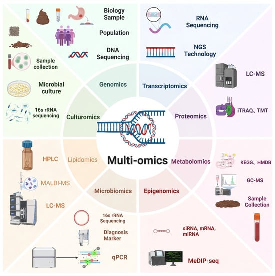
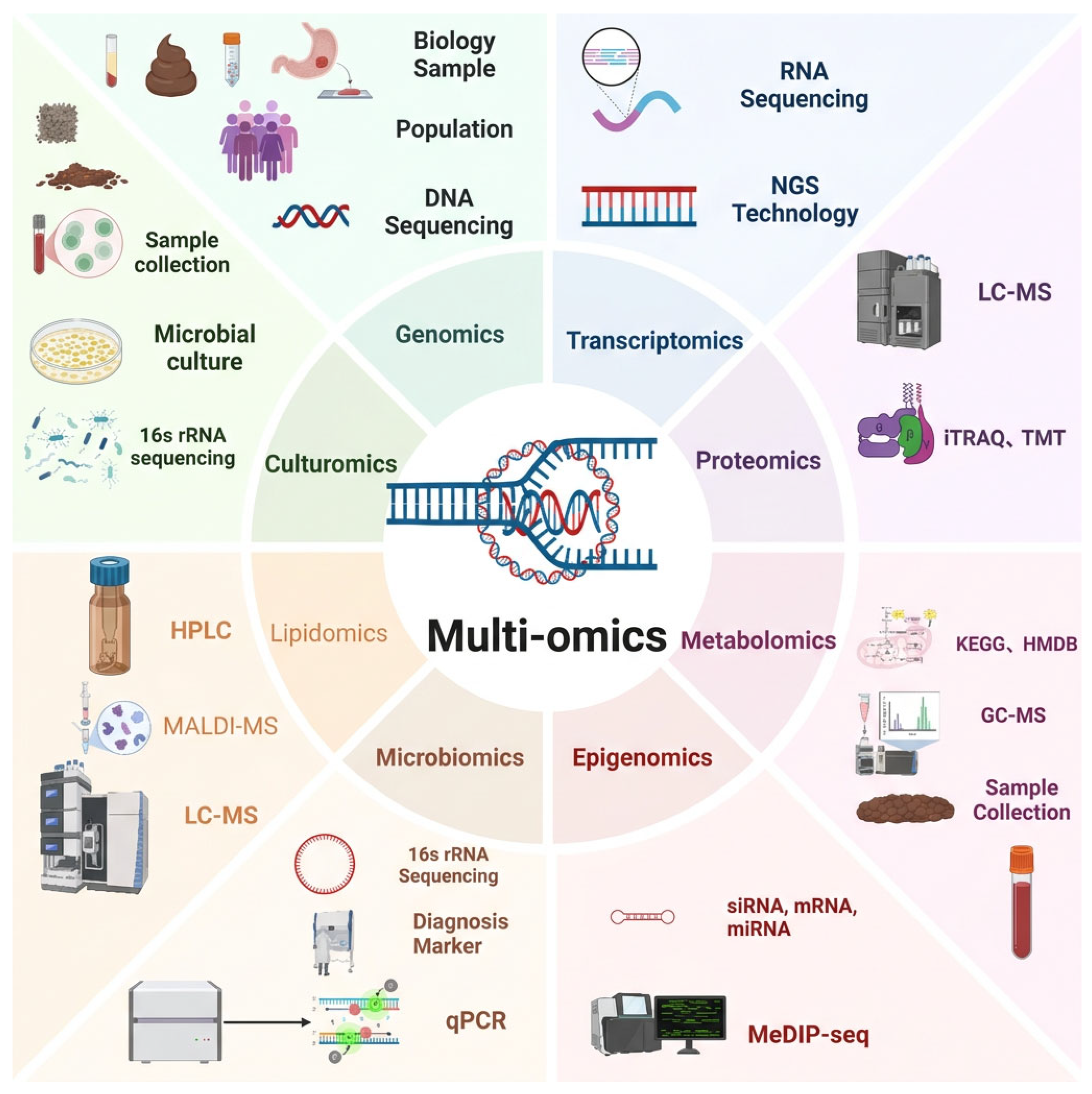
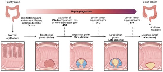
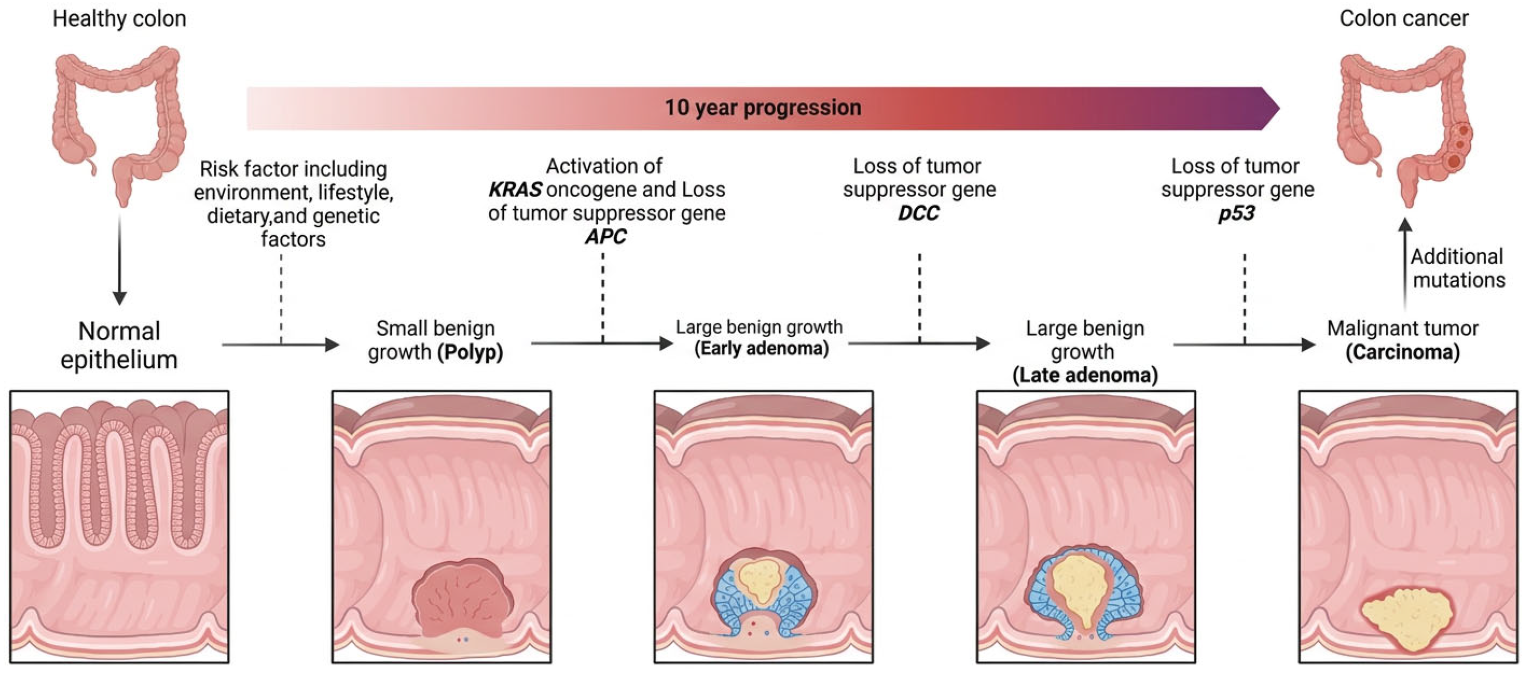
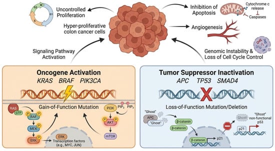
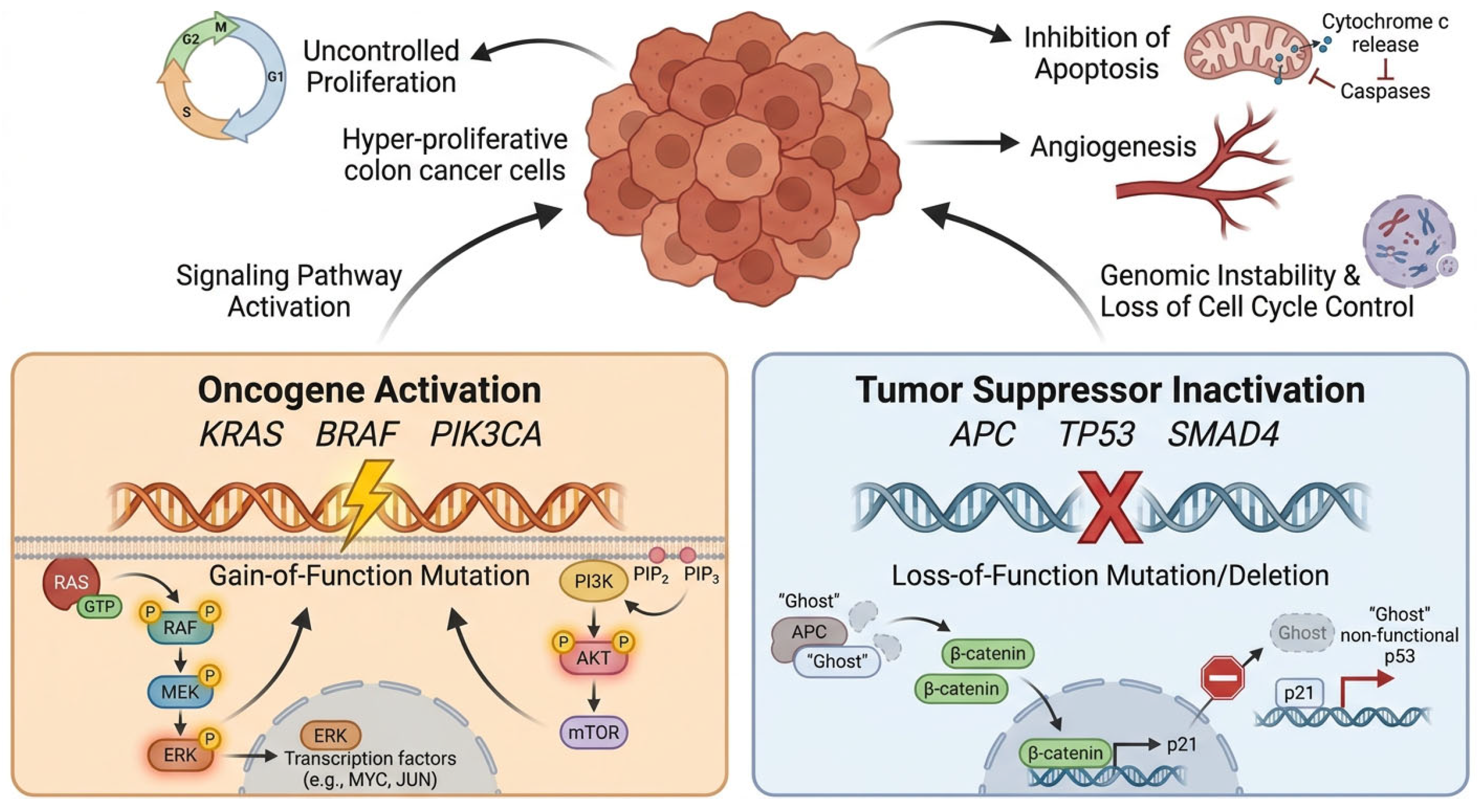
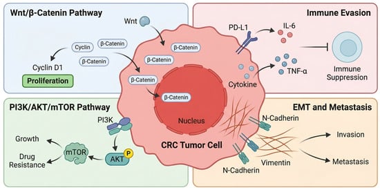
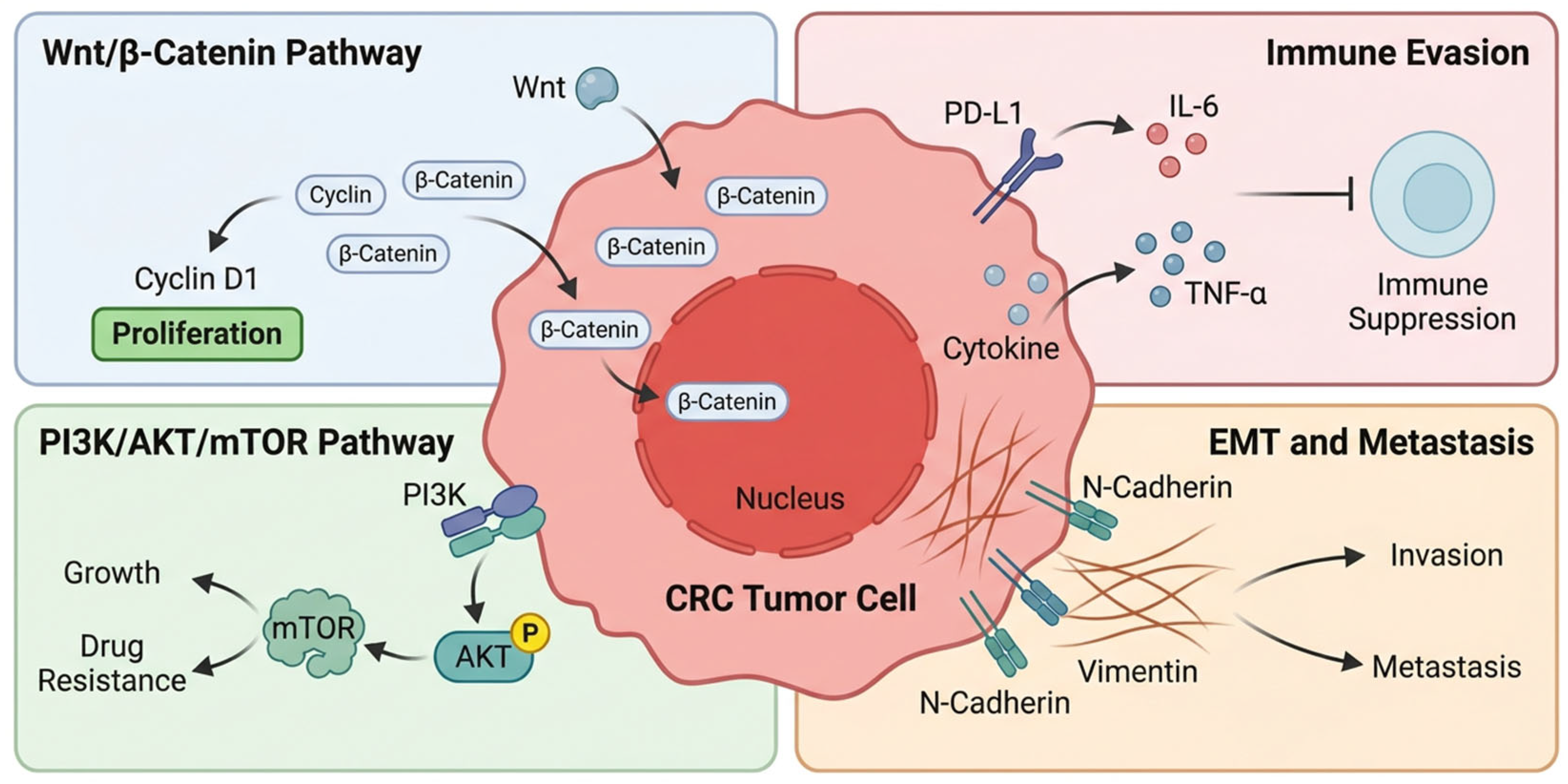
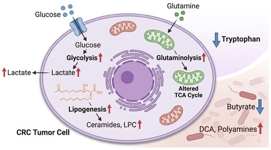
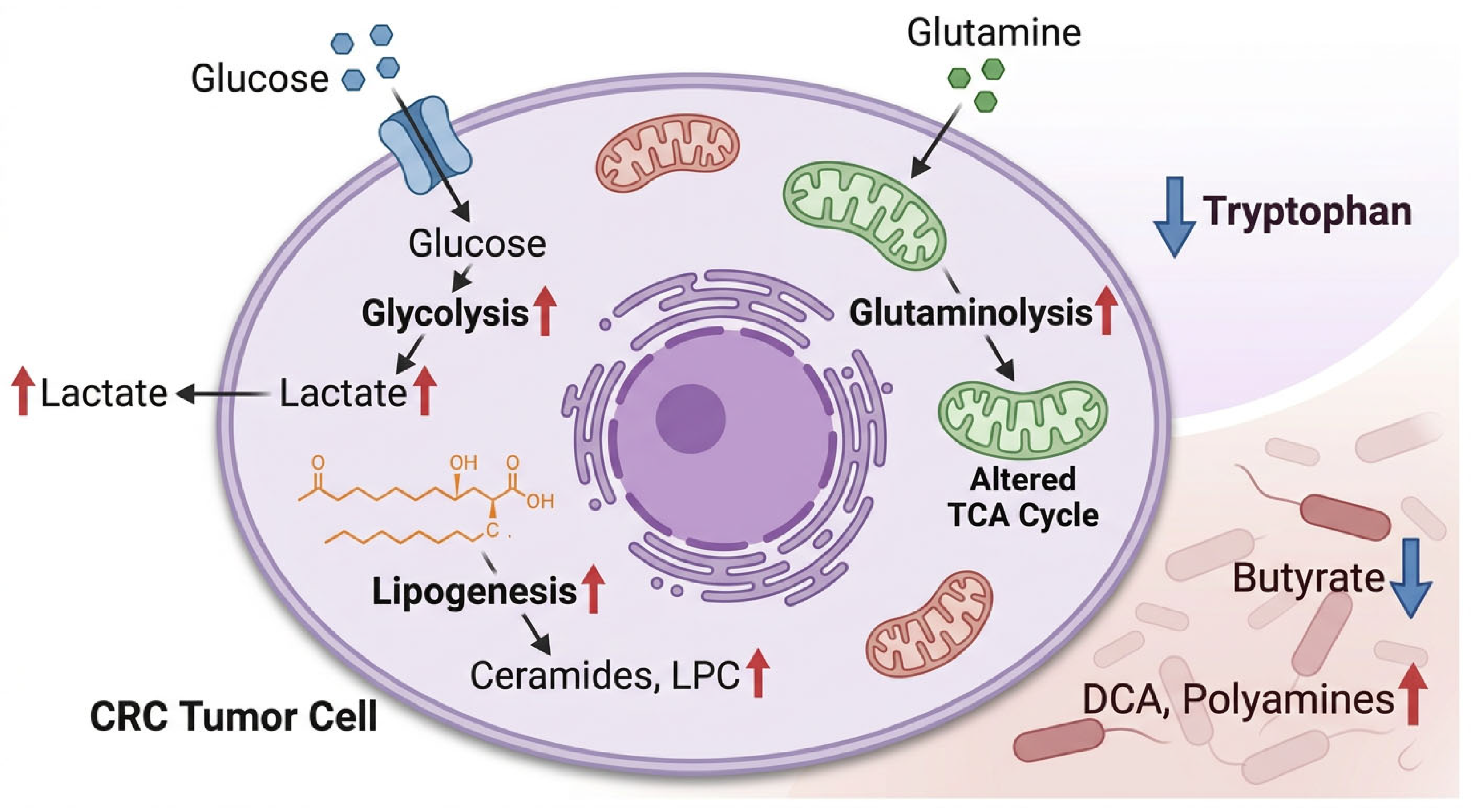
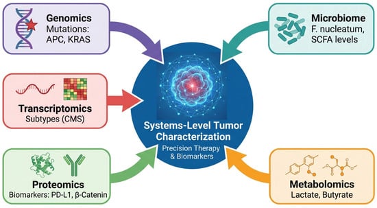
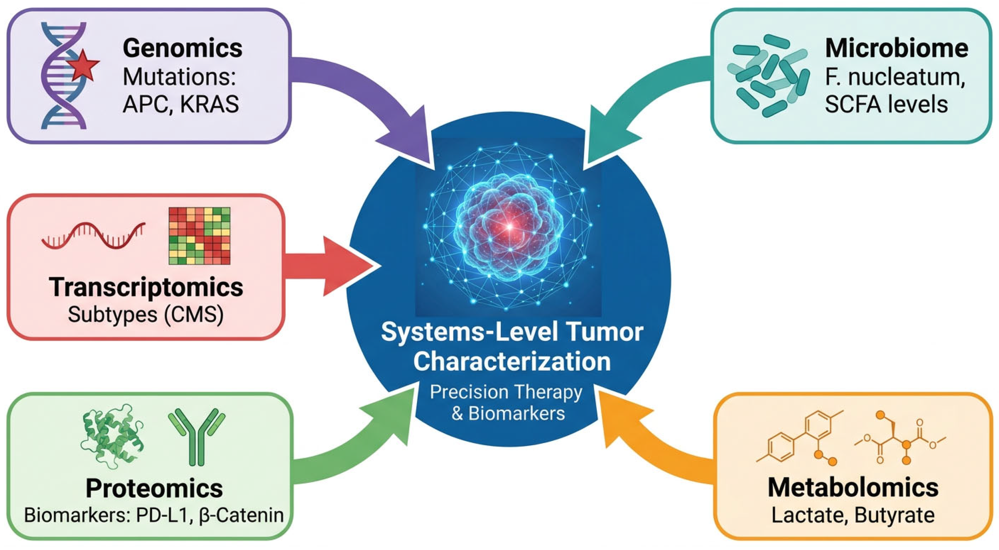
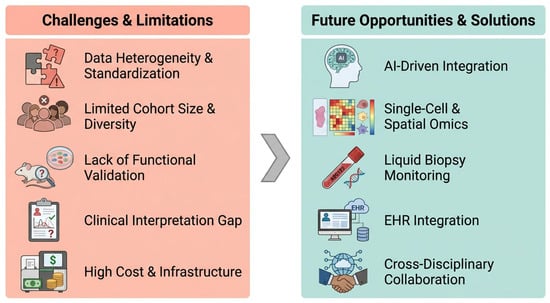
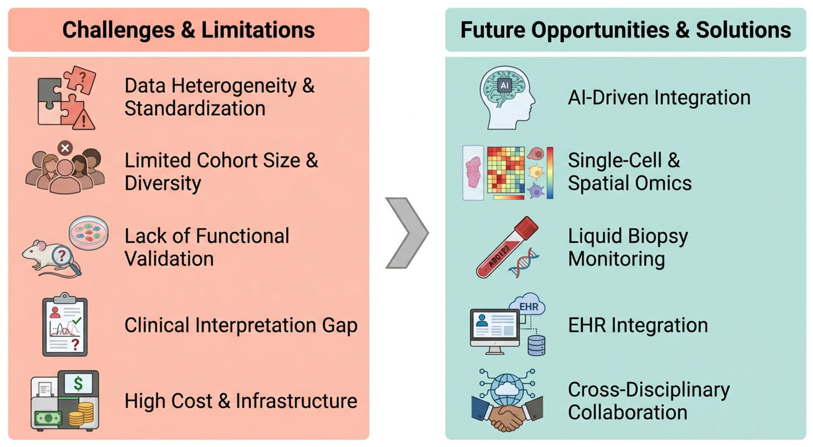

Disclaimer/Publisher’s Note: The statements, opinions and data contained in all publications are solely those of the individual author(s) and contributor(s) and not of MDPI and/or the editor(s). MDPI and/or the editor(s) disclaim responsibility for any injury to people or property resulting from any ideas, methods, instructions or products referred to in the content. |
© 2026 by the authors. Licensee MDPI, Basel, Switzerland. This article is an open access article distributed under the terms and conditions of the Creative Commons Attribution (CC BY) license.