


Abstract
This review evaluates recent progress in nanozyme-based biosensors for detecting circulating tumour cells, nucleic acids, and protein biomarkers, with particular attention to how peroxidase-, oxidase-, and catalase-like reactions enhance signal generation across electrochemical, optical, and microfluidic platforms. The roles of iron oxide–gold composites, silica nanostructures, quantum dots, and hybrid nanomaterials in improving analytical performance, enabling multiplexed detection, and facilitating assay miniaturization are critically assessed. Advances such as amplification-free detection approaches, smartphone-compatible point-of-care systems, and AI-assisted data analysis are discussed in relation to their translational potential. Key barriers, including regulatory requirements, reproducibility concerns, and manufacturing scalability, are also evaluated. By integrating mechanistic understanding with practical considerations for clinical deployment, this review outlines how next-generation nanozyme-based biosensors may strengthen early cancer detection, real-time monitoring, and precision oncology.
1. Introduction
Cancer remains a major global health burden, causing nearly 10 million deaths each year across both high-income and low-income regions [1]. Although imaging, histopathology, and molecular diagnostics have improved substantially, many cancers are still identified at advanced stages, when treatment options are limited and prognosis remains poor [2,3]. Improving early detection and enabling continuous monitoring are therefore essential for improving survival outcomes. However, current diagnostic approaches often remain invasive, costly, and insufficiently sensitive [4,5,6]. These limitations have accelerated efforts to develop technologies capable of delivering rapid, accurate, and minimally invasive cancer diagnostics.
Biosensors offer a promising pathway to address these challenges by enabling real-time detection of cancer-associated biomarkers with high analytical sensitivity and specificity [7]. By integrating biological recognition elements with diverse transduction mechanisms, biosensor platforms can quantitatively detect proteins, nucleic acids, and whole cells in complex biological samples [8,9]. In oncology, these capabilities have been applied to the detection of circulating tumour cells (CTCs), circulating tumour DNA (ctDNA), and exosomal markers, providing clinically relevant information on tumour burden and therapeutic response [10]. Continued miniaturization of biosensor systems further strengthens the clinical utility of point-of-care testing (POC) and expands access to diagnostic tools beyond centralized laboratories [11].
This review specifically evaluates nanozyme-enabled biosensors for detecting circulating tumour cells, nucleic acids, and protein biomarkers, with emphasis on how catalytic mechanisms drive signal amplification across electrochemical, optical, and microfluidic platforms. The section also outlines the translational relevance of these systems, including their potential for point-of-care deployment and integration with emerging digital diagnostics.
Nanomaterials have played a central role in advancing biosensor performance by enhancing signal amplification, improving surface functionalization, and enabling multiplexed detection [12]. Silica nanostructures, quantum dots, and hybrid composites offer tunable optical and electronic properties that can be tailored for specific biomarker targets [13,14]. These nanomaterial-based systems routinely achieve sensitivities surpassing those of conventional assays, including femtomolar detection limits and single-cell resolution in CTC analyses [15]. Such progress points to the growing influence of nanotechnology in cancer diagnostics.
As a distinct class of nanomaterials, nanozymes mimic the catalytic activity of natural enzymes whilst offering robustness, tunability, and cost effectiveness [16]. Unlike biological enzymes, which are often unstable under harsh conditions, nanozymes maintain catalytic activity across diverse environments, including variations in pH and temperature [17]. Nanozymes with peroxidase-like, oxidase-like, and catalase-like activities support diverse sensing modalities, from colorimetric assays to electrochemical detection [18]. The ability to engineer surface chemistry and particle morphology further enhances nanozyme catalytic efficiency and facilitates their integration into biosensor platforms [19].
Compared with natural enzymes, nanozymes offer several practical advantages: they are less expensive to manufacture, more stable during storage and operation, and readily engineered for multiplexed detection [20]. Catalytic properties can be precisely tuned through modifications in particle size, composition, and surface ligands, allowing control over biosensor performance [21]. Nanozymes also integrate effectively with optical, electrochemical, and microfluidic platforms, broadening their applicability across diverse diagnostic platforms [22,23]. Overall, these attributes position nanozymes as strong candidates for sensitive and reliable biomarker detection.
Recent progress in CTC research highlights the importance of integrating biosensors into cancer monitoring workflows to capture clinically meaningful signals from blood-based assays [24]. Previous studies have shown that Fe3O4–Au nanozymes can enhance catalytic amplification in cancer biomarker assays, supporting their integration into translational biosensor platforms [25]. Building on these foundational contributions, this review integrates emerging literature to provide a comprehensive evaluation of nanozyme-enabled biosensors in oncology. By positioning biosensor innovation within the broader landscape of global cancer diagnostics and the core principles of nanozyme science, we establish a framework for examining their translational potential from laboratory validation to clinical implementation and future applications.
2. Nanozyme Fundamentals
A central component of these advances is the emergence of nanozymes, which exhibit enzyme-like catalytic behaviour whilst offering enhanced robustness and design flexibility [26]. The combination of catalytic efficiency, stability, adaptability, and multifunctionality has expanded nanozyme relevance in biosensing applications [27]. These characteristics are particularly valuable in cancer diagnostics, where analytical sensitivity and reproducibility are essential [28].
Peroxidase-like catalysis in nanozymes is primarily governed by vacancy mediated adsorption of H2O2 and rapid electron shuttling between metal centres, enabling efficient generation of highly reactive •OH radicals that drive strong signal amplification in biosensing assays [29]. Iron oxide–gold (Fe3O4–Au) nanozyme composites demonstrate a dual catalytic and bio-interfacing role, combining the magnetic responsiveness of Fe3O4 with the plasmonic, catalytic, and bioconjugation advantages of Au. This synergy enhances signal output in colorimetric and electrochemical platforms and provides a robust scaffold for biomarker binding conjugate applications [30,31]. Nanozyme-driven signal amplification therefore holds strong potential to improve assay sensitivity and reproducibility in translational oncology. Nanozyme performance is also shaped by intrinsic physicochemical features such as surface defect density, oxygen vacancy distribution, and electron transfer efficiency, all of which influence catalytic turnover and substrate affinity.
Another important class of nanozymes is the oxidase-like, which catalyse oxygen-dependent reactions without requiring hydrogen peroxide, enabling simplified assay cascades [32]. Cerium oxide and manganese oxide nanostructures fall into this class. For example, cerium oxide and manganese oxide nanostructures can oxidize substrates such as o-phenylenediamine and dopamine, generating clear and measurable colorimetric signals [33]. Oxidase-like activity is supported by the ability of nanozymes to activate dissolved molecular oxygen at defect-rich surface sites, enabling multi-step electron transfer processes that drive substrate oxidation in the absence of exogenous H2O2, an advantage that enhances practicality for point-of-care biosensors [34].
Catalase-like nanozymes constitute another major class, decomposing hydrogen peroxide into water and oxygen in a manner analogous to natural catalase enzymes [35]. This catalytic mode is particularly relevant for monitoring reactive oxygen species (ROS) in tumour microenvironments and for biosensors designed to assess oxidative stress [36]. Platinum-based and iron oxide nanozymes frequently exhibit catalase-like behaviour, supporting applications in cancer diagnostics and therapeutic monitoring [37]. Catalase-like activity is facilitated by high-energy active sites that promote rapid H2O2 decomposition, supported by favourable electron donor/acceptor interactions and reduced activation barriers at engineered surface defects.
The catalytic performance of nanozymes is shaped by surface chemistry, electronic structure, and particle morphology [38]. As illustrated in Figure 1, nanozyme activity depends on electron transfer pathways influenced by transition metal ions, oxygen vacancies, and surface defects, all of which play central roles in driving catalytic reactions [39]. Furthermore, the mechanistic framework of Fe3O4–Au composites exemplify how synergistic interactions between the magnetic iron oxide core and the plasmonic gold shell accelerate catalytic kinetics, leading to higher turnover rates and enhanced analytical sensitivity [40]. A mechanistic understanding of these interactions is essential for the rational design and optimization of nanozyme-based biosensors.
Figure 1.
Mechanistic pathways of nanozyme catalytic activity across peroxidase-like, oxidase-like, and catalase-like modes. (A) Peroxidase-like mechanism (Fe3O4–Au nanozymes). Defect-rich Fe3O4–Au core–shell nanozymes catalyse Fenton-type reactions in the presence of H2O2. Surface-bound transition metal centres (Fe2+/Fe3+) and oxygen vacancy sites lower the activation barrier for O–O bond cleavage, generating highly reactive hydroxyl radicals (•OH) and high valent metal–oxo intermediates (Fe4+=O). These species oxidize chromogenic substrates such as TMB, producing a measurable colour change. Electron transfer pathways between Fe2+/Fe3+ and the Au shell accelerate redox cycling and enhance turnover. Symbols: oxygen vacancies (౦), metal–oxo intermediates (Fe4+=O), electron transfer arrows, and substrate-to-product conversion. Relevance to biosensing: this pathway highlights colorimetric and electrochemical amplification strategies, enabling ultrasensitive detection of cancer biomarkers through strong signal generation. (B) Oxidase-like mechanism (CeO2 nanozymes). Cerium oxide nanoparticles with mixed Ce3+/Ce4+ valence states activate dissolved O2 directly at defect-rich surfaces without requiring H2O2. Oxygen vacancies stabilize O2 adsorption and promote multi-step electron transfer, producing reactive oxygen species such as superoxide (O2•−) and singlet oxygen (1O2). These ROS oxidize substrates including o phenylenediamine and dopamine, generating quantifiable optical signals. Mixed valence cycling (Ce3+ ↔ Ce4+) supports continuous electron shuttling and sustained catalytic activity. Symbols: Ce3+/Ce4+ redox pairs, oxygen vacancy sites, ROS species (O2•−, 1O2), and substrate oxidation arrows. Relevance to biosensing: oxidase-like nanozymes eliminate the need for exogenous H2O2, simplifying assay design and improving compatibility with point-of-care platforms. (C) Catalase-like mechanism (Pt or MnO2 nanozymes). Platinum-based sheets and MnO2 nanosheets decompose H2O2 into H2O and O2 via redox cycling at high-energy active sites. Lattice strain and surface defects enhance electron flow between Pt2+/Pt0 (or Mn4+/Mn2+) states, preventing accumulation of harmful intermediates and enabling rapid turnover. The catalytic process produces molecular oxygen, which can be quantified directly or used to modulate downstream reactions. Symbols: active site redox pairs (Pt2+/Pt0), electron transfer arrows, O2 gas evolution, and defect-rich catalytic surfaces. Relevance to biosensing: catalase-like activity supports ROS-related biomarker detection, tumour microenvironment profiling, and oxygen-generation-based signal modulation in cancer diagnostics. Overall significance: across all three panels, the figure highlights how surface defects, oxygen vacancy density, lattice strain, and electron transfer pathways govern nanozyme catalytic behaviour. These mechanistic determinants directly influence signal amplification in nanozyme-enabled biosensors, enabling sensitive, robust, and tunable detection strategies for cancer biomarkers.
Recognizing the diversity of nanozyme activities enables researchers to match catalytic behaviour with specific diagnostic requirements. For example, peroxidase-like activity is well suited for colorimetric assays, oxidase-like activity reduces reagent complexity, and catalase-like activity provides access to oxidative stress-related biomarkers [41]. Leveraging these catalytic modes allows nanozyme-enabled biosensors to achieve high sensitivity, strong reproducibility, and broad adaptability across a range of cancer diagnostic applications [42], Table 1 compiles the summary of nanozyme classes and mode of catalytic activity, along with some of their specific applications. Additionally, a consolidated overview of these nanozyme classes, including catalytic functions, detection modalities, analytical performance, and clinical readiness, is presented in Table 2, which expands on these distinctions to guide selection for biomarker-specific biosensor design.
Table 1.
Comparison of nanozyme types, catalytic activities, and cancer biomarker uses.
Table 2.
Expanded comparison of nanozyme classes.
3. Nanomaterials in Biosensor Platforms
Within this broader framework, silica nanomaterials play a central role in biosensor engineering due to their biocompatibility, high surface area, and tunable porosity, features that make them effective scaffolds for molecular recognition [56]. When immobilised with antibodies, aptamers, and enzymes, they provide stable recognition interfaces for cancer biomarker detection [57]. Mesoporous silica nanoparticles (MSNs) further support controlled probe loading and diffusion regulated release, improving assay reproducibility and sensitivity in complex biological samples [58]. Silica nanostructures exhibit enhanced biosensing performance through defect-mediated surface chemistry, where silanol group density, pore-wall charge distribution, and engineered surface defects modulate probe loading, molecular diffusion, and electron transfer efficiency. These mechanistic features directly influence binding kinetics, signal amplification, and assay reproducibility in silica-based biosensors [59].
Figure 2 illustrates the multifunctional roles of silica nanostructures in nanomedicine, showing how surface-engineered silica nanoparticles (SNPs) participate in cellular uptake, intracellular delivery, and biosensing. Once internalized through endocytosis, SNPs can mediate stimuli-responsive release of therapeutic payloads and influence intracellular redox balance through interactions that modulate reactive oxygen species (ROS). The ROS enter cells through endocytosis, enabling controlled release of therapeutic payloads once internalized. In parallel, surface-functionalized silica nanostructures enhance biosensing performance by providing chemically tailored interfaces that stabilize biorecognition elements and improve signal transduction efficiency.
Figure 2.
Silica nanostructures for nanomedicine. Schematic representation of silica nanoparticle-mediated mechanisms in nanomedicine. Cellular uptake occurs via endocytosis, enabling intracellular delivery. Silica nanostructures facilitate ROS generation and therapeutic payload release, whilst also enhancing biosensing capabilities through surface functionalization and signal transduction.
Another important class of materials for biosensor development is quantum dots (QDs), which are a unique set of semiconductor nanocrystals with distinctive photophysical properties. A striking feature of the functional application of QDs is that they have a broad range of fluorescence emission that is size-dependent and has a high quantum yield and strong resistance to photobleaching, making QDs particularly suitable for optical biosensing, where signal stability and tunability are essential [60,61]. The photophysical behaviour of QDs is governed by quantum confinement effects, surface trap states, and engineered core–shell interfaces, which collectively regulate emission wavelength, quantum yield, and photostability. These mechanistic determinants underpin their sensitivity in fluorescence-based biosensors, enabling precise modulation of signal output in complex biological environments.
Significant advancements in graphene quantum dot (GQD) composites for cancer diagnosis and biosensing applications have reported enhanced functionalisation when combined with various materials, such as gold (AuNPs), carbon nanotubes (CNTs), polyaniline (PANI), and zeolitic imidazolate framework-8 (ZIF-8), to create composites with improved sensitivity, selectivity, and stability [62]. Recent advances in quantum dot imaging have shown how atomic-level structural control can improve emission stability and spectral tunability [63]. Hybrid GQD composites leverage synergistic charge transfer pathways, defect-rich interfaces, and plasmon–exciton coupling to enhance fluorescence intensity, reduce non-radiative recombination, and improve target binding specificity. Atomic-level structural control further stabilizes exciton dynamics, enabling highly tunable and robust optical responses for biosensing.
Silica-coated QDs represent another important evolution in QD engineering, offering improved stability and biocompatibility for biomedical applications [64]. Silica shells enhance physicochemical robustness, reduce cytotoxicity, and convert hydrophobic QD surfaces into hydrophilic interfaces suitable for aqueous biological systems. These coatings preserve high photoluminescence efficiency, minimize photobleaching, and extend fluorescence lifetimes, making silica-coated QDs valuable for imaging and sensing applications. Silica shells modulate QD behaviour by passivating surface trap states, stabilizing exciton recombination pathways, and creating controlled dielectric environments that enhance fluorescence lifetime and emission stability. These mechanistic improvements directly translate into higher signal fidelity and reduced photodegradation in biosensing applications.
The schematic in Figure 3 illustrates how excitation and emission pathways in QD systems are influenced by surface-bound biomolecules such as antibodies, DNA, and aptamers. These functional groups enable interactions including FRET, quenching, and charge transfer, each of which alters fluorescence output. Fluorescence modulation in QD systems is driven by nanoscale energy-transfer processes, including donor–acceptor distance-dependent FRET, surface-mediated charge-transfer quenching, and exciton–biomolecule coupling. These mechanistic interactions precisely tune emission intensity and spectral shifts, enabling highly selective biomarker detection. The resulting changes in signal intensity or wavelength form the basis for sensitive and selective detection in QD-based biosensors.
Figure 3.
QD fluorescence modulation mechanism. Advanced schematic illustrating fluorescence modulation of core-shell QDs (CdSe/ZnS) via excitation and emission pathways. Surface functionalization with biomolecules (antibodies, DNA, aptamers) enables FRET, quenching, and charge transfer interactions. These mechanisms modulate the fluorescence signal, producing measurable changes in signal output such as intensity shifts and wavelength variations for biosensor applications.
Altogether, the design of QD-based systems increasingly incorporates principles of self-assembly, in which nanoscale components autonomously organize into ordered structures through non-covalent interactions [65]. This strategy facilitates the creation of multifunctional architectures with enhanced optical, chemical, and biological performance. Self-assembly processes exploit electrostatic interactions, hydrogen bonding, and ligand-mediated coordination to organize QDs, silica matrices, and metallic nanoparticles into ordered architectures. These controlled nanoscale arrangements optimize energy transfer, probe accessibility, and multimodal signal integration in hybrid biosensor platforms.
A notable example of such hybrid engineering is the development of QD@MSN-EPI-Au-PEG-Apt nanoparticles for colorectal cancer theranostics [66]. These multimodal constructs combine QDs, MSNs, and Au nanoparticles to achieve targeted drug delivery, radiosensitization, and multimodal imaging. The platform incorporates epirubicin within the silica matrix, Au nanoparticles for enhanced radiotherapy response, PEGylation for improved circulation, and EpCAM aptamers for selective targeting of colorectal cancer cells. The QD@MSN–Au hybrid platform demonstrates how hierarchical nanoconstructs integrate optical, catalytic, and targeting mechanisms through coordinated energy transfer, enhanced radiative-decay modulation, and ligand-directed cellular uptake. These mechanistic synergies enable simultaneous imaging, drug delivery, and therapeutic enhancement.
In summary, advances in QD engineering, from intrinsic photophysical tuning to silica coating, controlled self-assembly, and hybrid nanoconstruct design, have substantially expanded the capabilities of nanomaterial-based biosensors. These innovations support ultrasensitive detection, multiplexed analysis, and device miniaturization, whilst enabling translational applications such as POC diagnostics and multimodal imaging [67,68]. Collectively, these engineering strategies refine exciton dynamics, surface-state energetics, and nanoscale charge-transfer pathways, enabling biosensor platforms with superior sensitivity, multiplexing capability, and translational robustness [69].
4. Cancer Biomarker Detection Using Nanozymes
Nanozyme-enabled biosensors have shown exceptional sensitivity in detecting protein biomarkers associated with cancer progression. Peroxidase-like iron oxide–gold (Fe3O4–Au) nanozymes catalyse colorimetric reactions that amplify signals from low-abundance proteins such as prostate-specific antigen (PSA) and carcinoembryonic antigen (CEA) [70]. Functionalization with antibodies or aptamers has enabled picomolar-level detection, surpassing the performance of conventional ELISA assays [71]. These developments demonstrate the potential of nanozymes to deliver rapid, cost-effective, and clinically relevant protein biomarker assays [72].
Furthermore, nanozymes have been applied to nucleic acid detection, including circulating tumour DNA (ctDNA) and microRNAs (miRNAs), which serve as key indicators of tumour burden and therapeutic response [73]. Catalytic amplification by Fe3O4–Au nanozymes enables sensitive detection of short nucleic acid sequences whilst reducing dependence on conventional amplification steps such as PCR, which are commonly but not universally used in nucleic acid-based diagnostic workflows [74]. By minimizing the need for these amplification procedures, nanozyme-enabled platforms lower assay complexity and shorten turnaround times, thereby improving suitability for POC applications [75]. When integrated with electrochemical biosensors, nanozyme-based systems can further discriminate single-nucleotide variants in cancer-associated genes, enhancing analytical specificity [76].
Moreso, nanozyme-based biosensors have also been adapted for whole cell detection, particularly circulating tumour cells (CTCs), which are critical indicators of metastasis and disease progression [77]. Magnetic Fe3O4–Au nanozymes enable both selective capture and catalytic amplification, supporting dynamic detection ranges from 10 to 105 cells/mL [78]. Advances in cytosensor design, such as three-site recognition strategies using branched Pt–Au nanospheres and MnO2–GO–Au nanosheets have further improved selectivity and accuracy in CTC detection [79]. These innovations demonstrate the value of nanozyme-functionalized biosensors as robust platforms for translational oncology, bridging laboratory assay development with clinical monitoring of tumour dissemination.
Case studies in breast cancer biomarker detection further illustrate the translational potential of advanced biosensor platforms. The development of label-free multiplex electrochemical biosensors capable of simultaneously detecting MUC1, CA15-3, and HER2 has demonstrated improved sensitivity and reproducibility compared with conventional assays [80]. Multiplexed biosensing continues to accelerate clinical translation by enabling simultaneous quantification of multiple disease-relevant biomarkers within a single assay cascade. Recent studies have expanded this capability to include additional breast cancer markers such as HER2, CA15-3, and other clinically significant proteins [81]. Altogether, these applications show how nanozymes can be harnessed for protein, nucleic acid, and cell-based detection, positioning them as key components of next-generation cancer diagnostics [82].
5. Integration with Biosensor Technologies
Electrochemical biosensors have become widely used in oncology due to their high sensitivity, rapid response, and compatibility with miniaturized diagnostic platforms [83]. Incorporating nanozymes into these systems enhances electron transfer and catalytic amplification, enabling detection of cancer biomarkers at ultra-low concentrations [84]. Iron oxide-based nanozymes have been integrated into electrochemical interfaces to strengthen catalytic signal output, supporting highly sensitive detection of clinically relevant biomarkers, including those associated with breast cancer [85].
Optical biosensors, including fluorescence and colorimetric platforms also benefit from nanozyme-mediated catalysis, which amplifies optical signals and improves assay reproducibility [86]. QD-based optical platforms extend these capabilities by enabling multiplexed detection, allowing simultaneous quantification of multiple biomarkers within complex biological samples [87].
Microfluidic biosensors represent another major advancement in cancer diagnostics, offering precise fluid manipulation, sample miniaturization, and high-throughput analysis [88]. Incorporation of nanozymes into microfluidic channels enables real-time catalytic amplification within confined volumes, reducing reagent consumption and shortening assay time [89]. These features make microfluidic–nanozyme systems particularly suitable for POC applications, where portability and automation are essential [90]. Integrating nanozymes into microfluidic architectures has been shown to enhance sensitivity whilst supporting scalable, clinically oriented device development [91].
Compared to traditional biosensor assays that often rely on nucleic acid amplification methods such as PCR, which increase workflow complexity and extend turnaround time, amplification-free detection strategies enabled by nanozyme catalysis simplify these workflows whilst maintaining high analytical sensitivity [92]. Notably, nanozyme-assisted CRISPR/Cas platforms where Fe3O4–Au nanozymes directly catalyse substrate reactions enable rapid nucleic acid detection without enzymatic amplification [93]. This approach streamlines assay design, reduces operational costs, minimizes error rates, and improves compatibility with POC devices [94]. Hence, amplification-free nanozyme biosensors represent a significant shift toward simplified, clinically viable cancer diagnostics.
Consistent with this, the Figure 4 schematic illustrates how portable biosensor platforms integrate sample handling, molecular recognition, and real-time signal processing. The figure depicts a compact system in which a blood sample is introduced into the device, passes through a sensor core functionalized with aptamers, DNA, and antibodies, and generates a measurable signal upon analyte interaction. This signal is processed electronically and transmitted to a handheld display for immediate interpretation, demonstrating how portable biosensors translate molecular recognition events into clinically actionable readouts.
Figure 4.
Portable biosensor system integrating sample input, molecular recognition, signal transduction, and real-time digital readout. This schematic illustrates the operational workflow and functional components of a portable biosensor designed for point-of-care cancer diagnostics. (1) Sample Input. A small volume of biological fluid (e.g., whole blood) is introduced into a disposable cartridge. The droplet symbol represents minimally invasive sampling, whilst the cartridge icon denotes a self-contained microfluidic chamber that guides the sample toward the sensing interface. This unit ensures controlled sample delivery, minimizes contamination, and enables rapid onsite testing. (2) Sensor Core with Surface-Functionalized Detection Elements. The central sensing region contains immobilized biorecognition molecules: aptamers, DNA probes, and antibodies, each represented by distinct labels. These elements selectively bind target analytes such as circulating tumour markers, nucleic acids, or proteins. Binding events modulate the physicochemical properties of the sensor surface, triggering a measurable signal. The geometric shapes adjacent to the recognition elements represent analytes interacting with the functionalized surface. This unit performs the critical molecular recognition step that determines assay specificity and sensitivity. (3) Signal Processor. The electronic module receives the raw signal generated by the sensor core, such as fluorescence modulation, electrochemical current changes, or impedance shifts, and converts it into a digital waveform. The waveform graphic symbolizes signal conditioning, amplification, noise filtering, and feature extraction. This processing step ensures that subtle biomolecular interactions are translated into robust, quantifiable outputs suitable for clinical interpretation. (4) Handheld Device Display. The processed signal is transmitted to a portable device (e.g., smartphone or dedicated reader) for real-time visualization. The display panel shows an example output: “Analyte detected: Positive,” accompanied by a concentration versus time plot and a confidence metric (“95% Confidence”). These elements represent quantitative reporting, trend analysis, and decision support features. This unit enables immediate interpretation by clinicians or end users, supporting decentralized diagnostics and rapid clinical decision making. Overall relevance to biosensing applications: together, these components illustrate how portable biosensors integrate molecular recognition, nanozyme-enhanced signal modulation, and digital analytics into a compact, user-friendly platform. The workflow supports point-of-care cancer biomarker detection, enabling rapid, sensitive, and accessible diagnostics outside centralized laboratories. The modular design also facilitates multiplexing, smartphone-based data management, and future integration with AI-assisted interpretation.
6. Translational and Clinical Perspectives
Despite substantial progress in laboratory research, the clinical translation of nanozyme-enabled biosensors continues to face significant challenges. Validation across diverse patient cohorts remains essential, as diagnostic performance must be demonstrated under real-world conditions to ensure reliability and reproducibility [95]. Regulatory approval also represents a major bottleneck, requiring rigorous assay standardization, robust quality-assurance frameworks, and long-term stability testing to meet international diagnostic compliance standards [96].
Furthermore, scalability of nanozyme synthesis and device manufacturing presents an additional hurdle, as cost-effective production is necessary for widespread adoption in healthcare systems [97]. Without comprehensive validation and regulatory clearance, the transition of nanozyme technologies from bench to bedside will remain constrained.
Nevertheless, nanozyme-enabled biosensors are well positioned to support POC diagnostics, offering rapid, portable, and user-friendly platforms for cancer detection [98]. Integration with smartphones and wearable devices enables decentralized testing, reducing dependence on centralized laboratories and improving access in resource-limited settings [99,100].
By integrating biosensors with mobile technologies, these systems empower both patients and clinicians through improved accessibility, rapid data acquisition, and enhanced clinical decision making in resource-limited settings. These opportunities align with global healthcare priorities to expand early detection and personalized monitoring, improving patient outcomes.
Thus, by linking biosensors with mobile technologies, these platforms empower both patients and clinicians, through improved accessibility, to rapidly acquire data, and enable enhanced clinical decision-making in settings where laboratory infrastructure is limited. Such capabilities align with global healthcare priorities focused on early detection, personalized monitoring, and improved patient outcomes [100].
Extending beyond proof-of-concept for POC applications, nanozyme technologies, including biosensors and therapeutic nanoplatforms, are moving towards personalized medicine, enabling patient stratification based on biomarker profiles for tailored therapeutic interventions in cancer and other diseases [101]. Multiplexed detection of proteins, nucleic acids, and circulating tumour cells provide comprehensive insights into tumour heterogeneity and clonal evolution [102].
A recent nanotheranostics review (2025) highlighted the convergence of diagnostic and therapeutic functions, demonstrating that nanozyme platforms can simultaneously detect biomarkers and deliver targeted interventions [103]. These translational perspectives, together with the expanding diagnostic potential of nanozyme-enabled biosensors, outline a rapidly evolving landscape in which the integration of laboratory innovation into clinical practice is becoming increasingly achievable.
The schematic workflow in Figure 5 illustrates the stepwise process used for CTC detection from whole blood. The diagram outlines each stage from blood collection and cellular preparation to immunoaffinity-based recognition using labelled antibodies or aptamers, followed by signal generation through fluorescence or electrochemical readouts and final data interpretation for clinical reporting.
Figure 5.
Workflow for CTC detection. Stepwise schematic illustrating the detection of circulating tumour cells (CTCs) from whole blood. The process includes blood collection, isolation and preparation of cellular components, immunoaffinity-based CTC recognition using labelled antibodies or aptamers, signal generation via fluorescence or electrochemical readout, and final data analysis for clinical reporting.
7. Discussion
Nanozyme-enabled biosensing increasingly relies on catalytic systems that combine enzyme-mimetic activity with robust structural design, engineered surface chemistry, and compatibility with miniaturized analytical platforms [104]. Across peroxidase-, oxidase-, and catalase-like nanozymes, signal amplification is governed primarily by defect-engineered active sites and controlled electron transfer pathways, rather than by particle composition itself [105,106]. This mechanistic perspective provides a more predictive framework for understanding biosensor performance and guides rational design strategies for next-generation diagnostic platforms [107].
7.1. Mechanistic Determinants of Catalytic Efficiency
The catalytic pathways illustrated in Figure 1 depict how nanozyme activity is fundamentally governed by surface defect density, oxygen-vacancy distribution, and redox-active metal centres [108]. Peroxidase-like nanozymes, particularly Fe3O4–Au composites, rely on vacancy-mediated adsorption of H2O2 and rapid electron shuttling between Fe2+/Fe3+ sites to generate hydroxyl radicals and high-valent metal–oxo intermediates that drive chromogenic oxidation [109]. These processes are highly sensitive to the density of coordinatively unsaturated metal atoms and the electronic coupling between the iron oxide core and gold shell, explaining why engineered Fe3O4–Au systems frequently achieve catalytic efficiencies comparable to or exceeding horseradish peroxidase [110].
Oxidase-like nanozymes operate through a distinct mechanism in which direct O2 activation is enabled by mixed-valence metal centres and defect-rich lattice edges [111]. Cerium oxide and manganese oxide nanozymes exemplify this behaviour, where Ce3+/Ce4+ redox cycling stabilizes partially reduced oxygen species such as O2•− and 1O2, enabling substrate oxidation without exogenous H2O2 [112]. This eliminates reagent-dependent steps and simplifies assay workflows, a key advantage for point-of-care deployment [113].
Catalase-like nanozymes, including Pt-based sheets and MnO2 nanosheets, catalyse H2O2 decomposition through two-step redox cycling at high-energy active sites [114]. Lattice strain and engineered surface defects modulate adsorption energies and stabilize reaction intermediates, accelerating turnover and preventing the accumulation of harmful ROS [115]. These mechanistic distinctions highlight how nanozyme classes differ not only in catalytic mode but also in their suitability for specific biosensing contexts [116].
7.2. Comparative Performance Across Nanozyme Classes
Table 1, Table 2 and Table 3 collectively demonstrate that nanozyme performance is strongly influenced by active site architecture, defect chemistry, and hybrid material integration [117]. Single-atom nanozymes (SANs) exhibit exceptional catalytic precision due to atomically dispersed M–N4 or M–N6 coordination environments, which lower activation barriers and enhance electron transfer kinetics [118]. The high turnover rates and stability under harsh conditions make SANs promising candidates for oxidative stress biomarkers and tumour microenvironment sensing [119].
MOF-based nanozymes leverage ordered porosity, high surface area, and tunable metal–ligand coordination to facilitate substrate preconcentration and controlled redox behaviour. Their modularity supports multiplexed detection of nucleic acids, proteins, and metabolites, although their clinical readiness remains limited by structural fragility and synthesis complexity [120].
Cascade nanozyme systems, which integrate sequential catalytic steps (e.g., oxidase–peroxidase or SOD–catalase), offer superior signal amplification and dynamic range by mimicking natural metabolic pathways. The spatially organized active sites enhance intermediate channelling and reduce side reactions, enabling robust performance in complex physiological matrices [121]. The recent advances summarized in Table 3 illustrate how hybrid cascade architectures can outperform single-enzyme mimics in both sensitivity and functional integration [122].
7.3. Integration with Biosensor Platforms and Device Miniaturization
The mechanistic advantages of nanozymes translate directly into improved performance across electrochemical, optical, and microfluidic biosensors [123]. Electrochemical platforms benefit from enhanced electron transfer kinetics and catalytic amplification at defect-rich nanozyme interfaces, enabling ultra-low detection limits for proteins, nucleic acids, and tumour metabolites [124]. Optical biosensors, including fluorescence and colorimetric systems, leverage nanozyme-driven ROS generation and substrate oxidation to produce strong, quantifiable signals with high reproducibility [125].
Microfluidic integration (Figure 6) further strengthens translational potential by enabling real-time catalytic amplification within confined volumes, reducing reagent consumption, and supporting automated workflows [126]. When combined with self-powered systems and AI-driven analytics, nanozyme-based microfluidic devices offer a pathway toward decentralized cancer diagnostics, continuous monitoring, and precision oncology [127].
Figure 6.
Schematic overview of an integrated wearable biosensing framework illustrating how self-powered modules (triboelectric nanogenerators and biofuel cells) drive continuous operation without external batteries; multimodal sensing arrays (nanozyme-based catalysis, optical, electrochemical, and impedance readouts) enable comprehensive biochemical and physiological monitoring; and AI-driven analytics support predictive modelling, early warning alerts, and personalized risk assessment. The system demonstrates a pathway toward continuous patient-centric monitoring and precision oncology within decentralized care environments.
7.4. Translation Barriers: Variability, Scalability, and Regulatory Pathways
Despite these advances, several barriers must be addressed before nanozyme enabled biosensors can achieve clinical adoption. Batch-to-batch variability remains a significant challenge, as small deviations in defect density, surface ligand distribution, or metal coordination environments can produce substantial fluctuations in catalytic activity [128,129]. This variability complicates assay standardization and undermines reproducibility across laboratories.
Scalability is another major limitation. Many high performing nanozymes are synthesized through laboratory scale routes that do not translate well to industrial manufacturing, where maintaining uniform defect structures and catalytic stability is difficult [130]. Additionally, regulatory frameworks for nanozyme-based diagnostics are still emerging, with stringent requirements for physicochemical characterization, long-term biocompatibility, environmental safety, and quality control [131]. Addressing these challenges will require coordinated efforts in materials engineering, process optimization, and regulatory science [132].
Table 3.
Recent advances in single-atom, MOF-based, and cascade nanozyme systems for cancer-relevant biosensing (last 3–5 years).
8. Outlook
The mechanistic insights synthesized in this review highlight the importance of defect engineering, electron transfer optimization, and hybrid material design in advancing nanozyme-enabled biosensing. Continued progress in single-atom catalysis, MOF-hybrid architectures, and cascade nanozyme systems will likely yield platforms with higher catalytic precision, improved robustness, and enhanced multiplexing capability. Integration with microfluidics, wearable systems, and AI-assisted analytics (Figure 2, Figure 3, Figure 4, Figure 5 and Figure 6) positions nanozyme-based biosensors as strong candidates for next-generation cancer diagnostics.
However, realizing their full clinical potential will depend on overcoming variability, achieving scalable manufacturing, and establishing regulatory pathways that ensure safety and reproducibility. With these challenges addressed, nanozyme-enabled biosensors could play a transformative role in early cancer detection, real-time monitoring, and decentralized precision oncology.
9. Conclusions
Nanozyme-enabled biosensors are entering a decisive translational phase, with clear progress across POC platforms, portable diagnostic systems, and multiplexed analytical workflows. These advances directly support global healthcare priorities by decentralizing diagnostic capacity, improving accessibility, and enabling more individualized clinical decision making. Emerging capabilities in circulating tumour cell detection, biomarker profiling, and amplification-free portable assays demonstrate how nanozyme technologies can effectively bridge laboratory innovation with clinical practice, accelerating the shift toward precision oncology.
This review establishes nanozyme-enabled biosensors as a coherent roadmap for next-generation diagnostic technologies. By integrating catalytic principles, nanomaterial engineering, translational workflow design, and emerging directions such as AI-driven analytics and multi-omics convergence, we outline a comprehensive framework for advancing the field. The long-term impact of nanozyme platforms will depend on sustained progress in materials optimization, scalable manufacturing, ethical deployment, and rigorous regulatory validation.
Author Contributions
Conceptualization, V.A. and I.E.C.; methodology, V.A.; image software, V.A.; validation, V.A. and I.E.C.; formal analysis, V.A.; investigation, V.A. and I.E.C.; resources, V.A.; data curation, V.A.; writing—original draft preparation, V.A. and I.E.C.; writing—review and editing, V.A. and I.E.C.; visualization, V.A.; supervision, I.E.C.; project administration, V.A. All authors have read and agreed to the published version of the manuscript.
Funding
No funding was received to support the conduct of this study or the preparation of this manuscript.
Informed Consent Statement
Not applicable.
Data Availability Statement
No new data were created or analyzed in this study.
Conflicts of Interest
The authors declare no conflicts of interest.
Abbreviations
The following abbreviations are used in this manuscript:
| AI | Artificial Intelligence |
| AuNPs | Gold Nanoparticles |
| CA15-3 | Cancer Antigen 15-3 |
| CEA | Carcinoembryonic Antigen |
| CDT | Chemodynamic Therapy |
| CeO2 | Cerium Oxide |
| CTC(s) | Circulating Tumour Cell(s) |
| ctDNA | Circulating Tumour DNA |
| CNTs | Carbon Nanotubes |
| DNA | Deoxyribonucleic Acid |
| ELISA | Enzyme-Linked Immunosorbent Assay |
| FRET | Förster Resonance Energy Transfer |
| Fe3O4-Au | Iron Oxide–Gold Composite |
| GO | Graphene Oxide |
| GQD | Graphene Quantum Dot |
| HER2 | Human Epidermal Growth Factor Receptor 2 |
| HRP | Horseradish Peroxidase |
| LOD | Limit of Detection |
| miRNA | MicroRNA |
| MOF(s) | Metal–Organic Framework(s) |
| MSNs | Mesoporous Silica Nanoparticles |
| MnO2 | Manganese Dioxide |
| MUC1 | Mucin-1 |
| PANI | Polyaniline |
| PB | Prussian Blue |
| POC | Point-of-Care |
| PSA | Prostate-Specific Antigen |
| QDs | Quantum Dots |
| QD@MSN-EPI-Au-PEG-Apt | QDS-Quantum Dots; MSN-Mesoporous Silica Nanoparticle Shell; EPI-Epirubicin; Au-Gold; PEG-Polyethylene Glycol; Apt-Aptamer |
| RBCs | Red Blood Cells |
| ROS | Reactive Oxygen Species |
| SNP | Silica Nanoparticle |
| SOD | Superoxide Dismutase |
| SANs | Single-Atom Nanozymes |
| TEM | Transmission Electron Microscopy |
| WBCs | White Blood Cells |
| ZIF-8 | Zeolitic Imidazolate Framework-8 |
References
- Sung, H.; Ferlay, J.; Siegel, R.L.; Laversanne, M.; Soerjomataram, I.; Jemal, A.; Bray, F. Global cancer statistics 2020: GLOBOCAN estimates of incidence and mortality worldwide for 36 cancers in 185 countries. CA Cancer J. Clin. 2021, 71, 209–249. [Google Scholar] [CrossRef] [Scilit]
- Hricak, H.; Mayerhoefer, M.E.; Herrmann, K.; Lewis, J.S. Advances and challenges in precision imaging. Lancet Oncol. 2025, 26, e34–e45. [Google Scholar] [CrossRef] [Scilit] [PubMed]
- Lawal, A.O.; Ogunniyi, T.J.; Oludele, O.I.; Olorunfemi, O.A.; Okesanya, O.J.; Ogaya, J.B.; Ahmed, M.M.; Lucero Prisno, D.E. Innovative laboratory techniques shaping cancer diagnosis and treatment in developing countries. Discov. Oncol. 2025, 16, 137. [Google Scholar] [CrossRef] [Scilit] [PubMed]
- Patriotis, C.; Srivastava, S. Early Cancer Detection: Challenges and Opportunities. In Circulating Tumour Cells; Cote, R.J., Lianidou, E., Eds.; In Current Cancer Research; Springer: Cham, Switzerland, 2023; pp. 619–631. [Google Scholar] [CrossRef] [Scilit]
- Etzioni, R.; Urban, N.; Ramsey, S.; McIntosh, M.; Schwartz, S.; Reid, B.; Radich, J.; Anderson, G.; Hartwell, L. The case for early detection. Nat. Rev. Cancer 2003, 3, 243–252. [Google Scholar] [CrossRef] [Scilit]
- Madar, S.; Amor, R.E.; Furman Assaf, S.; Friedman, E. Innovative Approaches to Early Detection of Cancer-Transforming Screening for Breast, Lung, and Hard-to-Screen Cancers. Cancers 2025, 17, 1867. [Google Scholar] [CrossRef] [Scilit]
- Raheja, I.K.; Kumar, P.; Rajashekarappa, K.K.; Mahadevan, G.D. Nanobiosensors for Early Detection of Cancer: A Recent Update. Biomed. Mater. Devices 2026, 4, 78–96. [Google Scholar] [CrossRef] [Scilit]
- Crnoglavac Popović, M.; Stanković, V.; Stanković, D.; Prodanović, R. Protein, Nucleic Acid, and Nanomaterial Engineering for Biosensors and Monitoring. Biosensors 2025, 15, 430. [Google Scholar] [CrossRef] [Scilit]
- Abuzeid, H.R.; Abdelaal, A.F.; Elsharkawy, S.; Ali, G.A.M. Basic Principles and Applications of Biological Sensors Technology. In Handbook of Nanosensors; Ali, G.A.M., Chong, K.F., Makhlouf, A.S.H., Eds.; Springer: Cham, Switzerland, 2023; pp. 1–45. [Google Scholar] [CrossRef] [Scilit]
- de Abreu, A.R.; Wyninckx, A.; Vandamme, T.; Op de Beeck, K.; Van Camp, G.; Peeters, M.; Laurent Puig, P.; Taieb, J.; Taly, V.; Benhaim, L. Circulating Tumour DNA detection in cancer: A comprehensive overview of current detection methods and prospects. Oncologist 2025, 30, oyaf204. [Google Scholar] [CrossRef] [Scilit]
- Wang, Y.; Jiang, H.; Zhang, Y.; Yang, Q.; Song, Y.; Gao, Y. Microfluidic platforms for CRISPR-based biosensing advancing molecular diagnostics from benchtop to point-of-care. Sens. Diagn. 2025, 5, 116–135. [Google Scholar] [CrossRef] [Scilit]
- Rajendran, J.V.; Elahi, A.; Scully, P. Toward next-generation BNP/NT-proBNP biosensors: Multiplexed detection, biofouling control, and digital health integration. Heart Fail. Rev. 2025, 31, 8. [Google Scholar] [CrossRef] [Scilit]
- Pham, X.H.; Park, S.M.; Ham, K.M.; Kyeong, S.; Son, B.S.; Kim, J.; Hahm, E.; Kim, Y.-H.; Bock, S.; Kim, W.; et al. Synthesis and Application of Silica-Coated Quantum Dots in Biomedicine. Int. J. Mol. Sci. 2021, 22, 10116. [Google Scholar] [CrossRef] [Scilit]
- Liang, Z.; Khawar, M.B.; Liang, J.; Sun, H. Bio Conjugated Quantum Dots for Cancer Research: Detection and Imaging. Front. Oncol. 2021, 11, 749970. [Google Scholar] [CrossRef] [Scilit] [PubMed]
- Lee, S.; Rafiq, S.; Kang, S.H. Nanobiosensors for Single-Molecule Diagnostics: Toward Integration with Super-Resolution Imaging. Biosensors 2025, 15, 705. [Google Scholar] [CrossRef] [Scilit]
- Sharma, M.V.; Ibeyaima, A.; Sharmah, B.; Afzal, N.U.; Manna, P. Natural Enzymes and Nanozymes: A Comparative Review of Structure, Mechanism and Kinetics. Top. Catal. 2025, 68, 243–265. [Google Scholar] [CrossRef] [Scilit]
- Li, T.; Wang, X.; Wang, Y.; Zhang, Y.; Liu, W.; Liu, S.; Li, S.; Liu, Y.; Xing, H.; Otake, K.-I.; et al. Microenvironmental modulation breaks intrinsic pH limitations of nanozymes to boost their activities. Nat. Commun. 2024, 15, 55163. [Google Scholar] [CrossRef] [Scilit]
- Feng, L.; Zhang, M.; Fan, Z. Current trends in colorimetric biosensors using nanozymes for detecting biotoxins (bacterial food toxins, mycotoxins, and marine toxins). Anal. Methods 2024, 16, 6771–6792. [Google Scholar] [CrossRef] [Scilit]
- Panferov, V.G.; Hendrickson, O.D.; Zherdev, A.V.; Dzantiev, B.B. Surface Modified Nanozymes: Production, Properties, and Analytical Application. Mosc. Univ. Chem. Bull. 2025, 80, 55–65. [Google Scholar] [CrossRef] [Scilit]
- Abdullah, K.A.; Tahir, T.F.; Qader, A.F.; Omer, R.A.; Othman, K.A. Nanozymes: Classification and Analytical Applications—A Review. J. Fluoresc. 2025, 35, 4973–4987. [Google Scholar] [CrossRef] [Scilit] [PubMed]
- Lu, Y.; Cao, C.; Pan, X.; Liu, Y.; Cui, D. Structure design mechanisms and inflammatory disease applications of nanozymes. Nanoscale 2023, 15, 14–40. [Google Scholar] [CrossRef] [Scilit] [PubMed]
- Zhang, X.; Chen, X.; Zhao, Y. Nanozymes: Versatile Platforms for Cancer Diagnosis and Therapy. Nano Micro Lett. 2022, 14, 95. [Google Scholar] [CrossRef] [Scilit]
- Xu, K.; Cui, Y.; Guan, B.; Qin, L.; Feng, D.; Abuduwayiti, A.; Wu, Y.; Li, H.; Cheng, H.; Li, Z. Nanozymes with biomimetically designed properties for cancer treatment. Nanoscale 2024, 16, 3555–3578. [Google Scholar] [CrossRef] [Scilit] [PubMed]
- Ferreira, M.M.; Ramani, V.C.; Jeffrey, S.S. Circulating tumour cell technologies. Mol. Oncol. 2016, 10, 374–394. [Google Scholar] [CrossRef] [Scilit]
- Akpe, V.; Shiddiky, M.J.A.; Kim, T.H.; Brown, C.L.; Yamauchi, Y.; Cock, I.E. Cancer biomarker profiling using nanozyme containing iron oxide loaded with gold particles. J. R. Soc. Interface 2020, 17, 20200180. [Google Scholar] [CrossRef] [Scilit]
- Patra, P. Nanozymes as Next-Generation Therapeutics: Synergizing Enzyme-Mimetic Activity with Nanoscale Features. Biomed. Mater. Devices 2025, 4, 388. [Google Scholar] [CrossRef] [Scilit]
- Huang, Y.; Ren, J.; Qu, X. Nanozymes: Classification, catalytic mechanisms, and applications. Chem. Rev. 2019, 119, 4357–4412. [Google Scholar] [CrossRef] [Scilit]
- Wang, K.; Lin, X.; Lv, X.; Xie, M.; Wu, J.; Wu, J.J.; Luo, Y. Nanozyme-based aptasensors for the detection of tumour biomarkers. J. Biol. Eng. 2025, 19, 13. [Google Scholar] [CrossRef] [Scilit]
- Celardo, I.; Pedersen, J.Z.; Traversa, E.; Ghibelli, L. Pharmacological potential of cerium oxide nanoparticles. Nanoscale 2011, 3, 1411–1420. [Google Scholar] [CrossRef] [Scilit]
- Zhang, B.; Wang, X.; Hu, W.; Liao, Y.; He, Y.; Dong, B.; Zhao, M.; Ma, Y. SPR-Enhanced Au@Fe3O4 Nanozyme for the Detection of Hydroquinone. Chemosensors 2023, 11, 392. [Google Scholar] [CrossRef] [Scilit]
- Gao, L.; Zhuang, J.; Nie, L.; Zhang, J.; Zhang, Y.; Gu, N.; Wang, T.; Feng, J.; Yang, D.; Perrett, S.; et al. Intrinsic peroxidase-like activity of ferromagnetic nanoparticles. Nat. Nanotechnol. 2007, 2, 577–583. [Google Scholar] [CrossRef] [Scilit]
- Waris, A.; Hasan, S.; Bano, S.; Sultana, S.; Ibhadon, A.O.; Khan, M.Z. Development of nanozyme based sensors as diagnostic tools in clinic applications: A review. J. Mater. Chem. B 2023, 11, 6112–6135. [Google Scholar] [CrossRef] [Scilit] [PubMed]
- Alsharif, N.B.; Viczián, D.; Szczes, A.; Szilagyi, I. Formulation of Antioxidant Composites by Controlled Heteroaggregation of Cerium Oxide and Manganese Oxide Nanozymes. J. Phys. Chem. C 2023, 127, 17201–17212. [Google Scholar] [CrossRef] [Scilit] [PubMed]
- Mahmoodpour, M.; Abedi Kiasari, B.; Karimi, M.; Abroshan, A.; Shamshirian, D.; Hosseinalizadeh, H.; Delavari, A.; Mirzei, H. Paper-based biosensors as point-of-care diagnostic devices for the detection of cancers: A review of innovative techniques and clinical applications. Front. Oncol. 2023, 13, 1131435. [Google Scholar] [CrossRef] [Scilit]
- Singh, S. Nanomaterials Exhibiting Enzyme-Like Properties (Nanozymes): Current Advances and Future Perspectives. Front. Chem. 2019, 7, 46. [Google Scholar] [CrossRef] [Scilit]
- Huang, W.; Zhang, P.; Zhao, E.; Xiong, J.; Wang, S.; Lv, Z. Potential application and prospects of ROS-sensitive biomaterials in cancer therapy: A immune microenvironment triggered nanomaterial. Discov. Oncol. 2025, 16, 185. [Google Scholar] [CrossRef] [Scilit]
- Tanawish, T.; Jahan, N.; Rasheed, K.; Iqbal, M.; Atif, M. Exploring the Advanced Synthesis Strategies and Biomedical Applications of Iron Oxide-Based Nanozymes: A Comprehensive Review. J. Clust. Sci. 2024, 35, 2637–2661. [Google Scholar] [CrossRef] [Scilit]
- Chen, X.; Willner, I. Surface-Modified Nanozymes for Enhanced and Selective Catalysis. ACS Appl. Mater. Interfaces 2025, 17, 44011–44029. [Google Scholar] [CrossRef] [Scilit]
- Chen, H.; Meng, X.; Liang, M.; Li, W.; Liu, T.; Liu, J.; Shi, Z.; Liu, J.; Liu, L. Facilitating reversible transition metal migration and expediting ion diffusivity via oxygen vacancies for high performance O3-type sodium layered oxide cathodes. J. Mater. Chem. A 2023, 11, 68–76. [Google Scholar] [CrossRef] [Scilit]
- Chen, Y.; Zhang, Y.; Kou, Q.; Liu, Y.; Han, D.; Wang, D.; Sun, Y.; Zhang, Y.; Xing, S. Enhanced Catalytic Reduction of 4-Nitrophenol Driven by Fe3O4-Au Magnetic Nanocomposite Interface Engineering: From Facile Preparation to Recyclable Application. Nanomaterials 2018, 8, 353. [Google Scholar] [CrossRef] [Scilit]
- Wu, J.; Wang, X.; Wang, Q.; Lou, Z.; Li, S.; Zhu, Y.; Qin, L.; Wei, H. Nanomaterials with enzyme-like characteristics (nanozymes): Next-generation artificial enzymes (II). Chem. Soc. Rev. 2019, 48, 1004–1076. [Google Scholar] [CrossRef] [Scilit]
- Wang, Y.; Sun, X.; Wang, S.; Zhang, Z.; Luo, C.; He, Z.; Zhang, S. Nanozyme Enabled Cancer Theranostics: Bridging Enzyme Mimicry and Material Intelligence. Mater. Horiz. 2025, 12, 8895–8919. [Google Scholar] [CrossRef] [Scilit] [PubMed]
- Yang, L.; Guo, H.; Hou, T.; Zhang, J.; Li, F. Metal-mediated Fe3O4@polydopamine-aptamer capture nanoprobe coupling multifunctional MXene@Au@Pt nanozyme for direct and portable photothermal analysis of circulating breast cancer cells. Biosens. Bioelectron. 2023, 234, 115346. [Google Scholar] [CrossRef] [Scilit] [PubMed]
- Feng, N.; Liu, Y.; Dai, X.; Wang, Y.; Guo, Q.; Li, Q. Advanced applications of cerium oxide based nanozymes in cancer. RSC Adv. 2022, 12, 1486–1493. [Google Scholar] [CrossRef] [Scilit]
- Dou, Y.; Li, Z.; Su, J.; Song, S. A Portable Biosensor Based on Au Nanoflower Interface Combined with Electrochemical Immunochromatography for POC Detection of Prostate-Specific Antigen. Biosensors 2022, 12, 259. [Google Scholar] [CrossRef] [Scilit]
- Pi, F.; Deng, X.; Xue, Q.; Zheng, L.; Liu, H.; Yang, F.; Chen, T. Alleviating the hypoxic tumour microenvironment with MnO2-coated CeO2 nanoplatform for magnetic resonance imaging guided radiotherapy. J. Nanobiotechnol. 2023, 21, 90. [Google Scholar] [CrossRef] [Scilit]
- Fan, L.; Lou, D.; Wu, H.; Zhang, X.; Zhu, Y.; Gu, N.; Zhang, Y. A Novel AuNP-Based Glucose Oxidase Mimic with Enhanced Activity and Selectivity Constructed by Molecular Imprinting and O2-Containing Nanoemulsion Embedding. Adv. Mater. Interfaces 2018, 5, 1801070. [Google Scholar] [CrossRef] [Scilit]
- Niu, J.; Sun, S.; Liu, P.; Zhang, X.; Mu, X. Copper-based Nanozymes: Properties and Applications in Biomedicine. J. Inorg. Mater. 2023, 38, 489–502. [Google Scholar] [CrossRef] [Scilit]
- He, Q.; Zhang, L. Bimetallic and multimetallic nanozymes: Synergistic catalysis for advanced biomedical and health applications. RSC Adv. 2025, 15, 47648–47665. [Google Scholar] [CrossRef] [Scilit] [PubMed]
- Niu, X.; Li, X.; Lyu, Z.; Pan, J.; Ding, S.; Ruan, X.; Du, D.; Lin, Y. Metal–organic framework based nanozymes: Promising materials for biochemical analysis. Chem. Commun. 2020, 56, 11338–11353. [Google Scholar] [CrossRef] [Scilit]
- Estelrich, J.; Busquets, M.A. Prussian Blue: A Nanozyme with Versatile Catalytic Properties. Int. J. Mol. Sci. 2021, 22, 5993. [Google Scholar] [CrossRef] [Scilit]
- Tripathi, A.; Rautela, S.S.; Kumari, P. Graphene-based nanozymes for revolutionizing biomedical research. RSC Adv. 2025, 15, 39092–39120. [Google Scholar] [CrossRef] [Scilit] [PubMed]
- Gutiérrez, F.A.; Rubianes, M.D.; Rivas, G.A. New bioanalytical platform based on the use of avidin for the successful exfoliation of multi-walled carbon nanotubes and the robust anchoring of biomolecules: Application for hydrogen peroxide biosensing. Anal. Chim. Acta 2019, 1065, 12–20. [Google Scholar] [CrossRef] [Scilit]
- Zhang, W.; Wu, Y.; Li, Z.; Dong, H.; Zhang, Y.; Gu, N. Prussian Blue and Other Metal–Organic Framework-Based Nanozymes. In Nanozymology; Yan, X., Ed.; Nanostructure Science and Technology; Springer: Singapore, 2020; pp. 141–170. [Google Scholar] [CrossRef] [Scilit]
- Xu, D.; Tu, Q.; San, X.; Zhu, A.; Li, X. CoO/Co-graphene quantum dots as an oxidative mimetic nanozyme for the colorimetric detection of l-cysteine. Anal. Methods 2024, 16, 2044–2050. [Google Scholar] [CrossRef] [Scilit]
- Tang, F.; Li, L.; Chen, D. Mesoporous Silica Nanoparticles: Synthesis, Biocompatibility and Drug Delivery. Adv. Mater. 2012, 24, 1504–1534. [Google Scholar] [CrossRef] [Scilit]
- Slowing, I.I.; Vivero-Escoto, J.L.; Wu, C.W.; Lin, V.S.Y. Mesoporous silica nanoparticles as controlled release drug delivery and gene transfection carriers. Adv. Drug Deliv. Rev. 2008, 60, 1278–1288. [Google Scholar] [CrossRef] [Scilit] [PubMed]
- Mamaeva, V.; Sahlgren, C.; Lindén, M. Mesoporous silica nanoparticles in medicine—Recent advances. Adv. Drug Deliv. Rev. 2013, 65, 689–702. [Google Scholar] [CrossRef] [Scilit] [PubMed]
- Zou, S.; Peng, G.; Ma, Z. Surface-Functionalizing Strategies for Multiplexed Molecular Biosensing: Developments Powered by Advancements in Nanotechnologies. Nanomaterials 2024, 14, 2014. [Google Scholar] [CrossRef] [Scilit]
- Alivisatos, A.P. Semiconductor Clusters, Nanocrystals, and Quantum Dots. Science 1996, 271, 933–937. [Google Scholar] [CrossRef] [Scilit]
- Medintz, I.L.; Uyeda, H.T.; Goldman, E.R.; Mattoussi, H. Quantum dot bioconjugates for imaging, labelling and sensing. Nat. Mater. 2005, 4, 435–446. [Google Scholar] [CrossRef] [Scilit]
- Iannazzo, D.; Espro, C.; Celesti, C.; Ferlazzo, A.; Neri, G. Smart Biosensors for Cancer Diagnosis Based on Graphene Quantum Dots. Cancers 2021, 13, 3194. [Google Scholar] [CrossRef] [Scilit] [PubMed]
- Torres, R.; Thal, L.B.; McBride, J.R.; Cohen, B.E.; Rosenthal, S.J. Quantum Dot Fluorescent Imaging: Using Atomic Structure Correlation Studies to Improve Photophysical Properties. J. Phys. Chem. C 2024, 128, 3632–3640. [Google Scholar] [CrossRef] [Scilit]
- Gerion, D.; Pinaud, F.; Williams, S.C.; Parak, W.J.; Zanchet, D.; Weiss, S.; Alivisatos, A.P. Synthesis and properties of biocompatible water soluble silica coated CdSe/ZnS semiconductor quantum dots. J. Phys. Chem. B 2001, 105, 8861–8871. [Google Scholar] [CrossRef] [Scilit]
- Whitesides, G.M.; Grzybowski, B. Self-assembly at all scales. Science 2002, 295, 2418–2421. [Google Scholar] [CrossRef] [Scilit] [PubMed]
- Abrishami, A.; Bahrami, A.R.; Nekooei, S.; Saljooghi, A.S.; Matin, M.M. Hybridized quantum dot, silica, and gold nanoparticles for targeted chemo-radiotherapy in colorectal cancer theranostics. Commun. Biol. 2024, 7, 393. [Google Scholar] [CrossRef] [Scilit]
- Bagheri, E.; Ansari, L.; Abnous, K.; Taghdisi, S.M.; Ramezani, P.; Ramezani, M.; Alibolandi, M. Silica–Quantum Dot Nanomaterials as a Versatile Sensing Platform. Crit. Rev. Anal. Chem. 2021, 51, 687–708. [Google Scholar] [CrossRef] [Scilit]
- Bahavarnia, F.; Nami, F.; Hasanzadeh, M.; Shadjou, N. Developing Point of Need (PON) Cancer Biosensors Using Microfluidic Paper Based Analytical Devices: Recent Advances. RSC Adv. 2025, 15, 48876–48912. [Google Scholar] [CrossRef] [Scilit]
- Rathore, R.; Yadav, A.; Suhag, D.; Thakur, A. Nanoscale Biosensors for Diagnostics. In Advancements in Nanobiology; CRC Press: Boca Raton, FL, USA; Taylor & Francis: Abingdon, UK, 2025; pp. 87–104. [Google Scholar] [CrossRef] [Scilit]
- Zhao, Y.; Wen, Y.; Hu, X.; Zhang, B. A Colorimetric Immunoassay Based on g-C3N4@Fe3O4 Nanocomposite for Detection of Carcinoembryonic Antigen. J. Anal. Methods Chem. 2022, 2022, 6966470. [Google Scholar] [CrossRef] [Scilit]
- Reaño, R.L.; Escobar, E.C. A review of antibody, aptamer, and nanomaterials synergistic systems for an amplified electrochemical signal. Front. Bioeng. Biotechnol. 2024, 12, 1361469. [Google Scholar] [CrossRef] [Scilit]
- Das, B.; Franco, J.L.; Logan, N.; Balasubramanian, P.; Kim, M.I.; Cao, C. Nanozymes in Point-of-Care Diagnosis: An Emerging Futuristic Approach for Biosensing. Nano Micro Lett. 2021, 13, 193. [Google Scholar] [CrossRef] [Scilit]
- Garehbaghi, S.; Richtera, L.; Altintas, Z.; Ashrafi, A.M. Tyrosine-Capped Pt Nanozyme Functionalized with cDNA: An Innovative Sensor Designed for One-Step Detection of the miRNA-21 Biomarker. ACS Omega 2025, 10, 5564–5576. [Google Scholar] [CrossRef] [Scilit] [PubMed]
- Zhang, Y.; Yu, W.; Wang, M.; Li, P. Nanozyme-assisted amplification-free CRISPR/Cas system realizes visual detection. Front. Bioeng. Biotechnol. 2024, 11, 1327498. [Google Scholar] [CrossRef] [Scilit] [PubMed]
- Chang, Z.; Fu, Q.; Wang, M.; Duan, D. Advances of Nanozyme-Driven Multimodal Sensing Strategies in Point-of-Care Testing. Biosensors 2025, 15, 375. [Google Scholar] [CrossRef] [Scilit]
- Liu, J.L.; Ma, Y.C.; Yang, T.; Hu, R.; Yang, Y.H. A single nucleotide polymorphism electrochemical sensor based on DNA-functionalized Cd-MOFs-74 as cascade signal amplification probes. Microchim. Acta 2021, 188, 266. [Google Scholar] [CrossRef] [Scilit] [PubMed]
- Xie, L.; He, L.; Fu, Y.; Wei, Y.; Zhang, K.; Chen, M. A three-site recognition cytosensor for accurate detection of circulating tumour cells based on novel branched Pt Au nanospheres and MnO2-GO-Au nanosheets. Microchim. Acta 2025, 192, 175. [Google Scholar] [CrossRef] [Scilit]
- Dong, M.; Gao, Z.; Zhang, Y.; Cai, J.; Li, J.; Xu, P.; Jiang, H.; Gu, J.; Wang, J. Ultrasensitive electrochemical biosensor for detection of circulating tumour cells based on a highly efficient enzymatic cascade reaction. RSC Adv. 2023, 13, 12966–12972. [Google Scholar] [CrossRef] [Scilit]
- Tang, S.; Shen, H.; Hao, Y.; Huang, Z.; Tao, Y.; Peng, Y.; Guo, Y.; Xie, G.; Feng, W. A novel cytosensor based on Pt@Ag nanoflowers and AuNPs/Acetylene black for ultrasensitive and highly specific detection of circulating tumour cells. Biosens. Bioelectron. 2018, 104, 72–78. [Google Scholar] [CrossRef] [Scilit]
- Kuntamung, K.; Jakmunee, J.; Ounnunkad, K. A label-free multiplex electrochemical biosensor for the detection of three breast cancer biomarker proteins employing dye/metal ion-loaded and antibody-conjugated polyethyleneimine-gold nanoparticles. J. Mater. Chem. B 2021, 9, 6576–6585. [Google Scholar] [CrossRef] [Scilit] [PubMed]
- Di Gioia, D.; Blankenburg, I.; Nagel, D.; Heinemann, V.; Stieber, P. Tumour markers in the early detection of tumour recurrence in breast cancer patients: CA 125, CYFRA 21-1, HER2 shed antigen, LDH and CRP in combination with CEA and CA 15-3. Clin. Chim. Acta 2016, 461, 1–7. [Google Scholar] [CrossRef] [Scilit]
- Jin, X.; Guo, F.; Wu, M.; Xu, C.; Li, Y.; Chen, G. Emerging versatile nanozymes for the therapeutic and diagnostic applications of diseases. J. Transl. Med. 2025, 23, 1375. [Google Scholar] [CrossRef] [Scilit]
- Noreena, S.; Ishaq, I.; Saleem, M.H.; Ali, B.; Ali, S.M.; Iqbal, J. Electrochemical biosensing in oncology: A review advancements and prospects for cancer diagnosis. Cancer Biol. Ther. 2025, 26, 2475581. [Google Scholar] [CrossRef] [Scilit] [PubMed]
- Mahmudunnabi, R.G.; Farhana, F.Z.; Kashaninejad, N.; Firoz, S.H.; Shim, Y.B.; Shiddiky, M.J.A. Nanozyme-based electrochemical biosensors for disease biomarker detection. Analyst 2020, 145, 4398–4420. [Google Scholar] [CrossRef] [Scilit]
- Kurup, C.P.; Ahmed, M.U. Nanozymes towards Personalized Diagnostics: A Recent Progress in Biosensing. Biosensors 2023, 13, 461. [Google Scholar] [CrossRef] [Scilit]
- Kashtiaray, A.; Karimi, M.; Ghafori Gorab, M.; Maleki, A. A comprehensive review on the recent applications of nanozymes in breast cancer therapy and diagnosis. Mater. Adv. 2025, 6, 3017–3042. [Google Scholar] [CrossRef] [Scilit]
- Mousavi, S.M.; Hashemi, S.A.; Yari Kalashgrani, M.; Omidifar, N.; Lai, C.W.; Rao, N.V.; Gholami, A.; Chiang, W.H. The Pivotal Role of Quantum Dots-Based Biomarkers Integrated with Ultra-Sensitive Probes for Multiplex Detection of Human Viral Infections. Pharmaceuticals 2022, 15, 880. [Google Scholar] [CrossRef] [Scilit] [PubMed]
- Liu, Z.; Zhou, Y.; Lu, J.; Gong, T.; Ibáñez, E.; Cifuentes, A.; Lu, W. Microfluidic biosensors for biomarker detection in body fluids: A key approach for early cancer diagnosis. Biomark. Res. 2024, 12, 97. [Google Scholar] [CrossRef] [Scilit] [PubMed]
- Gao, M.Y.; Wang, M.; Wang, Y.T.; Zhang, Z.L. Enhanced catalytic efficiency of nanozymes with a V-structured chip for microfluidic biosensing of S. typhimurium. J. Mater. Chem. B 2025, 13, 4471–4483. [Google Scholar] [CrossRef] [Scilit]
- Behera, P.P.; Kumar, N.; Kumari, M.; Kumar, S.; Mondal, P.K.; Arun, R.K. Integrated microfluidic devices for point-of-care detection of bio-analytes and disease. Sens. Diagn. 2024, 2, 1437–1459. [Google Scholar] [CrossRef] [Scilit]
- Gowers, S.A.N.; Rogers, M.L.; Booth, M.A.; Leong, C.L.; Samper, I.C.; Phairatana, T.; Jewell, S.L.; Pahl, C.; Strong, A.J.; Boutelle, M.G. Clinical translation of microfluidic sensor devices: Focus on calibration and analytical robustness. Lab Chip 2019, 19, 2537–2548. [Google Scholar] [CrossRef] [Scilit]
- Liang, M.; Yan, X. Nanozymes: From New Concepts, Mechanisms, and Standards to Applications. Acc. Chem. Res. 2019, 52, 2190–2200. [Google Scholar] [CrossRef] [Scilit]
- Li, H.; Xie, Y.; Chen, F.; Bai, H.; Xiu, L.; Zhou, X.; Guo, X.; Hu, Q.; Yin, K. Amplification-free CRISPR/Cas detection technology: Challenges, strategies, and perspectives. Chem. Soc. Rev. 2023, 52, 361–382. [Google Scholar] [CrossRef] [Scilit] [PubMed]
- Lin, Y.; Ren, J.; Qu, X. Catalytically Active Nanomaterials: A Promising Candidate for Artificial Enzymes. Acc. Chem. Res. 2014, 47, 1097–1105. [Google Scholar] [CrossRef] [Scilit]
- Ma, Q.; Liu, Y.; Zhu, H.; Zhang, L.; Liao, X. Nanozymes in Tumour Theranostics. Front. Oncol. 2021, 11, 666017. [Google Scholar] [CrossRef] [Scilit] [PubMed]
- Romanholo, P.V.V.; Paiva, J.V.F.; Sgobbi, L.F. Reliability Issues and Challenges in Biosensor Development. In Springer Tracts in Electrical and Electronics Engineering; Springer: Singapore, 2024; pp. 321–344. [Google Scholar] [CrossRef] [Scilit]
- Zhang, Y.; Wei, G.; Liu, W.; Li, T.; Wang, Y.; Zhou, M.; Liu, Y.; Wang, X.; Wei, H. Nanozymes for nanohealthcare. Nat. Rev. Methods Primers 2024, 4, 315. [Google Scholar] [CrossRef] [Scilit]
- Mansur, A.A.P.; Mansur, H.S. Nanozyme-Based Cancer Nanotheranostics: Emerging Applications and Challenges in Brain Cancer Therapeutics. J. Nanotheranostics 2025, 6, 4. [Google Scholar] [CrossRef] [Scilit]
- Lee, S.; Moussa, N.A.M.; Kang, S.H. Deep Learning-Enhanced Nanozyme-Based Biosensors for Next-Generation Medical Diagnostics. Biosensors 2025, 15, 571. [Google Scholar] [CrossRef] [Scilit] [PubMed]
- Akpe, V.; Cock, I. Decentralising Cancer Diagnostics: Advances in Point-of-Care Biosensor Technologies and Digital Health Integration. Preprints 2025. [Google Scholar] [CrossRef] [Scilit]
- Malla, P.; Wang, Y.M.; Su, C.H. New horizons for the therapeutic application of nanozymes in cancer treatment. J. Nanobiotechnol. 2025, 23, 130. [Google Scholar] [CrossRef] [Scilit] [PubMed]
- Jarockyte, G.; Karabanovas, V.; Rotomskis, R.; Mobasheri, A. Multiplexed Nanobiosensors: Current Trends in Early Diagnostics. Sensors 2020, 20, 6890. [Google Scholar] [CrossRef] [Scilit]
- Akpe, V.; Cock, I.E. Advances in Cancer Treatment Through Nanotheranostics and Emerging Therapies. J. Nanotheranostics 2025, 6, 29. [Google Scholar] [CrossRef] [Scilit]
- Wu, Y.; Darland, D.C.; Zhao, J.X. Nanozymes—Hitting the Biosensing “Target”. Sensors 2021, 21, 5201. [Google Scholar] [CrossRef] [Scilit]
- Jiao, L.; Xu, W.; Wu, Y.; Yan, H.; Gu, W.; Du, D.; Lin, Y.; Zhu, C. Single-Atom Catalysts Boost Signal Amplification for Biosensing. Chem. Soc. Rev. 2021, 50, 750–765. [Google Scholar] [CrossRef] [Scilit]
- Zhang, H.; Zhang, S.; Zhang, Z. Advancements and Applications of Single-Atom Nanozymes in Sensing Analysis. Chemosensors 2024, 12, 209. [Google Scholar] [CrossRef] [Scilit]
- Zhu, C.; Jiang, C.; Lee, H.K.; Zhang, Y.; Tang, S. All Nanozyme-Based Cascade Reactions for Biomedical Applications: From Self-Cascading Nanozyme to Immobilized Cascade Nanozyme. Adv. Sci. 2026, 13, e19656. [Google Scholar] [CrossRef] [Scilit]
- Mathur, P.; Kumawat, M.; Nagar, R.; Singh, R.; Daima, H.K. Tailoring Metal Oxide Nanozymes for Biomedical Applications: Trends, Limitations, and Perceptions. Anal. Bioanal. Chem. 2024, 416, 27. [Google Scholar] [CrossRef] [Scilit]
- Shi, X.; Zhang, Y.; Liu, H.; Wang, J.; Chen, Q.; Li, Z.; Zhao, X. Vacancy Rich Fe–Ce Mixed Oxides as Nanoenzymes for Efficient Antioxidant Activity In Vitro. Appl. Surf. Sci. 2024, 664, 160256. [Google Scholar] [CrossRef] [Scilit]
- Zeng, X.; Ruan, Y.; Chen, Q.; Yan, S.; Huang, W. Biocatalytic Cascade in Tumor Microenvironment with a Fe2O3/Au Hybrid Nanozyme for Synergistic Treatment of Triple Negative Breast Cancer. Chem. Eng. J. 2023, 452, 138422. [Google Scholar] [CrossRef] [Scilit]
- Shen, H.; Chen, S.; Mo, S.-C.; Huang, H.; Liang, H.; Zhang, J.; Xu, Z.-L.; Liu, W.; Liu, Y. Dual Active Centers Linked by Oxygen Transfer for Enhancing Proximity Orientation Effect of Nanozyme. Adv. Funct. Mater. 2025, 35, 2418360. [Google Scholar] [CrossRef] [Scilit]
- Tang, K.; Shen, H.; Wang, H.; Hu, Y.; Tang, A.; Liu, W.; Liu, Y. Metal–Organic Framework Based Nanozymes: From Fundamental Catalytic Mechanisms to Rational Design of Optical Sensors for Food Safety. Microchem. J. 2026, 224, 117600. [Google Scholar] [CrossRef] [Scilit]
- Jain, A.; Stavrakis, S.; DeMello, A. Droplet-Based Microfluidics and Enzyme Evolution. Curr. Opin. Biotechnol. 2024, 87, 103097. [Google Scholar] [CrossRef] [Scilit]
- Zhang, W.; Guo, Y.; Xu, G.; Li, X.; Chen, Y.; Wang, H.; Liu, Z.; Zhao, J. Engineering MnPt Bimetallic Nanozymes for Cascade Enzymatic Therapy and Enhanced Radio-Immunotherapy. Adv. Sci. 2026, 2026, e19300. [Google Scholar] [CrossRef] [Scilit] [PubMed]
- Hou, Z.; Cui, C.; Li, Y.; Gao, Y.; Zhu, D.; Gu, Y.; Pan, G.; Zhu, Y.; Zhang, T. Lattice-Strain Engineering for Heterogeneous Electrocatalytic Oxygen Evolution Reaction. Adv. Mater. 2023, 35, 2209876. [Google Scholar] [CrossRef] [Scilit]
- Yang, Z.; Guo, J.; Wang, L.; Zhang, J.; Ding, L.; Liu, H.; Yu, X. Nanozyme-Enhanced Electrochemical Biosensors: Mechanisms and Applications. Small 2024, 20, 2307815. [Google Scholar] [CrossRef] [Scilit]
- Yu, B.; Wang, W.; Sun, W.; Jiang, C.; Lu, L. Defect Engineering Enables Synergistic Action of Enzyme-Mimicking Active Centers for High-Efficiency Tumor Therapy. J. Am. Chem. Soc. 2021, 143, 8855–8865. [Google Scholar] [CrossRef] [Scilit]
- Peng, C.; Pang, R.; Li, J.; Wang, E. Current Advances on the Single-Atom Nanozyme and Its Bioapplications. Adv. Mater. 2024, 36, 2211724. [Google Scholar] [CrossRef] [Scilit]
- Hamed, E.M.; Elsaady, M.M.; Li, S.F. Single-Atom Nanozymes in Analytical Chemistry: Opportunities and Challenges. Anal. Chem. 2025, 97, 26313–26325. [Google Scholar] [CrossRef] [Scilit]
- Chakraborty, S. Democratizing Nucleic Acid-Based Molecular Diagnostic Tests for Infectious Diseases at Resource-Limited Settings—From Point of Care to Extreme Point of Care. Sens. Diagn. 2024, 3, 536–561. [Google Scholar] [CrossRef] [Scilit]
- Cai, X.; Huang, Y.; Zhu, C. Immobilized Multi-Enzyme/Nanozyme Biomimetic Cascade Catalysis for Biosensing Applications. Adv. Healthc. Mater. 2025, 14, 2401834. [Google Scholar] [CrossRef] [Scilit]
- Zhang, X.; Li, G.; Chen, G.; Wu, D.; Wu, Y.; James, T.D. Enzyme Mimics for Engineered Biomimetic Cascade Nanoreactors: Mechanism, Applications, and Prospects. Adv. Funct. Mater. 2021, 31, 2106139. [Google Scholar] [CrossRef] [Scilit]
- Wang, M.; Liu, H.; Fan, K. Signal Amplification Strategy Design in Nanozyme-Based Biosensors for Highly Sensitive Detection of Trace Biomarkers. Small Methods 2023, 7, 2301049. [Google Scholar] [CrossRef] [Scilit]
- Ren, G.; Dong, F.; Zhao, Z.; Li, K.; Lin, Y. Structure Defect Tuning of Metal–Organic Frameworks as a Nanozyme Regulatory Strategy for Selective Online Electrochemical Analysis of Uric Acid. ACS Appl. Mater. Interfaces 2021, 13, 52987–52997. [Google Scholar] [CrossRef] [Scilit]
- Wang, F.; Ju, E.; Guan, Y.; Ren, J.; Qu, X. Light-Mediated Reversible Modulation of ROS Level in Living Cells by Using an Activity-Controllable Nanozyme. Small 2017, 13, 1603051. [Google Scholar] [CrossRef] [Scilit]
- Huang, Y.; Wang, L.; Zhou, J.; Peng, X.; Li, Q.; Zhang, Y. An Integrated Centrifugal Microfluidic System for Automatic Nanozyme ELISA Lateral Flow Immunoassay at Point-of-Care Testing. Microfluid. Nanofluidics 2025, 29, 36. [Google Scholar] [CrossRef] [Scilit]
- Mejía-Salazar, J.R.; Rodrigues Cruz, K.; Materón Vásques, E.M.; de Novais Oliveira, O., Jr. Microfluidic Point-of-Care Devices: New Trends and Future Prospects for eHealth Diagnostics. Sensors 2020, 20, 1951. [Google Scholar] [CrossRef] [Scilit]
- Panferov, V.G.; Liu, J. Optical and Catalytic Properties of Nanozymes for Colorimetric Biosensors: Advantages, Limitations, and Perspectives. Adv. Opt. Mater. 2024, 12, 2401318. [Google Scholar] [CrossRef] [Scilit]
- Zandieh, M.; Liu, J. Nanozyme Catalytic Turnover and Self-Limited Reactions. ACS Nano 2021, 15, 15645–15655. [Google Scholar] [CrossRef] [Scilit]
- Abdel-Mageed, H.M. Frontiers in Nanoparticles Redefining Enzyme Immobilization: A Review Addressing Challenges, Innovations, and Unlocking Sustainable Future Potentials. Micro Nano Syst. Lett. 2025, 13, 7. [Google Scholar] [CrossRef] [Scilit]
- Ghorbani, M.; Izadi, Z.; Jafari, S.; Casals, E.; Rezaei, F.; Aliabadi, A.; Derakhshankhah, H. Preclinical Studies Conducted on Nanozyme Antioxidants: Shortcomings and Challenges Based on US FDA Regulations. Nanomedicine 2021, 16, 1133–1151. [Google Scholar] [CrossRef] [Scilit] [PubMed]
- Sheng, J.; Wu, Y.; Ding, H.; Feng, K.; Shen, Y.; Zhang, Y.; Gu, N. Multienzyme-Like Nanozymes: Regulation, Rational Design, and Application. Adv. Mater. 2024, 36, 2211210. [Google Scholar] [CrossRef] [Scilit]
- Jiao, L.; Yan, H.; Wu, Y.; Gu, W.; Zhu, C.; Du, D.; Lin, Y. When Nanozymes Meet Single-Atom Catalysis: From Structure Design to Biomedical Applications. Angew. Chem. Int. Ed. 2020, 59, 2565. [Google Scholar] [CrossRef] [Scilit] [PubMed]
- Li, X.; Yang, X.; Zhang, J.; Li, Y.; Wang, D.; Li, Y. Single-Atom Catalysts: Structural Design and Applications in Electrochemical Energy Conversion and Biosensing. Adv. Funct. Mater. 2020, 30, 1907379. [Google Scholar] [CrossRef] [Scilit]
- Huang, L.; Chen, J.; Gan, L.; Wang, J.; Dong, S. Single-Atom Nanozymes. Sci. Adv. 2019, 5, eaav5490. [Google Scholar] [CrossRef] [Scilit] [PubMed]
- Chandio, I.; Ai, Y.; Wu, L.; Liang, Q. Recent Progress in MOFs-Based Nanozymes for Biosensing. Nano Res. 2024, 17, 39–64. [Google Scholar] [CrossRef] [Scilit]
- Wen, Y.; Xu, M.; Hu, L.; Xu, M.; Gu, W.; Sun, H.; Zhu, C. Tuning Atomic-Scale Sites in Metal–Organic Framework-Based Nanozymes for Sensitive Biosensing. Sens. Diagn. 2023, 2, 120–144. [Google Scholar] [CrossRef] [Scilit]
- Shen, H.; Shi, H.; Feng, B.; Ding, C.; Yu, S.A. Versatile Biomimetic Multienzyme Cascade Nanoplatform Based on Boronic Acid-Modified Metal–Organic Framework for Colorimetric Biosensing. J. Mater. Chem. B 2022, 10, 3534–3545. [Google Scholar] [CrossRef] [Scilit]
- Liu, Y.; Cheng, Y.; Zhang, H.; Zhou, M.; Yu, Y.; Lin, S.; Jiang, B.; Zhao, X.; Miao, L.; Wei, C.-W.; et al. Integrated cascade nanozyme catalyzes in vivo ROS scavenging for anti-inflammatory therapy. Sci. Adv. 2020, 6, eabb2695. [Google Scholar] [CrossRef] [Scilit] [PubMed]
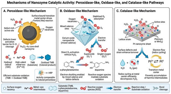
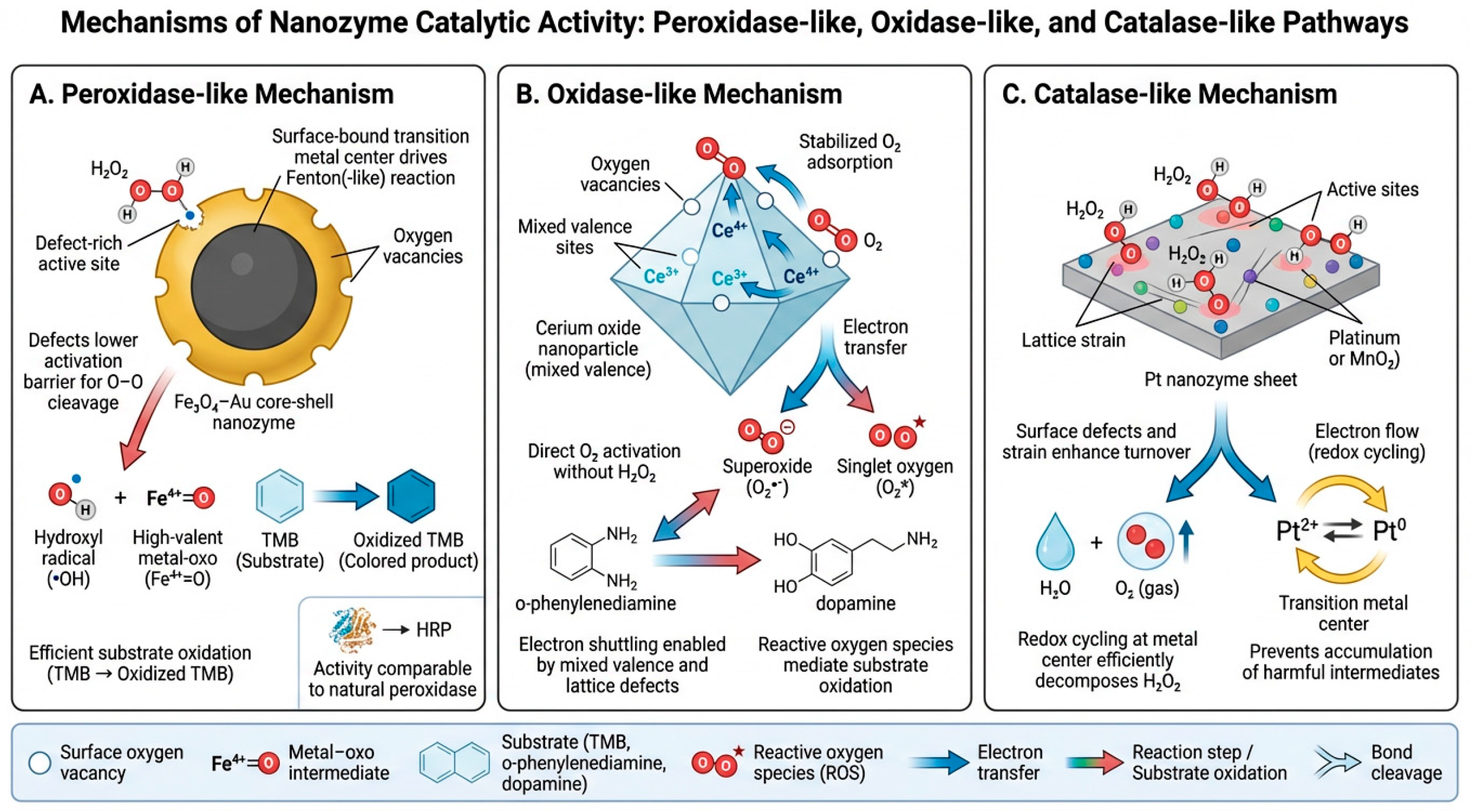
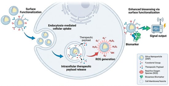
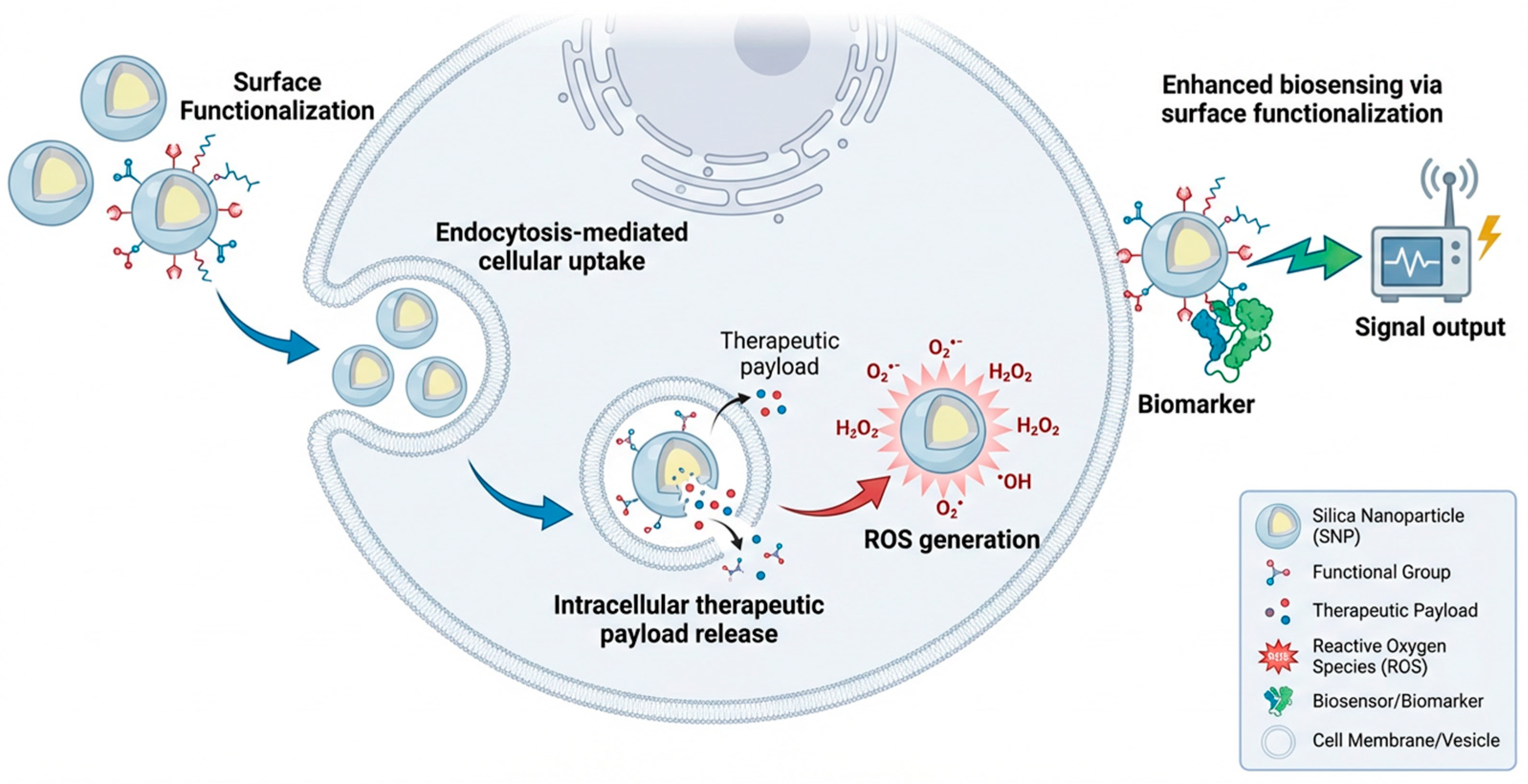
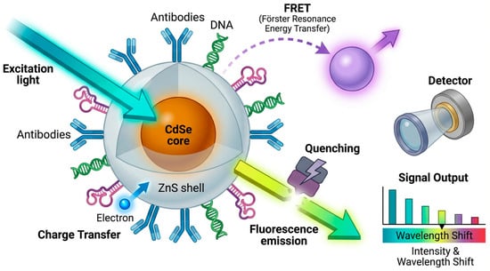
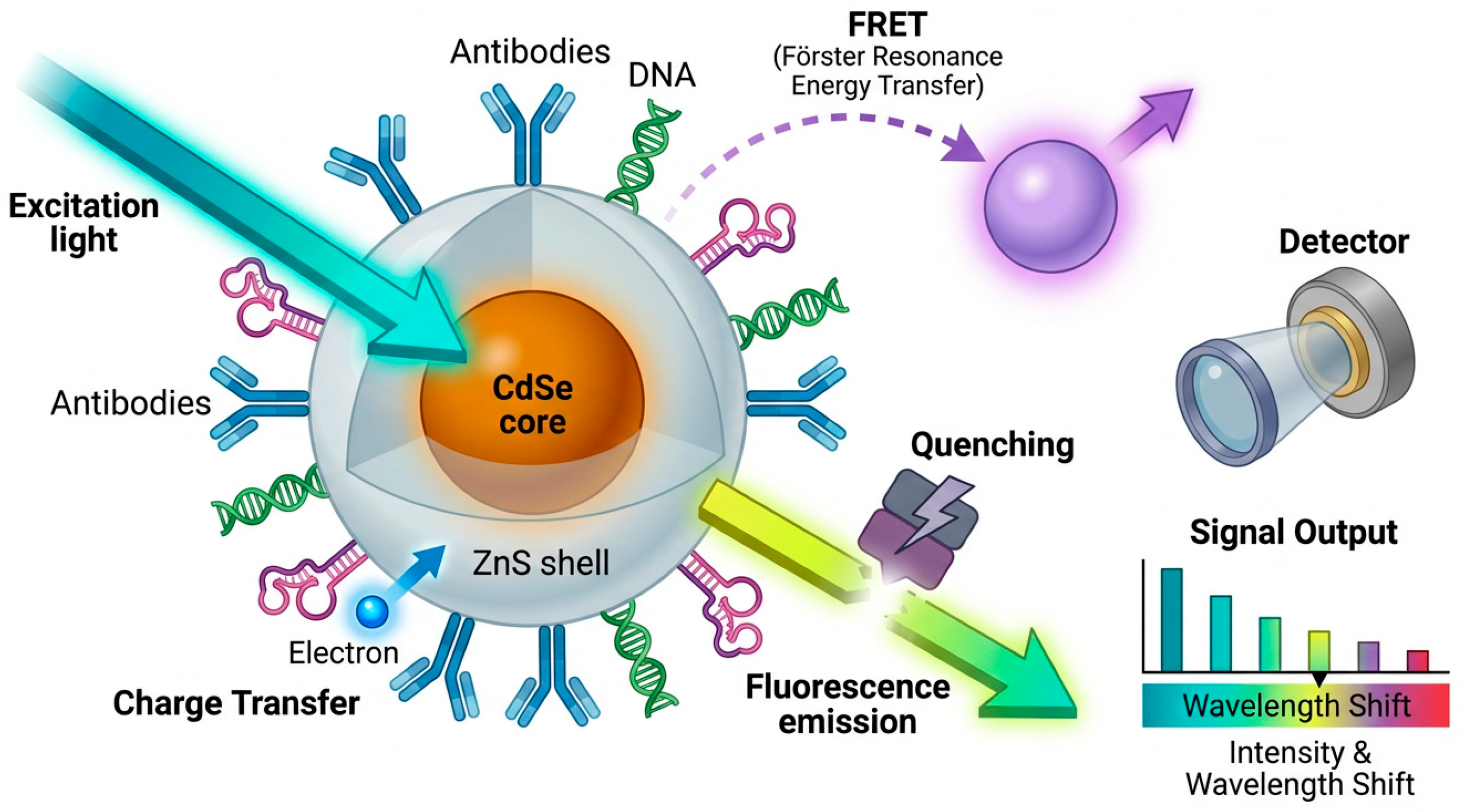
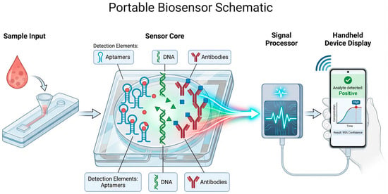
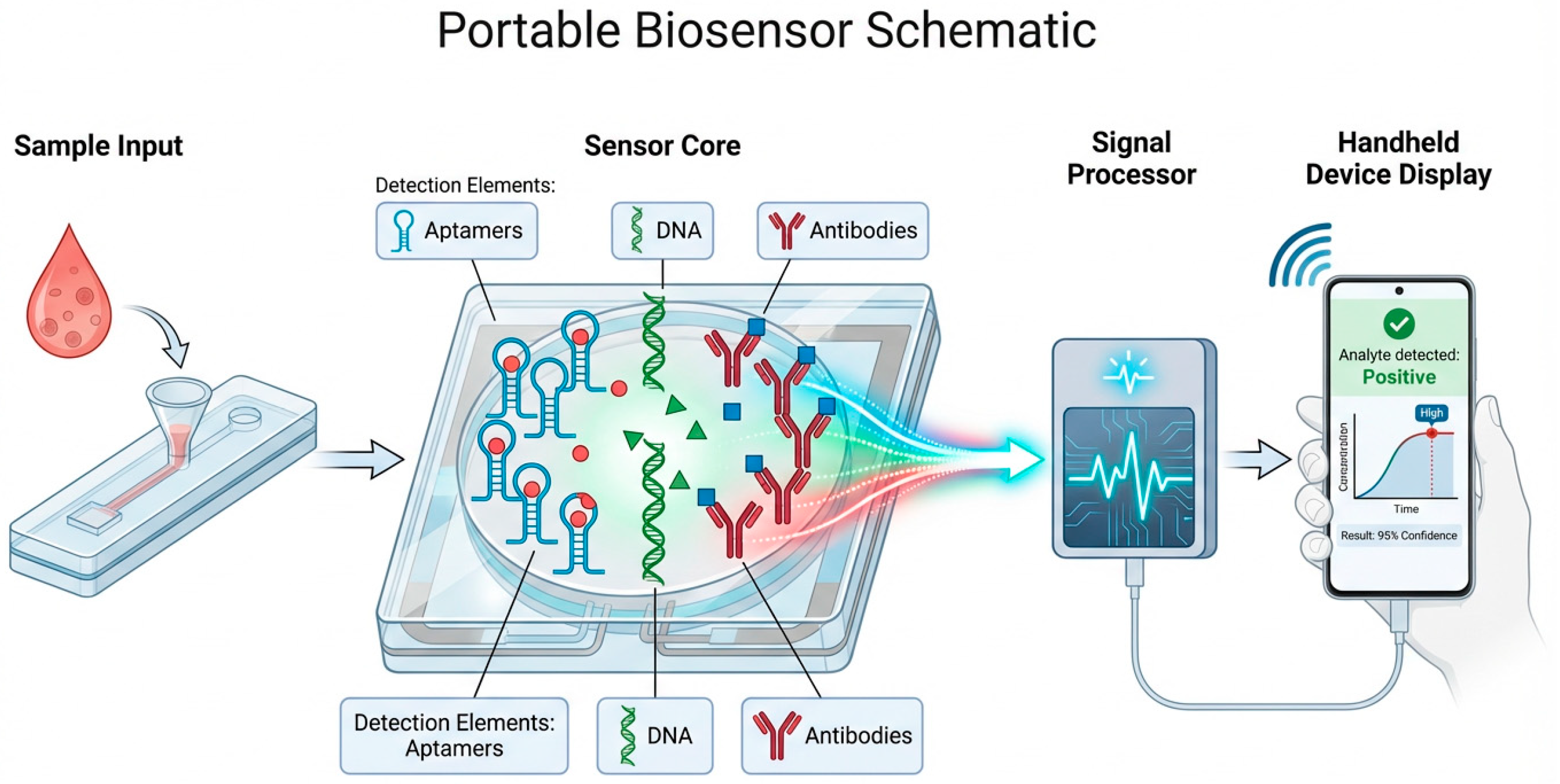
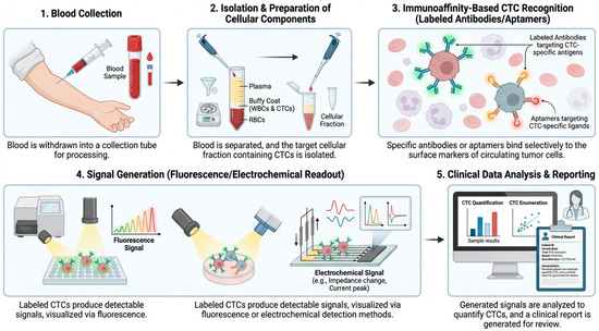
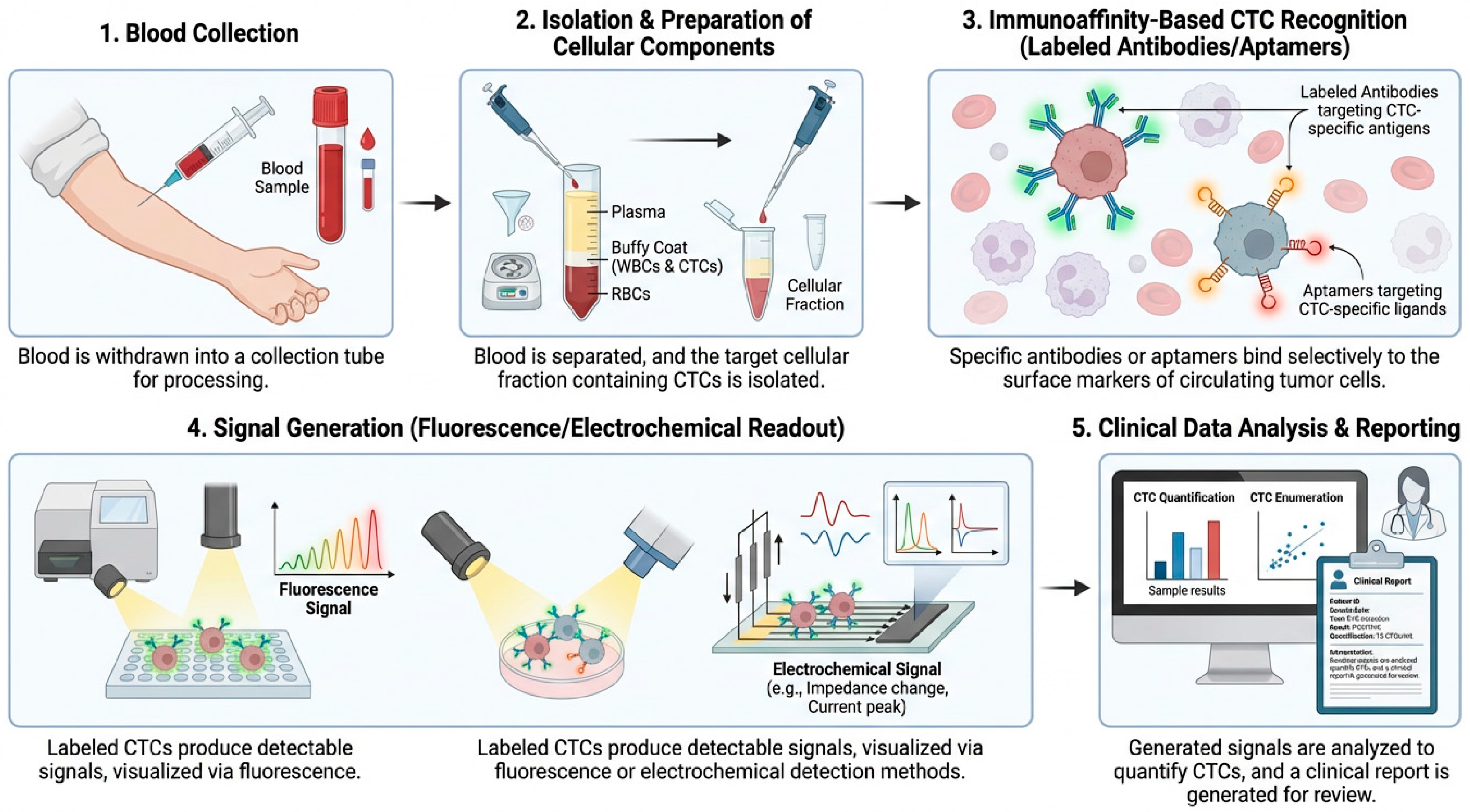
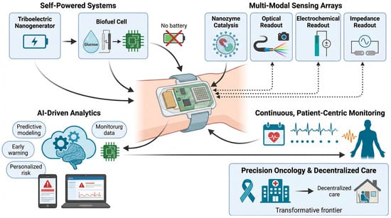
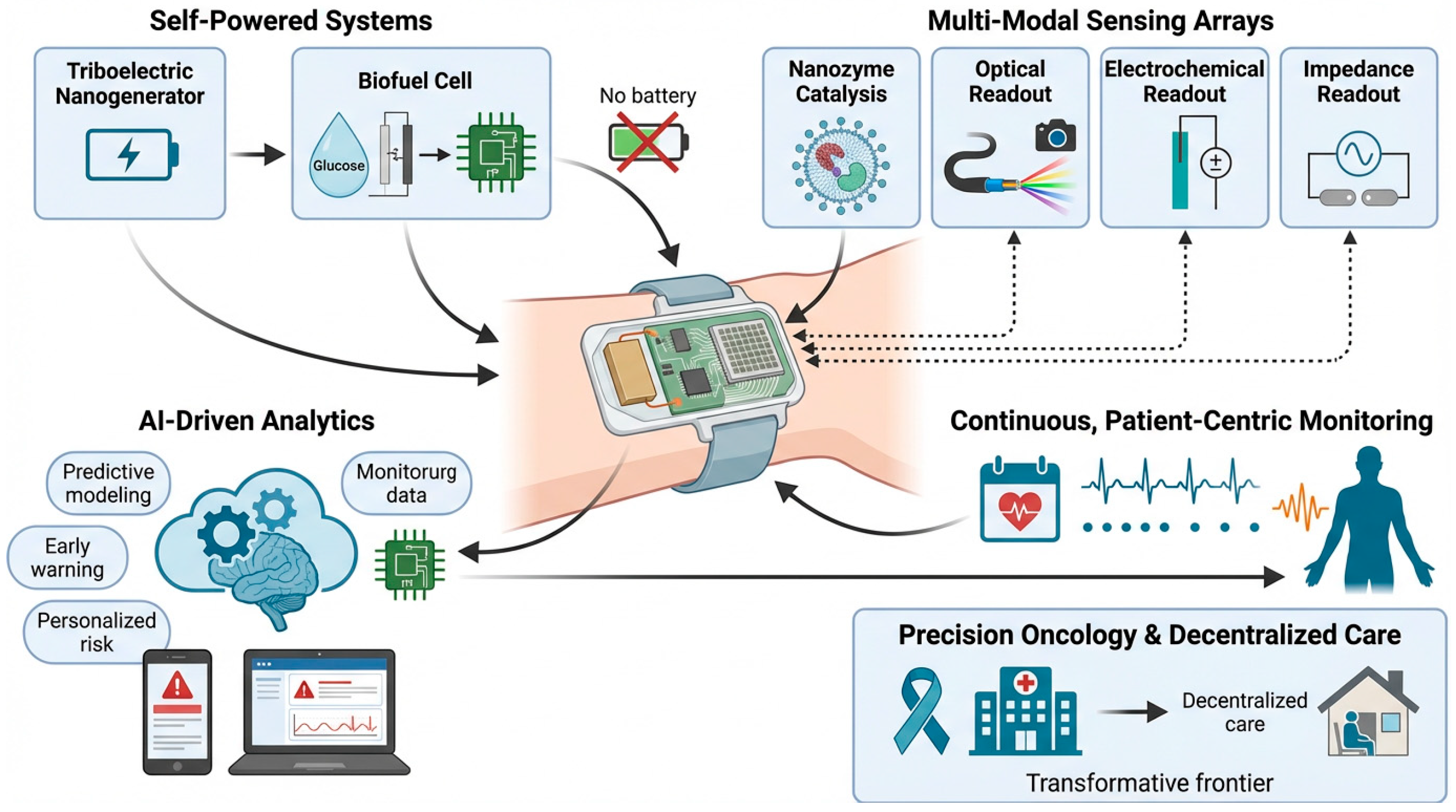

Disclaimer/Publisher’s Note: The statements, opinions and data contained in all publications are solely those of the individual author(s) and contributor(s) and not of MDPI and/or the editor(s). MDPI and/or the editor(s) disclaim responsibility for any injury to people or property resulting from any ideas, methods, instructions or products referred to in the content. |
© 2026 by the authors. Licensee MDPI, Basel, Switzerland. This article is an open access article distributed under the terms and conditions of the Creative Commons Attribution (CC BY) license.