


Abstract
Saxitoxins (STXs) are potent neurotoxins associated with harmful algal blooms (HABs) and represent a major concern for seafood safety. While monitoring programs mainly target bivalve molluscs, information on STXs occurrence in other aquatic organisms remains limited. This study investigated STXs occurrence in marine invertebrates from the Lesina and Varano coastal lagoons (southern Italy) and its relationship with phytoplankton composition and environmental conditions. STXs were determined by enzyme-linked immunosorbent assay (ELISA) in the Pacific oyster (Magallana gigas), the Atlantic blue crab (Callinectes sapidus), and gelatinous zooplankton. Potentially STXs-producing dinoflagellates were mainly represented by Gymnodinium spp. and Alexandrium spp., including Alexandrium minutum. Temperature showed the strongest association with the genus Alexandrium, whereas Gymnodinium spp. was negatively associated with salinity, although these relationships were not statistically significant. STXs were detected in all investigated matrices at concentrations below the EU maximum level for bivalve molluscs, which was used only as a contextual reference. Among the investigated matrices, C. sapidus showed both the highest occurrence of STXs-positive samples and the highest toxin concentrations. Tissue-specific analyses revealed STXs in all analysed anatomical compartments, with the highest concentrations detected in the hepatopancreas. These findings suggest that STXs circulation may extend beyond traditional bivalve vectors and provide preliminary evidence supporting a multi-compartment approach for investigating toxin dynamics in Mediterranean transitional ecosystems.
Keywords:
saxitoxins; Paralytic Shellfish Poisoning; Callinectes sapidus; Magallana gigas; gelatinous zooplankton; Mediterranean coastal lagoons; marine biotoxins; ecological compartments; ecosystem-based monitoring Key Contribution:
This study provides a multi-compartment assessment of saxitoxins (STXs) in organisms occupying distinct ecological compartments within Mediterranean coastal lagoons, including Pacific oysters, Atlantic blue crabs and gelatinous zooplankton. The results suggest that STXs circulation may extend beyond traditional filter-feeding bivalve vectors and highlight the value of integrating non-traditional biological matrices into ecosystem-based approaches for investigating marine biotoxin dynamics.
1. Introduction
Phycotoxins are naturally occurring marine biotoxins produced by a variety of aquatic microorganisms, including toxic microalgae (mainly dinoflagellates, diatoms) and cyanobacteria during phenomena collectively known as harmful algal blooms (HABs) [1,2,3]. HABs are increasingly recognized as a major environmental, ecological, and public health concern because of their impacts on marine ecosystems, aquaculture, fisheries, and seafood safety [1,2,4].
Among the best-known and most extensively studied marine phycotoxins are saxitoxins (STXs), a family of potent neurotoxic alkaloids responsible for Paralytic Shellfish Poisoning (PSP) [3]. Paralytic shellfish toxins (PSTs) comprise more than 50 structurally related saxitoxin analogues, whose content is expressed as STX equivalents for regulatory purposes [3,5]. In this study, “PSTs” is used when referring to this toxin group as a whole, as in the regulatory framework and the cited literature, whereas “STXs” refers to the toxins measured here, including saxitoxin and the cross-reacting analogues detected by enzyme-linked immunosorbent assay (ELISA), expressed as STX equivalents. STXs are produced primarily by marine dinoflagellates belonging to the genera Alexandrium, Gymnodinium, and Pyrodinium, as well as by several freshwater cyanobacteria, including Anabaena, Cylindrospermopsis, Aphanizomenon, Planktothrix and Lyngbya [1,2]. Among marine species, Alexandrium tamarense, A. fundyense, A. minutum, Gymnodinium catenatum, and Pyrodinium bahamense are recognized as the principal sources of PSP outbreaks worldwide [6].
Environmental conditions are recognized as key drivers of HABs, influencing the growth, distribution, and toxicity of potentially harmful dinoflagellates [1]. Among these factors, temperature, salinity, nutrient availability, and water column stability play a major role in regulating the physiology and bloom dynamics of species belonging to the genera Alexandrium, Gymnodinium, and Pyrodinium, although species-specific responses may vary considerably [1]. In particular, changes in temperature and salinity have been shown to affect both cell proliferation and STXs production, while nutrient availability can further modulate toxin content and bloom intensity [7,8,9]. Nevertheless, the relationships between environmental conditions, bloom development, and toxin production remain complex, reflecting the interaction of multiple abiotic and biotic factors rather than the effect of a single environmental driver [1,9].
Due to their high toxicity and implications for public health, STXs are among the most strictly regulated marine biotoxins worldwide. International monitoring programs have been established to prevent contaminated seafood from reaching consumers, with surveillance primarily focusing on bivalve molluscs because of their high filtration capacity and their recognized role as the main vectors of PSPs [2,3,5,10]. Current legislation regulates the monitoring of STXs primarily in filter-feeding bivalve molluscs, which represent the main vectors of human exposure through seafood consumption [2,3,4,11]. In the European Union, the maximum permitted level for PSTs (800 μg STX equivalents kg−1 shellfish flesh) is established under Regulation (EC) No 853/2004 [12], as amended by Commission Delegated Regulation (EU) 2021/1374 [13], while official controls are organized under Regulation (EU) 2017/625 [14]. However, increasing evidence indicates that STXs are not confined to filter-feeding bivalves but can occur in a wide range of marine organisms, reflecting their transfer through marine food webs [2,3,15]. Accordingly, numerous studies have documented the presence of PSTs and other marine biotoxins in gastropods, crustaceans, echinoderms, tunicates and other marine organisms, highlighting that marine biotoxin occurrence extends well beyond the traditionally monitored filter-feeding bivalves [15,16,17,18,19].
Consequently, increasing attention has been directed towards these non-traditional vectors to improve our understanding of STXs distribution and trophic transfer within marine ecosystems, while strengthening seafood safety monitoring and risk assessment beyond routinely monitored bivalve molluscs [15,19].
Among non-traditional vectors, gelatinous zooplankton has attracted increasing attention because of its central ecological role in pelagic food webs [20]. Gelatinous organisms constitute a taxonomically and functionally diverse group that actively participates in marine trophic interactions, linking planktonic communities with higher trophic levels through both predation and prey availability [21]. The interactions between toxic phytoplankton and zooplankton represent an important ecological process influencing HABs dynamics. In particular, toxin-mediated reductions in grazing pressure may promote bloom development through feedback mechanisms between toxin production and zooplankton feeding [22]. Beyond their ecological relevance, gelatinous organisms have recently attracted interest as novel food resources. Several Mediterranean jellyfish species are considered promising candidates for human consumption under the European Novel Food framework [23]. Previous studies have demonstrated their nutraceutical potential [24], established safety and quality parameters for their processing [25], and evaluated thermal stabilization protocols for Mediterranean edible jellyfish species, including Rhizostoma pulmo and Aurelia coerulea [26].
Consequently, assessing the occurrence of marine biotoxins such as STXs in these emerging food resources represents an additional food safety challenge, which has so far received little attention despite recent reviews highlighting the need for comprehensive risk assessment [27,28]. Beyond jellyfish, gelatinous zooplankton also includes ctenophores, which may represent additional vectors of marine biotoxins within pelagic food webs. In particular, the invasive ctenophore Mnemiopsis leidyi, an efficient zooplankton predator recorded in both Lesina and Varano lagoons [29], provides a complementary model for investigating toxin uptake and trophic transfer.
Besides gelatinous zooplankton, crustaceans have also received increasing attention as non-traditional vectors because numerous studies have documented the occurrence and accumulation of PSTs and other marine biotoxins in decapods and other crustacean groups, highlighting their potential role in toxin transfer through marine food webs [17,18,19].
The Atlantic blue crab (Callinectes sapidus) represents a particularly relevant biological model because it occupies a trophic position complementary to that of filter-feeding bivalves and pelagic gelatinous organisms within coastal food webs [30]. This invasive species has rapidly expanded throughout the Mediterranean Sea and is characterized by remarkable ecological plasticity, occupying coastal and transitional environments where it exhibits opportunistic feeding behaviour [30,31,32]. Owing to its omnivorous diet, which includes bivalves, gastropods, crustaceans, polychaetes, fish and detritus, C. sapidus is exposed to contaminants and biotoxins through multiple trophic pathways, making it a promising non-traditional model for investigating marine biotoxin transfer [30,32]. Stable and increasing populations of C. sapidus have been documented in the Lesina and Varano lagoons, with evidence of reproduction in Varano, supporting the establishment of the species in these ecosystems [33,34]. Moreover, its increasing commercial exploitation as an edible resource in the Mediterranean highlights the need to assess potential food safety risks [30]. Despite its growing ecological and commercial importance, studies investigating STXs occurrence and tissue-specific accumulation in C. sapidus remain scarce, particularly in Mediterranean transitional ecosystems [17,19]. The ecological context of the study area is also characterized by shellfish aquaculture activities in the Varano lagoon, including the Pacific oyster Magallana gigas. Previous research in the lagoon has documented its growth performance and quality under different hydrodynamic conditions, highlighting the importance of environmental conditions for oyster production and marketability [35]. As a suspension-feeding bivalve, M. gigas represents a particularly relevant organism for assessing the accumulation of water- and particle-associated toxins and their potential implications for shellfish food safety.
Against this background, the present study investigated STXs occurrence in ecologically distinct marine invertebrates from the Lesina and Varano lagoons to explore toxin uptake and trophic transfer across complementary ecological compartments. The investigated organisms comprised the ctenophore Mnemiopsis leidyi, the jellyfish R. pulmo and A. coerulea, the invasive Atlantic blue crab C. sapidus, and the filter-feeding Pacific oyster M. gigas. This combination provides an opportunity to investigate STXs occurrence and potential transfer across contrasting feeding strategies and habitats within Mediterranean transitional ecosystems.
To place biological findings within their environmental context, STXs occurrence was interpreted together with the environmental conditions and phytoplankton composition associated with HABs development in the two lagoons. This integrated ecosystem-based approach, combining environmental drivers with toxin occurrence across multiple ecological compartments, represents the main novelty of the present study.
Specifically, the objectives were to: (i) investigate STXs occurrence in marine invertebrates occupying different ecological compartments; (ii) evaluate the environmental and phytoplankton conditions associated with STXs occurrence in the investigated organisms; and (iii) provide preliminary information to support food safety assessments and future monitoring strategies for edible and emerging marine resources in Mediterranean transitional ecosystems.
2. Results
2.1. Inter-Lagoon Environmental and Phytoplankton Patterns
Environmental and phytoplankton patterns were compared between the Lesina and Varano lagoons over the sampling period (May–November 2024). Inter-lagoon comparisons of the environmental parameters (Figure 1) revealed largely overlapping conditions, with no significant differences detected (p > 0.05). In particular, water temperature ranged from 16.8–29.0 °C in Lesina and from 21.0–31.2 °C in Varano, while salinity was on average higher in Varano (mean 30.46 practical salinity units, PSU) than in Lesina (mean 28.04 PSU). Dissolved oxygen exhibited comparable distributions in both lagoons. Total phytoplankton abundance also showed comparable distributions between the two lagoons (Figure 1D), with no significant differences detected (p > 0.05), although greater variability was observed in Varano.
Figure 1.
Boxplots of environmental parameters and phytoplankton in the two coastal lagoons (Lesina and Varano). Panels (A–C) report temperature (°C), dissolved oxygen (mg/L), and salinity (PSU), respectively; panel (D) shows total phytoplankton as log10 cells L−1. Each plot shows the median (red line), interquartile range (IQR) and non-outlier distribution; open circles indicate outliers.
Temporal trends (Figure 2) highlighted a coherent seasonal evolution of environmental conditions in both lagoons. Temperature increased from late spring to a peak in summer (July), followed by a decline towards November, with slightly higher values generally observed in Varano. Dissolved oxygen showed more variable trends, with a general decrease during warmer months and an increase towards autumn, particularly evident in Lesina. Moreover, salinity displayed lagoon-specific patterns, increasing from May to late summer in both systems, but remaining consistently higher and less variable in Varano.
Figure 2.
Monthly variations in (A) temperature, (B) dissolved oxygen, and (C) salinity, and in the abundance (log10 cells L−1) of (D) diatoms, (E) dinoflagellates, and (F) other phytoplankton in Lesina (black circles) and Varano (grey squares) lagoons. Points represent monthly observed values; no sampling was carried out in August and October.
Monthly variations in the abundance of the main phytoplankton groups are shown in Figure 2D–F. Diatom abundance showed alternating monthly fluctuations in both systems, with the highest values occurring in June and September in Varano, whereas Lesina displayed more moderate oscillations, with slight increases in July and November. Dinoflagellates followed similar overall seasonal trends in the two lagoons, increasing from May to early summer before showing slight differences in the timing of fluctuations during July and September, and converging to comparable abundances by November. The remaining phytoplankton fraction remained comparatively stable over time in both lagoons, with only slight month-to-month variations.
Overall, differences between lagoons were limited and mainly reflected slight shifts in the timing of monthly fluctuations rather than marked differences in phytoplankton abundance patterns.
2.2. Composition of Potential STXs-Producing Dinoflagellates and Environmental Correlations
The relative composition of the main potential STXs-producing dinoflagellates exhibited marked temporal variability in both lagoons (Figure 3A,B). For each sampling event, the relative contribution of the four selected taxa (A. minutum, Alexandrium spp., Gymnodinium spp., and P. bahamense) was calculated as percentage of their combined abundance. Absolute abundances are also reported to provide a quantitative assessment of the temporal variation in these taxa (Figure 3A,B).
Figure 3.
Abundance and relative composition of the main potentially STXs-producing dinoflagellates in the Lesina (A) and Varano (B) lagoons during the study period. The tables report the abundance (cells L−1) of A. minutum, Alexandrium spp., Gymnodinium spp. and P. bahamense, and their total, at each sampling event (-, not detected). The bars show the relative contribution (%) of each taxon to the total abundance (100%) of the selected taxa in each lagoon and sampling event.
In the Lesina lagoon (Figure 3A), Gymnodinium spp. dominated the selected assemblage in May (54.5%) and reached its highest relative contribution in June (97.6%), before declining in July (37.5%). Its contribution increased again in November, when it accounted for 66.7% of the assemblage. Conversely, A. minutum was most prominent in July (62.5%), after contributing 36.3% in May and only 2.4% in June. Alexandrium spp. was the dominant taxon in September (80%), following its low contribution in May (4.5%), and accounted for the remaining 33.3% in November. P. bahamense was detected only in May, representing 4.5% of the selected taxa.
In the Varano lagoon (Figure 3B), Gymnodinium spp. dominated the selected assemblage in May (100%) and remained prevalent in June (74.8%) and July (82.6%), despite the contribution of other taxa. Its relative contribution declined sharply in September (1.8%), when A. minutum accounted for 98.2% of the assemblage. In November, Gymnodinium spp. regained its predominance (77.3%), while A. minutum represented the remaining 22.7%. Alexandrium spp. contributed to the assemblage in June (25.2%) and July (5.8%), but was not detected in the subsequent sampling events.
A comprehensive overview of the phytoplankton community, including diatoms, dinoflagellates, and other phytoplankton groups across all sampling events and both lagoons, is provided in Supplementary Table S1.
Spearman correlation analyses between algal abundances and environmental parameters showed weak to moderate, non-significant relationships in the pooled lagoon data (n = 10; p > 0.05 for all comparisons). Among the tested associations (Figure 4), the strongest correlation was observed between temperature and total Alexandrium (A. minutum plus other Alexandrium spp.; ρ = 0.59), followed by a weaker positive correlation with A. minutum abundance alone (ρ = 0.45). In contrast, Gymnodinium spp. abundance was negatively associated with salinity (ρ = −0.39). Analyses performed separately for each lagoon showed generally similar directions of association, although effect sizes varied between lagoons. However, none of the observed correlations remained significant after FDR correction. Given the limited sample size (n = 10 pooled observations; n = 5 observations per lagoon), the statistical power of these analyses was likely low, and the observed correlations should therefore be regarded as preliminary trends requiring confirmation with larger datasets.
Figure 4.
Pooled relationships between log-transformed algal concentration (log10[x + 1]) and the main environmental drivers showing the strongest associations: A. minutum and Alexandrium (overall) vs. temperature, and Gymnodinium vs. salinity. Points represent individual observations from Lesina (black) and Varano (grey). Spearman rank correlation coefficients (ρ) are reported for the pooled dataset in each panel.
No consistent or relevant relationships were observed between algal abundances and dissolved oxygen, and the remaining correlations were close to zero (Supplementary Table S2).
2.3. Sample Characteristics and Biometric Relationships of the Analysed Organisms
The organisms investigated in this study differ substantially in their biology and ecology, ranging from a sessile filter-feeding bivalve to a mobile decapod crustacean and soft-bodied gelatinous taxa. Biometric measurements were evaluated for M. gigas and C. sapidus to characterize the size range of the analysed specimens. The morphometric relationships between total length (TL, mm) and whole weight (WW, g) were evaluated for both species (Figure 5). The non-linear regression analyses showed positive relationships between body size and whole weight in both C. sapidus (N = 60; R2 = 0.80) and M. gigas (N = 100; R2 = 0.86). In both species, the exponent of the length–weight relationship (b) was significantly different from zero (p ≤ 0.01).
Figure 5.
Length-weight relationships (TL/WW) in C. sapidus (WW = 0.004 × TL2.56; R2 = 0.80) on the left and M. gigas (WW = 0.02 × TL1.70; R2 = 0.86) on the right, respectively.
Conversely, no formal biometric characterization was performed for gelatinous organisms (M. leidyi, A. coerulea, and R. pulmo), given the ecological and biological constraints inherent to this matrix.
2.4. STXs Detection, Occurrence and Accumulation Patterns in Aquatic Organisms
2.4.1. Assay Performance in C. sapidus and Gelatinous Zooplankton
The ELISA assay employed in this study is a commercial screening method validated for bivalve molluscs (mussels, clams and oysters); its applicability to crustaceans and gelatinous organisms was therefore verified. Matrix effects were within the ± 20% acceptance criterion in all matrices (M. gigas, −11%; C. sapidus, −19.0%; gelatinous zooplankton, −1.4%), indicating limited interference from matrix components on the ELISA response. Extraction recovery was further assessed only for C. sapidus and gelatinous zooplankton, matrices not covered by the original validation of the assay, by fortifying tissue before extraction at 20, 40 and 80 ng g−1. Mean recoveries ranged from 84–120% (RSDr 8–21%) (Table 1). The LOQ was set at 20 ng g−1, corresponding to the lowest fortification level meeting the acceptance criteria (recovery: 70–120%, repeatability RSDr ≤ 20%), which also corresponded to the lowest calibration level.
Table 1.
Extraction recovery (%) and repeatability (RSDr, %) of the ELISA method in C. sapidus and gelatinous zooplankton fortified before extraction at three levels (n = 3 replicates per level).
2.4.2. Occurrence in C. sapidus, M. gigas, and Gelatinous Zooplankton
Overall, at least one positive sample was recorded for each investigated biological matrix (C. sapidus, M. gigas, and gelatinous zooplankton), although STXs occurrence varied among lagoons and sampling events. All concentrations, expressed as STX equivalents, were at least eight times lower than the EU maximum level for bivalve molluscs (800 µg STX eq. kg−1, corresponding to 800 ng g−1) [12,13]. This comparison is provided for context only, in the absence of specific regulatory limits for crustaceans or gelatinous zooplankton. The limit applies to bivalve molluscs, and ELISA screening results cannot establish regulatory compliance or food-safety status, which require the official reference methods [11].
For each matrix, STXs occurrence was expressed as the percentage of sampling events in which at least one positive sample was detected (Table 2). For M. gigas and gelatinous zooplankton, one composite sample was analysed per sampling event; for C. sapidus, six samples (five tissues and one whole-organism composite) were analysed per event, and an event was considered positive when at least one of them was positive. Positive samples were recorded only in May, September and November. In the Varano lagoon, STXs were detected in blue crabs in all three months and in oysters in May and November. In the Lesina lagoon, positive samples were detected only in September, in blue crabs and gelatinous zooplankton.
Table 2.
STXs occurrence and concentrations (ng g−1 STX eq.) in the investigated matrices. Occurrence: percentage of sampling events with at least one positive sample (for C. sapidus, any tissue or the whole-organism composite). Concentrations were calculated on positive samples only.
Mean concentrations, calculated when more than one concentration value was available, ranged from 23.3 ± 6.4 ng g−1 in M. gigas to 46.1 ± 28.5 ng g−1 in C. sapidus from the Lesina lagoon (Table 2). For C. sapidus, these values summarise tissue-specific and whole-organism measurements from the same sampling events and are therefore descriptive only (Section 5.4.4). Only one gelatinous zooplankton sample (M. leidyi, Lesina, September) resulted positive (20% occurrence), showing a concentration of 45.0 ng g−1; therefore, no standard deviation could be calculated.
2.4.3. Tissue Distribution in C. sapidus
Within blue crabs, the frequency of STXs detection differed among anatomical compartments (Figure 6). Positive samples were more frequently recorded in the stomach and in the whole-organism composite sample (30% of the ten sampling events, five per lagoon), followed by the hepatopancreas (20%). Gills, gonads, and muscle each yielded a single positive sample, corresponding to an occurrence of 10%.
Figure 6.
STXs occurrence (%) in the different anatomical compartments of the blue crab C. sapidus (gills, gonads, muscle, stomach, hepatopancreas, and whole-organism composite sample).
Mean STXs concentrations (±standard deviation, when applicable) differed among the anatomical compartments of the blue crab C. sapidus (Figure 7). Gills, gonads and muscle each yielded a single positive sample (n = 1), so no standard deviation could be calculated for these compartments. The hepatopancreas exhibited the highest mean STXs concentration (74.4 ± 30.3 ng g−1, n = 2), whereas the lowest value was recorded in muscle tissue (17.7 ng g−1, semi-quantitative). Mean concentrations in the remaining anatomical compartments were 27.3 ng g−1 in the gills, 29.4 ng g−1 in the gonads, 36.1 ± 18.7 ng g−1 in the stomach, and 36.2 ± 15.6 ng g−1 in the whole-organism composite sample.
Figure 7.
Mean STXs concentrations (ng g−1) in the different anatomical compartments of the blue crab C. sapidus, calculated on positive samples only. Colour intensity is proportional to STXs concentration. Error bars represent the standard deviation (±SD) among positive samples collected in different sampling events and lagoons (stomach and whole-organism, n = 3; hepatopancreas, n = 2); no SD is shown for gills, gonads and muscle, as only one positive sample was recorded (n = 1). The value for muscle is below the LOQ and should be considered semi-quantitative.
3. Discussion
The present study provides preliminary evidence of STXs occurrence across multiple ecological compartments within Mediterranean transitional lagoons. Although toxin concentrations remained below the EU maximum level for bivalve molluscs, used here only as a contextual reference, STXs were detected in all investigated matrices, including the filter-feeding bivalve M. gigas, the brachyuran crab C. sapidus, and gelatinous organisms. This finding extends the current understanding of STXs occurrence beyond the traditional bivalve compartment and is consistent with previous evidence that marine biotoxins may occur in a broader range of marine invertebrates, including crustaceans and gastropods [16,17,18,19]. Studies on marine food webs have shown that phycotoxins can be detected across organisms occupying different trophic positions, with trophic transfer and dilution patterns varying among toxin groups and biological compartments [37]. The occurrence of STXs across organisms with different ecological roles and feeding strategies highlights the value of considering multiple biological compartments when investigating toxin distribution in transitional ecosystems.
In the filter-feeding bivalve M. gigas, the occurrence of STXs was consistent with the established role of bivalve molluscs as major vectors of marine biotoxins. Through filtration and ingestion of suspended particles, bivalves can efficiently accumulate algal toxins, although toxin uptake, retention and elimination may vary according to feeding activity, physiological condition and characteristics of the toxin-producing organisms [37,38]. Experimental studies in M. gigas have also shown that exposure to toxic A. minutum can activate detoxification and antioxidant responses, highlighting the physiological processes involved in toxin handling [39]. In the Mediterranean, the occurrence of PSTs in bivalves has been documented in association with potentially toxic Alexandrium spp. [40].
The relevance of M. gigas extends beyond its ecological role, as the species is widely cultivated for human consumption and represents an established component of shellfish aquaculture. Its occurrence in the present study therefore provides not only a reference point for comparison with the less routinely investigated biological compartments, but also a direct food-safety context for STXs surveillance in farmed shellfish. The detection of STXs confirms that the lagoon systems provided conditions compatible with toxin exposure, but should not be interpreted as a direct proxy for toxin production in the surrounding waters, since accumulation in bivalves reflects the balance between uptake and elimination processes [38,41].
Within the non-traditional vectors investigated, C. sapidus represents a particularly relevant compartment because of its opportunistic and generalist feeding behaviour, which may expose the species to toxins through multiple dietary pathways. The blue crab is an invasive species in several areas of the Mediterranean, and its ecological plasticity has facilitated its establishment across coastal and transitional habitats [30,31,32]. Its broad trophic connectivity may favour the integration of toxin exposure over time rather than reflecting direct interaction with potentially toxic phytoplankton alone.
In the present study, the occurrence of STXs in C. sapidus from both lagoons, together with their detection in different anatomical compartments, supports the relevance of this species as a complementary matrix for investigating toxin occurrence beyond filter-feeding bivalves. The comparatively higher occurrence observed in blue crabs than in M. gigas should nevertheless be interpreted cautiously, as differences in feeding strategy, tissue composition and toxin processing may influence both uptake and retention. Evidence from other similar crab species supports the potential accumulation of naturally occurring algal toxins in this group. Algal toxins have been detected in Carcinus maenas [42], while evidence from Portunus pelagicus further supports toxin exposure in closely related brachyuran taxa [19]. In addition, cyanotoxin accumulation in C. sapidus under natural environmental conditions has been documented [43]. Although these studies concern toxin groups other than STXs, they provide complementary evidence that crabs can accumulate naturally occurring toxins. Together with previous reports of PSTs in non-traditional crustacean vectors [16,17,18,19], these findings support the biological plausibility of STXs occurrence in C. sapidus.
Of particular interest was the highest concentration of STXs observed in the hepatopancreas of C. sapidus, consistent with the recognised role of this organ in digestion, assimilation, storage and detoxification processes in decapod crustaceans, and with tissue-specific toxin accumulation reported in other crab species exposed to algal toxins [16,17,18]. The detection of STXs in the whole-organism composite sample further indicates that toxin occurrence was not confined to a specific anatomical compartment. Although pooling prevents the assessment of tissue-specific accumulation, this matrix provides a complementary measure of the overall toxin burden of the animal and may be relevant for evaluating exposure when whole-organism material is considered.
Although the present data do not allow discrimination between direct exposure and trophic transfer pathways, nor do they establish the persistence or depuration kinetics of STXs in the species, the occurrence of STXs across multiple tissues, combined with the broad trophic niche of C. sapidus [30,31,32], supports its consideration as a complementary biological matrix for investigating marine biotoxin occurrence in transitional ecosystems. Moreover, the invasive status and increasing commercial interest of C. sapidus also provide an additional perspective for its potential use in environmental and food-safety studies. Its exploitation as a seafood resource may contribute to the valorisation of an invasive species, while its broad trophic niche could make it a potentially informative species for characterising toxin occurrence in transitional ecosystems. Further studies combining repeated sampling with targeted analysis of individual tissues would therefore be necessary to clarify the mechanisms underlying toxin uptake and retention in C. sapidus.
Gelatinous organisms represent a comparatively under-investigated compartment in the context of marine biotoxins. The group includes organisms with distinct feeding strategies and trophic roles, and therefore potentially different pathways of toxin exposure [21]. Previous studies have shown that gelatinous organisms can interact with harmful microalgae, highlighting their potential involvement in processes related to the transfer or retention of algal-derived compounds within pelagic food webs [44]. In the present study, the gelatinous compartment included the predatory ctenophore M. leidyi and the scyphozoan jellyfish A. coerulea and R. pulmo. STXs were detected in only one sample, corresponding to M. leidyi, whereas no positive samples were recorded for the two jellyfish species. Although this isolated detection does not allow conclusions regarding species-specific accumulation or trophic transfer, it provides preliminary evidence that STXs may occur in gelatinous predators and supports the inclusion of these organisms in multi-compartment investigations of marine biotoxins.
The detection in M. leidyi is also of ecological interest considering its role as an efficient pelagic predator and its capacity to influence zooplankton communities and pelagic trophic interactions [45]. More broadly, the occurrence of STXs in gelatinous organisms may deserve attention in the context of their increasing consideration as potential food resources. Mediterranean jellyfish, including A. coerulea and R. pulmo, have been investigated as novel food resources, with studies addressing their nutritional properties, processing and safety [24,25,26,27,28]. However, information on marine phycotoxins in edible jellyfish remains limited, indicating a need for further investigation of toxin occurrence in these emerging food matrices.
The occurrence of STXs should be interpreted within the seasonal environmental variability characterising both lagoons. Although Lesina and Varano differ in their hydrological and morphological features, previous studies have shown that both systems are strongly influenced by seasonal forcing, freshwater inputs, and seawater exchange, which contribute to marked temporal and spatial variability in their environmental conditions [46,47,48]. In Lesina, seasonal changes in hydrological conditions have also been associated with marked variations in phytoplankton abundance and composition [49]. In the present study, the two lagoons showed broadly comparable seasonal environmental patterns, with temporal variability more pronounced than spatial differences. These conditions provide the ecological background for phytoplankton dynamics and toxin exposure, but do not by themselves allow STXs occurrence in the investigated organisms to be attributed to a specific environmental driver.
The composition of potentially STXs-producing dinoflagellates provides a more specific ecological context for interpreting toxin occurrence. Among the taxa detected, A. minutum deserves particular attention because it was the only Alexandrium taxon identified to species level in the present study and is a recognised PSTs-producing species widely reported from Mediterranean coastal and transitional waters [7,50,51]. Its occurrence in both lagoons indicates the presence of a potentially relevant PSTs-producing taxon throughout the study area. A positive trend between temperature and the abundance of total Alexandrium and A. minutum was observed, although not statistically significant, likely due to the limited number of observations. This pattern is broadly consistent with the recognised influence of temperature on the growth and seasonal dynamics of A. minutum [7,52,53,54].
Gymnodinium spp. was also frequently detected and, in some periods, represented an important component of the potentially toxic assemblage. However, the genus was not resolved to species level in the present study, and therefore the occurrence of G. catenatum, a recognised PSTs-producing species reported from Mediterranean waters, cannot be established [8,9]. Experimental studies on G. catenatum indicate that salinity may influence both growth and toxin content, although these responses can vary depending on strain and environmental conditions [9]. In the present study, Gymnodinium spp. abundance showed a negative association with salinity, although not statistically significant. However, given that the organisms were not identified to species level, this pattern should be interpreted cautiously and cannot be directly related to STXs production.
The temporal distribution of potentially toxic dinoflagellates did not show a consistent correspondence with STXs occurrence in the investigated organisms. Periods characterised by higher contributions of Alexandrium spp. and/or A. minutum were not systematically associated with STXs-positive samples, while the frequent occurrence of Gymnodinium spp. was likewise not accompanied by a consistent detection of STXs. This contrasts with observations in Australian abalone, where PSTs accumulation was more clearly associated with G. catenatum blooms and supported by complementary evidence of exposure [55]. The lack of a consistent correspondence in the present study is nevertheless biologically plausible, as toxin accumulation depends not only on the abundance of potentially toxic phytoplankton, but also on their actual toxin content, the timing of toxin production and the feeding and toxin-processing characteristics of exposed organisms [8,52]. P. bahamense was detected only once and represented a negligible component of the potentially toxic assemblage, suggesting limited relevance during the study period.
Overall, the phytoplankton data indicate the presence of potentially STXs-producing dinoflagellates in both lagoons, but their abundance alone does not explain the occurrence of STXs in the investigated organisms. Since STXs were not measured in phytoplankton samples, the abundance of potentially STX-producing taxa does not necessarily reflect active toxin production, as toxin content can vary widely among species, strains and environmental conditions [1,6,9]; consequently, the actual source of the STXs detected in the biological matrices cannot be identified. Moreover, the absence of winter sampling prevents assessment of the full annual cycle. STXs occurrence during the cold season, when some PSTs-producing species such as G. catenatum may also bloom in the Mediterranean [9], cannot therefore be excluded. The discontinuous sampling schedule may also have limited the detection of short-term fluctuations in phytoplankton composition and STXs occurrence. Nevertheless, the co-occurrence of potentially toxigenic dinoflagellates and STXs-positive organisms provides an important ecological context for toxin exposure in these transitional ecosystems [52,56] and supports the need for higher-frequency environmental monitoring and species-specific toxin characterization in future studies.
The environmental and phytoplankton results provide the ecological context in which STXs occurrence in the investigated organisms should be interpreted, but the observed patterns do not support a direct causal relationship between environmental variables, potentially toxigenic taxa, and toxin occurrence. Despite the exploratory nature of the work, the results provide novel evidence that STXs may occur simultaneously in filter-feeding bivalves, gelatinous zooplankton and decapod crustaceans, supporting the usefulness of a multi-compartment approach for investigating toxin circulation within lagoon food webs. Such an approach may provide complementary information on toxin occurrence across biological compartments with different ecological roles and exposure pathways, as previously highlighted for non-traditional toxin vectors [17,19].
Current surveillance programmes are largely centred on filter-feeding bivalve molluscs, which remain the primary indicators for regulatory purposes [11]. However, the occurrence of STXs in M. gigas, C. sapidus and M. leidyi suggests that toxin circulation may extend to biological compartments that are not routinely included in conventional monitoring frameworks [16,19]. The occurrence of STXs in non-traditional matrices therefore supports the potential value of complementary monitoring approaches that consider organisms with different ecological roles and exposure pathways. In particular, C. sapidus may represent a useful complementary sentinel species in transitional ecosystems, although further validation is required before considering its application in routine monitoring. This broader monitoring perspective is also relevant from a food-safety standpoint, as both M. gigas and C. sapidus are potentially consumed by humans. The increasing recognition of non-traditional biotoxin vectors supports the development of monitoring approaches that complement, rather than replace, established surveillance based on bivalve molluscs [15,19].
The ELISA assay employed in the present study enabled the investigation of toxin occurrence in crustaceans and gelatinous organisms, biological matrices that are less frequently investigated than traditional bivalve molluscs and for which analytical data remain comparatively limited [17,19]. ELISA-based approaches provide useful screening information but do not resolve individual congeners of PSTs, and their performance may depend on antibody cross-reactivity and matrix effects [57,58].
In the present study, matrix effects and extraction recovery in C. sapidus and gelatinous zooplankton met the acceptance criteria (Section 2.4.1), supporting the applicability of the assay as a screening tool in these matrices; nevertheless, concentrations close to the LOQ should be regarded as semi-quantitative.
Instrumental methods, such as pre-column oxidation HPLC-FLD (EN 14526 [59]) and LC-MS/MS, validated through an international collaborative study [60], allow the identification and quantification of individual analogues, but their validation is currently limited to bivalve molluscs. The development and validation of such methods for C. sapidus and gelatinous zooplankton were beyond the scope of this preliminary screening study and will be addressed in future work, in order to confirm STXs occurrence and characterise toxin profiles in these matrices [15].
The present study should be considered a preliminary assessment of STXs occurrence across multiple biological compartments in Mediterranean transitional ecosystems. The limited number of sampling events, conducted over a single year, and the relatively small number of STXs-positive samples, particularly among gelatinous organisms, limit the strength of inferences regarding temporal variability, species-specific accumulation and toxin transfer pathways. Nevertheless, the multi-compartment approach provides a useful basis for further investigations combining more extensive temporal and spatial sampling with targeted analyses of toxin occurrence.
Future studies should therefore combine higher-frequency and longer-term environmental and biological sampling with species-level identification and direct toxin characterization in phytoplankton. Such approaches would help clarify the mechanisms underlying toxin occurrence and retention and further assess the potential relevance of non-traditional vectors for food safety and environmental monitoring.
4. Conclusions
This study provides an integrated assessment of STXs occurrence across environmental, phytoplanktonic and biological compartments in the Mediterranean coastal lagoons of Lesina and Varano. The environmental conditions showed broadly comparable seasonal patterns between the two lagoons, with temporal variability more pronounced than spatial differences. Although temperature showed the strongest association with the abundance of total Alexandrium and A. minutum, the observed relationships were not statistically significant, likely reflecting, at least in part, the limited number of observations. These findings provide a preliminary characterization of the environmental and phytoplankton context associated with STXs occurrence.
STXs were detected in all three investigated biological compartments (M. gigas, C. sapidus and gelatinous organisms), at concentrations consistently below the EU maximum level for bivalve molluscs, used only as a contextual reference. The detection of STXs in organisms with different ecological roles and feeding strategies highlights the occurrence of toxin contamination beyond the traditionally monitored bivalve compartment. Among the investigated organisms, the blue crab showed the highest frequency of STXs-positive samples and the highest toxin concentrations, with the hepatopancreas representing the main site of accumulation. These findings support the potential value of C. sapidus as a complementary biological matrix for investigating STXs occurrence in transitional ecosystems, although further studies are required to assess its suitability for routine monitoring. The detection of STXs in M. leidyi, although limited to a single sample, also provides preliminary evidence of toxin occurrence in gelatinous organisms and supports further investigation of these less-studied compartments.
Overall, the results support the value of a multi-compartment approach combining environmental and phytoplankton observations with toxin surveillance in different biological matrices. The present study should be considered a preliminary assessment providing evidence of STXs occurrence across multiple ecological compartments in Mediterranean transitional lagoons. Future studies based on higher-frequency and longer-term sampling, species-level identification and toxin characterization in phytoplankton, together with confirmatory instrumental analyses such as LC-MS/MS, will be important to clarify toxin sources, accumulation patterns and the potential relevance of non-traditional vectors for environmental and food-safety monitoring.
5. Materials and Methods
5.1. Study Area and Sampling Activity
The study was conducted in the Lesina and Varano coastal lagoons (Apulia, southern Italy), situated along the northern margin of the Gargano Promontory in the Southern Adriatic Sea. Both are non-tidal transitional water bodies (0.7–1.5 and 2–5 m depth, respectively) with an area of 51 and 65 km2, respectively [47], classified under the European Water Framework Directive (2000/60/EC) as transitional waters [61]. They are located within the Gargano National Park and are part of the Natura 2000 network of protected areas [62].
One sampling station was established in each lagoon (Lesina, 41.897501° N, 15.512744° E; Varano, 41.908438° N, 15.796772° E; Figure 8). Station selection was based on the availability and accessibility of the target species within their respective production or harvesting areas. The Lesina station was located near the Schiapparo inlet, where blue crabs and gelatinous organisms were expected to be more abundant, and the Varano station near the Varano inlet, close to oyster farming areas. Both stations were thus located in areas with local fishing and aquaculture activities and should be regarded as targeted sites associated with the main production or harvesting areas, rather than as representative of all lagoon habitats.
Figure 8.
Study area and sampling site localization. The map illustrates the morphology of the Lesina and Varano lagoons, highlighting their respective inlets (Acquarotta, Schiapparo, Capoiale, and Varano) and the Adriatic Sea coastline. Black dots indicate the sampling stations. Spatial data processing and cartographic rendering were performed using QGIS v.3.44.8 ‘Solothurn’ LTR (WGS 84/EPSG:4326).
Sampling was performed on five occasions between May and November 2024 (May, June, July, September and November), scheduled according to weather conditions and the availability of local fishing operations. The sampling period was selected to cover the months of highest water temperature and phytoplankton productivity, when blooms of Alexandrium spp. and Gymnodinium spp. are most frequently reported in Mediterranean coastal and transitional waters [7,49,50,55], encompassing the main seasonal phases potentially associated with the development of harmful algal blooms (HABs); winter months were not sampled. During each sampling event, environmental parameters were measured in situ, water samples were collected for phytoplankton analyses, and biological samples were simultaneously collected for STXs determination. This integrated sampling design allowed the concurrent assessment of environmental conditions, potentially toxigenic phytoplankton, and toxin occurrence in different biological matrices.
5.2. Water Parameters and Phytoplankton Analysis
At each sampling event, physicochemical parameters of the water column were measured in situ at both sampling stations using a multiparameter probe (Thermo Scientific Orion Star A329, Thermo Fisher Scientific, Waltham, MA, USA). Temperature (°C), salinity (PSU), and dissolved oxygen (mg L−1) were recorded at approximately 30–50 cm below the water surface.
To identify and quantify phytoplankton taxa, 1-L water samples were collected in clean polyethylene bottles. Each sample was divided into two aliquots: 500 mL were preserved in situ with acid Lugol’s iodine solution for subsequent microscopic identification and enumeration, whereas the remaining 500 mL were kept unpreserved for live-cell observations. Samples were transported to the laboratory in dark, cooled conditions to minimize photo-oxidation and degradation.
Phytoplankton identification and cell counts were performed according to the European reference method UNI EN 15204:2006 and the Utermöhl sedimentation technique [63,64]. Prior to analysis, samples were acclimated to room temperature for approximately 12 h and gently homogenized. Aliquots of 25 mL were transferred into calibrated sedimentation chambers (inner diameter 2.4 cm) and allowed to settle for 24–72 h, depending on sample turbidity [65]. Phytoplankton were identified and counted using a Nikon Eclipse Ti-S inverted microscope (Nikon Corporation, Tokyo, Japan) equipped for phase-contrast microscopy at 200×, 600×, and 1000× magnification. Taxa were identified to the lowest possible taxonomic level based on morphological characteristics. When species-level identification was not feasible, organisms were assigned to the lowest reliable taxonomic category.
5.3. Biological Sampling and Biometric Measurements
During the sampling events, biological samples were collected from different invertebrate species inhabiting the Lesina and Varano lagoons. The investigated species included M. gigas, C. sapidus and gelatinous zooplankton.
Specimens of M. gigas were collected at Varano lagoon (100 individuals in total) and immediately measured for biometric parameters, including total length (TL; ±0.1 mm), and whole weight (WW; ±0.1 g). Soft tissues together with the intervalvular liquid were removed and pooled in batches of 20 individuals, with each pool representing one sampling unit for STXs analysis.
Similarly, samples of C. sapidus were collected from both lagoons (60 individuals in total) using traditional gillnets. Specimens were measured for their total length of carapace (TL; ±0.1 mm) and whole weight (WW; ±0.1 g) (Table 3). At each sampling event, six crabs were assigned to each analytical sampling unit. Five individuals were used for tissue-specific analyses (gills, gonads, muscle, stomach, and hepatopancreas), whereas the sixth individual was analysed as a whole-organism composite sample obtained by pooling all major tissues.
Table 3.
Monthly biometric data (mean ± SD) of the C. sapidus and M. gigas sampled from Lesina and Varano lagoons for STXs analysis. Total Length (TL, mm); WW, whole weight (g); N, number of specimens.
Gelatinous zooplankton, including the ctenophore M. leidyi (commonly known as the sea walnut) and the scyphozoan jellyfish A. coerulea and R. pulmo, were collected using a hand net following the procedure described by Leone et al. (2019) [26]. In the Lesina lagoon, M. leidyi was collected during all five sampling events. In the Varano lagoon, only one of the three gelatinous species was collected at each sampling event, depending on its availability; no gelatinous organisms were, instead, recorded during the November sampling event. At each sampling station, approximately 1000 ± 200 g (fresh weight) of gelatinous organisms were collected. Each gelatinous sample consisted of a single species. In the laboratory, the whole biomass (~1000 g) was homogenized as one composite sample, from which analytical aliquots were taken (Section 5.4); each sample thus represents one composite per station and sampling event. The number of individuals per sample was <100 individuals for M. leidyi, 3–5 individuals for A. coerulea and for R. pulmo. The gelatinous samples were immediately transferred to 500 mL food-grade plastic containers, equipped with screw caps and inner seals to prevent leakage. Because the number of individuals varied considerably according to species-specific size and morphology, sample adequacy was defined on the basis of fresh biomass rather than specimen count. This weight-based criterion was considered appropriate for standardizing analyses across gelatinous taxa characterized by large differences in body size and water content and for achieving the exploratory, matrix-oriented objectives of the present study [24,26].
All biological pooled samples were kept refrigerated at 4 °C immediately after collection and transported to the ISPA-CNR laboratory (Bari) within 2 h for subsequent STXs determination. On arrival, samples were stored at −20 °C until analysis, for a maximum of one week. All samples underwent a single freeze–thaw cycle.
5.4. STXs Determination
5.4.1. Sample Preparation and Extraction
Each sample (oyster pool, crab tissue pool, whole-organism crab composite or gelatinous composite) was thawed at 4 °C. For C. sapidus, each tissue was pooled from the five crabs of the sampling unit before homogenization. Each sample was then homogenised in its entirety with a T25 digital ULTRA-TURRAX (IKA-Werke GmbH & Co. KG, Staufen, Germany) until a visually uniform homogenate was obtained. Two 1.0 g aliquots (analytical duplicates) were taken from each homogenate. The same aliquot mass was used for all matrices. Samples were then extracted and detected according to the instruction of Abraxis® Saxitoxin (PSP) ELISA kit (Gold Standard Diagnostics, Warminster, PA, USA).
Briefly, 1.0 g of homogenized sample was mixed with 6 mL of methanol/deionized water (80:20, v/v) and centrifuged at 3000× g for 10 min. The supernatant was collected and the residual pellet was subsequently re-extracted with 2 mL of the extraction solvent, vortexed, and centrifuged again under identical conditions. The supernatants were pooled and brought to a final volume of 10 mL with extraction solvent. The resulting supernatant was filtered (0.45 µm) and diluted 1:100 to reach a final dilution factor of 1000.
5.4.2. ELISA Analysis
For quantification, samples and standards were added to antibody-coated microplates and incubated with STXs-enzyme conjugate and rabbit anti-STXs antibodies. After washing and color development, the final absorbance of the standard curve and samples was measured at 450 nm using a Thermo Scientific Varioskan Flash Multimode Reader (Thermo Fisher Scientific, Waltham, MA, USA). Data acquisition and analysis were carried out using the SkanIt software (version 2.4.5.9, Research Edition; Thermo Fisher Scientific, Waltham, MA, USA).
STXs concentrations were calculated using a standard curve (0.02–0.40 ng mL−1, corresponding to 20–400 ng g−1 in the sample). A positive control (0.075 ± 0.015 ng mL−1) provided with the kit was included in each run to ensure assay validity. The ELISA detects saxitoxin and, to different extents, other congeners of PSTs (e.g., dcSTX 29%, GTX2&3 23%, GTX5 23%, according to the manufacturer’s instructions); results are therefore expressed as ng g−1 STX equivalents, based on the STX calibration standards. All analyses were performed in duplicate.
5.4.3. Assay Verification in C. sapidus and Gelatinous Zooplankton
Since the ELISA kit is validated for bivalve molluscs, its applicability to crustacean and gelatinous matrices was verified. As STXs concentrations in the analysed samples fell in the lower part of the kit calibration range, verification experiments were performed using a restricted calibration curve (0.02–0.10 ng mL−1, corresponding to 20–100 ng g−1). Matrix effects were assessed by post-extraction spiking of blank extracts of crab and gelatinous zooplankton, and of oyster as a reference matrix covered by the kit validation. Blank extracts were diluted in assay buffer and fortified with STX working solutions at levels corresponding to 20, 40, 80 and 100 ng g−1, each analysed in triplicate. The matrix effect was calculated from the slopes of the matrix-matched and solvent calibration curves (log–log regression) as:
with ±20% as the acceptance criterion.
ME (%) = (Slopematrix/Slopesolvent − 1) × 100
Recovery and repeatability were assessed for C. sapidus and gelatinous zooplankton by fortifying 1.0 g of sample before extraction at 20, 40 and 80 ng g−1 (n = 3 per level), followed by the procedure described above. Recovery was calculated as the ratio between measured and nominal concentration, and repeatability as the relative standard deviation (RSDr). The LOQ was defined as the lowest fortification level meeting the acceptance criteria for recovery (70–120%) and repeatability (RSDr ≤ 20%).
5.4.4. Data Treatment
Concentrations were quantified only within the calibration range (20–400 ng g−1). Replicates below the lowest calibration level (LOQ, 20 ng g−1) were not quantified and were treated as left-censored. A sample was considered positive when at least one of the two analytical replicates was ≥LOQ. The concentration of a positive sample was calculated as the mean of the two replicates, assigning a left-censored replicate a value of LOQ/2 (10 ng g−1), corresponding to the middle-bound approach described by the European Food Safety Authority (EFSA) [36]. Means below the LOQ are reported as estimated (semi-quantitative) values. Lower-bound (zero) and upper-bound (LOQ) estimates are reported in Table S3.
For each matrix and lagoon, STXs occurrence was expressed as the percentage of sampling events in which at least one positive sample was detected; for C. sapidus, an event was considered positive when at least one tissue-specific or whole-organism composite sample was positive (Table 2).
Mean concentration, standard deviation and range were calculated for each matrix and lagoon on positive samples only, including both tissue-specific and composite samples. Because crab measurements derive from different tissues of the same specimens and sampling events, they are not independent. They therefore provide only a descriptive summary of the STXs levels detected in the crab matrix and do not represent a whole-body toxin burden. Tissue-specific results are reported separately (Figure 6 and Figure 7).
5.5. Statistical Analysis
Descriptive and exploratory statistical analyses were performed using Microsoft Excel (Microsoft 365; Microsoft Corporation) and RStudio software (version 4.3.3; R Core Team, 2024). Temporal patterns of environmental parameters, phytoplankton groups and STXs concentrations were explored graphically across the sampling period. Monthly trends in temperature, dissolved oxygen and salinity, as well as in the abundance of the main phytoplankton groups, were visualised to describe temporal variation in the two lagoons. Phytoplankton abundances were log10-transformed for graphical representation.
Left-censored STXs data and descriptive statistics of STXs concentrations (Table 2, Figure 7) were handled as described in Section 5.4.4. STXs concentrations were not included in the correlation analyses.
For the selected potentially STXs-producing dinoflagellates (A. minutum, Alexandrium spp., Gymnodinium spp., and P. bahamense) both the absolute abundance (cells L−1) and the relative composition (%) of each taxon to the total abundance of the selected taxa were reported for each lagoon and sampling event.
Differences between Lesina and Varano were evaluated for environmental parameters (temperature, dissolved oxygen, and salinity) and total phytoplankton abundance. Normality was assessed using the Shapiro–Wilk test. Environmental parameters were analysed on the original scale, whereas phytoplankton abundance data were transformed as log10(x + 1) to accommodate zero values and reduce skewness. Following assessment of distributional assumptions, comparisons between the two lagoons were performed using Welch’s t-test for normally distributed variables and the Mann–Whitney U test for non-normally distributed variables. Statistical significance was assessed at p < 0.05. Boxplots were used to visualise the distribution of environmental parameters and total phytoplankton abundance in the two lagoons.
Length–weight relationships of the sampled Pacific oysters and blue crabs were assessed by fitting non-linear allometric models according to the equation:
where WW is the whole body weight, TL represents total length for oysters and crabs, and a and b are species-specific parameters. Model fitting was performed directly on untransformed data using non-linear least squares regression. Model fit was assessed using the coefficient of determination (R2), calculated as 1 − SSE/SST, where SSE is the residual sum of squares and SST is the total sum of squares of the observed weights.
WW = a × TLb
Relationships between environmental drivers and the abundance of potentially STXs-producing microalgae were explored using a pairwise correlation matrix approach, assessing associations between microalgal abundance and the investigated environmental variables (temperature, salinity, and dissolved oxygen). Abundances of the target dinoflagellate taxa were analysed on a log10(x + 1) scale, with a + 1 offset to accommodate zero values. Prior to correlation analyses, variables were inspected for distributional assumptions; Shapiro–Wilk tests were used as an exploratory assessment of normality. Monotonic relationships between each taxon and each environmental variable were quantified using Spearman’s rank correlation coefficient (ρ) with two-sided hypothesis tests and complete-case handling of missing values. To account for multiple comparisons, p-values were adjusted using the Benjamini–Hochberg false discovery rate (FDR) procedure within each taxon across the three environmental tests. Uncertainty around correlation estimates was quantified using nonparametric bootstrap 95% confidence intervals (5000 resamples). As a sensitivity analysis, the main pairwise correlations were also computed separately for each lagoon (n = 5 per lagoon).
Supplementary Materials
The following supporting information can be downloaded at: https://www.mdpi.com/article/10.3390/toxins18110477/s1, Table S1: Phytoplankton composition recorded in the Lesina and Varano lagoons during the study period (May–November 2024); Table S2: Spearman rank correlation analyses between the abundance of selected potential STX-producing dinoflagellates and environmental variables. Table S3: Lower-bound (LB), middle-bound (MB) and upper-bound (UB) STXs concentrations (ng g−1 STX eq.) in the investigated matrices.
Author Contributions
Conceptualization, V.G. and M.D.; methodology, V.G., M.D. and B.C.; software, M.D.; validation, V.G., M.D. and B.C.; formal analysis, V.G. and M.D.; investigation, V.G., M.D., B.C., A.L., L.C. and M.F.; resources, P.B. and N.U.; data curation, V.G. and M.D.; writing—original draft preparation, V.G. and M.D.; writing—review and editing, A.L., B.C., V.G., L.C. and M.D.; visualization, M.D.; supervision, A.L. and L.C.; project administration, A.L.; funding acquisition, A.M., L.C., A.L. All authors have read and agreed to the published version of the manuscript.
Funding
This research was funded by PRIN 2022 The TROphic geograPHY of marine bioinvaders: the Atlantic blue crab, Callinectes sapidus, in the Mediterranean Sea as a model species (TROPHYC), cod. 2022PKSCNT, CUP B53D23012250006; National Recovery and Resilience Plan (NRRP), Project title “National Biodiversity Future Center—NBFC”, Project code CN_00000033, Concession Decree No. 1034 of 17 June 2022, adopted by the Italian Ministry of University and Research, CUP B83C22002930006.
Institutional Review Board Statement
Not applicable.
Informed Consent Statement
Not applicable.
Data Availability Statement
The original contributions presented in this study are included in the article/Supplementary Material. Further inquiries can be directed to the corresponding authors.
Acknowledgments
The authors are grateful to the local fishermen for their support in sample collection: Domenico Di Mauro, Primiano Augelli and Salvatore Biscotti (Lesina Lagoon), and Vincenzo Falco, Giuseppe Biscotti, Giuseppe Morritti and Michele Falco (Varano Lagoon).
Conflicts of Interest
The authors declare no conflicts of interest.
Abbreviations
The following abbreviations are used in this manuscript:
| HABs | harmful algal blooms |
| STXs | saxitoxins |
| PSP | Paralytic Shellfish Poisoning |
| PSTs | paralytic shellfish toxins |
| LOQ | limit of quantification |
| TL | Total Length |
| WW | whole weight |
| EFSA | European Food Safety Authority |
| ELISA | Enzyme-linked immunosorbent assay |
| PSU | Practical salinity units |
References
- Anderson, D.M.; Cembella, A.D.; Hallegraeff, G.M. Progress in Understanding Harmful Algal Blooms: Paradigm Shifts and New Technologies for Research, Monitoring, and Management. Annu. Rev. Mar. Sci. 2012, 4, 143–176. [Google Scholar] [CrossRef] [Scilit] [PubMed]
- Louzao, M.C.; Vilariño, N.; Vale, C.; Costas, C.; Cao, A.; Raposo-Garcia, S.; Vieytes, M.R.; Botana, L.M. Current Trends and New Challenges in Marine Phycotoxins. Mar. Drugs 2022, 20, 198. [Google Scholar] [CrossRef] [Scilit] [PubMed]
- Wang, Y.; Javeed, A.; Jian, C.; Zeng, Q.; Han, B. Precautions for Seafood Consumers: An Updated Review of Toxicity, Bioaccumulation, and Rapid Detection Methods of Marine Biotoxins. Ecotoxicol. Environ. Saf. 2024, 274, 116201. [Google Scholar] [CrossRef] [Scilit] [PubMed]
- Food and Agriculture Organization of the United Nations (FAO). Marine Biotoxins. In FAO Food and Nutrition Paper 80; FAO: Rome, Italy, 2004. [Google Scholar]
- Wiese, M.; D’Agostino, P.M.; Mihali, T.K.; Moffitt, M.C.; Neilan, B.A. Neurotoxic Alkaloids: Saxitoxin and Its Analogs. Mar. Drugs 2010, 8, 2185–2211. [Google Scholar] [CrossRef] [Scilit] [PubMed]
- Anderson, D.M.; Alpermann, T.J.; Cembella, A.D.; Collos, Y.; Masseret, E.; Montresor, M. The Globally Distributed Genus Alexandrium: Multifaceted Roles in Marine Ecosystems and Impacts on Human Health. Harmful Algae 2012, 14, 10–35. [Google Scholar] [CrossRef] [Scilit] [PubMed]
- Guallar, C.; Bacher, C.; Chapelle, A. Global and Local Factors Driving the Phenology of Alexandrium minutum (Halim) Blooms and Its Toxicity. Harmful Algae 2017, 67, 44–60. [Google Scholar] [CrossRef] [Scilit] [PubMed]
- Lin, Z.-R.; Geng, H.-X.; Zhang, Q.-C.; Chen, Z.-F.; Dai, L.; Yu, R.-C. Toxin Production of Dinoflagellate Gymnodinium catenatum Isolated from the East China Sea. Harmful Algae 2022, 113, 102188. [Google Scholar] [CrossRef] [Scilit] [PubMed]
- Aboualaalaa, H.; Rijal Leblad, B.; Elkbiach, M.L.; Ibghi, M.; Boutaib, R.; Maamour, N.; Savar, V.; Masseret, E.; Abadie, E.; Rolland, J.L.; et al. Effect of Temperature, Salinity and Nutrients on the Growth and Toxin Content of the Dinoflagellate Gymnodinium catenatum from the Southwestern Mediterranean. Sci. Total Environ. 2024, 945, 174094. [Google Scholar] [CrossRef] [Scilit] [PubMed]
- FAO; IOC; WHO. Report of the Joint FAO/IOC/WHO ad hoc Expert Consultation on Biotoxins in Bivalve Molluscs, Oslo, Norway, 26–30 September 2004; IOC Information Document No. 1215; UNESCO: Paris, France, 2005.
- Visciano, P.; Schirone, M.; Berti, M.; Milandri, A.; Tofalo, R.; Suzzi, G. Marine Biotoxins: Occurrence, Toxicity, Regulatory Limits and Reference Methods. Front. Microbiol. 2016, 7, 1051. [Google Scholar] [CrossRef] [Scilit] [PubMed]
- European Parliament and Council of the European Union. Regulation (EC) No 853/2004 of the European Parliament and of the Council of 29 April 2004 Laying Down Specific Hygiene Rules for Food of Animal Origin. Off. J. Eur. Union 2004, L139, 55–205. [Google Scholar]
- European Commission. Commission Delegated Regulation (EU) 2021/1374 of 12 April 2021 Amending Annex III to Regulation (EC) No 853/2004 of the European Parliament and of the Council as Regards Specific Hygiene Requirements for Food of Animal Origin. Off. J. Eur. Union 2021, L297, 1–15. [Google Scholar]
- European Parliament and Council of the European Union. Regulation (EU) 2017/625 of the European Parliament and of the Council of 15 March 2017 on Official Controls and Other Official Activities Performed to Ensure the Application of Food and Feed Law, Rules on Animal Health and Welfare, Plant Health and Plant Protection Products. Off. J. Eur. Union 2017, L95, 1–142. [Google Scholar]
- Otero, P.; Silva, M. Emerging Marine Biotoxins in European Waters: Potential Risks and Analytical Challenges. Mar. Drugs 2022, 20, 199. [Google Scholar] [CrossRef] [Scilit] [PubMed]
- Shumway, S.E. Phycotoxin-Related Shellfish Poisoning: Bivalve Molluscs Are Not the Only Vectors. Rev. Fish. Sci. 1995, 3, 1–31. [Google Scholar] [CrossRef] [Scilit]
- Deeds, J.R.; Landsberg, J.H.; Etheridge, S.M.; Pitcher, G.C.; Longan, S.W. Non-Traditional Vectors for Paralytic Shellfish Poisoning. Mar. Drugs 2008, 6, 308–348. [Google Scholar] [CrossRef] [Scilit]
- Silva, M.; Barreiro, A.; Rodriguez, P.; Otero, P.; Azevedo, J.; Alfonso, A.; Botana, L.; Vasconcelos, V. New Invertebrate Vectors for PST, Spirolides and Okadaic Acid in the North Atlantic. Mar. Drugs 2013, 11, 1936–1960. [Google Scholar] [CrossRef] [Scilit] [PubMed]
- Costa, P.R.; Costa, S.T.; Braga, A.C.; Rodrigues, S.M.; Vale, P. Relevance and Challenges in Monitoring Marine Biotoxins in Non-Bivalve Vectors. Food Control 2017, 76, 24–33. [Google Scholar] [CrossRef] [Scilit]
- Zampardi, S.; Perzia, P.; Cillari, T.; Castriota, L.; Spada, E.; Borrello, P.; Scannella, D.; Falautano, M.; Maggio, T.; Farella, G.; et al. CIMPAL-Based Assessment of Cumulative Biological Pressures from Invasive Alien Species, Jellyfish Blooms, and Harmful Algal Blooms in the Italian Adriatic Sea. Ocean Coast. Manag. 2026, 278, 108237. [Google Scholar] [CrossRef] [Scilit]
- Jaspers, C.; Acuña, J.L.; Brodeur, R.D. Interactions of Gelatinous Zooplankton within Marine Food Webs. J. Plankton Res. 2015, 37, 985–988. [Google Scholar] [CrossRef] [Scilit]
- Chakraborty, S.; Moorthi, S.D.; Karnatak, R.; Feudel, U. Irregular Harmful Algal Blooms Triggered by Feedback between Toxin Production and Zooplankton Feeding. Ecol. Model. 2022, 473, 110120. [Google Scholar] [CrossRef] [Scilit]
- European Parliament and Council of the European Union. Regulation (EU) 2015/2283 of the European Parliament and of the Council of 25 November 2015 on Novel Foods, Amending Regulation (EU) No 1169/2011 and Repealing Regulation (EC) No 258/97 and Commission Regulation (EC) No 1852/2001. Off. J. Eur. Union 2015, L327, 1–22. [Google Scholar]
- Leone, A.; Lecci, R.; Durante, M.; Meli, F.; Piraino, S. The Bright Side of Gelatinous Blooms: Nutraceutical Value and Antioxidant Properties of Three Mediterranean Jellyfish (Scyphozoa). Mar. Drugs 2015, 13, 4654–4681. [Google Scholar] [CrossRef] [Scilit] [PubMed]
- Bleve, G.; Ramires, F.A.; Gallo, A.; Leone, A. Identification of Safety and Quality Parameters for Preparation of Jellyfish Based Novel Food Products. Foods 2019, 8, 263. [Google Scholar] [CrossRef] [Scilit] [PubMed]
- Leone, A.; Lecci, R.M.; Milisenda, G.; Piraino, S. Mediterranean Jellyfish as Novel Food: Effects of Thermal Processing on Antioxidant, Phenolic, and Protein Contents. Eur. Food Res. Technol. 2019, 245, 1611–1627. [Google Scholar] [CrossRef] [Scilit]
- Bonaccorsi, G.; Garamella, G.; Cavallo, G.; Lorini, C. A Systematic Review of Risk Assessment Associated with Jellyfish Consumption as a Potential Novel Food. Foods 2020, 9, 935. [Google Scholar] [CrossRef] [Scilit] [PubMed]
- Raposo, A.; Alasqah, I.; Alfheeaid, H.; Alsharari, Z.; Alturki, H.; Raheem, D. Jellyfish as Food: A Narrative Review. Foods 2022, 11, 2773. [Google Scholar] [CrossRef] [Scilit] [PubMed]
- Cilenti, L.; Scirocco, T.; D’Adamo, R. Prima Segnalazione di Mnemiopsis leidyi (Ctenophora, Lobata, Mnemiidae) nelle Lagune di Lesina e Varano. In Proceedings of the LAGUNET 2016 Congress of the Italian Network for Lagoon Studies: Gli Ambienti di Transizione Italiani e le Aree Adiacenti verso la Blue Economy. Stato delle Conoscenze e Prospettive, Lesina, Italy, 21–23 November 2016. [Google Scholar]
- Chkili, O.; Massimino, M.; Garrido, M.; Tarkan, A.S.; Giannetto, D.; Balzani, P.; Duley, J.; Mayot, N.; Marchessaux, G. Mapping the Trophic Ecology of the Invasive American Blue Crab Callinectes sapidus: A Global Scoping Review across Native and Invaded Ecosystems. Estuar. Coast. Shelf Sci. 2026, 337, 109901. [Google Scholar] [CrossRef] [Scilit]
- De Giorgi, R.; Bardelli, R.; Cilenti, L.; Falco, S.; Fanizzi, F.P.; Guerra, M.T.; Katselis, G.; Kevrekidis, K.; Mancini, F.; Doria, L.; et al. Opportunistic Omnivory Impairs the Use of the Atlantic Blue Crab Callinectes sapidus as a Trace Metal Biomonitor in Invaded Mediterranean Coastal Waters. Mar. Pollut. Bull. 2024, 206, 116715. [Google Scholar] [CrossRef] [Scilit] [PubMed]
- Mancinelli, G.; Bardelli, R.; Zenetos, A. A Global Occurrence Database of the Atlantic Blue Crab Callinectes sapidus. Sci. Data 2021, 8, 111. [Google Scholar] [CrossRef] [Scilit] [PubMed]
- Cilenti, L.; Pazienza, G.; Scirocco, T.; Fabbrocini, A. First Record of Ovigerous Callinectes sapidus (Rathbun, 1896) in the Gargano Lagoons (South-West Adriatic Sea). BioInvasions Rec. 2015, 4, 281–287. [Google Scholar] [CrossRef] [Scilit]
- Mancinelli, G.; Lago, N.; Scirocco, T.; Lillo, O.A.; De Giorgi, R.; Doria, L.; Mancini, E.; Mancini, F.; Potenza, L.; Cilenti, L. Abundance, Size Structure, and Growth of the Invasive Blue Crab Callinectes sapidus in the Lesina Lagoon, Southern Adriatic Sea. Biology 2024, 13, 1051. [Google Scholar] [CrossRef] [Scilit] [PubMed]
- Cilenti, L.; Scirocco, T.; Specchiulli, A.; Vitelli, M.L.; Manzo, C.; Fabbrocini, A.; Santucci, A.; Franchi, M.; D’Adamo, R. Quality Aspects of Crassostrea gigas (Thunberg, 1793) Reared in the Varano Lagoon (Southern Italy) in Relation to Marketability. J. Mar. Biol. Assoc. U. K. 2018, 98, 71–79. [Google Scholar] [CrossRef] [Scilit]
- European Food Safety Authority (EFSA). Management of Left-Censored Data in Dietary Exposure Assessment of Chemical Substances. EFSA J. 2010, 8, 1557. [Google Scholar] [CrossRef] [Scilit]
- Li, J.; Ruan, Y.; Mak, Y.L.; Zhang, X.; Lam, J.C.W.; Leung, K.M.Y.; Lam, P.K.S. Occurrence and Trophodynamics of Marine Lipophilic Phycotoxins in a Subtropical Marine Food Web. Environ. Sci. Technol. 2021, 55, 8829–8838. [Google Scholar] [CrossRef] [Scilit] [PubMed]
- Bricelj, V.M.; Shumway, S.E. Paralytic Shellfish Toxins in Bivalve Molluscs: Occurrence, Transfer Kinetics, and Biotransformation. Rev. Fish. Sci. 1998, 6, 315–383. [Google Scholar] [CrossRef] [Scilit]
- Fabioux, C.; Sulistiyani, Y.; Haberkorn, H.; Hégaret, H.; Amzil, Z.; Soudant, P. Exposure to Toxic Alexandrium minutum Activates the Detoxifying and Antioxidant Systems in Gills of the Oyster Crassostrea gigas. Harmful Algae 2015, 48, 55–62. [Google Scholar] [CrossRef] [Scilit] [PubMed]
- Bazzoni, A.M.; Cangini, M.; Mudadu, A.G.; Lorenzoni, G.; Arras, I.; Sanna, G.; Pino, F.; Milandri, A.; Virgilio, S. Recent Findings of Paralytic Shellfish Toxins Linked to the Genus Alexandrium Halim in Mediterranean Mollusc Production Areas. Toxicon 2020, 174, 48–56. [Google Scholar] [CrossRef] [Scilit] [PubMed]
- Pousse, É.; Flye-Sainte-Marie, J.; Alunno-Bruscia, M.; Hégaret, H.; Jean, F. Sources of Paralytic Shellfish Toxin Accumulation Variability in the Pacific Oyster Crassostrea gigas. Toxicon 2018, 144, 14–22. [Google Scholar] [CrossRef] [Scilit] [PubMed]
- Vale, P.; Sampayo, M.A.D.M. First Confirmation of Human Diarrhoeic Poisonings by Okadaic Acid Esters after Ingestion of Razor Clams (Solen marginatus) and Green Crabs (Carcinus maenas) in Aveiro Lagoon, Portugal and Detection of Okadaic Acid Esters in Phytoplankton. Toxicon 2002, 40, 989–996. [Google Scholar] [CrossRef] [Scilit] [PubMed]
- Garcia, A.C.; Bargu, S.; Dash, P.; Rabalais, N.N.; Sutor, M.; Morrison, W.; Walker, N.D. Evaluating the Potential Risk of Microcystins to Blue Crab (Callinectes sapidus) Fisheries and Human Health in a Eutrophic Estuary. Harmful Algae 2010, 9, 134–143. [Google Scholar] [CrossRef] [Scilit]
- Giussani, V.; Costa, E.; Pecorino, D.; Berdalet, E.; De Giampaulis, G.; Gentile, M.; Fuentes, V.; Vila, M.; Penna, A.; Chiantore, M.; et al. Effects of the Harmful Dinoflagellate Ostreopsis cf. ovata on Different Life Cycle Stages of the Common Moon Jellyfish Aurelia sp. Harmful Algae 2016, 57, 49–58. [Google Scholar] [CrossRef] [Scilit] [PubMed]
- Tiselius, P.; Møller, L.F. Community Cascades in a Marine Pelagic Food Web Controlled by the Non-Visual Apex Predator Mnemiopsis leidyi. J. Plankton Res. 2017, 39, 271–279. [Google Scholar] [CrossRef] [Scilit]
- Roselli, L.; Fabbrocini, A.; Manzo, C.; D’Adamo, R. Hydrological Heterogeneity, Nutrient Dynamics and Water Quality of a Non-Tidal Lentic Ecosystem (Lesina Lagoon, Italy). Estuar. Coast. Shelf Sci. 2009, 84, 539–552. [Google Scholar] [CrossRef] [Scilit]
- Roselli, L.; Cañedo-Argüelles, M.; Costa Goela, P.; Cristina, S.; Rieradevall, M.; D’Adamo, R.; Newton, A. Do Physiography and Hydrology Determine the Physico-Chemical Properties and Trophic Status of Coastal Lagoons? A Comparative Approach. Estuar. Coast. Shelf Sci. 2013, 117, 29–36. [Google Scholar] [CrossRef] [Scilit]
- Specchiulli, A.; Focardi, S.; Renzi, M.; Scirocco, T.; Cilenti, L.; Breber, P.; Bastianoni, S. Environmental Heterogeneity Patterns and Assessment of Trophic Levels in Two Mediterranean Lagoons: Orbetello and Varano, Italy. Sci. Total Environ. 2008, 402, 285–298. [Google Scholar] [CrossRef] [Scilit] [PubMed]
- Caroppo, C.; Roselli, L.; Di Leo, A. Hydrological Conditions and Phytoplankton Community in the Lesina Lagoon (Southern Adriatic Sea, Mediterranean). Environ. Sci. Pollut. Res. 2018, 25, 1784–1799. [Google Scholar] [CrossRef] [Scilit] [PubMed]
- Lugliè, A.; Giacobbe, M.G.; Riccardi, E.; Bruno, M.; Pigozzi, S.; Mariani, M.A.; Satta, C.T.; Stacca, D.; Bazzoni, A.M.; Caddeo, T.; et al. Paralytic Shellfish Toxins and Cyanotoxins in the Mediterranean: New Data from Sardinia and Sicily (Italy). Microorganisms 2017, 5, 72. [Google Scholar] [CrossRef] [Scilit] [PubMed]
- Bravo, I.; Vila, M.; Masó, M.; Figueroa, R.I.; Ramilo, I. Alexandrium catenella and Alexandrium minutum Blooms in the Mediterranean Sea: Toward the Identification of Ecological Niches. Harmful Algae 2008, 7, 515–522. [Google Scholar] [CrossRef] [Scilit]
- Laabir, M.; Collos, Y.; Masseret, E.; Grzebyk, D.; Abadie, E.; Savar, V.; Sibat, M.; Amzil, Z. Influence of Environmental Factors on the Paralytic Shellfish Toxin Content and Profile of Alexandrium catenella (Dinophyceae) Isolated from the Mediterranean Sea. Mar. Drugs 2013, 11, 1583–1601. [Google Scholar] [CrossRef] [Scilit] [PubMed]
- Lim, P.T.; Leaw, C.P.; Usup, G.; Kobiyama, A.; Koike, K.; Ogata, T. Effects of Light and Temperature on Growth, Nitrate Uptake, and Toxin Production of Two Tropical Dinoflagellates: Alexandrium tamiyavanichii and Alexandrium minutum (Dinophyceae). J. Phycol. 2006, 42, 786–799. [Google Scholar] [CrossRef] [Scilit]
- Lim, P.T.; Leaw, C.P.; Kobiyama, A.; Ogata, T. Growth and Toxin Production of Tropical Alexandrium minutum Halim (Dinophyceae) under Various Nitrogen to Phosphorus Ratios. J. Appl. Phycol. 2010, 22, 203–210. [Google Scholar] [CrossRef] [Scilit]
- McLeod, C.; Dowsett, N.; Hallegraeff, G.; Harwood, D.T.; Hay, B.; Ibbott, S.; Malhi, N.; Murray, S.; Smith, K.; Tan, J.; et al. Accumulation and Depuration of Paralytic Shellfish Toxins by Australian Abalone Haliotis rubra: Conclusive Association with Gymnodinium catenatum Dinoflagellate Blooms. Food Control 2017, 73, 971–980. [Google Scholar] [CrossRef] [Scilit]
- Zingone, A.; Escalera, L.; Aligizaki, K.; Fernández-Tejedor, M.; Ismael, A.; Montresor, M.; Mozetič, P.; Taş, S.; Totti, C. Toxic Marine Microalgae and Noxious Blooms in the Mediterranean Sea: A Contribution to the Global HAB Status Report. Harmful Algae 2021, 102, 101843. [Google Scholar] [CrossRef] [Scilit] [PubMed]
- McCall, J.R.; Holland, W.C.; Keeler, D.M.; Hardison, D.R.; Litaker, R.W. Improved Accuracy of Saxitoxin Measurement Using an Optimized Enzyme-Linked Immunosorbent Assay. Toxins 2019, 11, 632. [Google Scholar] [CrossRef] [Scilit] [PubMed]
- Wharton, R.E.; Feyereisen, M.C.; Gonzalez, A.L.; Abbott, N.L.; Hamelin, E.I.; Johnson, R.C. Quantification of Saxitoxin in Human Blood by ELISA. Toxicon 2017, 133, 110–115. [Google Scholar] [CrossRef] [Scilit] [PubMed]
- EN 14526:2017; Foodstuffs—Determination of Saxitoxin-Group Toxins in Shellfish—HPLC Method Using Pre-Column Derivatization with Peroxide or Periodate Oxidation. European Committee for Standardization (CEN): Brussels, Belgium, 2017.
- Turner, A.D.; Dhanji-Rapkova, M.; Fong, S.Y.T.; Hungerford, J.; McNabb, P.S.; Boundy, M.J.; Harwood, D.T. Ultrahigh-Performance Hydrophilic Interaction Liquid Chromatography with Tandem Mass Spectrometry Method for the Determination of Paralytic Shellfish Toxins and Tetrodotoxin in Mussels, Oysters, Clams, Cockles, and Scallops: Collaborative Study. J. AOAC Int. 2020, 103, 533–562. [Google Scholar] [CrossRef] [Scilit] [PubMed]
- European Parliament and Council of the European Union. Directive 2000/60/EC of the European Parliament and of the Council of 23 October 2000 Establishing a Framework for Community Action in the Field of Water Policy. Off. J. Eur. Communities 2000, L327, 1–73. Available online: https://eur-lex.europa.eu/eli/dir/2000/60/oj (accessed on 28 July 2026).
- Gargano National Park Authority. Laguna di Lesina; Laguna di Varano. Available online: https://www.parcogargano.it/ (accessed on 28 July 2026).
- Utermöhl, H. Zur Vervollkommnung der Quantitativen Phytoplankton-Methodik. Mitt. Int. Ver. Theor. Angew. Limnol. 1958, 9, 1–38. [Google Scholar] [CrossRef] [Scilit]
- UNI EN 15204:2006; Water Quality—Guidance Standard on the Enumeration of Phytoplankton Using Inverted Microscopy (Utermöhl Technique). European Committee for Standardization (CEN): Brussels, Belgium, 2006.
- Druart, J.-C.; Rimet, F. Protocoles d’Analyse du Phytoplancton de l’INRA: Prélèvement, Dénombrement et Biovolumes; INRA: Paris, France, 2008. [Google Scholar]
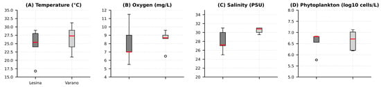
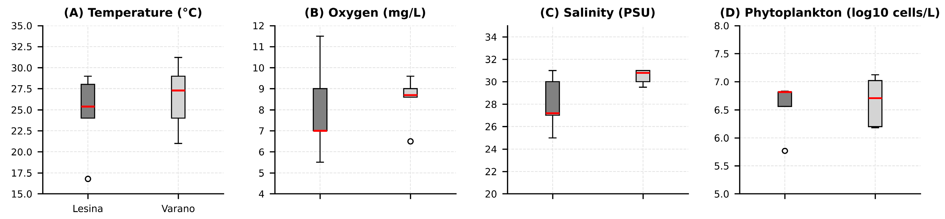
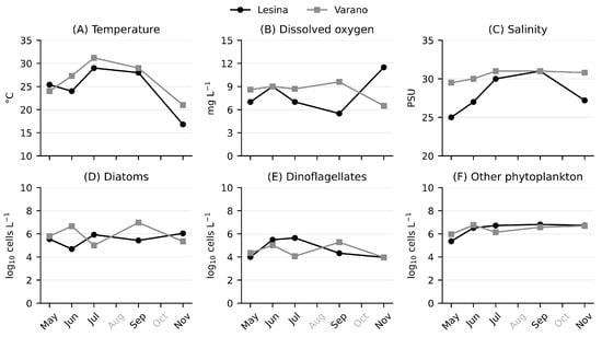
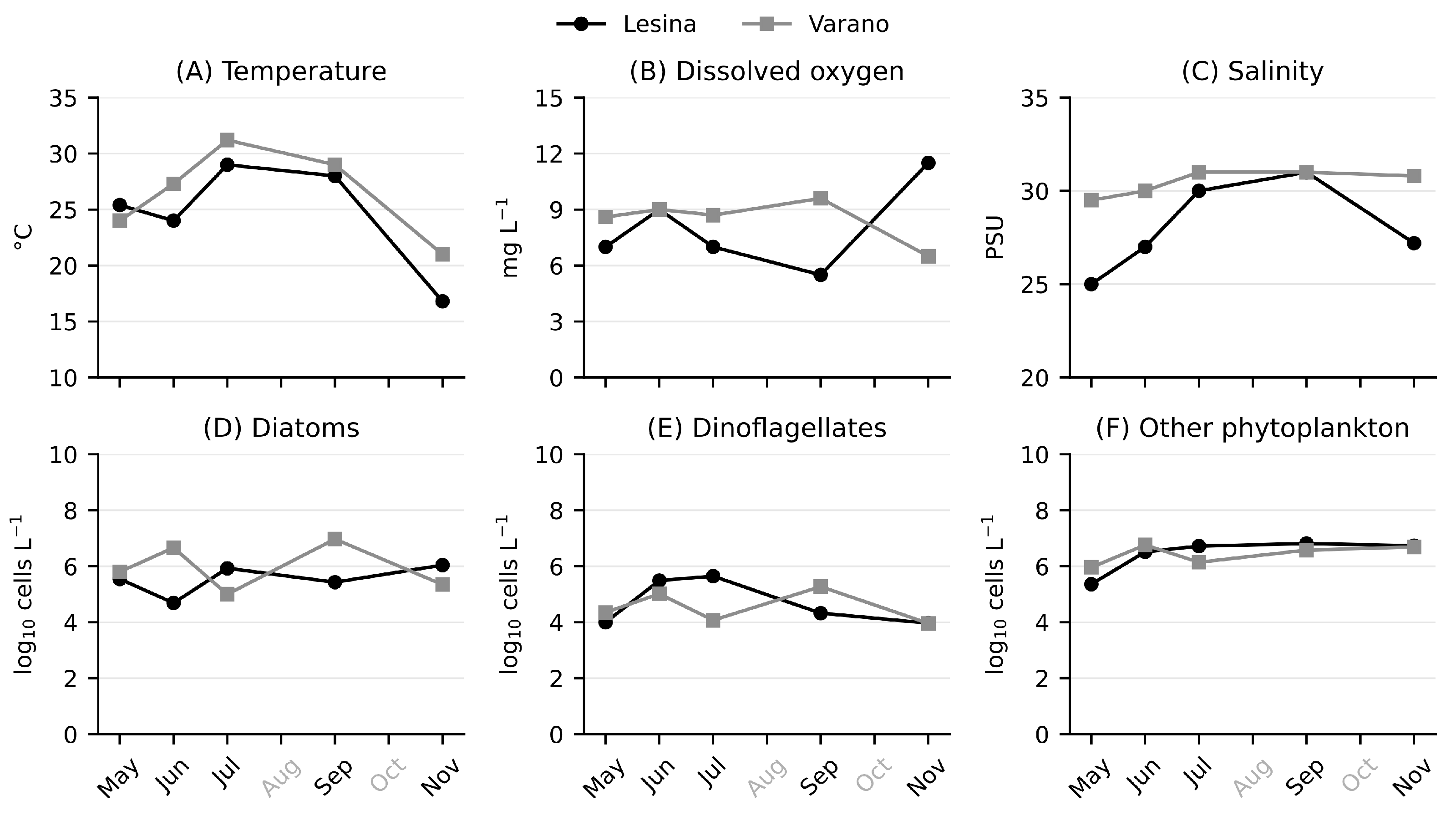
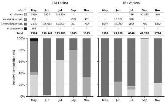
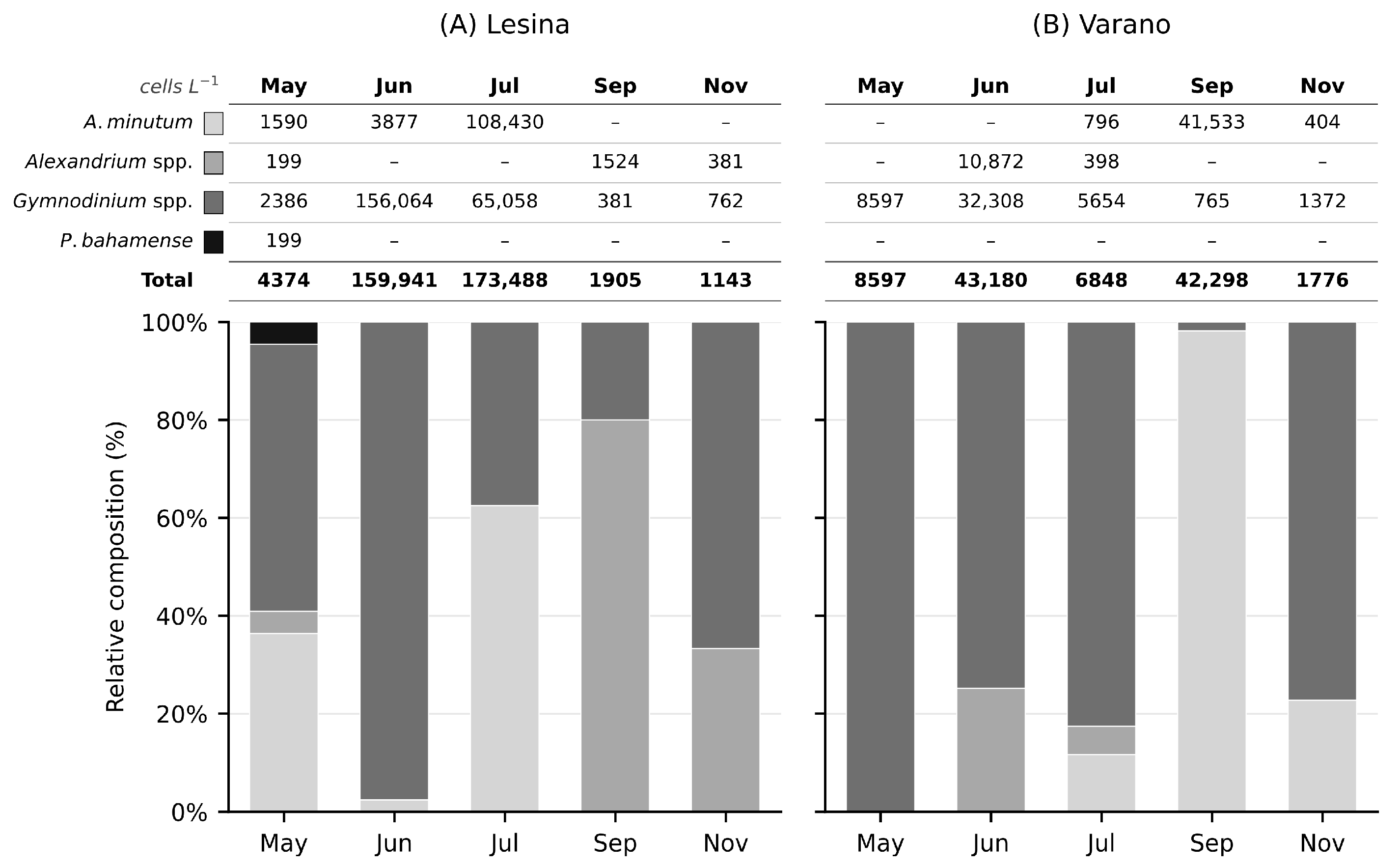
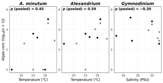
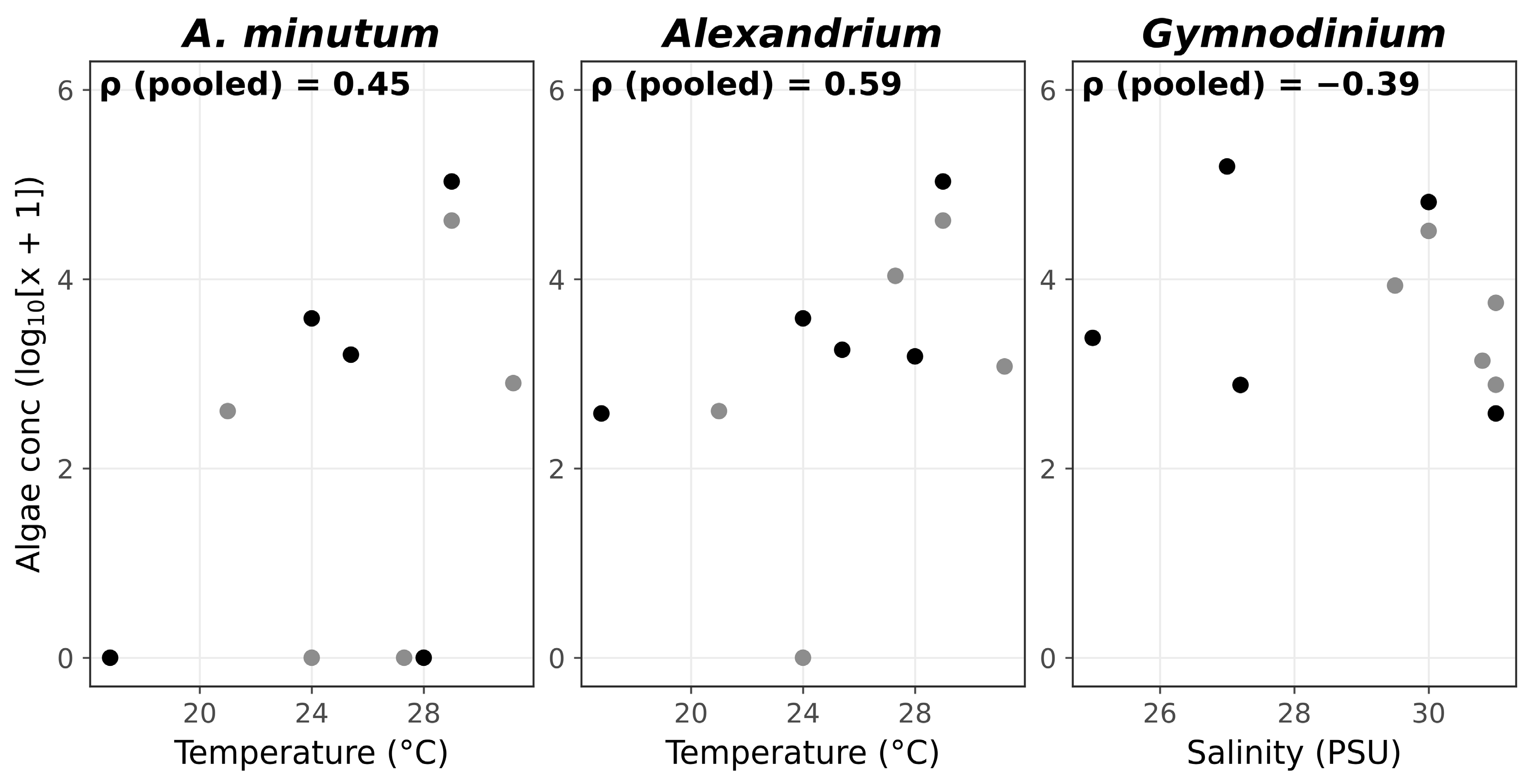
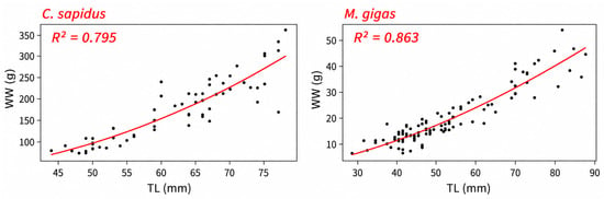
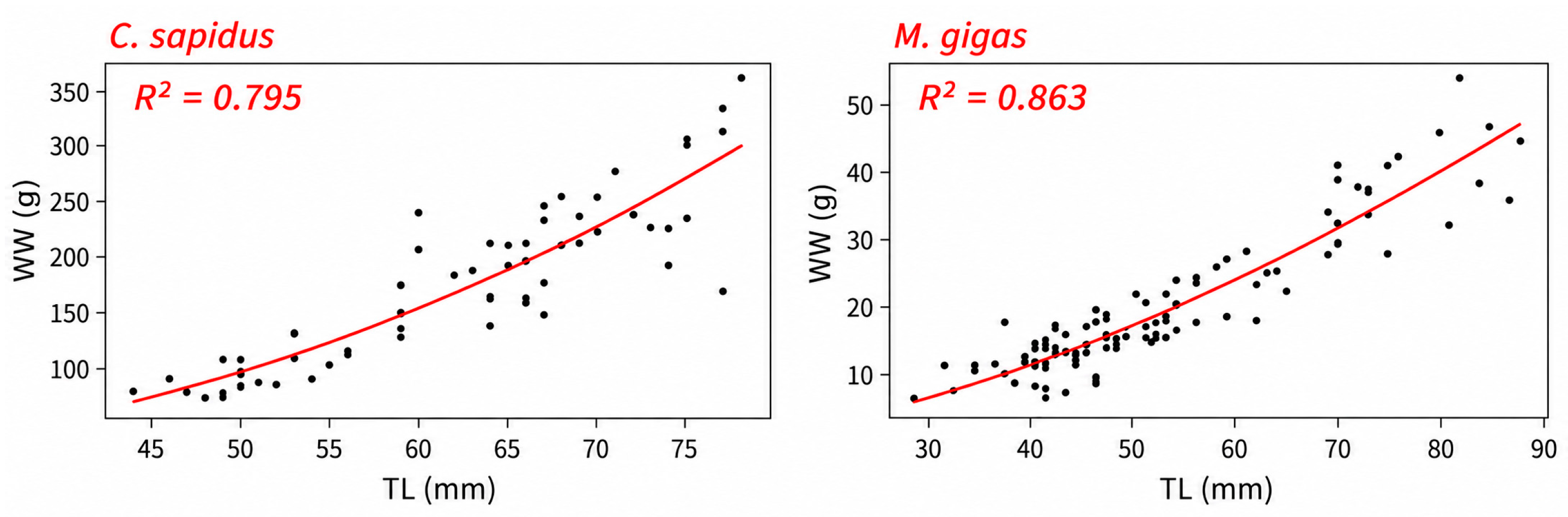
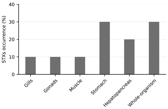
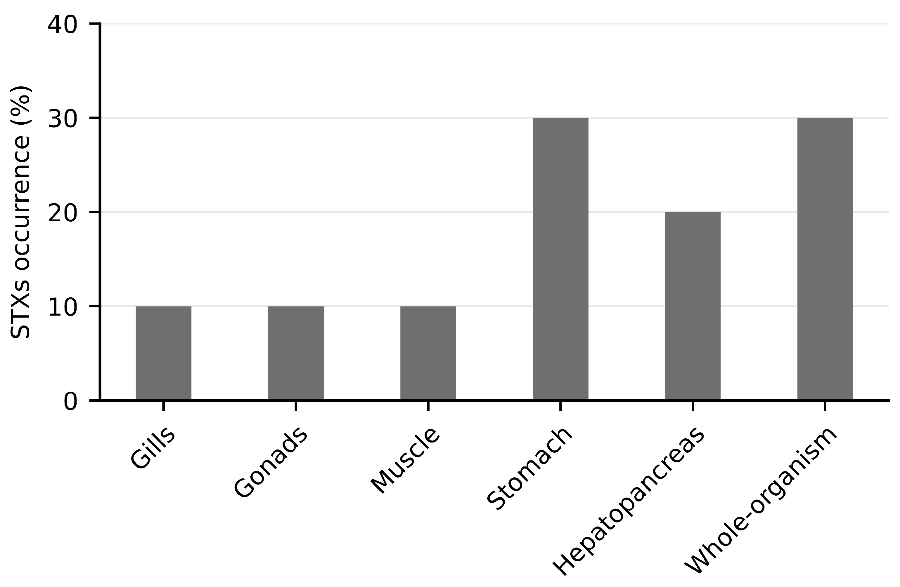
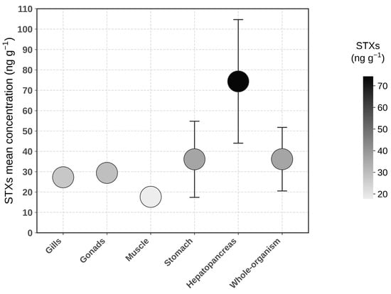
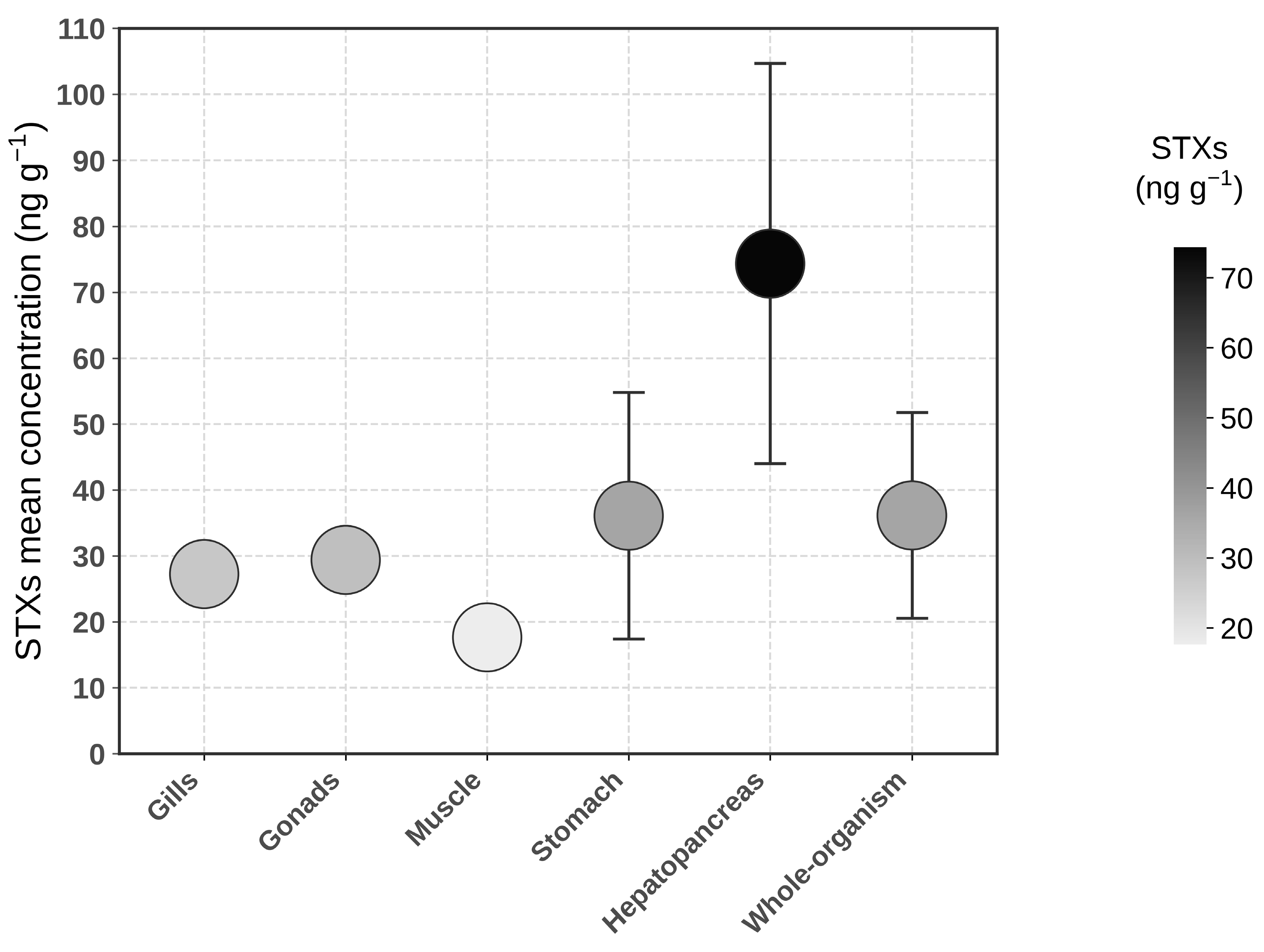
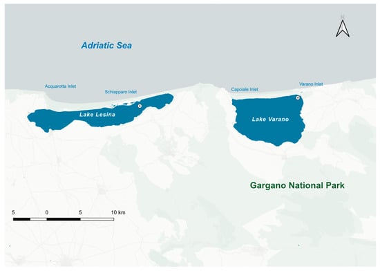
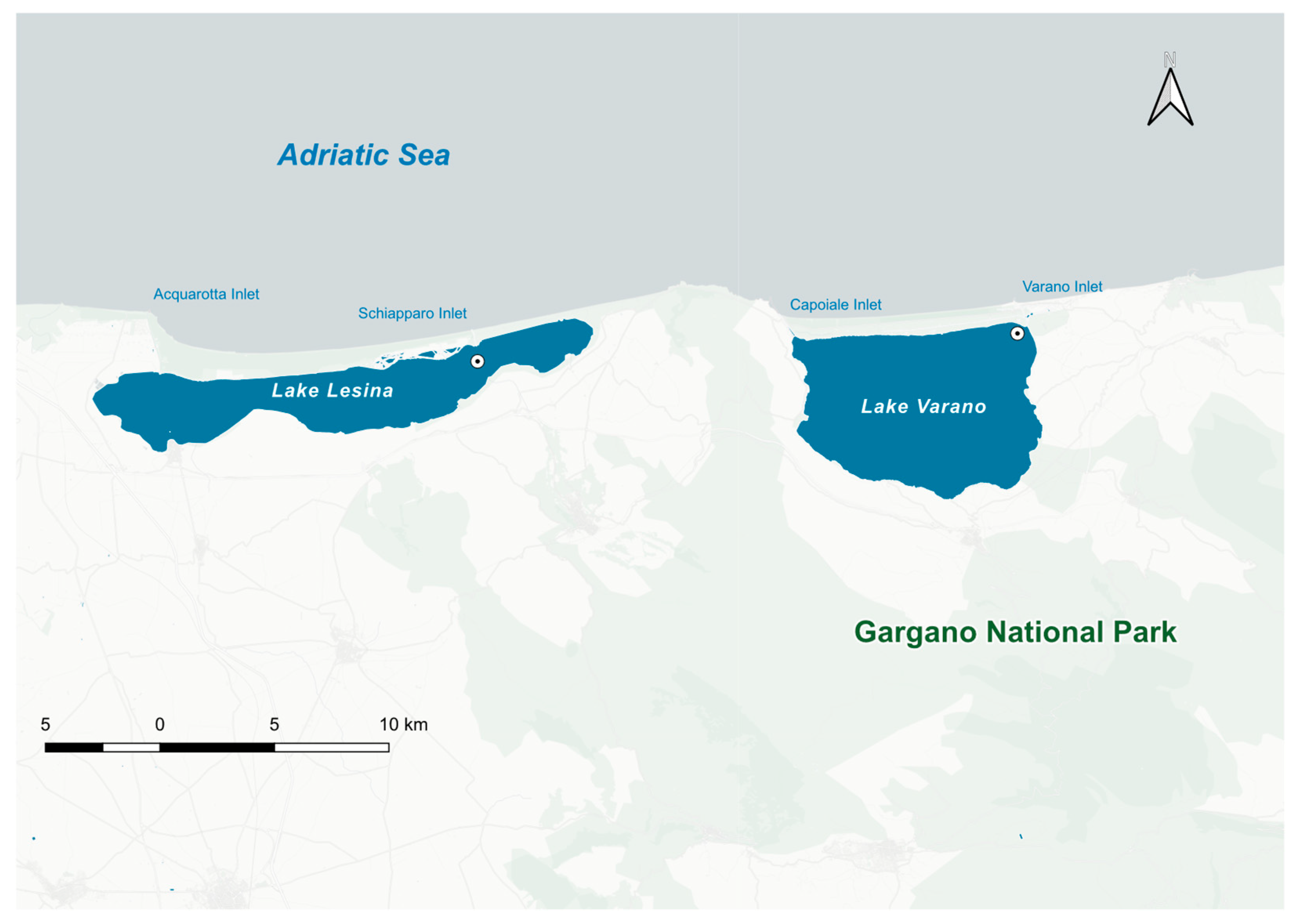

Disclaimer/Publisher’s Note: The statements, opinions and data contained in all publications are solely those of the individual author(s) and contributor(s) and not of MDPI and/or the editor(s). MDPI and/or the editor(s) disclaim responsibility for any injury to people or property resulting from any ideas, methods, instructions or products referred to in the content. |
© 2026 by the authors. Licensee MDPI, Basel, Switzerland. This article is an open access article distributed under the terms and conditions of the Creative Commons Attribution (CC BY) license.