


Abstract
Objectives: Patients with thalassemia major are at high risk of developing blood-borne infections, including toxoplasmosis, due to their dependence on frequent blood transfusions and underlying immune system disorders. This study was designed to investigate this hidden risk and provide data for policymaking in blood transfusion services in a region with a high endemicity. Methods: A total of 300 blood samples from thalassemia patients in northern Iran were collected. Serological testing was conducted to detect IgG and IgM antibodies. DNA extraction followed, with molecular screening performed via PCR. Finally, genotyping of T. gondii was carried out using nested PCR focused on the GRA6 gene. Results: The serological analysis revealed 59.7% of patients exhibited IgG against T. gondii, while only 0.6% tested positive for IgM. The results of the molecular screening revealed 2.7% of patients had DNA of T. gondii. The results of genetic analysis showed 75% had type II, 12.5% had type I, and 12.5% belonged to type III. Conclusions: This study provides serological and molecular evidence of a high chronic Toxoplasma gondii burden in thalassemia patients from northern Iran, an endemic region. A significant association between blood transfusion history and seropositivity, along with parasite DNA detection, suggests elevated exposure risk, though direct transfusion transmission remains unproven. Finding’s support integrating nested PCR with routine serology for diagnosing infection in this population.
1. Introduction
Thalassemia comprises a group of autosomal recessive disorders affecting globin chain production, resulting in chronic hemolytic anemia. Patients inherit these genetic abnormalities from asymptomatic carrier parents. Contemporary medical interventions have significantly enhanced long-term survival outcomes for those with the major form of the disease. Notably, the global distribution of thalassemia is uneven, with prevalence rates varying substantially between different countries and ethnic groups [1]. Due to impaired hemoglobin synthesis, individuals with thalassemia experience a constellation of symptoms, including debilitating fatigue, muscular asthenia, shortness of breath, cardiac irregularities, and marked pallor [2]. Management of severe anemia necessitates ongoing blood transfusion therapy, which must be accompanied by iron chelation to address the inevitable iron accumulation resulting from chronic transfusion support [3]. Blood transfusion provokes immunological responses in recipients that encompass both immune activation and immunosuppressive phenomena. Such transfusion-associated immune modulation, particularly evident in patients receiving repeated transfusions for anemia management, may precipitate cardiac, hepatic, and endocrinological complications when therapeutic intervention is delayed or inadequate [4]. Moreover, transfusion represents a recognized route for transmitting numerous infectious agents, including bacteria, viruses, and parasites. The extensive utilization of blood products in major surgical procedures, combined with the direct patient exposure to blood components, substantially increases the potential for transmission of these infectious agents [5].
Toxoplasmosis is a widespread zoonotic infection caused by the intracellular protozoan parasite T. gondii. It is estimated that up to one-quarter of the global human population has been infected with this parasite, though most people do not experience symptoms [6,7].
Although Toxoplasma gondii infection is typically self-limiting in immunocompetent individuals, it poses a significant clinical threat to immunocompromised populations [8,9]. Among these, recipients of solid organ and hematopoietic stem cell transplants are at particularly high risk for severe toxoplasmosis, which can manifest as life-threatening conditions such as meningoencephalitis and disseminated disease. This elevated susceptibility primarily results from either the reactivation of a latent infection or direct transmission from an infected donor [10,11,12]. In addition to donor-derived transmission via transplantation, transfusion-associated toxoplasmosis represents another critical concern for immunocompromised patients. This mode of transmission may occur when blood products or leukocytes obtained from asymptomatic immunocompetent donors are administered to vulnerable recipients.
In vitro investigations have shown that T. gondii tachyzoites can survive in whole blood containing the anticoagulant citrate, stored at 4 °C, for up to 50 days [13]. This survival capacity represents a significant risk of parasite transmission through blood products, especially leukocyte-rich components donated by asymptomatic infected individuals. Other evidence suggests that infected human blood, anticoagulated with citrate or heparin and stored for 28 days under similar conditions, still retains the ability to transmit infection to an animal model (rabbit) [14].
In the distant past, direct evidence of transfusion-transmitted toxoplasmosis (TTT) has been limited, with only four confirmed cases reported in the literature, all associated with granulocyte transfusions, no definitive transmission of red blood cells or plasma, and one possible case involving platelets [13,15].
However, recent evidence strongly suggests that blood transfusions are an important route of transmission of this parasite, particularly in immunocompromised populations and those requiring multiple transfusions [16,17].
Despite this clear risk, the focus of blood screening services worldwide has remained largely on viruses such as HIV and hepatitis viruses [18]. This approach has created a critical immunity gap against blood-borne pathogens such as T. gondii. The risk of transmission through blood transfusion is significantly increased, especially among frequent blood recipients, such as patients with thalassemia major, who often also have some degree of immunosuppression [19,20]. However, insufficient attention to this threat has led to a systematic underestimation of the true risk of blood-borne toxoplasmosis in these high-risk patients.
The substantial progress achieved in screening blood products for HIV and hepatitis viruses has contributed to a perception that transfusion-transmitted infections are largely confined to these viral pathogens. However, this perspective overlooks the potential for parasitic agents, including T. gondii, to be transmitted through transfusion and cause serious complications in immunocompromised hosts [18]. Notably, blood donors with detectable IgM antibodies may be experiencing recent or acute toxoplasma infection, rendering their blood components potentially hazardous for susceptible recipients [21].
The host response to T. gondii infection typically involves a robust innate pro-inflammatory cascade, recognized as one of the most intense immunological reactions to any infectious agent [22,23]. In the context of thalassemia, however, underlying immune dysfunction, including lymphocyte reduction and impaired phagocytosis and chemotaxis, compromises host defense mechanisms [24].
Thus, the presence of immune dysfunction may potentiate the clinical course of toxoplasmosis, elevating the likelihood of mortality among patients with advanced thalassemic pathology. Regarding the Iranian context, thalassemia gene frequency demonstrates notable inter-regional variation, with estimates ranging between 4% and 8% depending upon geographical location and underlying genetic architecture. Effective management of at-risk individuals necessitates accurate and timely diagnostic assessment. Such evaluation may be accomplished through various technical approaches, including serological analysis, biological assays, histopathological examination, and molecular investigation applied either as individual tests or in combination to achieve reliable detection of thalassemia [25,26]. For patients in this vulnerable population, prompt diagnosis of Toxoplasma infection is essential to guide clinical management and prevent disease progression. Serological evaluation using enzyme-linked immunosorbent assay (ELISA) has traditionally served as the cornerstone for the detection of anti-T. gondii antibodies [27,28,29]. More recently, molecular techniques, notably nested PCR, have been deployed to achieve superior analytical sensitivity by targeting multicopy genetic elements, thereby enhancing the capacity to detect parasitic DNA even in low-burden samples [30,31]. The considerable burden imposed by toxoplasmosis on thalassemia patients, together with the lack of epidemiological data from northern Iran regarding this comorbidity, provided the impetus for the current study. Accordingly, we sought to delineate the serological and molecular features of T. gondii infection in thalassemia patients and to assess factors predisposing this population to infection.
2. Materials and Methods
2.1. Ethics Approval and Consent to Participate
This study was conducted following approval from the Ethics Committee of Mazandaran University of Medical Sciences (Certificate number: IR.MAZUMS.REC.1401.14034). The research protocol conformed to the ethical standards articulated in the Declaration of Helsinki (2013 revision). All participants, or their parents/guardians in the case of minors, provided written informed consent before inclusion. To ensure anonymity and protect participant identity, all personal identifiers were removed from the dataset.
2.2. Study Design and Population
This cross-sectional investigation was conducted at Mazandaran University of Medical Sciences, northern Iran, between January 2021 and January 2023. The study population comprised 300 patients with thalassemia (230 with thalassemia major and 70 with thalassemia minor) recruited from hospitals across multiple cities in Mazandaran Province, including Sari, Babol, Chalous, Nur, Tonekabon, and Ramsar. Prior to enrollment, the objectives and methodology of the study were thoroughly explained to all participants, after which written informed consent was obtained. A standardized questionnaire was administered to each participant to collect demographic data, including age, occupation, educational attainment, and place of residence (urban or rural).
2.3. Blood Sampling and Processing
Under aseptic conditions, two blood samples were collected from the antecubital vein of each participant: 5 mL of whole blood without anticoagulant for serological analysis, and an additional 5 mL collected into CBC tubes containing 2% EDTA for molecular analysis. All specimens were transported to the Medical Research Laboratory of the Department of Parasitology, Mazandaran University of Medical Sciences, under strict cold chain maintenance. Samples exhibiting lipemia or hemolysis were excluded from the analysis, and appropriate replacement samples were obtained. Following centrifugation for serum separation, all specimens were stored at −20 °C until subsequent analysis.
2.4. Identification of Anti-Toxoplasma Specific Antibodies (IgM and IgG)
To determine the serum titers of IgG and IgM antibodies against T. gondii, commercial Pishtaz Teb® ELISA kits (Pishtaz Teb Diagnostics, Tehran, Iran; https://pishtazteb.com/en/ (accessed on 20 May 2026) were utilized according to the manufacturer’s instructions. These kits are ISO 13485, Pishtaz Teb Diagnostics, Tehran, Iran, 2023 certified and have been extensively validated in previous epidemiological studies in Iran. The optical density of both control and test samples was measured at 450 nm using a DANA® ELISA reader (DANA, Tehran, Iran), which was calibrated according to the manufacturer’s specifications [32].
2.5. DNA Extraction
The buffy coat obtained from each of the 300 blood samples was subjected to DNA extraction using the DNG PLUS Kit (DNA Blood Mini Extraction Kit, Sinaclone company, Tehran, Iran), with all procedures performed as specified by the manufacturer [33]. Post-extraction, the purified DNA was kept at −20 °C until required for downstream applications. To ensure reliable detection, a frozen aliquot of T. gondii RH strain DNA was prepared and used as the positive control in each experimental run.
2.6. Conventional PCR Assay
To detect T. gondii DNA, a conventional PCR assay targeting the RE gene was performed. This assay amplifies a 529 bp region corresponding to a repetitive element present in 200–300 copies per parasite genome. Each 25-μL reaction consisted of 12.5 μL of 2x Master Mix, 5 μL of DNA template, 5.5 μL of PCR-grade water, and 1 μL each of forward and reverse primers (10 pmol/μL). The primers used were: forward, 5′-CGCTGCAGGGAGGAAGACGAAAGTTG-3′; reverse, 5′-CGCTGCAGACACAGTGCATCTGGATT-3′. The amplification program comprised an initial denaturation at 94 °C for 5 min, followed by 35 cycles of 94 °C for 30 s, 55 °C for 30 s, and 72 °C for 30 s, with a final extension at 72 °C for 5 min. Following amplification, PCR products were subjected to electrophoresis on 1.5% agarose gels, and the presence of 529 bp fragments was considered indicative of T. gondii DNA.
2.7. Genotyping of T. gondii via Nested-PCR Assay
To determine the genotype, we examined the positive samples obtained from conventional PCR using a gene marker. Nested PCR consists of two successive amplification rounds using outer and inner primers. In the initial step, the first round of nested PCR (PCR 1) is carried out with a set of outer primers. The reaction mixture for the nested PCR was prepared in a total volume of 25 μL, containing 12.5 μL of Ampliqon PCR Master Mix, 5 μL of template DNA, 2 μL of PCR-grade water, and 1 μL of each outer primer (10 pmol/μL). PCR amplification was performed under the following conditions: initial denaturation at 95 °C for 5 min; 30 cycles of 94 °C for 20 s, 53 °C for 20 s, and 72 °C for 20 s.
For the nested PCR assay, a second amplification (PCR 2) was performed employing inner primers specific to the T. gondii GRA6 gene. The 25-μL reaction mixture consisted of 12.5 μL of Ampliqon PCR Master Mix, 1 μL of each inner primer (10 pmol/μL), 2 μL of the primary PCR product, and 7.5 μL of PCR-grade water. Cycling conditions for the second round were as follows: initial denaturation at 95 °C for 5 min; 40 cycles of 94 °C for 20 s, 53 °C for 20 s, and 72 °C for 20 s; and a final extension at 72 °C for 10 min. PCR products were subsequently analyzed by electrophoresis on 1.5% agarose gels, and the expected 344 bp fragments were observed in positive samples. The inner primer sequences targeting the GRA6 locus were based on those reported by Hosseini and colleagues (2019) [34].
2.8. Nucleic Acid Sequencing
To determine the sequence of the isolated Toxoplasma types, a number of PCR2 products (approximately 20 µL) that had a different cleavage pattern and were free of additional bands and had strong desired bands were sent to Pishgam Company (Tehran, Iran) along with internal round-trip primers for sequencing and sequenced using the Sanger sequencing method.
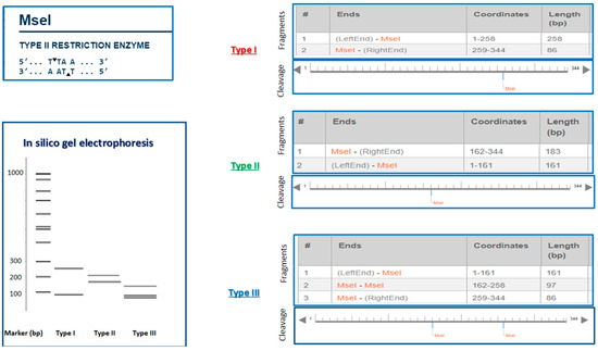
To determine genotypes based on cleavage patterns, the sequences obtained from the samples in the present study were subjected to in silico restriction digestion using the endonuclease MseI via the online tool NEB cutter (https://nc3.neb.com/NEBcutter/) (accessed on 20 May 2026). Genotypes of Toxoplasma parasites were subsequently assigned according to the resulting restriction sites and fragment patterns generated by the enzymatic digestion.
2.9. Data Analysis Using Statistics
The statistical evaluation of the study data was carried out employing SPSS software (version 21). The Chi-square test was utilized to analyze associations between qualitative variables, and the independent samples t-test was applied for the comparison of quantitative variables. In all analyses, a p-value of less than 0.05 was regarded as statistically significant.
3. Results
3.1. Demographic Characteristics and Serological Assay
A total of 300 serum samples were obtained from individuals with thalassemia residing in Mazandaran province. Of the samples, 179 (66.59%) were positive for IgG and 2 (0.66%) for IgM antibodies against T. gondii. The samples were divided into the east and west logic of the province, with 150 samples from the west region (Ramsar, Tonekabon, and Chalus) and 150 samples from the eastern (Babol, Qaemshahr, Sari, Behshahr) of Mazandaran province. The serologic findings indicated a 63% prevalence of anti-T. gondii IgG antibodies in the eastern region, compared to 56% in the western region. Statistical analysis revealed no significant association between the prevalence of T. gondii in thalassemia patients and the type of region (p = 0.19).
As shown in Table 1, analysis of gender distribution among thalassemia patients showed that 122 (40.66%) of the total patients were male and 178 (59.33%) were female. The prevalence of IgG antibodies against Toxoplasma was 50% in males and 66.29% in females. Statistical analysis indicated that there was no significant association between Toxoplasma prevalence and gender among thalassemia patients (p = 0.27).
Table 1.
Population attributes of thalassemia patients based on the T. gondii seropositivity.
Analysis of blood group distribution demonstrated that IgG anti-T. gondii antibody prevalence was greatest in blood group O (65.87%), exceeding rates observed in other groups. The frequency of IgG anti-T. gondii antibodies were higher in Rh-positive individuals, with 136 positive cases out of 213 samples (63.85%), compared to Rh-negative individuals, who had 43 positive cases out of 87 samples (49.42%). Statistical analysis revealed no significant association between the seroprevalence of T. gondii in individuals with thalassemia and their blood group or Rh factor (p = 0.69).
Within this research, the participants consisted of 117 individuals under 30 years old, 102 individuals between 31 and 39 years old, and 81 individuals aged over 40. The results showed that the prevalence of IgG antibodies against T. gondii was higher in individuals over 40 years old (72.83%) compared to the other age groups. Data analysis demonstrated a significant association between T. gondii seroprevalence and age among individuals with thalassemia (p = 0.001).
Furthermore, the prevalence of anti-T. gondii IgG antibodies were significantly higher (63.04%) in persons with major thalassemia or a history of blood transfusion compared to those with minor thalassemia or no transfusion history (48.57%), with the difference reaching statistical significance (p = 0.03).
Serological findings from the study further revealed a statistically significant association between educational level and T. gondii seroprevalence among patients with thalassemia. The prevalence of anti-T. gondii IgG antibodies were notably higher in individuals with less than a high school diploma (76.47%) compared to those with education beyond the high school level (51.01%). The relationship between T. gondii seroprevalence and educational attainment in this patient population was statistically significant (p = 0.00), as confirmed by the analytical results.
Based on the results of Table 1, the division of patients according to place of residence shows that out of the total sample of people with thalassemia, 127 people lived in rural areas and 173 people lived in urban areas. Serological analysis revealed that the prevalence of IgG antibodies against T. gondii was significantly higher in individuals residing in villages (93 out of 127, 73.22%) compared to those living in cities (86 out of 173, 49.71%) (p = 0.00). The demographic characteristics of thalassemia patients based on T. gondii seropositivity are presented in Table 1.
3.2. Molecular Identification
Conventional PCR molecular testing was conducted on all serum samples from individuals with thalassemia, irrespective of whether anti-T. gondii antibodies were present, by amplifying a 529 bp fragment of the RE gene. Out of 300 whole blood samples collected from individuals with thalassemia, 8 samples (2.7%) tested positive for toxoplasmosis, including 2 M+/G+ and 6 M-/G+ samples, and showed a band of 529 base pairs in a 1.5% agarose gel.
3.3. Sequencing Analysis
The datasets generated and analyzed during the current study are available in the NCBI repository, [ACCESSION NUMBER: PV796110, PV796111, PV796112, PV796113, PV796114, PV796115, PV796116, and PV796117].
To ascertain the genotype, we examined the positive samples obtained from conventional PCR using the GRA6 gene marker. The results of genotyping of Toxoplasma in samples of individuals with thalassemia using the Nested PCR technique and the GRA6 gene showed that six out of eight positive samples (75%) had type II [ACCESSION NUMBER: PV796110, PV796111, PV796114, PV796115, PV796116 and PV796117], one sample (12.5%) had type I [ACCESSION NUMBER: PV796112], and one sample (12.5%) belonged to type III [ACCESSION NUMBER: PV796113] (Figure 1).
Figure 1.
PCR-RFLP patterns of Toxoplasma gondii genotypes isolated from thalassemia patients in northern Iran. For genotyping of positive Toxoplasma gondii samples, the GRA6 gene was utilized while the MseI enzyme was applied for enzyme digestion. For T. gondii type I, this enzyme yields one cut, producing fragments of 86 and 258 bps, respectively. Type II, on the other hand, yields fragments of 161 and 183 bps. Meanwhile, type III consists of two cuts, producing fragments of 86, 97, and 161 bps).
4. Discussion
Thalassemia is one of the most common human genetic diseases, with the highest prevalence in Southeast Asia, Africa, and Mediterranean countries. Thalassemia genes are widely distributed globally, particularly in regions within the thalassemia belt, such as Iran [35]. Blood transfusion is recognized as a valuable medical treatment; however, it is important to be aware of the potential side effects associated with it. Blood transfusions can lead to various adverse effects, including allergic reactions, chills, fever, urticaria, and the potential transmission of infectious diseases. Severe opportunistic infections such as toxoplasmosis can be associated with immunodeficiency and chronic immune activation resulting from repeated blood transfusions, iron overload, and splenectomy [26,36]. Toxoplasmosis is recognized as an infection that can be transmitted via blood transfusion, particularly from asymptomatic seropositive donors who are in the acute phase of infection. This infection adds extra strain on thalassemia patients, as T. gondii is an opportunistic parasite that impacts multiple body systems. Thalassemia patients face a greater risk of acquiring toxoplasma infection compared to healthy individuals, primarily because of their frequent blood transfusions and compromised immune systems.
Although toxoplasmosis is rarely symptomatic in healthy hosts, it poses a high risk to patients with immunodeficiency [37]. The level of seropositivity varies in different regions of the world and has been reported to be between 30 and 60% in most countries [38]. Considering the high prevalence of human toxoplasmosis infection in northern Iran and its significant impact on immunocompromised individuals, this research was designed to evaluate the serological and molecular detection of T. gondii in patients with thalassemia.
The serological findings of the present study revealed that anti-T. gondii IgG antibodies were present in 59.7% of thalassemia patients, reflecting a high burden of chronic infection within this population. IgM antibodies, indicative of recent or active infection, were detected in only two participants (0.67%). When comparing subgroups based on transfusion exposure, the prevalence of Toxoplasma infection was notably higher among transfused patients (63.04%) than among those without a history of transfusion (48.57%).
The seroprevalence estimate for toxoplasmosis among thalassemia patients in northern Iran, as obtained in the present study, is notably higher than values reported in earlier investigations conducted both within Iran and internationally. To contextualize these findings, El-Tantawy and colleagues (2019) examined T. gondii infection in Egyptian children with thalassemia and reported IgM and IgG seropositivity rates of 23.2% and 53.6%, respectively. Furthermore, avidity testing in that study revealed that 65.5% of IgG-positive patients exhibited low-avidity antibodies, indicating recent infection acquisition [39].
The findings align with Karakaş et al. (2012), who explored potential associations between toxoplasmosis and blood transfusion practices in severe thalassemia patients in Turkey. The researchers found that the studied group exhibited a higher seropositivity rate for anti-T. gondii antibodies compared to the control group (19.4% versus 14%), although this difference did not reach statistical significance [38].
Supporting evidence comes from the work of Saki et al. (2019), who evaluated T. gondii seroprevalence in healthy blood donors from southwest Iran. Among 380 participants, anti-T. gondii IgG antibodies were detected in 34.47% (131 individuals). Moreover, 0.5% (two donors) tested positive exclusively for IgM antibodies, and 2.9% (11 donors) exhibited seropositivity for both immunoglobulin classes, suggesting recent or acute infection in a subset of the donor population [40].
In contrast, Moghimi et al. (2015) reported that routine screening for T. gondii was not warranted at the Yazd transfusion center in Iran, as the prevalence of Toxoplasma IgG antibodies in thalassemia patients was found to be 16%, a relatively low rate compared to infections such as Cytomegalovirus [27].
As noted earlier, the elevated levels of anti-T. gondii IgG antibodies in patients with thalassemia major may result from a greater risk of Toxoplasma infection associated with frequent blood transfusions. In Karbala, Iraq, women with thalassemia exhibited the highest Toxoplasma infection rate at 18.3%, compared to 15.6% among men. Individuals receiving blood transfusions twice or more monthly showed a higher prevalence of Toxoplasma infection than those transfused once per month [41].
In general, one of the ways to transmit toxoplasmosis is through blood bags. This parasite can survive for up to 50 days at 4 degrees Celsius in a refrigerator and can be transmitted [13]. Patients with thalassemia major require regular blood transfusions, which increases the risk of exposure to blood contaminated with Toxoplasma. Although routine screening for Toxoplasma is not standard practice in many blood centers worldwide, some national regulations do recognize the risk of transfusion-transmitted toxoplasmosis. A resolution by the Cabinet of Ministers of the Azerbaijan Republic, for example, classifies toxoplasmosis alongside HIV and viral hepatitis as a ‘haemo transmissible disease’ that permanently excludes individuals from donation, highlighting its official recognition as a transfusion risk in certain contexts) https://cis-legislation.com/document.fwx?rgn=14989#A000000002 (accessed on 12 May 2026)). On the other hand, thalassemia patients often have a weakened immune system. This condition makes people vulnerable to opportunistic infections such as toxoplasmosis. In addition, in communities with a high prevalence of toxoplasmosis, the possibility of contamination of donated blood and, as a result, its transmission to thalassemia major patients is higher. Our analysis revealed that blood transfusion recipients exhibited higher toxoplasmosis seroprevalence than non-recipients. Despite the fact that this difference was statistically significant, it should be considered that, given the prevalence of Toxoplasma in blood transfusion samples, there is a possibility of infection during blood transfusion, and it is recommended that toxoplasmosis testing be performed in addition to screening tests before blood transfusion.
The prevalence and pathogenesis of T. gondii infection in humans are influenced by a complex interplay of environmental, host, and parasite-related factors. Host determinants include age, immune status, dietary habits, educational level, place of residence, and other demographic variables. Among these, age may indirectly affect disease prevalence by modulating individual and social behaviors. Consequently, the seroprevalence of toxoplasmosis varies across different age groups. Accumulating evidence indicates that advancing age is directly associated with an increased likelihood of T. gondii infection. Although the higher seroprevalence observed in older populations remains incompletely understood, it may be attributable to prolonged cumulative exposure to risk factors and potential transmission routes over time [42]. The frequency of T. gondii infection rises with age, reaching 90% by the fourth decade of life [43]. Consistent with expectations, this study revealed a significant association between age and toxoplasmosis seroprevalence. Specifically, individuals aged ≤30 years exhibited the lowest seropositivity rates, while those ≥40 years demonstrated the highest prevalence of infection. The findings of the current study align with those reported by Siyadatpanah et al. (2013), who observed a statistically significant difference associated with increasing age [44].
Inadequate awareness of toxoplasmosis as a source of infection, along with poor hygiene practices and unfamiliarity with transmission methods, are critical risk factors for contracting the disease. Increasing knowledge through higher education and its consequences on life and behavioral patterns may lead to a decrease in infection and have an indirect effect on environmental and cultural factors involved in toxoplasmosis infection. Similarly to the data published in review studies conducted in Iran by Mizani, the outcomes of the current study show that education is inversely related to positivity of toxoplasmosis infection [45]. Therefore, individuals with lower levels of education are more vulnerable to this infection because they often are not aware of transmission methods and proper personal hygiene practices. In the present study, the prevalence of T. gondii between people with education below and above a diploma was statistically significant (p < 0.05).
The serological findings of this study revealed that the frequency of IgG antibodies against Toxoplasma was significantly higher in individuals residing in rural areas than in those living in urban settings (p < 0.05). It appears that people in rural areas are more exposed to T. gondii infection due to limited health facilities and increased contact with animals. Environmental factors such as humidity and temperature suitable for the survival of oocysts, as well as the presence of various animal reservoirs, moreover, have a major impact on the incidence and progression of toxoplasmosis [46].
Although serological methods are widely employed for the diagnosis of Toxoplasma infection and are generally considered cost-effective, they may pose significant risks for certain high-risk patient populations [47,48]. For decades, serology has remained the most commonly used laboratory approach for diagnosing toxoplasmosis. However, the increasing prevalence of immunocompromised conditions has drawn attention to the diverse clinical manifestations of toxoplasmosis, which can lead to severe and irreversible complications. A primary limitation of serological diagnosis is the potential absence of detectable antibodies during the early stages of infection, as well as the prolonged persistence of IgM antibodies in the circulation. Given the high seroprevalence of toxoplasmosis among immunocompromised individuals, there is an urgent need for alternatives to highly sensitive serological methods. In this context, nested PCR has been shown to provide favorable sensitivity and specificity for identifying Toxoplasma gondii infection, even in immunocompetent individuals [30]. The reliability of this molecular diagnostic tool is contingent upon several technical considerations, among which are the selection of DNA target characteristics, the volume of clinical specimen analyzed, the efficacy of the extraction process, and the specifics of the amplification protocol [38]. When compared to conventional diagnostic modalities like serological testing, nested PCR demonstrates appreciably higher sensitivity and specificity, positioning it as a powerful technique for recognizing acute toxoplasmosis across human and animal hosts. The complementary nature of nested PCR relative to serology enables the acquisition of richer diagnostic information, particularly in cases where antibody responses are equivocal or absent. Evidence suggests that nested PCR attains sensitivity and specificity levels of up to 94%, conferring superior detection capabilities relative to standard PCR for analyzing laboratory samples. In addition, this method offers considerable economic advantages, being significantly more cost-effective than many alternative molecular techniques [49]. Nested PCR analysis was undertaken to determine the molecular frequency of T. gondii infection among thalassemia patients. Evaluation of 300 whole blood samples demonstrated that eight individuals harbored detectable parasitic DNA, representing a prevalence rate of 2.66%. It is noteworthy that the identification of T. gondii nucleic acid in clinical specimens signifies the presence of the organism, with potential implications for acute infection, chronic persistence, or reactivation of latent disease [50].
Evidence from Hanifehpour [26] provides additional context for interpreting our findings. In that study, which employed both molecular and serological diagnostics for toxoplasmosis in thalassemia patients, the molecular detection rate was 9.78%, showing no meaningful difference from the control population. However, seropositivity for anti-Toxoplasma IgG antibodies was considerably higher, affecting 51.9% of thalassemia patients.
The inability to detect Toxoplasma DNA in many seropositive individuals has been observed across different population groups [51]. This phenomenon has been previously attributed to the low levels of DNA existing in blood samples, particularly during chronic T. gondii infections. Parasites are rarely found in the blood during asymptomatic stages, and the lack of detectable DNA does not necessarily mean that an infection is absent [52].
The study by El-Latif et al. (2023) discovered that Toxoplasma DNA was detected in six persons who had negative serological test results. This discrepancy can be attributed to either the timing of blood sample collection, which might have occurred before antibodies had a chance to develop, or the individuals’ immune systems being incapable of producing enough immunoglobulins for detection by ELISA due to immunodeficiency [53]. Given the high sensitivity of PCR in immunocompromised persons, it is strongly recommended for diagnosing Toxoplasma infections when serological tests yield negative results [54]. Combining both methods would provide a more accurate diagnosis.
In the case of Toxoplasma, both the severity of infection and genetic diversity play crucial roles in determining the pathogenicity of the disease. To understand the epidemiology of Toxoplasma, genetic classification is essential. T. gondii is primarily categorized into three main types with varying levels of pathogenicity. The most common types of this parasite belong to three clonal lineages. Type I strains are extremely virulent and deadly in experimental mouse models, whereas type II strains are less virulent and usually form cysts. Type III strains display a combination of traits from both type I and type II strains.
Despite many studies in Iran examining the genotyping of T. gondii across different hosts, most have focused on only a few genetic markers. As a result, there remains a lack of comprehensive data on the genotypes of T. gondii in the country, especially in human samples.
In the present study, the results of Toxoplasma genotyping in samples from individuals with thalassemia revealed that out of the eight positive samples, six belonged to type II (75%), one sample belonged to type I (25%), and one sample belonged to type III (25%) of the Toxoplasma parasite. In 2013, Asgari et al. prepared 542 samples from paraffin blocks of aborted fetuses in Fars Province. They then used the SAG2 gene to determine the genotypes of the positive samples. The results showed that 14% of the samples tested positive, with 83% of these identified as type II and 17% as type I [55]. Behzadi et al. (2003) used the PCR-RFLP method based on the SAG2 gene to identify T. gondii in 21 isolates from human and mouse brains. The results showed that 85.5% of the isolates were classified as type II, while 14.3% were classified as type I [56]. The systematic review by Hosseini et al. (2018) revealed that genotype II was the most dominant strain of T. gondii in human toxoplasmosis cases from 1995 to August 2017. This genotype was particularly prevalent in human samples from Europe and America, including countries such as the USA, France, and Portugal. Based on the literature, type II strains of T. gondii are known for their low virulence and strong capacity to form cysts, which explains their predominance in these areas [57].
Type II strains of T. gondii are characterized by low virulence and high cyst formation. According to this review, Europe has the highest prevalence of the type II genotype, accounting for 69.4% of cases. Globally, type II is the most common genotype found in clinical samples. Notably, type II is less virulent than type I, with a lethal dose in mice of LD > 103, whereas type I has a lethal dose of just one organism (LD100 = 1) [57].
5. Conclusions
This study provides a comprehensive serological and molecular overview of T. gondii infection among thalassemia patients in northern Iran, a region with high toxoplasmosis prevalence, revealing a substantial burden of chronic infection in this vulnerable group. A key finding is the statistically significant association between blood transfusion history and higher T. gondii seropositivity, which, alongside the detection of parasitic DNA through nested PCR, strongly suggests that these immunocompromised patients face an elevated risk of exposure to the parasite. While our data cannot definitively prove that blood transfusions are the direct source of infection, the alignment of our findings with the existing literature on parasite survival in stored blood and transmission from asymptomatic donors lends weight to this possibility, particularly as genotyping revealed the predominance of the less virulent, cyst-forming type II strain, consistent with global trends in human toxoplasmosis. Therefore, the implications of this study underscore the critical need for enhanced diagnostic accuracy in this high-risk population, supporting the implementation of nested PCR as a routine complement to serology for thalassemia patients with suspected active infection. Our findings provide a strong foundation for future research, including parallel screening of blood donors in high-prevalence regions to quantify transfusion-transmission risk and genotype analysis to establish epidemiological links. In conclusion, this work maps the current landscape of toxoplasmosis in Iranian thalassemia patients with a level of molecular detail that distinguishes it from prior studies, highlighting the urgent need for more vigilant diagnostic approaches for these patients.
Author Contributions
Conduct experiments and acquire data, A.M., B.B. and M.S.; Writing—original draft, M.S., D.A. and S.A.S.; Writing—review and editing, S.S. and S.G. (Shirzad Gholami); Project administration, S.A.H. and S.G. (Sara Gholami); Conceptualization and Investigation, M.N.S. and H.K.; Formal analysis, S.A.A.; Study concept and Methodology, S.A.H. and A.D.; Study supervision, S.A.H. and A.D. All authors have read and agreed to the published version of the manuscript.
Funding
This research received no external funding.
Institutional Review Board Statement
This study was supported by the Vice-Chancellor for Research and Technology, Mazandaran University of Medical Sciences, Sari, Iran (IR.MAZUMS.REC.1401.14034) and the RA Higher Education and Science Committee (Research Project No. 23RL-1F014).
Informed Consent Statement
Not applicable.
Data Availability Statement
All data is provided in the manuscript and in additional files. All data are available on request.
Acknowledgments
This article is part of a project approved by the Vice Chancellor for Research of Mazandaran University of Medical Sciences, Sari, Iran, and this study was conducted in collaboration with the staff and professors of Mazandaran Toxoplasmosis Research Center (TRC). The authors thank Mazandaran University of Medical Sciences for their cooperation, as well as the assistance and cooperation of individuals with thalassemia and the control group, and the Higher Education and Science Committee of RA (Research project No. 23RL-1F014).
Conflicts of Interest
The authors declare no conflict of interest.
References
- Brancaleoni, V.; Di Pierro, E.; Motta, I.; Cappellini, M. Laboratory diagnosis of thalassemia. Int. J. Lab. Hematol. 2016, 38, 32–40. [Google Scholar] [CrossRef] [Scilit] [PubMed]
- Munkongdee, T.; Chen, P.; Winichagoon, P.; Fucharoen, S.; Paiboonsukwong, K. Update in laboratory diagnosis of thalassemia. Front. Mol. Biosci. 2020, 7, 74. [Google Scholar] [CrossRef] [Scilit] [PubMed]
- Safabakhsh, H.; Tehranian, F.; Tehranian, B.; Hatami, H.; Karimi, G.; Shahabi, M. Prevalence of anti-CMV antibodies in blood donors in Mashhad, Iran. Iran. J. Epidemiol. 2013, 9, 52–57. [Google Scholar]
- Viprakasit, V.; Ekwattanakit, S. Clinical classification, screening and diagnosis for thalassemia. Hematol. Oncol. Clin. 2018, 32, 193–211. [Google Scholar] [CrossRef] [Scilit] [PubMed]
- Eivazi-Ziaei, J.; Movassaghpour, A.; Asgharzadeh, M.; Dastgiri, S. Seroprevalence of cytomegalovirus in blood donors in the northwest of Iran. J. Res. Clin. Med. 2013, 1, 96–100. [Google Scholar]
- Smith, J.R.; Ashander, L.M.; Arruda, S.L.; Cordeiro, C.A.; Lie, S.; Rochet, E.; Belfort, R.; Furtado, J.M. Pathogenesis of ocular toxoplasmosis. Prog. Retin. Eye Res. 2021, 81, 100882. [Google Scholar] [CrossRef] [Scilit] [PubMed]
- Nazari, N.; Shojaee, S.; Mohebali, M.; Teimouri, A.; Ghadiri, K.; Raeghi, S.; Shiee, M.R.; Azarakhsh, Y.; Bozorgomid, A. Toxoplasma gondii and Neospora caninum in brain tissue of rodents in North-West Iran. Vet. Med. Res. Rep. 2019, 10, 223–227. [Google Scholar] [CrossRef] [Scilit] [PubMed]
- Sadeghi, M.; Hosseini, S.A.; Sarvi, S.; Nayeri, T.; Sharif, M.; Pagheh, A.S.; Amouei, A.; Montazeri, M.; Daryani, A. More than Seventy Years of Research (1948–November 2021) on Toxoplasma gondii in Iran: A Narrative Review. Iran. J. Parasitol. 2022, 17, 124. [Google Scholar] [CrossRef] [Scilit] [PubMed]
- Tork, M.; Sadeghi, M.; Sarvi, S.; Khazaei, F.; Basirpour, B.; Ahmadi, S.; Shariatzadeh, S.A.; Hosseini, S.A.; Zamaniyan, M.; Daryani, A.; et al. Vitamin D3 status and seroprevalence of Toxoplasma gondii in reproductive-aged women in Northern Iran. BMC Womens Health 2025, 25, 544. [Google Scholar] [CrossRef] [Scilit] [PubMed]
- de la Mata Navazo, S.; Slöcker Barrio, M.; García-Morín, M.; Beléndez, C.; Escobar Fernández, L.; Rincón-López, E.M.; Aguilera Alonso, D.; Guinea, J.; Marín, M.; Butragueño-Laiseca, L.; et al. Case report: Severe ARDS in a pediatric hematopoietic stem-cell transplantation recipient caused by disseminated toxoplasmosis. Front. Pediatr. 2022, 9, 810718. [Google Scholar] [CrossRef] [Scilit] [PubMed]
- Adekunle, R.O.; Sherman, A.; Spicer, J.O.; Messina, J.A.; Steinbrink, J.M.; Sexton, M.E.; Lyon, G.M.; Mehta, A.K.; Phadke, V.K.; Woodworth, M.H. Clinical characteristics and outcomes of toxoplasmosis among transplant recipients at two US academic medical centers. Transpl. Infect. Dis. 2021, 23, e13636. [Google Scholar] [CrossRef] [Scilit] [PubMed]
- Mansouri, A.; Mojarad, M.R.A.; Badfar, G.; Abasian, L.; Rahmati, S.; Kooti, W.; YektaKooshali, M.H.; Soleymani, A.; Azami, M. Epidemiology of Toxoplasma gondii among blood donors in Iran: A systematic review and meta-analysis. Transfus. Apher. Sci. 2017, 56, 404–409. [Google Scholar] [CrossRef] [Scilit] [PubMed]
- Siegel, S.E.; Lunde, M.N.; Gelderman, A.H.; Halterman, R.H.; Brown, J.A.; Levine, A.S.; Graw, R.G., Jr. Transmission of toxoplasmosis by leukocyte transfusion. Blood 1971, 37, 388–394. [Google Scholar] [CrossRef] [Scilit]
- Räisänen, S. Toxoplasmosis transmitted by blood transfusions. Transfusion 1978, 18, 329–332. [Google Scholar] [CrossRef] [Scilit] [PubMed]
- Roth, J.A.; Siegel, S.E.; Levine, A.S.; Berard, C.W. Fatal recurrent toxoplasmosis in a patient initially infected via a leukocyte transfusion. Am. J. Clin. Pathol. 1971, 56, 601–605. [Google Scholar] [CrossRef] [Scilit] [PubMed]
- Kantzanou, M.; Kostares, E.; Kostare, G.; Papagiannopoulou, E.; Kostares, M.; Tsakris, A. Prevalence of Toxoplasma gondii Infection Among Blood Donors: A Systematic Review and Meta-Analysis. Cureus 2025, 17, 531. [Google Scholar] [CrossRef] [Scilit] [PubMed]
- Hosseini, S.A.; Golchin, E.; Sharif, M.; Sarvi, S.; Ahmadpour, E.; Rostamian, A.; Gholami, S.; Amouei, A.; Daryani, A. A serological investigation and genotyping of Toxoplasma gondii among Iranian blood donors indicates threat to health of blood recipients. Transfus. Apher. Sci. 2020, 59, 102723. [Google Scholar] [CrossRef] [Scilit] [PubMed]
- Amoo, A.; Njaanake, K.; Dada-Adegbola, O.; Omosa-Manyonyi, G. Toxoplasmosis among blood donors: Unsafe blood transfusion in Ibadan, Southwest Nigeria. J. Appl. Hematol. 2019, 10, 120–125. [Google Scholar] [CrossRef] [Scilit] [PubMed]
- Wesołowski, R.; Pawłowska, M.; Smoguła, M.; Szewczyk-Golec, K. Advances and challenges in diagnostics of toxoplasmosis in HIV-infected patients. Pathogens 2023, 12, 110. [Google Scholar] [CrossRef] [Scilit] [PubMed]
- Stopić, M.; Štajner, T.; Marković-Denić, L.; Nikolić, V.; Djilas, I.; Srzentić, S.J.; Djurković-Djaković, O.; Bobić, B. Epidemiology of Toxoplasmosis in SERBIA: A Cross-Sectional Study on Blood Donors. Microorganisms 2022, 10, 492. [Google Scholar] [CrossRef] [Scilit] [PubMed]
- Asfaram, S.; Rezaei, R.; Fakhar, M.; Ghezelbash, B.; Nakhaei, M.; Hezarjaribi, H.Z.; Mardani, A.; Teshnizi, S.H. High occurrence of Toxoplasma gondii infection among blood donors in Ardabil Province as main focus of zoonotic visceral leishmaniosis, northwestern Iran. Ann. Parasitol. 2021, 67, 611–617. [Google Scholar] [CrossRef] [Scilit] [PubMed]
- Rahmati-Balaghaleh, M.; Hosseini Farash, B.R.; Zarean, M.; Hatami-Pourdehno, S.; Mirahmadi, H.; Jarahi, L.; Sharifi, Y.; Ahmadi, O.; Shamsian, S.A.; Moghaddas, E.; et al. Diagnosis of acute toxoplasmosis by IgG avidity method in pregnant women referred to health centers in south-eastern Iran. J. Parasit. Dis. 2019, 43, 517–521. [Google Scholar] [CrossRef] [Scilit] [PubMed]
- Miller, C.M.; Boulter, N.R.; Ikin, R.J.; Smith, N.C. The immunobiology of the innate response to Toxoplasma gondii. Int. J. Parasitol. 2009, 39, 23–29. [Google Scholar] [CrossRef] [Scilit] [PubMed]
- Asadov, C.D. Immunologic abnormalities in β-thalassemia. J. Blood Disord. Transfus. 2014, 5, 1–5. [Google Scholar]
- Sotiriadou, I.; Karanis, P. Evaluation of loop-mediated isothermal amplification for detection of Toxoplasma gondii in water samples and comparative findings by polymerase chain reaction and immunofluorescence test (IFT). Diagn. Microbiol. Infect. Dis. 2008, 62, 357–365. [Google Scholar] [CrossRef] [Scilit] [PubMed]
- Hanifehpour, H.; Shariat, S.K.S.; Ghafari, M.S.; Kheirandish, F.; Saber, V.; Fallahi, S. Serological and molecular diagnosis of Toxoplasma gondii infections in thalassemia patients. Iran. J. Parasitol. 2019, 14, 20. [Google Scholar] [CrossRef] [Scilit]
- Moghimi, M.; Doosti, M.; Vahedian-Ardakani, H.A.; Talebi, A.; Akhavan-Ghalibaf, M.; Najafi, A.; Aminorroaya, M.M.; Yazdani, S.; Shayestehpour, M.; Bahrami, H.; et al. Serological study on cytomegalovirus and Toxoplasma gondii in thalassemia major patients of Yazd, Iran. Iran. J. Ped Hematol. Oncol. 2015, 5, 149. [Google Scholar] [CrossRef] [Scilit]
- Raeghi, S.; Sedeghi, S.; Sedeghi, S. Prevalence of Toxoplasma gondii antibodies in cats in Urmia, northwest of Iran. J. Anim. Plant Sci. 2011, 21, 132–134. [Google Scholar]
- Sarkari, B.; Shafiei, R.; Zare, M.; Sohrabpour, S.; Kasraian, L. Seroprevalence and molecular diagnosis of Toxoplasma gondii infection among blood donors in southern Iran. J. Infect. Dev. Ctries. 2014, 8, 543–547. [Google Scholar] [CrossRef] [Scilit] [PubMed]
- Mousavi, M.; Saravani, R.; Modrek, M.J.; Shahrakipour, M.; Sekandarpour, S. Detection of Toxoplasma gondii in diabetic patients using the nested PCR assay via RE and B1 genes. Jundishapur J. Microbiol. 2016, 9, e29493. [Google Scholar] [CrossRef] [Scilit] [PubMed]
- Mirahmadi, H.; Hasanzadeh, R.; Raeesi, H.M.; Fallahi, S.; Shahraki, M.K.; Badirzadeh, A. Loop-mediated isothermal amplification (Lamp) assay to detect toxoplasmosis in schizophrenia patients. Iran. J. Parasitol. 2020, 15, 299. [Google Scholar] [PubMed]
- ISO 13485: 2016; Medical Devices—Qualitymanagement Systems—Requirementsforregulatory Purposes. ISO: Geneva, Switzerland, 2024.
- Masoori, L.; Kheirandish, F.; Haghighi, A.; Mohebali, M.; Akhoundi, B.; Taghipour, N.; Gachkar, L.; Chegeni-Sharafi, A.; Moin-Vaziri, V. Molecular-based detection of Leishmania infantum in human blood samples in a new focus of visceral Leishmaniasis in Lorestan Province, Iran. J. Arthropod Borne Dis. 2018, 12, 67. [Google Scholar] [PubMed]
- Hosseini, S.A.; Sharif, M.; Sarvi, S.; Abediankenari, S.; Hashemi-Soteh, M.B.; Amouei, A.; Montazeri, M.; Aghayan, S.A.; Gholami, S.; Shaker, D.; et al. Genetic characterization of Toxoplasma gondii in Iranian HIV positive patients using multilocus nested-PCR-RFLP method. Parasitology 2020, 147, 322–328. [Google Scholar] [PubMed]
- Shah, F.T.; Sayani, F.; Trompeter, S.; Drasar, E.; Piga, A. Challenges of blood transfusions in β-thalassemia. Blood Rev. 2019, 37, 100588. [Google Scholar] [CrossRef] [Scilit] [PubMed]
- Haghighi, J.D.; Hosseini, A.; Shafiei, R.; Mehravaran, A.; Alijani, E.; Mirahmadi, H. Evaluation of Toxoplasma gondii antibodies in addicted and non-addicted women in Zahedan, Southeast ofIran. Int. J. High. Risk Behav. Addict. 2020, 9, e106458. [Google Scholar] [CrossRef] [Scilit]
- Rashno, M.M.; Fallahi, S.; Kheirandish, F.; Bagheri, S.; Kayedi, M.; Birjandi, M. Seroprevalence of Toxoplasma gondii infection in patients with Alzheimer’s disease. Arch. Clin. Infect. Dis. 2016, 11, e37205. [Google Scholar] [CrossRef] [Scilit]
- Karakas, S.; Özlem, S.; Tellioglu, A.M.; Ertabaklar, H.; Ertug, S. Investigation of anti-Toxoplasma gondii IgG and IgM antibodies in beta thalaseamia major patients in Aydin province. Turk. Parazitol. Derg. 2012, 36, 133. [Google Scholar] [CrossRef] [Scilit] [PubMed]
- El-Tantawy, N.; Darwish, A.; Eissa, E. Seroprevalence of Toxoplasma gondii infection among β-thalassemia major pediatric population: Implications for transfusion transmissible toxoplasmosis. Pediatr. Infect. Dis. J. 2019, 38, 236–240. [Google Scholar] [CrossRef] [Scilit] [PubMed]
- Saki, J.; Foroutan, M.; Khodkar, I.; Khodadadi, A.; Nazari, L. Seroprevalence and molecular detection of Toxoplasma gondii in healthy blood donors in southwest Iran. Transfus. Apher. Sci. 2019, 58, 79–82. [Google Scholar] [CrossRef] [Scilit] [PubMed]
- Saleh, A.Y.; Al-Numan, A.Y.S. Investigation of the incidence of Toxoplasmosis and cytomegalovirus in patients with thalassemia. Tik J. Pure Sci. 2019, 23, 14–18. [Google Scholar] [CrossRef] [Scilit]
- Daryani, A.; Sarvi, S.; Aarabi, M.; Mizani, A.; Ahmadpour, E.; Shokri, A.; Rahimi, M.T.; Sharif, M. Seroprevalence of Toxoplasma gondii in the Iranian general population: A systematic review and meta-analysis. Acta Trop. 2014, 137, 185–194. [Google Scholar] [CrossRef] [Scilit] [PubMed]
- Robert-Gangneux, F.; Dardé, M.-L. Epidemiology of and diagnostic strategies for toxoplasmosis. Clin. Microbiol. Rev. 2012, 25, 264–296. [Google Scholar] [CrossRef] [Scilit] [PubMed]
- Siyadatpanah, A.; Tabatabaie, F.; Oormazdi, H.; Meamar, A.R.; Razmjou, E.; Hadighi, R.; Akhlaghi, L. Comparison of anti-Toxoplasma IgG and IgM antibodies determined by ELISA method in diabetic and non-diabetic individuals in west Mazandaran province, Iran, 2011–2012. Ann. Biol. Res. 2013, 4, 281–285. [Google Scholar]
- Mizani, A.; Alipour, A.; Sharif, M.; Sarvi, S.; Amouei, A.; Shokri, A.; Rahimi, M.T.; Hosseini, S.A.; Daryani, A. Toxoplasmosis seroprevalence in Iranian women and risk factors of the disease: A systematic review and meta-analysis. Trop. Med. Health 2017, 45, 7. [Google Scholar] [CrossRef] [Scilit] [PubMed]
- Yan, C.; Liang, L.-J.; Zheng, K.-Y.; Zhu, X.-Q. Impact of environmental factors on the emergence, transmission and distribution of Toxoplasma gondii. Parasit. Vectors 2016, 9, 137. [Google Scholar] [CrossRef] [Scilit] [PubMed]
- Khan, A.; Grigg, M.E. Toxoplasma gondii: Laboratory maintenance and growth. Curr. Protoc. Microbiol. 2017, 44, 20C.1.1–20C.1.17. [Google Scholar] [CrossRef] [Scilit] [PubMed]
- Arab-Mazar, Z.; Fallahi, S.; Koochaki, A.; Mirahmadi, H.; Tabaei, S.J.S. Cloning, expression and immunoreactivity of recombinant Toxoplasma gondii GRA5 protein. Iran. J. Microbiol. 2016, 8, 331. [Google Scholar] [CrossRef] [Scilit] [PubMed]
- Jafari Modrek, M.; Hasanzadeh, R.; Foroutan, M.; Mirahmadi, H.; Rahmati-Balaghaleh, M.; Zarean, M. Seroprevalence and molecular evaluation of Toxoplasma gondii in schizophrenic patients hospitalized in Sistan and Baluchestan province, Southeast of Iran. Trop. Biomed. 2018, 36, 422–429. [Google Scholar]
- Liu, Q.; Wang, Z.-D.; Huang, S.-Y.; Zhu, X.-Q. Diagnosis of toxoplasmosis and typing of Toxoplasma gondii. Parasit. Vectors 2015, 8, 292. [Google Scholar] [CrossRef] [Scilit] [PubMed]
- Nakashima, F.; Pardo, V.S.; Miola, M.P.; Murata, F.H.A.; Paduan, N.; Longo, S.M.; Brandão de Mattos, C.C.; Pereira-Chioccola, V.L.; Ricci, O., Jr.; de Mattos, L.C. Serum IgG anti-Toxoplasma gondii antibody concentrations do not correlate nested PCR results in blood donors. Front. Cell Infect. Microbiol. 2020, 9, 461. [Google Scholar] [CrossRef] [Scilit] [PubMed]
- Wang, L.; He, L.Y.; Meng, D.D.; Chen, Z.W.; Wen, H.; Fang, G.S.; Luo, Q.L.; Huang, K.Q.; Shen, J.L. Seroprevalence and genetic characterization of Toxoplasma gondii in cancer patients in Anhui Province, Eastern China. Parasit. Vectors 2015, 8, 162. [Google Scholar] [CrossRef] [Scilit] [PubMed]
- Abd El-Latif, N.F.; Salem, A.I.; Sadek, N.A.; Salah, S.A.; Shalaby, D.G.; Elhadad, H. Serological and molecular detection of Toxoplasma gondii in ß. thalassemia patients. J. Parasit. Dis. 2023, 47, 778–786. [Google Scholar] [CrossRef] [Scilit] [PubMed]
- Martino, R.; Bretagne, S.; Einsele, H.; Maertens, J.; Ullmann, A.J.; Parody, R.; Schumacher, U.; Pautas, C.; Theunissen, K.; Schindel, C. Early detection of Toxoplasma infection by molecular monitoring of Toxoplasma gondii in peripheral blood samples after allogeneic stem cell transplantation. Clin. Infect. Dis. 2005, 40, 67–78. [Google Scholar] [CrossRef] [Scilit] [PubMed]
- Asgari, Q.; Fekri, M.; Monabati, A.; Kalantary, M.; Mohammadpour, I.; Motazedian, M.H.; Sarkari, B. Molecular genotyping of Toxoplasma gondii in human spontaneous aborted fetuses in Shiraz, Southern Iran. Iran. J. Public. Health 2013, 42, 620. [Google Scholar]
- Behzadi, R.; Razavi, M.; Hovanessian, A.; Asmar, M.; Rouhvand, F. Genotyping of Toxoplasma gondii strains isolated from patients and mice by PCR-RFLP assay. Iran. J. Biotechnol. 2003, 1, 82–86. [Google Scholar]
- Hosseini, S.A.; Amouei, A.; Sharif, M.; Sarvi, S.; Galal, L.; Javidnia, J.; Pagheh, A.S.; Gholami, S.; Mizani, A.; Daryani, A. Human toxoplasmosis: A systematic review for genetic diversity of Toxoplasma gondii in clinical samples. Epidemiol. Infect. 2018, 147, e36. [Google Scholar] [CrossRef] [Scilit] [PubMed]

Disclaimer/Publisher’s Note: The statements, opinions and data contained in all publications are solely those of the individual author(s) and contributor(s) and not of MDPI and/or the editor(s). MDPI and/or the editor(s) disclaim responsibility for any injury to people or property resulting from any ideas, methods, instructions or products referred to in the content. |
© 2026 by the authors. Licensee MDPI, Basel, Switzerland. This article is an open access article distributed under the terms and conditions of the Creative Commons Attribution (CC BY) license.