


Abstract
Background: Vestibular migraine (VM) and benign paroxysmal positional vertigo (BPPV) frequently present with overlapping positional symptoms, making their differential diagnosis challenging. This study aimed to characterize positional vestibular manifestations in patients with suspected VM. Methods: In this observational study, 85 adult patients presenting with positional vertigo or dizziness in whom typical BPPV could not be demonstrated were evaluated. All patients underwent detailed clinical and neurotological assessment, videonystagmography, and standardized positional testing. Results: Definite VM was diagnosed in 51/85 patients (60.0%) and probable VM in 23/85 (27.1%), accounting together for 87.1% of the selected cohort. Positional nystagmus was documented in 52 patients (61.2%), of whom 38/52 (73.1%) showed at least one atypical characteristic. Pupillary Nystagmus (PNy) was detected in 61/85 patients (71.8%). Among 14 patients with an initially BPPV-like positional pattern, five responded to canalith repositioning maneuvers, whereas nine had persistent or recurrent symptoms requiring diagnostic reassessment. At 3 months, 66/74 patients (89.2%) with definite or probable VM reported marked overall clinical improvement, while complete resolution of positional vertigo or dizziness occurred in 49/74 (66.2%). Positional nystagmus resolved in 36/47 patients (76.6%) who had presented with this finding at baseline. Conclusions: VM should be considered in patients with recurrent positional vestibular symptoms when positional findings are atypical, variable, or inconsistent with a single semicircular canal disorder, particularly when repositioning maneuvers are ineffective. PNy may provide an additional supportive finding, although its diagnostic value requires validation in controlled studies. Treatment response may support the overall clinical interpretation but should not be regarded as an independent diagnostic criterion.
1. Introduction
Benign paroxysmal positional vertigo (BPPV) and vestibular migraine (VM) are among the most common causes of recurrent vertigo and frequently present with overlapping positional symptoms [1,2,3,4]. Although BPPV is characterized by brief episodes of positional vertigo associated with canal-specific nystagmus and generally responds to canalith repositioning maneuvers, VM may also manifest with positionally triggered vertigo or dizziness, sometimes accompanied by nystagmus resembling that observed in peripheral otolithic disorders [2,3,5,6]. According to the Bárány Society and International Headache Society diagnostic criteria, VM is defined by recurrent vestibular episodes associated with a current or previous history of migraine and characteristic migrainous features [7,8]. However, the positional nature of vestibular symptoms may complicate the differential diagnosis, particularly when the clinical findings do not conform to a typical BPPV pattern.
Positional nystagmus associated with VM may exhibit characteristics that differ from those expected in classical BPPV, including absent or minimal latency, prolonged persistence, poor fatigability, variability in direction, and lack of congruence with the anatomical plane of a single semicircular canal [3,9]. Although these features may suggest a non-BPPV positional mechanism, they are not specific to VM and must be interpreted within the overall clinical context. Moreover, migraine and BPPV are not mutually exclusive conditions. Previous investigations have demonstrated an association between the two disorders and suggested that concomitant peripheral and migraine-related vestibular mechanisms may contribute to positional symptoms in individual patients [2,5,6].
This diagnostic overlap is particularly relevant in patients presenting with recurrent positional vertigo despite canalith repositioning maneuvers or with positional dizziness in the absence of reproducible canal-specific nystagmus. Previous studies have reported that some patients initially considered to have refractory BPPV may subsequently fulfill the diagnostic criteria for VM, whereas others may have coexisting migraine-related and peripheral positional vestibular manifestations [2,3,4]. Consequently, accurate diagnostic interpretation requires integration of clinical history, migraine-associated symptoms, repeated positional testing, and detailed neurotological and video-oculographic assessment rather than reliance on positional symptoms alone.
Despite increasing recognition of the relationship between VM and BPPV, the clinical characterization of patients presenting with diagnostically ambiguous positional vestibular manifestations remains challenging. In particular, the interpretation of positional nystagmus patterns, the possible contribution of pupillary nystagmus, and the evolution of symptoms following diagnostic reassessment and treatment warrant further clinical investigation.
The aim of the present observational study was to characterize positional vestibular manifestations in patients with suspected VM in whom typical BPPV could not be demonstrated at the initial neurotological evaluation. Particular attention was given to positional nystagmus characteristics, pupillary nystagmus, final diagnostic classification, and clinical outcomes following migraine preventive treatment.
2. Materials and Methods
A retrospective, observational, single-center study was conducted at the Vestibular Unit, Department of Otorhinolaryngology, Azienda Ospedaliero-Universitaria Pisana, Pisa, Italy, between November 2023 and November 2025.
During the study period, 220 consecutive adult patients referred to the Vestibular Unit for vertigo and/or dizziness were screened for eligibility.
Patients included in the final analysis underwent a comprehensive neurotological evaluation during the symptomatic phase or within 48 h of symptom onset or recurrence. This early assessment was performed to maximize the likelihood of documenting vestibular signs and ocular motor abnormalities before their spontaneous attenuation or resolution.
Following the application of predefined inclusion and exclusion criteria, 85 patients (58 females and 27 males; mean age 46.3 ± 13.1 years, range 18–75 years) were included in the final analysis. No a priori sample size calculation was performed, as the study was observational and descriptive in nature and was not designed to test a predefined statistical hypothesis. The sample size was determined by the number of eligible patients identified during the predefined study period (November 2023 to November 2025), following application of the inclusion and exclusion criteria.
The study population consisted of patients presenting with positional vestibular symptoms in whom the differential diagnosis between vestibular migraine (VM) and benign paroxysmal positional vertigo (BPPV) remained uncertain after the initial neurotological evaluation. The rationale of the study was to investigate this diagnostic overlap, where conventional positional testing alone is often insufficient to establish the correct diagnosis. The included patients presented with different clinical phenotypes. Some complained of positional vertigo associated with positional nystagmus elicited during diagnostic positioning maneuvers, whereas others reported positional vertigo or dizziness without observable positional nystagmus or objective evidence of canalolithiasis or cupulolithiasis. In these patients, symptoms were consistently triggered or exacerbated by changes in head position despite the absence of findings compatible with a typical semicircular canal disorder. Patients with unequivocal BPPV were excluded. Typical BPPV was defined by the presence of positional vertigo associated with canal-specific positional nystagmus demonstrating the expected direction, latency, duration and temporal characteristics according to the involved semicircular canal. Consequently, patients with typical posterior-, lateral- or anterior-canal BPPV were not included in the final study cohort. Additional exclusion criteria included definite Ménière’s disease, acute unilateral vestibular hypofunction, other peripheral vestibular disorders explaining the clinical presentation, clinical or ocular motor signs suggestive of central nervous system involvement, recent head trauma, concomitant cochleovestibular disorders, inability to complete the diagnostic protocol, and evaluation performed more than 48 h after symptom onset or recurrence.
Brain magnetic resonance imaging (MRI) was performed when clinically indicated, particularly in patients presenting with neurological or ocular motor findings raising suspicion of an alternative central vestibular disorder, to exclude structural central nervous system abnormalities.
Neuroimaging findings were considered together with the clinical and neurotological examination in the differential diagnostic assessment.
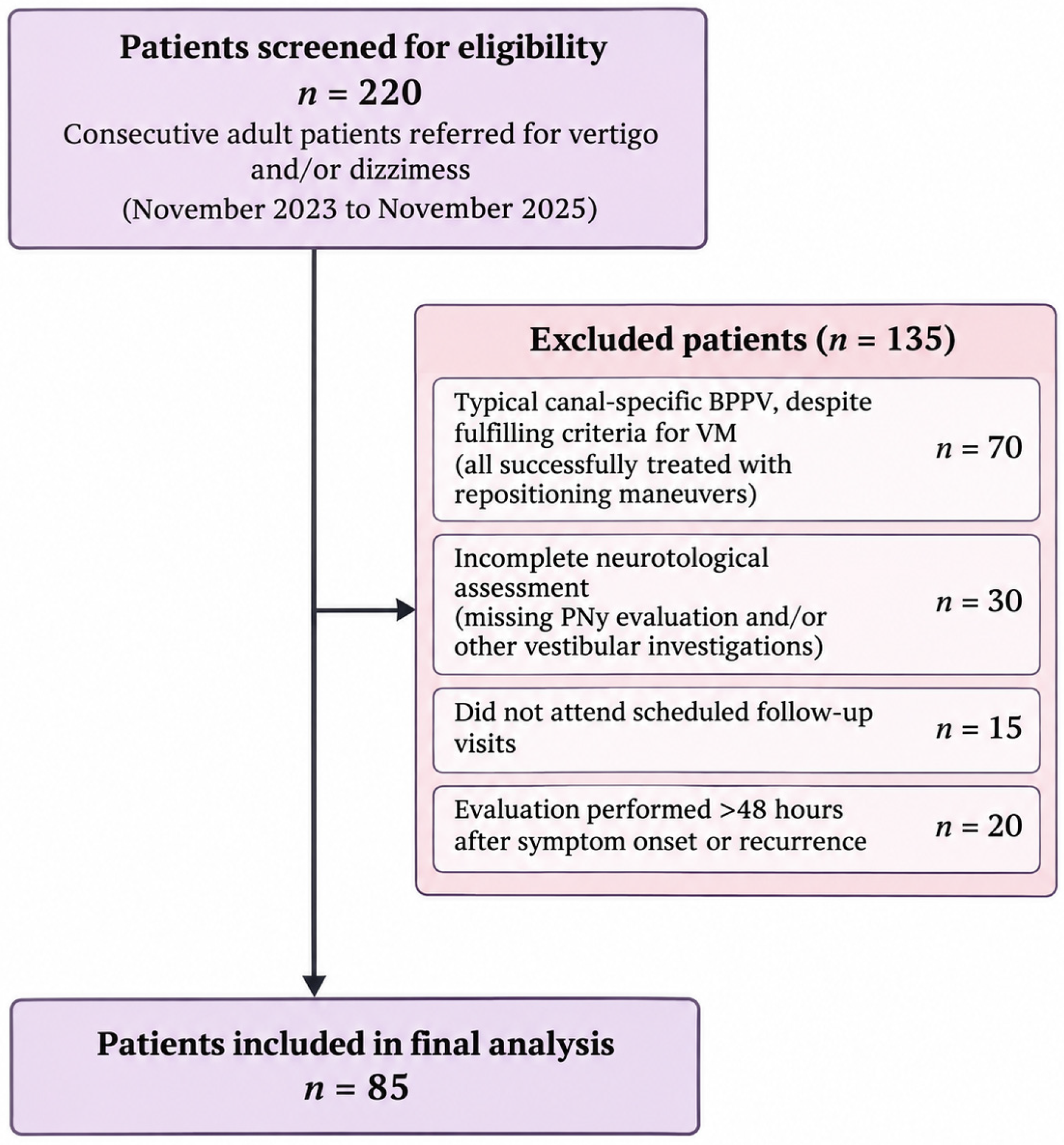
Of the 220 patients initially screened, 135 were excluded from the final study population. Specifically, 70 patients presented with unequivocal, canal-specific benign paroxysmal positional vertigo (BPPV), despite also fulfilling the diagnostic criteria for vestibular migraine (VM). All these patients were successfully treated with canal-specific repositioning maneuvers, resulting in resolution of their positional symptoms. These patients were excluded because their positional manifestations were clearly attributable to typical BPPV, rather than representing a diagnostically ambiguous positional vestibular presentation.
An additional 30 patients were excluded because of incomplete neurotological assessment, including missing pupillary nystagmus (PNy) evaluation or other vestibular investigations necessary to exclude alternative vestibular disorders. Fifteen patients were excluded because they did not attend the scheduled follow-up visits required to complete the clinical and diagnostic assessment. Finally, 20 patients were excluded because their initial neurotological evaluation was performed more than 48 h after symptom onset or recurrence, exceeding the predefined assessment interval.
Consequently, 85 patients fulfilled the eligibility criteria and were included in the final study population. The patient selection process is summarized in Figure 1.
Figure 1.
Patient selection flow diagram. Of 220 consecutive patients screened for eligibility, 135 were excluded according to the predefined exclusion criteria, and 85 were included in the final analysis.
All participants underwent a standardized neurotological evaluation including a detailed medical history and bedside vestibular examination. Demographic and clinical variables included age, sex, symptom onset, duration and frequency of vestibular episodes, triggering factors, relationship between symptoms and head position changes, previous vestibular disorders and family history of migraine. Particular attention was paid to migraine history and migraine-associated features, including headache characteristics, photophobia, phonophobia, visual aura, nausea, vomiting, motion sickness, and symptom exacerbation related to stress, sleep deprivation or other recognized migraine triggers. Clinical history also aimed to distinguish the brief positional attacks typically associated with otolithic disorders from longer-lasting or clinically heterogeneous vestibular episodes suggestive of vestibular migraine. However, symptom duration alone was not considered sufficient to establish the final diagnosis. All patients underwent a standardized vestibular examination, including assessment of spontaneous, gaze-evoked, positional, and positioning nystagmus. Eye movements were recorded using videonystagmography (VNG–Nystalyze system®, Inventis SRL, Padova, Italy) under fixation-denied conditions. Video-oculographic assessment primarily focused on the qualitative and temporal characteristics of nystagmus, including direction, latency, duration, persistence, fatigability, direction reversal, occurrence in multiple head positions, and congruence with the anatomical plane of a single semicircular canal. Quantitative measures of nystagmus intensity, such as slow-phase velocity (SPV), were not included among the reported outcome measures, as the primary objective was to characterize positional vestibular phenotypes rather than quantify nystagmus intensity.
As part of the standardized neurotological protocol, pupillary nystagmus (PNy) was systematically assessed in all patients according to the methodology previously described by our group [10]. Pupillary oscillations were evaluated under fixation-denied conditions while patients maintained primary gaze under constant ambient illumination for at least 10 s. Rhythmic oscillations in pupillary diameter occurring independently of eye movements and unrelated to changes in illumination or accommodation were considered positive. The presence or absence of PNy was recorded for each patient and included among the variables analyzed. PNy assessment was independently performed by two experienced examiners, who were blinded to the patients’ final clinical diagnoses and to each other’s evaluations. Complete agreement was observed between the two examiners regarding the presence or absence of PNy. However, no formal test–retest reproducibility assessment or quantitative interobserver reliability analysis was performed.
PNy assessment was qualitative and based on the identification of visible rhythmic oscillations in pupil diameter under constant illumination. Particular attention was paid to excluding pupillary changes associated with variations in illumination, accommodation, or eye movements. However, because pupillary hippus may also occur under physiological conditions, the presence of PNy was not considered sufficient to distinguish physiological from potentially migraine-associated pupillary oscillations. No quantitative thresholds based on oscillation amplitude or frequency were applied in the present study. PNy was therefore investigated as an exploratory clinical finding and was not used as an independent diagnostic criterion for vestibular migraine.
Positional testing systematically included bilateral Dix–Hallpike maneuvers, the straight head-hanging test and the supine roll test. Each position was maintained long enough to evaluate the onset, evolution and persistence of positional nystagmus, and ocular motor responses were also assessed after returning to the sitting position. For each spontaneous or positional nystagmus, the direction of the fast phase, horizontal, vertical and torsional components, latency, duration, persistence, fatigability, habituation, possible direction reversal, occurrence in different head positions and congruence with the anatomical plane of a single semicircular canal were systematically recorded. The subjective perception of vertigo during each maneuver was also documented. For the purposes of the retrospective analysis, positional nystagmus was classified according to its temporal and directional characteristics observed during the initial neurotological assessment. Two descriptive categories were identified: positional nystagmus atypical for BPPV and BPPV-like positional nystagmus. These categories were applied retrospectively and were not prospectively predefined. Atypical positional nystagmus was characterized by the presence of one or more features inconsistent with typical canal-specific BPPV, including absent or minimal latency, prolonged persistence, poor fatigability, direction variability, occurrence in multiple head positions, or lack of congruence with the expected response of a single semicircular canal. Conversely, BPPV-like positional nystagmus referred to positional responses predominantly resembling those observed in peripheral canalithiasis, although the overall clinical and neurotological findings were insufficient to establish an unequivocal diagnosis of BPPV.
Latency was defined as the interval between completion of the positioning maneuver and the onset of observable nystagmus. Absent or minimal latency referred to nystagmus appearing immediately or within a few seconds after positioning. Prolonged or persistent nystagmus was defined as a response lasting more than 60 s while maintaining the provoking position. Poor fatigability referred to the absence of a clear reduction in nystagmus intensity or duration during repeated positional testing. Direction variability was defined as changes in nystagmus direction during repeated examinations that could not be explained by the expected response of a single semicircular canal. Single-canal congruence was assessed by evaluating whether nystagmus direction, temporal characteristics, and positional dependence were consistent with the expected response of a specific semicircular canal.
For descriptive purposes, patients were assigned to mutually exclusive categories according to the predominant positional nystagmus pattern observed at the initial assessment. Accordingly, 38 patients were classified as presenting predominantly atypical positional nystagmus, whereas 14 patients exhibited an initially BPPV-like positional pattern. However, this classification did not imply that the underlying vestibular mechanisms or individual nystagmus characteristics were necessarily mutually exclusive. Indeed, some patients initially presenting with BPPV-like positional findings subsequently exhibited atypical characteristics during repeated neurotological examinations. Therefore, this classification reflected the predominant initial clinical phenotype rather than a fixed or immutable vestibular disorder. These positional characteristics were considered supportive elements in the differential diagnostic assessment and were not used as independent diagnostic criteria for vestibular migraine.
Patients presenting positional vertigo or dizziness without observable positional nystagmus were analyzed as a distinct clinical phenotype. The final diagnosis was established by integrating clinical history, neurotological examination and video-oculographic findings. Vestibular migraine was diagnosed according to the Bárány Society/International Headache Society diagnostic criteria. Definite vestibular migraine required at least five episodes of vestibular symptoms of moderate or severe intensity lasting between 5 min and 72 h, a current or previous diagnosis of migraine, and the presence of migraine-associated features during at least 50% of vestibular episodes, in the absence of a more appropriate alternative diagnosis. Probable vestibular migraine was diagnosed when patients fulfilled the vestibular episode criteria but met only one of the two migraine-related requirements: a current or previous diagnosis of migraine or migraine-associated features during at least 50% of vestibular episodes.
The diagnostic classification of definite and probable vestibular migraine was established according to the Bárány Society/International Headache Society criteria, independently of pupillary nystagmus (PNy) and subsequent treatment response. Positional nystagmus characteristics were evaluated as part of the differential diagnostic assessment, particularly to exclude typical canal-specific BPPV, but were not considered independent positive diagnostic criteria for vestibular migraine. PNy was investigated as an exploratory neurotological finding and was not used to determine the final diagnostic classification.
The presence of positional vertigo or positional nystagmus alone was not considered sufficient to establish a diagnosis of BPPV. Particular attention was paid to positional nystagmus that was not congruent with a single semicircular canal, occurred in multiple head positions, showed prolonged persistence, poor fatigability, limited habituation or variability in direction. After exclusion of alternative peripheral and central vestibular disorders, these findings were considered suggestive of a migraine-related positional phenotype. Following completion of the diagnostic work-up, patients were classified according to their presenting phenotype as (1) positional vertigo associated with positional nystagmus or (2) positional vertigo or dizziness without observable positional nystagmus. They were subsequently classified according to the final diagnosis as definite vestibular migraine, probable vestibular migraine, or other/unclassified positional vestibular disorders.
The primary variables analyzed included demographic characteristics, migraine history and migraine-associated symptoms, the presence or absence of positional vertigo, positional nystagmus and pupillary nystagmus, the qualitative and temporal characteristics of positional nystagmus, its congruence with a single semicircular canal, the duration and frequency of vestibular episodes, the presence of spontaneous vestibular symptoms in addition to positional complaints, and the final diagnostic classification. Following completion of the diagnostic work-up, patients fulfilling the diagnostic criteria for definite or probable vestibular migraine, according to the Bárány Society and the International Headache Society, were offered preventive migraine therapy. All patients received standardized counseling regarding lifestyle modifications, including regular sleep habits, adequate hydration, regular meals, stress management, and avoidance of recognized migraine triggers. Preventive treatment consisted of oral magnesium oxide (400–500 mg/day), according to the recommendations of the American Headache Society for migraine prevention, together with flunarizine 5 mg once daily, which represented the first-line prophylactic treatment routinely adopted at our Vestibular Unit. Patients underwent scheduled follow-up visits at 1 and 3 months after treatment initiation. At each visit, a complete neurotological examination was repeated, including positional testing and video-oculographic assessment whenever clinically indicated. Clinical outcome measures included the frequency, duration and severity of vestibular episodes, persistence or resolution of positional vertigo, presence or absence of positional nystagmus, persistence of subjective dizziness, and the patient’s overall perception of clinical improvement. Particular attention was paid to patients who initially presented with positional symptoms suggestive of benign paroxysmal positional vertigo but showed absent or only partial improvement following appropriate canalith repositioning maneuvers. In these patients, persistence of symptoms prompted further clinical reassessment and review of the patient’s migraine history and associated migrainous features. When the overall clinical picture fulfilled the diagnostic criteria for vestibular migraine, preventive migraine therapy was initiated. Response to migraine preventive treatment was evaluated during follow-up as a clinical outcome. Clinical improvement was interpreted in the context of the overall clinical course but was not used to establish or modify the initial diagnostic classification of definite or probable vestibular migraine.
The statistical approach was exclusively descriptive. Data were summarized using absolute frequencies, percentages, and means with standard deviations, as appropriate. The primary descriptive outcomes were the characterization of positional vestibular manifestations and final diagnostic classification. Secondary outcomes included the prevalence of pupillary nystagmus and clinical evolution at 1 and 3 months. No inferential statistical comparisons between diagnostic groups or longitudinal hypothesis testing were performed.
Clinical improvement was assessed through patient-reported changes in the frequency, duration, and severity of vestibular symptoms, together with repeated neurotological examinations. Complete resolution was defined as the absence of positional vertigo or dizziness, whereas substantial improvement referred to a marked reduction in symptoms with occasional residual manifestations. Limited or no improvement indicated the absence of clinically relevant symptomatic benefit. All 74 patients receiving migraine preventive treatment completed the scheduled follow-up assessments at 1 and 3 months, with no loss to follow-up. Complete adherence to the prescribed therapeutic regimen was reported throughout the observation period.
3. Results
A total of 85 patients (58 females and 27 males; mean age 46.3 ± 13.1 years, range 18–75 years) were included in the final analysis. All patients presented with positional vertigo or dizziness in whom a definite diagnosis of typical benign paroxysmal positional vertigo could not be established at the initial neurotological assessment. A previous history of migraine was reported by 64 patients (75.3%).
Migraine-associated symptoms were frequently observed, including photophobia in 44 patients (51.8%), phonophobia in 39 (45.9%), nausea in 52 (61.2%), motion sickness in 35 (41.2%), and visual aura in 18 (21.2%). Positional vertigo or dizziness associated with observable positional nystagmus was documented in 52 patients (61.2%), whereas 33 patients (38.8%) reported positional symptoms without detectable positional nystagmus. In the 33 patients presenting with positional symptoms without observable nystagmus, the clinical manifestations were predominantly characterized by positional unsteadiness and subjective instability rather than the brief episodes of rotational vertigo typically associated with BPPV. Standardized positional testing did not reveal reproducible canal-specific nystagmus, and canalith repositioning maneuvers did not produce significant symptomatic improvement. These findings, considered together with the clinical history and neurotological assessment, made BPPV a less likely explanation for the positional symptoms, although its possibility could not be excluded solely on the basis of absent nystagmus.
Among these patients, 17 fulfilled the diagnostic criteria for definite vestibular migraine and 10 for probable vestibular migraine, whereas six were classified as having other or unclassified positional vestibular disorders. Migraine-associated clinical features and supportive instrumental findings contributed to the overall diagnostic evaluation, while the final classification of vestibular migraine was established according to the Bárány Society/International Headache Society criteria.
The main demographic and clinical characteristics of the study population according to the final diagnostic classification are summarized in Table 1.
Table 1.
Summary of the main demographic and clinical characteristics of the study population.
In 14 patients (26.9% of those presenting with positional nystagmus), positional findings initially showed features partially resembling BPPV. Five patients experienced complete resolution of positional vertigo and nystagmus following canalith repositioning maneuvers. Specifically, three patients underwent two Epley maneuvers each, one patient achieved complete resolution after three Semont maneuvers, and one patient required four Semont maneuvers. All treatment sessions were performed at 48-h intervals.
Among these five responders, three were ultimately classified as having other/unclassified positional vestibular disorders, whereas two fulfilled the diagnostic criteria for probable vestibular migraine. The favorable response to repositioning maneuvers supported the presence of a peripheral positional component, potentially coexisting with migraine-related vestibular manifestations.
In the remaining nine patients, positional symptoms persisted or recurred despite repeated repositioning procedures. Five patients underwent four Epley maneuvers each, whereas four patients underwent four Semont maneuvers each, with consecutive treatment sessions separated by 48 h. The persistence of symptoms prompted further neurotological reassessment, including repeated positional testing and evaluation of migraine-associated clinical features. According to the Bárány Society/International Headache Society criteria, six of these patients were ultimately diagnosed with definite vestibular migraine and three with probable vestibular migraine. Migraine preventive treatment was subsequently initiated.
Repeated positional testing showed that nystagmus characteristics could vary over time in a subset of patients, including changes in direction, intensity, or the position in which nystagmus was elicited. These findings, particularly when associated with migrainous symptoms, raised suspicion of a migraine-related positional phenotype rather than a typical otolithic disorder. The distribution of positional nystagmus characteristics according to final diagnosis is shown in Table 2.
Table 2.
Characteristics of positional nystagmus according to final diagnosis.
Atypical positional findings were particularly frequent among patients with definite VM. Absent or minimal latency, poor fatigability, occurrence in multiple head positions and lack of congruence with a single semicircular canal were the most frequently observed features. Conversely, a BPPV-like positional pattern was less frequently observed among patients with definite VM and was more common in patients who did not fulfill the diagnostic criteria for VM. The diagnostic course, therapeutic management and treatment response of patients presenting with BPPV-like or migraine-related positional phenotypes are summarized in Table 3. Among the 74 patients classified as having definite or probable vestibular migraine, preventive treatment with flunarizine 5 mg/day and magnesium oxide 400–500 mg/day was initiated together with standardized lifestyle counseling. Clinical improvement was already observed at the 1-month follow-up and became more evident after 3 months of treatment.
Table 3.
Initial positional phenotype, clinical management, treatment response, and final diagnostic classification.
At 1 month, 47 patients (63.5%) reported a marked reduction in the frequency and severity of vestibular episodes, while complete resolution of positional vertigo or dizziness was observed in 28 patients (37.8%). Among patients presenting with positional nystagmus at baseline, disappearance of positional nystagmus was documented in 21 patients. At the 3-month follow-up, 66 of 74 patients (89.2%) experienced either complete resolution or substantial improvement of positional vestibular symptoms. Complete resolution of positional vertigo or dizziness was observed in 49 patients (66.2%), whereas 17 patients (23.0%) reported substantial improvement with occasional residual symptoms. The remaining eight patients (10.8%) showed limited or no clinically relevant improvement.
Among the 47 patients with definite or probable VM who presented with positional nystagmus at baseline, positional nystagmus was no longer detectable in 36 patients (76.6%) at the 3-month assessment. Persistent positional nystagmus was observed in 11 patients (23.4%), although in several of these patients its intensity and reproducibility were reduced compared with baseline.
Clinical improvement was generally accompanied by a reduction in migraine-associated symptoms and subjective dizziness. Response to preventive treatment was interpreted as a clinical follow-up outcome and was not used to establish or modify the initial diagnostic classification.
Clinical outcomes at 1 and 3 months are summarized in Table 4.
Table 4.
Clinical outcome of patients with definite or probable vestibular migraine during follow-up.
4. Discussion
The present study highlights the diagnostic complexity of positional vestibular symptoms in patients in whom typical benign paroxysmal positional vertigo (BPPV) cannot be clearly demonstrated. In this selected population, vestibular migraine (VM) represented the most frequent final diagnosis, with definite or probable VM accounting for 87.1% of cases. This finding should be interpreted in the context of the specific selection criteria of our cohort, which excluded patients with unequivocal canal-specific BPPV and specifically included subjects in whom the distinction between VM and BPPV remained uncertain after the initial neurotological assessment. Previous studies have demonstrated that VM may closely mimic BPPV, with positional vertigo and positional nystagmus representing prominent manifestations in a subset of patients [2,3]. Patients with migraine-related positional vertigo may therefore initially be diagnosed as having BPPV, particularly when positional symptoms are accompanied by an elicitable nystagmus. Patients presenting with an apparently BPPV-like positional phenotype but showing persistent or recurrent symptoms despite appropriate canalith repositioning maneuvers should undergo diagnostic reassessment, as the positional manifestations may reflect an underlying migraine-related vestibular mechanism rather than a purely peripheral otolithic disorder [3]. More recently, the substantial clinical overlap between VM and BPPV has been further emphasized, supporting the need to integrate symptom characteristics with detailed neurotological and video-oculographic assessment rather than relying solely on the positional nature of vertigo [4]. One of the most relevant observations in our cohort concerned the characteristics of positional nystagmus. Among the 52 patients in whom positional nystagmus was documented, 38 (73.1%) were classified as presenting a predominantly atypical positional pattern at the initial neurotological assessment.
These findings were particularly frequent among patients ultimately classified as having definite VM, in whom 28 of 34 patients with positional nystagmus showed an atypical pattern. Absent or minimal latency, prolonged or persistent nystagmus, poor or absent fatigability, direction variability or reversal, occurrence in multiple head positions and lack of congruence with a single semicircular canal were the principal abnormalities observed. These observations are consistent with previous descriptions of positional nystagmus in VM, in which the elicited eye movements may be persistent, variable and poorly compatible with the expected response of an isolated semicircular canal [2,9]. However, none of these characteristics should be regarded as independently diagnostic of VM. Positional nystagmus may result from different peripheral and central vestibular mechanisms, and its interpretation requires systematic evaluation of direction, temporal profile, response to positional testing and relationship with head position [11]. Accordingly, the diagnostic relevance of these findings appears to derive primarily from their overall pattern and their integration with the clinical history rather than from any single video-oculographic feature. An additional clinically relevant phenotype was represented by patients reporting reproducible positional vertigo or dizziness in the absence of observable positional nystagmus. This phenotype accounted for 33 of the 85 patients in our cohort. In such patients, particularly when repeated positional testing fails to demonstrate objective findings compatible with canalolithiasis or cupulolithiasis, the positional nature of symptoms alone should not be considered sufficient to establish a diagnosis of BPPV. The presence of migraine-associated symptoms, spontaneous vestibular episodes and a temporal pattern not typical of an otolithic disorder may provide additional information supporting a migraine-related mechanism [4]. At the same time, the presence of migraine features does not exclude a peripheral positional disorder. Migraine and BPPV may coexist, and migraine has been identified as an important comorbidity in patients with BPPV [6]. This relationship is particularly relevant in clinical practice because it argues against a rigid distinction between exclusively peripheral and exclusively migraine-related positional syndromes. A patient with migraine may develop genuine canalolithiasis requiring repositioning maneuvers, whereas a patient with recurrent positional symptoms attributed to BPPV may instead present a migraine-related positional phenotype. Repeated assessment of the positional nystagmus pattern and its canal congruence therefore remains essential. This clinical overlap was also reflected in our cohort. Among the 14 patients with an initially BPPV-like positional pattern, five showed resolution of positional vertigo and nystagmus following canalith repositioning maneuvers, supporting the presence of a peripheral positional component. Conversely, nine patients showed persistent or recurrent positional symptoms despite repositioning, prompting further diagnostic reassessment and consideration of a migraine-related positional phenotype when criteria for definite or probable VM were fulfilled. A further aspect of the present investigation was the systematic assessment of pupillary nystagmus (PNy). Previous observations from our group identified PNy as a potentially useful objective finding in patients with VM [10]. In the present cohort, PNy was detected in 61 of 85 patients (71.8%), with the highest prevalence among patients fulfilling the criteria for definite VM, in whom it was observed in 45 of 51 cases (88.2%). By comparison, PNy was detected in 13 of 23 patients with probable VM and in 3 of 11 patients (27.3%) classified as having other/unclassified positional vestibular disorders. These findings are consistent with our subsequent quantitative pupillometric investigation, which further supported the potential role of PNy as an objective neuro-otological marker in VM [12]. Nevertheless, the present results should not be interpreted as establishing PNy as an independent diagnostic biomarker. An important consideration concerns the physiological occurrence of pupillary hippus, which may share qualitative characteristics with the oscillations identified as PNy. Consequently, qualitative bedside observation alone does not allow a definitive distinction between physiological pupillary unrest and potentially pathological oscillatory activity. Although complete agreement was observed between the two independent blinded examiners, formal interobserver reliability statistics and test–retest reproducibility assessments were not performed. Therefore, the reproducibility of PNy detection requires further investigation in prospective studies.
Furthermore, the retrospective design and absence of dedicated analyses of diagnostic accuracy and incremental diagnostic value limit the interpretation of the present findings. Although previous investigations have provided encouraging evidence regarding the association between PNy and vestibular migraine, the present observations should be regarded as exploratory and hypothesis-generating. Further prospective studies incorporating blinded examinations, standardized quantitative pupillometry, and reproducibility assessments are warranted.
Rather, PNy may represent an additional supportive finding when interpreted together with migraine history, associated migrainous features and conventional neurotological and video-oculographic findings. Further prospective controlled studies directly comparing VM with typical BPPV and other vestibular disorders are required to determine its diagnostic accuracy and potential incremental value. Treatment response provided an additional element in the longitudinal assessment of these patients. Most patients classified as having definite or probable VM showed progressive improvement during follow-up, with complete resolution of positional vertigo or dizziness in 66.2% of patients at 3 months. Among patients with positional nystagmus at baseline, nystagmus was no longer detectable in 76.6% at the 3-month assessment. These findings suggest that improvement of the underlying migraine-related vestibular condition may be accompanied not only by subjective clinical improvement but also by changes in objective positional findings. In the present cohort, preventive treatment consisted of flunarizine combined with magnesium oxide and standardized lifestyle counseling; therefore, the observed clinical improvement should be interpreted as the response to the overall preventive strategy rather than attributed to either component individually. Available randomized evidence suggests that preventive migraine treatments, including flunarizine, may improve dizziness-related disability in patients with VM [13]. However, therapeutic response should not be regarded as proof of the diagnosis. In the present study, improvement following preventive treatment was considered supportive of the overall clinical interpretation only when consistent with the patient’s history, migrainous features and neurotological findings. This cautious interpretation is particularly important because the evidence supporting pharmacological prophylaxis in VM remains limited, with systematic reviews highlighting the small number of randomized controlled trials and the low or very low certainty of the available evidence [14]. From a clinical perspective, our findings suggest that the diagnosis should be reconsidered in patients with persistent positional symptoms when positional nystagmus is atypical, variable or inconsistent with the expected pattern of a single semicircular canal, particularly following unsuccessful canalith repositioning maneuvers. Integration of clinical history with repeated positional testing and video-oculographic assessment may help distinguish migraine-related positional manifestations from typical BPPV and may reduce unnecessary repetition of repositioning procedures. Conversely, because migraine and BPPV may coexist [6], the presence of a migraine history should not prevent appropriate repositioning treatment when a reproducible canal-specific BPPV pattern is demonstrated.
Several limitations should be acknowledged. Furthermore, the sample size was determined by the number of eligible patients during the study period, without an a priori power calculation. The relatively small size of certain diagnostic subgroups may limit the generalizability of the findings.
First, the observational single-center design and recruitment from a tertiary vestibular unit may introduce referral and selection bias. In particular, the high proportion of definite or probable VM observed in the present cohort cannot be extrapolated to an unselected population of patients with positional vertigo, since typical BPPV was deliberately excluded by the study design. Second, the absence of a control group with unequivocal BPPV prevents direct assessment of the diagnostic sensitivity and specificity of the investigated positional findings and of PNy. Third, positional and ocular motor abnormalities may fluctuate over time, potentially affecting their reproducibility. Furthermore, the retrospective assignment of patients to mutually exclusive categories based on their predominant initial positional nystagmus pattern involves a degree of subjectivity and may introduce misclassification bias. Indeed, atypical and BPPV-like features may coexist or fluctuate over time, potentially limiting the accuracy and stability of the initial phenotypic classification.
Finally, treatment outcomes should be interpreted cautiously because of the absence of a randomized control group and the possibility of spontaneous fluctuations in VM symptoms.
Although the diagnostic classification was based on established Bárány Society/International Headache Society criteria, potential diagnostic incorporation bias cannot be entirely excluded, particularly because positional nystagmus characteristics contributed to the differential diagnostic assessment. Therefore, the associations between vestibular migraine and the investigated neurotological findings, including PNy, should be interpreted cautiously and require confirmation in independent controlled studies.
Furthermore, the possibility of misclassifying subjective BPPV in patients presenting with positional symptoms without observable nystagmus cannot be completely excluded, despite comprehensive clinical assessment and repeated positional testing when indicated. Another limitation concerns the reproducibility of qualitative PNy assessment. Although agreement between independent examiners was observed, formal test–retest reproducibility analyses were not performed. These methodological limitations, together with the relatively small size of certain diagnostic subgroups, should be considered when interpreting the present findings.
An additional limitation concerns the absence of quantitative video-oculographic measurements of nystagmus intensity, such as slow-phase velocity. Although videonystagmography was systematically performed, the analysis primarily focused on qualitative and temporal characteristics. Future studies incorporating quantitative nystagmus measurements may provide further insight into the differentiation between migraine-associated positional nystagmus and BPPV.
Future prospective controlled studies directly comparing patients with definite VM, typical BPPV and combined VM–BPPV are needed to determine the diagnostic accuracy of individual and combined positional nystagmus characteristics. Particular attention should be paid to whether the combination of atypical positional findings and PNy provides greater diagnostic information than either finding alone. Longitudinal quantitative assessment of PNy and positional nystagmus may also clarify whether changes in these objective findings parallel clinical activity and response to preventive treatment.
5. Conclusions
Vestibular migraine should be considered in patients with recurrent positional vertigo or dizziness when positional findings are atypical or inconsistent with the expected pattern of a single semicircular canal disorder. Careful analysis of positional nystagmus, particularly its latency, duration, fatigability, direction, reproducibility and canal congruence, may provide useful information in the differential diagnosis with BPPV. No single positional finding should be considered diagnostic of VM. Rather, the combination of clinical history, migraine-associated symptoms and detailed video-oculographic assessment appears to provide the most appropriate diagnostic approach.
The observed clinical improvement following migraine preventive treatment should be interpreted cautiously, given the absence of an untreated or comparator group, and should not be regarded as independent confirmation of vestibular migraine diagnosis. Further prospective controlled studies are needed to validate these exploratory findings and clarify their potential diagnostic and therapeutic implications.
Author Contributions
Conceptualization, A.P.C. and N.D.; methodology, A.P.C. and N.D.; validation, A.P.C. and N.D.; formal analysis, A.P.C.; investigation, N.D.; data curation, N.D.; writing—original draft preparation, N.D.; writing—review and editing, A.P.C. All authors have read and agreed to the published version of the manuscript.
Funding
This research was partially funded by Fondazione Mario Marianelli, via Verdi 26, 56029 S. Croce sull’Arno, Pisa, Italy, grant number n°3/2025.
Institutional Review Board Statement
This retrospective observational study was conducted in accordance with the ethical principles of the Declaration of Helsinki. Formal ethics committee approval was not sought, as the investigation was based exclusively on clinical data collected during routine neurotological practice. No experimental diagnostic techniques or investigational procedures were introduced. All therapeutic interventions were performed according to established clinical practice and Italian guidelines for the management of benign paroxysmal positional vertigo and vestibular migraine.
Informed Consent Statement
Written informed consent was obtained from all patients prior to the diagnostic assessments and therapeutic procedures performed as part of routine clinical practice.
Data Availability Statement
The original contributions presented in this study are included in the article. Further inquiries can be directed to the corresponding author.
Acknowledgments
We would like to thank Nicola Vernassa for his technical support.
Conflicts of Interest
The authors declare no conflicts of interest. The funders had no role in the design of the study; in the collection, analyses, or interpretation of data; in the writing of the manuscript; or in the decision to publish the results.
References
- Lempert, T.; von Brevern, M. Vestibular migraine. Neurol. Clin. 2019, 37, 695–706. [Google Scholar] [CrossRef] [Scilit]
- Tian, E.; Li, F.; Liu, D.; Wang, J.; Guo, Z.; Chen, J.; Guo, J.; Zhang, S. Dispelling mist that obscures positional vertigo in vestibular migraine. Brain Sci. 2023, 13, 1487. [Google Scholar] [CrossRef] [Scilit]
- Mahrous, M.M. Vestibular migraine and benign paroxysmal positional vertigo, close presentation dilemma. Acta Otolaryngol. 2020, 140, 741–744. [Google Scholar] [CrossRef] [Scilit]
- Mok, B.; Welgampola, M.S.; Rosengren, S.M. Vestibular migraine as a mimic of benign paroxysmal positioning vertigo and Meniere’s disease. J. Vestib. Res. 2025, 35, 30–38. [Google Scholar] [CrossRef] [Scilit]
- Shih, I.A.; Hsu, C.Y.; Li, T.C.; Wang, S.J. Benign paroxysmal positional vertigo is associated with an increased risk for migraine diagnosis: A nationwide population-based cohort study. Int. J. Environ. Res. Public Health 2023, 20, 3563. [Google Scholar] [CrossRef] [Scilit]
- Alolayet, H.; Murdin, L. Benign paroxysmal positional vertigo: A systematic review of the effects of comorbidities. Front. Neurol. 2025, 16, 1595693. [Google Scholar] [CrossRef] [Scilit]
- Lempert, T.; Olesen, J.; Furman, J.; Waterston, J.; Seemungal, B.; Carey, J.; Bisdorff, A.; Versino, M.; Evers, S.; Newman-Toker, D. Vestibular migraine: Diagnostic criteria. J. Vestib. Res. 2012, 22, 167–172. [Google Scholar] [CrossRef] [Scilit]
- Headache Classification Committee of the International Headache Society (IHS). The International Classification of Headache Disorders, 3rd edition. Cephalalgia 2018, 38, 1–211. [Google Scholar] [CrossRef] [Scilit]
- El-Badry, M.M.; Samy, H.; Kabel, A.M.; Rafat, F.M.; Sanyelbhaa, H. Clinical criteria of positional vertical nystagmus in vestibular migraine. Acta Otolaryngol. 2017, 137, 720–722. [Google Scholar] [CrossRef] [Scilit]
- Gufoni, M.; Casani, A.P. “The Pupillary (Hippus) Nystagmus”: A possible clinical hallmark to support the diagnosis of vestibular migraine. J. Clin. Med. 2023, 12, 1957. [Google Scholar] [CrossRef] [Scilit]
- Eggers, S.D.Z.; Bisdorff, A.; von Brevern, M.; Zee, D.S.; Kim, J.S.; Perez-Fernandez, N.; Welgampola, M.S.; Della Santina, C.C.; Newman-Toker, D.E. Classification of vestibular signs and examination techniques: Nystagmus and nystagmus-like movements. J. Vestib. Res. 2019, 29, 57–87. [Google Scholar] [CrossRef] [Scilit]
- Casani, A.P.; Ducci, N.; Califano, L.; Gufoni, M. Pupillary nystagmus as an objective neuro-otological biomarker in vestibular migraine: A quantitative pupillometric study. Audiol. Res. 2026, 16, 79. [Google Scholar] [CrossRef] [Scilit]
- Chu, H.; Wang, Y.; Ling, X.; Li, K.; Yang, X. Prophylactic treatments for vestibular migraine: A systematic review and network meta-analysis of randomized clinical trials. Front. Pharmacol. 2023, 14, 1332973. [Google Scholar] [CrossRef] [Scilit]
- Webster, K.E.; Dor, A.; Galbraith, K.; Haj Kassem, L.; Harrington-Benton, N.A.; Judd, O.; Kaski, D.; Maarsingh, O.R.; MacKeith, S.; Ray, J.; et al. Pharmacological interventions for prophylaxis of vestibular migraine. Cochrane Database Syst. Rev. 2023, 2023, CD015187. [Google Scholar] [CrossRef] [Scilit]

Disclaimer/Publisher’s Note: The statements, opinions and data contained in all publications are solely those of the individual author(s) and contributor(s) and not of MDPI and/or the editor(s). MDPI and/or the editor(s) disclaim responsibility for any injury to people or property resulting from any ideas, methods, instructions or products referred to in the content. |
© 2026 by the authors. Licensee MDPI, Basel, Switzerland. This article is an open access article distributed under the terms and conditions of the Creative Commons Attribution (CC BY) license.