


Highlights
What are the main findings?
- Vestibular migraine was associated with distinct peripheral and central vestibular abnormalities.
- Vestibular recruitment and caloric hyperexcitability appeared as partially independent phenotypes.
What are the implications of the main findings?
- Combined caloric and vHIT testing provides insights into the heterogeneous vestibular pathophysiology of vestibular migraine.
- Improved diagnostic accuracy leads to more effective treatment.
Abstract
Background/Objective: To characterize vestibular phenotypes associated with vestibular migraine (VM) and explore the potential underlying pathophysiological mechanisms. Methods: This consecutive observational comparative study included 98 participants (63 with VM and 35 healthy controls). All participants underwent caloric testing and the video head impulse test (vHIT). Vestibular recruitment, unilateral weakness, caloric hyperreflexia, caloric dysrhythmia, and high gain on vHIT were assessed. Associations between VM and vestibular variables were evaluated using logistic regression. Caloric responses were compared between groups using multivariate analysis of covariance (MANOVA), adjusted for age and sex. The association between vestibular recruitment and markers of vestibular hyperexcitability was assessed using contingency tables, Pearson’s chi-square test and Fisher’s exact test. Results: Patients with VM exhibited higher frequencies of vestibular recruitment (p = 0.0009), unilateral weakness (p = 0.0017), and caloric dysrhythmia (p = 0.0076). Multivariate analysis demonstrated significantly greater caloric responses in patients with VM across all irrigations (p = 0.0129). High gain on vHIT was not associated with VM (p = 0.6878). No association was observed between vestibular recruitment and caloric dysrhythmia (p = 0.3320) or between vestibular recruitment and caloric hyperreflexia (p = 0.3725). Conclusions: These findings suggest that VM is associated with both peripheral vestibular abnormalities and vestibular hyperexcitability. The absence of associations between vestibular recruitment and markers of hyperexcitability suggests that these abnormalities may represent distinct vestibular phenotypes, supporting an integrative model in which peripheral and central mechanisms coexist in a partially independent manner in the vestibular pathophysiology of VM.
1. Introduction
Vestibular migraine (VM) is a neurological disorder characterized by recurrent episodes of vestibular symptoms in individuals with a current or previous history of migraine. Although its pathophysiology remains incompletely understood, VM has been associated with altered central sensory processing and neuronal hyperexcitability involving cortical, brainstem, and vestibular networks. These mechanisms may contribute to abnormal vestibular responsiveness and altered integration of vestibular information, potentially influencing the functional responses observed during vestibular testing [1,2,3,4].
Several abnormalities in vestibular testing have been described in patients with VM involving both peripheral vestibular function and central vestibular processing mechanisms [2,3,5]. However, these findings are highly heterogeneous across studies and may coexist in different combinations, making their pathophysiological interpretation challenging. It remains unclear whether these abnormalities reflect different manifestations of a shared neurobiological mechanism or distinct vestibular phenotypes associated with VM.
Experimental and neurophysiological studies have demonstrated that migraine and VM is associated with glutamatergic hyperexcitability, neuroinflammation mediated by neuropeptides such as the calcitonin gene-related peptide (CGRP), and impairment of central inhibitory pathways [1,6]. Within the vestibular system, the cerebellum, particularly the flocculus, nodulus, and uvula, plays a pivotal role in modulating the vestibulo-ocular reflex through the inhibitory influence of Purkinje cells on the vestibular nuclei [3,7]. Reduced cerebellar inhibition may impair the velocity storage mechanism, leading to amplification of low-frequency vestibular responses [7,8]. In contrast, vestibular recruitment and unilateral weakness are findings suggestive of peripheral vestibular dysfunction, although they may reflect different functional processes [9,10]. Unilateral weakness indicates asymmetry in peripheral vestibular responsiveness, whereas vestibular recruitment has been interpreted as a possible manifestation of incomplete central compensation following peripheral vestibular dysfunction. These findings may therefore provide complementary information about peripheral vestibular function [9].
Despite these observations, few studies have simultaneously investigated vestibular recruitment, unilateral weakness, caloric hyperreflexia, caloric dysrhythmia, and vHIT hypergain within the same cohort to explore whether these findings may represent distinct vestibular phenotypes associated with VM.
Based on these observations, we hypothesized that VM is associated with at least two distinct vestibular phenotypes: a central phenotype, characterized by vestibular hyperexcitability manifested as caloric hyperreflexia, caloric dysrhythmia, and, occasionally, high gain on the video head impulse test (vHIT); and a peripheral phenotype, characterized by the presence of vestibular recruitment and/or unilateral weakness.
Therefore, the present study aimed to characterize vestibular phenotypes associated with VM by investigating vestibular recruitment, unilateral weakness, caloric hyperreflexia, caloric dysrhythmia, and high gain on vHIT, while exploring the potential pathophysiological mechanisms linking these findings.
2. Material and Methods
This consecutive observational comparative study was approved by the Ethics Committee for the Analysis of Research Projects on 8 November 2018 (number 3.008.904), and was performed in accordance with the Declaration of Helsinki.
Sample
The study group (SG) comprised 63 consecutive participants with VM diagnosed according to the Bárány Society diagnostic criteria [11]. The control group (CG) comprised 35 healthy volunteers without vestibular complaints or a current or previous history of migraine. Individuals reporting any history of migraine, with or without vestibular symptoms, were excluded from the control group. Controls were recruited according to the same predefined eligibility criteria but were not individually matched to the SG by age or sex.
The inclusion criteria for both groups were age >18 years, ability to understand the study procedures and perform the tests, and provision of written informed consent. Exclusion criteria were other vestibular disorders, use of prophylactic medication for migraine during the testing period or within the three months preceding the tests; chronic otitis media; previous ear surgery; somatoform or psychiatric disorders; and ocular, cervical, rheumatic, or orthopedic conditions that could interfere with or preclude test performance. No patient was assessed during an acute vestibular episode. The duration of vestibular migraine was not systematically recorded or controlled for in this study and was therefore not included as a study variable. No formal a priori sample size calculation was performed; the study sample was based on the consecutive recruitment of eligible participants during the study period. Patients were recruited between 2018 and 2026 at the three participating institutions: the State University of Campinas, University of Paraná, and University of São Paulo School of Medicine. Patient recruitment and vestibular examinations were conducted at the participating institutions, and the test results were jointly interpreted by the three researchers. Before enrollment, the technical quality of both caloric and vHIT recordings was assessed. Only individuals with technically acceptable recordings from both tests were included in the study. Recordings showing technical artifacts or insufficient quality for reliable interpretation were considered unsuitable for inclusion. All enrolled participants had complete and technically acceptable vestibular test recordings, and no participants were excluded after enrollment.
Method
All participants underwent otoneurological assessment, caloric testing, and vHIT during the same week. Otoneurological assessment included medical history, otorhinolaryngological and cranial nerve examination, static and dynamic balance assessment, and coordination tests.
Caloric testing was performed using video-oculography (ICS Chartr 200, Otometrics®, Copenhagen, Denmark or Visual Eyes VO 525, Interacoustics, Middelfart, Denmark). The stimulation was performed with air at 50 °C and 24 °C, at a flow rate of 8 L/min for one minute or with water at 44 °C and 30 °C for 40 s. These temperature settings are standardized for air- and water-caloric testing and are designed to provide comparable vestibular stimulation [12]. Before each stimulation, the water temperature was checked with three thermometers, and the first jet of water was discarded before starting the irrigation of the external auditory canal.
The parameters obtained in the caloric test were post-caloric recruitment index (PCRI), unilateral weakness (UW), hyperreflexia and dysrhythmia of the responses.
The PCRI was calculated for each ear separately using the formula [9]
AVSP C: angular velocity of the slow phase after cold irrigation.
AVSP W: angular velocity of the slow phase after warm irrigation.
A PCRI value > 17% was considered abnormal and used to define the presence of recruitment. This threshold was previously established in our population [9].
The Jongkees formula was used to calculate the UW:
RW: angular velocity of the slow phase after warm irrigation of the right ear.
LW: angular velocity of the slow phase after warm irrigation of the left ear.
RC: angular velocity of the slow phase after cold irrigation of the right ear.
LC: angular velocity of the slow phase after cold irrigation of the left ear.
Values above 19% and 20% were considered abnormal for air and water stimulation, respectively, based on modality-specific normative reference values established for the Brazilian population [10,13].
The reference values used to determine caloric hyperreflexia in air stimulation were [13]:
- AVSP in a single irrigation > 46°/s.
- Unilateral: Sum of the AVSP in the cold and warm insufflation on either side: >62°/s.
- Bilateral: Sum of the AVSP in the four insufflations > 122°/s.
For water irrigation, the criteria for hyperreflexia were [14]:
- AVSP in a single irrigation > 50°/s.
- Unilateral: Sum of the AVSP in the cold and warm irrigation on either side > 75°/s.
- Bilateral: Sum of the AVSP in the four irrigations > 150°/s.
A caloric response was considered dysrhythmic if the examiner observed significant variations in the amplitude and frequency of the response without corresponding changes in the slow-phase angular velocity in at least three of the four stimulations.
The quantitative vestibular test parameters were analyzed by a single examiner who was not blinded to group allocation. Dysrhythmia was assessed independently by three examiners based on visual inspection of the caloric tracings. Two examiners were blinded to group allocation. Each examiner classified the tracings independently and without discussion, and complete agreement was reached among the three examiners.
vHIT was performed using the Interacoustics EyeSeeCam (Middelfart, Denmark) or Otometrics ICS impulse system® (Copenhagen, Denmark). Manual unpredictable head impulses were administered with a peak amplitude of 15–20°, a mean velocity of 200°/s and a mean acceleration of 3000°/s2 in the horizontal plane to assess the lateral semicircular canals. The mean VOR gain calculated by the software was used for quantitative analysis. Recordings showing elevated VOR gain were visually reviewed to assess the quality and consistency of eye and head velocity traces and to identify potential technical artifacts. Head impulses with suspected artifacts were repeated during testing. High gain was classified when the mean VOR gain exceeded 1.2 [15], the recordings were technically acceptable, and reverse saccades were present as an additional confirmatory criterion.
Statistical analysis
Multivariable logistic regression analysis was performed to investigate the association between VM status and the laboratory findings. VM status was entered as the dependent variable, whereas sex, age, vestibular recruitment, unilateral weakness, caloric dysrhythmia, caloric hyperreflexia, and high gain on vHIT were included as independent variables. Caloric responses, measured as AVSP during RW, LW, LC, and RC irrigations, were compared between groups using multivariate analysis of covariance (MANOVA), with adjustment for age and sex. Associations between vestibular recruitment and caloric dysrhythmia or hyperreflexia were evaluated in the entire study population, including patients with VM and healthy controls, using Pearson’s chi-square test and Fisher’s exact test. There were no missing data in the study dataset; therefore, no data imputation or other missing-data handling procedures were required. All statistical analyses were performed using JMP Pro version 19.1 (SAS Institute Inc., Cary, NC, USA).
3. Results
The study group (SG) comprised 63 participants, of whom 14 were men (22%) and 49 were women (78%), with a mean age of 45 years. The control group consisted of 35 participants, including 18 men (51%) and 17 women (49%), with a mean age of 40 years. The distribution of the variables in both groups is presented in Table 1.
Table 1.
Distribution of vestibular findings in the study and control groups.
Vestibular recruitment (OR = 6.42; CI 95%: 1.97–20.89; p = 0.0009), unilateral weakness (OR = 9.83; CI 95%: 1.81–53.37; p = 0.0017), and dysrhythmia (OR = 7.11; CI 95%: 1.46–34.48; p = 0.0076) were significantly more common in patients with VM. Caloric hyperreflexia was more common in the study group, although it did not reach statistical significance (OR = 7.96; CI 95%: 0.72–87.30; p = 0.0534). In contrast, high gain on vHIT (OR = 0.79; CI 95%: 0.25–2.46; p = 0.6878) and age (OR = 1.02; CI 95%: 0.98–1.07; p = 0.2045) were not significantly associated with VM. Women with VM exhibited a higher frequency of vestibular recruitment, unilateral weakness, caloric hyperreflexia, dysrhythmia, and high gain on vHIT than men (Table 1).
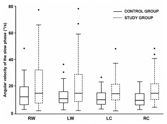
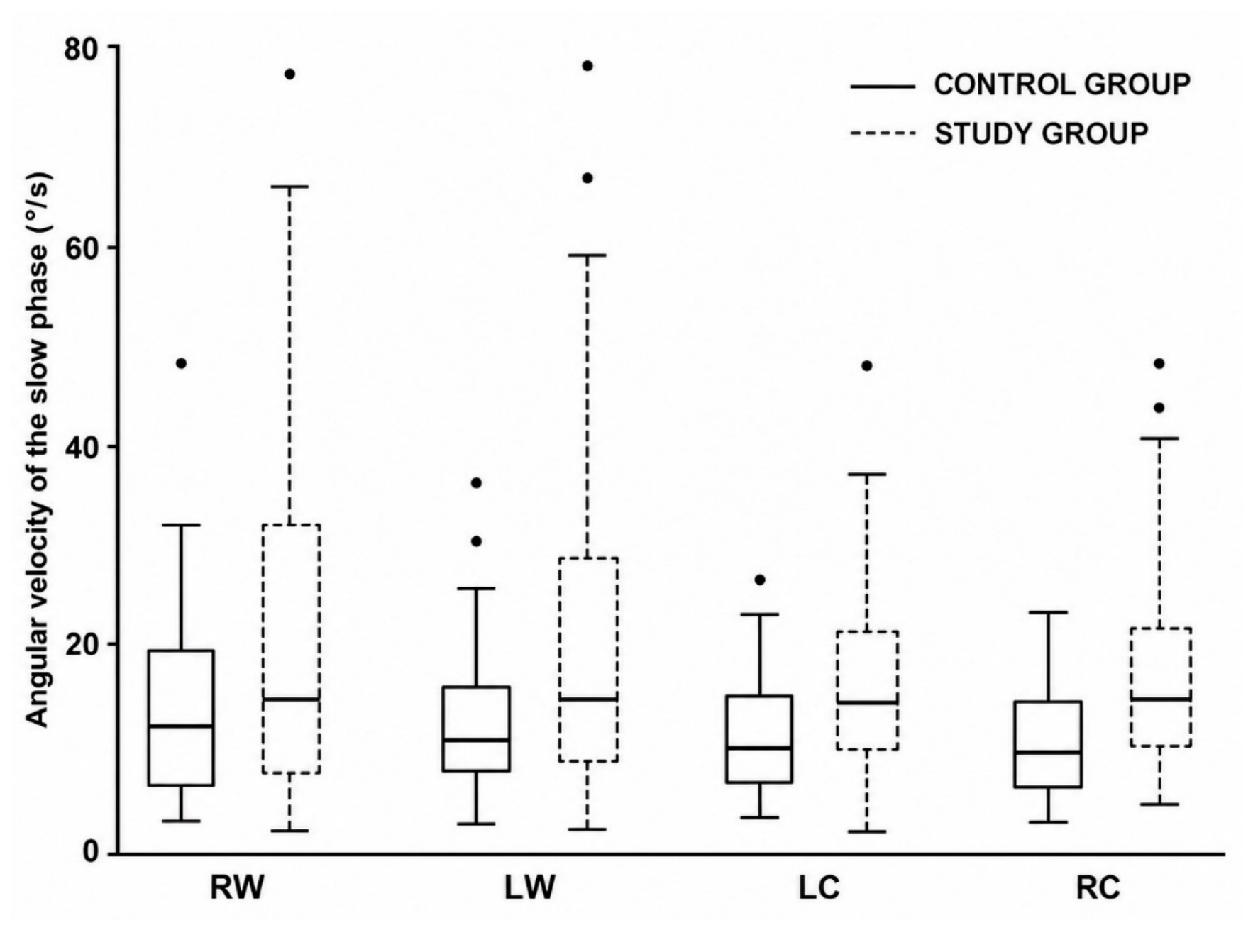
Multivariate analysis revealed a significant difference in the overall AVSP response profile between patients with VM and controls after accounting for age, sex, and their interactions with VM status (p = 0.0129). Mean AVSP values were consistently higher in the VM group across all four caloric irrigations (Table 2 and Figure 1).
Table 2.
Angular velocity of the slow phase values for each caloric stimulus in the study and control groups.
Figure 1.
Sequential analysis of AVSP during caloric testing across consecutive irrigations. RW: right warm; LW: left warm; LC: left cold; RC: right cold.
Within the study group, vestibular recruitment was not significantly associated with caloric dysrhythmia (Pearson’s chi-square test, p = 0.2851; Fisher’s exact test, p = 0.3320) or caloric hyperreflexia (Pearson’s chi-square test, p = 0.2562; Fisher’s exact test, p = 0.3725) (Table 3, Table 4, Table 5 and Table 6).
Table 3.
Association between the combined presence of vestibular migraine and caloric vestibular recruitment and caloric dysrhythmia in the entire study population (n = 98).
Table 4.
Results of Pearson’s chi-square test and Fisher’s exact test assessing the association between caloric dysrhythmia and vestibular recruitment.
Table 5.
Association between the combined presence of vestibular migraine and caloric vestibular recruitment and caloric hyperreflexia in the entire study population (n = 98).
Table 6.
Results of Pearson’s chi-square test and Fisher’s exact test assessing the association between caloric hyperreflexia and vestibular recruitment.
4. Discussion
The present study investigated the relationship between VM and vestibular recruitment, unilateral weakness, caloric dysrhythmia and hyperreflexia, and high gain on the video head impulse test (vHIT). The predominance of women and the mean age of participants with VM, which was within the fourth to fifth decades of life, are consistent with the epidemiological profile of VM. Although age was not significantly associated with VM in our analysis, the potential influence of hormonal status during the menopausal transition may be relevant based on previous literature; however, this factor was not systematically assessed in our cohort and therefore cannot be evaluated in the present study [16].
The association observed between VM and unilateral weakness adds to the growing evidence that peripheral vestibular abnormalities may contribute to the pathophysiology of the disorder. Although VM is generally regarded as a disorder of central sensory processing, accumulating evidence over the past two decades suggests that peripheral vestibular abnormalities may also contribute to its pathophysiology [2,17]. Several studies have reported an increased prevalence of unilateral vestibular hypofunction, vestibular asymmetries, and otolithic abnormalities in patients with VM [18,19]. Proposed mechanisms underlying peripheral involvement include transient vasospasm of the labyrinthine circulation, neuroinflammation mediated by trigeminovascular peptides, and direct modulation of peripheral vestibular nerve endings by CGRP [20,21,22]. From this perspective, unilateral caloric weakness may be the functional expression of recurrent episodes of peripheral vestibular dysfunction throughout the course of the disease. This concept is further supported by a previous study that found no association between unilateral weakness and vestibular recruitment, suggesting that the magnitude of vestibular asymmetry does not necessarily determine the state of vestibular compensation [9].
One should also note the independent association between VM and vestibular recruitment. Even after adjustment for other vestibular markers, patients with VM had a significantly higher likelihood of exhibiting vestibular recruitment. This finding is particularly relevant because vestibular recruitment has traditionally been considered a manifestation of incomplete vestibular compensation following peripheral vestibular injury [9,23,24]. It may indicate reduced efficacy of compensatory mechanisms or insufficient neural reorganization following a reduction in peripheral vestibular input. Impaired compensation may be related to dysfunction of glutamatergic neurotransmission and intracellular calcium homeostasis, which are directly involved in vestibular adaptation and, consequently, central compensation. The relationship between VM and central compensation can be understood in light of experimental evidence demonstrating anatomical and functional connectivity among the trigeminovascular system (TVS), vestibular nuclei, vestibulocerebellum, and thalamic structures, forming a network capable of integrating nociceptive and vestibular information [20,25]. Recurrent alterations within these circuits could interfere with the neural plasticity mechanisms required for effective vestibular compensation.
Caloric dysrhythmia was also significantly associated with VM, suggesting that alterations in the temporal organization of vestibular responses may represent another relevant component of the vestibular pathophysiology of the disorder. The vestibulocerebellum, particularly the flocculus, nodulus, and uvula, exerts continuous control over the vestibular nuclei through the inhibitory activity of Purkinje cells, regulating vestibular gain, vestibulo-ocular reflex adaptation, and the velocity storage mechanism [26]. Loss of this modulation may result not only in increased vestibular excitability but also in temporally unstable or irregular responses. Thus, the dysrhythmia observed in this study may represent an indirect functional marker of disorganization within the vestibulocerebellar circuits.
Caloric responses in VM were significantly greater than those of controls in the multivariate analysis of AVSP, and caloric hyperreflexia was more common in migraineurs, although this finding did not reach statistical significance. The study sample may have been insufficient to detect a statistically significant difference in the frequency of caloric hyperreflexia. Nevertheless, this pattern is compatible with the concept of VM as a disorder involving altered neuronal excitability, including changes in glutamatergic neurotransmission, GABAergic modulation, and ion channel function [1,3,27,28,29,30,31]. Given the modulatory role of the vestibulocerebellum over the vestibular nuclei, alterations in these mechanisms may potentially contribute to the amplification of low-frequency vestibular responses elicited during caloric testing. However, these mechanisms were not directly evaluated in the present study and therefore should be regarded as possible pathophysiological explanations rather than mechanisms demonstrated by our findings.
One of the most relevant results of this study was the absence of an association between vestibular recruitment and the markers of vestibular hyperexcitability, represented by caloric dysrhythmia and hyperreflexia. These observations do not appear to represent different stages of the same pathophysiological process, but rather distinct functional states of the vestibular system. Whereas vestibular recruitment indicates reduced peripheral vestibular input associated with incomplete compensation, caloric hyperreflexia and dysrhythmia may reflect central vestibular hyperexcitability or disorganization of central vestibular circuits.
This interpretation is consistent with current models of VM, which propose extensive functional interactions among the trigeminovascular system, vestibular nuclei, and vestibulocerebellum [1,6,17,32]. CGRP receptors have been identified in peripheral vestibular structures and vestibular nuclei, suggesting a mechanism capable of simultaneously influencing peripheral and central vestibular function [22]. Furthermore, vestibular symptoms have been reported to improve following treatment with anti-CGRP monoclonal antibodies, further supporting the involvement of this neuropeptide in the pathophysiology of VM [33]. Thus, the coexistence of unilateral weakness, vestibular recruitment, and dysrhythmia, along with the lack of association between vestibular recruitment and caloric dysrhythmia or caloric hyperreflexia, could reinforce the concept that VM should not be interpreted exclusively as a central or a peripheral disorder. Rather, our findings support an integrative model in which peripheral vestibular abnormalities coexist with dysfunction of central vestibular modulatory mechanisms [34]. This model can be investigated more effectively using caloric testing, as it is the only vestibular test capable of delivering unilateral, sustained vestibular stimulus. The evoked response reflects not only the function of the peripheral vestibular end-organ but also the activity within the internuclear commissural pathways and the integrity of central mechanisms involved in vestibular response modulation.
Conversely, high gain on vHIT was not significantly associated with VM. This observation is particularly interesting because it contrasts with recent hypotheses proposing that high vHIT gain could be a marker of central vestibular hyperexcitability [3]. One possible explanation is that alterations in glutamatergic neurotransmission might disrupt the firing patterns of Purkinje cells. Reduced cerebellar inhibition could impair control over the vestibular nuclei and the velocity storage mechanism, which is responsible for integrating and prolonging low-frequency vestibular responses [26]. Consequently, slow and sustained stimuli, such as those delivered during caloric testing, may become excessively amplified or temporally disorganized, resulting in hyperreflexia and potential temporal dysregulation of caloric responses. In contrast, vHIT predominantly assesses the high-frequency vestibulo-ocular reflex, which has minimal dependence on the velocity storage mechanism. Thus, alterations restricted to cerebellar control and velocity storage may produce exaggerated caloric responses without substantially affecting vHIT gain. In cases in which high vHIT gain is observed, it is plausible that hyperexcitability involves not only the velocity storage mechanism but also the intrinsic excitability of the vestibular nuclei, which may influence both low- and high-frequency reflexes [8,35,36]. This may explain why, in our clinical practice, high vHIT gain is frequently observed during VM attacks, whereas gain tends to return to normal or near-normal levels during the intercritical interval. Thus, the coexistence of caloric hyperreflexia with preserved vHIT responses may reflect a predominantly central alteration in vestibular modulation, potentially related to migraine-associated glutamatergic hyperexcitability and vestibulocerebellar dysfunction. This interpretation is also consistent with evidence of subclinical vestibular abnormalities in individuals with VM without dizziness, suggesting that dysfunction of central vestibular processing may represent a persistent pathophysiological phenotype of the disorder, even between acute attacks.
Taken together, these findings allow us to propose an integrative pathophysiological model to explain the heterogeneity of vestibular abnormalities described in VM. Rather than representing an exclusively central or peripheral disorder, VM appears to affect different levels of the vestibular system, resulting in partially independent functional phenotypes. We speculate that a predominantly peripheral phenotype may be characterized by unilateral weakness, vestibular recruitment, and incomplete vestibular compensation, reflecting peripheral vestibular dysfunction potentially related to activation of the TVS. The second phenotype may represent a predominantly central phenotype involving altered cerebellar modulation of vestibular nuclei, potentially reflecting changes in Purkinje cell firing patterns and GABAergic inhibitory control. This altered central vestibular modulation may contribute to the hyperexcitability reflected by caloric hyperreflexia and dysrhythmia and, in a subset of patients, by high gain on vHIT. However, these underlying mechanisms were not directly evaluated in the present study and should therefore be regarded as possible pathophysiological explanations for the observed vestibular findings rather than mechanisms directly demonstrated by our data.
This model helps explain the considerable variability in vestibular findings reported in the literature and provides a unified interpretation of abnormalities that have thus far largely been considered in isolation. It also suggests that patients classified under the same clinical diagnosis may exhibit different predominant pathophysiological mechanisms, which may contribute to differences in clinical presentation, disease course, and therapeutic response.
This study has some limitations. The sample size was based on consecutive recruitment rather than an a priori sample size calculation, and the study and control groups were not individually matched for age or sex. In addition, both air and water caloric stimulation were used, although standardized reference values were applied. Finally, the proposed pathophysiological mechanisms were not directly assessed, and the study did not include comparison with other vestibular disorders.
Longitudinal studies integrating vestibular assessment with functional neuroimaging, molecular biomarkers, and clinical follow-up will be important to validate the proposed phenotypes, determine their stability over the course of the disease, and explore their potential prognostic and therapeutic relevance. Comparisons with other vestibular disorders will also be important to determine the specificity of these phenotypes and further clarify their clinical and pathophysiological relevance, including their potential contribution to differential diagnosis.
5. Conclusions
This study identified distinct vestibular abnormalities associated with VM that involve both peripheral vestibular dysfunction and vestibular hyperexcitability. The lack of association between vestibular recruitment and caloric hyperreflexia or dysrhythmia is consistent with the hypothesis that these abnormalities represent partially distinct vestibular phenotypes. These findings may contribute to a more refined clinical characterization of VM and may provide relevant features for its differentiation from other vestibular disorders. However, their diagnostic utility and specificity require confirmation in studies directly comparing VM with other vestibular disorders.
Author Contributions
Conceptualization, R.M., L.R.L.M. and R.S.M.B.; Methodology, R.M., L.R.L.M. and R.S.M.B.; Formal analysis, R.M., L.R.L.M. and R.S.M.B.; Investigation, R.M., L.R.L.M. and R.S.M.B.; Data curation, R.M., L.R.L.M. and R.S.M.B.—original draft, R.M., L.R.L.M. and R.S.M.B.; Writing—review & editing, R.M., L.R.L.M. and R.S.M.B.; Project administration, R.M., L.R.L.M. and R.S.M.B. All authors participated equally in all phases of this work. All authors have read and agreed to the published version of the manuscript.
Funding
This research received no external funding.
Institutional Review Board Statement
This consecutive observational comparative study was approved by the Ethics Committee for the Analysis of Research Projects on 8 November 2018 (number 3.008.904), and was performed in accordance with the Declaration of Helsinki.
Informed Consent Statement
Informed consent was obtained from all subjects involved in the study.
Data Availability Statement
De-identified data supporting the findings of this study are available from the corresponding author upon reasonable request, subject to institutional approvals and applicable data protection regulations.
Acknowledgments
José Ferreira de Carvalho.
Conflicts of Interest
The authors declare no conflicts of interest.
References
- Goadsby, P.J.; Holland, P.R.; Martins-Oliveira, M.; Hoffmann, J.; Schankin, C.; Akerman, S. Pathophysiology of Migraine: A Disorder of Sensory Processing. Physiol. Rev. 2017, 97, 553–622. [Google Scholar] [CrossRef] [Scilit] [PubMed] [PubMed Central]
- Lempert, T.; Von Brevern, M. Vestibular Migraine. Neurol. Clin. 2019, 37, 695–706. [Google Scholar] [CrossRef] [Scilit] [PubMed]
- Bernetti, L.; Pellegrino, C.; Corbelli, I.; Caproni, S.; Eusebi, P.; Faralli, M.; Ricci, G.; Calabresi, P.; Sarchielli, P. Subclinical vestibular dysfunction in migraineurs without vertigo: A Clinical study. Acta Neurol. Scand. 2018, 138, 270–277. [Google Scholar] [CrossRef] [Scilit] [PubMed]
- Mangia, L.R.L.; Bittar, R.S.M. Clinical Features of Midlife Women with Isolated Episodic Vestibular Syndrome and Their Relationship with Migraine and Menopause Transition. Otol. Neurotol. 2026, 47, 716–723. [Google Scholar] [CrossRef] [Scilit] [PubMed]
- da Fonseca Neto, F.C.; Mezzalira, R.; Domene, V.; Chone, C.T. Sensitivity and specificity of the video head impulse test in the diagnosis of vestibular migraine—A systematic review and meta-analysis. Braz. J. Otorhinolaryngol. 2026, 92, 101806. [Google Scholar] [CrossRef] [Scilit] [PubMed] [PubMed Central]
- Zhang, Y.; Zhang, Y.; Wang, Y.; Zhang, X.; Qin, G.; Zhang, D.; Chen, L.; Zhou, J. Inhibition of glutamatergic trigeminal nucleus caudalis- vestibular nucleus projection neurons attenuates vestibular dysfunction in the chronic-NTG model of migraine. J. Headache Pain 2023, 24, 77. [Google Scholar] [CrossRef] [Scilit] [PubMed] [PubMed Central]
- Leigh, R.J.; Zee, D.S. The Neurology of Eye Movements, 6th ed.; Oxford University Press: New York, NY, USA, 2021. [Google Scholar]
- Laurens, J.; Angelaki, D.E. The functional significance of velocity storage and its dependence on gravity. Exp. Brain Res. 2011, 210, 407–422. [Google Scholar] [CrossRef] [Scilit] [PubMed] [PubMed Central]
- Bittar, R.S.M.; Mezzalira, R.; Ramos, A.C.M.; Risso, G.H.; Real, D.M.; Grasel, S.S. Vestibular recruitment: New application for an old concept. Braz. J. Otorhinolaryngol. 2021, 88, S91–S96. [Google Scholar] [CrossRef] [Scilit] [PubMed] [PubMed Central]
- Gonçalves, D.U.; Felipe, L.; Lima, T.M. Interpretation and use of caloric testing. Braz. J. Otorhinolaryngol. 2015, 74, 440–446. [Google Scholar] [CrossRef] [Scilit] [PubMed] [PubMed Central]
- Lempert, T.; Olesen, J.; Furman, J.; Waterston, J.; Seemungal, B.; Carey, J.; Bisdorff, A.; Versino, M.; Evers, S.; Newman-Toker, D. Vestibular migraine: Diagnostic criteria. J. Vestib. Res. 2012, 22, 167–172. [Google Scholar] [CrossRef] [Scilit] [PubMed]
- Felipe, L.; Cavazos, R. Caloric Stimulation with Water and Air: Responses by Age and Gender. Iran. J. Otorhinolaryngol. 2021, 33, 71–77. [Google Scholar] [CrossRef] [Scilit] [PubMed] [PubMed Central]
- Albertino, S.; Bittar, R.S.M.; Bottino, M.A.; Ganança, M.M.; Gonçalves, D.U.; Greters, M.E.; Mezzalira, R.; Ganança, F.F. Air caloric test reference values. Braz. J. Otorhinolaryngol. 2015, 78, 2. [Google Scholar] [CrossRef] [Scilit] [PubMed] [PubMed Central]
- Ganança, M.M.; Caovilla, H.H.; Munhoz, M.S.L.; Silva, M.L.G.; Frazza, M.M. As etapas da equilibriometria. In Equilibriometria clínica; Caovilla, H.H., Ganança, M.M., Munhoz, M.S.L., Silva, M.L.G., Eds.; Atheneu: São Paulo, Brazil, 1999; pp. 41–44. [Google Scholar]
- Hougaard, D.D.; Abrahamsen, E.R. Functional Testing of all Six Semicircular Canals with Video Head Impulse Test Systems. J. Vis. Exp. 2019, 146, e59012, Erratum in J. Vis. Exp. 2019, 143. https://doi.org/10.3791/6418. [Google Scholar] [CrossRef] [Scilit] [PubMed]
- Mangia, L.R.L.; Bittar, R.S.M. Is there any relationship between the menopause transition and dizziness? Braz. J. Otorhinolaryngol. 2026, 92, 101763. [Google Scholar] [CrossRef] [Scilit] [PubMed]
- Furman, J.M.; A Marcus, D.; Balaban, C.D. Vestibular migraine: Clinical aspects and pathophysiology. Lancet Neurol. 2013, 12, 706–715. [Google Scholar] [CrossRef] [Scilit] [PubMed]
- Neuhauser, H.; Leopold, M.; von Brevern, M.; Arnold, G.; Lempert, T. The interrelations of migraine, vertigo, and migrainous vertigo. Neurology 2001, 56, 436–441. [Google Scholar] [CrossRef] [Scilit] [PubMed]
- Zaleski, A.; Bogle, J.; Starling, A.; Zapala, D.A.; Davis, L.; Wester, M.; Cevette, M. Vestibular evoked myogenic potentials in patients with vestibular migraine. Otol. Neurotol. 2015, 36, 295–302. [Google Scholar] [CrossRef] [Scilit] [PubMed]
- Balaban, C.D. Migraine, vertigo and migrainous vertigo: Links between vestibular and pain mechanisms. J. Vestib. Res. 2011, 21, 315–321. [Google Scholar] [CrossRef] [Scilit] [PubMed]
- Sharon, J.D.; Hullar, T.E. Motion sensitivity and caloric responsiveness in vestibular migraine and Meniere’s disease. Laryngoscope 2013, 124, 969–973. [Google Scholar] [CrossRef] [Scilit] [PubMed] [PubMed Central]
- Marano, E.; Marcelli, V.; Di Stasio, E.; Bonuso, S.; Vacca, G.; Manganelli, F.; Marciano, E.; Perretti, A. Trigeminal stimulation elicits a peripheral vestibular imbalance in migraine patients. Headache J. Head Face Pain 2005, 45, 325–331. [Google Scholar] [CrossRef] [Scilit] [PubMed]
- Smith, P.F.; Curthoys, I.S. Mechanisms of recovery following unilateral labyrinthectomy: A review. Brain Res. Rev. 1989, 14, 155–180. [Google Scholar] [CrossRef] [Scilit] [PubMed]
- Lacour, M.; Helmchen, C.; Vidal, P.P. Vestibular compensation: The neuro-otologist’s best friend. J. Neurol. 2016, 263, 54–64. [Google Scholar] [CrossRef] [Scilit] [PubMed] [PubMed Central]
- Espinosa-Sanchez, J.M.; Lopez-Escamez, J.A. New insights into pathophysiology of vestibular migraine. Front. Neurol. 2015, 6, 12. [Google Scholar] [CrossRef] [Scilit] [PubMed] [PubMed Central]
- Bertolini, G.; Ramat, S.; Bockisch, C.J.; Marti, S.; Straumann, D.; Palla, A. Is vestibular self-motion perception controlled by the velocity storage? Insights from patients with chronic degeneration of the vestibulo-cerebellum. PLoS ONE 2012, 7, e36763. [Google Scholar] [CrossRef] [Scilit] [PubMed] [PubMed Central]
- Forrest, M.D.; Wall, M.J.; Press, D.A.; Feng, J. The sodium-potassium pump controls the intrinsic firing of the cerebellar Purkinje neuron. PLoS ONE 2012, 7, e51169. [Google Scholar] [CrossRef] [Scilit] [PubMed] [PubMed Central]
- Katoh, A.; Shin, S.; Kimpo, R.R.; Rinaldi, J.M.; Raymond, J.L. Purkinje cell responses during visually and vestibularly driven smooth eye movements in mice. Brain Behav. 2015, 5, e00310. [Google Scholar] [CrossRef] [Scilit] [PubMed] [PubMed Central]
- Blazquez, P.M.; Hirata, Y.; Heiney, S.A.; Green, A.M.; Highstein, S.M. Cerebellar signatures of vestibulo-ocular reflex motor learning. J. Neurosci. 2003, 23, 9742–9751. [Google Scholar] [CrossRef] [Scilit] [PubMed] [PubMed Central]
- Payne, H.L.; French, R.L.; Guo, C.C.; Nguyen-Vu, T.B.; Manninen, T.; Raymond, J.L. Cerebellar Purkinje cells control eye movements with a rapid rate code that is invariant to spike irregularity. eLife 2019, 8, e37102. [Google Scholar] [CrossRef] [Scilit] [PubMed] [PubMed Central]
- De Zeeuw, C.I.; Berrebi, A.S. Postsynaptic targets of Purkinje cell terminals in the cerebellar and vestibular nuclei of the rat. Eur. J. Neurosci. 1995, 7, 2322–2333. [Google Scholar] [CrossRef] [Scilit] [PubMed]
- Dieterich, M.; Obermann, M.; Celebisoy, N. Vestibular migraine: The most frequent entity of episodic vertigo. J. Neurol. 2016, 263, 82–89. [Google Scholar] [CrossRef] [Scilit] [PubMed] [PubMed Central]
- Edvinsson, L. The CGRP Pathway in Migraine as a Viable Target for Therapies. Headache J. Head Face Pain 2018, 58, 33–47. [Google Scholar] [CrossRef] [Scilit] [PubMed]
- Harriott, A.M.; Ayata, C. Spreading depolarization as a therapeutic target in migraine. Nat. Rev. Neurol. 2025, 21, 529–543. [Google Scholar] [CrossRef] [Scilit] [PubMed] [PubMed Central]
- Curthoys, I.S.; Zee, D.S.; Dumas, G.; Pastras, C.J.; Dlugaiczyk, J. Skull vibration induced nystagmus, velocity storage and self-stability. Front. Neurol. 2025, 16, 1533842. [Google Scholar] [CrossRef] [Scilit] [PubMed] [PubMed Central]
- Manzari, L. Velocity Storage Integrator (VSI): Mechanisms, circuits, and clinical implications A narrative review. Am. J. Otolaryngol. 2026, 47, 104834. [Google Scholar] [CrossRef] [Scilit] [PubMed]

Disclaimer/Publisher’s Note: The statements, opinions and data contained in all publications are solely those of the individual author(s) and contributor(s) and not of MDPI and/or the editor(s). MDPI and/or the editor(s) disclaim responsibility for any injury to people or property resulting from any ideas, methods, instructions or products referred to in the content. |
© 2026 by the authors. Licensee MDPI, Basel, Switzerland. This article is an open access article distributed under the terms and conditions of the Creative Commons Attribution (CC BY) license.