


Abstract
Background/Objectives: The variability of the internal acoustic meatus (IAM) and its morphological peculiarities are of high clinical significance in otologic interventions and neurosurgery. Considering the complexity of the neurovascular structures housed by the IAM, including passage of the facial nerve, the aim of the study was to highlight the morphological peculiarities of the IAM and their clinical significance. Methods: The age peculiarities of the IAM course were studied on 18 serially, transversely sectioned human embryos and fetuses, as well as on 20 newborn and 20 adult formalin-fixed hemiheads. The morphometric parameters of the IAM (width, height, length, and angles between the anterior and posterior walls of the IAM and the longitudinal axis of the petrous ridge (LAPR) were evaluated on 82 dry temporal bones. Results: In embryos and early fetal stages of development, the course of the IAM formed an acute, posteromedially open angle with the longitudinal axis of the petrous part of the temporal bone (LAP); in newborns, it was a right angle; in adults, it was an acute, anteromedially open angle. Various shapes of the IAO, such as oval (51.2%), semioval (23.2%), quadrangular (13.4%), heart-like (3.7%), and triangular (8.5%), were highlighted. The mean width of the internal acoustic opening (IAO) was 7.7 ± 2.11 mm (right/left—8.2 ± 2.22 mm/7.2 ± 1.89 mm), with a statistically significant difference (p = 0.031). The mean height of the IAO was 5.3 ± 1.43 mm (right/left—5.6 ± 1.58 mm/5.0 ± 1.22 mm), p = 0.082. The mean length of the IAM was 10.1 ± 2.83 (right/left—10.5 ± 2.96 mm/9.7 ± 2.67 mm), p = 0.214. A partial septum was revealed in 3.6% of cases, while total septation of the IAM was observed in 2.4%. The mean angle between the anterior wall of the IAM and the longitudinal axis of the petrous ridge (AWIAM/LAPR) was 21.4 ± 13.67° (right/left—22.2 ± 14.62°/20.5 ± 12.78°), p = 0.564. The mean angle between the posterior wall of the IAM and the longitudinal axis of the petrous ridge (PWIAM/LAPR) was 82.4 ± 8.02° (right/left—84.3 ± 7.93°/80.5 ± 7.76°); p = 0.032. Conclusions: In the initial stages of an individual’s ontogenesis, the IAM forms an acute, posteromedially open angle with the LAP; in newborns—a right angle; in adults—an acute, anteromedially open angle. Both morphological characteristics and morphometric parameters of the IAO and IAM vary bilaterally. The oval, semioval, quadrangular, heart-like, and triangular shapes of the IAO were highlighted. Partial and total septation of the IAM were revealed. Only the TDAIO and PWIAM/LAPR angle were statistically significant, p < 0.05, while the bilateral correlation was negligible. The established morphological variants of the IAM could predispose to facial and vestibulocochlear nerve impairments.
1. Introduction
The internal acoustic meatus is an important anatomical structure that ensures passage, protection, and function of the vestibular ganglion, vestibulocochlear nerve, facial nerve, and labyrinthine vessels [1,2,3]. The common origin of the vestibulocochlear and facial nerves, from the facioacoustic primordium [4,5], and their vicinity within the internal acoustic meatus and at the bulbopontine angle, are predisposing factors for involvement of both nerves in various pathologies, including tumors.
Over the past few decades, the incidence of vestibular schwannomas has increased [6,7], with an intrameatal to extrameatal ratio of 1:3 [8], and an occurrence between 5 and 10% of all the intracranial tumors [6,9,10], accounting for around 80% of tumors of the cerebellopontine angle [10]. Historical rates of 0.7–1.0 per 100,000 population [6,11] have risen to 3–5 cases per 100,000 person-year, with rates reaching approximately 20 cases per 100,000 person-year among adults aged 70 and older [6,7]. The latest data reported an incidence of vestibular schwannoma exceeding 1 case among 500 people, while sporadic unilateral schwannomas account for about 95% of all cases, and about 90% of bilateral schwannomas develop in people with neurofibromatosis type 2 [6]. It should be noted that vestibular schwannoma can remain small and stable for a long time, or to grow rapidly with an unfavorable outcome [12]. In older patients, small schwannomas predominate, while in married [13] and young people [6], a larger size was reported. Along with tumor detection, computed tomography and magnetic resonance imaging allow identification of cochlear nerve hypoplasia, IAM stenosis, IAM duplication and malformations [14,15,16].
Advances in genomics, high-resolution imaging and functional studies have led to the identification of the major genes involved in anatomical and clinical variability of the IAM, vestibulocochlear and facial nerves. Their development is controlled by a complex of genes that coordinate otic placode induction, neurosensory differentiation, neural crest migration, and temporal bone osteogenesis.
The IAM develops by remodeling the petrous part of the temporal bone, and its development is influenced by both osteogenesis and the presence of the facial and vestibulocochlear nerves, which explains the association between cochlear nerve hypoplasia and stenosis of the IAM [14]. The genes RUNX2, FGFR2, TWIST1 and BMP4 are involved in osteogenesis and skull base remodeling [17,18,19,20]. Changes in these genes may influence the caliber and morphology of the IAM independently of the development of the vestibulocochlear nerve, explaining certain forms of stenosis or asymmetry of the IAM.
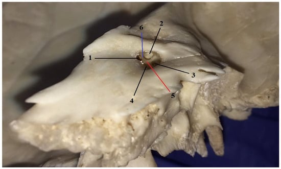
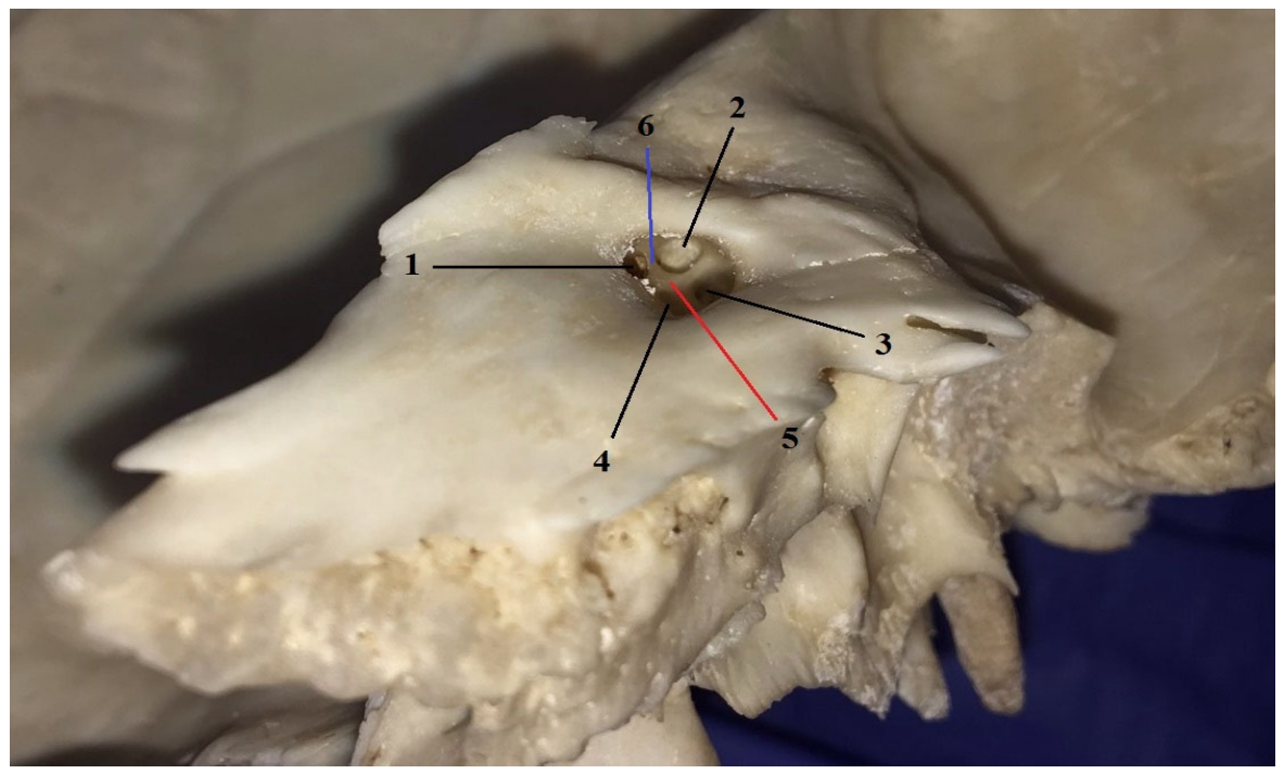
Anatomically, the internal acoustic meatus is a smooth, short, and rounded canal that starts on the posterior surface of the petrous part of the temporal bone at the internal acoustic opening and ends in a bony plate with cribriform areas, which separates it from the internal ear. It is approximately 10 mm in length and narrows towards its fundus. Its shape and size may be variable even in the same individual [21]. The bottom of the IAM is divided by the crista falciformis and Bill’s bar [16,22,23] into four areas (Figure 1).
Figure 1.
The bottom of the internal acoustic meatus of a right temporal bone: 1—anterosuperior area—inlet into the facial canal; 2—posterosuperior area or superior vestibular area; 3—posteroinferior area or inferior vestibular area; 4—anteroinferior or cochlear area; 5—crista falciformis or transverse crest; 6—Bill’s bar or vertical crest.
In a clockwise way, the anterosuperior area transmits the facial nerve; through the posterosuperior area passes the superior branch of vestibular nerve, consisting of the utricular, anterior ampullary and lateral ampullary nerves; the posteroinferior area serves for passage of the inferior branch of vestibular nerve, formed by the saccular and the posterior ampullary nerves (the last nerve passing through the singular foramen); and the anteroinferior area transmits the cochlear nerve [16,22]. Along with the facial and vestibulocochlear nerves, the orifices transmit the arteries and veins of the organ of hearing and balance.
Abnormalities of the IAM and inner ear are identified in about 20% of patients with congenital sensorineural loss [14], and among those anomalies, in approximately 12% of cases, a narrow IAM was reported. Stenosis and other anatomical variations in the IAM can compromise both the nerves and labyrinthine vessels, resulting in nerve compression, tinnitus, and hearing loss [24,25]. The components of the IAM are highly susceptible to iatrogenic injuries in otologic surgery, particularly in vestibular schwannoma. The anatomical peculiarities, variants, and abnormalities of the IAM are of great clinical importance in terms of surgical access.
In the surgical approach from the middle cranial fossa, as a primary landmark, the greater petrosal nerve and its relationships with the neighboring anatomical structures is often used. Among the reliable landmarks were reported the angle between the IAM and longitudinal axis of the greater petrosal nerve [26]; the angles formed between the point of intersection of the greater petrosal nerve with the mandibular nerve and the base of the cochlea; the distance between the mandibular nerve at its origin from the trigeminal ganglion and fundus of the IAM [27]; the angle formed at the union of the foramen ovale with foramen spinosum and of the last one with the internal acoustic opening [28]. According to Rai et al. [29], the distances between the trigeminal ganglion and the medial surface of the posterior root of the zygoma; the lateral end of the petrous ridge; the arcuate eminence; and the hiatus of the greater petrosal nerve were statistically significant depending on laterality (p < 0.05).
One of the main problems in otologic surgery is preservation of the facial nerve function [10]. In various genetic syndromes associated with cochlear malformations, aberrant facial nerve pathways are at high risk of lesion, particularly in the context of cochlear implantation [14,30]. Due to multiple connections with the neighboring cranial and spinal nerves, the facial nerve might be involved in the pathology of those nerves [5,31,32], especially in impairments of the vestibulocochlear nerve. In order to avoid iatrogenic injuries to the facial nerve and labyrinthine artery, particularly in their variations [31,33], a cautious surgical approach is required.
Considering that after vestibular schwannoma surgery, the most common clinical symptom is sensorineural hearing loss [25], and about 30% of patients develop delayed facial nerve paralysis [34], the aim of the study was to highlight the anatomical specific features and morphological variations in the IAO and IAM, and their clinical significance.
2. Materials and Methods
The microscopic part of our research project was conducted in Minsk, on the basis of the Bilateral Agreement between the Department of Anatomy and Clinical Anatomy of Nicolae Testemitanu State University of Medicine and Pharmacy of the Republic of Moldova and the Department of Normal Anatomy of the Belarusian State Medical University from Minsk. The macroscopic observations and the morphometry were performed at the Department of Anatomy and Clinical Anatomy of Nicolae Testemitanu State University of Medicine and Pharmacy of the Republic of Moldova.
The age-specific features of the IAM were investigated in 18 groups of serially and transversely sectioned human embryos and fetuses from the historical embryo-fetal collection at the Department of Normal Anatomy of the Belarusian State Medical University. We studied 12 embryological groups, each including from 3 to 5 embryos (totaling 48 embryos), and six fetal groups, each including from 3 to 4 fetuses (totaling 22 fetuses).
The crown-rump length (CRL) of the embryos and fetuses varied from 9 mm to 57 mm. According to the available data, for fixation of the embryos and fetuses, a 10% formalin solution was used. The histological samples were stained using hematoxylin–eosin and Bielschowsky–Bucke silver impregnation methods. The morphometric and staining data of the samples were taken from the archived database of the Department of Normal Anatomy in Minsk. For protocol description, the OLYMPUS CX31 binocular microscope (SKU: OLY-CX31RBSFA/R-0326, Olympus Corporation, Ina Plant, Ina, Japan) (eyepiece 10×, objectives 4×; 10×; 40×; 100×) and Nikon DS-Fi1, DS-L2 (Nikon Corporation, Tokyo, Japan) were used.
Additionally, age peculiarities were examined on 20 newborn formalin-fixed hemiheads (10 right and 10 left) of the same individuals and on 20 adult formalin-fixed hemiheads (10 right and 10 left) of the same individuals from the Anatomy and Clinical Anatomy Department of Nicolae Testemitanu State University of Medicine and Pharmacy.
The morphometry was performed on 82 dry temporal bones (41 right and 41 left) from adult individuals, of unknown sex and age. Out of 82 dry temporal bones, 24 bones (12 right and 12 left) belonged to the same individuals, while 58 temporal bones (29 right and 29 left) belonged to different people. The transverse diameter of the internal acoustic opening (TDIAO); the vertical diameter of the internal acoustic opening (VDIAO); the length of the internal acoustic meatus (IAM length); the angle between the anterior wall of the internal acoustic meatus and the longitudinal axis of the petrous ridge (AWIAM/LAPR); the angle between the posterior wall of the internal acoustic meatus and the longitudinal axis of the petrous ridge (PWIAM/LAPR) were measured. The longitudinal and transverse diameters of the internal acoustic opening were measured with the Vernier caliper. The length of the internal acoustic meatus was measured using an anatomic needle, introducing it through the internal acoustic opening into the internal acoustic meatus until it reached the fundus. Upon extraction from the meatus, the needle was measured with the caliper. After measuring the length of the internal acoustic meatus, the anatomic needle was reintroduced into the IAM and fixed to its anterior wall. The angle between the needle and the longitudinal axis of the petrous ridge was measured with a protractor. Then, the needle was fixed to the posterior wall of the IAM, and the angle between the needle and the longitudinal axis of the petrous ridge was also measured. For intra-observer variability, the same observer took all the measurements twice, and for the final analysis, the average of the two measurements was used. The quantitative variables were analyzed using Excel “Descriptive statistics” option in “Tool—Data Analysis”, such as AVERAGE, STDEV, CONFIDENCE, CORREL, SKEW, etc. For the arithmetic mean of the quantitative variables, the confidence interval (CI95), with a safety level (p) of 0.95 and significance level (α) of 0.05, was calculated. To compare the variances between the two groups of measured parameters, the F-test was used. For comparison of the means of two independent samples, Student’s t-test was applied. The distribution of the variables and their symmetry were determined using the Excel SKEW function. For determining the bilateral correlation, the Pearson coefficient was used.
3. Results
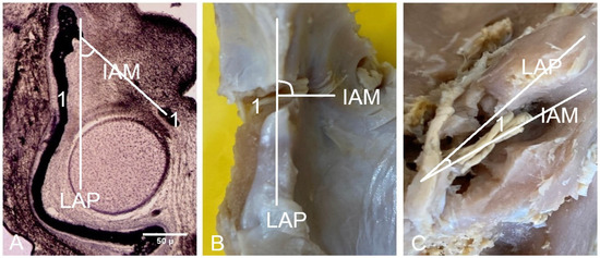
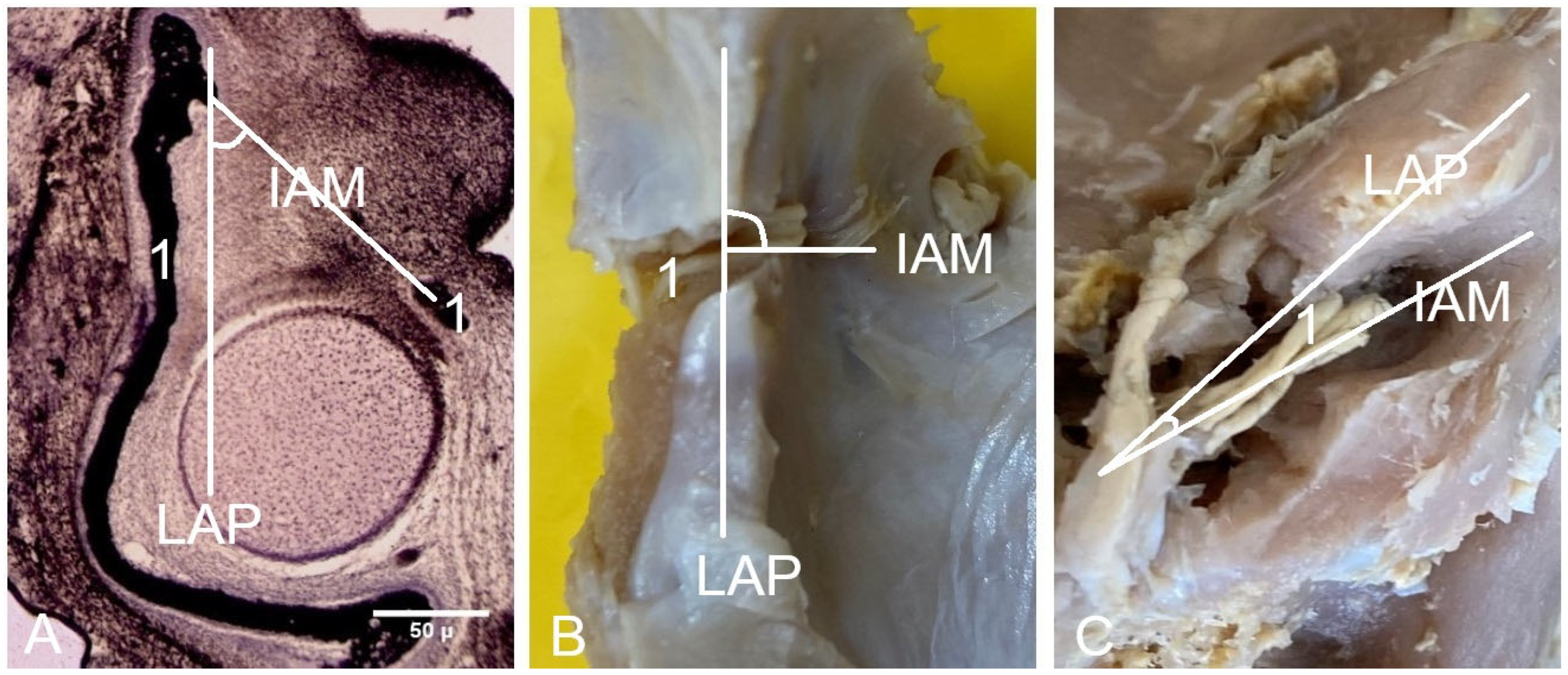
The angle between the internal acoustic meatus and the longitudinal axis of the petrous part of the temporal bone changes during ontogenesis. The inlet into the internal auditory meatus and the facial nerve within it were first identifiable at Carnegie stage 20, by which point the angle between the IAM and the longitudinal axis of the petrous part of the temporal bone was already well defined. In embryos with a CRL of 20 mm and in fetuses with a CRL of 40 mm, that angle was acute and open posteromedially. In newborns, a right angle between the IAM and longitudinal axis of the petrous part of the temporal bone was identified, while in adults that angle was acute and directed anteromedially (Figure 2).
Figure 2.
Angle between the internal acoustic meatus and longitudinal axis of the petrous part of the temporal bone. (A) Fetus with a CRL of 40 mm; (B) Newborn; (C) Adult; IAM—internal acoustic meatus; LAP—longitudinal axis of the petrous part of the temporal bone; 1—facial nerve.
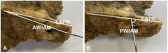
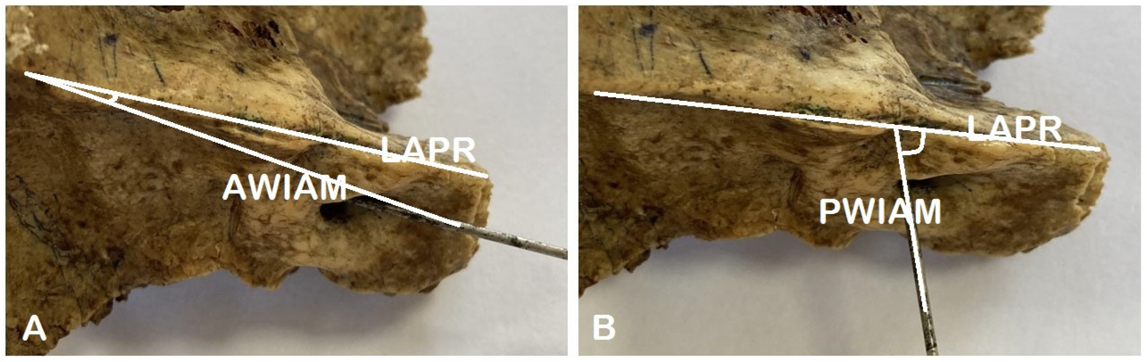
A thorough examination of the temporal bones revealed a range of macroscopic specific features and variants of both internal acoustic opening and internal acoustic meatus. These included shape variants of the IAO, partial and total septation of the IAM, and variation in the angle between the anterior wall of the IAM and the longitudinal axis of the petrous ridge (AWIAM/LAPR). The angle between the posterior wall of the IAM and the longitudinal axis of the petrous ridge (PWIAM/LAPR) was also variable (Figure 3).
Figure 3.
Angles between the anterior and posterior walls of the IAM and longitudinal axis of the petrous ridge. (A) Angle between the anterior wall of the internal acoustic meatus and the longitudinal axis of the petrous ridge (AWIAM/LAPR). (B) Angle between the posterior wall of the internal acoustic meatus and the longitudinal axis of the petrous ridge (PWIAM/LAPR).
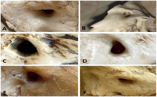
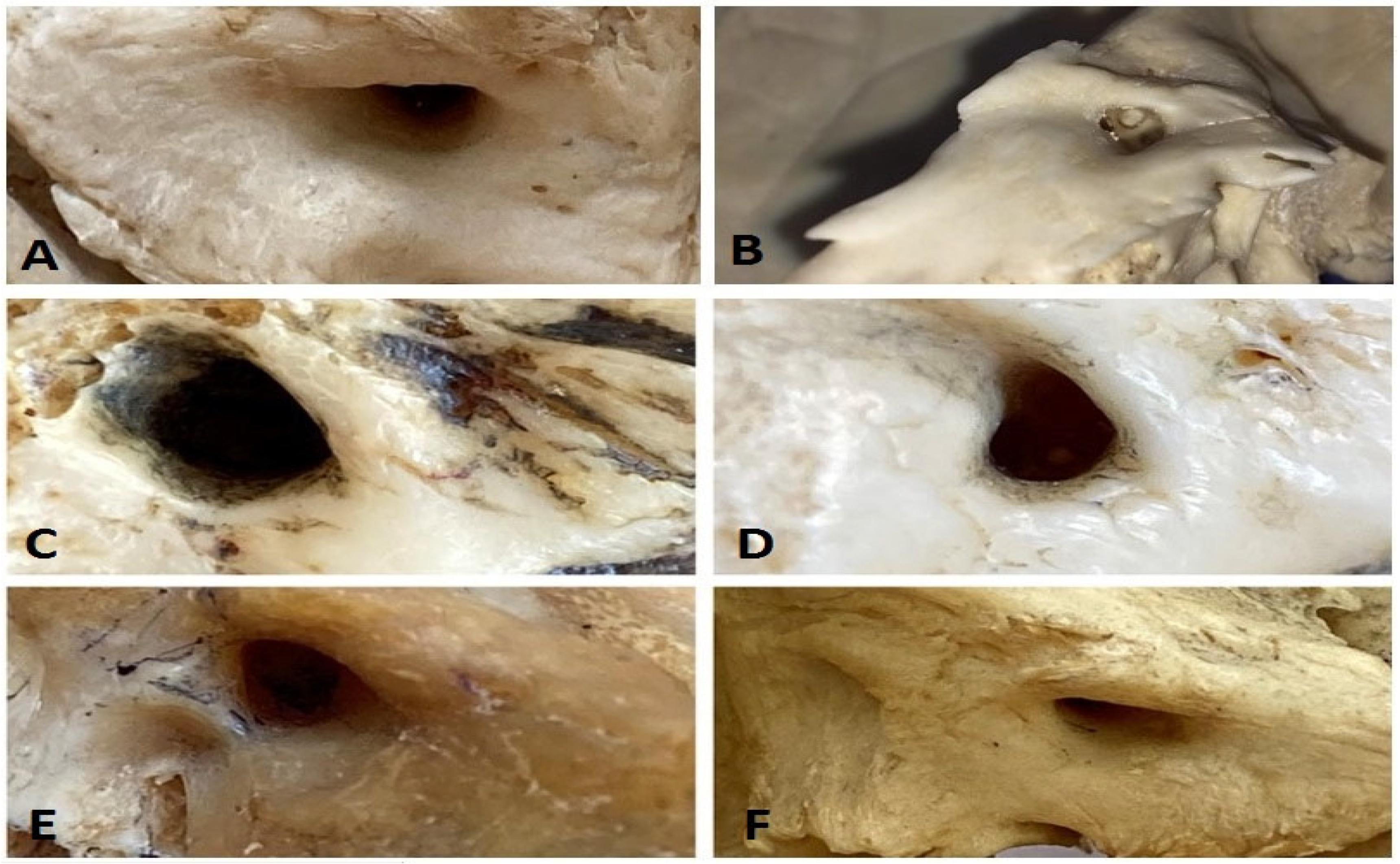
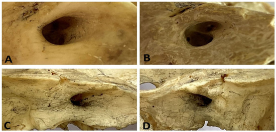
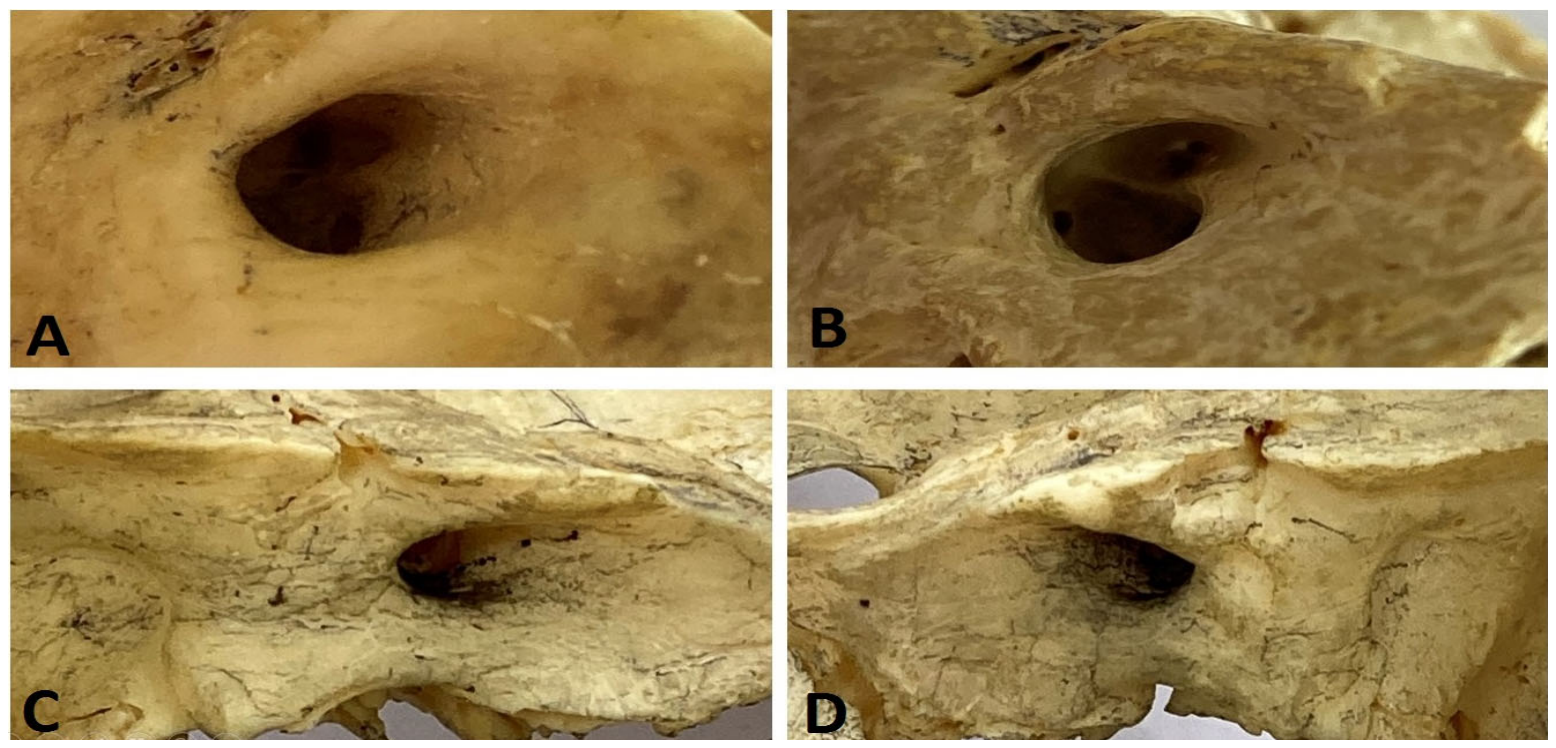
A few shape variants were characteristic of the internal acoustic opening: oval, semioval, quadrangular, heart-like, and triangular (V-shape) (Figure 4). The oval shape was determined in 42 bones (51.2%), semioval—in 19 bones (23.2%); quadrangular shape—in 11 bones (13.4%); heart-like shape—in 3 bones (3.7%); triangular shape—in 7 bones (8.5%).
Figure 4.
Variants of the internal acoustic opening. (A)—semioval; (B,C,E)—quadrangular; (D) heart-like; (F)—triangular (V-shaped).
It should be noted that along with a straight and smooth course of the IAM, 3 cases of partial (3.7%) and 2 cases of complete septation (2.4%) were identified (Figure 5).
Figure 5.
Septa of the internal acoustic meatus. (A,B) complete septum; (C,D) partial septum.
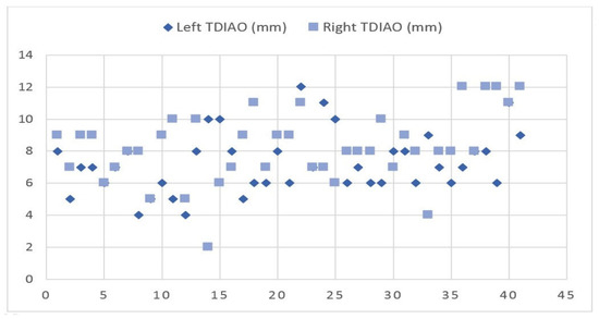
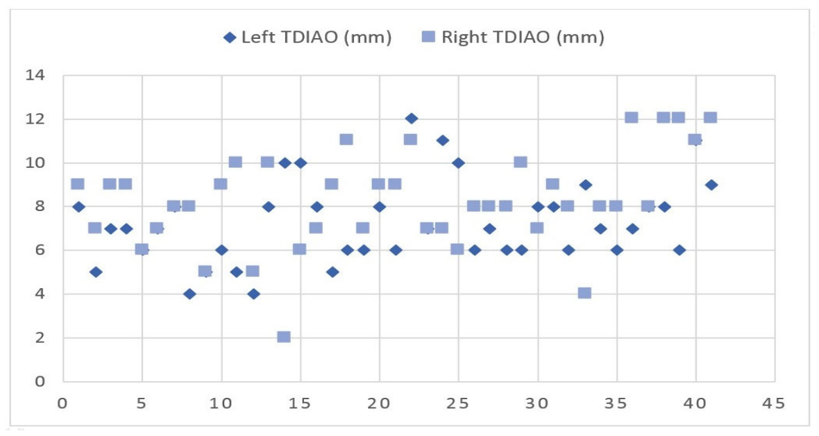
The mean transverse diameter of the internal acoustic opening (TDIAO) in the entire group was 7.7 ± 2.11 mm (max 12 mm–min 2 mm). For the right-side samples, the mean TDIAO was 8.2 ± 2.22 (max 12 mm–min 2 mm), and for the left-side samples, it was 7.2 ± 1.89 mm (max 12 mm–min 4 mm), with a statistically significant difference p = 0.031 (Table 1).
Table 1.
The morphometric parameters of the internal acoustic opening and internal acoustic meatus.
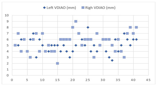
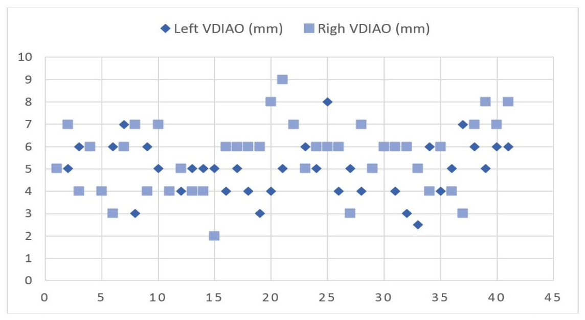
The mean vertical diameter of the internal acoustic opening (VDIAO) for the whole group was 5.3 ± 1.43 mm (max 9 mm–min 2 mm). For the right temporal bones, the mean VDIAO was 5.6 ± 1.58 (max 9 mm–min 2 mm), and for the left temporal bones, it was 5.0 ± 1.22 mm (max 8 mm–min 2.5 mm), p = 0.082 (Table 1).
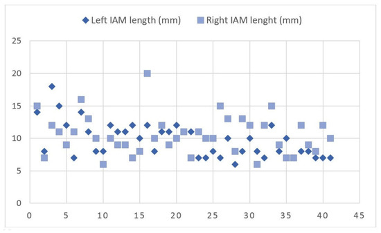
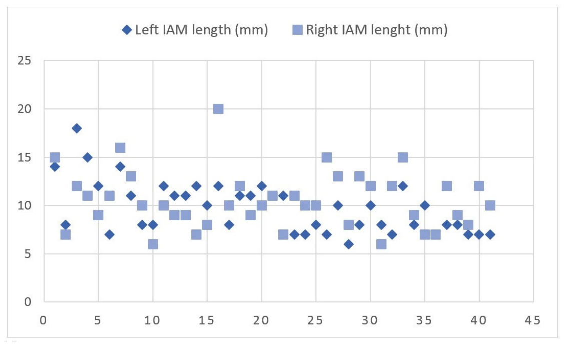
The mean length of the IAM was 10.1 ± 2.83 (max 20 mm–min 6 mm) across all the examined temporal bones. On the right bones, the mean length of the IAM was 10.5 ± 2.96 (max 20 mm–min 6 mm), and on the left bones, it was 9.7 ± 2.67 mm (max 18 mm–min 6 mm), p = 0.214 (Table 1).
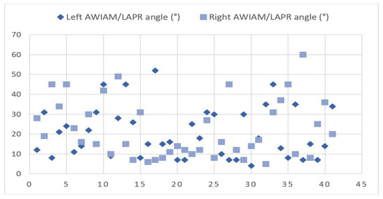
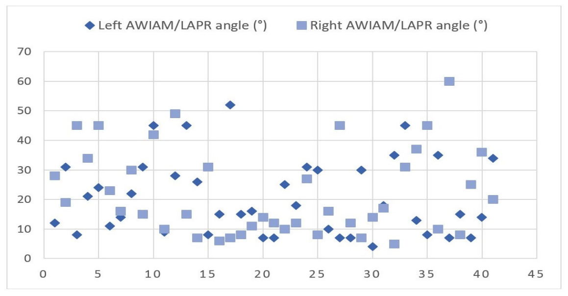
For the entire cohort, the mean angle between the anterior wall of the internal acoustic meatus and the longitudinal axis of the petrous ridge (AWIAM/LAPR) was 21.4 ± 13.67° (max 60–min 4°). When analyzed by side, the mean AWIAM/LAPR was 22.2 ± 14.62° (max 60–min 5°) on the right, and 20.5 ± 12.78° (max 52–min 4°) on the left, p = 0.564 (Table 1).
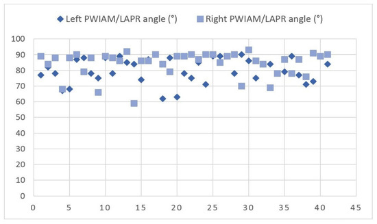
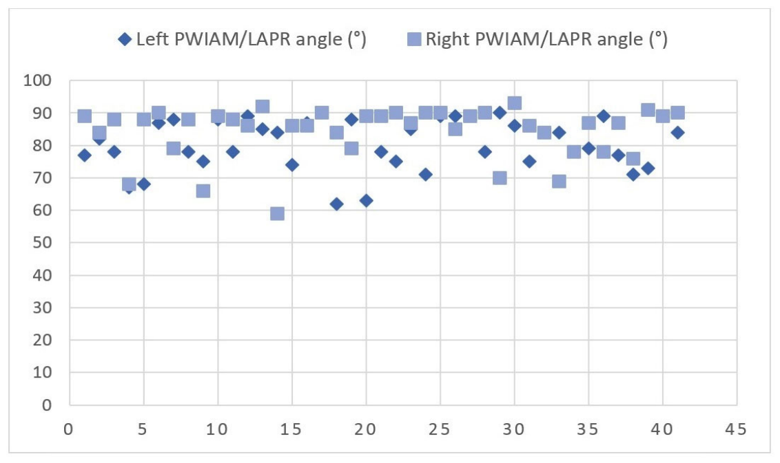
The mean angle between the posterior wall of the internal acoustic meatus and the longitudinal axis of the petrous ridge (PWIAM/LAPR) for the entire group was 82.4 ± 8.02° (max 93–min 59°). On the right side, the mean value was 84.3 ± 7.93 (max 93–min 59°), and on the left side, it was 80.5 ± 7.76° (max 90–min 62°), with a statistically significant difference depending on laterality, p = 0.032 (Table 1).
The Pearson correlation coefficient depending on laterality was very low for all the studied parameters: TDIAO (r = 0.003), IAM length (r = 0.312), PWIAM/LAP angle (r = 0.017), while for two of them the correlation was negative: VDIAO (r= −0.094) and AWIAM/LAPR angle (−0.171) (Table 1).
Our data showed a statistically significant difference between the right and left TDAIO (p = 0.031), while a negligible association with laterality (r = 0.003) was revealed. The PWIAM/LAPR angle (p = 0.032) was also statistically significant, showing a weak association (r = 0.017) depending on laterality (Figure 6, Figure 7, Figure 8, Figure 9 and Figure 10).
Figure 6.
Correlation between the left TDIAO and right TDIAO.
Figure 7.
Correlation between the left VDIAO and right VDIAO.
Figure 8.
Correlation between the left IAM length and right IAM length.
Figure 9.
Correlation between the left AWIAM/LAPR and right AWIAM/LAPR.
Figure 10.
Correlation between the left PWIAM/LAPR and right PWIAM/LAPR.
4. Discussion
The internal acoustic opening and internal acoustic meatus are characterized by various shapes, presence of septa, stenosis, partial duplication, which occur due to disturbances of embryological development, or they can be acquired as a result of disrupted osteogenesis and some bone diseases [1,3,25,35].
The oval, irregular, fissure-shaped, U-shape, V-shape variants of the IAO [1,35] and elliptical IAO were reported [35]. In the current study, oval, semioval, quadrangular, heart-like, and triangular shapes of the IAO were identified.
The main morphological changes in the IAO occur until 10 years of age, preponderantly increasing its transverse diameter, while the vertical diameter continues its growth until 15–16 years of age. The IAO transverse diameter increases from 5 mm in a newborn up to 10 mm at ages of 15–17 years [35], while the IAM fundus attains adult dimensions at 21 weeks of intrauterine development [36]. The postnatal modifications of the IAM occur due to growth of the bone around the otic capsule [36], and the shape of the IAO and IAM could be influenced by the pneumatization of the temporal bone [35,36], as a result of which, the medial part of the IAM lengthens, and the medial edge of the IAO elevates [35]. The transverse diameter of the IAO is completed earlier than the vertical one [35].
According to Mamatha et al. [37], the mean height of the IAO on the right side was 3.56 mm, and the mean transverse diameter was 3.86 mm. On the left side, the vertical diameter had a mean of 3.44 mm, and the mean of the transverse diameter was 3.47 mm. Saygin et al. [1] reported a width of the right IAO of 6.83 ± 1.59 mm and of the left IAO of 6.31 ± 1.47 mm. The height of the right IAO was 4.12 ± 1.01 mm and of the left one, it was 4.54 ± 0.88 mm.
Marques et al. [15] obtained via CT examination a mean width of the IAO of 7.10 mm and a mean height of 4.47 mm, while Gözen et al. [38] reported a mean diameter of the right IAO of 7.91 ± 1.55 mm and of the left IAO of 8.10 ± 1.46 mm [38]. In the current study, the mean transverse diameter of the IAO was 7.7 ± 2.11 mm (right/left—8.2 ± 2.22 mm/7.2 ± 1.89 mm), and the mean vertical diameter was 5.3 ± 1.43 mm (right/left—5.6 ± 1.58 mm/5.0 ± 1.22 mm). Our morphometric parameters are consistent with those of Gözen et al. [38] and Marques et al. [15]. Nevertheless, Gözen et al. [38] reported a higher width of the left IAO, while we have obtained a higher width of the right IAO.
Some studies highlighted a slight predominance of the IAO width over its height [21,37], while in our study the ratio width/height (TDIAO/VDIAO) was 1.45:1. A range of authors reported asymmetric data of the IAM with prevalence of the left side parameters [21,38,39], while in our study the right parameters were higher. The asymmetry between the right and left sides in the current study could be conditioned by the heterogeneity of the temporal bones and a possible cranial asymmetry.
The most common normal variants of the IAM are the cylindrical and the funnel-shaped ones [15,40,41]. Mntungwa et al. [41] found the cylindrical shape in 57.14%, funnel shape in 40.48%, and bud shape in 2.38% of cases, while Marques et al. [15] reported a prevalence of funnel shape in both children and adults with a rate of 74%/55.3%, respectively, followed by cylindrical shape—22%/30.9% and the bud shape—4%/10.8%.
Özandaç et al. [21] obtained a mean length of the right IAM of 9.71 mm and a mean length of the left IAM of 9.92 mm. In our study, the IAM length was 10.1 ± 2.83 mm. Slightly higher values were obtained via CT by Gözen et al. [38], who determined on the right side a mean of 11.03 ± 1.63 mm and on the left side a mean of 11.13 ± 1.62 mm, and by Marques et al. [15], who obtained on CT a mean of the IAM length of 12.63 mm.
According to Özandaç et al. [21], in females, the length of the IAM on the right and left sides was 9.71 ± 1.46 mm and 9.92 ± 1.45 mm, respectively; in males, the length on the right and left sides was 9.61 ± 1.58 mm and 9.87 ± 1.64 mm, respectively. In our study, the mean length of the right IAM was 10.5 ± 2.96 mm and of the left IAM, it was 9.7 ± 2.67 mm. Our findings reveal a longer mean length of the right IAM compared with the left, a result that is inconsistent with the cited study.
In females, Özandaç et al. [21] reported a height of the right IAM of 4.65 ± 0.69 mm and of the left IAM of 4.66 ± 0.69 mm, while in males, the right and left mean values were 5.13 ± 0.85 mm and 5.13 ± 0.90 mm, respectively. In females, the IAM width on the right and left sides was 3.97 ± 0.6 mm and 3.95 ± 0.61 mm, respectively; in males, it was 4.18 ± 0.74 mm and 4.15 ± 0.71 mm, respectively.
Gibelli et al. [39] found that the left IAM was wider compared with the right one, and in females the lateral angle was higher than in males, correlating with widths of the IAM in both sexes. Even if the lateral angle in females was higher (45.5 ± 7.1) than in males (41.0 ± 6.7), and statistically significant (p < 0.01) [42], its cautious usage in sex differentiation was recommended [38,39,42].
The postnatal morphological changes mainly influence the width and the length of the IAM [43], while its volume gradually increases throughout an individual’s life, rising from 76.30 ± 24.24 (mm3) in early childhood (1 < 3 years), to 189.89 ± 61.75 (mm3) in adulthood (20–67 years) [2,36], significantly increasing until age of 10 years and without significant changes after late adolescence [36]. Conversely, the diameter of the IAM fundus remains constant across different age groups [43]. Maturation of the internal acoustic meatus is largely complete by 12–15 years of age [44]. Analyzing the data reported in the literature, we have noted that the morphometric parameters of the IAM vary among different ethnic groups, depending on sex and on age.
Variations in the configuration and size of the IAM are closely associated with the development of the vestibulocochlear nerve, which aplasia correlates with IAM abnormalities in 92% [45]. The hypoplasia or aplasia of the VIII pair of cranial nerves is frequently associated with congenital stenosis [46,47], agenesis [46], enlarged IAM, enlarged and narrowed fundus, defects of the lateral end of the IAM, hypoplasia or duplication of the IAM, suggesting a direct morphogenetic relationship between vestibulocochlear nerve and temporal bone development [14,46,48,49]. Stenosis of the IAM might be associated with the aberrant course of the facial nerve [47,50] and agenesis of the cisternal and intracanalicular parts of the vestibulochochlear nerve [47]. In cases of a dilated vestibule and malformation of the lateral semicircular canal, the facial nerve is displaced antero-medially, which increases its iatrogenic risk [50]. Along with shape and size variants, the IAM fundus is also variable. Double transverse crests, singular anterior crest splitting posteriorly into two branches, variable number of transverse crest foramina from one to four, an anterior crest structure narrowing the entrance into the facial canal, and a crest between foramina of the same nerve were highlighted [51]. In a rudimentary otocyst, the agenesis of the internal ear occurs, resulting in a common cavity communicating with the IAM [52], or an incomplete, extremely small oval or round otic capsule with agenesis of the IAM. The bulbous shape of the IAM and an incomplete separation of the lateral end of the IAM might result in communication of the subarachnoid space with the perilymphatic space of the cochlea [50,53,54], which can lead to recurrent meningitis [52,54].
Various landmarks were reported to be feasible in surgical access of vestibular schwannomas; nevertheless, the postoperative complications and mortality cannot be entirely avoided. Despite the possibility of facial nerve preservation, the use of the translabyrinthine approach in vestibular schwannomas results in total hearing loss [55,56,57], and surgeons often prefer the retrosigmoid approach with cerebellar retraction [57].
In anterior petrosectomy, for safe surgical access of the IAM as a reliable landmark, the angle formed between the lines connecting the foramen spinosum with the foramen ovale and with the internal acoustic meatus was reported. The mean value of that angle, on both dry skulls and human heads, measured radiologically, was 95 ± 1.97° [28]. An important peculiarity of the temporal bone is its pneumatization; the more extensive it is, the more laterally the labyrinth is located, facilitating the drilling of the IAM medially to the transverse crest [40].
Considering that the middle cranial fossa comprises anatomical structures highly susceptible to iatrogenic injuries and whose preservation is challenging in large vestibular schwannomas, the Kawase and extended Kawase approaches with a temporal lobe and Labbé’s vein retraction can be used to provide better exposure volume and area when the manipulation angle does not exceed 135° [58]. Gözen et al. [38] stated that an orientation angle around 180° provides a better translabyrinthine view during posterior cranial fossa surgical approach, while a lower angle would result in a narrower exposure. Even though the risk of iatrogenic injuries to the cavernous part of the internal carotid artery, oculomotor, trochlear, abducens, and facial nerves is still high [58]. Due to good exposure of the IAM, the facial nerve, and the geniculate ganglion, in tumors confined to the IAM, the middle cranial fossa access is the safest in terms of facial nerve protection, achieving a 60–80% rate in hearing preservation [59].
As feasible landmarks in the middle cranial fossa approach were reported, the distance between the point of the greater petrosal nerve intersection with the mandibular nerve and the base of the cochlea was measured, with a mean value of 10.46 ± 1.13 mm. Another reliable landmark is considered the distance between the origin of the mandibular nerve from the trigeminal ganglion and the fundus of the IAM, with a mean of 11.92 ± 1.71 mm [27]. Lesser et al. [30] established that the distance between the cochlea and adjacent anatomical structures, including IAM, does not differ between children older than 4 years and adults. However, in children younger than 4 years, those landmarks were significantly smaller.
According to Akansel et al. [42], the lateral angle of the IAM was higher in patients up to 17 years (45.8 ± 9.1°), with a non-significant decrease in patients older 18 years (42.9 ± 6.3°). Afacan et al. [60] found that the lateral angle is subjected to changes during postnatal ontogenesis. In children aged 0–2 years, the mean value of the lateral angle was 52.80 ± 9.95°, decreasing in children aged 5–12 years (46.49 ± 5.14°), and no significant changes were determined after 12 years of age.
A mean of 40.31 ± 8.01° was determined for the right lateral angle and a mean of 40.68 ± 8.92° for the left lateral angle by Gözen et al. [38], while the value of the orientation longitudinal axis of the IAM was 155.66 ± 11.19°. In our study, the mean values of both angles, AWIAM/LAPR (right/left—22.2 ± 14.62°/20.5 ± 12.78°) and PWIAM/LAPR (right/left—84.3 ± 7.93°/80.5 ± 7.76°), were higher on the right temporal bones, while the lateral angles evaluated by Gözen et al. [38] showed higher values on the left side. It should be noted that AWIAM/LAPR and the PWIAM/LAPR angles could not be used as essential surgical landmarks, while, along with the lateral angle, they can serve as additional landmarks to avoid the overextension of the neurovascular bundle when the neurinoma involves both the intracanalicular and the middle cranial fossa parts of the vestibulocochlear or facial nerves.
The current study highlighted important age-related peculiarities that characterize changes in the IAM course throughout an individual’s life. In embryonic and early fetal development, the course of the IAM formed a sharp posteromedially opened angle with the longitudinal axis of the petrous part of the temporal bone, while in newborns that angle was right and in adults it turned into a sharp anteromedially opened angle. To the best of our knowledge, there are no comprehensive studies about these peculiarities, and only Noren et al. [61] have noted that the angle between the facial and vestibulocochlear nerves and the brain stem with growth of the cephalic part of the embryo becomes sharper.
The otic capsule is a relatively stable structure, while the midline components of the skull elongate during intrauterine development. According to Hirano-Kawamoto et al. [62], at early embryonic stages the medial occipitopetrosal junction was located near the glossopharyngeal nerve, while at midterm of the intrauterine life, that junction was located in the immediate proximity of the internal acoustic meatus. The authors made a supposition that the growth of the skull might contribute to the sliding of the medial occipitopetrosal junction towards the spheno-occipital junction. During the first year of life, there is a rapid growth of the brain, which results in an increase in the neurocranium to about 65% of the adult skull, with predominant expenditure of the width of the petrous part of the temporal bone [63]. In fetuses at the age of 12 weeks, the developing tympanic cavity is almost horizontally positioned. In 19-week-old fetuses, a lateral movement of the temporal squama [64] and an increase in transverse diameter of the skull occur [65], resulting in lateral rotation of the inferior wall of the tympanic cavity and its shifting into a vertical position [64]. The petrous part of the temporal bone grows rapidly during the first year of life, both in width and in length [63,65], and the postnatal growth of the IAM occurs mainly due to growth of the bone tissue around the otic capsule [36].
In our opinion, those changes might contribute to rotation of the longitudinal axis of the IAM within the petrous part of the temporal bone, from the posterolateral to transverse and later, to anterolateral course. Considering that the orientation and position of the IAM can affect the visualization of the facial nerve in the translabyrinthine approach [44], our findings regarding ontogenetic morphological changes in the IAM course, towards the longitudinal axis of the petrous part of the temporal bone, are both of theoretical and clinical importance.
Some limitations of the current study should be noted, namely the low number of samples and the heterogeneity of the temporal bones, which were randomly collected from various people; the lack of sex and age information; the manual measurement of the investigated parameters; the modified edges of IAO on some temporal bones; and the evaluation of the parameters, based solely on laterality. Although the shape of the IAO was clearly distinguishable on the examined samples, the shape of the IAM was visible only in its initial part. This, combined with the inability to manually measure parameters within the IAM compared with CT imaging, represents a serious limitation of the current study.
5. Conclusions
An important ontogenetic peculiarity of the internal acoustic meatus course was highlighted in the current study. In embryonic and early fetal stages of development, the course of the IAM crossed the longitudinal axis of the petrous part of the temporal bone at an acute angle open posteromedially, while in newborns the IAM crossed the LAP at a right angle and in adults that angle became acute and open anteromedially. Both the internal acoustic opening and the internal acoustic meatus were bilaterally variable, and their mean morphometric parameters were higher on the right side. Five variants of the IAO shape were established: oval (51.2%), semioval (23.2%), quadrangular (13.4%), heart-like (3.7%), and triangular (8.5%). In 3.6% of cases, a partial septum and in 2.4% total septation of the IAM was observed. Only the TDAIO and PWIAM/LAPR angle were statistically significant, p < 0.05, while the bilateral correlation was negligible. The established morphological variants of the IAM could predispose to facial and vestibulocochlear nerve impairments.
Author Contributions
Conceptualization, A.B. and L.A.; methodology, A.B., L.A., Z.Z., S.C. and I.C. (Ilia Catereniuc); software, A.B., S.S. (Stanislav Strisca) and A.M.; validation, A.B., L.A., S.C. and Z.Z.; formal analysis, A.B., L.A., Z.Z., S.L. (Sofia Lehtman), G.M. and E.S.; investigation, A.B. and L.A.; resources, A.B., L.A., Z.Z., S.S. (Silvia Stratulat), S.C. and S.L. (Sofia Lehtman); data curation, A.B. and L.A.; writing—original draft preparation, A.B., L.A. and S.C.; writing—review and editing, A.B., L.A., Z.Z., S.C., S.S. (Silvia Stratulat), I.C. (Ilia Catereniuc), N.C., L.G., D.B., M.B., I.C. (Igor Cemortan), S.L. (Svetlana Lozovanu), A.M., G.M., S.S. (Stanislav Strisca), M.D., I.D., S.B., L.N., E.S., O.C. and S.L. (Sofia Lehtman); visualization, A.B., L.A., Z.Z. and A.M.; supervision, A.B., L.A., Z.Z. and S.L. (Sofia Lehtman); project administration, A.B. and S.L. (Sofia Lehtman). All authors have read and agreed to the published version of the manuscript.
Funding
This research received no external funding.
Institutional Review Board Statement
The research project was approved by the Ethics Committee of Nicolae Testemitanu State University of Medicine and Pharmacy, based on Minute no. 1 dated 19 September 2014. The study was conducted according to the ethical principles of the Declaration of Helsinki for medical research. Code of the research: CZU: [611.831.7+616.833.17] (043.2), approved by the Scientific Council of Nicolae Testemitanu State University of Medicine and Pharmacy, Minute no. 2 of 2 October 2014.
Informed Consent Statement
Patient consent was waived due to the following reasons: 1. The current study was conducted on histological samples from the historical embryo-fetal collection of the Department of Normal Anatomy of the Belarusian State Medical University (BSMU) from Minsk, based on the Bilateral Agreement between Nicolae Testemitanu State University of Medicine and Pharmacy of the Republic and BSMU, Minsk. A copy of the Agreement was sent to the editorial Office. 2. The morphological part of the study was carried out on dry temporal bones and on formalized cadaveric material at the Anatomy and Clinical Anatomy Department of Nicolae Testemitanu State University of Medicine and Pharmacy. Copies of the documents regarding the collection of cadaveric material were sent to the editorial Office.
Data Availability Statement
The data reported in this study is available if required. The inquiry should be addressed to the corresponding author.
Acknowledgments
We would like to express our gratitude to the Head of the Department of Normal Anatomy of the Belarusian State Medical University from Minsk, Natalya Trushel, and to Ecaterina Shestakovich for their help and support.
Conflicts of Interest
The authors declare no conflicts of interest.
Abbreviations
The following abbreviations are used in this manuscript:
| IAM | Internal Acoustic Meatus |
| IAO | Internal Acoustic Opening |
| CRL | Crown-rump length |
References
- Saygin, D.A.; Türkoglu, F.N.; KabakçI, A.D.A.; Alpa, S. Internal Acoustic Opening: Different Osseous Landmarks and Their Clinical Implications. Int. J. Morphol. 2022, 40, 1368–1375. [Google Scholar] [CrossRef]
- Song, S.W.; Chang, K.-H.; Jun, B.C. Morphometric Analysis of the Internal Auditory Canal by Developmental Change Using Three-Dimensional Computed Tomography. Surg. Radiol. Anat. 2024, 47, 4. [Google Scholar] [CrossRef] [PubMed]
- Thaneeru, A.S.; Suresh, R.; Ujjappa, J. Narrow Internal Auditory Canal With Duplication in a Patient With Sensorineural Hearing Loss and Facial Palsy: A Case Report. Cureus 2024, 16, e68957. [Google Scholar] [CrossRef] [PubMed]
- Weglowski, M.; Woźniak, W.; Piotrowski, A.; Bruska, M.; Weglowska, J.; Sobański, J.; Grzymisławska, M.; Łupicka, J. Early Development of the Facial Nerve in Human Embryos at Stages 13–15. Folia Morphol. 2015, 74, 252–257. [Google Scholar] [CrossRef] [PubMed][Green Version]
- Babuci, A.; Zorina, Z.; Catereniuc, I.; Truseli, N.; Bendelic, A.; Ostahi, N.; Lehtman, S. Development of the Facial Nerve and Its Specific Features. Mold. J. Health Sci. 2025, 12, 12–19. [Google Scholar] [CrossRef]
- Carlson, M.L.; Link, M.J. Vestibular Schwannomas. N. Engl. J. Med. 2021, 384, 1335–1348. [Google Scholar] [CrossRef] [PubMed]
- Marinelli, J.P.; Lohse, C.M.; Carlson, M.L. Incidence of Vestibular Schwannoma over the Past Half-Century: A Population-Based Study of Olmsted County, Minnesota. Otolaryngol.-Head Neck Surg. 2018, 159, 717–723. [Google Scholar] [CrossRef] [PubMed]
- Reznitsky, M.; Petersen, M.M.B.S.; West, N.; Stangerup, S.-E.; Cayé-Thomasen, P. Epidemiology Of Vestibular Schwannomas—Prospective 40-Year Data From An Unselected National Cohort. Clin. Epidemiol. 2019, 11, 981–986. [Google Scholar] [CrossRef] [PubMed]
- Foley, R.W.; Shirazi, S.; Maweni, R.M.; Walsh, K.; McConn Walsh, R.; Javadpour, M.; Rawluk, D. Signs and Symptoms of Acoustic Neuroma at Initial Presentation: An Exploratory Analysis. Cureus 2017, 9, e1846. [Google Scholar] [CrossRef] [PubMed]
- Zhang, S.; Liu, W.; Hui, X.; You, C. Surgical Treatment of Giant Vestibular Schwannomas: Facial Nerve Outcome and Tumor Control. World Neurosurg. 2016, 94, 137–144. [Google Scholar] [CrossRef] [PubMed]
- Vetricean, S.; Antohi, A.; Işciucov, M.; Eremia, C.; Popa, V. Neurinomul de Acustic. Actualitaţi în Diagnostic. Analele Ştiinţifice Ale USMF „N. Testemiţanu” 2008, 9, 198–201. (In Romanian) [Google Scholar]
- Buracovschi, M.; Borysenko, O.; Zapuhlîh, G.; Vetricean, S.; Moraru, V. Translabyrinthine Approach in Acoustic Neuroma Surgery—Case Report. Mold. J. Health Sci. 2024, 11, 74–80. [Google Scholar] [CrossRef]
- Ostler, B.; Killeen, D.E.; Reisch, J.; Barnett, S.; Kutz, J.W.; Isaacson, B.; Hunter, J.B. Patient Demographics Influencing Vestibular Schwannoma Size and Initial Management Plans. World Neurosurg. 2020, 136, e440–e446. [Google Scholar] [CrossRef] [PubMed]
- Brotto, D.; Sorrentino, F.; Cenedese, R.; Avato, I.; Bovo, R.; Trevisi, P.; Manara, R. Genetics of Inner Ear Malformations: A Review. Audiol. Res. 2021, 11, 524–536. [Google Scholar] [CrossRef] [PubMed]
- Marques, S.R.; Ajzen, S.; D’Ippolito, G.; Alonso, L.; Isotani, S.; Lederman, H. Morphometric Analysis of the Internal Auditory Canal by Computed Tomography Imaging. Iran. J. Radiol. 2012, 9, 71–78. [Google Scholar] [CrossRef] [PubMed]
- Juliano, A.F.; Ginat, D.T.; Moonis, G. Imaging Review of the Temporal Bone: Part I. Anatomy and Inflammatory and Neoplastic Processes. Radiology 2013, 269, 17–33. [Google Scholar] [CrossRef] [PubMed]
- Komori, T. Whole Aspect of Runx2 Functions in Skeletal Development. Int. J. Mol. Sci. 2022, 23, 5776. [Google Scholar] [CrossRef] [PubMed]
- Rigueur, D.; Roberts, R.R.; Bobzin, L.; Merrill, A.E. A Requirement for Fgfr2 in Middle Ear Development. Genesis 2019, 57, e23252. [Google Scholar] [CrossRef] [PubMed]
- Dhiman, S.; Panigrahi, I.; Sharma, M.; Chaudhry, C.; Garg, M. TWIST1 Gene Variants Cause Craniosynostosis with Limb Abnormalities in Asian Patients. J. Pediatr. Genet. 2024, 13, 258–262. [Google Scholar] [CrossRef] [PubMed]
- Waqas, M.; Sun, S.; Xuan, C.; Fang, Q.; Zhang, X.; Islam, I.; Qi, J.; Zhang, S.; Gao, X.; Tang, M.; et al. Bone Morphogenetic Protein 4 Promotes the Survival and Preserves the Structure of Flow-Sorted Bhlhb5+ Cochlear Spiral Ganglion Neurons in Vitro. Sci. Rep. 2017, 7, 3506. [Google Scholar] [CrossRef] [PubMed]
- Özandaç Polat, S.; Uygur, A.G.; Öksüzler, F.Y.; Öksüzler, M.; Yücel, A.H. Morphometric Measurements of the Internal Acoustic Meatus. Cukurova Med. J. 2019, 44, 419–426. [Google Scholar] [CrossRef]
- Kozerska, M.; Skrzat, J. Anatomy of the Fundus of the Internal Acoustic Meatus—Micro-Computed Tomography Study. Folia Morphol. 2015, 74, 352–358. [Google Scholar] [CrossRef] [PubMed]
- Hovland, N.; Phuong, A.; Lu, G.N. Anatomy of the Facial Nerve. Oper. Tech. Otolaryngol.-Head Neck Surg. 2021, 32, 190–196. [Google Scholar] [CrossRef]
- Zaidi, S.A.A.; Amanullah; Jafri, S.K.K.; Sharif, S. Glioblastoma Multiforme at Internal Auditory Canal. Surg. Neurol. Int. 2023, 14, 2. [Google Scholar] [CrossRef] [PubMed]
- Massetto Junior, I.; Dell’aringa, A.R.; Nardi, J.C.; Kobari, K.; Brandão, L.A.F.; Fernandes, L.B.V. Internal Auditory Meatus Stenosis--Case Report. Braz. J. Otorhinolaryngol. 2008, 74, 318. [Google Scholar] [CrossRef] [PubMed]
- Tanriover, N.; Sanus, G.Z.; Ulu, M.O.; Tanriverdi, T.; Akar, Z.; Rubino, P.A.; Rhoton, A.L. Middle Fossa Approach: Microsurgical Anatomy and Surgical Technique from the Neurosurgical Perspective. Surg. Neurol. 2009, 71, 586–596. [Google Scholar] [CrossRef] [PubMed]
- Anania, P.; Mirapeix-Lucas, R.; Zona, G.; Prior, A.; Cortes, C.A.; Muñoz Hernandez, F. Middle Cranial Fossa Approach: Anatomical Study on Skull Base Triangles as a Landmark for a Safe Anterior Petrosectomy. J. Neurol. Surg. Part B Skull Base 2021, 82, 202–207. [Google Scholar] [CrossRef] [PubMed]
- Sahana, D.; Rathore, L.; Kumar, S.; Jain, A.; Tawari, M.; Sahu, R. Internal Acoustic Meatus Localization During Anterior Petrosectomy: A Novel Technique. World Neurosurg. 2023, 175, e1255–e1264. [Google Scholar] [CrossRef] [PubMed]
- Rai, A.; L, B.; Mittal, S.; Tyagi, Y.; Arora, J.; Bansal, J. Morphometric Analysis of Petrous Bone With Emphasis on Landmarks Utilized in Lateral Skull Base Approaches. Cureus 2024, 16, e71760. [Google Scholar] [CrossRef] [PubMed]
- Lesser, J.C.C.; Brito Neto, R.V.d.; Martins, G.d.S.Q.; Bento, R.F. Cochlear Implantation through the Middle Fossa Approach: A Review of Related Temporal Bone Studies and Reported Cases. Int. Arch. Otorhinolaryngol. 2017, 21, 102–108. [Google Scholar] [CrossRef] [PubMed]
- Babuci, A.; Ashkar, L.; Zorina, Z.; Catereniuc, I.; Gavriliuc, M.; Chele, N.; Lehtman, S.; Motelica, G.; Dabija, I. Anatomical Features of the Mastoid Segment of the Facial Canal. Folia Morphol. 2025, 84, 117–126. [Google Scholar] [CrossRef] [PubMed]
- Babuci, A.; Stratulat, S.; Zorina, Z.; Catereniuc, I.; Bendelic, A.; Trushel, N.; Solomon, O.; Stepco, E.; Calancea, S.; Lehtman, S.; et al. Facial Nerve Trunk Surgical Identification: A New Approach. Folia Morphol. 2025, 84, 902–914. [Google Scholar] [CrossRef] [PubMed]
- Jeican, I.I.; Siserman, C.V.; Toader, C.; Trombitaș, V.E. Microsurgical Anatomy of the Labyrinthine Artery: A Cadaveric Microdissection Study of Number, Origin and Course at the Cerebellopontine Angle. Medicina 2026, 62, 1073. [Google Scholar] [CrossRef] [PubMed]
- Simon, M.; Althaus, L.; Burggraf, M.; Albrecht, A.; Schipper, J.; Kristin, J. Delayed Facial Nerve Palsy after Vestibular Schwannoma Resection: Risk Factors, Extent and Prognosis. Eur. Arch. Otorhinolaryngol. 2024, 281, 6385–6390. [Google Scholar] [CrossRef] [PubMed]
- Sekerci, R.; Ogut, E.; Keles-Celik, N. The Influences of Porus Acusticus Internus on Ethnicity and Importance in Preoperative and Intraoperative Approaches. Surg. Radiol. Anat. 2021, 43, 1829–1838. [Google Scholar] [CrossRef] [PubMed]
- Sakashita, T.; Sando, I. Postnatal Development of the Internal Auditory Canal Studied by Computer-Aided Three-Dimensional Reconstruction and Measurement. Ann. Otol. Rhinol. Laryngol. 1995, 104, 469–475. [Google Scholar] [CrossRef] [PubMed]
- Mamatha, Y.; Trisha, K.R.; Kumar, V. Anthropometry of Internal Acoustic Meatus in Dry Adult Human Skull Using Casting Method. Int. J. Anat. Res. 2019, 7, 6113–6118. [Google Scholar] [CrossRef]
- Gözen, E.D.; Tevetoğlu, F.; Ertaş, A.; Yener, H.M.; Kızılkılıç, O.; Soyluoğlu, A.İ. Computerized Tomography Morphometric Assessment of the Internal Acoustic Meatus: Sex Differences, Orientation Angles, and Surgical Implications. J. Clin. Med. 2026, 15, 1312. [Google Scholar] [CrossRef] [PubMed]
- Gibelli, D.; Cellina, M.; Gibelli, S.; Termine, G.; Oliva, G.; Sforza, C.; Cattaneo, C. Relationship between Lateral Angle and Shape of Internal Acoustic Canal: Cautionary Note for Diagnosis of Sex. Int. J. Leg. Med. 2021, 135, 687–692. [Google Scholar] [CrossRef] [PubMed]
- Damam, S.K.; Harugop, A.K.S. Mastering the Trans-Labrynthine Approach to Internal Acoustic Meatus: A Cadaveric Temporal Bone Study. Indian J. Otolaryngol. Head Neck Surg. 2023, 75, 127–132. [Google Scholar] [CrossRef] [PubMed]
- Mntungwa, N.; Human-Baron, R.; Hanekom, T. Morphology of the Internal Auditory Canal: Deriving Parameters from Computer Tomography Scans. An Observational STROBE-MR Study. Ear Nose Throat J. 2025, 104, 355–361. [Google Scholar] [CrossRef] [PubMed]
- Akansel, G.; Inan, N.; Kurtas, O.; Sarisoy, H.T.; Arslan, A.; Demirci, A. Gender and the Lateral Angle of the Internal Acoustic Canal Meatus as Measured on Computerized Tomography of the Temporal Bone. Forensic Sci. Int. 2008, 178, 93–95. [Google Scholar] [CrossRef] [PubMed]
- Bonaldi, L.V.; Lago, A.D.; Crema, L.C.; Fukuda, Y.; Smith, R.L. Internal Auditory Canal: Pre- and Postnatal Growth. J. Otolaryngol. 2004, 33, 243–247. [Google Scholar] [CrossRef] [PubMed]
- Rennert, R.C.; Brandel, M.G.; Steinberg, J.A.; Friedman, R.A.; Couldwell, W.T.; Fukushima, T.; Day, J.D.; Khalessi, A.A.; Levy, M.L. Maturation of the Internal Auditory Canal and Posterior Petrous Bone with Relevance to Lateral and Posterolateral Skull Base Approaches. Sci. Rep. 2022, 12, 3489. [Google Scholar] [CrossRef] [PubMed]
- Hosri, J.; Abou Chaar, J.; Tamer, C.; Barazi, R. Congenital Facial and Cochlear Nerve Hypoplasia with Cochlear Dysplasia Causing Sensorineural Hearing Loss and Facial Palsy: A Case Report. Otolaryngol. Case Rep. 2024, 32, 100618. [Google Scholar] [CrossRef]
- Sennaroglu, L.; Saatci, I. A New Classification for Cochleovestibular Malformations. Laryngoscope 2002, 112, 2230–2241. [Google Scholar] [CrossRef] [PubMed]
- Araujo Uchoa, L.R.; Luna, L.P.; Brasil Gadelha Farias, L.A.; Nascimento Lima, D.L.; De Araujo Coimbra, P.P. Internal Auditory Canal Hypoplasia Associated with Bilateral Vestibulocochlear Nerve Aplasia and Deviant Facial Nerve Course: A Case Report and MRI Findings. J. Radiol. Case Rep. 2021, 15, 17–23. [Google Scholar] [CrossRef] [PubMed]
- Takanashi, Y.; Kawase, T.; Tatewaki, Y.; Suzuki, J.; Yahata, I.; Nomura, Y.; Oda, K.; Miyazaki, H.; Katori, Y. Duplicated Internal Auditory Canal with Inner Ear Malformation: Case Report and Literature Review. Auris Nasus Larynx 2018, 45, 351–357. [Google Scholar] [CrossRef] [PubMed]
- Li, Y.; Yang, J.; Liu, J.; Wu, H. Restudy of Malformations of the Internal Auditory Meatus, Cochlear Nerve Canal and Cochlear Nerve. Eur. Arch. Otorhinolaryngol. 2015, 272, 1587–1596. [Google Scholar] [CrossRef] [PubMed]
- Grover, M.; Sharma, S.; Samdani, S.; Gupta, G.; Preetam, C.; Gera, K.; Vaishnav, J.K.; Hada, M.K. New SMS Classification of Cochleovestibular Anomalies: Our Experience with 25 Cases of Type I Anomaly. Indian J. Otolaryngol. Head Neck Surg. 2021, 73, 333–339. [Google Scholar] [CrossRef] [PubMed]
- Sarı, E.; Nteli Chatzioglou, G.; Temirbekov, D.; Aliyeva, A.; Öztürk, A.; Gürses, I.A. The Variations of Osseous Structure of the Internal Acoustic Canal: An Anatomical Study. Braz. J. Otorhinolaryngol. 2024, 90, 101414. [Google Scholar] [CrossRef] [PubMed]
- Haber, K.; Konopka, W.; Amernik, K.; Mierzwiński, J. Inner Ear Malformations—Overview, Current Classifications, Challenges in the Management. Pol. Prz. Otorynolaryngol. 2020, 9, 1–11. [Google Scholar] [CrossRef]
- Phelps, P.D.; Reardon, W.; Pembrey, M.; Bellman, S.; Luxom, L. X-Linked Deafness, Stapes Gushers and a Distinctive Defect of the Inner Ear. Neuroradiology 1991, 33, 326–330. [Google Scholar] [CrossRef] [PubMed]
- Al-Busaidi, R.S.; Habib, S.J.; Al-Lawati, A.M.; Tahhan, K.; Al-Saidi, Y. Incomplete Partition Type III: Computed Tomography Features and Cochlear Implantation Complications. Oman Med. J. 2021, 36, e286. [Google Scholar] [CrossRef] [PubMed]
- Goyal, S.; Natarajan, K.; Devarasetty, A.; Sarankumar, T.; Chauhan, N.; Kameswaran, M. Translabyrinthine Approach to Internal Auditory Meatus: A Retrospective Study. Med. J. Armed Forces India 2018, 74, 65–71. [Google Scholar] [CrossRef] [PubMed]
- Ahsan, S.; Bojrab, D.; Standring, R. Partial Hearing Preservation after Translabyrinthine Vestibular Schwannoma Resection: Case Report and Review of the Literature. J. Neurol. Surg. Rep. 2015, 76, e211–e215. [Google Scholar] [CrossRef] [PubMed]
- Gabbita, A.C.; Das, K.K.; Keshri, A.K.; Sahu, V.; Mehrotra, A.; Manogaran, R.; Bhaisora, K.S.; Srivastava, A.K.; Jaiswal, A.K.; Kumar, R. Translabyrinthine Approach for Resection of Vestibular Schwannoma: “Neurosurgeon’s Path Less Travelled”. Neurol. India 2022, 70, 1813–1818. [Google Scholar] [CrossRef] [PubMed]
- Yan, Y.; Xu, T.; Zhao, Y.; Mei, Q.; Jiang, L.; Hou, L. Exposure Region of the Kawase Approach and Its Correlation with Skull Base Anatomy: An Evaluation with Digital Models. Front. Surg. 2023, 9, 1047949. [Google Scholar] [CrossRef] [PubMed]
- Fisher, M.; McKinnon, B. Middle Cranial Fossa Approach with and without Anterior Petrosectomy. Oper. Tech. Otolaryngol.-Head Neck Surg. 2013, 24, 146–148. [Google Scholar] [CrossRef]
- Afacan, G.O.; Onal, T.; Akansel, G.; Arslan, A.S. Is the Lateral Angle of the Internal Acoustic Canal Sexually Dimorphic in Non-Adults? An Investigation by Routine Cranial Magnetic Resonance Imaging. HOMO 2017, 68, 393–397. [Google Scholar] [CrossRef] [PubMed]
- Norén, A.; Lynnerup, N.; Czarnetzki, A.; Graw, M. Lateral Angle: A Method for Sexing Using the Petrous Bone. Am. J. Phys. Anthropol. 2005, 128, 318–323. [Google Scholar] [CrossRef] [PubMed]
- Hirano-Kawamoto, A.; Honkura, Y.; Li, C.; Verdugo-López, S.; Murakami, G.; Rodríguez-Vázquez, J.F.; Katori, Y. Development of the Occipitopetrosal Junctions: A Histological Study Using Human Fetuses. Anat. Rec. 2026, 309, 333–345. [Google Scholar] [CrossRef] [PubMed]
- Libby, J.; Marghoub, A.; Johnson, D.; Khonsari, R.H.; Fagan, M.J.; Moazen, M. Modelling Human Skull Growth: A Validated Computational Model. J. R. Soc. Interface 2017, 14, 20170202. [Google Scholar] [CrossRef] [PubMed]
- Ars, B.; Ars-Piret, N. The Morphogenesis of the Tympanic Part of the Temporal Bone. Clin. Otolaryngol. Allied Sci. 1986, 11, 9–13. [Google Scholar] [CrossRef] [PubMed]
- Hilding, D.A. Petrous Apex and Subarcuate Fossa Maturation. Laryngoscope 1987, 97, 1129–1135. [Google Scholar] [CrossRef] [PubMed]

Disclaimer/Publisher’s Note: The statements, opinions and data contained in all publications are solely those of the individual author(s) and contributor(s) and not of MDPI and/or the editor(s). MDPI and/or the editor(s) disclaim responsibility for any injury to people or property resulting from any ideas, methods, instructions or products referred to in the content. |
© 2026 by the authors. Licensee MDPI, Basel, Switzerland. This article is an open access article distributed under the terms and conditions of the Creative Commons Attribution (CC BY) license.