


Highlights
What are the main findings?
- Intrathoracic glands present in varied mediastinal sites, confirming the anatomical heterogeneity of ectopic parathyroid disease.
- Minimally invasive thoracic surgery achieves consistent biochemical cure in patients with intrathoracic ectopic parathyroid glands.
What are the implications of the main findings?
- Advanced imaging improves localization and supports timely referral for thoracic management when cervical studies are negative.
- The exploratory correlation between biochemical parameters and operative duration observed in this small cohort should be interpreted cautiously, as operative complexity is primarily driven by anatomical factors rather than biochemical disease severity.
Abstract
Background/Objectives: Primary hyperparathyroidism (PHPT) is most commonly caused by a single cervical parathyroid adenoma; however, a minority of cases originate from ectopic glands, including intrathoracic localizations. Intrathoracic ectopic parathyroid glands (EPGs) are rare and pose significant diagnostic and therapeutic challenges due to their anatomical variability and overlap with other mediastinal pathologies. Methods: We retrospectively analyzed 13 consecutive patients with PHPT caused by intrathoracic EPGs, who underwent minimally invasive surgical resection between 2015 and 2024 at two Italian tertiary referral centers. Demographic characteristics, biochemical profiles, imaging findings, surgical approaches, intraoperative parathyroid hormone (ioPTH) kinetics, and postoperative outcomes were evaluated. Results: All patients underwent successful resection using video-assisted thoracoscopic surgery (VATS) or robotic-assisted thoracoscopic surgery (RATS). Ectopic glands were identified in thymic, mediastinal, retroesophageal, paratracheal, and aortopulmonary locations. Postoperative normalization of calcium, phosphate, and PTH levels confirmed biochemical cure in all patients. No intraoperative complications or 30-day mortality were observed. Higher preoperative PTH levels were associated with longer operative times only in an exploratory, non-significant manner. Conclusions: Intrathoracic EPGs should be suspected in patients with PHPT and negative cervical imaging. Minimally invasive thoracic surgery provides safe and effective treatment with excellent biochemical outcomes. Elevated preoperative PTH levels may reflect increased surgical complexity, a hypothesis that warrants validation in larger cohorts.
1. Introduction
Primary hyperparathyroidism (PHPT) is a frequent endocrine disorder characterized by the autonomous overproduction of parathyroid hormone, leading to persistent hypercalcemia and a broad spectrum of clinical manifestations. These may involve the skeletal, renal, gastrointestinal, and neuropsychiatric systems, reflecting the widespread physiological influence of calcium homeostasis. The reported incidence of PHPT varies considerably across regions and populations, largely influenced by differences in biochemical screening practices and demographic factors. A marked predominance in postmenopausal women is consistently observed. In the majority of cases—approximately 80–85%—PHPT results from a single parathyroid adenoma located in the typical cervical position, making focused cervical exploration the standard surgical approach [1,2,3].
Ectopic parathyroid glands (EPGs), however, represent a distinct diagnostic and therapeutic challenge. Arising from aberrant embryological migration of the parathyroid primordia, EPGs are identified in roughly 2–5% of patients with PHPT. Among these, intrathoracic localizations constitute nearly one third of all ectopic presentations. The anterior mediastinum and thymus are the most common sites, although deeper mediastinal or paraesophageal positions may also occur. Intrathoracic EPGs are particularly problematic because they may evade detection on routine cervical imaging and can radiologically mimic other mediastinal lesions, including thymomas, lymphadenopathy, or substernal goiters. Consequently, patients may undergo delayed diagnosis, repeated imaging, or even unsuccessful cervical exploration before the ectopic source is identified [3,4].
Over the past decade, significant advances in functional imaging and minimally invasive thoracic surgery have transformed the management of intrathoracic EPGs. High-resolution computed tomography (CT) and magnetic resonance imaging (MRI) provide detailed anatomical characterization, while nuclear medicine modalities—such as technetium 99m sestamibi scintigraphy and fluorocholine positron emission tomography/computed tomography (PET/CT)—have markedly improved preoperative localization accuracy [5]. Parallel progress in surgical techniques, particularly video-assisted thoracoscopic surgery (VATS) and robotic-assisted thoracic approaches (RATS), has reduced the morbidity historically associated with sternotomy or thoracotomy, enabling safer and more targeted resections [6].
The aim of this study is to describe the clinical presentation, localization patterns, biochemical profiles, and surgical outcomes of patients with intrathoracic ectopic parathyroid glands treated using minimally invasive thoracic surgery. Furthermore, we seek to contextualize our findings within the existing literature, highlighting evolving diagnostic strategies and contemporary operative management of this uncommon but clinically significant entity.
2. Materials and Methods
2.1. Study Design and Patient Selection
We included 13 patients with biochemically confirmed PHPT, caused by intrathoracic ectopic parathyroid glands, who underwent minimally invasive surgical resection between 2015 and 2024 at the University Hospital of Padua and Santa Maria della Misericordia University Hospital in Udine. Patients were eligible for inclusion if they had biochemically confirmed primary hyperparathyroidism, a single intrathoracic ectopic parathyroid gland identified on cross-sectional and/or functional imaging, and underwent minimally invasive thoracic surgery at one of the participating centers.
Exclusion criteria included: multiglandular disease or parathyroid hyperplasia, hereditary syndromes such as MEN1 or MEN2, and patients requiring open thoracic surgery. Patients with prior cervical parathyroid surgery were included if imaging demonstrated a clearly localized intrathoracic gland. Patients with prior thoracic surgery were included only when minimally invasive access remained feasible.
All patients provided written informed consent. The study was conducted in accordance with the Declaration of Helsinki and, given its retrospective nature and use of anonymized routine clinical data, did not require formal ethical committee approval according to hospital regulations.
2.2. Preoperative Evaluation
All patients underwent comprehensive biochemical assessments, including serum calcium, phosphate, and PTH levels. Preoperative localization studies included contrast-enhanced CT, technetium-based scintigraphy, and, in selected cases, gallium or fluorocholine PET/CT. Surgical indication was established based on biochemical evidence of PHPT and radiological suspicion of ectopic parathyroid tissue, in accordance with international guidelines.
2.3. Surgical Technique
All procedures were performed using minimally invasive thoracic surgery, either VATS or RATS. The choice between VATS and RATS was based on the following algorithm: VATS was preferred for anterior mediastinal or thymic lesions, small (<25 mm) glands, and cases without prior thoracic surgery. RATS was selected for lesions in anatomically complex regions (aortopulmonary window, retroesophageal space, paratracheal area) or when enhanced dexterity and 3D visualization were expected to facilitate dissection. Surgeon expertise and availability of the robotic platform also influenced approach selection.
VATS was performed through a triportal Copenhagen’s approach, with a utility port of 4 cm in the 5th anterior intercostal space (anterior axillary line), the camera port in the 8th intercostal space, alongside the scapular line, and the third port placed in the 8th intercostal space, a few centimeters at the bottom the scapular spine. We routinely use a 30°, 10 mm, 3D optical camera inserted through the lower access. The utility and the posterior accesses are used to perform the resection.
RATS is approached using three robotic arms out of four. Three skin incisions are performed: the first alongside the midclavicular line, in the fifth intercostal space. Another one is in the same space, but laterally, crossing the anterior axillary line. The ultimate access is performed at the third intercostal space, a few centimeters laterally from the second one. For improved maneuverability, the optic is placed in the intermediate access; then, the grasper, scissors, and a cautery can be placed in the other two arms, according to necessities. The first surgeon sits at the console while a second surgeon stands at the operatory table, helped by a senior scrub nurse.
Definitive histology was performed in all cases to confirm the presence of parathyroid tissue in the removed specimen. Hormone levels were also measured postoperatively in different time frames to assess their trend toward reduction.
2.3.1. Intraoperative PTH Monitoring Protocol
Intraoperative PTH (ioPTH) monitoring was performed using a standardized protocol at both centers. Four sequential samples were collected:
- –
- P1 (baseline): before surgical intervention;
- –
- P2 (10–15 min post-excision): used as the primary biochemical success indicator;
- –
- P3 (end of procedure): confirming stabilization of PTH decline;
- –
- P4 (postoperative day 1): verifying early postoperative biochemical normalization.
Biochemical success was defined as a ≥50% reduction from baseline (P1) at 10–15 min (P2), consistent with the Miami criterion [7]. If the PTH decline at P2 was <50%, the surgical field was re-explored to identify residual hyperfunctioning tissue.
2.3.2. Perioperative Calcium Management Protocol
All patients underwent biochemical optimization before surgery. In cases of significant hypercalcemia (serum calcium > 2.85 mmol/L), patients received intravenous hydration with isotonic saline. Bisphosphonates or calcimimetics were used selectively in patients with severe biochemical disease or delayed surgical scheduling. Vitamin D deficiency was corrected preoperatively when needed. Serum calcium, phosphate, magnesium, and PTH were measured postoperatively, and, to prevent hungry bone syndrome, all patients received routine oral calcium supplementation (1–2 g elemental calcium/day) and cholecalciferol (800–1000 IU/day). Patients were discharged with instructions for daily calcium and vitamin D supplementation and were scheduled for biochemical reassessment at 1–2 weeks postoperatively. Additional follow-up was arranged based on clinical and biochemical status.
2.4. Statistical Analysis
Descriptive statistics were reported as mean and standard deviation or with median and interquartile ranges. Normality of the distribution was assessed via running a Shapiro–Wilk test for normality. Homoschedasticity was investigated via the Breusch–Pagan test. According to the lack of significance for the Shapiro–Wilk test, statistical comparisons among groups were carried out using Student’s t-test or the linear model for ANOVA (Analysis of Variance) when appropriate. Comparisons were ruled out with the Pearson’s Chi-squared test. Linear regression was used to check relations between relevant continuous variables. Significance level for statistics was fixed at p ≤ 0.05. Analyses and plots were performed using R Statistical software 4.6.0 and Jamovi free open source 2.7.30.
3. Results
3.1. Descriptive Statistics
The cohort included 13 patients (7 women, 6 men) with a mean age of 58.4 ± 14.2 years and a mean body mass index of 23.9 ± 3.6 kg/m2. Patient blood tests were performed at a maximum of one month preoperatively. Mean preoperative serum calcium was 2.75 ± 0.30 mmol/L, phosphorus 0.87 ± 0.46 mmol/L, and PTH 201.3 ± 115.7 pg/mL (Table 1). Gallium PET/CT was performed in 61.5% of patients, while technetium-based scintigraphy identified the ectopic gland in 76.9% of cases. Ectopic parathyroid glands were located in the mediastinum (38.5%), thymus (15.4%), retroesophageal space (15.4%), aortopulmonary window (15.4%), and paratracheal region (15.4%). Median gland diameter was 20.8 mm (IQR 18–24 mm). Final pathology confirmed ectopic parathyroid tissue in all cases. Eight patients underwent VATS and five underwent RATS. Mean operative time was 82.3 ± 44.9 min. No conversion to median sternotomy or open thoracotomy occurred. Median chest drainage duration was 1.1 days, and mean hospital stay was 3.0 ± 2.0 days. No major intraoperative complications or 30-day mortality were observed, including bleeding requiring reintervention, phrenic or recurrent laryngeal nerve injury, vascular injury, or pleural complications.
Table 1.
Descriptive statistics.
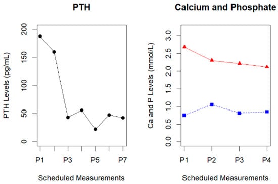
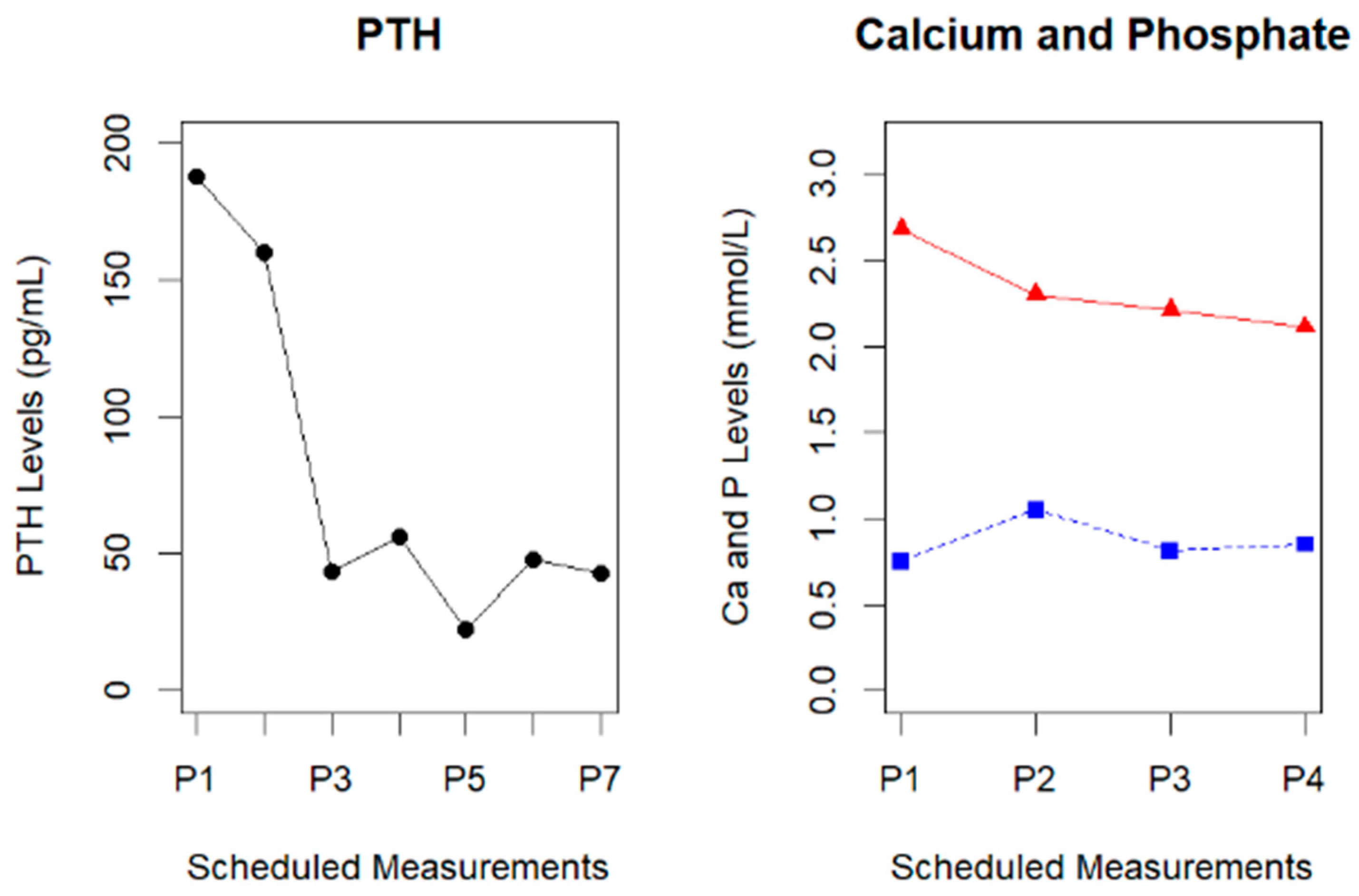
Among the intraoperative parameters, we registered a median of 202.75 pg/mL (IQR25–75 116–267 pg/mL) for the first point of PTH, 60.67 pg/mL (IQR25–75 24–78 pg/mL) for the second point, and 56.40 pg/mL (IQR25–75 45–67 pg/mL) for the third point. The perioperative PTH levels showed a progressive decrease in serum concentration. Initially (P1), the highest average level of PTH was registered before surgical intervention to the gland, with a further dropping value 15 min after resection (P2), confirming its adequate removal. In postoperative day 1 (P3), the median PTH was under 50 pg/mL, and it remained under this threshold in the postoperative surveillance (P4 to P7). All patients met the predefined biochemical success criterion of a ≥50% decline at P2. PTH levels remained stable at low values at the end of surgery (P3) and on postoperative day 1 (P4). No discordant ioPTH results were observed. In our sample, the plummeted value of the hormone did not reflect, in turn, a similar dramatic change in blood calcium and phosphate, with a slightly decreased value of Ca (from between 2.5 and 3.0 mmol/L to approximately 2.0 mmol/L) and P (a moderate increase in P2, from roughly 0.75 mmol/L to 1.0 mmol/L, and then a stable blood concentration) (Figure 1). Median follow-up was 23 months (range 9–50 months). At last evaluation, biochemical cure persisted in all patients, with a median value of PTH of 36 pg/mL (IQR25–75 31–71 pg/mL) and serum calcium of 2.11 mmol/L (IQR25–75 2.04–2.4 mmol/L).
Figure 1.
On the left side, the line graph shows the decreasing serum levels of PTH in seven different blood samples, defined as Points (P). After surgery (measurement from P3 onwards), PTH levels leveled to be stable in postoperative measurements (P4 to P7). On the right side, a decreasing level of Ca (red line with triangles) and the increasing of P (blue line with squares) during preoperative and intraoperative measurements.
3.2. Relation Between PTH Levels and Surgical Time
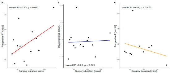
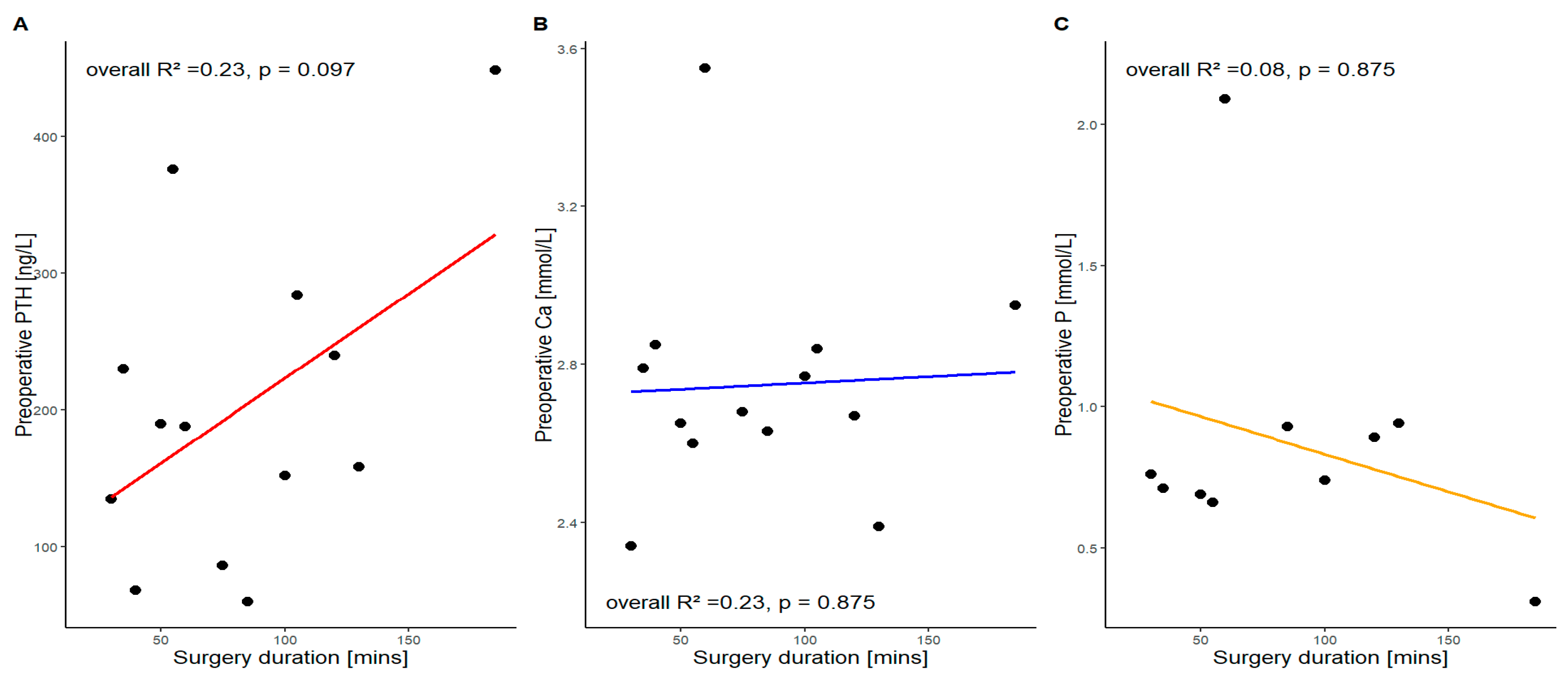
Due to the long operative time for some patients, we investigated the role of the preoperative hormone and its related ions’ levels. Given the limited sample size, the analysis did not demonstrate a statistically significant association between preoperative PTH levels and operative duration (R = 0.23, p = 0.097). This observation should be interpreted as exploratory only, and no inferential conclusions can be drawn. According to our preliminary data, preoperative Ca and P were not associated with the surgery duration (p = 0.875) (Figure 2).
Figure 2.
Scatter plots show the individual associations between preoperative PTH, serum calcium, and serum phosphate concentrations and the total operative time. (A) preoperative PTH; (B) Preoperative Ca; (C) Preoperative P. Each point represents a single patient (n = 13). Trend lines are shown solely as descriptive visual aids and do not indicate a meaningful correlation between biochemical parameters and operative complexity.
4. Discussion
Intrathoracic ectopic parathyroid glands (EPGs) represent a rare but clinically significant subset of primary hyperparathyroidism (PHPT), and their management continues to challenge even experienced endocrine and thoracic surgeons. Although PHPT is most commonly caused by a single eutopic cervical adenoma, the presence of ectopic mediastinal tissue introduces diagnostic uncertainty, increases the risk of failed cervical exploration, and necessitates specialized surgical approaches. The present study reinforces the growing consensus that minimally invasive thoracic surgery—whether video-assisted or robot-assisted—offers a safe, effective, and durable treatment option for intrathoracic EPGs when performed in high-volume centers with appropriate expertise. High biochemical cure rates, low complication rates, and short hospital stays underscore the value of these approaches in contemporary practice [1,2,3,4,5].
4.1. Embryogenesis
The inferior parathyroid glands and thymus share a common origin in the third pharyngeal pouch and descend together during embryogenesis. Variations in this migration pathway can result in parathyroid tissue becoming sequestered within the mediastinum and explains why most mediastinal EPGs are found in or near thymic tissue. Importantly, many patients with intrathoracic EPGs lack overt endocrine symptoms, and hypercalcemia may be discovered incidentally, making accurate localization even more critical [8,9,10,11].
4.2. Diagnostic Techniques
Preoperative localization remains a cornerstone of successful surgical management. While cervical ultrasonography is the firstline modality for most patients with PHPT, its utility diminishes substantially for lesions located below the thoracic inlet. Cross-sectional imaging with contrast-enhanced CT or MRI provides valuable anatomical detail, particularly regarding the relationship of the lesion to major vascular structures, the trachea, and the esophagus. However, anatomical imaging alone may be insufficient in cases of small adenomas or in patients with prior surgery [12].
Functional imaging plays a pivotal role in these scenarios. Technetium-99m sestamibi scintigraphy has historically been the workhorse of parathyroid localization, but its sensitivity decreases in the setting of multigland diseases, small adenomas, or ectopic locations. The emergence of fluorine-18 fluorocholine PET/CT has been transformative. Its superior spatial resolution and high sensitivity—often exceeding 90% in ectopic or reoperative cases—make it particularly valuable for identifying deep mediastinal lesions that may be missed with conventional imaging. The growing body of evidence supporting fluorocholine PET/CT suggests that it may become the preferred modality for complex or atypical presentations, and its integration into diagnostic algorithms warrants further study [13,14].
4.3. Surgical Approaches and Mini-Invasive Techniques
Surgical resection remains the definitive treatment for intrathoracic EPGs. Indications mirror those for cervical PHPT, encompassing symptomatic patients and asymptomatic individuals who meet established guideline criteria. Historically, median sternotomy or posterolateral thoracotomy were required to access mediastinal parathyroid tissue. Although effective, these approaches carry substantial morbidity, including postoperative pain, prolonged recovery, and increased risk of complications. The evolution of minimally invasive thoracic surgery has dramatically reshaped this landscape. Video-assisted thoracoscopic surgery (VATS) provides excellent visualization of the mediastinum through small incisions, reducing surgical trauma and facilitating rapid recovery. Robotic-assisted thoracoscopic surgery (RATS) further enhances these advantages by offering three-dimensional visualization, tremor filtration, and wristed instruments that allow precise dissection in confined spaces [6,13]. Although minimally invasive thoracic surgery avoids the morbidity associated with open thoracic approaches, our study does not include a comparator group (e.g., sternotomy).
In our multicenter series, both VATS and RATS achieved successful gland excision with no intraoperative complications or mortality. These findings align with the published literature demonstrating that minimally invasive thoracic parathyroidectomy yields high cure rates and low morbidity. A recent case report further supports this evidence base, describing long-standing primary hyperparathyroidism caused by a mediastinal ectopic parathyroid adenoma successfully managed through minimally invasive thoracic surgery. The prolonged diagnostic delay and severe metabolic complications observed in that case underscore the challenges inherent to ectopic mediastinal disease and highlight the importance of accurate preoperative localization and timely surgical referral [15]. Although our sample size limits direct comparison between VATS and RATS, robotic assistance may offer particular benefits for lesions situated in anatomically complex regions such as the aortopulmonary window, retroesophageal space, or paratracheal area. Ultimately, the choice of approach should be individualized, balancing lesion location, surgeon expertise, and institutional resources.
Intraoperative parathyroid hormone (ioPTH) monitoring has become an indispensable tool in modern parathyroid surgery. A rapid decline in PTH levels following gland excision provides real-time confirmation of biochemical cure and reduces the risk of persistent disease. While ioPTH monitoring is well established in cervical procedures, its application in thoracic parathyroidectomy is equally valuable. In our series, consistent intraoperative PTH decline confirmed complete excision in all cases, supporting its routine use in intrathoracic EPG surgery. Given the potential for anatomical complexity and the difficulty of re-exploration in the mediastinum, ioPTH monitoring adds an important layer of intraoperative assurance [16,17].
An exploratory observation in our cohort suggested a possible relationship between higher preoperative PTH levels and longer operative times; however, this association did not reach statistical significance and must be interpreted with caution, as it lacks a plausible mechanistic basis. Operative time in thoracic parathyroidectomy is determined by anatomical factors such as lesion location, adhesions, prior surgery, and the chosen minimally invasive approach, rather than biochemical disease severity. Therefore, this finding should be considered hypothesis-generating rather than indicative of a true clinical trend. Nevertheless, this finding raises intriguing questions about the relationship between biochemical disease burden and surgical complexity. Higher PTH levels may correlate with larger adenomas, more fibrotic or adherent lesions, or atypical anatomical positions that require more meticulous dissection. Similar associations have been described in cervical parathyroidectomy, where elevated PTH levels have been linked to increased operative difficulty [18]. If validated in larger studies, preoperative PTH levels could serve as a useful adjunct in preoperative planning, helping surgeons anticipate technical challenges and allocate operative resources more effectively.
When comparing thoracic parathyroidectomy with conventional cervical exploration, several important distinctions emerge. In appropriately selected patients, minimally invasive thoracic surgery achieves biochemical cure rates comparable to those of cervical surgery. However, thoracic approaches offer a distinct advantage in cases where imaging clearly localizes disease below the thoracic inlet. Attempting repeated or extended cervical exploration in such cases increases operative time, risk of complications, and patient morbidity. Early referral for thoracic evaluation—particularly when advanced imaging supports an intrathoracic location—can streamline care and improve outcomes. Nonetheless, cervical exploration remains the standard of care for the vast majority of patients with PHPT, and thoracic intervention should be reserved for cases with definitive or strongly suspected mediastinal disease [19].
4.4. Statistical and Methodological Issues
This study has several limitations that warrant consideration. Its retrospective design introduces inherent biases, including variability in imaging protocols, surgical decision-making, and follow-up practices. The small sample size reflects the rarity of intrathoracic EPGs but strongly limits the statistical power of exploratory analyses, including the widening of the confidence intervals, which preclude meaningful interpretation of the observed association between PTH levels and operative time. Additionally, all patients were treated at tertiary centers with significant experience in minimally invasive thoracic surgery, which may limit the generalizability of our findings to lower-volume institutions. Despite these limitations, the consistency of surgical success and biochemical cure across two high-volume centers strengthens the validity of our conclusions.
Looking ahead, future research should focus on refining diagnostic pathways and optimizing surgical strategies for intrathoracic EPGs. Prospective multicenter studies are needed to evaluate the comparative effectiveness of different imaging modalities, particularly the role of fluorocholine PET/CT in firstline localization. Fluorocholine PET/CT was frequently used in our cohort and facilitated the precise localization of intrathoracic glands. However, our series does not include a formal comparison with sestamibi scintigraphy, and therefore no conclusions can be drawn regarding relative diagnostic performance. Standardized criteria for selecting VATS versus RATS approaches would also be valuable, as would studies assessing cost-effectiveness and long-term outcomes. Collaborative registries may play a crucial role in advancing knowledge, enabling larger datasets and more robust analyses of this uncommon but clinically important condition.
5. Conclusions
Minimally invasive thoracic surgery (VATS/RATS) provides a safe and effective short-term treatment for intrathoracic ectopic parathyroid glands. In this multicenter series, all patients achieved early biochemical cure, with no major intraoperative complications and a short postoperative recovery. The cohort encompassed a broad spectrum of intrathoracic locations, demonstrating the versatility of minimally invasive access. Intraoperative PTH monitoring proved highly reliable, offering real-time confirmation of complete excision and supporting surgical decision-making in anatomically complex regions. While long-term outcomes require further studies, these findings reinforce the role of minimally invasive thoracic surgery as a targeted and effective option for well-localized intrathoracic parathyroid disease. Continued multicenter collaboration, standardized imaging pathways, and prospective data collection will be essential to refine patient selection, optimize surgical strategies, and deepen the understanding of this distinctive clinical entity.
Author Contributions
Conceptualization, A.B. (Alberto Busetto), A.B. (Alessandro Bonis) and V.V.; methodology, A.B. (Alessandro Bonis), G.C. and F.L.; software, A.B. (Alberto Busetto) and C.A.; validation, W.G., E.d.F., A.D. and A.Z.; formal analysis, A.B. (Alberto Busetto), A.B. (Alessandro Bonis), W.G. and V.V.; investigation, C.A., E.d.F. and F.L.; resources, A.D. and A.Z.; data curation, V.V., C.A. and G.C.; writing—original draft preparation, A.B. (Alberto Busetto); writing—review and editing, W.G., F.L., A.Z., A.D. and G.C.; visualization, E.d.F. and V.V.; supervision, G.C., A.Z. and A.D.; project administration, A.D. and A.Z. All authors have read and agreed to the published version of the manuscript.
Funding
This research received no external funding.
Institutional Review Board Statement
This study was conducted in accordance with the Declaration of Helsinki. Ethical review and approval were waived by University of Padua and Udine University Hospital for this study due to its retrospective design, the use of anonymized routine clinical data, and the absence of any intervention beyond standard of care. According to institutional and national regulations, retrospective studies not involving identifiable personal information do not require formal Istitutional Review Board approval.
Informed Consent Statement
Informed consent was obtained from all subjects involved in the study.
Data Availability Statement
The raw data supporting the findings of this study are available from the corresponding author upon reasonable request.
Conflicts of Interest
The authors declare no conflicts of interest.
Abbreviations
The following abbreviations are used in this manuscript:
| CT | Computed Tomography |
| EPG | Ectopic Parathyroid Glands |
| ioPTH | intraoperative PTH |
| IQR | Interquartile Range |
| MRI | Magnetic Resonance Imaging |
| PET/CT | Positron Emission Tomography/Computed Tomography |
| PHPT | Primary Hyperparathyroidism |
| PTH | Parathyroid Hormone |
| RATS | Robotic-Assisted Thoracic Surgery |
| VATS | Video-Assisted Thoracic Surgery |
References
- Liu, X.; Sun, L.; Shao, M.; Li, P.; Liu, W.; Zhang, X.; Zhang, L.; Ma, Y.; Li, W. Primary hyperparathyroidism due to ectopic parathyroid adenoma in an adolescent. Endocrine 2019, 64, 38–42. [Google Scholar] [CrossRef] [Scilit] [PubMed]
- Walker, M.D.; Silverberg, S.J. Primary hyperparathyroidism. Nat. Rev. Endocrinol. 2018, 14, 115–125. [Google Scholar] [PubMed]
- Levine, D.S.; Wiseman, S.M. Fusion imaging for parathyroid localization in primary hyperparathyroidism. Expert Rev. Anticancer Ther. 2010, 10, 353–363. [Google Scholar] [CrossRef] [Scilit] [PubMed]
- Boccalatte, L.A.; Peña, L.B.; Pezzotto, S.M.; De Francesco, M.V.; Rizzi, R.; Saba, G. Hiperparatiroidismo ectópico. Medicina 2020, 80, 39–47. [Google Scholar] [PubMed]
- Phitayakorn, R.; McHenry, C.R. Incidence and location of ectopic abnormal parathyroid glands. Am. J. Surg. 2006, 191, 418–423. [Google Scholar] [CrossRef] [Scilit] [PubMed]
- Iacobone, M.; Citton, M.; Viel, G.; Mondi, I.; Tropea, S.; Favia, G. Thoracoscopic Removal of Mediastinal Parathyroid Glands: A Systematic Review. Surg. Endosc. 2015, 29, 1381–1389. [Google Scholar]
- Rajamanickam, S.; Agarwal, A.; Senthilkumar, S.; Chatzoudis, D.; Curran, R.; Balasubaramaniam, V.; Williams, S. Chasing the Drop: Miami Versus Dual Criteria for Intraoperative Parathyroid Hormone Monitoring in Primary Hyperparathyroidism. Cureus 2025, 17, e96720. [Google Scholar] [CrossRef] [Scilit] [PubMed]
- Akerström, G.; Malmaeus, J.; Bergström, R. Surgical anatomy of human parathyroid glands. Surgery 1984, 95, 14–21. [Google Scholar] [PubMed]
- Cao, L.; Sun, X.; Zhang, T.; Niu, Y.; Suo, H.; Zhao, Z.; Wang, C.; Bai, J. Risk factors for early hypocalcemia after parathyroidectomy. Updat. Surg. 2022, 74, 1961–1970. [Google Scholar] [CrossRef] [Scilit]
- Salido, S.; Gómez-Ramírez, J.; Bravo, J.M.; Martín-Pérez, E.; Fernández-Díaz, G.; Muñoz de Nova, J.L.; Auza, J.; Larrañaga, E. Parathyroid adenoma in third pharyngeal pouch cyst as a rare case of primary hyperparathyroidism. Ann. R. Coll. Surg. Engl. 2014, 96, e8–e10. [Google Scholar] [CrossRef] [Scilit] [PubMed]
- Sellitri, F.; Tamburrini, A.; Tacconi, F.; Bollero, P.; Ortensi, A.; Mineo, T.C. Intrathymic ectopic parathyroid adenoma. Thorac. Cancer 2015, 6, 101–104. [Google Scholar] [CrossRef] [Scilit] [PubMed]
- Johnson, N.A.; Tublin, M.E.; Ogilvie, J.B. Parathyroid imaging: Technique and role in the preoperative evaluation of primary hyperparathyroidism. AJR Am. J. Roentgenol. 2007, 188, 1706–1715. [Google Scholar] [CrossRef] [Scilit] [PubMed]
- Kluijfhout, W.P.; Pasternak, J.D.; Gosnell, J.E.; Shen, W.T.; Duh, Q.Y. Fluorine-18 Fluorocholine PET/MR Imaging in Patients with Primary Hyperparathyroidism and Inconclusive Conventional Imaging. Surgery 2017, 161, 77–83. [Google Scholar]
- Thanseer, N.; Bhadada, S.K.; Sood, A.; Mittal, B.R.; Bhansali, A. Comparative Effectiveness of 18F-Fluorocholine PET/CT and 99mTc-Sestamibi SPECT/CT in Localization of Parathyroid Adenomas. Clin. Endocrinol. 2017, 86, 475–482. [Google Scholar]
- Topolnitskiy, Y.B.; Milovanova, T.A.; Apalkov, D.G.; Melnichenko, O.A.; Mokrysheva, N.G. Primary Hyperparathyroidism Caused by an Ectopic Parathyroid Adenoma in the Mediastinum (Case Report). Endocr. Surg. 2024, 18, 36–42. [Google Scholar] [CrossRef] [Scilit]
- Irvin, G.L.; Molinari, A.S.; Figueroa, C.; Carneiro, D.M. Intraoperative parathyroid hormone monitoring improves cure rates in hyperparathyroidism. Am. Surg. 1999, 65, 643–648. [Google Scholar]
- Khan, Z.F.; Lew, J.I. Intraoperative PTH monitoring. Endocrinol. Metab. 2019, 34, 327–339. [Google Scholar] [CrossRef] [Scilit]
- Randhawa, P.S.; Mace, A.D.; Nouraei, S.A.; Stearns, M.P. Primary hyperparathyroidism: Do perioperative biochemical variables correlate with operative difficulty? Clin. Otolaryngol. 2007, 32, 179–184. [Google Scholar] [CrossRef] [Scilit] [PubMed]
- Haciyanli, M.; Lal, G.; Morita, E.; Duh, Q.Y.; Clark, O.H. Accuracy of preoperative localization studies and intraoperative parathyroid hormone assay in patients with primary hyperparathyroidism and previous neck surgery. Arch. Surg. 2002, 137, 948–953. [Google Scholar]

Disclaimer/Publisher’s Note: The statements, opinions and data contained in all publications are solely those of the individual author(s) and contributor(s) and not of MDPI and/or the editor(s). MDPI and/or the editor(s) disclaim responsibility for any injury to people or property resulting from any ideas, methods, instructions or products referred to in the content. |
© 2026 by the authors. Licensee MDPI, Basel, Switzerland. This article is an open access article distributed under the terms and conditions of the Creative Commons Attribution (CC BY) license.