


Abstract
(1) Background and introduction: High-demand lumbosacral fusions are often supplemented with sacral-alar-iliac (SAI) screws. The idealized SAI trajectory was estimated to traverse 35 mm of sacrum before crossing the sacroiliac (SI) joint. However, there is debate on how much osseous purchase SAI screws achieve. The goal of this study was to determine the amount of osseous engagement achieved using a porous fusion–fixation screw (PFFS) when placed in a stacked SAI configuration. (2) Materials and methods: We retrospectively reviewed 40 consecutive patients who underwent sacropelvic fixation with stacked PFFS at our institution from 1 June 2022 to 30 June 2023, using intraoperative computed tomography (CT)-based computer navigation. A snapshot of each screw was taken and the length of purchase within the sacrum and ilium was measured on the axial image along the anterior and posterior aspect of each screw. Nineteen patients did not have adequate images available for review and were excluded. (3) Results: The overall mean anterior sacral engagement was 38.6 mm (±8.2 mm), which was found to be statistically significantly greater than the hypothesized threshold of 35 mm (p < 0.001), while posterior sacral engagement was 28.1 mm (±8.6 mm), which was not found to be statistically significantly greater than the hypothesized threshold of 35 mm (p = 1). The mean difference in sacral engagement between the anatomical location for the cephalad screws was 10.3 mm (p < 0.001) and 10.6 mm (p < 0.001) for the caudal screws. The total sacral surface area available for bone ingrowth for bilateral stacked PFFS was calculated to be 3338.3 mm2, while the total iliac surface area available for bone ingrowth was 4364.8 mm2. A mean difference in surface area availability between anatomical locations was −689.5 mm2 (p < 0.001) for the sacrum and 689.5 mm2 (p < 0.001) for the ilium. (4) Discussion and conclusions: The SAI trajectory screws in this cohort of patients achieved approximately 39 mm of sacrum engagement anteriorly and 28 mm posteriorly. This is consistent with prior estimates based on the idealized SAI pathway through the sacrum. PFFSs allow for simultaneous sacropelvic fixation and SI joint fusion, which may reduce the incidence of de novo SI joint pain in patients with long fusion constructs.
1. Introduction
Pelvic fixation is commonly employed in high-demand surgical scenarios such as long fusions extending to the sacrum and pelvis, high-grade spondylolisthesis, and other complex deformities requiring extensive stabilization [1]. However, despite its frequent use, the failure rate of pelvic fixation remains unacceptably high. A 5% rate of acute pelvic fixation failure has been observed in previous studies [2,3], indicating that a notable proportion of cases may experience early mechanical failure [4,5]. Fusions that extend to the sacrum have also been shown to elevate stress on the sacroiliac (SI) joint [6] often leading to an increase in SI joint pain when compared to fusions that incorporate pelvic fixation [7,8]. A systematic review recently highlighted the significant variability in reported rates of de novo SI joint pain following lumbosacral fusion, with an average occurrence of 24% [7].
To address these issues, numerous strategies have emerged to enhance the durability of pelvic fixation constructs. The evolution of pelvic fixation has increasingly incorporated multiple points of fixation and multi-rod constructs to the pelvis, demonstrating a reduction in mechanical failure rates [9,10,11]. In particular, one promising approach involves the use of porous fusion–fixation screws (PFFS) that feature a bony ingrowth surface designed to promote osseointegration and thus reduce screw loosening [12,13,14]. These PFFS are typically placed using a sacral-alar-iliac (SAI) trajectory, where they ideally traverse 35 mm of the sacrum before crossing the SI joint, and extend into the ilium, providing fixation across the sacropelvic region [13,15].
The importance of achieving adequate osseous engagement of both the sacrum and ilium is critical for the long-term success of sacropelvic fixation. Although placing screws in the SAI trajectory has gained significant popularity as a preferred method for pelvic fixation in long spinal fusions [16,17], concerns remain regarding whether PFFS engage sufficient bone and provide an adequate surface area to support bone ingrowth and successful fusion. These concerns are amplified by the increased size and strength of PFFS, which, while advantageous in terms of stability, present new challenges.
The purpose of this study was to analyze a cohort of patients who underwent sacropelvic fixation using stacked PFFS. The primary goal was to assess the extent of bony engagement achieved within the sacrum and ilium, as well as the surface area available for bone ingrowth.
2. Materials and Methods
Institutional review board approval was received prior to initiation of this study (STUDY00020316). Due to the retrospective design, it was determined to provide no more than minimum risk to subjects, and thus a waiver of patient consent was granted.
2.1. Patient Population
We retrospectively reviewed 40 consecutive patients who underwent sacropelvic fixation with bilateral stacked PFFS (iFuse Bedrock Granite Implant System, SI-BONE, Inc., Santa Clara, CA, USA) at our institution from 1 June 2022 to 30 June 2023, using intraoperative computed tomography (CT)-based computer navigation (O-arm/StealthStation v7/8, Medtronic, Minneapolis, MN, USA). Nineteen patients did not have adequate images available for review and were excluded. A total of 21 patients (6 males, 15 females) were included with a mean age of 61.8 ± 14.3 years, BMI 32.0 ± 4.5 kg/m2, and mean height of 1.7 ± 0.1 m. All 84 PFFS analyzed in the study had a diameter of 10.5 mm. Screw lengths included 80 mm (n = 8, 9.5%), 90 mm (n = 47, 56.0%), and 100 mm (n = 29, 34.5%) (Table 1). Post-operative clinic visit notes and radiographs were reviewed to assess if any instances of acute pelvic fixation failure occurred in patients.
Table 1.
Patient demographics and screw specifications for PFFS cohort.
2.2. Imaging
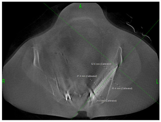
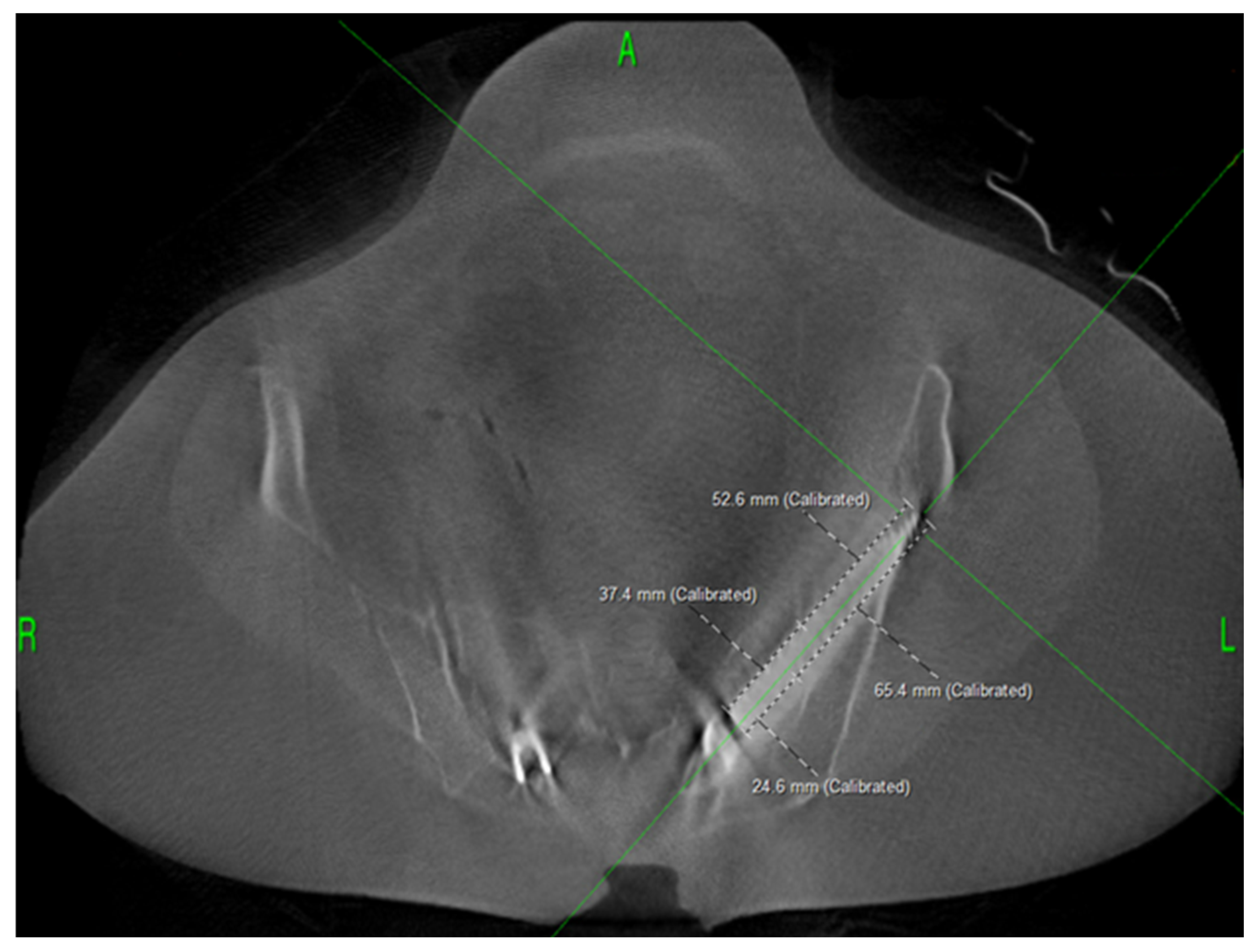
Computer-assisted navigation software was used to visualize the PFFS in the axial, sagittal, and coronal planes. After screw placement and the intraoperative check-spin were complete, a snapshot of each screw was taken and the length of purchase within the sacrum and ilium was measured on the axial image along the anterior and posterior aspect of each screw (Figure 1).
Figure 1.
Axial CT image of a 90 mm PFFS with sacrum and ilium engagement measurements.
2.3. Statistical Methods
Descriptive statistics were calculated for demographic information, implant sizes, and osseous engagement measurements. The surface area of each implant’s osseous engagement was calculated using the following equation:
(2 × (22/7) × [implant diameter] × [length of engagement]) + (2 × (22/7) × [implant diameter]2).
One-sample t-tests were performed to determine whether anterior and posterior engagement of the cephalad and caudal screws reached a depth of 35 mm. Independent t-tests were performed to compare the mean difference in bone engagement between the anterior and posterior caudal groups and the anterior and posterior cephalad groups as well as the surface area. All analyses were completed using commercially available software (SPSS Statistics version 28, IBM, Armonk, NY, USA).
3. Results
3.1. Sacral Engagement
The overall mean anterior sacral engagement was 38.6 ± 8.2 mm (range 17.7–56.4 mm), which was found to be statistically significantly greater than the hypothesized threshold of 35 mm (p < 0.001). A one-sample t-test revealed that anterior sacral engagement in cephalad screws (2.2 mm mean engagement difference, p = 0.11) was not statistically significantly greater than 35 mm, whereas caudal screws demonstrated a significantly greater anterior engagement compared to the threshold (4.9 mm mean engagement difference, p < 0.001) (Table 2).
Table 2.
Anterior sacral screw engagement lengths versus 35 mm engagement threshold (H0).
The overall mean posterior sacral engagement was 28.1 ± 8.6 mm (range 6.3–50.1 mm; p = 1). A one-sample t-test revealed that posterior sacral engagement in cephalad screws (−8.1 mm mean engagement difference, p = 1) was not statistically significantly greater than 35 mm, whereas caudal screws did demonstrate a greater posterior engagement compared to the cephalad screws but were not statistically significant (−5.7 mm mean engagement difference, p = 1) (Table 3).
Table 3.
Posterior sacral screw engagement lengths versus 35 mm engagement threshold (H0).
For the 42 cephalad screws, the average anterior sacral engagement was 37.2 ± 8.6 mm, while the posterior engagement measured 26.9 ± 9.5 mm. In contrast, the 42 caudal screws demonstrated a mean anterior engagement of 39.9 ± 7.6 mm, with a posterior engagement of 29.3 ± 7.5 mm. An independent t-test demonstrated a mean difference of 10.3 mm (p < 0.001) in sacral engagement between anatomical locations for the cephalad screws, while the caudal screws demonstrated a mean difference in sacral engagement between anatomical locations of 10.6 mm (p < 0.001) (Table 4).
Table 4.
Comparison of cephalad and caudal sacral screw engagement.
3.2. Iliac Engagement
The mean anterior engagement within the ilium was 54.0 ± 9.3 mm (range 32.9–73.6 mm), while the mean posterior engagement was 64.4 ± 9.5 mm (range 40.3–83.7 mm). Cephalad screws demonstrated an average iliac engagement of 52.6 ± 9.1 mm anteriorly and 62.9 ± 9.4 mm posteriorly, while caudal screws showed 55.3 ± 9.4 mm anterior engagement and 65.9 ± 9.4 mm posterior engagement. An independent t-test demonstrated a mean difference of 10.3 mm (p < 0.001) in sacral engagement between anatomical locations for the cephalad screws, while the caudal screws demonstrated a mean difference in sacral engagement between anatomical locations of 10.6 mm (p < 0.001) (Table 5).
Table 5.
Comparison of cephalad and caudal iliac screw engagement.
3.3. Surface Area
The total sacral surface area available for bone ingrowth for bilateral stacked PFFS was calculated to be 3338.3 ± 552.8 mm2, while the total iliac surface area available for bone ingrowth was 4364.8 mm2. An independent t-test demonstrated a mean difference in surface area availability between anatomical locations for the sacrum of −689.5 mm2 (p < 0.001), while the ilium demonstrated a mean difference in surface area availability between anatomical locations of 689.5 mm2 (p < 0.001) (Table 6).
Table 6.
Surface area available for bone ingrowth in sacral and iliac regions.
4. Discussion
This study demonstrates the engagement length of stacked SAI PFFS in a sacropelvic fixation, showing an average anterior sacral engagement of 38.6 ± 8.2 mm and posterior engagement of 28.1 ± 8.6 mm. While there have been numerous studies examining the mechanical properties and biomechanical benefits of SAI screws, this study is unique in its quantification of the actual bone engagement in the sacrum and ilium. Previous research has focused on identifying optimal trajectories and the available anatomical corridors for SAI screws to avoid neurovascular structures and maximize fixation [11,13,15,18]. However, few studies have specifically focused on measuring engagement lengths within the sacrum, which is a critical metric for understanding how effectively SAI screws contribute to long-term stability and fusion success [5,13]. There has been significant debate surrounding whether or not the amount of engagement of the S2AI screw in the sacrum is enough for ingrowth. While there have been multiple virtual studies looking at screw trajectories, this is an actual analysis of spine patients with these specific implants, allowing direct assessment of the amount of sacral osseous engagement. To date, there have been limited publications about the use of these screw fusion devices [12].
In this study, stacked screw fusion devices with a 10.5 mm outer diameter and a minimum length of 80 mm were used, with the majority being 90 and 100 mm in length. PFFSs feature a larger diameter (10.5 mm), a thicker screw neck (6.35 mm), and a more robust set plug (T30 hexalobe torx drive) with a higher locking torque (115 in lbs) than traditional pedicle screws, which effectively reduces the available corridor size for screw placement. The use of intraoperative cone beam CT scanning with computer navigation was instrumental in ensuring the accuracy of their placement. Previous studies13 have similarly shown that intraoperative navigation with cone beam CT scans and computer navigation significantly reduces the malposition rates and improves the overall screw placement in complex procedures. Despite these benefits, the malposition of cephalad screws was still reported in their study. The higher incidence of malposition in the cephalad screws, likely due to reduced corridor size when caudal screws are placed first, further restricting the available bony corridor for the cephalad screws and complicating the optimal placement, emphasizes the importance of precise preoperative planning and navigation. In addition to these findings, when malposition did occur, it typically involved ventral iliac wing breach, but all screw fusion devices were successfully repositioned without further complications [11].
While the biomechanical advantages of stacked SAI screws have been well documented, the long-term effects of crossing the sacroiliac (SI) joint remain a topic of debate. Previous research has raised concerns about placing screws across a joint that remains mobile. However, in other studies, crossing the SI joint with screws was not associated with increased pain or complications in the short term [7,8]. In fact, crossing the joint appears to decrease motion at the SI joint, which is beneficial for fusion stability. Preoperatively, SI joint pain is not uncommon; a recent randomized controlled trial found that 16% of patients undergoing long fusions for spinal deformity reported SI joint pain before surgery [19]. Approximately 24% of patients undergoing sacropelvic fusion report postoperative SI joint pain, and fusion of the SI joint has been shown to significantly reduce this pain [4]. As a result, the concept of simultaneously fusing the SI joint during sacropelvic fixation continues to gain support in the clinical community.
There has been a notable shift in pelvic fixation strategies over recent years, with advancements aimed at enhancing stability and reducing hardware-related complications [7]. The results of this study also reinforce the biomechanical advantages of combining stacked SAI screws with multi-rod constructs, which have been shown to offer superior rigidity and decrease motion at critical points in the spine [9,11]. Multi-rod constructs reduce the strain on S1 pedicle screws and minimize movement at the L5-S1 junction, where pseudarthrosis is most likely to occur [14]. The reduction in mechanical stress not only protects the hardware but also plays a crucial role in maintaining spinal alignment. This biomechanical advantage is supported by previous studies that have found stacked screw configurations to enhance construct rigidity and reduce the risk of hardware-related failures [11].
However, these benefits must be weighed against the potential risk of adjacent segment disease (ASD). The decreased motion across the SI joint may result in increased forces transferred to adjacent joints, particularly the hip joints, which could accelerate wear and lead to the need for hip arthroplasty. Previous research has shown an increased rate of total hip arthroplasty in patients undergoing pelvic fixation for adult spinal deformity [20]. While this was not directly evaluated in the present study, it remains an important consideration for future investigations, especially for patients who may already be predisposed to joint degeneration.
A key aspect of this study is the large surface area available for osseointegration, provided by the stacked SAI screw configuration. The importance of achieving successful bone ingrowth and fusion cannot be overstated, as it is essential for short-term stability and minimizing the risk of hardware failure. The idealized SAI screw trajectory is estimated to traverse approximately 35 mm of the sacrum before crossing the SI joint, providing substantial osseous purchase within the sacral bone [15]. In this study, we observed an anterior sacral engagement of 38.6 ± 8.2 mm, which was found to be statistically significantly greater than the idealized estimate of 35 mm (t = 3.99, p < 0.001), as demonstrated by our t-test analysis. Additionally, mean ilium engagement measured 52.6 ± 9.1 mm anteriorly and 62.9 ± 9.4 mm posteriorly, which is expected to offer sufficient fixation and robust osseous integration. This confirms that PFFSs, when placed in a stacked SAI configuration, achieve a significant degree of osseous purchase, ensuring adequate surface area for bone ingrowth, as inadequate osseous engagement may significantly contribute to increased rates of implant failure and the need for revision surgeries in patients undergoing complex spinal fusions [21,22,23]. This robust engagement in both the sacrum and ilium supports the hypothesis that the PFFS design, coupled with the SAI trajectory, can provide sufficient fixation. Moreover, clinical outcomes are encouraging, with PROMs showing marked improvement following the use of PFFS [13]. Importantly, no instances of acute pelvic fixation failure were recorded in this cohort, further supporting the safety and efficacy of this technique in the short-term postoperative period [13]. These findings suggest that PFFS may offer enhanced stability while minimizing complications during the early stages of recovery.
However, determining whether osseointegration has occurred remains a significant challenge. Standard imaging modalities, including plain radiographs and CT scans, are often limited in their ability to accurately assess the bone–implant interface due to artifacts or limited resolution. Future studies could benefit from the incorporation of advanced imaging techniques or direct histologic analysis to more definitively evaluate osseointegration. In animal models, such as those using ovine subjects, increased removal torque and histological evidence of bone ingrowth have been demonstrated [24]. Moreover, recent human specimen retrieval studies have provided further confirmation of osseointegration using micro-CT and clinical photographic evidence [25].
This study is not without limitations. First, the retrospective design limits the ability to establish causality and control for confounding factors, a common challenge in retrospective cohort studies. Second, the relatively small sample size of 21 patients, while sufficient for a preliminary analysis, limits the generalizability of the findings. However, there has been significant discussion at spine meetings surrounding the amount of osseous engagement in the sacrum with a screw fusion device and whether it is adequate for fusion to occur within the sacrum. As such, we wanted to provide real world data about how much engagement actually occurs. Larger prospective studies are needed to confirm these results and provide more robust statistical power. Another limitation is the heterogeneity of the patient population. The patients in this study had varying levels of spinal deformity and different histories of previous spinal surgeries, which could influence the outcomes and screw placement accuracy. Our intention was to determine how much surface area was actually engaged. This is not intended to be an outcomes study, as the heterogeneity of the population makes claims about efficacy certainly challenging. We simply wanted to indicate the area available for ingrowth in a real-world population. There is basic science histology evidence of bony ingrowth in these devices in an animal model [24]. There is also a published human explanation study where the implants were removed due to infection that has micro-CT evidence of bony ingrowth [25]. Future studies should consider stratifying patients based on the severity of spinal deformity and prior surgical interventions to better understand how these factors influence outcomes.
Additionally, the study only followed patients for a limited postoperative period, which restricts the ability to assess long-term outcomes such as adjacent segment disease, hardware failure, and the true success of osseointegration. This study has the limited goal of providing real world data for surface area available for ingrowth. There are other studies demonstrating the rates of acute and chronic pelvic fixation failure [2,3,4,5]. Emerging data indicates that the trend points more towards pelvic fixation being used [10]. We are not advocating that this is better at this point in time. We are simply responding to the debate that there is not enough surface area for ingrowth, with a real-world data set demonstrating how much area is available in the sacrum. Long-term follow-up with regular imaging and clinical assessments will be critical for understanding the full impact of stacked SAI screw fusion devices on spinal deformity correction and pelvic fixation durability.
5. Conclusions
In conclusion, this study demonstrates that PFFSs, when placed in a SAI trajectory, achieve substantial engagement within both the sacrum and ilium. This robust osseous purchase provides a large surface area for bone ingrowth, which is critical for ensuring mechanical stability and promoting successful short-term fusion outcomes. The combination of enhanced biomechanical stability, reduced strain on critical spinal structures, and a substantial surface area for bone ingrowth suggests that this strategy may help reduce the rates of pelvic fixation failure. However, the retrospective nature of the study, small sample size, and short-term follow-up limit the generalizability of the results. Future studies with larger cohorts and longer follow-up periods are necessary to fully evaluate the long-term efficacy of PFFS in sacropelvic fixation.
Author Contributions
J.J.H.: Investigation, Formal Analysis, Writing—Reviewing and Editing, Visualization. T.J.P.: Formal Analysis, Data Curation, Writing—Reviewing and Editing. K.O.: Formal Analysis, Writing—Reviewing and Editing, Visualization, Supervision, Project Administration. J.N.S.: Resources, Writing—Reviewing and Editing, Supervision, Project Administration. N.R.H.: Conceptualization, Resources, Writing—Reviewing and Editing, Supervision, Project Administration. C.T.M.: Resources, Investigation, Writing—Reviewing and Editing, Supervision, Project Administration. K.E.J.: Resources, Writing—Reviewing and Editing, Supervision, Project Administration. D.W.P.J.: Conceptualization, Methodology, Resources, Writing—Reviewing and Editing, Supervision, Project Administration. All authors have read and agreed to the published version of the manuscript.
Funding
The authors’ institution received financial support from SI-BONE Inc. (Santa Clara, CA, USA) (award #CON000000109123) for the submitted work. The funding for this study was through the SI-BONE external research program. They approved the funding, but had no role in data collection, data analysis, manuscript preparation. They were provided a copy of the manuscript, but did not provide any input for its submission.
Institutional Review Board Statement
This retrospective cohort study was conducted in compliance of the Declaration of Helsinki. Institutional review board (IRB) STUDY#00020316 approval for this project was obtained from the University of Minnesota IRB. Approval date: 18 January 2024.
Informed Consent Statement
Patients were reviewed in EPIC for their willingness to participate in research. Patients who “opted out” of research participation were excluded. We requested a waiver of consent and received it, as the research was no greater than the minimal risk, procedures were in place to prevent breach of confidentiality, and consent could not practicably be obtained.
Data Availability Statement
The original contributions presented in this study are included in the article. Further inquiries can be directed to the corresponding author.
Conflicts of Interest
J.J.H., K.O. and T.J.P. have nothing to disclose. N.R.H. declares consulting fees from Medtronic. J.N.S. receives support of non-study-related clinical or research effort from Orthofix, NuVasive, and AO Spine. C.T.M. has consultancies at Medtronic, institutional research support from SI-Bone, and industry funding to travel to cadaveric surgical training sessions from Medtronic, NuVasive, and Accutech, and is a KF Degenerative committee member for AO Spine. K.E.J. has consultancies at SI Bone and Medtronic. D.W.P.J. declares consulting fees from Globus Medical and Alexion; institutional grant/research support from Medtronic and Mizuho OSI; consulting fees, royalties, and honoraria from SI Bone; and royalties from Springer.
References
- Shen, F.H.; Mason, J.R.; Shimer, A.L.; Arlet, V.M. Pelvic fixation for adult scoliosis. Eur. Spine J. 2013, 22, S265–S275. [Google Scholar] [CrossRef] [Scilit] [PubMed]
- Odland, K.; Chanbour, H.; Zuckerman, S.L.; Polly, D.W. Spinopelvic fixation failure in the adult spinal deformity population: Systematic review and meta-analysis. Eur. Spine J. 2024, 33, 2751–2762. [Google Scholar] [CrossRef] [Scilit] [PubMed]
- Eastlack, R.K.; Soroceanu, A.; Mundis, G.M., Jr.; Daniels, A.H.; Smith, J.S.; Line, B.; Passias, P.; Nunley, P.D.; Okonkwo, D.O.; Than, K.D.; et al. Rates of Loosening, Failure, and Revision of Iliac Fixation in Adult Deformity Surgery. Spine 2022, 47, 986–994. [Google Scholar] [CrossRef] [Scilit] [PubMed]
- Martin, C.T.; Polly, D.W.; Holton, K.J.; Miguel-Ruiz, J.E.S.; Albersheim, M.; Lender, P.; Sembrano, J.N.; Hunt, M.A.; Jones, K.E. Acute failure of S2-alar-iliac screw pelvic fixation in adult spinal deformity: Novel failure mechanism, case series, and review of the literature. J. Neurosurg. Spine 2021, 36, 53–61. [Google Scholar] [CrossRef] [Scilit]
- Martin, C.T.; Holton, K.J.; Elder, B.D.; Fogelson, J.L.; Mikula, A.L.; Kleck, C.J.; Calabrese, D.; Burger, E.L.; Ou-Yang, D.; Patel, V.V.; et al. Catastrophic acute failure of pelvic fixation in adult spinal deformity requiring revision surgery: A multicenter review of incidence, failure mechanisms, and risk factors. J. Neurosurg. Spine 2022, 38, 98–106. [Google Scholar] [CrossRef] [Scilit]
- Ivanov, A.A.; Kiapour, A.; Ebraheim, N.A.; Goel, V. Lumbar fusion leads to increases in angular motion and stress across sacroiliac joint: A finite element study. Spine 2009, 34, E162–E169. [Google Scholar] [CrossRef] [Scilit]
- Manzetti, M.; Ruffilli, A.; Barile, F.; Fiore, M.; Viroli, G.; Cappello, L.; Faldini, C. Sacroiliac Joint Degeneration and Pain After Spinal Arthrodesis: A Systematic Review. Clin. Spine Surg. 2023, 36, 169–182. [Google Scholar] [CrossRef] [Scilit]
- Unoki, E.; Miyakoshi, N.; Abe, E.; Kobayashi, T.; Abe, T.; Kudo, D.; Shimada, Y. Sacropelvic Fixation with S2 Alar Iliac Screws May Prevent Sacroiliac Joint Pain After Multisegment Spinal Fusion. Spine 2019, 44, E1024–E1030. [Google Scholar] [CrossRef] [Scilit] [PubMed]
- Lee, N.J.; Marciano, G.; Puvanesarajah, V.; Park, P.J.; Clifton, W.E.; Kwan, K.; Morrissette, C.R.; Williams, J.L.; Fields, M.; Hassan, F.M.; et al. Incidence, mechanism, and protective strategies for 2-year pelvic fixation failure after adult spinal deformity surgery with a minimum six-level fusion. J. Neurosurg. Spine 2022, 38, 208–216. [Google Scholar] [CrossRef] [Scilit]
- Ormseth, A.F.; Odland, K.; Haselhuhn, J.J.; Holton, K.J.; Polly, D.W. Pelvic Fixation Construct Trends in Spinal Deformity Surgery. Indian J. Orthop. 2024, 58, 396–401. [Google Scholar] [CrossRef] [Scilit]
- Polly, D.W., Jr.; Holton, K.J.; Soriano, P.O.; Sembrano, J.N.; Martin, C.T.; Hendrickson, N.R.; Jones, K.E. Multiple Points of Pelvic Fixation: Stacked S2-Alar-Iliac Screws (S2AI) or Concurrent S2AI and Open Sacroiliac Joint Fusion with Triangular Titanium Rod. JBJS Essent. Surg. Tech. 2022, 12, e21.00044. [Google Scholar] [CrossRef] [Scilit] [PubMed]
- Alan, N.; Lee, K.E.; Zhou, J.J.; Farber, S.H.; DiDomenico, J.D.; O’nEill, L.K.; Turner, J.D.; Uribe, J.S. Bilateral sacroiliac joint fusion in long constructs using self-harvesting porous S2-alar iliac screws with an integrated tulip: Technical considerations and early clinical and radiographic experience. Neurosurg. Focus 2023, 55, E5. [Google Scholar] [CrossRef] [Scilit] [PubMed]
- Hendrickson, N.R.; Haselhuhn, J.J.; Odland, K.; Sembrano, J.N.; Martin, C.T.; Jones, K.E.; Polly, D.W. Sacropelvic Fixation with Porous Fusion/Fixation Screws: A Technical Note and Retrospective Review. World Neurosurg. 2024, 190, 172–180. [Google Scholar] [CrossRef] [Scilit] [PubMed]
- Panico, M.; Chande, R.D.; Lindsey, D.P.; Mesiwala, A.; Polly, D.W.; Villa, T.; Yerby, S.A.; Brayda-Bruno, M.; Galbusera, F. Stability and Instrumentation Stresses Among Sacropelvic Fixation Techniques with Novel Porous Fusion/Fixation Implants: A Finite Element Study. Int. J. Spine Surg. 2023, 17, 598–606. [Google Scholar] [CrossRef] [Scilit]
- Chang, T.L.; Sponseller, P.D.; Kebaish, K.M.; Fishman, E.K. Low-profile pelvic fixation: Anatomic parameters for sacral alar-iliac fixation versus traditional iliac fixation. Spine 2009, 34, 436–440. [Google Scholar] [CrossRef] [Scilit]
- Sponseller, P.D.; Zimmerman, R.M.; Ko, P.S.; ter Gunne, A.F.P.; Mohamed, A.S.M.; Chang, T.-L.; Kebaish, K.M. Low-profile pelvic fixation with the sacral alar iliac technique in the pediatric population improves results at two-year minimum follow-up. Spine 2010, 35, 1887–1892. [Google Scholar] [CrossRef] [Scilit]
- Hasan, M.Y.; Liu, G.; Wong, H.K.; Tan, J.-H. Postoperative complications of S2AI versus iliac screw in spinopelvic fixation: A meta-analysis and recent trends review. Spine J. 2020, 20, 964–972. [Google Scholar] [CrossRef] [Scilit]
- Jain, A.; Brooks, J.T.; Kebaish, K.M.; Sponseller, P.D. Sacral Alar Iliac Fixation for Spine Deformity. JBJS Essent. Surg. Tech. 2016, 6, e10. [Google Scholar] [CrossRef] [Scilit]
- Polly, D.; Mundis, G.; Eastlack, R.; Leveque, J.-C.; Elder, B.D.; Martin, C.; Kent, R.; Snowden, R.; Kim, H.J.; Sembrano, J.; et al. Randomized Trial of Augmented Pelvic Fixation in Patients Undergoing Thoracolumbar Fusion for Adult Spine Deformity: Initial Results from a Multicenter Randomized Trial. World Neurosurg. 2024, 187, e15–e27. [Google Scholar] [CrossRef] [Scilit]
- Kress, D.J.; Morgan, P.M.; Thomas, D.C.; Haselhuhn, J.J.; Polly, D.W. Prevalence of total joint arthroplasty in the adult spine deformity population. Spine Deform. 2024, 12, 1421–1429. [Google Scholar] [CrossRef] [Scilit]
- Fradet, L.; Bianco, R.J.; Tatsumi, R.; Coleman, J.; Aubin, C. Biomechanical comparison of sacral and transarticular sacroiliac screw fixation. Spine Deform. 2020, 8, 853–862. [Google Scholar] [CrossRef] [Scilit]
- Hyun, S.J.; Jung, J.M.; Kim, K.J.; Jahng, T.-A. Durability and Failure Types of S2-Alar-Iliac Screws: An Analysis of 312 Consecutive Screws. Oper. Neurosurg. 2020, 20, 91–97. [Google Scholar] [CrossRef] [Scilit]
- Nakashima, H.; Kanemura, T.; Satake, K.; Ito, K.; Ishikawa, Y.; Ouchida, J.; Segi, N.; Yamaguchi, H.; Imagama, S. The Prevalence and Risk Factors for S2 Alar-Iliac Screw Loosening with a Minimum 2-Year Follow-up. Asian Spine J. 2020, 14, 177–184. [Google Scholar] [CrossRef] [Scilit]
- Polly, D.W.; Gardner, M.J.; Wills, D.; Crowley, J.; Pelletier, M.; Wang, T.; Bailey, I.; Yerby, S.; Walsh, W.R. Osseointegration and Fixation Opportunities of Sacroiliac Hardware: A Preclinical Evaluation in a Large Animal Model. J. Spine Surg. 2024, 10, 616–626. [Google Scholar] [CrossRef] [Scilit]
- Spence, B.S.; Godlewski, M.W.; Haselhuhn, J.J.; Jones, K.E.; Polly, D.W. Removal of infected bone in-growth pelvic fixation implants and microcomputed tomography analysis of osseous integration: Illustrative case. J. Neurosurg. Case Lessons. 2024, 8, CASE24274. [Google Scholar] [CrossRef] [Scilit]

Disclaimer/Publisher’s Note: The statements, opinions and data contained in all publications are solely those of the individual author(s) and contributor(s) and not of MDPI and/or the editor(s). MDPI and/or the editor(s) disclaim responsibility for any injury to people or property resulting from any ideas, methods, instructions or products referred to in the content. |
© 2026 by the authors. Licensee MDPI, Basel, Switzerland. This article is an open access article distributed under the terms and conditions of the Creative Commons Attribution (CC BY) license.