


Abstract
Introduction: Gastrointestinal basidiobolomycosis is an uncommon invasive fungal infection caused by Basidiobolus ranarum, an environmental mould endemic to the arid south-west of Saudi Arabia and to a handful of other hot, dry regions. Its non-specific presentation routinely invites misdiagnosis as inflammatory bowel disease, tuberculosis or malignancy, and extension to the biliary tree is decidedly rare. Case Presentation: We report a 25-year-old Saudi woman who developed chronic watery diarrhoea and pronounced weight loss two months after her first delivery. Severe microcytic anaemia, leukocytosis, a very high erythrocyte sedimentation rate and multisegmental colonic wall thickening were initially attributed to inflammatory bowel disease complicated by Clostridioides difficile colitis. She subsequently required cholecystectomy for acalculous cholecystitis and then deteriorated, with gastric outlet obstruction and collections at the gallbladder bed. Re-examination of the gallbladder specimen revealed broad, pauci-septate hyphae enveloped by the Splendore–Hoeppli phenomenon, and culture yielded an isolate phenotypically identified as B. ranarum. Voriconazole followed by itraconazole achieved near-complete clinical and radiological recovery. Discussion: We hypothesise that the early puerperium could represent a possible, though as yet unproven, window of susceptibility to this infection. Conclusions: In endemic regions, gastrointestinal basidiobolomycosis should be considered when presumed inflammatory bowel disease behaves atypically; deep tissue sampling is often decisive, and prolonged triazole therapy can achieve an excellent outcome.
1. Introduction
Basidiobolus ranarum is a filamentous environmental fungus of the order Entomophthorales that is recovered from soil, decaying vegetation and the gut of amphibians, reptiles and insectivorous animals. Human disease was first documented in 1956 as a subcutaneous infection in Indonesia [1], and for several decades the organism was regarded almost exclusively as a cause of chronic subcutaneous mycosis in the tropics. The first gastrointestinal case followed in 1964 [2], and the gastrointestinal form—now termed gastrointestinal basidiobolomycosis (GIB)—has since become recognised as a distinct, if rare, clinical entity.
Reported cases cluster conspicuously in three arid corridors: the south-western provinces of Saudi Arabia, the south-western United States (chiefly Arizona) and southern Iran [3,4]. In these settings, warm temperatures, low rainfall and an abundant reptile population appear to favour environmental persistence of the fungus. Wall lizards native to the Saudi highlands carry B. ranarum in their intestinal tract, lending weight to an environmental or zoonotic route of acquisition through contaminated soil, water or food [5,6]. Sporadic cases have nonetheless been described well beyond these zones, including the central region of Saudi Arabia [7].
Clinically, GIB is a notorious mimic. It typically produces indolent abdominal pain, weight loss and a palpable mass that are readily mistaken for Crohn disease, intestinal tuberculosis or carcinoma [8,9,10]. Diagnosis is further hindered by the predominantly submucosal location of the organism, so that conventional endoscopic biopsies—which sample only the mucosa—are frequently unrevealing [6]. Spread beyond the bowel wall to the liver, retroperitoneum and, exceptionally, the biliary tree has occasionally been reported [11,12]. Here we describe a young woman who developed multifocal GIB with biliary tract involvement in the early postpartum period, and we use the case to frame a practical review of the diagnostic and therapeutic challenges that this infection poses in endemic regions.
2. Case Presentation
2.1. Patient Information and Clinical Findings
A 25-year-old Saudi woman was referred to Aseer Central Hospital, a tertiary centre in the highlands of south-western Saudi Arabia, for evaluation of three months of diarrhoea together with colonic wall thickening seen on an outside computed tomography (CT) scan. Her illness had begun roughly two months after the uncomplicated vaginal delivery of her first child. The earliest symptom was mild left lower-quadrant discomfort, which settled after she was treated empirically for Helicobacter pylori infection at a private clinic. Over the following weeks she developed watery, non-bloody diarrhoea about twice daily that occurred both day and night, bore no relation to meals, and was accompanied by an involuntary loss of 10–12 kg. She reported no fever, vomiting, rectal bleeding or night sweats.
Her medical, surgical and drug histories were unremarkable, and she had no known allergies. She was a lifelong non-smoker who kept house and had no recognised tuberculosis contact. There was a family history of H. pylori infection but none of inflammatory bowel disease (IBD), coeliac disease or gastrointestinal cancer. On examination she looked chronically unwell but was alert and haemodynamically stable. The abdomen was soft, non-tender and free of palpable mass, organomegaly, jaundice or peripheral lymphadenopathy.
2.2. Initial Investigations
Initial laboratory testing disclosed a constellation of abnormalities (Table 1): severe microcytic, hypochromic anaemia (haemoglobin 6.7 g/dL), neutrophilic leukocytosis (17.9 × 10³/µL), reactive thrombocytosis (602 × 103/µL), a markedly raised erythrocyte sedimentation rate (ESR, 120 mm/h) and hypoalbuminaemia (2.75 g/dL). A stool nucleic-acid amplification test (PCR/NAAT) for toxigenic Clostridioides difficile was positive; glutamate dehydrogenase antigen and toxin A/B enzyme-immunoassay results are not documented in the available records. Tuberculin skin testing and mycobacterial PCR were negative, while a mildly elevated anti-tissue-transglutaminase IgA raised the possibility of coexisting coeliac disease. The differential white-cell count performed during the initial admission showed marked peripheral eosinophilia, with an absolute eosinophil count of 2.62 × 10³/µL (21.7% of a total leukocyte count of 12.07 × 10³/µL in that sample), and the peripheral blood smear confirmed moderate eosinophilia without eosinophil atypia—a finding of particular interest given the recognised association of basidiobolomycosis with peripheral eosinophilia. In view of the marked eosinophilia, stool examinations for ova and parasites were also performed and were negative.
Table 1.
Laboratory findings at presentation.
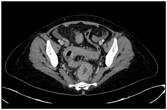
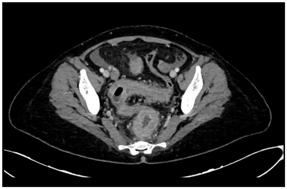
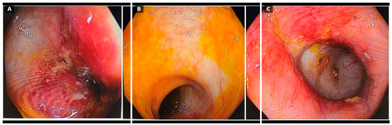
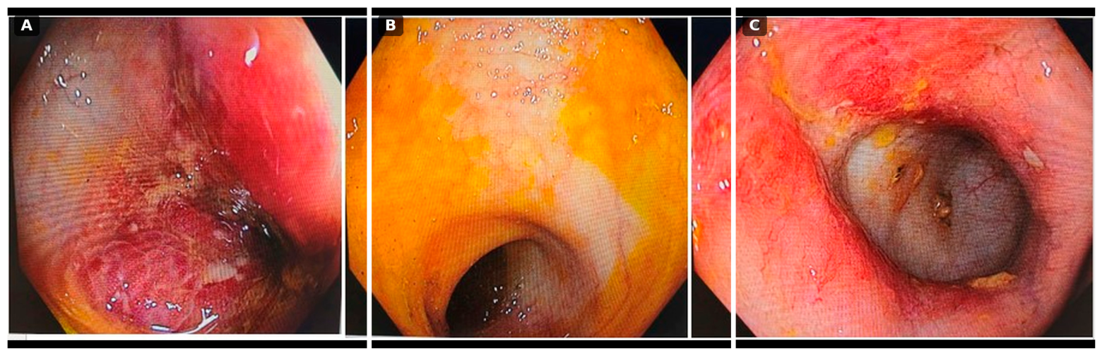
Contrast-enhanced CT of the abdomen confirmed circumferential, multisegmental mural thickening of the large bowel with surrounding fat stranding, a pattern most in keeping with IBD (Figure 1). Colonoscopy showed patchy mucosal erythema, oedema, friability and shallow ulceration across several colonic segments; histology of the mucosal biopsies returned chronic active colitis without granulomas or malignancy, interpreted as compatible with either IBD or an infective colitis (Figure 2).
Figure 1.
Initial contrast-enhanced CT of the abdomen and pelvis (axial). There is circumferential, multisegmental mural thickening of the large bowel with adjacent inflammatory fat stranding, an appearance initially attributed to inflammatory bowel disease.
Figure 2.
Initial colonoscopy. (A) Sigmoid colon with mucosal erythema, oedema and a shallow ulcer; (B) descending colon coated with yellowish exudate; (C) a more severely inflamed segment with luminal narrowing and contact friability.
2.3. Hospital Course and Evolution
The patient received two units of packed red cells, which raised her haemoglobin to 9.7 g/dL, together with a course of oral vancomycin (125 mg four times daily) for C. difficile colitis. Her diarrhoea resolved completely and she was discharged with a plan for outpatient re-endoscopy.
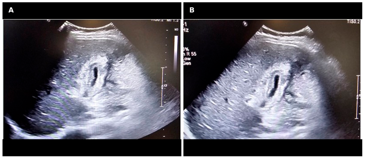
She returned about one month later with severe right upper-quadrant pain, nausea and vomiting. Ultrasonography demonstrated a contracted, markedly thick-walled gallbladder (wall thickness 1.87 cm) surrounded by inflammatory change but without calculi (Figure 3). She underwent cholecystectomy at an external facility. According to the operative record, the gallbladder was densely adherent to the stomach, colon and omentum, contained neither bile, pus nor stones, and was described by the surgeon as a “white solid structure”; histopathology reported active chronic inflammation with fibrosis and suppurative, epithelioid granulomas.
Figure 3.
Abdominal ultrasonography of the gallbladder in two planes (A,B). The gallbladder is contracted with marked, diffuse wall thickening measured at 1.87 cm (callipers) and surrounding inflammatory change; no calculi are seen.
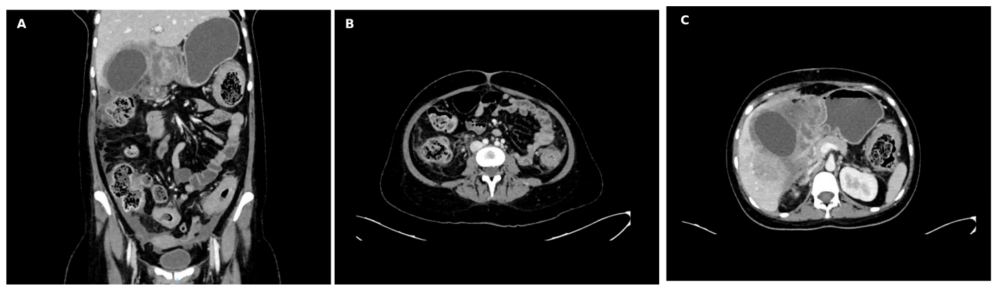
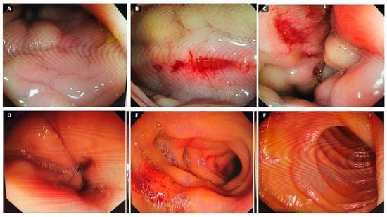
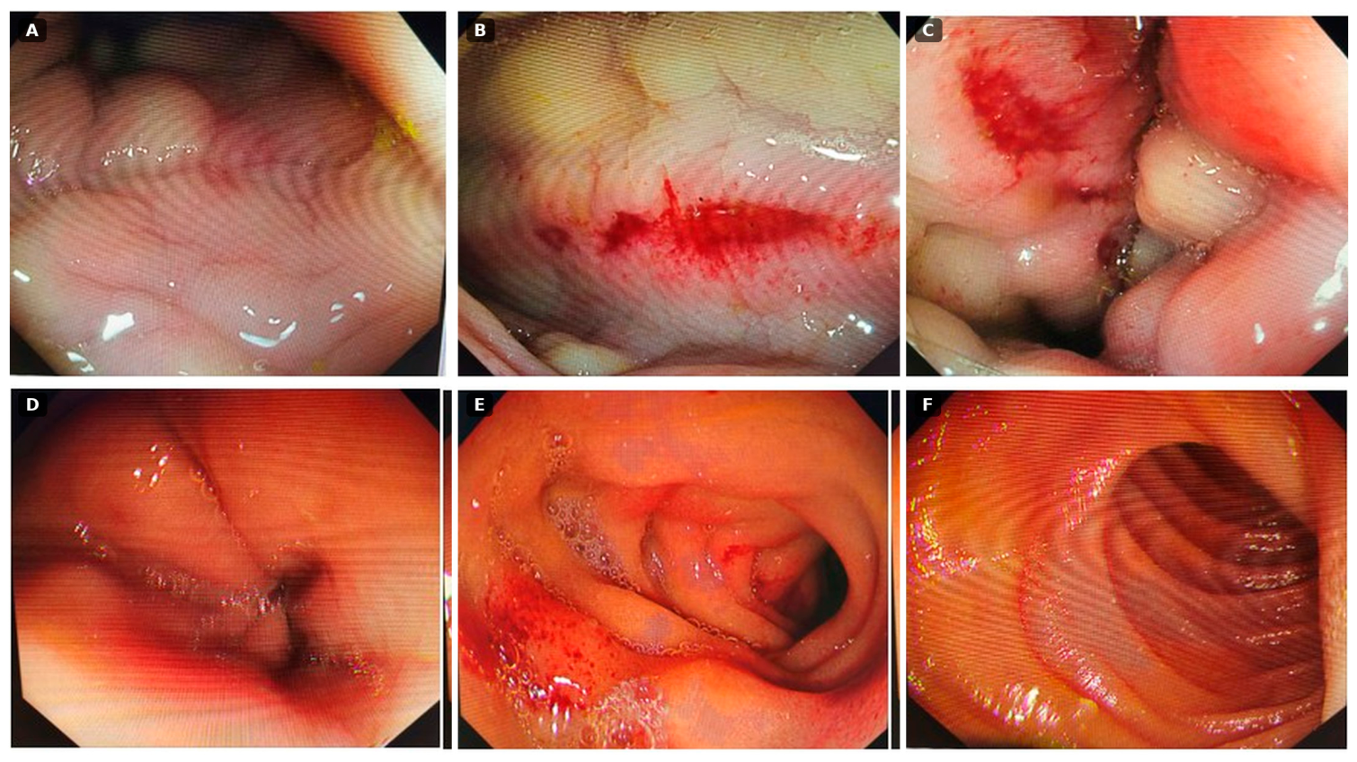
Rather than improving, her pain intensified within a week of surgery and she was readmitted to our hospital. Repeat CT revealed loculated fluid collections in the gallbladder fossa, distension of the stomach consistent with gastric outlet obstruction, and persistent multisegmental bowel-wall thickening (Figure 4). Percutaneous pigtail drainage of the collection yielded thick yellow-green pus. Upper endoscopy showed florid inflammation of the gastric antrum and duodenum, with mucosal oedema, erythema, nodularity, contact bleeding and superficial ulceration (Figure 5).
Figure 4.
Repeat CT after cholecystectomy. (A) Coronal reformat showing a loculated collection in the gallbladder fossa and a distended, fluid-filled stomach reflecting gastric outlet obstruction; (B) axial image showing persistent multisegmental colonic wall thickening; (C) axial image of the upper abdomen showing the loculated subhepatic collection at the gallbladder bed.
Figure 5.
Upper gastrointestinal endoscopy. (A–C) Gastric antrum and body with mucosal oedema, erythema, nodularity, an area of active bleeding and superficial ulceration; (D–F) duodenal involvement with mucosal irregularity, friability and narrowing.
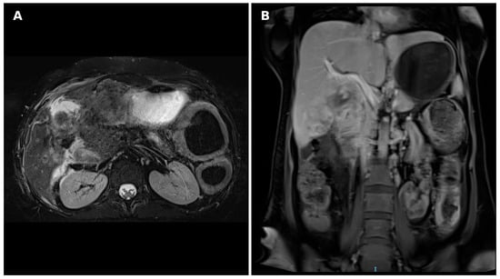
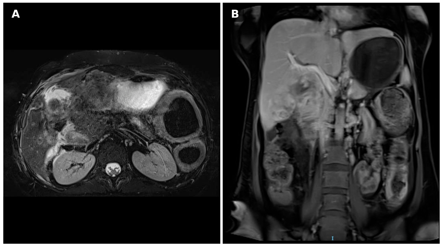
Contrast-enhanced magnetic resonance imaging (MRI) of the abdomen demonstrated circumferential thickening of the gastric antrum containing submucosal microabscesses, a skip-pattern of small- and large-bowel involvement, inflammatory infiltration of the gallbladder bed extending along the porta hepatis, and a focal lesion at the upper pole of the right kidney; the appearances were considered characteristic of an invasive fungal process, specifically basidiobolomycosis (Figure 6).
Figure 6.
Contrast-enhanced MRI of the abdomen. (A) Axial image showing persistent inflammatory change at the gallbladder bed after cholecystectomy, extending into the adjacent hepatic parenchyma and the thickened gastric antrum; (B) coronal image showing periportal extension, skip-pattern bowel-wall thickening and a focal right renal lesion.
2.4. Diagnosis
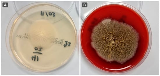
Prompted by the imaging, the cholecystectomy specimen was retrieved and re-examined with fungal stains. This disclosed broad, thin-walled, sparsely septate (pauci-septate) hyphae cuffed by intensely eosinophilic material—the Splendore–Hoeppli phenomenon—set within a dense eosinophilic and granulomatous infiltrate, findings that are highly characteristic of basidiobolomycosis. Culture of the drained pus on Sabouraud dextrose agar grew Basidiobolus ranarum, producing the expected waxy, glabrous, radially folded buff colonies that acquired a greyish-brown surface on subculture (Figure 7). Species-level identification was made by the clinical microbiology laboratory on the basis of these characteristic colonial and growth features, taken together with the histopathological demonstration of broad, pauci-septate hyphae with the Splendore–Hoeppli phenomenon; molecular confirmation (panfungal PCR with sequencing) was not available at our centre and could not be performed. A diagnosis of multifocal gastrointestinal basidiobolomycosis with biliary tract involvement was thereby established. Throughout this report, “biliary tract involvement” denotes histologically confirmed infection of the gallbladder with contiguous inflammatory extension along the gallbladder bed and porta hepatis, rather than demonstrated invasion of the bile ducts, and the multifocal distribution most plausibly reflects contiguous or intramural spread, as haematogenous dissemination was not microbiologically proven. Because the cholecystectomy and initial histopathological processing were performed at an external facility, photomicrographs of the stained sections were unfortunately not retrievable for publication.
Figure 7.
Mycological culture of the drained pus. (A) Sabouraud dextrose agar showing a waxy, glabrous, buff-coloured colony with characteristic radial folds; (B) blood agar showing the greyish-brown surface that develops on subculture—features consistent with Basidiobolus ranarum.
2.5. Treatment and Outcome
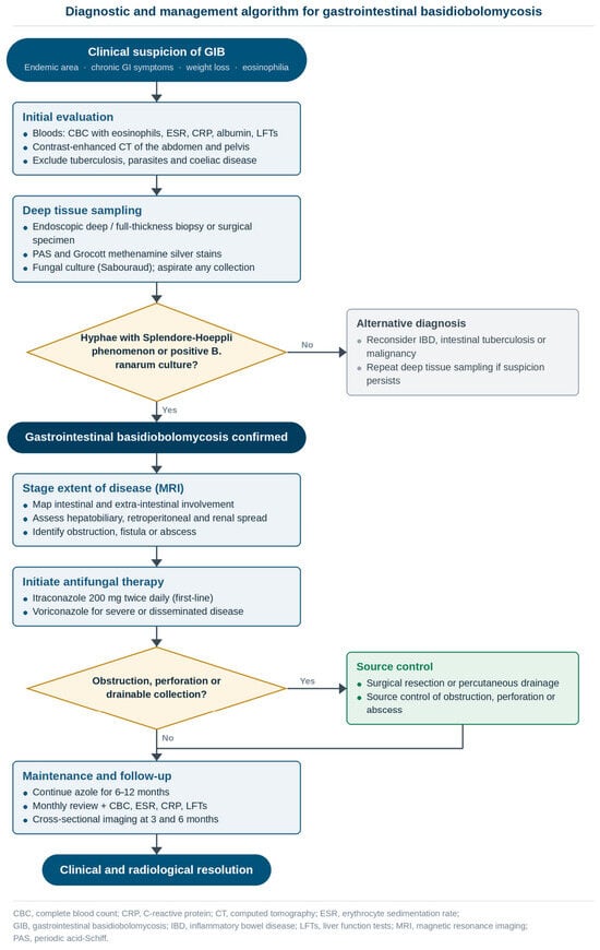
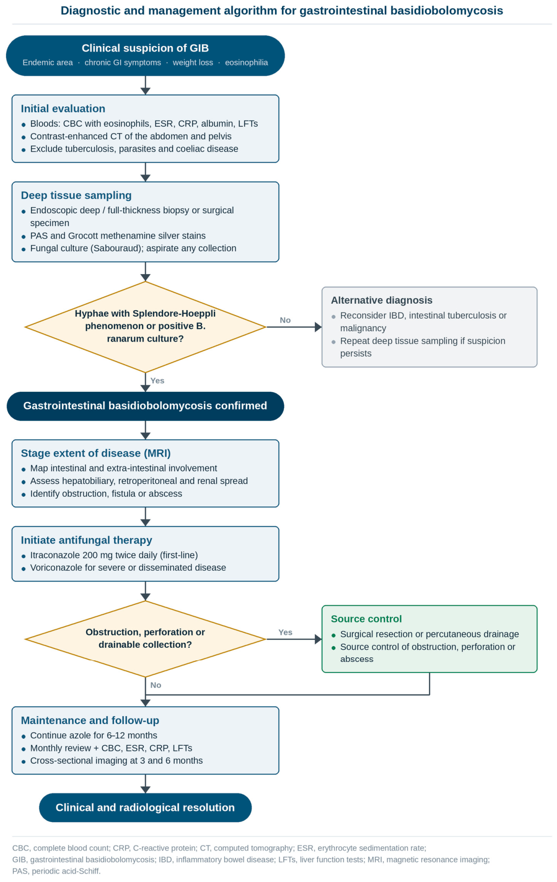
Systemic antifungal therapy was started with intravenous voriconazole (a loading dose of 6 mg/kg every 12 h for one day, then 4 mg/kg every 12 h). Once she had stabilised, treatment was switched to oral itraconazole 200 mg twice daily for prolonged maintenance. Her symptoms abated steadily, the inflammatory markers normalised, and serial imaging showed near-complete resolution of the previously extensive disease. A practical synthesis of the diagnostic and management steps that this case illustrates is presented as an algorithm in the Discussion (Figure 8).
Figure 8.
Proposed stepwise diagnostic and management algorithm for gastrointestinal basidiobolomycosis in endemic regions, emphasising early clinical suspicion, deep tissue sampling, mycological confirmation and prolonged triazole therapy with surgery reserved for complications. This pathway is a pragmatic synthesis of the available literature and the present case; it has not been prospectively validated and is not intended as a formal guideline.
3. Discussion
For the accompanying narrative literature review, we searched PubMed/MEDLINE and Google Scholar from inception to June 2026 using combinations of the terms “basidiobolomycosis”, “Basidiobolus ranarum”, “gastrointestinal”, “hepatobiliary” and “biliary”, limited to English-language publications, and hand-searched the reference lists of retrieved articles for additional cases. This was a narrative rather than a systematic review; accordingly, the case counts and clinical frequencies cited below are approximate, are drawn from the referenced series and reviews, and may overlap between sources.
3.1. Epidemiology
Since the first gastrointestinal case in 1964 [2], GIB has been reported across several continents but remains heavily concentrated in a few hot, dry regions. In their landmark review, Vikram and colleagues assembled 44 cases worldwide and observed that roughly 43% originated in the United States—overwhelmingly Arizona—and about 25% in Saudi Arabia [3]. Within the Kingdom, the southern Aseer and Jazan provinces account for the great majority of reports (Table 2), a distribution that mirrors the local climate and the regional reptile reservoir of B. ranarum [5,13]. Recent Saudi series have reinforced this pattern and broadened the recognised spectrum, with contemporary bicentric and multicentre cohorts of children and adults documenting extensive colonic, hepatic and multifocal disease [14,15]. Our patient, from the Aseer highlands, fits the established geography of the disease.
Table 2.
Geographic distribution of reported cases of gastrointestinal basidiobolomycosis.
3.2. Pathogenesis and Risk Factors
The precise route by which humans acquire GIB remains unproven, but ingestion of fungal elements in contaminated soil, water or food is the most widely accepted mechanism [5]. A distinctive and clinically important feature is that, unlike most invasive mycoses, GIB predominantly affects immunocompetent hosts. Recognised associations include residence in an endemic area, contact with soil or reptiles, and the use of acid-suppressing medication, which may weaken the gastric barrier to ingested spores. Pregnancy and the puerperium have occasionally featured among reported patients, and the temporal association in our case—symptoms beginning some two months postpartum—is noteworthy, although it may be coincidental. The physiological immune re-modelling of late pregnancy and the early postpartum period—characterised by a relative shift away from cell-mediated (T-helper-1) immunity—could plausibly open a transient window of vulnerability to an organism that immunocompetent hosts would ordinarily contain. We advance this as a hypothesis rather than a demonstrated causal link.
3.3. Clinical Presentation
The clinical hallmark of GIB is its lack of specificity. Abdominal pain, a palpable mass, altered bowel habit, weight loss and low-grade fever predominate, and these features overlap almost completely with Crohn disease, intestinal tuberculosis and colorectal carcinoma (Table 3) [8,9,10]. Peripheral eosinophilia, present in the large majority of cases, is among the few clues that should prompt consideration of a fungal cause. The colon—particularly the caecum and ascending colon—is the segment most often involved, although disease may arise anywhere from the stomach to the rectum; the prominent gastroduodenal involvement in our patient is a reminder of this range. Extension beyond the bowel to the liver, retroperitoneum and biliary tree is uncommon. Biliary tract involvement of the kind seen here—histologically confirmed gallbladder infection with contiguous peri-portal extension on imaging, rather than proven intraductal disease—has been described in only a small number of patients [11,12], and whether this reflects genuine rarity or simply under-recognition—given how difficult the diagnosis is to secure—remains unclear.
Table 3.
Frequency of clinical features of gastrointestinal basidiobolomycosis reported in the literature.
3.4. Diagnostic Approach
Three factors conspire to delay the diagnosis: the non-specific clinical picture, the submucosal habitat of the organism, and limited clinician familiarity with the disease [6,19]. Cross-sectional imaging usually shows circumferential bowel-wall thickening, an inflammatory mass or submucosal abscesses; MRI is especially valuable for mapping the extent of disease and for revealing the characteristic skip pattern, as it did in our patient [20]. The diagnosis ultimately rests on tissue. Histology characteristically shows chronic granulomatous inflammation, a dense eosinophilic infiltrate, and broad, pauci-septate hyphae surrounded by the Splendore–Hoeppli phenomenon [21]. Because the fungus lies deep to the mucosa, superficial endoscopic biopsies are frequently falsely negative, and—as in this case—the diagnosis may only declare itself on a surgical specimen. Culture supports mycological identification: B. ranarum characteristically grows on Sabouraud dextrose agar at 25–37 °C within two to three days, forming waxy, glabrous colonies with radial folds [17]. Where available, panfungal PCR and sequencing can provide definitive species-level confirmation and shorten the diagnostic interval.
3.5. Management
Treatment combines prolonged systemic antifungal therapy with surgery reserved for specific complications (Table 4). Triazoles are the mainstay: itraconazole is the most widely used first-line agent, with reported response rates of 70–80%, while voriconazole—used here—is an effective option for severe or disseminated disease [18,22,23]. Therapy is necessarily long, usually six to twelve months, and should continue until both symptoms and imaging have resolved. The place of surgery has narrowed over time. Although earlier reports relied heavily on resection, accumulating experience indicates that many patients can be managed medically once the diagnosis is secured, with operation reserved for obstruction, perforation, abscess or diagnostic uncertainty [22,24]; in children, mechanical complications such as intussusception have also mandated operative intervention [25]. Saturated potassium iodide solution, long used for subcutaneous basidiobolomycosis, is a further inexpensive and readily available option: it has produced complete resolution of gastrointestinal disease refractory to amphotericin B and itraconazole in at least one paediatric case, and it features in several of the combination regimens reported in recent series [24,26]. Statements about amphotericin B should be interpreted cautiously: in vitro susceptibility of B. ranarum is variable and clinical failures have been reported, but the evidence base rests on small numbers of heterogeneous cases. A recent qualitative review of 24 cases reaffirmed the central role of azoles and the variable, often adjunctive, contribution of surgery [24]. In our patient, source control of the drainable collection together with extended triazole therapy produced an excellent outcome without further bowel resection.
Table 4.
Antifungal agents used in the treatment of gastrointestinal basidiobolomycosis.
3.6. Key Learning Points
Three aspects of this case merit emphasis. First, the onset in the early puerperium raises the hypothesis—unproven, and possibly coincidental—that the immunological changes of pregnancy and the postpartum period might create a transient susceptibility to invasive fungal infection; this association is worth bearing in mind in endemic regions. Second, histologically documented biliary tract involvement, reported in only a handful of previous patients, extends the recognised anatomical reach of GIB. Third, the early course—dominated by C. difficile colitis and a mildly positive coeliac serology—illustrates how readily GIB is mistaken for commoner conditions, and how a deep tissue specimen, rather than a superficial biopsy, is often the key to the diagnosis. A pragmatic, stepwise approach to suspected GIB in endemic areas is summarised in Figure 8.
4. Conclusions
We describe an unusual case of multifocal gastrointestinal basidiobolomycosis with biliary tract involvement arising in the early postpartum period in a young woman from the Aseer region of Saudi Arabia. The case suggests three lessons: the puerperium may possibly represent a window of susceptibility to this infection, although this remains a hypothesis; biliary tract involvement should be considered when GIB extends beyond the bowel; and a high index of suspicion is indispensable in endemic settings. Because superficial biopsies so often miss the submucosal organism, early deep tissue sampling and the prompt initiation of triazole therapy are decisive in achieving a favourable outcome.
Author Contributions
Conceptualisation, A.M.A. and K.A.A.; clinical management and data collection, A.M.A., A.H.A. and A.K.A.; writing—original draft preparation, K.A.A. and A.M.A.; writing—review and editing, A.H.A., K.A.A. and A.K.A.; supervision, A.H.A. and K.A.A. is the guarantor of the article. All authors have read and agreed to the published version of the manuscript.
Funding
This research received no external funding.
Institutional Review Board Statement
Ethical review and approval were waived for this study in accordance with the policy of the Aseer Health Cluster Research Ethics Committee (registration no. H-06-B-091), as it reports a single, retrospective, de-identified patient case arising from routine clinical care and does not constitute research requiring formal ethics review under the Law of Ethics of Research on Living Creatures and its Implementing Regulations (NCBE, KACST).
Informed Consent Statement
Written informed consent was obtained from the patient for the publication of this case report and the accompanying clinical and radiological images.
Data Availability Statement
The data supporting the findings of this case report are contained within the article; further detail is available from the corresponding author upon reasonable request.
Acknowledgments
The authors thank the Departments of Histopathology and Microbiology at Aseer Central Hospital for their assistance with diagnostic confirmation. During the preparation of this manuscript, the authors used Claude (Anthropic, San Francisco, CA, USA; accessed via https://claude.ai on 20 June 2026) and Grammarly (Grammarly Inc., San Francisco, CA, USA; accessed via https://app.grammarly.com/ on 20 June 2026) for English-language editing and for the preparation and labelling of figures; the authors reviewed and edited all content and take full responsibility for the published article.
Conflicts of Interest
The authors declare no conflicts of interest.
Abbreviations
The following abbreviations are used in this manuscript:
| CT | computed tomography |
| ESR | erythrocyte sedimentation rate |
| GIB | gastrointestinal basidiobolomycosis |
| GMS | Grocott methenamine silver |
| IBD | inflammatory bowel disease |
| MRI | magnetic resonance imaging |
| PAS | periodic acid–Schiff |
| PCR | polymerase chain reaction |
References
- Kian Joe, L.; Pohan, A.; Tjoei Eng, N.I.; Van Der Meulen, H. Basidiobolus ranarum as a cause of subcutaneous mycosis in Indonesia. AMA Arch. Derm. 1956, 74, 378–383. [Google Scholar] [CrossRef] [Scilit] [PubMed]
- Edington, G.M. Phycomycosis in Ibadan, Western Nigeria: Two postmortem reports. Trans. R. Soc. Trop. Med. Hyg. 1964, 58, 242–245. [Google Scholar] [PubMed]
- Vikram, H.R.; Smilack, J.D.; Leighton, J.A.; Crowell, M.D.; De Petris, G. Emergence of gastrointestinal basidiobolomycosis in the United States, with a review of worldwide cases. Clin. Infect. Dis. 2012, 54, 1685–1691. [Google Scholar] [CrossRef] [Scilit] [PubMed]
- Lyon, G.M.; Smilack, J.D.; Komatsu, K.K.; Pasha, T.M.; Leighton, J.A.; Guarner, J.; Colby, T.V.; Lindsley, M.D.; Phelan, M.; Warnock, D.W.; et al. Gastrointestinal basidiobolomycosis in Arizona: Clinical and epidemiological characteristics and review of the literature. Clin. Infect. Dis. 2001, 32, 1448–1455. [Google Scholar] [CrossRef] [Scilit] [PubMed]
- Gugnani, H.C. A review of zygomycosis due to Basidiobolus ranarum. Eur. J. Epidemiol. 1999, 15, 923–929. [Google Scholar] [CrossRef] [Scilit] [PubMed]
- El-Shabrawi, M.H.; Kamal, N.M. Gastrointestinal basidiobolomycosis in children: An overlooked emerging infection? J. Med. Microbiol. 2011, 60, 871–880. [Google Scholar] [CrossRef] [Scilit] [PubMed]
- Fathaddin, A.; Alobaid, S.; Alhumoudi, D.; Almarshoud, G.; Alsubaie, A.; Alotaibi, N.H. Gastrointestinal basidiobolomycosis: A rare manifestation of Basidiobolus ranarum in a non-endemic region. J. Surg. Case Rep. 2024, 2024, rjae289. [Google Scholar] [CrossRef] [Scilit] [PubMed]
- Geramizadeh, B.; Heidari, M.; Shekarkhar, G. Gastrointestinal basidiobolomycosis, a rare and under-diagnosed fungal infection in immunocompetent hosts: A review article. Iran. J. Med. Sci. 2015, 40, 90–97. [Google Scholar] [PubMed]
- Nemenqani, D.; Yaqoob, N.; Khoja, H.; Al Saif, O.; Mahfouz, R.; Ein Eldin, K. Gastrointestinal basidiobolomycosis: An unusual fungal infection mimicking colon cancer. Arch. Pathol. Lab. Med. 2009, 133, 1938–1942. [Google Scholar] [CrossRef] [Scilit] [PubMed]
- Meeralam, Y.; Alsulami, H.; Aljoaid, A.M.; Khayat, M.; Zahrani, S.; Khairo, M.; Alotaibi, S. Basidiobolomycosis mimicking fistulizing Crohn’s disease: A case report from Saudi Arabia. Cureus 2023, 15, e37981. [Google Scholar] [CrossRef] [Scilit] [PubMed]
- Ejtehadi, F.; Anushiravani, A.; Bananzadeh, A.; Geramizadeh, B. Gastrointestinal basidiobolomycosis accompanied by liver involvement: A case report. Iran. Red. Crescent Med. J. 2014, 16, e14109. [Google Scholar] [CrossRef] [Scilit] [PubMed]
- Van den Berk, G.E.; Noorduyn, L.A.; Van Ketel, R.J.; Van Leeuwen, J.; Bemelman, W.A.; Prins, J.M. A fatal pseudo-tumour: Disseminated basidiobolomycosis. BMC Infect. Dis. 2006, 6, 140. [Google Scholar] [CrossRef] [Scilit] [PubMed]
- Al-Shanafey, S.; AlRobean, F.; Bin Hussain, I. Surgical management of gastrointestinal basidiobolomycosis in pediatric patients. J. Pediatr. Surg. 2012, 47, 949–951. [Google Scholar] [CrossRef] [Scilit] [PubMed]
- Dhayhi, N.S.; Aljarbou, A.Z.; Alhazmi, A.H.; Arishi, H.M.; Yatimi, A.A.; Mashi, H.M.; Althamer, H.M.; Humedi, R.A.; Alhamoud, A.H.; Ghazwani, S.; et al. Pediatric gastrointestinal basidiobolomycosis: A descriptive bicentric retrospective study. Sci. Rep. 2025, 15, 27211. [Google Scholar] [CrossRef] [Scilit] [PubMed]
- Mohammed, M.; Albishri, A.; Alabbas, A.; Abdelmogeit, S.E.; Alasmari, B.G.; Alqahtani, J.A.; Mohammed, S.E.; Hawan, A.; Hussein, M.; Saeed, M.; et al. Gastrointestinal basidiobolomycosis in pediatric cases with multiple-site involvement: A case series. Cureus 2024, 16, e76451. [Google Scholar] [CrossRef] [Scilit] [PubMed]
- Shreef, K.; Saleem, M.; Saeedd, M.A.; Eissa, M. Gastrointestinal basidiobolomycosis: An emerging and confusing disease in children. Eur. J. Pediatr. Surg. 2018, 28, 194–199. [Google Scholar] [CrossRef] [Scilit] [PubMed]
- Geramizadeh, B.; Foroughi, R.; Keshtkar-Jahromi, M.; Malek-Hosseini, S.A.; Alborzi, A. Gastrointestinal basidiobolomycosis: A report of 14 patients. J. Med. Microbiol. 2012, 61, 1770–1774. [Google Scholar] [CrossRef] [Scilit] [PubMed]
- Khan, Z.U.; Khoursheed, M.; Makar, R.; Al-Waheeb, S.; Al-Bader, I.; Al-Muzaini, A.; Chandy, R.; Mustafa, A.S. Basidiobolus ranarum as an etiologic agent of gastrointestinal zygomycosis. J. Clin. Microbiol. 2001, 39, 2360–2363. [Google Scholar] [CrossRef] [Scilit] [PubMed]
- El-Shabrawi, M.H.; Kamal, N.M.; Kaerger, K.; Voigt, K. Diagnosis of gastrointestinal basidiobolomycosis: A mini-review. Mycoses 2014, 57, 138–143. [Google Scholar] [CrossRef] [Scilit] [PubMed]
- Flicek, K.T.; Vikram, H.R.; De Petris, G.D.; Johnson, C.D. Abdominal imaging findings in gastrointestinal basidiobolomycosis. Abdom. Imaging 2015, 40, 246–250. [Google Scholar] [CrossRef] [Scilit] [PubMed]
- Almoosa, Z.; Alsuhaibani, M.; AlDandan, S.; Alshahrani, D. Pediatric gastrointestinal basidiobolomycosis mimicking malignancy. Med. Mycol. Case Rep. 2017, 18, 31–33. [Google Scholar] [CrossRef] [Scilit] [PubMed]
- Albaradi, B.A.; Babiker, A.M.; Al-Qahtani, H.S. Successful treatment of gastrointestinal basidiobolomycosis with voriconazole without surgical intervention. J. Trop. Pediatr. 2014, 60, 476–479. [Google Scholar] [CrossRef] [Scilit] [PubMed]
- Al-Naemi, A.Q.; Khan, L.A.; Al-Naemi, I.; Amin, K.; Athlawy, Y.A.; Awad, A.; Sun, Z. A case report of gastrointestinal basidiobolomycosis treated with voriconazole: A rare emerging entity. Medicine 2015, 94, e1430. [Google Scholar] [PubMed]
- Pouladfar, G.; Jahangiri, S.; Shahpar, A.; Nakhaie, M.; Nezhad, A.M.; Jafarpour, Z.; Dashti, A.S. Navigating treatment for basidiobolomycosis: A qualitative review of 24 cases. BMC Infect. Dis. 2024, 24, 816. [Google Scholar] [CrossRef] [Scilit] [PubMed]
- Aljehani, S.M.; Zaidan, T.I.D.; AlHarbi, N.O.; Allahyani, B.H.; Zouaoui, B.R.; Alsaidalan, R.H.; Aljohani, S.M. Pediatric intussusception due to basidiobolomycosis: A case report and literature review. BMC Pediatr. 2022, 22, 427. [Google Scholar] [CrossRef] [Scilit] [PubMed]
- Sanaei Dashti, A.; Nasimfar, A.; Hosseini Khorami, H.; Pouladfar, G.; Kadivar, M.R.; Geramizadeh, B.; Khalifeh, M. Gastro-intestinal basidiobolomycosis in a 2-year-old boy: Dramatic response to potassium iodide. Paediatr. Int. Child Health 2018, 38, 150–153. [Google Scholar] [CrossRef] [Scilit] [PubMed]

Disclaimer/Publisher’s Note: The statements, opinions and data contained in all publications are solely those of the individual author(s) and contributor(s) and not of MDPI and/or the editor(s). MDPI and/or the editor(s) disclaim responsibility for any injury to people or property resulting from any ideas, methods, instructions or products referred to in the content. |
© 2026 by the authors. Licensee MDPI, Basel, Switzerland. This article is an open access article distributed under the terms and conditions of the Creative Commons Attribution (CC BY) license.