


Abstract
Pancreatic ductal adenocarcinoma (PDAC) remains a highly lethal malignancy. The identification and management of precursor lesions, particularly the increasingly common intraductal papillary mucinous neoplasms (IPMNs), pose a significant challenge, creating a profound clinical dilemma between intercepting pancreatic ductal adenocarcinoma and avoiding surgical overtreatment. This literature review aims to synthesize the latest evidence to facilitate a transition from purely morphology-based surveillance toward a biologically informed risk stratification paradigm. This approach could provide a personalized risk-stratification algorithm that optimizes therapeutic management and enables timely intervention for pancreatic cancer. By using PubMed, Embase, Scopus, and Web of Science, we analyzed and summarized key findings from recent literature (2020–2025), including cohort studies, mechanistic analyses, evidence-based guidelines, and systematic reviews on cyst fluid biomarkers (CEA panels, DNA/RNA sequencing), and emerging AI applications. Prospective and multicenter studies consistently report that NOD is independently associated with high-risk stigmata, cyst progression, and malignant transformation. Mechanistic research suggests a bidirectional interplay between the evolving neoplasia and pancreatic endocrine dysfunction. Updated guidelines underscore the need for more precise diagnostic algorithms. Recent work demonstrates that advanced cyst fluid markers—CEA panels, DNA/RNA sequencing, and multi-omic signatures—significantly improve diagnostic accuracy. Furthermore, explainable AI models show encouraging performance in predicting malignancy and assisting patient triage. Risk stratification in PCLs is shifting from morphology-based assessment toward integrated, multimodal approaches combining clinical, endocrine, imaging, molecular, and computational data. Recent evidence positions new-onset diabetes as a clinically accessible and biologically plausible marker of high-risk IPMNs. Similarly, molecular assays and AI-enhanced analytics provide an additional layer of diagnostic precision. The development of personalized risk prediction algorithms could improve early detection of malignancy while reducing unnecessary surgical resections.
1. Introduction
Pancreatic ductal adenocarcinoma (PDAC) remains one of the most lethal malignancies worldwide, largely due to late diagnosis and limited therapeutic options at advanced stages. Consequently, increasing attention has been directed toward the early detection and management of precursor lesions, particularly pancreatic cystic lesions (PCLs), which are now frequently identified due to the widespread use of high-resolution cross-sectional imaging. Incidental pancreatic cysts are detected in up to 15–20% of abdominal imaging studies in older adults, creating a growing clinical challenge regarding surveillance strategies and intervention thresholds [1]. Recent radiological data synthesized in 2025 indicates that the incidental detection rate of PCLs ranges from a modest 1.2% to 2.6% on computed tomography (CT) scans, but escalates dramatically to between 2.4% and 49.1% on magnetic resonance imaging (MRI) [2].
Among PCLs, intraductal papillary mucinous neoplasms (IPMNs) and mucinous cystic neoplasms (MCNs) are of particular importance because of their malignant potential. However, the natural history of these lesions is highly heterogeneous, ranging from indolent cysts that remain stable for years to lesions that progress to high-grade dysplasia (HGD) or invasive carcinoma (Figure 1). The clinical management of PCLs has undergone a paradigm shift in the last decade, transitioning from a focus on incidental detection to a sophisticated, multi-layered approach to risk stratification. In the contemporary medical landscape, the identification of a pancreatic cyst often marks the beginning of a long-term surveillance journey or a complex surgical decision-making process. The central clinical dilemma lies in distinguishing cysts that warrant surgical resection from those that could be safely monitored, as pancreatic surgery carries substantial morbidity and mortality, while delayed intervention risks missing a window for curative treatment.
Figure 1.
Malignant progression pathway of pancreatic cystic lesions. Schematic representation of the stepwise transformation of intraductal papillary mucinous neoplasms (IPMNs) into pancreatic ductal adenocarcinoma (PDAC). Early stages are characterized by KRAS and GNAS mutations, followed by progressive genomic alterations including TP53, SMAD4, and CDKN2A in high-grade dysplasia. Clinical indicators such as cyst growth rate and new-onset diabetes may precede the development of invasive carcinoma.
The recent systematic review and meta-analysis by Samuel Tanner and colleagues represents the most definitive quantification of this surgical burden, revealing that a substantial proportion of surgically resected PCLs were entirely benign or represented low-grade lesions that should ideally be managed conservatively [3].
Current risk stratification strategies rely predominantly on morphologic features identified by computed tomography (CT), magnetic resonance imaging (MRI), and endoscopic ultrasound (EUS). Over time, international guidelines have progressively refined the management of PCLs as knowledge of their natural history and malignant potential has improved, having defined “high-risk stigmata” (HRS) and “worrisome features” (WFs) to guide management decisions [4,5].
Although these frameworks have improved clinical standardization, their diagnostic performance remains suboptimal. Overtreatment and undertreatment persist, partly due to limited sensitivity for early malignant transformation and variability in imaging interpretation. These shortcomings underscore the need for additional biomarkers that reflect the biological behavior of pancreatic cysts beyond structural features alone. Recent research supports a shift from morphology-based evaluation to biologically informed risk stratification. In this context, endocrine alterations—particularly new-onset diabetes (NOD)—have emerged as clinically accessible and biologically plausible markers associated with pancreatic neoplasia [6,7,8]. Endocrine dysfunction is now recognized as a potential indicator of malignant transformation in pancreatic cysts. This association suggests that the tumor microenvironment might affect the endocrine pancreas long before a detectable mass appears on imaging, reflecting tumor-induced metabolic changes, endocrine dysfunction, or paraneoplastic phenomena [9]. The development of NOD during pancreatic cyst progression is multifactorial, involving both direct islet cell destruction by an expanding tumor and the secretion of paraneoplastic mediators [10,11,12]. Furthermore, the link between metabolic syndrome, hypertension, hyperglycemia, and increased pancreatic cancer risk highlights the systemic nature of the disease.
Furthermore, parallel advances in cyst fluid diagnostics and molecular profiling have further expanded the landscape of PCLs evaluation. Analyses of DNA mutations, RNA expression, and multi-omic signatures in cyst fluid have demonstrated improved accuracy in differentiating mucinous from non-mucinous lesions and in identifying high-risk neoplasia. Advances in cyst fluid diagnostics have significantly expanded the evaluation of PCLs, moving beyond conventional cytology and carcinoembryonic antigen (CEA) measurements [13]. Traditional fluid analysis has primarily focused on distinguishing mucinous from non-mucinous cysts; however, its ability to predict dysplasia grade or malignant potential has been limited. Recent technological developments now enable more detailed biochemical, proteomic, and molecular characterization of cyst fluid, offering improved diagnostic precision and risk stratification. Molecular profiling of cyst fluid has emerged as a particularly transformative tool. The detection of recurrent somatic mutations—such as KRAS and GNAS in mucinous cysts, and TP53, SMAD4, or CDKN2A in advanced neoplasia—has demonstrated strong diagnostic and prognostic value [14]. These molecular alterations not only help classify cyst subtype but also provide insight into the likelihood of high-grade dysplasia or invasive carcinoma. The integration of next-generation sequencing (NGS) panels into clinical practice has therefore enhanced the ability to identify high-risk lesions that warrant surgical intervention [15,16,17,18]. For instance, the PancreaSeq platform represents the pinnacle of current molecular stratification [19,20].
At the same time, artificial intelligence (AI) and radiomic approaches offer new opportunities to integrate multimodal data and generate individualized risk predictions. AI-driven models offer the potential to integrate high-dimensional data and detect subtle patterns not appreciable to the human eye, thereby improving diagnostic consistency and predictive accuracy [21].
Given this rapidly evolving field, a comprehensive synthesis of recent evidence is needed to clarify how emerging clinical, endocrine, molecular, and computational markers can be incorporated into contemporary risk stratification. The present review aims to summarize current data from 2020 onward on predictors of malignant transformation in pancreatic cystic lesions, with a particular focus on new-onset diabetes, molecular cyst fluid analysis, and AI-based tools. By examining these developments within an integrated framework, we seek to outline a more precise and personalized approach to the management of PCLs.
2. Methods
2.1. Literature Search Protocol
This literature review is based on a comprehensive literature search, conducted to identify relevant studies addressing risk stratification in pancreatic cystic lesions (PCLs), with a particular focus on intraductal papillary mucinous neoplasms (IPMNs), malignant transformation, endocrine alterations, cyst fluid biomarkers, and emerging diagnostic technologies. We analysed electronic databases including PubMed/MEDLINE, Embase, Scopus, and Web of Science searching for studies published between January 2020 and January 2026, with additional landmark studies published earlier included when considered essential for contextual understanding of pancreatic cyst pathogenesis and management. The search strategy combined keywords and Medical Subject Headings (MeSH) related to PCLs and risk stratification. Representative search terms included: “pancreatic cystic lesions” or “pancreatic cyst” or “IPMN” or “intraductal papillary mucinous neoplasm” or “mucinous cystic neoplasm”) and “malignant transformation” or “risk stratification” or “high-grade dysplasia” or “pancreatic cancer” and “new-onset diabetes” or “cyst fluid biomarkers” or “molecular analysis” or “molecular profiling” or “next-generation sequencing” or “genetic mutations” or “artificial intelligence”. Reference lists of relevant review articles and guidelines were also manually screened to identify additional studies not captured by the database search.
2.2. Study Selection
Studies were screened by all authors in two stages (Figure 2): first, titles and abstracts were evaluated to exclude clearly irrelevant publications; full-text articles were subsequently reviewed through a joint assessment by all co-authors to determine final eligibility. Eligible study designs included prospective or retrospective cohort studies, multicenter studies, systematic reviews, meta-analyses, and international guideline publications.
Figure 2.
Literature selection process for PCLs risk stratification (2020–2025).
The selection process is summarized using a PRISMA-style flow diagram (Figure 3) [22].
Figure 3.
PRISMA-style flow diagram of literature selection.
After removal of duplicates, titles and abstracts were screened, followed by full-text eligibility assessment. A total of 98 studies were included in the qualitative synthesis addressing risk stratification in pancreatic cystic lesions (Figure 3). Studies were included if they: evaluated clinical, imaging, endocrine, or molecular biomarkers, addressed cyst fluid analysis or genomic profiling, or assessed artificial intelligence or radiomic approaches in pancreatic cyst evaluation and if they investigated risk factors or predictors of malignant transformation in pancreatic cystic lesions. On the other hand, studies were excluded if they: were not available in English, involved pediatric populations, focused exclusively on non-cystic pancreatic tumors, lacked clear relevance to risk stratification or disease progression or if they were conference abstracts without full manuscripts.
In addition, international consensus statements and clinical guidelines—including the Fukuoka guidelines, American Gastroenterological Association (AGA), American College of Gastroenterology (ACG), European evidence-based guidelines, and the Kyoto 2024 update—were reviewed to contextualize the evolution of diagnostic and surveillance strategies for pancreatic cystic lesions.
2.3. Thematic Synthesis
Relevant data from the selected studies were extracted and qualitatively synthesized. Key variables included study design, patient population, type of pancreatic cystic lesion, diagnostic modality, evaluated biomarkers, and reported outcomes related to malignant transformation or high-grade dysplasia. Given the heterogeneity of study designs and outcomes, we decided to interpret the findings through a narrative review rather than performing a quantitative meta-analysis. The evidence was synthesized qualitatively and organized into thematic sections covering:
- Clinical and epidemiological predictors, including age, symptoms, and family history;
- Imaging and endoscopic findings, including CT, MRI, and endoscopic ultrasound (EUS) features defined by international guidelines;
- Endocrine markers, particularly the role of new-onset diabetes (NOD) as an indicator of malignant progression;
- Cyst fluid biomarkers and molecular profiling, including genomic alterations identified through next-generation sequencing;
- Emerging approaches including artificial intelligence and radiomics.
The review methodology was designed to ensure transparency and reproducibility while allowing comprehensive integration of emerging diagnostic approaches in pancreatic cystic lesions.
2.4. Classification of Pancreatic Cystic Lesions
Pancreatic cysts are being identified with increasing frequency due to the widespread use of high-resolution imaging techniques, and are detected in approximately 2–13% of patients without a prior history of pancreatic disease [1,23]. These lesions encompass a broad and heterogenous spectrum of entities, ranging from benign cysts without malignant potential to premalignant and malignant lesions, including pancreatic adenocarcinoma. Pancreatic cysts might be benign, mucinous cystic lesions with malignant transformation potential—such as intraductal papillary mucinous neoplasms (IPMNs) and mucinous cystic neoplasms (MCNs)—pancreatic pseudocysts, as well as malignant cystic lesions resulting from the cystic degeneration of solid pancreatic tumors (Figure 4, Figure 5 and Figure 6) [1,24]. Pancreatic pseudocysts and serous cystadenomas (SCA) are generally benign and lack malignant potential. Intraductal papillary mucinous neoplasms (IPMN) and mucinous cystic neoplasms (MCN) are mucin-producing lesions with a recognized risk of malignant transformation, with higher risk in main-duct IPMN compared with branch-duct IPMN. Solid pseudopapillary tumors (SPT) and cystic pancreatic neuroendocrine tumors (PanNET) represent less common cystic pancreatic neoplasms with variable malignant potential. Optimal management of patients with pancreatic cysts requires integration of clinical, imaging, and endoscopic ultrasound findings, with EUS representing an essential tool for accurate risk stratification and therapeutic decision-making [5,25].
Figure 4.
Classification and management of pancreatic cystic lesions.
Figure 5.
Major pancreatic cystic lesions: clinical characteristics, imaging findings, and malignant potential.
Figure 6.
Endoscopic ultrasound (EUS) images showing different pancreatic cystic lesions: (A) Pseudocyst showing a unilocular anechoic cyst. (B) Cystadenocarcinoma demonstrating a complex cystic lesion with thick irregular walls, internal solid components, and heterogeneous echogenic content. (C) Intraductal papillary mucinous neoplasm (IPMN) showing cystic dilation communicating with the pancreatic duct, with mural nodule. (D) Serous cystadenoma with microcystic “honeycomb” appearance. (E) Mucinous cystic neoplasm (MCN) showing a macrocystic lesion with thick walls, septations, and internal echogenic material.
2.5. Imaging-Based Risk Stratification and Current Limitations
Despite their essential role for the initial evaluation, computed tomography (CT) and magnetic their ability to precisely characterize the nature of a pancreatic cyst remains limited whereas the reliance on structural features alone often fails to capture the underlying molecular and pathological heterogeneity of these lesions [26]. For example, serous cystadenomas (SCAs), mucinous cystic neoplasms (MCNs), and branch-duct intraductal papillary mucinous neoplasms (IPMNs) may all appear as unilocular or oligocystic lesions on CT or MRI. Similarly, SCAs with oligocystic morphology or atypical features may mimic mucinous lesions, leading to diagnostic uncertainty. This overlap can lead to inappropriate surgical indications when benign lesions are mistaken for premalignant or malignant cysts [3]. The magnitude of this problem is illustrated by recent evidence demonstrating that a substantial proportion of resected pancreatic cysts ultimately prove to be benign [3].
Endoscopic ultrasound (EUS) plays a substantial role in the assessment of these lesions due to its superior resolution and its ability to provide detailed evaluation of cyst morphology, the presence of mural nodules, communication with the pancreatic duct, and surrounding structures [27,28]. In addition, EUS allows cyst aspiration and cyst fluid analysis, thereby facilitating a more accurate approach [4,23,28,29]. However, despite its widespread use, EUS has not fully resolved the problem of diagnostic uncertainty. Recent evidence indicates that increasing use of EUS has not significantly reduced the rate of surgical resections for IPMNs with low-grade dysplasia [3].
In the absence of HRS, surveillance strategies are guided by cyst size and the presence of WFs. Smaller cysts are typically monitored at longer imaging intervals, while larger lesions require closer follow-up with MRI and/or EUS. When WFs are identified, EUS with cytology and detailed morphologic assessment plays a central role in determining further management. Overall, risk level dictates surveillance intensity, with the goal of balancing early cancer detection against avoidance of unnecessary surgery.
The most prominent guidelines include the International Consensus Guidelines (Fukuoka), the American Gastroenterological Association guidelines, the American College of Gastroenterology guidelines, and the European Evidence-Based Guidelines [5] (Table 1). The Fukuoka guidelines, first introduced in 2006 and revised in 2012 and 2017, represented a major milestone by standardizing the management of MCNs and IPMNs [30]. MCNs are generally recommended for surgical resection given their significant malignant potential and frequent occurrence in surgically fit patients. For IPMNs, surgery is advised in the presence of HRS, including obstructive jaundice related to a cyst in the pancreatic head, an enhancing solid component, or main pancreatic duct dilation ≥10 mm [23,30].
American guidelines have generally favored a more conservative and cost-conscious approach, recommending surveillance intervals primarily based on cyst size and stability over time, and allowing discontinuation of follow-up in selected low-risk cases after prolonged stability [31]. European guidelines have similarly promoted risk-adapted management but emphasize multidisciplinary decision-making and the integration of clinical and biological markers such as CA 19-9 [4,32].
The most recent Kyoto 2024 guidelines retain the high-risk stigmata (HRS) and worrisome features (WFs) framework while refining clinical, imaging, and biological risk criteria. Firstly, regarding the redefinition of HRS, Kyoto 2024 explicitly incorporates EUS and EUS-guided fine-needle aspiration (EUS-FNA) cytology into the initial HRS assessment framework. Under the new guidelines, suspicious or positive cytology for high-grade dysplasia or invasive carcinoma is now classified as an independent absolute indication for surgery (an HRS), elevating the role of cyto-molecular acquisition [2]. The morphological thresholds for HRS remain strictly defined: main pancreatic duct (MPD) dilation must be 10 mm, and enhancing mural nodules must measure 5 mm to trigger automatic surgical referral in fit patients [4]. Secondly, the expansion of WF in the Kyoto 2024 update represents a significant leap forward in dynamic risk stratification. The guidelines introduce a highly specific growth kinetic threshold, defining a cyst growth rate of 2.5 mm/year as a distinct WF, replacing older and more ambiguous velocity metrics [2,4]. Furthermore, new-onset or acute exacerbation of diabetes mellitus within the past year is formally codified as a WF, fundamentally linking endocrine dysfunction to morphological surveillance algorithms. Other WFs include a cyst size ≥30 mm, enhancing mural nodules measuring <5 mm, thickened or enhancing cyst walls, an MPD diameter between 5 and 9.9 mm, an abrupt change in the MPD caliber with distal pancreatic atrophy, lymphadenopathy, and an elevated serum level of CA 19-9 in the absence of biliary obstruction. Last but not least, the guidelines now uniquely permit the discontinuation of surveillance for small, branch-duct IPMNs (BD-IPMNs) measuring <20 mm that have remained morphologically unchanged without the development of any WFs for a continuous period of five years [18,33]. This represents a major paradigm departure from previous IAP guidelines that mandated lifelong surveillance, and it aligns much more closely with the cost-conscious, resource-sparing approach originally championed by the 2015 American Gastroenterological Association (AGA) guidelines.
Collectively, these updates reflect a shift toward multimodal assessment and personalized management strategies in PCLs.
Table 1.
Evolution of major international guidelines for pancreatic cystic lesions.
Contrast-enhanced ultrasound (CEUS) has emerged as a useful adjunct imaging modality in the evaluation of PCLs, particularly for the assessment of features associated with malignant transformation [35]. Its main advantage lies in the real-time evaluation of vascularity within cyst walls, septa, and mural nodules, allowing differentiation between enhancing solid components and avascular intracystic debris or mucin [35,36]. This distinction is clinically important because enhancing mural nodules represent one of the most significant imaging predictors of high-grade dysplasia or invasive carcinoma in mucinous cystic lesions such as IPMNs and MCNs. Contrast-enhanced ultrasound has shown high sensitivity for detecting vascular mural nodules and might improve risk stratification in indeterminate pancreatic cysts detected on CT or MRI [35]. Additionally, CEUS could be particularly useful in patients with contraindications to iodinated or gadolinium-based contrast agents, serving as a problem-solving tool before proceeding to EUS-FNA [35]. Although CEUS does not replace MRI/MRCP for ductal communication assessment or EUS for cyst fluid analysis and molecular testing, it represents a valuable complementary technique in the multimodality diagnostic approach to PCLs, especially for evaluating malignant potential, hence guiding the need for EUS-FNA or surgical management in indeterminate cysts.
An important limitation of imaging is the considerable variability in interpretation among clinicians. Differences in experience among radiologists and endosonographers may influence the identification and characterization of cyst features [26]. For example, mural nodules, septations, and ductal communication might be interpreted differently across observers, particularly when lesions are small or exhibit subtle morphological characteristics. Endoscopic ultrasound, although considered one of the most sensitive modalities for cyst evaluation, is also subject to operator-dependent variability [30,31]. Factors such as imaging angle, technical expertise, and experience in pancreatic imaging could influence diagnostic performance. Consequently, the same cyst may be classified differently by different clinicians, contributing to inconsistent management decisions.
Even when the cyst subtype is correctly identified, imaging has limited ability to determine the degree of dysplasia within mucinous cystic lesions. Perhaps the most fundamental limitation of imaging is that it evaluates structural changes rather than biological processes. Malignant transformation in pancreatic cysts is driven by molecular and cellular alterations that may occur long before visible morphological changes become apparent. Consequently, there is growing recognition that imaging must be complemented by clinical factors, endocrine markers, molecular profiling, and artificial intelligence tools that might provide a more accurate prediction of cyst behavior.
2.6. Endocrine Markers—The Role of New-Onset Diabetes in Pancreatic Cystic Lesions
The pancreas serves both exocrine and endocrine functions, and neoplastic transformation within the pancreatic parenchyma could disrupt the endocrine microenvironment well before structural changes become detectable on imaging. Hence, the recognition that metabolic disturbances might precede overt malignant transformation has prompted increasing interest in incorporating endocrine markers into risk assessment algorithms so as, in recent years, studies focused on metabolic and endocrine alterations as potential biomarkers for pancreatic neoplasia [37]. Among these, new-onset diabetes (NOD) has emerged as a particularly promising clinical indicator associated with pancreatic cancer and its precursor lesions [9,33,35]. However, rather than representing only a general risk factor for PDAC, NOD in patients with pancreatic cystic lesions may function as a dynamic biomarker of cyst progression and malignant transformation.
Increasing evidence suggests that endocrine dysfunction may represent an early biological manifestation of neoplastic progression in pancreatic cystic lesions (PCLs), particularly intraductal papillary mucinous neoplasms (IPMNs). While current surveillance strategies rely predominantly on morphologic imaging criteria, recent studies indicate that metabolic alterations may precede overt radiologic progression. Consequently, NOD has emerged as a clinically accessible and biologically plausible marker associated with high-risk cyst evolution and malignant transformation. This concept is further supported by the Kyoto 2024 guidelines, which formally incorporated NOD as a worrisome feature in IPMN surveillance algorithms, reflecting the growing recognition that endocrine dysfunction may provide additional prognostic information beyond imaging alone. Several cohort studies demonstrated that patients with pancreatic cysts who develop NOD exhibit significantly higher rates of cyst progression, high-grade dysplasia, and pancreatic cancer compared with non-diabetic cyst surveillance populations [6,38,39]. Importantly, these findings suggest that NOD in the context of PCLs could function not merely as a metabolic comorbidity, but rather as a dynamic biomarker of neoplastic progression.
The relationship between pancreatic neoplasia and endocrine dysfunction appears multifactorial. Progressive cyst-associated neoplastic transformation might impair pancreatic endocrine function through local disruption of islet architecture, ductal obstruction, and chronic inflammatory remodeling [40,41]. In parallel, experimental studies suggest that neoplastic pancreatic tissue could induce systemic metabolic alterations through secretion of diabetogenic mediators, inflammatory cytokines, exosomes, and paraneoplastic signaling molecules [10,11]. These mechanisms may contribute to peripheral insulin resistance and β-cell dysfunction, potentially explaining why metabolic abnormalities can develop before clear morphologic evidence of invasive transformation becomes detectable on imaging [10,11]. Overall, the integration of endocrine markers into pancreatic cyst surveillance represents an important step toward precision medicine in PCL management. Future multimodal algorithms integrating endocrine, imaging, molecular, and computational data may further improve early malignancy detection while reducing unnecessary surgical intervention and excessive surveillance.
Multiple epidemiological and cohort studies have established a profound association between newly diagnosed metabolic dysfunction and underlying pancreatic neoplasia, demonstrating that patients with pancreatic cysts who develop new-onset diabetes progress to malignancy at nearly three times the rate of non-diabetic cyst surveillance populations [8,36,38,39,42,43,44]. This elevated metabolic risk is most pronounced within a critical 0-to-36-month window prior to cancer diagnosis, particularly in individuals aged 50 years or older with newly diagnosed diabetes who represent the highest-risk group [10,11,32,33,41]. Furthermore, significant unexplained weight loss exceeding 10% of body weight or more than 2 kg in older individuals occurring around the time of diabetes diagnosis, combined with a rapid deterioration of glycemic control, represents an important clinical phenotype that distinguishes pancreatic cancer-associated diabetes from typical type 2 diabetes [7,39,42]. Therefore, the development of NOD in a patient undergoing cyst surveillance constitutes a dynamic clinical indicator of advanced neoplastic progression rather than a standard metabolic comorbidity.
The relationship between pancreatic neoplasia and diabetes appears to be complex and bidirectional, involving both local pancreatic damage and systemic tumor-mediated metabolic effects [10,13,45]. One proposed mechanism is the direct disruption of pancreatic endocrine tissue, whereby expanding neoplastic lesions impair β-cell function through destruction of islet architecture or ductal obstruction, ultimately reducing insulin secretion [12]. However, pancreatic cancer-associated diabetes often develops even in patients with relatively small tumors, suggesting that additional mechanisms are involved. Increasing evidence supports the concept of tumor-induced paraneoplastic diabetes, in which pancreatic tumors release diabetogenic factors that alter systemic glucose metabolism [10,11,12]. Experimental studies have identified several tumor-derived mediators—including inflammatory cytokines, exosomes, and peptides such as adrenomedullin—that may induce peripheral insulin resistance or impair β-cell function [11,39]. These tumor-secreted factors can disrupt insulin signaling pathways and promote hepatic glucose production, contributing to rapid metabolic deterioration. Furthermore, chronic inflammation associated with tumor development could exacerbate metabolic dysregulation through cytokine-mediated pathways and alterations in adipokine signaling. Together, these mechanisms suggest that diabetes arising in the context of pancreatic neoplasia represents a distinct metabolic phenotype, often referred to as pancreatogenic or tumor-associated diabetes, which might precede the clinical diagnosis of pancreatic cancer by several months and serve as an early biological manifestation of malignancy [9,10,37,45].
These mechanisms suggest that pancreatic neoplasia induces detectable metabolic alterations prior to radiologic progression, prompting investigation into additional metabolic biomarkers beyond NOD. For instance, glycemic abnormalities might precede the diagnosis of pancreatic cancer by several months.
In many cases, elevations in fasting glucose or HbA1c can be detected approximately 7–8 months before the clinical diagnosis of pancreatic cancer, reinforcing the concept that metabolic disturbances might represent early biological signals of malignant evolution [38]. In large prospective cohorts, glycemic changes identifiable through electronic health records occurred on average 8 months before the clinical cancer diagnosis. This has led experts to recommend that monitoring gradual increases in HbA1c and fasting glucose could serve as early metabolic warning signals suggesting cyst progression or malignant transformation and should be integrated in PCLs surveillance protocols [38,42,43,46,47,48].
Furthermore, metabolic syndrome and insulin resistance have been associated with increased pancreatic cancer risk through mechanisms involving chronic hyperinsulinemia and activation of insulin-like growth factor signaling pathways which stimulate cellular proliferation and inhibit apoptosis [49,50]. Emerging research has also explored the role of adipokines and metabolomic signatures as potential biomarkers reflecting systemic metabolic changes linked to tumor development [51]. Moreover, among patients with pancreatic cysts, the presence of obesity, hypertension, dyslipidemia, and insulin resistance induces a pro-inflammatory and pro-oncogenic metabolic environment. Although these factors are not specific to pancreatic neoplasia, they could interact with cyst-related molecular changes to accelerate tumor progression. Therefore, metabolic syndrome should be considered a relevant component of the overall clinical risk profile in patients undergoing cyst surveillance.
Adipose tissue-derived signaling molecules, known as adipokines, have also been implicated in pancreatic tumor biology [9]. Alterations in circulating adipokines such as leptin, adiponectin, and resistin influence inflammatory pathways, insulin sensitivity, and tumor microenvironment dynamics. For example, decreased adiponectin levels and elevated leptin concentrations have been associated with increased pancreatic cancer risk in several observational studies [52,53]. Although their clinical utility in PCLs surveillance remains investigational, adipokine profiling could reflect systemic metabolic inflammation that promotes carcinogenesis.
Several studies have identified advances in metabolomics which enabled the identification of metabolic signatures associated with pancreatic cancer [54,55,56,57,58]. High-throughput techniques could detect alterations in circulating metabolites, including branched-chain amino acids, lipids, and energy metabolism intermediates, which occur during early tumor development.
Risk stratification models have been developed to identify high-risk individuals. One of the most widely studied tools is the Enriching New-Onset Diabetes for Pancreatic Cancer (ENDPAC) score, which aims to identify patients with NOD who are at increased risk of PDAC and could benefit from targeted pancreatic imaging or surveillance [59]. Recent 2025 prospective and retrospective validations of the ENDPAC score demonstrate that it is a critical triage tool. The score integrates three clinical parameters: the age of diabetes onset, the degree of unintentional weight change, and the rapidity of blood glucose escalation (or HbA1c increase) in the year immediately prior to the formal diagnosis of NOD [57,59]. An ENDPAC score of ≥3 indicates a profoundly high risk for short-term PDAC development—yielding an estimated 3.6% probability of diagnosing pancreatic cancer within 3 years [7]. The authors must argue that an ENDPAC score of ≥3 should automatically trigger intensified screening protocols, circumventing standard morphological waiting periods and proceeding directly to EUS and dynamic MRI [7,59,60]. However, while NOD is biologically profound, its clinical utility is currently bottlenecked by fragmented electronic health record data and inconsistent primary care metabolic tracking; an exhaustive 2025 British cohort study analyzing 197.092 NOD patients sourced from the Clinical Practice Research Datalink revealed that complete primary care longitudinal information necessary to calculate the true ENDPAC score was only available in 9.2% of patients [60]. Although the score alone is not sufficient for definitive screening, it represents an important clinical tool that can be combined with biomarkers, imaging, and molecular testing to improve early detection of pancreatic cancer in patients with new-onset diabetes.
2.7. Cyst Fluid Analysis and Molecular Profiling
Endoscopic ultrasound-guided fine-needle aspiration (EUS-FNA) allows direct sampling of pancreatic cyst fluid and has become a key adjunct to imaging in the diagnostic evaluation of PCLs [30,31,61].
2.7.1. Emerging Tissue Acquisition Techniques—EUS-Guided Through-the-Needle Biopsy (TTNB)
Although EUS-guided fine-needle aspiration (EUS-FNA) and cyst fluid analysis have improved the evaluation of pancreatic cystic lesions (PCLs), important diagnostic limitations remain, particularly in cysts with indeterminate imaging or inconclusive cytology. Although current international guidelines do not yet recommend TTNB routinely, increasing evidence suggests that it may become an important adjunctive tool within integrated risk stratification algorithms, especially for diagnostically challenging PCLs [62,63].
In this context, TTNB has emerged as a promising technique enabling direct histological sampling of the cyst wall, septa, or mural nodules and appears particularly useful in lesions with atypical morphology, insufficient cyst fluid, or discordant imaging and molecular findings [64]. Compared with conventional cyst fluid cytology, it seems that TTNB provides substantially higher diagnostic yield and improved cyst subtype characterization, particularly in differentiating mucinous from non-mucinous lesions and identifying high-grade dysplasia. Several studies demonstrated that TTNB improves diagnostic concordance with surgical pathology and may reduce the proportion of indeterminate cysts.
This technique is performed using microforceps introduced through a 19-gauge EUS needle, allowing tissue acquisition for histopathological and immunohistochemical evaluation. Furthermore, tissue sampling enables assessment of epithelial architecture and stromal characteristics that cannot be evaluated through fluid analysis alone [64].
However, TTNB remains associated with procedure-related risks, including intracystic bleeding, pancreatitis, and infection, with adverse event rates generally higher than those reported for standard EUS-FNA [63]. Consequently, patient selection and procedural expertise remain essential.
2.7.2. Conventional Cyst Fluid Biomarkers
Historically, cyst fluid evaluation has relied primarily on cytological examination and measurement of biochemical markers such as carcinoembryonic antigen (CEA). Cytology is highly specific when malignant or high-grade dysplastic cells are identified; however, its sensitivity is limited because cyst fluid often contains few cellular elements [61,65,66]. Cytology yields diagnostic material in approximately 30–40% of cases and has an overall accuracy of 30–50% in differentiating mucinous from non-mucinous cysts [66,67,68,69]. As a result, cytology alone frequently fails to provide definitive diagnostic information.
CEA remains the most widely used biomarker for distinguishing mucinous from non-mucinous cystic lesions (an overall diagnostic accuracy of approximately 79%). Elevated cyst fluid CEA levels—commonly using a threshold of approximately 192 ng/mL—suggest a mucinous etiology, including IPMNs and MCNs [50,70,71]. Although CEA demonstrates reasonable diagnostic accuracy for identifying mucinous cysts, it does not reliably predict the degree of dysplasia or malignant potential [65]. Consequently, CEA measurement is primarily used as a classification tool rather than a prognostic marker. Other biochemical markers have also been explored [51,54]. High cyst fluid amylase levels may indicate communication between the cyst and the pancreatic ductal system, which is commonly observed in pseudocysts and IPMNs [69,72]. However, similar to CEA, amylase levels lack sufficient specificity to independently guide management decisions.
In recent years, additional biomarkers—including glucose, CA 19-9, and molecular alterations detected through next-generation sequencing (NGS)—have significantly improved the diagnostic performance of cyst fluid analysis (Table 2) [21,67,68,70,73]. Intracystic glucose measurement has emerged as a simple, inexpensive, and highly accurate biomarker for identifying mucinous pancreatic cysts [74]. Multiple studies and meta-analyses have demonstrated that in clinical practice, low intracystic glucose levels strongly suggest a mucinous cystic lesion, whereas higher glucose concentrations are more consistent with non-mucinous cysts (cyst fluid glucose levels ≤50 mg/dL are strongly associated with mucinous lesions, with reported sensitivity of 90–92%, specificity of 85–88%, and an overall diagnostic accuracy approaching 90–94%) [73,74,75]. Notably, glucose testing requires only small volumes of cyst fluid and can be performed rapidly, even using point-of-care glucometers, with results comparable to laboratory-based measurements. Given these advantages, glucose measurement is increasingly recommended as a complementary test alongside CEA analysis and cytological examination [34].
Table 2.
Cyst Fluid Biomarkers and Molecular Analysis Used in the Evaluation of PCLs.
2.7.3. Serum and Urine Biomarkers
Beyond cyst fluid analysis, increasing interest has focused on non-invasive serum and urine biomarkers that could complement imaging and endoscopic evaluation in pancreatic cyst surveillance. Serum carbohydrate antigen 19-9 (CA 19-9) remains the most widely used circulating biomarker and is currently incorporated into several international guidelines as a relative risk factor for malignant transformation in IPMNs [18]. Elevated CA 19-9 levels have been associated with high-grade dysplasia and invasive carcinoma, although sensitivity and specificity remain suboptimal.
Recent research has explored additional circulating biomarkers, including circulating tumor DNA (ctDNA), exosomes, microRNAs (miRNAs), inflammatory mediators, and metabolomic signatures. Circulating KRAS and GNAS mutations, as well as specific miRNA expression profiles, have shown potential for identifying high-risk mucinous lesions and early malignant transformation [76]. Similarly, exosome-based biomarkers may reflect tumor-associated molecular alterations before overt radiological progression becomes apparent.
Urine biomarkers have also emerged as a promising non-invasive diagnostic approach. Preliminary studies evaluating urinary proteins, metabolomic signatures, and miRNA panels demonstrated encouraging results for differentiating benign from high-risk pancreatic lesions [77]. In particular, combinations of urinary biomarkers with serum CA 19-9 and imaging findings might improve diagnostic accuracy and patient stratification.
Although most serum and urine biomarkers remain investigational and are not yet integrated into routine clinical practice, they represent an important component of future multimodal and minimally invasive surveillance strategies [78].
2.7.4. Molecular Analysis of Cyst Fluid
Advances in molecular diagnostics have significantly improved the evaluation of pancreatic cystic lesions by enabling the detection of genetic alterations within cyst fluid. Molecular analysis has emerged as a valuable adjunct to conventional diagnostic methods, including cytology and biochemical markers, particularly in cases where imaging findings have reached their limit in diagnostic accuracy and cyst fluid biomarkers yield inconclusive results [14,18,19]. In last years, technological advances, particularly the implementation of next-generation sequencing (NGS) platforms, have enabled comprehensive genomic profiling of cyst fluid samples using minimal DNA quantities [15,16]. This technique allows detailed genomic profiling of cyst fluid, enabling the detection of driver mutations associated with specific cyst types and stages of neoplastic progression [21,61,79]. It has been highlighted that NGS-based panels simultaneously detect multiple oncogenic mutations and chromosomal alterations, thereby improving diagnostic accuracy and allowing more precise differentiation among cyst subtypes [16,17,18].
Platforms such as PancreaSeq represent the pinnacle of current molecular stratification by integrating DNA mutation analysis with RNA expression profiling to generate comprehensive molecular signatures of pancreatic cysts and risk profile [20]. These assays evaluate a broad panel (74 genes) of oncogenic mutations and gene expression markers, enabling simultaneous assessment of cyst subtype and malignant potential. In a multicenter validation, the genomic classifier demonstrated a sensitivity of 83% and a specificity of 98% for identifying high-grade dysplasia and cancer, significantly outperforming existing clinical guidelines based solely on imaging [17]. Moreover, the integration of mRNA expression of CEACAM5 (which encodes CEA) within the NGS panel provides an internal verification of mucinous differentiation, supporting the genomic findings.
Among the most frequently identified mutations in pancreatic cystic lesions are alterations in the KRAS and GNAS genes, which are strongly associated with mucinous cystic neoplasms (Table 2). Activating mutations in KRAS occur early in pancreatic tumorigenesis and are commonly detected in both IPMNs and MCNs [13,14,80]. Similarly, mutations in GNAS, which encodes the stimulatory G-protein alpha subunit, are highly characteristic of IPMNs and are rarely found in non-mucinous cysts [81]. The presence of either KRAS or GNAS mutations in cyst fluid therefore strongly supports a mucinous etiology [82,83].
Additionally to mutations associated with mucinous differentiation, several genetic alterations have been linked to high-grade dysplasia and malignant transformation. These include mutations in tumor suppressor genes such as TP53, SMAD4, and CDKN2A, as well as alterations in PIK3CA and other oncogenic pathways [17,18,84,85] (Table 2). Detection of these mutations might indicate advanced neoplastic progression and has been proposed as a marker of increased malignancy risk within pancreatic cystic lesions, providing valuable prognostic information and facilitating the identification of cysts requiring surgical resection. Furthermore, molecular testing could also assist in identifying other pancreatic cyst subtypes. For instance, alterations in the VHL gene are highly characteristic of serous cystadenomas, whereas mutations in CTNNB1 are associated with solid pseudopapillary tumors [61,65,66] (Table 2). These genetic signatures contribute to a more precise classification of cystic lesions when imaging findings are inconclusive.
2.8. Emerging Multi-Omic and Proteomic Approaches
Beyond genomic sequencing, emerging technologies are exploring proteomic, metabolomic, and transcriptomic profiling of cyst fluid. These high-dimensional analyses aim to identify biomarker signatures associated with early malignant transformation. For instance, proteomic studies have identified candidate markers involved in inflammation, extracellular matrix remodeling, and tumor signaling pathways. Similarly, metabolomic analyses have revealed alterations in amino acid and lipid metabolism that may reflect early neoplastic changes [86]. Recent advances in proteomic cyst fluid analysis have begun to reach clinical translation and regulatory maturity. The PanCystPro assay (Amplified Sciences), which obtained Clinical Laboratory Improvement Amendments (CLIA) certification and College of American Pathologists (CAP) accreditation in 2025, represents one of the most advanced proteomic platforms currently under clinical evaluation [87]. Unlike NGS approaches, PanCystPro uses a Surface Enhanced Raman Spectroscopy (SERS)-based optical reporter platform to analyze a tri-analyte biomarker panel consisting of cyst fluid glucose, carcinoembryonic antigen (CEA), and the protease gastricsin. This approach requires minimal cyst fluid volume and has demonstrated excellent diagnostic performance, with reported sensitivity of approximately 99% and a negative predictive value exceeding 95% for identifying non-mucinous pancreatic cystic lesions [88]. A prospective multicenter clinical utility study (PanAMP) is currently ongoing and is expected to further evaluate the clinical role of this proteomic liquid biopsy platform in pancreatic cyst risk stratification.
Although these approaches remain largely investigational, they illustrate the growing potential of cyst fluid analysis to capture the biological processes underlying cyst progression. In the future, integrated multi-omic profiling might allow clinicians to move beyond morphology-based classification toward a biologically informed risk stratification model.
2.9. Artificial Intelligence and Predictive Models
The growing availability of high-resolution imaging data and molecular biomarkers has created an opportunity for the application of artificial intelligence (AI) and machine learning (ML) techniques in the evaluation of PCLs [72,89]. AI-based approaches aim to improve diagnostic accuracy and risk stratification by analyzing complex datasets that integrate radiologic, clinical, biochemical, and molecular information so as to identify subtle patterns and correlations that might not be readily apparent through conventional clinical assessment [21,88,90].
One of the most promising applications of AI in PCLs evaluation is radiomics, a technique that extracts large numbers of quantitative features from imaging modalities such as CT and MRI [91,92,93]. Radiomic features could include texture, shape, intensity, and spatial relationships within the lesion, allowing for more detailed characterization of cyst morphology. Machine learning algorithms trained on these features have demonstrated the ability to differentiate mucinous from non-mucinous cysts, as well as to identify lesions with high-grade dysplasia or malignant transformation [93]. Several studies have reported that radiomic models might outperform conventional imaging criteria and improve the prediction of malignancy risk [91,92,93,94,95]. In addition to radiomics, deep learning models based on convolutional neural networks (CNNs) have been applied to pancreatic imaging to automatically classify cyst types and detect worrisome features such as mural nodules or main pancreatic duct dilation [89,90,95]. These algorithms evaluate large imaging datasets and continuously improve their performance as more annotated data become available. Early studies suggest that deep learning systems might achieve diagnostic accuracies comparable to those of experienced radiologists and may serve as valuable decision-support tools in clinical practice [89,94].
Beyond imaging analysis, AI has also been used to develop integrated predictive models that combine clinical variables, cyst fluid biomarkers, molecular alterations, and radiologic features. Such models could generate individualized risk scores for malignant progression and maybe help guide decisions regarding surveillance versus surgical resection. By incorporating multiple data sources, AI-driven algorithms have the potential to overcome the limitations of existing guidelines, which primarily rely on imaging criteria and may lead to both overtreatment and undertreatment.
Despite these promising advances, the clinical implementation of AI in pancreatic cyst management remains limited. Current studies are often based on retrospective datasets and relatively small patient cohorts, and external validation across diverse populations is still required. Furthermore, issues related to model interpretability, data standardization, and regulatory approval must be addressed before AI-based tools can be widely adopted in routine clinical practice. However, artificial intelligence represents a rapidly evolving field with significant potential to transform the management of PCLs. By enabling more accurate diagnosis and personalized risk assessment, AI-driven approaches might improve the early detection of malignancy, optimized surveillance strategies, and more precise patient selection for surgical intervention.
3. Discussions
The management of pancreatic cystic lesions remains challenging because currently available diagnostic tools, when used in isolation, are frequently insufficient to distinguish benign, premalignant, and malignant cysts with adequate precision. Existing international guidelines rely predominantly on morphologic and radiologic criteria, such as cyst size, mural nodules, and main pancreatic duct dilation, to estimate malignancy risk and guide surveillance or surgical referral. Although these parameters are clinically useful, they do not fully capture the biological heterogeneity of pancreatic cystic lesions leading to both overtreatment and undertreatment.
3.1. Future Directions—Toward Personalized Integrative Algorithms for Risk Stratification in PCLs
Given the limitations of isolated imaging criteria and single biomarkers, an important area of discussion is whether personalized, multimodal algorithms could provide a more accurate and clinically useful framework for risk stratification in pancreatic cystic lesions.
Recent advances in endocrine profiling, molecular diagnostics, and artificial intelligence support a transition from morphology-based surveillance toward integrated and biologically informed risk stratification in PCLs (Figure 7). The proposed multimodal workflow combines clinical, imaging, endocrine, molecular, and computational parameters into a sequential decision-making algorithm designed to improve early identification of high-risk lesions while reducing unnecessary surgical resections.
Figure 7.
The Multimodal PCL Stratification Workflow.
Such models might overcome the limitations of current guideline-based approaches by providing a more nuanced estimation of malignant potential and supporting individualized management decisions. The development and prospective validation of these multimodal strategies should therefore be considered a major priority in the field.
Concretely, a stepwise multimodal pathway could be as follows:
- Primary Stratification (Morphologic and Endocrine Assessment): Initial evaluation should combine high-resolution MRI/MRCP with clinical and metabolic assessment according to the updated Kyoto 2024 criteria, including cyst growth kinetics (≥2.5 mm/year). Morphologic evaluation should assess cyst size, mural nodules, main pancreatic duct dilation, and other worrisome features or high-risk stigmata. Simultaneously, endocrine assessment should screen for new-onset diabetes (NOD), rapid HbA1c escalation, and unexplained weight loss. Importantly, endocrine dysfunction should modify risk stratification independently of cyst morphology. An ENDPAC score ≥3 or rapidly progressive NOD should prompt expedited EUS evaluation regardless of whether the cyst meets the conventional 30 mm threshold, reflecting the possibility that metabolic alterations may precede overt radiologic progression.
- Secondary Evaluation (EUS and Multimodal Fluid Analysis): Patients with indeterminate lesions or worrisome imaging features should undergo EUS-guided fine-needle aspiration (EUS-FNA) with sequential cyst fluid analysis. Initial biochemical and proteomic assessment should include intracystic glucose measurement and high-negative-predictive-value proteomic platforms such as PanCystPro. Cyst fluid glucose levels ≤ 50 mg/dL strongly support mucinous differentiation and may serve as rapid triage tools for excluding low-risk non-mucinous cysts. For lesions remaining clinically relevant or molecularly indeterminate, targeted DNA/RNA next-generation sequencing (NGS) should be performed using comprehensive platforms such as the 74-gene PancreaSeq panel. Molecular analysis enables identification of KRAS and GNAS mutations associated with mucinous lineage, as well as TP53, SMAD4, CDKN2A, and CTNNB1 alterations linked to advanced neoplasia and high-grade dysplasia.
- Tertiary Integration (Multidisciplinary Decision-Anchored Assessment): Final management decisions should integrate morphologic, endocrine, molecular, and clinical data within a multidisciplinary framework involving gastroenterologists, pancreatic surgeons, radiologists, pathologists, and endocrinologists. Risk assessment should incorporate genomic classifier results, ENDPAC trajectory, cyst growth kinetics, patient age, comorbidities, and surgical fitness to guide individualized surveillance or surgical intervention. Importantly, the algorithm should also include a formal surveillance “off-ramp” strategy. Stable branch-duct IPMNs measuring <20 mm, lacking molecular markers of high-grade dysplasia, and remaining morphologically stable for 5 years may be considered for surveillance discontinuation in selected patients. This approach may reduce unnecessary long-term surveillance and help address the substantial burden of surgical overtreatment reported in pancreatic cystic lesions.
3.2. Avoiding Unnecessary Surgical Resection in PCLs
A central challenge in the management of PCLs is balancing the early detection of malignant transformation against the risks associated with pancreatic surgery. Although surgical resection remains the only curative option for lesions harboring high-grade dysplasia or invasive carcinoma, accumulating evidence indicates that a substantial proportion of patients undergo surgery for lesions with limited or no malignant potential. In the systematic review and meta-analysis by Tanner et al., a considerable proportion of resected PCLs were ultimately found to represent IPMNs with low-grade dysplasia or benign cysts, highlighting a persistent gap between pre-operative risk assessment and final histopathologic diagnosis [3]. The study encompassed 16 high-quality, globally distributed studies involving a total of 5830 sub-jects who underwent surgical resection for PCLs. The findings revealed that exactly 44% of all surgically resected PCLs were either entirely benign or represented IPMNs with low-grade dysplasia, which are lesions that should ideally be managed conservatively. When exclusively examining the subset of resected IPMNs, the meta-analysis demonstrated that 62% harbored only low-grade dysplasia (95% CI: 51–71%), meaning that nearly two-thirds of patients undergoing highly morbid pancreatic re-sections for IPMN derived no immediate oncological survival benefit [3].
This finding is particularly relevant given the non-negligible morbidity and mortality associated with pancreatic surgery, which has been reported to reach approximately 30% postoperative morbidity and around 2% mortality even in high-volume centers [3]. Consequently, the current paradigm—largely based on morphologic imaging criteria—might lead to overtreatment in a subset of patients whose lesions could be safely managed with surveillance. Crucially, there is a central diagnostic paradox identified by this 2026 meta-analysis regarding the utility of endoscopic ultrasound (EUS). While the increasing utilization of pre-operative EUS over the past two decades has successfully and significantly reduced the rate of inappropriate resections for SCAs—largely because EUS excels at identifying the typical microcystic, honeycomb morphological pattern characteristic of serous lesions—EUS has completely failed to impact or reduce the rate of resections for IPMNs harboring only low-grade dysplasia. The global pooled prevalence of pre-operative PCLs misclassification (defined as a mismatch between the presumed pre-operative diagnosis and the final surgical histopathology) remains unacceptably high at 24% (95% CI: 16–34%), despite a historical decrease from 50% prior to the year 2000 down to 22% in the 2011–2021 cohort [3].
These findings reinforce the need for more precise multimodal risk stratification strategies capable of identifying patients who would truly benefit from surgical intervention whereas by improving diagnostic precision is essential not only to optimize oncologic outcomes but also to minimize the burden of avoidable pancreatic surgery.
Limitations of Current Evidence and Risk Stratification Strategies
However, it is still worth considering that despite significant advances in imaging, cyst fluid analysis, molecular profiling, and artificial intelligence-based prediction models, several limitations remain in the current risk stratification of pancreatic cystic lesions. First, most available studies evaluating clinical, endocrine, molecular, and radiomic predictors are retrospective or based on relatively small cohorts, limiting the generalizability of their findings. Prospective multicenter studies with standardized diagnostic criteria and long-term follow-up are still required to validate many proposed biomarkers, including NOD, metabolomic markers, and multi-omic cyst fluid signatures.
Second, although molecular analysis of cyst fluid has demonstrated promising diagnostic accuracy, its widespread clinical implementation remains limited by cost, availability, and lack of standardized testing protocols. Furthermore, the interpretation of molecular alterations and their association with malignant transformation risk still varies across studies, and clear thresholds for clinical decision-making have not yet been universally established.
Third, artificial intelligence and radiomic models show encouraging diagnostic performance; however, most algorithms are developed using retrospective datasets and lack external validation across different institutions and imaging platforms. Variability in imaging protocols, data quality, and annotation methods could affect model reproducibility and clinical applicability. In addition, the interpretability of AI models remains a challenge, which might limit their integration into routine clinical decision-making.
Another important limitation relates to the heterogeneity of pancreatic cystic lesions themselves. The natural history of IPMNs, MCNs, and other cystic neoplasms is highly variable, and progression to malignancy may occur over many years. Consequently, many studies rely on surrogate endpoints such as high-grade dysplasia rather than invasive carcinoma, which may influence risk prediction models.
Finally, current international guidelines are largely based on imaging morphology and expert consensus rather than high-level prospective evidence. As a result, both overtreatment and undertreatment remain important clinical problems, highlighting the need for more precise and biologically informed risk stratification tools.
In clinical practice, the routine implementation of this personalized integrative workflow faces significant operational challenges, including operator-dependent variability during endosonographic evaluation, cellular scarcity within aspirated cyst fluid that frequently limits conventional cytological assessment, and substantial financial costs or limited healthcare availability of advanced next-generation sequencing platforms. Furthermore, given that a considerable proportion of resected lesions ultimately prove to be benign or exhibit low-grade dysplasia, establishing a precise framework for the type and timing of conservative follow-up is essential to balance early cancer interception against surgical overtreatment. For low-risk, small cysts lacking worrisome features, longitudinal monitoring should be performed non-invasively using cross-sectional imaging, focusing primarily on magnetic resonance imaging (MRI/MRCP) to minimize cumulative radiation exposure during multi-year surveillance, or computed tomography (CT) when MRI is contraindicated or unavailable, at extended intervals guided by initial cyst dimensions and structural stability. Conversely, intermediate-risk lesions presenting with structural worrisome features or relative surgical indications necessitate intensified, close-interval follow-up alternating between high-resolution cross-sectional imaging and endoscopic ultrasound to maximize diagnostic precision and capture early disease progression. Crucially, high-risk lesions demonstrating absolute surgical indications, such as main pancreatic duct dilation ≥ 10 mm, enhancing mural nodules ≥ 5 mm, suspicious cytology, or obstructive jaundice, as well as those presenting with severe endocrine risk signatures like an ENDPAC score ≥ 3, must bypass standard observational surveillance pathways entirely and proceed directly to urgent multidisciplinary tumor board review and surgical evaluation to minimize critical therapeutic delays. Lastly, this resource-intensive surveillance strategy can be safely discontinued, thereby providing a definitive clinical off-ramp, exclusively for small branch-duct intraductal papillary mucinous neoplasms (BD-IPMNs) measuring less than 20 mm that remain completely asymptomatic, morphologically unchanged, and free of advanced molecular alterations or worrisome features for a continuous period of five years.
Overall, while emerging clinical, endocrine, molecular, and computational markers offer promising opportunities to improve risk prediction, further prospective validation and cost-effectiveness analyses are necessary before these approaches should be fully integrated into routine clinical practice.
4. Conclusions
Taking everything into account, the management of pancreatic cystic lesions continues to represent a complex clinical dilemma, requiring clinicians to balance the early detection of malignant transformation with the avoidance of unnecessary surgical intervention. Evidence from recent literature highlights that a substantial proportion of pancreatic resections are performed for lesions ultimately found to have low-grade dysplasia or benign histology, underscoring the limitations of current risk stratification strategies based predominantly on imaging criteria. Consequently, there is an increasing need to transition toward more integrative, precise, biology-driven diagnostic approaches. Emerging tools—including cyst fluid biochemical markers, molecular profiling through next-generation sequencing, and artificial intelligence-based predictive models—offer promising opportunities to improve diagnostic accuracy and better characterize the malignant potential of PCLs. The integration of these modalities with clinical variables such as NOD, serum biomarkers, and detailed EUS morphology might enable the development of personalized risk stratification algorithms capable of distinguishing indolent cysts suitable for surveillance from lesions requiring timely surgical intervention. Ultimately, the implementation of such multimodal and individualized strategies has the potential to optimize patient outcomes by reducing unnecessary pancreatic resections while preserving the opportunity for curative treatment in patients with high-risk disease.
Author Contributions
Conceptualization, R.-I.D., M.I. and C.P. (Catalina Poiana); methodology, R.-I.D., O.-M.P. and C.P. (Christopher Pavel); software, R.-I.D. and V.R.; validation, R.-I.D., D.G. and G.C.; formal analysis, R.-I.D. and B.M.; investigation, R.-I.D.; data curation, R.-I.D. and O.-M.P.; writing—original draft preparation, R.-I.D.; writing—review and editing, R.-I.D. and M.I.; visualization, R.-I.D. and G.C.; supervision, M.M.D. and C.P. (Catalina Poiana). All authors have read and agreed to the published version of the manuscript.
Funding
This research received no external funding.
Institutional Review Board Statement
This review includes anonymized clinical imaging examples shown in Figure 6, derived from clinical data collected under Research Protocol No. 15044/23 March 2026, approved by the Ethics Committee of Clinical Emergency Hospital of Bucharest.
Informed Consent Statement
Written informed consent has been obtained from the patients to publish Figure 6.
Data Availability Statement
No new data were created or analyzed in this study. Data sharing is not applicable to this article.
Acknowledgments
During the preparation of this study, we used ChatGPT 5.5 for the purposes of creating diagrams and images that illustrate content produced by the authors. The authors have reviewed and edited the output and take full responsibility for the content of this publication.
Conflicts of Interest
The authors declare no conflicts of interest.
Abbreviations
The following abbreviations are used in this manuscript:
| PDAC | pancreatic ductal adenocarcinoma |
| PCLs | pancreatic cystic lesions |
| IPMN | intraductal papillary mucinous neoplasm |
| EUS | endoscopic ultrasound |
| NOD | new-onset diabetes |
| MCNs | mucinous cystic neoplasms |
| HGD | high-grade dysplasia |
| HRS | high-risk stigmata |
| WFs | worrisome features |
| MPD | main pancreatic duct |
| BD | branch-duct |
| PanNET | cystic pancreatic neuroendocrine tumors |
| SCA | serous cystadenomas |
| SPT | solid pseudopapillary tumors |
| NGS | next-generation sequencing |
References
- Patel, N.; Asafo-Agyei, K.O.; Osueni, A.; Mukherjee, S. Pancreatic Cysts. In StatPearls [Internet]; StatPearls Publishing: Treasure Island, FL, USA, 2025. Available online: http://www.ncbi.nlm.nih.gov/books/NBK525979/ (accessed on 17 February 2026). [PubMed]
- Rahmatullah, Z.F.; Nia, I.Y.; Afghani, E.; Zaheer, A. Exploring the new Kyoto guidelines for managing pancreatic cysts: An overview and comparison with previous guidelines. Abdom. Radiol. 2025, 50, 2660–2675. [Google Scholar] [CrossRef] [Scilit] [PubMed]
- Tanner, S.; Dutta, P.; Quingalahua, E.; Chalhoub, J.M.; Hawa, F.; Facciorusso, A.; Capurso, G.; Lee, U.; Machicado, J.D. High Burden of Pancreatic Surgery for Intraductal Papillary Mucinous Neoplasia with Low-Grade Dysplasia and Benign Cysts: A Systematic Review and Meta-Analysis. United Eur. Gastroenterol. J. 2026, 14, e70145. [Google Scholar] [CrossRef] [Scilit] [PubMed]
- Guilabert, L.; Nikolìc, S.; de-Madaria, E.; Vanella, G.; Capurso, G.; Tacelli, M.; Maida, M.; Vladut, C.; Knoph, C.S.; Quintini, D.; et al. Endoscopic ultrasound for pancreatic cystic lesions: A narrative review. BMJ Open Gastroenterol. 2025, 12, e001893. [Google Scholar] [CrossRef] [Scilit]
- Buerlein, R.C.D.; Shami, V.M. Management of pancreatic cysts and guidelines: What the gastroenterologist needs to know. Ther. Adv. Gastrointest. Endosc. 2021, 14, 26317745211045769. [Google Scholar] [CrossRef] [Scilit] [PubMed] [PubMed Central]
- Baydogan, S.; Mohindroo, C.; Hasanov, M.; Montiel, M.F.; Quesada, P.; Cazacu, I.M.; Chavez, A.A.L.; Mork, M.E.; Dong, W.; Feng, L.; et al. New-onset diabetes is a predictive risk factor for pancreatic lesions in high-risk individuals: An observational cohort study. Endosc. Ultrasound 2024, 13, 83–88. [Google Scholar] [CrossRef] [Scilit] [PubMed] [PubMed Central]
- Tao, L.V.; Karasinska, J.M.; Souza, V.G.P.; Loree, J.M.; Johnson, J.D.; Renouf, D.J.; Schaeffer, D.F. New-onset diabetes as an emerging risk group for early detection of pancreatic cancer: Current evidence, clinical challenge, and future directions. Front. Gastroenterol. 2026, 4, 1645459. [Google Scholar] [CrossRef] [Scilit]
- Pannala, R.; Basu, A.; Petersen, G.M.; Chari, S.T. New-onset Diabetes: A Potential Clue to the Early Diagnosis of Pancreatic Cancer. Lancet Oncol. 2009, 10, 88–95. [Google Scholar] [CrossRef] [Scilit] [PubMed] [PubMed Central]
- Hill, J.L.E.; Hill, T.G.; Parslow, D.; Hill, D.J. Pancreatic Cancer and Diabetes: Insights, Hypotheses, and Next Steps. Int. J. Mol. Sci. 2025, 26, 10245. [Google Scholar] [CrossRef] [Scilit]
- Korc, M. Pancreatic cancer-associated diabetes is an “exosomopathy”. Clin. Cancer Res. 2015, 21, 1508–1510. [Google Scholar] [CrossRef] [Scilit] [PubMed] [PubMed Central]
- Perera, C.J.; Falasca, M.; Chari, S.T.; Greenfield, J.R.; Xu, Z.; Pirola, R.C.; Wilson, J.S.; Apte, M.V. Role of Pancreatic Stellate Cell-Derived Exosomes in Pancreatic Cancer-Related Diabetes: A Novel Hypothesis. Cancers 2021, 13, 5224. [Google Scholar] [CrossRef] [Scilit] [PubMed] [PubMed Central]
- Aggarwal, G.; Ramachandran, V.; Javeed, N.; Arumugam, T.; Dutta, S.; Klee, G.G.; Klee, E.W.; Smyrk, T.C.; Bamlet, W.; Han, J.J.; et al. Adrenomedullin is up-regulated in patients with pancreatic cancer and causes insulin resistance in β cells and mice. Gastroenterology 2012, 143, 1510–1517.e1. [Google Scholar] [CrossRef] [Scilit] [PubMed] [PubMed Central]
- Faias, S.; Duarte, M.; Albuquerque, C.; da Silva, J.P.; Fonseca, R.; Roque, R.; Pereira, A.D.; Chaves, P.; Cravo, M. Clinical Impact of KRAS and GNAS Analysis Added to CEA and Cytology in Pancreatic Cystic Fluid Obtained by EUS-FNA. Dig. Dis. Sci. 2018, 63, 2351–2361. [Google Scholar] [CrossRef] [Scilit] [PubMed]
- McCarty, T.R.; Paleti, S.; Rustagi, T. Molecular analysis of EUS-acquired pancreatic cyst fluid for KRAS and GNAS mutations for diagnosis of intraductal papillary mucinous neoplasia and mucinous cystic lesions: A systematic review and meta-analysis. Gastrointest. Endosc. 2021, 93, 1019–1033.e5. [Google Scholar] [CrossRef] [Scilit] [PubMed]
- Arner, D.M.; Corning, B.E.; Ahmed, A.M.; Ho, H.C.; Weinbaum, B.J.; Siddiqui, U.; Aslanian, H.; Adams, R.B.; Bauer, T.W.; Andrew, Y.W.; et al. Molecular analysis of pancreatic cyst fluid changes clinical management. Endosc. Ultrasound 2018, 7, 29–33. [Google Scholar] [CrossRef] [Scilit]
- Jones, M.; Zheng, Z.; Wang, J.; Dudley, J.; Albanese, E.; Kadayifci, A.; Dias-Santagata, D.; Le, L.; Brugge, W.R.; Castillo, C.F.-D.; et al. Impact of next-generation sequencing on the clinical diagnosis of pancreatic cysts. Gastrointest. Endosc. 2016, 83, 140–148. [Google Scholar] [CrossRef] [Scilit] [PubMed]
- Paniccia, A.; Polanco, P.M.; Boone, B.A.; Wald, A.I.; McGrath, K.; Brand, R.E.; Khalid, A.; Kubiliun, N.; O’BRoin-Lennon, A.M.; Park, W.G.; et al. Prospective, Multi-Institutional, Real-Time Next-Generation Sequencing of Pancreatic Cyst Fluid Reveals Diverse Genomic Alterations That Improve the Clinical Management of Pancreatic Cysts. Gastroenterology 2023, 164, 117–133.e7. [Google Scholar] [CrossRef] [Scilit] [PubMed] [PubMed Central]
- Singhi, A.D.; McGrath, K.; Brand, R.E.; Khalid, A.; Zeh, H.J.; Chennat, J.S.; Fasanella, K.E.; Papachristou, G.I.; Slivka, A.; Bartlett, D.L.; et al. Preoperative next-generation sequencing of pancreatic cyst fluid is highly accurate in cyst classification and detection of advanced neoplasia. Gut 2018, 67, 2131–2141. [Google Scholar] [CrossRef] [Scilit] [PubMed] [PubMed Central]
- Nikiforova, M.N.; Wald, A.I.; Spagnolo, D.M.; Melan, M.A.; Grupillo, M.; Lai, Y.T.; Rand, R.E.; O’bRoin-Lennon, A.M.; McGrath, K.; Park, W.G.; et al. A Combined DNA/RNA-based Next-Generation Sequencing Platform to Improve the Classification of Pancreatic Cysts and Early Detection of Pancreatic Cancer Arising from Pancreatic Cysts. Ann. Surg. 2023, 278, e789–e797. [Google Scholar] [CrossRef] [Scilit] [PubMed] [PubMed Central]
- Ardor, G.D.; Hanna, H.; Ozalp, B.; Nassar, A. Molecular analysis with pancreaseq® in evaluation and management of pancreatic cysts: A cohort of 28 patients. CytoJournal 2023, 20, 23. [Google Scholar] [CrossRef] [Scilit] [PubMed] [PubMed Central]
- Lee, D.; Jesry, F.; Maliekkal, J.J.; Goulder, L.; Huntly, B.; Smith, A.M.; Khaled, Y.S. Application of Artificial Intelligence in Pancreatic Cyst Management: A Systematic Review. Cancers 2025, 17, 2558. [Google Scholar] [CrossRef] [Scilit] [PubMed] [PubMed Central]
- Page, M.J.; McKenzie, J.E.; Bossuyt, P.M.; Boutron, I.; Hoffmann, T.C.; Mulrow, C.D.; Shamseer, L.; Tetzlaff, J.M.; Akl, E.A.; Brennan, S.E.; et al. The PRISMA 2020 statement: An updated guideline for reporting systematic reviews. BMJ 2021, 372, n71. [Google Scholar] [CrossRef] [Scilit] [PubMed]
- Gardner, T.B.; Park, W.G.; Allen, P.J. Diagnosis and Management of Pancreatic Cysts. Gastroenterology 2024, 167, 454–468. [Google Scholar] [CrossRef] [Scilit] [PubMed]
- Lyons, R.; Maher, S.G.; Lysaght, J. Cystic lesions and their role in pancreatic cancer risk stratification. Transl. Oncol. 2026, 66, 102704. [Google Scholar] [CrossRef] [Scilit] [PubMed] [PubMed Central]
- Giannone, F.; Crippa, S.; Aleotti, F.; Palumbo, D.; Belfiori, G.; Partelli, S.; Lena, M.S.; Capurso, G.; Petrone, M.C.; De Cobelli, F.; et al. Improving diagnostic accuracy and appropriate indications for surgery in pancreatic cystic neoplasms: The role of EUS. Gastrointest. Endosc. 2022, 96, 648–656.e2. [Google Scholar] [CrossRef] [Scilit] [PubMed]
- Danti, G.; Scalzone, L.; Mattolini, L.; Anichini, M.; Treballi, F.; Calistri, L.; Cozzi, D.; Miele, V. Pancreatic Cystic Lesions: From Basic Knowledge to Recent Guidelines. J. Clin. Med. 2026, 15, 585. [Google Scholar] [CrossRef] [Scilit] [PubMed] [PubMed Central]
- Gesualdo, M.; Savino, F.; Pedote, M.; Affatato, V.; Castellano, F.; Iannone, A.; Mezzapesa, M.; Contaldo, A.; Losurdo, G.; Principi, M. Pancreatic Cystic Lesions and Endoscopic Ultrasound Diagnostic Equipment: A Literature Review. J. Clin. Med. 2026, 15, 1765. [Google Scholar] [CrossRef] [Scilit] [PubMed]
- Aziz, H.; Acher, A.W.; Krishna, S.G.; Cloyd, J.M.; Pawlik, T.M. Comparison of Society Guidelines for the Management and Surveillance of Pancreatic Cysts: A Review. JAMA Surg. 2022, 157, 723–730. [Google Scholar] [CrossRef] [Scilit] [PubMed]
- Keane, M.G.; Afghani, E. A Review of the Diagnosis and Management of Premalignant Pancreatic Cystic Lesions. J. Clin. Med. 2021, 10, 1284. [Google Scholar] [CrossRef] [Scilit] [PubMed] [PubMed Central]
- Tanaka, M.; Fernández-del Castillo, C.; Kamisawa, T.; Jang, J.Y.; Levy, P.; Ohtsuka, T.; Salvia, R.; Shimizu, Y.; Tada, M.; Wolfgang, C.L. Revisions of international consensus Fukuoka guidelines for the management of IPMN of the pancreas. Pancreatology 2017, 17, 738–753. [Google Scholar] [CrossRef] [Scilit]
- Elta, G.H.; Enestvedt, B.K.; Sauer, B.G.; Lennon, A.M. ACG Clinical Guideline: Diagnosis and Management of Pancreatic Cysts. Am. J. Gastroenterol. 2018, 113, 464. [Google Scholar] [CrossRef] [Scilit]
- The European Study Group on Cystic Tumours of the Pancreas. European evidence-based guidelines on pancreatic cystic neoplasms. Gut 2018, 67, 789–804. [CrossRef] [Scilit] [PubMed] [PubMed Central]
- Oza, V.M.; Yekula, A.; Kothari, T.H. Recent Advances in Endoscopic Ultrasound (EUS) for Pancreatic Cystic Lesions. J. Dig. Endosc. 2026, 17, 81–93. [Google Scholar] [CrossRef] [Scilit]
- Hamada, T.; Oyama, H.; Tange, S.; Hakuta, R.; Ishigaki, K.; Kanai, S.; Kawaguchi, Y.; Noguchi, K.; Saito, T.; Sato, T.; et al. The Revised Kyoto Criteria and Risk of Malignancy Among Patients with Intraductal Papillary Mucinous Neoplasms. Clin. Gastroenterol. Hepatol. 2024, 22, 2413–2423.e18. [Google Scholar] [CrossRef] [Scilit] [PubMed]
- Yamashita, Y.; Kawaji, Y.; Shimokawa, T.; Yamazaki, H.; Tamura, T.; Hatamaru, K.; Itonaga, M.; Ashida, R.; Kawai, M.; Kitano, M. Usefulness of Contrast-Enhanced Harmonic Endoscopic Ultrasonography for Diagnosis of Malignancy in Intraductal Papillary Mucinous Neoplasm. Diagnostics 2022, 12, 2141. [Google Scholar] [CrossRef] [Scilit] [PubMed] [PubMed Central]
- Lisotti, A.; Napoleon, B.; Facciorusso, A.; Cominardi, A.; Crinò, S.F.; Brighi, N.; Gincul, R.; Kitano, M.; Yamashita, Y.; Marchegiani, G.; et al. Contrast-enhanced EUS for the characterization of mural nodules within pancreatic cystic neoplasms: Systematic review and meta-analysis. Gastrointest. Endosc. 2021, 94, 881–889.e5. [Google Scholar] [CrossRef] [Scilit] [PubMed]
- Menini, S.; Iacobini, C.; Vitale, M.; Pesce, C.; Pugliese, G. Diabetes and Pancreatic Cancer—A Dangerous Liaison Relying on Carbonyl Stress. Cancers 2021, 13, 313. [Google Scholar] [CrossRef] [Scilit] [PubMed] [PubMed Central]
- Schweber, A.B.; Agarunov, E.; Brooks, C.; Hur, C.; Gonda, T.A. New-Onset Diabetes Is a Potential Marker for the Malignant Transformation of Pancreatic Cysts: A Real-World Population Cohort Study. Pancreas 2022, 51, 1186–1193. [Google Scholar] [CrossRef] [Scilit] [PubMed]
- Mellenthin, C.; Balaban, V.D.; Dugic, A.; Cullati, S. Risk Factors for Pancreatic Cancer in Patients with New-Onset Diabetes: A Systematic Review and Meta-Analysis. Cancers 2022, 14, 4684. [Google Scholar] [CrossRef] [Scilit]
- Frank, R.C.; Shim, B.; Lo, T.; Pandya, D.; Krebs, T.L.; Ma, C.; Labow, D.; Denowitz, J.; Anand, N.; Krumholtz, P.; et al. Pancreatic Cancer Screening in New-onset and Deteriorating Diabetes: Preliminary Results from the PANDOME Study. J. Clin. Endocrinol. Metab. 2026, 111, e148–e155. [Google Scholar] [CrossRef] [Scilit]
- Rozek, M.; Lipinski, M.; Jozefik, E.; Znajdek, Z.; Kiziak, M.; Sznurkowska, M.; Tatur, J.; Degowska, M.; Rydzewska, G. Pancreatic cystic lesions in diabetes mellitus patients. Gastroenterol. Rev. 2021, 16, 62–66. [Google Scholar] [CrossRef] [Scilit] [PubMed] [PubMed Central]
- Jensen, M.H.; Cichosz, S.L.; Hejlesen, O.; Henriksen, S.D.; Drewes, A.M.; Olesen, S.S. Risk of Pancreatic Cancer in People with New-onset Diabetes: A Danish Nationwide Population-based Cohort Study. Pancreatology 2023, 23, 642–649. [Google Scholar] [CrossRef] [Scilit]
- Clift, A.K.; Tan, P.S.; Patone, M.; Liao, W.; Coupland, C.; Bashford-Rogers, R.; Sivakumar, S.; Hippisley-Cox, J. Predicting the risk of pancreatic cancer in adults with new-onset diabetes: Development and internal–external validation of a clinical risk prediction model. Br. J. Cancer 2024, 130, 1969–1978. [Google Scholar] [CrossRef] [Scilit] [PubMed] [PubMed Central]
- Shen, B.; Li, Y.; Sheng, C.S.; Liu, L.; Hou, T.; Xia, N.; Sun, S.; Miao, Y.; Pang, Y.; Gu, K.; et al. Association between age at diabetes onset or diabetes duration and subsequent risk of pancreatic cancer: Results from a longitudinal cohort and mendelian randomization study. Lancet Reg. Health—West. Pac. 2023, 30, 100596. [Google Scholar] [CrossRef] [Scilit] [PubMed]
- Moreland, R.; Arredondo, A.; Dhasmana, A.; Dhasmana, S.; Shabir, S.; Siddiqua, A.; Banerjee, B.; Yallapu, M.M.; Behrman, S.W.; Chauhan, S.C.; et al. Current paradigm and futuristic vision on new-onset diabetes and pancreatic cancer research. Front. Pharmacol. 2025, 16, 1543112. [Google Scholar] [CrossRef] [Scilit]
- Oyama, H.; Tada, M.; Takagi, K.; Tateishi, K.; Hamada, T.; Nakai, Y.; Hakuta, R.; Ijichi, H.; Ishigaki, K.; Kanai, S.; et al. Long-term Risk of Malignancy in Branch-Duct Intraductal Papillary Mucinous Neoplasms. Gastroenterology 2020, 158, 226–237.e5. [Google Scholar] [CrossRef] [Scilit] [PubMed]
- Dănilă, M.; Ghiuchici, A.M.; Bende, R.; Rațiu, I.; Bende, F. Pancreatic Cancer Screening in Patients with Type 2 Diabetes Mellitus: A Narrative Review. Medicina 2025, 62, 67. [Google Scholar] [CrossRef] [Scilit] [PubMed]
- Balaban, D.V.; Coman, L.; Balaban, M.; Zoican, A.; Pușcașu, D.A.; Ayatollahi, S.; Mihălțeanu, E.; Costache, R.S.; Ioniță-Radu, F.; Jinga, M. Glycemic Abnormalities in Pancreatic Cystic Lesions—A Single-Center Retrospective Analysis. Gastroenterol. Insights 2023, 14, 191–203. [Google Scholar] [CrossRef] [Scilit]
- Xu, C.X.; Zhu, H.H.; Zhu, Y.M. Diabetes and cancer: Associations, mechanisms, and implications for medical practice. World J. Diabetes 2014, 5, 372–380. [Google Scholar] [CrossRef] [Scilit] [PubMed] [PubMed Central]
- Pollak, M. Insulin and insulin-like growth factor signalling in neoplasia. Nat. Rev. Cancer 2008, 8, 915–928. [Google Scholar] [CrossRef] [Scilit] [PubMed]
- Esposito, K.; Chiodini, P.; Colao, A.; Lenzi, A.; Giugliano, D. Metabolic Syndrome and Risk of Cancer. Diabetes Care 2012, 35, 2402–2411. [Google Scholar] [CrossRef] [Scilit] [PubMed] [PubMed Central]
- Sun, Y.; Hu, C.; Hu, S.; Xu, H.; Gong, J.; Wu, Y.; Fan, Y.; Lv, C.; Song, T.; Lou, J.; et al. Predicting Pancreatic Cancer in New-Onset Diabetes Cohort Using a Novel Model with Integrated Clinical and Genetic Indicators: A Large-Scale Prospective Cohort Study. Cancer Med. 2024, 13, e70388. [Google Scholar] [CrossRef] [Scilit] [PubMed] [PubMed Central]
- Yuan, C.; Babic, A.; Khalaf, N.; Nowak, J.A.; Brais, L.K.; Rubinson, D.A.; Ng, K.; Aguirre, A.J.; Pandharipande, P.V.; Fuchs, C.S.; et al. Diabetes, Weight Change, and Pancreatic Cancer Risk. JAMA Oncol. 2020, 6, e202948. [Google Scholar] [CrossRef] [Scilit]
- Krechler, T.; Zeman, M.; Vecka, M.; Macasek, J.; Jachymova, M.; Zima, T.; Zak, A. Leptin and adiponectin in pancreatic cancer: Connection with diabetes mellitus. Neoplasma 2011, 58, 58–64. [Google Scholar] [CrossRef] [Scilit] [PubMed]
- Bao, Y.; Giovannucci, E.L.; Kraft, P.; Stampfer, M.J.; Ogino, S.; Ma, J.; Buring, J.E.; Sesso, H.D.; Lee, I.-M.; Gaziano, J.M.; et al. A prospective study of plasma adiponectin and pancreatic cancer risk in five US cohorts. J. Natl. Cancer Inst. 2013, 105, 95–103. [Google Scholar] [CrossRef] [Scilit] [PubMed] [PubMed Central]
- Di Gangi, I.M.; Mazza, T.; Fontana, A.; Copetti, M.; Fusilli, C.; Ippolito, A.; Mattivi, F.; Latiano, A.; Andriulli, A.; Vrhovsek, U.; et al. Metabolomic profile in pancreatic cancer patients: A consensus-based approach to identify highly discriminating metabolites. Oncotarget 2016, 7, 5815–5829. [Google Scholar] [CrossRef] [Scilit]
- Long, N.P.; Yoon, S.J.; Anh, N.H.; Nghi, T.D.; Lim, D.K.; Hong, Y.J.; Hong, S.-S.; Kwon, S.W. A systematic review on metabolomics-based diagnostic biomarker discovery and validation in pancreatic cancer. Metabolomics 2018, 14, 109. [Google Scholar] [CrossRef] [Scilit] [PubMed]
- Kaoutari, A.E.; Fraunhoffer, N.A.; Hoare, O.; Teyssedou, C.; Soubeyran, P.; Gayet, O.; Roques, J.; Lomberk, G.; Urrutia, R.; Dusetti, N.; et al. Metabolomic profiling of pancreatic adenocarcinoma reveals key features driving clinical outcome and drug resistance. eBioMedicine 2021, 66, 103332. [Google Scholar] [CrossRef] [Scilit] [PubMed] [PubMed Central]
- Sharma, A.; Kandlakunta, H.; Nagpal, S.J.S.; Feng, Z.; Hoos, W.; Petersen, G.M.; Chari, S.T. Model to Determine Risk of Pancreatic Cancer in Patients with New-Onset Diabetes. Gastroenterology 2018, 155, 730–739.e3. [Google Scholar] [CrossRef] [Scilit] [PubMed]
- Mellenthin, C.; Meier, C.R.; Jick, S.S.; Bühler, L.H.; Adamina, M.; Schneider, C. Performance of the EndPAC risk score for pancreatic cancer in new onset diabetes patients with missing data—A database study in the British population. Pancreatology 2025, 25, 1434–1441. [Google Scholar] [CrossRef] [Scilit]
- Iwashita, T.; Uemura, S.; Shimizu, M. Endoscopic ultrasound-guided fine-needle aspiration for pancreatic cystic lesions: A comprehensive review. J. Med. Ultrason. 2024, 51, 219–226. [Google Scholar] [CrossRef] [Scilit] [PubMed]
- Basar, O.; Yuksel, O.; Yang, D.J.; Samarasena, J.; Forcione, D.; DiMaio, C.J.; Wagh, M.S.; Chang, K.; Casey, B.; Castillo, C.F.-D.; et al. Feasibility and safety of microforceps biopsy in the diagnosis of pancreatic cysts. Gastrointest. Endosc. 2018, 88, 79–86. [Google Scholar] [CrossRef] [Scilit] [PubMed]
- Westerveld, D.R.; Ponniah, S.A.; Draganov, P.V.; Yang, D. Diagnostic yield of EUS-guided through-the-needle microforceps biopsy versus EUS-FNA of pancreatic cystic lesions: A systematic review and meta-analysis. Endosc. Int. Open 2020, 8, E656–E667. [Google Scholar] [CrossRef] [Scilit] [PubMed] [PubMed Central]
- Crinò, S.F.; Bernardoni, L.; Brozzi, L.; Barresi, L.; Malleo, G.; Salvia, R.; Frulloni, L.; Sina, S.; Parisi, A.; Remo, A.; et al. Association between macroscopically visible tissue samples and diagnostic accuracy of EUS-guided through-the-needle microforceps biopsy sampling of pancreatic cystic lesions. Gastrointest. Endosc. 2019, 90, 933–943. [Google Scholar] [CrossRef] [Scilit] [PubMed]
- Masuda, S.; Koizumi, K.; Shionoya, K.; Jinushi, R.; Makazu, M.; Nishino, T.; Kimura, K.; Sumida, C.; Kubota, J.; Ichita, C.; et al. Comprehensive review on endoscopic ultrasound-guided tissue acquisition techniques for solid pancreatic tumor. World J. Gastroenterol. 2023, 29, 1863–1874. [Google Scholar] [CrossRef] [Scilit] [PubMed] [PubMed Central]
- Thornton, G.D.; McPhail, M.J.W.; Nayagam, S.; Hewitt, M.J.; Vlavianos, P.; Monahan, K.J. Endoscopic ultrasound guided fine needle aspiration for the diagnosis of pancreatic cystic neoplasms: A meta-analysis. Pancreatology 2013, 13, 48–57. [Google Scholar] [CrossRef] [Scilit] [PubMed]
- Kitagawa, K.; Mitoro, A.; Tomooka, F.; Asada, S.; Fujinaga, Y.; Nishimura, N.; Kaji, K.; Kawaratani, H.; Akahane, T.; Ozutsumi, T.; et al. Diagnostic yield of liquid-based cytology in serial pancreatic juice aspiration cytological examination. DEN Open 2022, 3, e177. [Google Scholar] [CrossRef] [Scilit] [PubMed] [PubMed Central]
- Chari, S.T.; Wu, B.; Lopez, C.; Lustigova, E.; Chen, Q.; Eeden, S.K.V.D.; Leimpeter, A.D.; Fisher, W.; Wood, A.; Alexander, A.S.; et al. Risk of Pancreatic Cancer in Glycemically Defined New-Onset Diabetes: A Prospective Cohort Study. Gastroenterology 2026, 170, 106–117. [Google Scholar] [CrossRef] [Scilit] [PubMed]
- van der Waaij, L.A.; van Dullemen, H.M.; Porte, R.J. Cyst fluid analysis in the differential diagnosis of pancreatic cystic lesions: A pooled analysis. Gastrointest. Endosc. 2005, 62, 383–389. [Google Scholar] [CrossRef] [Scilit]
- Meziani, J.; de Jong, J.G.Y.; Fuhler, G.M.; Koopmann, B.D.M.; Levink, I.J.M.; Fockens, P.; Vleggaar, F.P.; Bruno, M.J.; Cahen, D.L. Assessment of Glucose and HbA1c Monitoring in a Pancreatic Cancer Surveillance Program for High-Risk Individuals. Clin. Transl. Gastroenterol. 2024, 15, e00777. [Google Scholar] [CrossRef] [Scilit] [PubMed] [PubMed Central]
- Ngamruengphong, S.; Lennon, A.M. Analysis of pancreatic cyst fluid. Surg. Pathol. Clin. 2016, 9, 677–684. [Google Scholar] [CrossRef] [Scilit] [PubMed] [PubMed Central]
- Brugge, W.R.; Lewandrowski, K.; Lee-Lewandrowski, E.; Centeno, B.A.; Szydlo, T.; Regan, S.; del Castillo, C.F.; Warshaw, A.L. Diagnosis of pancreatic cystic neoplasms: A report of the cooperative pancreatic cyst study. Gastroenterology 2004, 126, 1330–1336. [Google Scholar] [CrossRef] [Scilit] [PubMed]
- Carr, R.A.; Yip-Schneider, M.T.; Simpson, R.E.; Dolejs, S.; Schneider, J.G.; Wu, H.; Ceppa, E.P.; Park, W.; Schmidt, C.M. Pancreatic cyst fluid glucose: Rapid, inexpensive, and accurate diagnosis of mucinous pancreatic cysts. Surgery 2018, 163, 600–605. [Google Scholar] [CrossRef] [Scilit] [PubMed]
- Bruni, A.; Eusebi, L.H.; Lisotti, A.; Ricci, C.; Maida, M.; Fusaroli, P.; Barbara, G.; Sadik, R.; Pagano, N.; Hedenström, P.; et al. Intracystic Glucose Measurement for On-Site Differentiation Between Mucinous and Non-Mucinous Pancreatic Cystic Lesions. Cancers 2024, 16, 4198. [Google Scholar] [CrossRef] [Scilit] [PubMed] [PubMed Central]
- Gyimesi, G.; Keczer, B.; Rein, P.; Horváth, M.; Szűcs, Á.; Marjai, T.; Szijártó, A.; Hritz, I. Diagnostic performance of intracystic carcinoembryonic antigen (CEA) versus glucose in differentiation of mucinous and non-mucinous pancreatic cysts. Pathol. Oncol. Res. 2024, 30, 1611881, Erratum in Pathol. Oncol. Res. 2026, 32, 1612408. https://doi.org/10.3389/pore.2026.1612408. [Google Scholar] [CrossRef] [Scilit] [PubMed] [PubMed Central]
- Debernardi, S.; Massat, N.J.; Radon, T.P.; Sangaralingam, A.; Banissi, A.; Ennis, D.P.; Dowe, T.; Chelala, C.; Pereira, S.P.; Kocher, H.M.; et al. Noninvasive urinary miRNA biomarkers for early detection of pancreatic adenocarcinoma. Am. J. Cancer Res. 2015, 5, 3455–3466. [Google Scholar] [PubMed] [PubMed Central]
- Ardeshna, D.R.; Cao, T.; Rodgers, B.; Onongaya, C.; Jones, D.; Chen, W.; Koay, E.J.; Krishna, S.G. Recent advances in the diagnostic evaluation of pancreatic cystic lesions. World J. Gastroenterol. 2022, 28, 624–634. [Google Scholar] [CrossRef] [Scilit] [PubMed] [PubMed Central]
- Turner, R.C.; Melnychuk, J.T.; Chen, W.; Jones, D.; Krishna, S.G. Molecular Analysis of Pancreatic Cyst Fluid for the Management of Intraductal Papillary Mucinous Neoplasms. Diagnostics 2022, 12, 2573. [Google Scholar] [CrossRef] [Scilit]
- Lin, Z.; Adeniran, E.A.; Cai, Y.; Qureshi, T.A.; Li, D.; Gong, J.; Li, J.; Pandol, S.J.; Jiang, Y. Early Detection of Pancreatic Cancer: Current Advances and Future Opportunities. Biomedicines 2025, 13, 1733. [Google Scholar] [CrossRef] [Scilit] [PubMed] [PubMed Central]
- Kadayifci, A.; Atar, M.; Wang, J.L.; Forcione, D.G.; Casey, B.W.; Pitman, M.B.; Brugge, W.R. Value of adding GNAS testing to pancreatic cyst fluid KRAS and carcinoembryonic antigen analysis for the diagnosis of intraductal papillary mucinous neoplasms. Dig. Endosc. 2017, 29, 111–117. [Google Scholar] [CrossRef] [Scilit] [PubMed]
- Schmitz, D.; Kazdal, D.; Allgäuer, M.; Trunk, M.; Vornhusen, S.; Nahm, A.M.; Doll, M.; Weingärtner, S.; Endris, V.; Penzel, R.; et al. KRAS/GNAS-testing by highly sensitive deep targeted next generation sequencing improves the endoscopic ultrasound-guided workup of suspected mucinous neoplasms of the pancreas. Genes Chromosomes Cancer 2021, 60, 489–497. [Google Scholar] [CrossRef] [Scilit] [PubMed]
- Gyimesi, G.; Keczer, B.; Rein, P.; Horváth, M.; Gellért, B.; Marjai, T.; Tóth, E.; Szűcs, Á.; Szijártó, A.; Barbai, T.; et al. Clinical Benefits of KRAS/GNAS Gene Mutation Analysis in Addition to Morphology and Conventional Cyst Fluid Testing in Differentiating Pancreatic Cysts. J. Clin. Med. 2025, 14, 8671. [Google Scholar] [CrossRef] [Scilit]
- Springer, S.; Wang, Y.; Molin, M.D.; Masica, D.L.; Jiao, Y.; Kinde, I.; Blackford, A.; Raman, S.P.; Wolfgang, C.L.; Tomita, T.; et al. A Combination of Molecular Markers and Clinical Features Improve the Classification of Pancreatic Cysts. Gastroenterology 2015, 149, 1501–1510. [Google Scholar] [CrossRef] [Scilit]
- Wu, J.; Jiao, Y.; Dal Molin, M.; Maitra, A.; de Wilde, R.F.; Wood, L.D.; Eshleman, J.R.; Goggins, M.G.; Wolfgang, C.L.; Canto, M.I.; et al. Whole-exome sequencing of neoplastic cysts of the pancreas reveals recurrent mutations in components of ubiquitin-dependent pathways. Proc. Natl. Acad. Sci. USA 2011, 108, 21188–21193. [Google Scholar] [CrossRef] [Scilit] [PubMed] [PubMed Central]
- Kane, L.E.; Mellotte, G.S.; Mylod, E.; Dowling, P.; Marcone, S.; Scaife, C.; Kenny, E.M.; Henry, M.; Meleady, P.; Ridgway, P.F.; et al. Multi-omic biomarker panel in pancreatic cyst fluid and serum predicts patients at a high risk of pancreatic cancer development. Sci. Rep. 2025, 15, 129. [Google Scholar] [CrossRef] [Scilit]
- Amplified Sciences Inc. Clinical Utility Study to Determine Impact of PanCystPro Assay on Physician Decision-Making in the Management of Pancreatic Cystic Lesions [Clinical Trial Registration] [Internet]. Clinicaltrials.Gov. Clinical Trial Registration No.: NCT06894329. 2026. Available online: https://clinicaltrials.gov/study/NCT06894329 (accessed on 25 March 2026).
- PanCystProTM. Amplified Sciences [Internet]. Available online: https://amplifiedsciences.com/pancystpro/ (accessed on 25 March 2026).
- Balduzzi, A.; Janssen, B.V.; De Pastena, M.; Pollini, T.; Marchegiani, G.; Marquering, H.; Stoker, J.; Verpalen, I.; Bassi, C.; Besselink, M.G.; et al. Artificial intelligence-based models to assess the risk of malignancy on radiological imaging in patients with intraductal papillary mucinous neoplasm of the pancreas: Scoping review. Br. J. Surg. 2023, 110, 1623–1627. [Google Scholar] [CrossRef] [Scilit]
- Seyithanoglu, D.; Durak, G.; Keles, E.; Medetalibeyoglu, A.; Hong, Z.; Zhang, Z.; Yaktak, T.B.; Cebeci, T.; Tiwari, P.; Velichko, Y.S.; et al. Advances for Managing Pancreatic Cystic Lesions: Integrating Imaging and AI Innovations. Cancers 2024, 16, 4268. [Google Scholar] [CrossRef] [Scilit]
- Jiang, J.; Chao, W.L.; Culp, S.; Krishna, S.G. Artificial Intelligence in the Diagnosis and Treatment of Pancreatic Cystic Lesions and Adenocarcinoma. Cancers 2023, 15, 2410. [Google Scholar] [CrossRef] [Scilit]
- Lavista Ferres, J.M.; Oviedo, F.; Robinson, C.; Chu, L.; Kawamoto, S.; Afghani, E.; He, J.; Klein, A.P.; Goggins, M.; Wolfgang, C.L.; et al. Performance of explainable artificial intelligence in guiding the management of patients with a pancreatic cyst. Pancreatology 2024, 24, 1182–1191. [Google Scholar] [CrossRef] [Scilit] [PubMed]
- Machicado, J.D.; Koay, E.J.; Krishna, S.G. Radiomics for the Diagnosis and Differentiation of Pancreatic Cystic Lesions. Diagnostics 2020, 10, 505. [Google Scholar] [CrossRef] [Scilit]
- Mao, K.Z.; Ma, C.; Song, B. Radiomics advances in the evaluation of pancreatic cystic neoplasms. Heliyon 2024, 10, e25535. [Google Scholar] [CrossRef] [Scilit] [PubMed] [PubMed Central]
- Torra-Ferrer, N.; Duh, M.M.; Grau-Ortega, Q.; Cañadas-Gómez, D.; Moreno-Vedia, J.; Riera-Marín, M.; Aliaga-Lavrijsen, M.; Serra-Prat, M.; López, J.G.; González-Ballester, M.Á.; et al. Machine Learning-Driven Radiomics Analysis for Distinguishing Mucinous and Non-Mucinous Pancreatic Cystic Lesions: A Multicentric Study. J. Imaging 2025, 11, 68. [Google Scholar] [CrossRef] [Scilit] [PubMed] [PubMed Central]
- Yuan, X.; Chen, C.; Shi, Z.; Liu, W.; Zhang, X.; Yang, M.; Zhu, M.; Yu, J.; Liu, F.; Li, J.; et al. Deep learning CT model for stratified diagnosis of pancreatic cystic neoplasms: Multicenter development, validation, and real-world clinical impact. npj Digit. Med. 2025, 8, 609. [Google Scholar] [CrossRef] [Scilit] [PubMed] [PubMed Central]
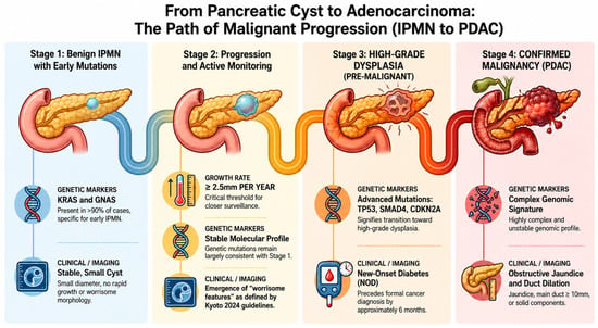
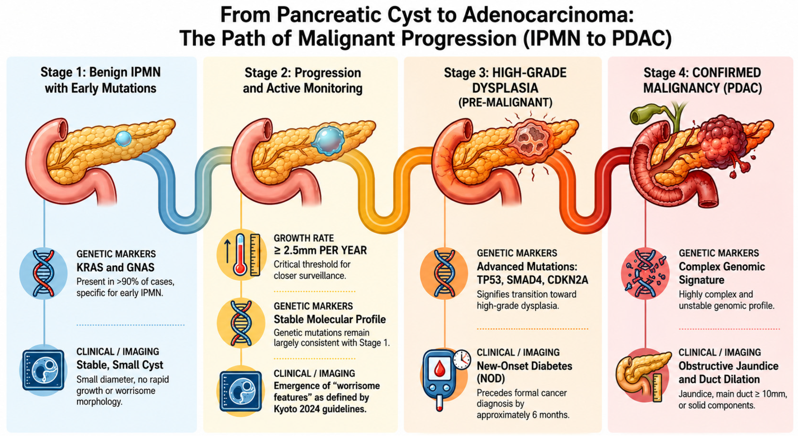
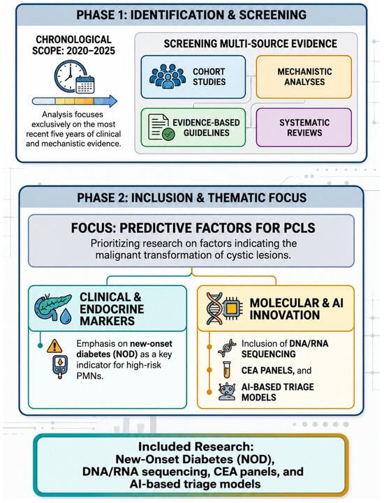
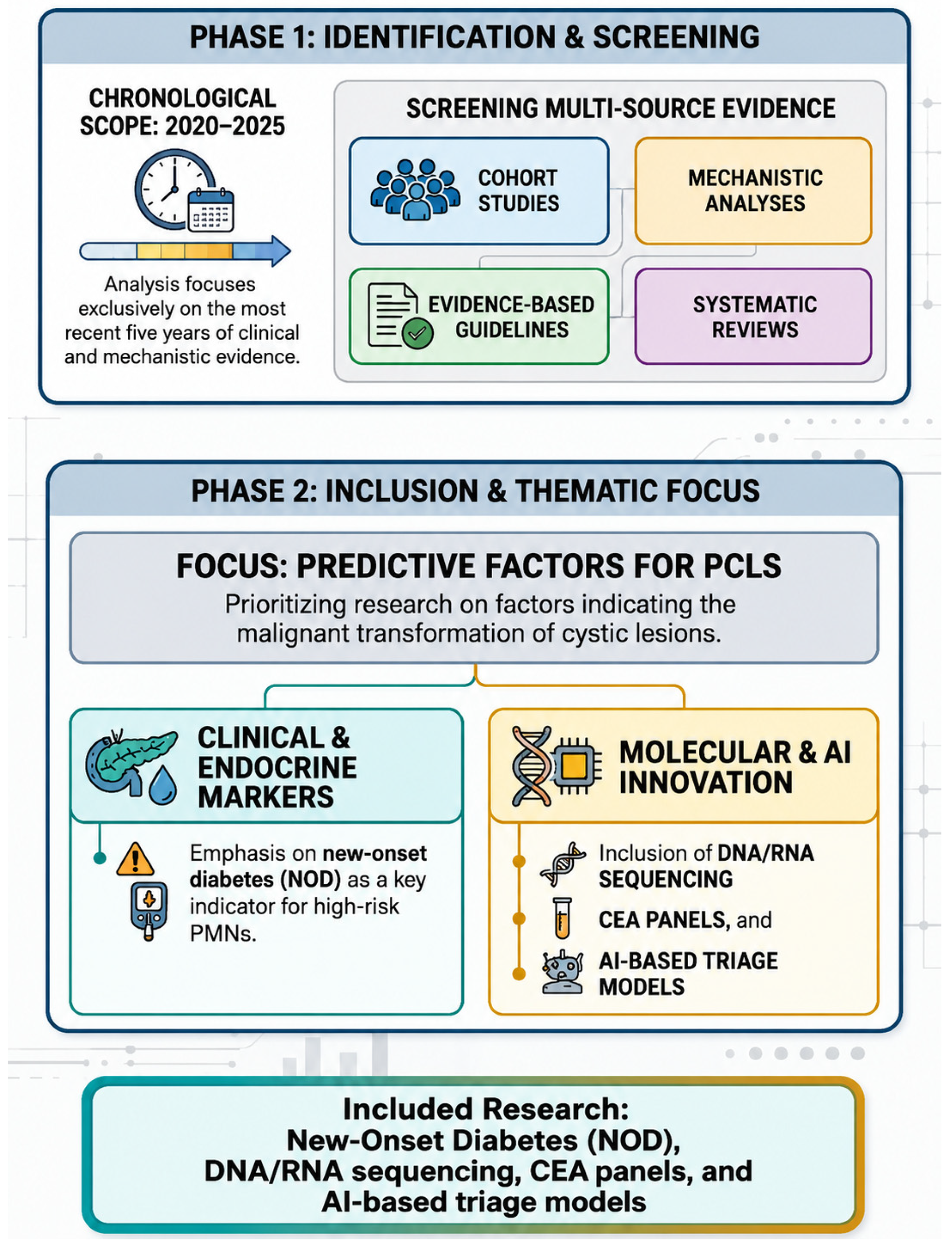
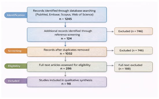
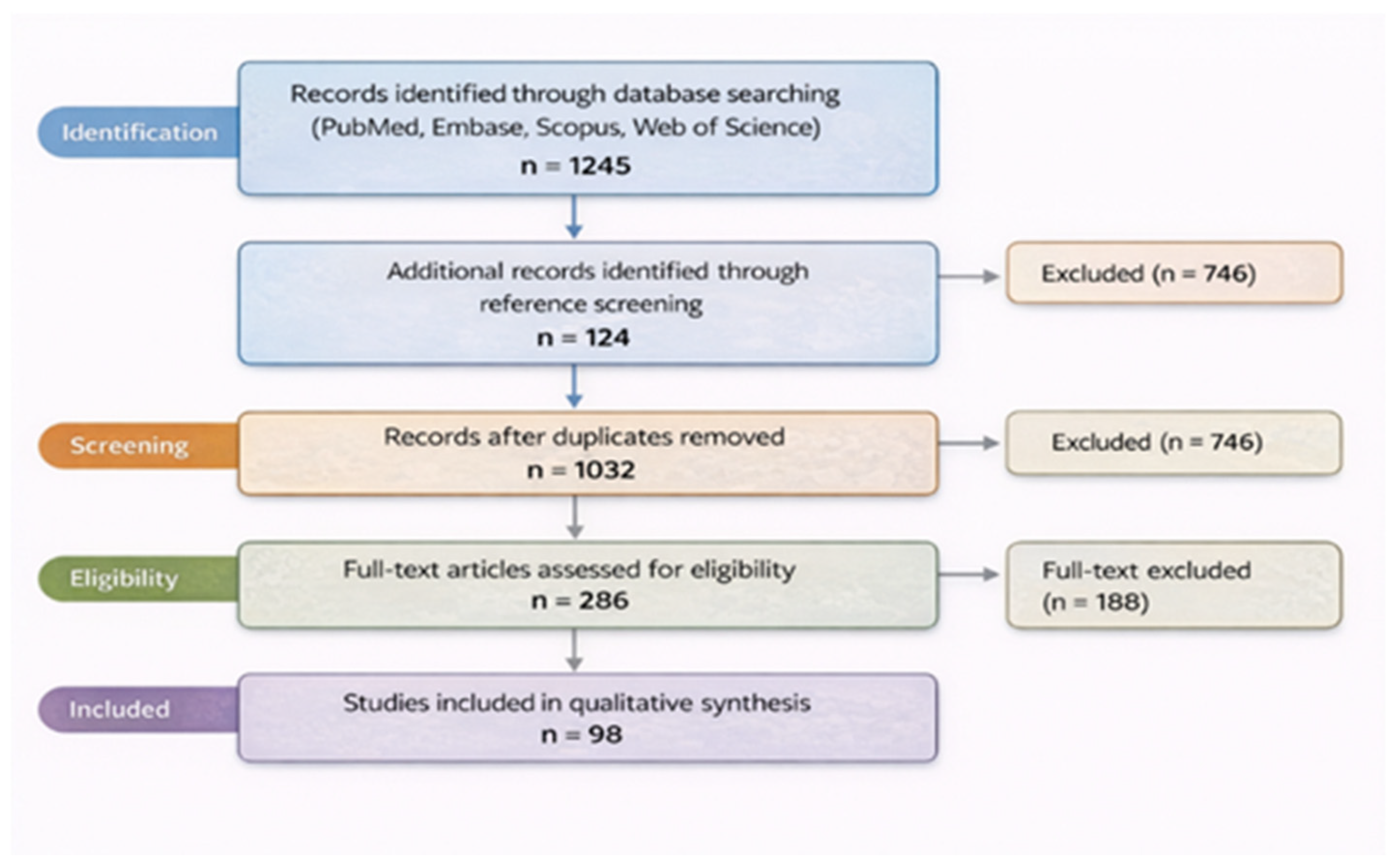
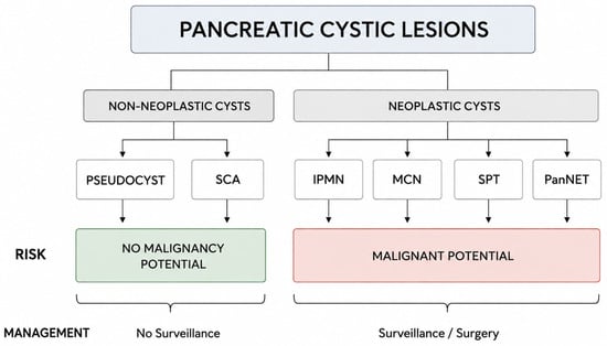
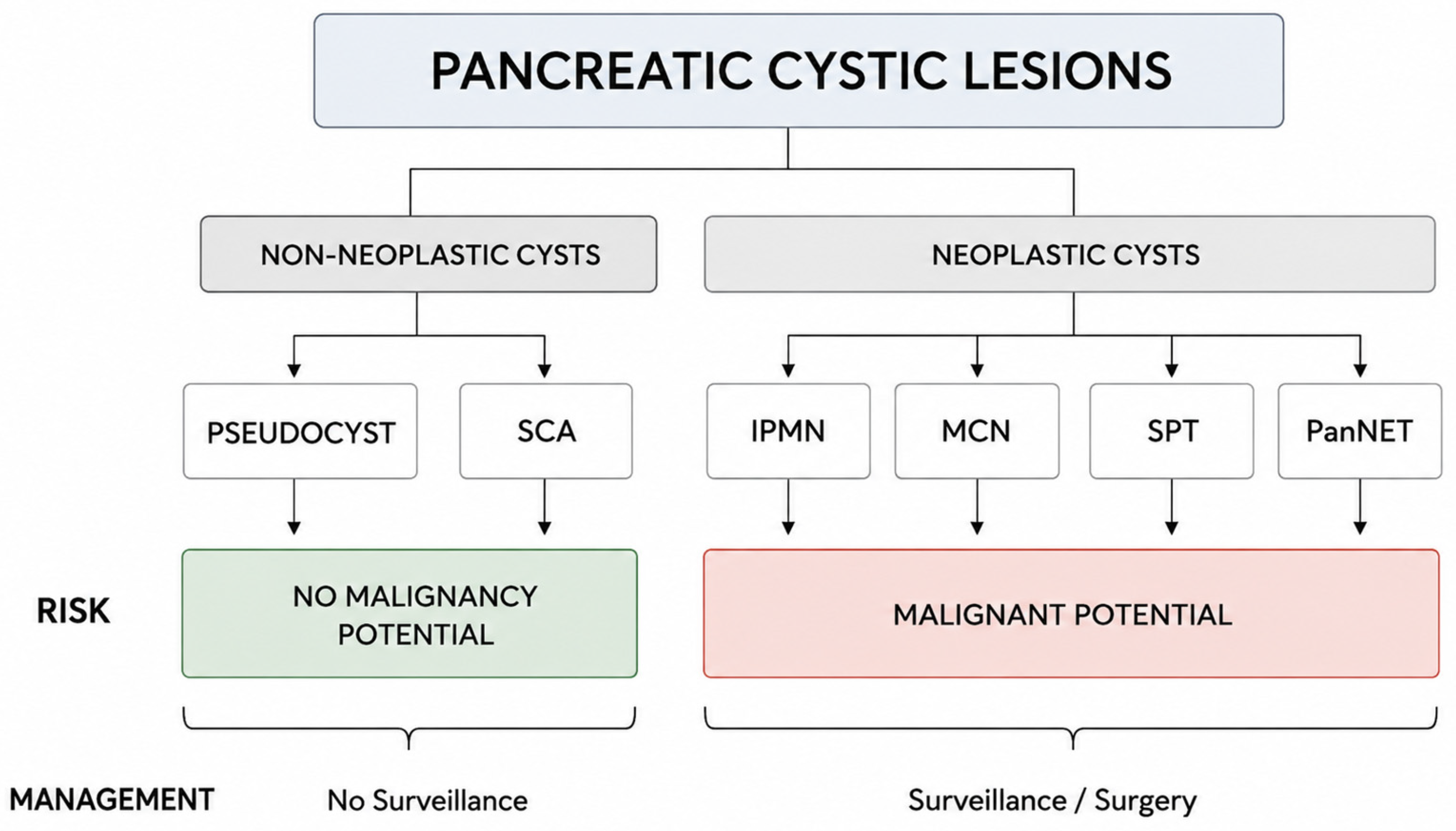
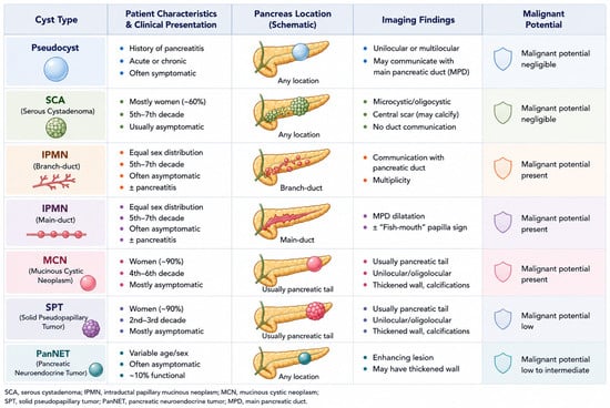
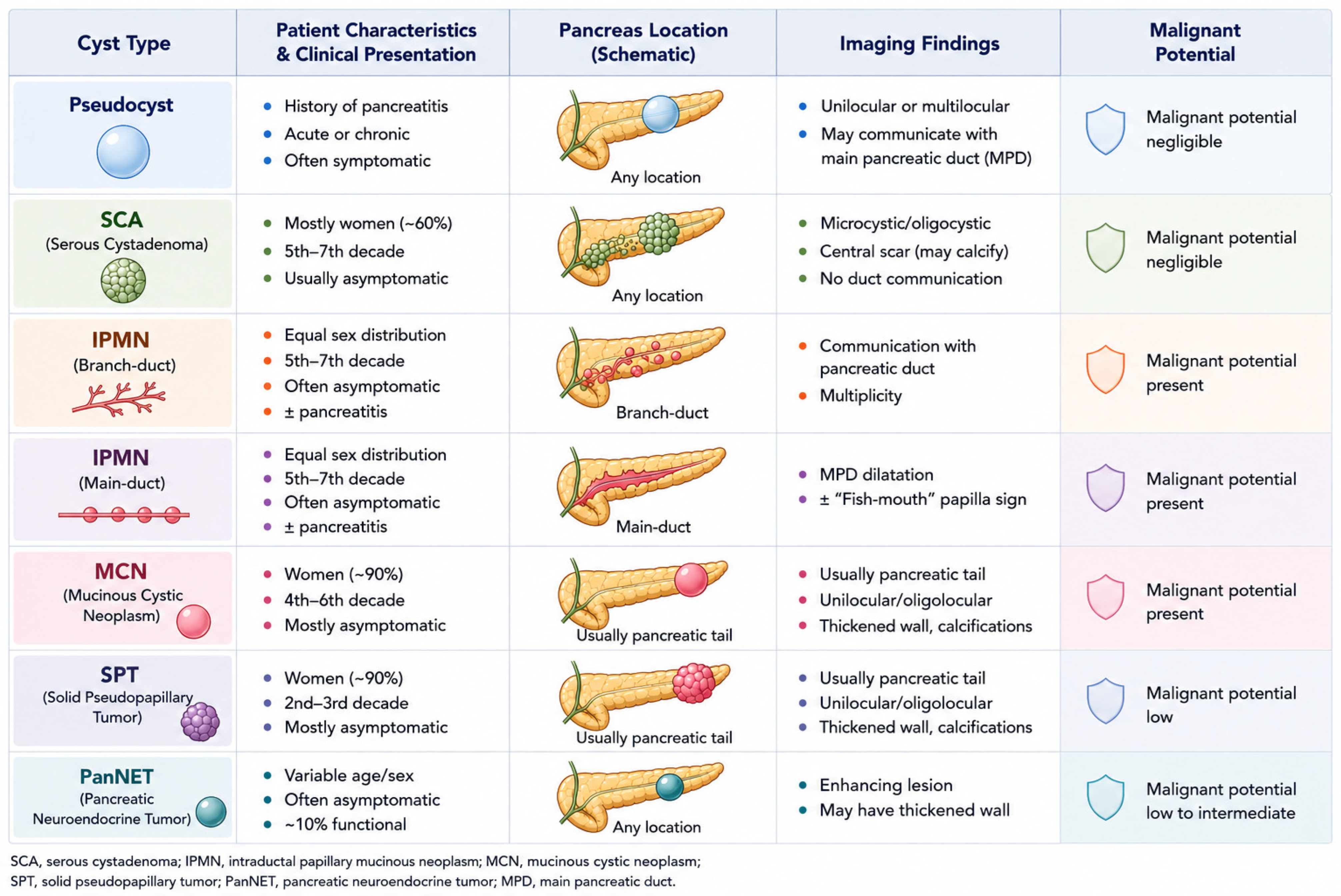
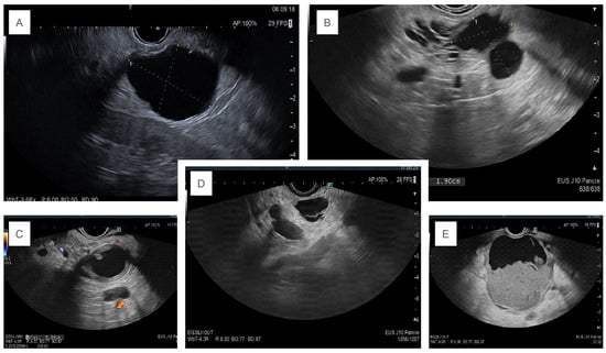
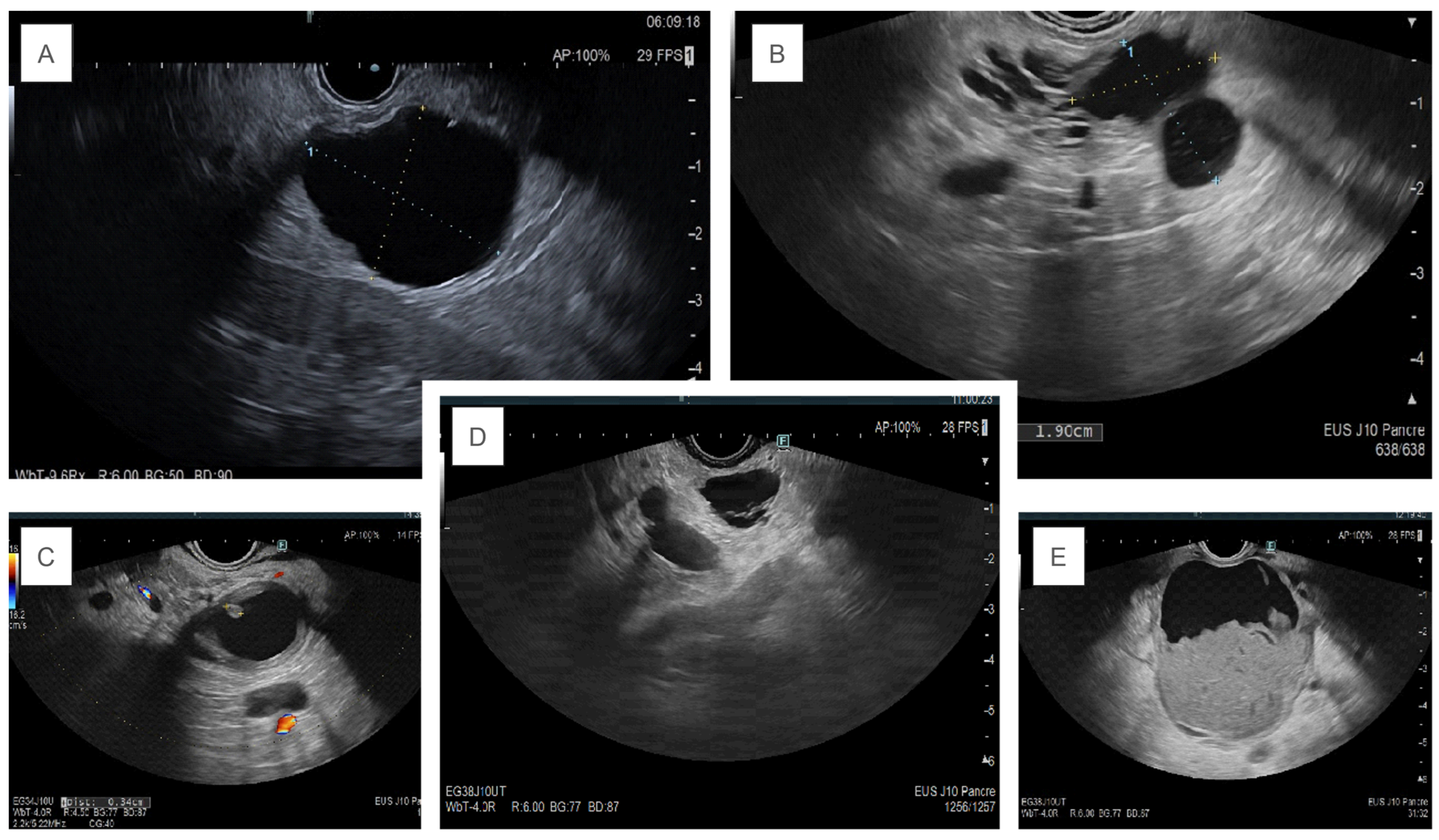
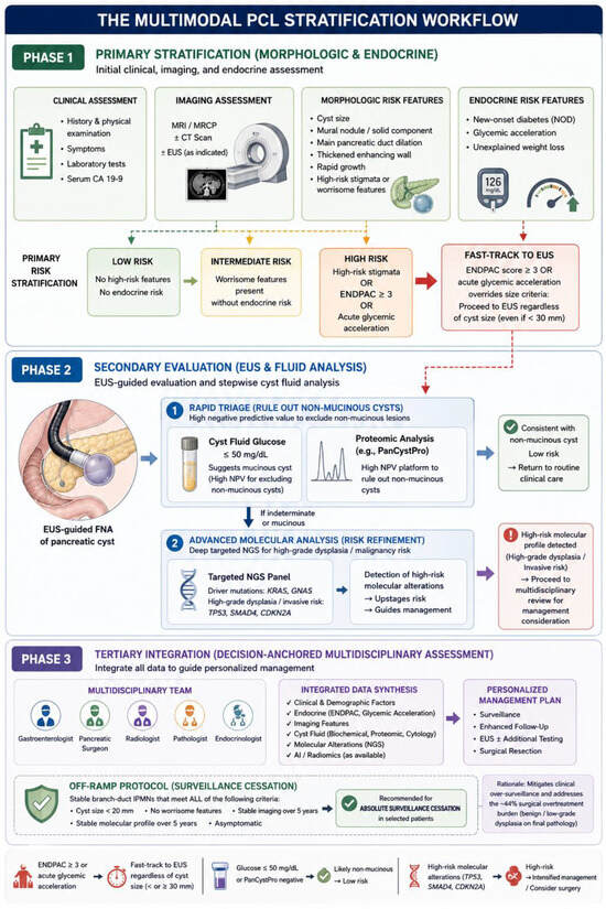
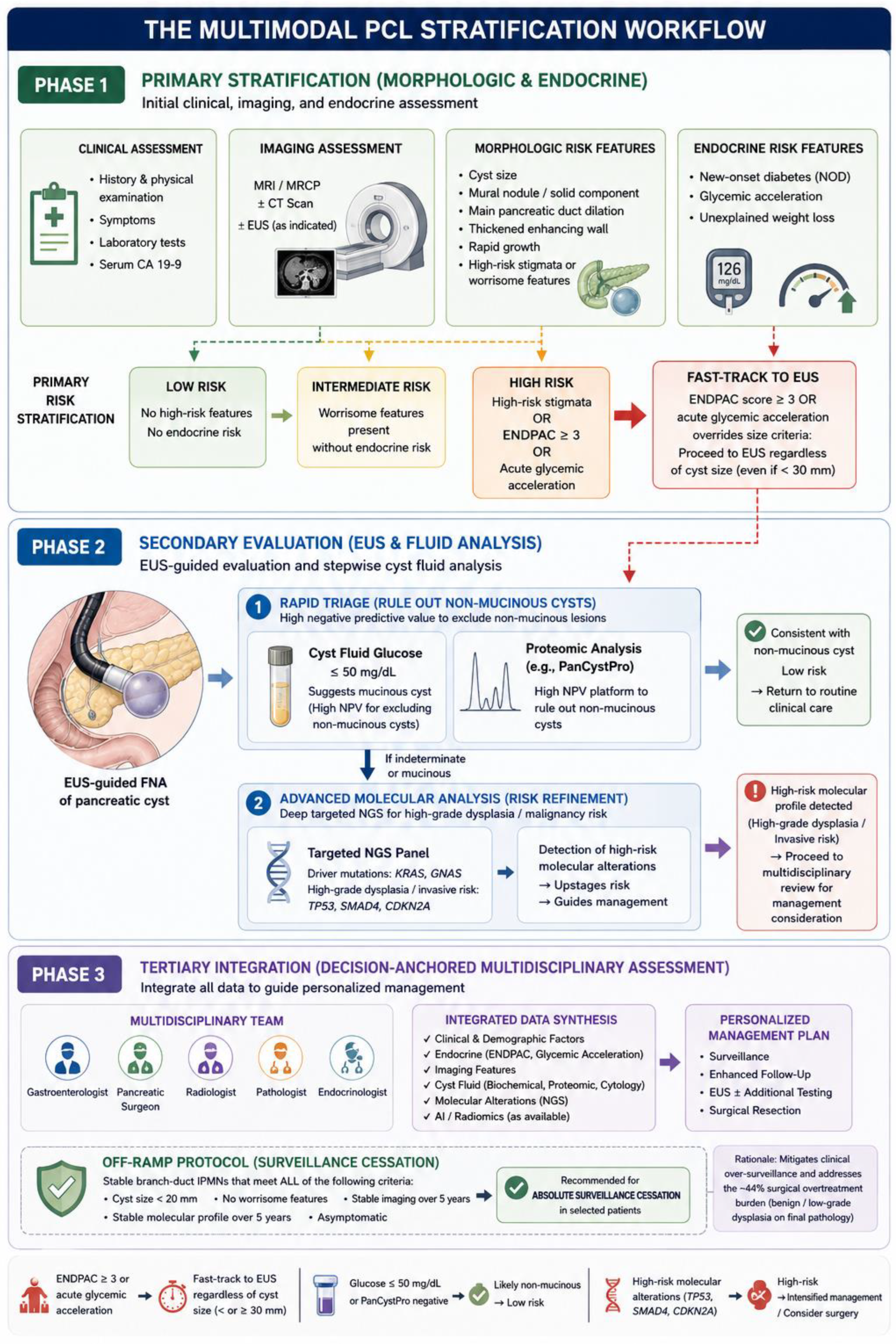

Disclaimer/Publisher’s Note: The statements, opinions and data contained in all publications are solely those of the individual author(s) and contributor(s) and not of MDPI and/or the editor(s). MDPI and/or the editor(s) disclaim responsibility for any injury to people or property resulting from any ideas, methods, instructions or products referred to in the content. |
© 2026 by the authors. Licensee MDPI, Basel, Switzerland. This article is an open access article distributed under the terms and conditions of the Creative Commons Attribution (CC BY) license.