


Abstract
Ferroptosis, an iron-dependent form of regulated cell death driven by phospholipid peroxidation, has emerged as a potential contributor to neuronal dysfunction and degeneration in Alzheimer’s disease (AD). This review critically examines the biochemical basis, AD-specific evidence, chemical modulators, and therapeutic prospects of ferroptosis, emphasizing the distinction between mechanistic compatibility and causal demonstration. Ferroptotic susceptibility is governed by the balance among polyunsaturated phospholipid substrates, redox-active iron, lipid-peroxyl radical propagation, and enzymatic or chemical termination systems, including glutathione peroxidase 4 (GPX4), FSP1, and radical-trapping antioxidants. Human and experimental AD studies show brain iron accumulation, enhanced lipid peroxidation, impaired glutathione/GPX4 defenses, and disrupted iron export, but none establishes neuronal ferroptosis as the predominant mode of cell death. Two randomized trials of brain-penetrant iron chelation lowered the bulk brain iron signal and worsened clinical outcomes, which refutes the proposition that depleting bulk brain iron is therapeutic; because neither trial measured the kinetically labile iron pool, the mechanism of that harm remains undetermined. Among emerging strategies, membrane-active radical-trapping antioxidants and substrate-directed approaches offer mechanistically rational alternatives, whereas many natural products and multitarget agents remain difficult to interpret pharmacologically. Future studies should integrate cell-type-resolved redox lipidomics, direct target engagement, and rigorous causal intervention to determine whether ferroptosis or chronic sublethal lipid peroxidation is therapeutically actionable in AD.
1. Introduction
Alzheimer’s disease (AD) is the leading cause of dementia and, on current demographic trajectories, will affect well over 100 million people by mid-century [1,2]. Two anti-amyloid monoclonal antibodies have now demonstrated statistically significant slowing of decline in early symptomatic disease, but the absolute effect sizes are small, the safety burden is real, and neither agent addresses the downstream events that actually destroy neurons [3,4]. The wider development pipeline reflects the same shift of attention toward non-amyloid mechanisms [5]. The persistent gap between removing a pathological aggregate and preserving a cell has renewed interest in the terminal execution mechanisms of neuronal death, and ferroptosis has become the most heavily cited candidate.
Ferroptosis was named in 2012 to describe an iron-dependent, caspase-independent death that could be blocked by iron chelators and by a small aromatic amine, ferrostatin-1, but not by inhibitors of apoptosis, necroptosis, or autophagy [6]. Its defining biochemistry is the accumulation of phospholipid hydroperoxides in cellular membranes beyond the capacity of the cell to reduce them [7,8]. It is, at bottom, a chemistry problem: a free-radical chain reaction running in a two-dimensional lipid solvent, initiated by hydrogen-atom abstraction from a bis-allylic methylene, propagated by peroxyl radicals, and terminated enzymatically by reduction of the hydroperoxide, chemically by radical trapping, or by excision of the oxidized acyl chain [9]. How the resulting membrane damage is converted into loss of plasma—membrane integrity is a separate and still-active question, with osmotic and pore-forming models both supported, ESCRT-III-dependent repair opposing the process, and lipid peroxidation increasing membrane tension sufficiently to gate Piezo1 [10,11,12]. Every therapeutic strategy discussed below is an attempt to change one of the rate constants in that scheme.
The mapping of this chemistry onto AD looks, at first sight, unusually clean. The AD brain accumulates iron in the regions that degenerate, over and above the regional accumulation that accompanies normal aging [13,14,15]. Cerebrospinal fluid ferritin predicts subsequent cognitive decline and is modified by APOE genotype [16]. Both amyloid precursor protein (APP) and tau participate in neuronal iron export, so their loss of function traps iron intracellularly [17,18]. Glutathione falls early [19]. Lipid peroxidation end products, 4-hydroxynonenal and F2-isoprostanes, are elevated in AD brain and biofluids [20,21]. Conditional deletion of Gpx4 from forebrain neurons produces hippocampal degeneration and memory impairment that is rescued by a lipophilic radical-trapping antioxidant [22]. Each observation is compatible with ferroptosis.
Compatibility is not demonstration, and this is where the field has been insufficiently careful. Almost every one of those findings is equally compatible with generic oxidative stress, with iron acting as a bystander marker of tissue injury, or with a death mode that shares markers with ferroptosis without sharing its mechanism. More pointedly, the two randomized controlled trials that directly tested the causal claim, by lowering brain iron in patients with a brain-permeable chelator, both produced clinical worsening [23,24]. A review that treats those results as an implementation detail rather than as data is not a critical review.
Existing reviews of ferroptosis in AD are numerous and largely concordant [25,26,27,28], which is itself a warning sign; they tend to recount the pathway, list correlative findings from the AD literature, and enumerate compounds that were protective in a cell or mouse model. Our aim differs in three respects. First, we treat ferroptosis as chemistry rather than as a pathway diagram because the therapeutic decisions that matter, which node to attack and with what pharmacophore, follow from rate constants and partition coefficients rather than from arrow direction. Second, we grade the AD-specific evidence explicitly against the hallmark criteria the field has agreed on, and we report where the evidence is absent rather than smoothing over the gaps. Third, we take the failed chelation trials seriously as mechanistic information about which iron pool matters in the human brain, and we use them to derive design rules for the next generation of central nervous system (CNS) anti-ferroptotic chemistry.
2. Materials and Methods
This narrative review was based on searches of PubMed/MEDLINE, Scopus, and Web of Science conducted through May 2026, using combinations of the terms ferroptosis, lipid peroxidation, radical-trapping antioxidant, GPX4, FSP1, ACSL4, iron chelation, deferiprone, quantitative susceptibility mapping, and Alzheimer’s disease, supplemented by Boolean operators and citation chaining from relevant primary mechanistic studies. We prioritized primary research over secondary reviews, favored physical—organic and kinetic studies where mechanism was at issue, and required clinical claims to rest on randomized controlled trials where these exist. Where a mechanistic claim is contested in the primary literature, for example, the role of DHODH, the attribution of protective effects to lipoxygenase inhibition, or the molecular target of RSL3, the disagreement is presented rather than a side selected. Studies were excluded if the sole evidence for ferroptosis was rescue by a single compound with no orthogonal confirmation, except where cited, specifically to illustrate that limitation. The evidence grading applied in Table 2 follows the hallmark criteria set out in the cell death nomenclature and ferroptosis reproducibility recommendations [29,30].
3. Ferroptosis as a Chemical Process
It is useful to decompose ferroptosis into three chemically distinct variables that can be manipulated independently: the substrate, the catalyst, and the kinetics of chain propagation versus termination (Figure 1). Confusion in the therapeutic literature can usually be traced to conflating them.
Figure 1.
The chemistry of ferroptosis resolved into substrate, catalyst, and chain kinetics. (A) Initiation requires abstraction of a bis-allylic hydrogen from a polyunsaturated acyl chain esterified into a membrane phospholipid; ACSL4 and LPCAT3 set this substrate load, and MBOAT1/2 and monounsaturated fatty acids reduce it. (B) Propagation proceeds through peroxyl radicals; the labile iron pool converts the resulting hydroperoxide into an alkoxyl radical, branching the chain. Lipoxygenases (with PEBP1) and the flavin oxidoreductases POR and CYB5R1 can also generate the initial hydroperoxide. (C) Termination is enzymatic, through GPX4 reduction of the phospholipid hydroperoxide to the alcohol, or chemical, through radical-trapping antioxidants regenerated by FSP1 (ubiquinol, vitamin K), by GCH1 (tetrahydrobiopterin), or supplied by the distal cholesterol pathway (7-dehydrocholesterol). Drug classes are placed at the node they act on.
3.1. Substrate: Which Lipids Can Burn
Autoxidation requires a bis-allylic C–H bond. The bond dissociation enthalpy at that position, roughly 75 kcal mol−1, is low enough for abstraction by a peroxyl radical, whereas the allylic C–H of a monounsaturated chain, at about 88 kcal mol−1, is not [31,32]. The rate of chain propagation therefore scales with the number of bis-allylic methylenes: linoleate has one; arachidonate, three; and docosahexaenoate, five. Peroxyl radical clock measurements put numbers on this ordering. At 37 °C in organic solution, the propagation rate constant kp rises from 62 M−1 s−1 for linoleic acid to 197 ± 13 M−1 s−1 for arachidonic acid and 334 ± 37 M−1 s−1 for docosahexaenoic acid, and the ranking is preserved for the corresponding phospholipids in phosphatidylcholine liposomes [31,32,33]. This single physical—organic fact determines much of ferroptosis biology. Cells become sensitive when polyunsaturated fatty acids (PUFAs) are esterified into membrane phospholipids and resistant when they are not.
The esterification step is enzymatically controlled. Acyl-CoA synthetase long-chain family member 4 (ACSL4) preferentially activates arachidonic and adrenic acid, and lysophosphatidylcholine acyltransferase 3 (LPCAT3) incorporates them into phosphatidylethanolamine; loss of either confers resistance [34,35]. Conversely, exogenous monounsaturated fatty acids displace PUFAs from phospholipids and produce a ferroptosis-resistant state in an ACSL3-dependent manner [36]. More recent work has refined the substrate picture considerably: phospholipids bearing two polyunsaturated acyl chains are disproportionately potent drivers of ferroptosis [37], and the essential sites of peroxidation have been narrowed to specific phospholipid species rather than to bulk membrane PUFA content [38]. MBOAT1 and MBOAT2 remodel membranes toward monounsaturated species and constitute a GPX4-independent surveillance arm that is under sex-hormone control [39].
The brain is an outlier on this axis. Gray matter phospholipids are among the most polyunsaturated in the body, with docosahexaenoate abundant in neuronal and synaptic membranes. If substrate loading alone determined risk, the brain would be the most ferroptosis-prone organ in the body, which it manifestly is not under physiological conditions. That discrepancy is informative: it implies that brain tissue is protected not by substrate scarcity but by surveillance capacity and by control of the catalyst. Reports that PUFA content, and docosahexaenoate in particular, is reduced in AD cortex [40,41] are therefore ambiguous. The loss is usually read as evidence of peroxidative consumption, and it may be; but a lower bis-allylic burden should, all else equal, lower ferroptosis susceptibility. Whether AD cortex is enriched or depleted in the specific oxidizable phospholipid species that matter remains unsettled by modern redox lipidomics, a tractable gap.
3.2. Catalyst: Which Iron Matters
Iron participates in ferroptosis in at least three chemically distinguishable ways, and the therapeutic implications differ sharply between them. First, ferrous iron reduces pre-existing lipid hydroperoxides to alkoxyl radicals, a branching step that multiplies chain-carrying radicals and is far more kinetically consequential than the classical Fenton reaction with hydrogen peroxide [42,43]. Second, iron is a structural cofactor of the lipoxygenases (ALOX15, ALOX15B, ALOX5), which can generate the initial hydroperoxides enzymatically and, via the scaffold protein PEBP1, be directed toward phosphatidylethanolamine substrates [35,44,45]. Third, iron is required for the flavin-dependent oxidoreductases cytochrome P450 oxidoreductase (POR) and CYB5R1, which contribute to phospholipid peroxidation independently of the lipoxygenases [46,47].
Only a small, kinetically labile fraction of cellular iron is available for any of this. The bulk is sequestered in ferritin, in iron—sulfur clusters, in heme, and in mineral phases; the partition between these compartments is itself age-dependent, and senescent cells accumulate iron with impaired ferritin turnover [48]. Ferritinophagy, mediated by nuclear receptor coactivator 4 (NCOA4), which was identified as the ferritin cargo receptor by quantitative proteomics [49], converts storage iron into labile iron and is a genuine sensitizing mechanism [50,51]. Brain iron transport, storage, and regional distribution have been reviewed in detail elsewhere [52,53,54]. The distinction between the storage and labile pools is the single most important and most frequently elided point in the AD ferroptosis literature, and it is revisited in Section 7.2 in connection with the failure of chelation.
The mechanistic weight given to lipoxygenases deserves particular scrutiny. Physical—organic analysis by Pratt and co-workers established that ferrostatin-1 and liproxstatin-1 are potent radical-trapping antioxidants in phospholipid bilayers and are not effective inhibitors of 15-LOX-1 catalysis [55]. A follow-up study showed that many compounds used as lipoxygenase inhibitors in ferroptosis experiments, including baicalein, suppress lipid autoxidation directly, so their protective effects cannot be attributed to enzyme inhibition [56]. A substantial portion of the published evidence for a lipoxygenase requirement in ferroptosis, including in neuronal systems, therefore rests on pharmacology that has been reinterpreted. Lipoxygenases can initiate; the claim that they execute is not secure.
3.3. Propagation and Termination: Where the Kinetics Are Decided
Once a chain is running, its length is set by the ratio of the propagation rate constant to the rate at which chain-carrying peroxyl radicals are intercepted. This is the classical inhibited autoxidation problem, and it is quantitatively well understood [31,57]. A radical-trapping antioxidant (RTA) is characterized by its inhibition rate constant, kinh, and by its stoichiometric factor, and, critically for membrane chemistry, by how it partitions and diffuses within the bilayer. α-Tocopherol reacts with peroxyl radicals at roughly 3 × 10⁶ M−1 s−1 in homogeneous solution [57]; ferrostatin-1 and liproxstatin-1 are somewhat slower in solution yet markedly more effective in phospholipid bilayers, which Zilka and colleagues attributed to superior partitioning and mobility in the lipid phase rather than to intrinsic reactivity [55]. The practical lesson is that intrinsic antioxidant potency measured in an organic solvent is a poor predictor of anti-ferroptotic potency in a membrane.
Termination can also be achieved enzymatically. GPX4 is the only mammalian enzyme that reduces phospholipid hydroperoxides in situ within the membrane, converting the branching substrate into an inert alcohol before iron can act on it [58,59,60]. Its loss was shown, before ferroptosis was named, to translate oxidative stress into 12/15-lipoxygenase-dependent death [61], which is the historical origin of the lipoxygenase interpretation discussed above. The three known GPX4-independent systems all work by regenerating small-molecule RTAs: FSP1 reduces ubiquinone to ubiquinol [62,63], GCH1 supplies tetrahydrobiopterin [64], and the distal cholesterol biosynthesis intermediate 7-dehydrocholesterol acts as an endogenous RTA in its own right [65,66]. FSP1 additionally participates in a non-canonical vitamin K cycle that suppresses ferroptosis [67] and can regenerate α-tocopherol. A further, structurally distinct route exists: the calcium-independent phospholipase iPLA2β excises the oxidized acyl chain outright, removing the redox lipid death signal rather than reducing it [68]. Ferroptosis, viewed chemically, is what happens when the enzymatic, chemical, and repair capacity of a membrane are exceeded simultaneously.
One much-cited element of this scheme remains formally contested. Mitochondrial dihydroorotate dehydrogenase (DHODH) was proposed as an additional defense arm operating through mitochondrial CoQ [69], but a subsequent analysis attributed the observed effects of DHODH inhibitors to off-target FSP1 inhibition, and the original authors have replied in defense of their assignment; the exchange has not converged [70,71]. It is therefore presented here as unresolved rather than as established, and any AD-directed strategy premised on DHODH would inherit that uncertainty.
4. Surveillance Systems Re-Weighted for the Brain
The termination and repair systems described above were characterized largely in transformed cell lines chosen for tractability, and their relative importance is not transferable to brain tissue without argument. Neurons are post-mitotic, highly oxidative, poorly glycolytic, and dependent on astrocytes for cysteine supply; oligodendrocytes carry the highest iron content in the CNS; microglia handle iron-rich debris; and astrocytes have their own ferroptotic route through NOX4-driven lipid peroxidation in AD [72]. Cell-type weighting is not a detail; it is the substance (Table 1). Two experiments make the point directly. Ferroptotic stress confined to microglia kills neurons non-cell-autonomously, so the cell that dies need not be the cell in which the chemistry ran [73]. Conversely, oligodendrocyte death in Pelizaeus-Merzbacher disease is rescued by iron chelation, showing that the same node can be protective in one CNS cell type, while, as discussed in Section 7, it is harmful when addressed indiscriminately across the whole brain [74].
Table 1.
Core ferroptosis nodes, their chemical function, and the status of the evidence in AD. Direction indicates the reported change in AD brain or AD models relative to control. AD evidence tier: H, human tissue or biofluid; M, mammalian AD model; C, cell model only; N, not examined in AD. A dash indicates that no direction can be assigned because the node has not been measured in AD.
4.1. GPX4, Selenium, and the Cysteine Supply Chain
GPX4 uses a selenocysteine residue in its active site. Ingold and colleagues showed that replacing it with cysteine yields an enzyme that retains catalytic activity but is irreversibly overoxidized under peroxide stress. The in vivo consequence is strikingly selective and is itself dependent on genetic background. Mice carrying the cysteine variant on a mixed C57BL/6J × 129S6SvEv background are born at the expected Mendelian ratio but selectively lose cortical parvalbumin-positive GABAergic interneurons, sparing calbindin- and calretinin-positive populations, and die of spontaneous seizures in the 3rd postnatal week; on a congenic C57BL/6J background, the allele is lethal in mid-gestation [88]. The chemical rationale is that the selenolate is both more nucleophilic and, once oxidized, more readily reduced back to the active state, so selenocysteine confers resistance to inactivation rather than simply higher turnover. Deletion of Gpx4 is, moreover, embryonic lethal [87]. This makes GPX4 an intrinsically selenium-limited enzyme and gives the brain’s well-documented prioritization of selenium a direct mechanistic reading.
The upstream constraint is cysteine. System xc−, the SLC7A11/SLC3A2 cystine-glutamate antiporter, supplies the cysteine for glutathione synthesis, and its inhibition by erastin or by high extracellular glutamate produces the death originally described in neuronal cells as oxytosis, a decade before ferroptosis was named [82,83]. Oxytosis and ferroptosis are now generally regarded as the same process approached from two literatures [94], a point of some importance because the neuronal literature is older, was conducted in primary cells, and used different readouts. Brain glutathione is reduced in mild cognitive impairment and AD as measured by magnetic resonance spectroscopy [19], and GPX4 protein is reported to fall in AD cortex [85], but the two observations are rarely made in the same tissue.
4.2. FSP1-Ubiquinol and the GPX4-Independent Arms
FSP1 (AIFM2) is a myristoylated, membrane-associated NAD(P)H oxidoreductase that maintains a pool of ubiquinol acting as a lipophilic RTA at the plasma membrane [62,63]. Its activity is also regulated by condensate formation [95], and selective inhibitors are now available [96]. For AD, this arm has an obvious attraction because it can in principle be reinforced pharmacologically with ubiquinone or with vitamin K, both of which FSP1 can recycle [67], and because it is glutathione-independent and therefore insensitive to the cysteine bottleneck that limits GPX4 in an aged, astrocyte-compromised brain. It has also been almost entirely unexplored in AD tissue: no study appears to have quantified FSP1 protein, ubiquinol redox state, and lipid hydroperoxides together in human AD cortex with cell-type resolution. That is a striking omission given how prominent the FSP1 axis has become elsewhere.
The GCH1-tetrahydrobiopterin arm is similarly under-examined in AD, although it is of particular interest because tetrahydrobiopterin is also the cofactor for the aromatic amino acid hydroxylases and for nitric oxide synthase, so its depletion couples ferroptosis susceptibility to monoamine synthesis and to nitrergic signaling [64]. Any GCH1-directed intervention in the brain carries obvious pleiotropy.
4.3. The 7-Dehydrocholesterol Axis: An Unexploited CNS Node
The identification of 7-dehydrocholesterol (7-DHC) as an endogenous RTA is the most interesting recent development from a CNS perspective, and it has been almost ignored in the AD ferroptosis literature [65,66]. Two independent genome-wide screens converged on DHCR7, the terminal enzyme of the Kandutsch-Russell branch of cholesterol biosynthesis. Loss of DHCR7 causes 7-DHC to accumulate, and 7-DHC, whose conjugated diene in the B-ring makes it an unusually good hydrogen—atom donor, suppresses ferroptosis by trapping peroxyl radicals in the membrane. The apparent paradox is worth stating: peroxyl radical clock measurements had already established 7-DHC as one of the most oxidizable lipids known, with a propagation rate constant of 2260 ± 40 M−1 s−1 at 37 °C, some 200-fold that of cholesterol itself at 11 ± 2 M−1 s−1 [31,33], so it protects precisely because it is sacrificially consumed, diverting the chain away from the phospholipid pool.
Three features make this node CNS-relevant. Brain cholesterol is synthesized locally because lipoproteins do not cross the blood—brain barrier, so the distal pathway is constitutively active in the tissue of interest. Cholesterol metabolism is already implicated in AD through APOE and through several genome-wide association loci. And DHCR7 is inhibited, as an off-target activity, by several widely used psychotropic drugs, which offers an unusual repurposing entry point. The caution is severe and must be stated with the proposal: profound 7-DHC accumulation is the biochemical lesion of Smith-Lemli-Opitz syndrome, and 7-DHC-derived oxysterols are themselves neurotoxic. The therapeutic window, if one exists, is a partial and probably transient elevation. This node is therefore flagged as a hypothesis worth testing, not as a recommendation.
4.4. APOE, Ferritinophagy, and a Genuine AD-Specific Link
One recent finding does connect the dominant genetic risk factor for late-onset AD to ferroptosis directly rather than by analogy. Apolipoprotein E was shown to inhibit ferroptosis potently in cultured neurons by activating PI3K/AKT signaling and thereby suppressing NCOA4-mediated ferritinophagy, which restrains the conversion of storage iron into the labile pool and so limits iron-driven lipid peroxidation [92]. The nature of the isoform dependence requires care because it is frequently misstated. In that study, the common apoE isoforms did not differ from one another in their capacity to protect against ferroptosis in vitro; the anti-ferroptotic activity is a property of the protein rather than of the variant. What differs between genotypes is the context in which that activity operates: apoE abundance, which is lower in ε4 carriers, and the lipid environment of the ε4 brain, which is comparatively enriched in polyunsaturated species. Consistent with a context-dependent rather than an activity-dependent effect, the same study found in a post-mortem series of 608 participants that the association between brain iron and clinically confirmed AD was stronger in ε4 carriers [92]. The mechanism therefore links APOE to ferroptotic susceptibility through apoE dose and membrane substrate loading, not through an isoform-intrinsic difference in protein efficacy. This dovetails with the earlier observation that cerebrospinal fluid ferritin is regulated by APOE and predicts cognitive outcomes [16], and with the finding that iron-associated lipid peroxidation in lipid rafts is most severe in ε4 carriers [86]. Confirmed with cell-type resolution in human tissue, the dose-and-substrate account would be the strongest AD-specific ferroptosis link available, tying an established genetic risk term to a defined step in the cascade rather than to a generic oxidative phenotype.
5. What Is Actually Established in AD
The field has converged on a hallmark set for claiming ferroptosis: iron dependence demonstrated by chelation, lipid peroxidation demonstrated directly rather than by a surrogate aldehyde, perturbation of the GPX4-glutathione or FSP1 axes, and rescue by a structurally distinct RTA that is not itself a chelator [29,30]. Applying that standard to the AD literature produces a much shorter list than a reading of recent reviews would suggest (Table 2).
Table 2.
Evidence grading for the principal claims made about ferroptosis in AD. Tier A, demonstrated in human AD tissue or in a randomized clinical trial with orthogonal confirmation; Tier B, demonstrated in a mammalian AD model with genetic and pharmacological support; Tier C, demonstrated in cell models or by correlation only; Tier D, asserted in the review literature without primary support meeting the hallmark criteria.
5.1. Human Tissue and Imaging
Iron accumulation in AD brain is not in dispute. Histochemical and synchrotron studies place iron in and around plaques, in glia, and in the neuropil, and demonstrate mineralized phases, including magnetite, that are chemically distinct from ferritin-bound iron [97,98,104,105]. The wider case for biometals as therapeutic targets in AD has been argued at length [106,107]. In vivo, quantitative susceptibility mapping (QSM) shows regional iron elevation that tracks with amyloid burden and predicts subsequent cognitive decline [13,14,108], and a large post-mortem series relates regional iron to the rate of deterioration [99]. Cortical iron also tracks tau aggregation measured by PET [109], and cerebrospinal fluid ferritin predicts brain hypometabolism in amyloid-positive individuals [110]. Human tissue work has also reported iron dyshomeostasis, lipid peroxidation, and altered cystine/glutamate antiporter expression together in the same AD brains, which is closer to a hallmark set than most reports achieve [84]. These are strong, replicated data. What they do not establish is that the accumulated iron is in a redox-active, chelatable, chain-branching form. QSM reports bulk magnetic susceptibility, a quantity dominated by ferritin-bound and mineral iron, and no available human measurement resolves the small labile fraction that performs chain branching at the membrane (Figure 2).
Figure 2.
Iron handling in the Alzheimer brain and the pool that chelation actually reaches. Left: neuronal iron export requires soluble tau to traffic APP to the plasma membrane, where APP stabilizes ferroportin; loss of soluble tau, amyloidogenic APP processing, or ferroportin downregulation all cause iron retention. APP supports export by maintaining surface ferroportin and is not itself a ferroxidase [18,101]. Centre: the intracellular iron budget is dominated by ferritin, heme, and iron-sulfur clusters, with only a small labile pool competent for chain branching; NCOA4-mediated ferritinophagy converts storage iron into labile iron and apoE restrains it. Right: quantitative susceptibility mapping is dominated by ferritin-bound and mineralized iron, so a fall in the QSM signal after chelation establishes that bulk tissue iron was lowered. It is not a measurement of the labile pool, which was not assayed in either trial, and the mechanism by which chelation worsened outcomes therefore remains undetermined.
Direct evidence of lipid peroxidation in AD tissue is also solid at the level of end products: 4-hydroxynonenal adducts are increased [20], and F2-isoprostanes are elevated in brain and cerebrospinal fluid [21], although peripheral F2-isoprostanes are not [111], which is a useful caution for anyone proposing a plasma peroxidation biomarker. Changes in antioxidant enzyme activity accompany these end products in the same cortical tissue [85]. The most informative recent human study localized iron-associated lipid peroxidation to lipid rafts, with reduced GPX4 and glutathione synthetic capacity in the same fractions and the most severe changes in APOE ε4 carriers [86]. Subcellular resolution of this kind is what the field needs more of because bulk homogenate measurements cannot distinguish a membrane in which a chain reaction ran from a tissue containing dying cells of any cause.
At the transcriptomic level, oxytosis/ferroptosis signatures are enriched in AD brain [112], and single-nucleus datasets show iron-handling and lipid-remodeling genes altered in specific glial populations [113,114]. Signature enrichment is a hypothesis-generating result. It cannot distinguish a cell that died by ferroptosis from a surviving cell mounting an anti-ferroptotic response, and in post-mortem tissue the cells that died are precisely the ones not sampled. This survivorship problem is intrinsic to human ferroptosis research in the brain and is rarely acknowledged.
5.2. Genetic and Model Evidence
The strongest causal evidence is genetic and comes from mice. Conditional deletion of Gpx4 in forebrain neurons produces hippocampal neurodegeneration, elevated lipid peroxidation, neuroinflammation, and spatial memory deficits; withdrawing vitamin E from the diet markedly accelerates the phenotype, and the ferroptosis inhibitor liproxstatin-1 reduces the neurodegeneration [22]. The direction of the dietary experiment deserves emphasis because it is often reported the other way round: vitamin E was removed rather than supplemented, and the acceleration that followed is evidence that endogenous radical-trapping capacity restrains the lesion rather than evidence that supplementation reverses it. Broader neuronal deletion causes motor neuron degeneration and paralysis [100], and inducible whole-body deletion is lethal with neuronal loss [115]. These experiments prove that neurons require GPX4 and that they die by an RTA-suppressible mechanism when it is removed. They do not prove that this is what happens in AD; they establish sufficiency of the lesion, not its presence in the disease.
Deletion of ferroportin in principal neurons produces hippocampal atrophy, memory deficits, and ferroptotic markers, and ferroportin is downregulated in APPswe/PS1dE9 mice and in human AD brain [76]. Tau-deficient mice accumulate brain iron because tau is required to traffic APP to the cell surface, where APP supports iron export [17,78,80,81]. The nature of that support should be stated precisely because the original description has been revised. APP was first reported to possess an intrinsic, zinc-inhibitable ferroxidase activity that oxidized Fe(II) for loading onto transferrin [77]. On re-examination, the apparent oxidase activity was traced to phosphate in the assay buffer rather than to the protein and purified soluble APP did not oxidize Fe(II) under conditions controlled for pH and charge [18]. What survived re-examination is the cell—biological function: endogenous APP is required to maintain ferroportin at the neuronal surface, so APP promotes iron export without itself being a ferroxidase [18]. Even this role is not uncontested, since in primary hippocampal neurons, the ferroxidase activity required for ferroportin-supported efflux was attributed to hephaestin, with APP knockdown leaving surface ferroportin unchanged [101]. Amyloidogenic processing of APP itself induces cellular iron retention [78]. Human neurons carrying familial AD presenilin mutations show selective ferroptosis vulnerability [93], and unbiased CRISPR interference and activation screens in human neurons converge on ferroptosis as a principal death route, linking it to lysosomal failure [116]. This is a coherent set, and it is the best argument that the ferroptosis machinery is genuinely wired into AD-relevant biology rather than merely co-occurring with it.
Two caveats attach. First, most of these models create iron retention by removing a protein, which is a much larger perturbation than the graded changes seen in sporadic disease. Second, several of the AD models in which ferrostatin-1 or liproxstatin-1 improved outcomes did not report the control experiments that would exclude a generic antioxidant effect, in particular a comparison with an RTA-inactive structural analogue. Ferrostatin-1 rescue is often treated as diagnostic of ferroptosis, but ferrostatin-1 will also suppress non-ferroptotic lipid peroxidation, and circularity follows quickly if the same compound defines the phenotype and demonstrates the mechanism.
5.3. The Direction of Causality Remains Open
The literature commonly asserts that ferroptosis is upstream of amyloid and tau pathology. The evidence for direction is thin in both directions. Iron promotes amyloid aggregation in vitro and amyloid-β can generate reactive oxygen species in the presence of redox-active metals; equally, amyloidogenic APP processing reduces surface ferroportin and causes iron retention [78], and soluble tau loss impairs iron export [17]. The amyloid-ferroptosis link has been reviewed in detail and remains bidirectional on the available data [117]. The system is reciprocal, which means correlational human data cannot resolve it and only staged interventional experiments can. In practice, the honest statement is that ferroptosis in AD is at least partly a consequence of the proteinopathy and may additionally be a driver, and the relative weights are unknown.
There is one further consideration that the field tends to avoid. Ferroptosis is, in most systems, a rapid death: hours, not years. AD removes neurons over a decade or more. Either ferroptosis in AD is a rare, stochastic event in a small fraction of vulnerable cells, almost undetectable by bulk assays, with an open cumulative contribution, or the relevant process is chronic sublethal lipid peroxidation that damages synapses and mitochondria without executing the cell, a distinct process that should be named differently. Sublethal membrane oxidation may well be the more important process in AD, and it would still be treatable with the same chemistry, but conflating it with ferroptosis has caused the field to search for an execution event where it should be measuring a chronic flux.
6. Chemical Modulators: Structures, Mechanisms, and CNS Suitability
Modulators are grouped here by the chemical variable each manipulates rather than by their provenance because compounds from very different sources converge on the same node and are subject to the same constraints (Table 3, Figure 3). For a CNS indication, three properties dominate: whether the compound acts at the propagation step or elsewhere, whether it survives long enough in plasma and tissue to act, and whether it reaches the brain at a free concentration above its effective potency.
Table 3.
Chemical modulators of ferroptosis relevant to AD, grouped by the chemical variable each manipulates. CNS exposure summarizes reported brain penetration after systemic dosing. Strongest evidence indicates the most advanced setting in which the compound has been tested for a neurological indication; RCT, randomized controlled trial. Limitations are the principal chemical or translational objection to each agent.
Figure 3.
Chemical modulator landscape positioned by node of action against readiness for a central nervous system indication. The vertical axis combines demonstrated unbound brain exposure with clinical stage; the horizontal axis is the chemical variable each agent manipulates. Agents in the upper region have human exposure data; agents in the lower region remain tool compounds. The two agents with completed randomized trials in neurodegeneration that lowered the bulk brain iron signal are marked as refuted at that node, while the only randomized signal consistent with the hypothesis sits at the termination node.
6.1. Radical-Trapping Antioxidants: The Best-Understood Class
Ferrostatin-1 is an arylamine bearing an ester, and both features matter. The arylamine is the pharmacophore, donating a hydrogen atom to a chain-carrying peroxyl radical to give a relatively stable aminyl radical [55]. Its cytoprotective mechanism has been dissected in detail [143]. The ester is a liability: it is hydrolytically and metabolically labile, which is why ferrostatin-1 is an excellent tool compound and a poor drug despite protecting in a wide range of disease models [144]. Second- and third-generation ferrostatins addressed this, culminating in UAMC-3203, in which the ester is replaced, and the compound acquires sufficient stability and exposure for in vivo use [118]. Further optimization has produced oxazole-containing RTAs with oral bioavailability and high brain concentrations, explicitly designed for CNS indications [119]. Liproxstatin-1, a spiro-fused quinoxalinamine, follows the same logic with a different scaffold [55,59].
The structure-activity relationship for this class is unusually well grounded because it derives from inhibited autoxidation kinetics rather than from cell-based screening alone. Good RTAs share a heteroatom-substituted aromatic that stabilizes the radical formed after hydrogen—atom transfer, sufficient lipophilicity to partition into the bilayer, and no strongly electron-withdrawing substituent adjacent to the donating group. Phenoxazines and related tricyclic amines are exceptionally reactive on this basis [120], and 2,2,6,6-tetramethylpiperidine-1-oxyl-type nitroxides are catalytic rather than stoichiometric: they reduce peroxyl radicals by electron transfer and are regenerated through hydride transfer to the derived oxoammonium ion followed by hydrogen—atom transfer from the resulting hydroxylamine, a cycle that operates in liposomes and in cells [121]. A related cross-dismutation of hydroperoxyl and alkylperoxyl radicals accounts for their unusual efficiency at low radical flux, which is the regime a chronic neurodegenerative process actually presents, and the same catalytic behavior has since been demonstrated for the most potent ferroptosis inhibitors acting on phospholipid-derived peroxyl radicals [145,146]. Copper(II) and nickel(II) bis(thiosemicarbazone) complexes are a chemically distinct entry to the same node: Zilka and co-workers demonstrated that their anti-ferroptotic activity derives from radical-trapping antioxidant behavior of the intact complex rather than from metal delivery or chelation [123], which explains and reframes the earlier observation that CuII(atsm) inhibits ferroptosis [122]. Because CuII(atsm) has already entered phase 1 clinical study in amyotrophic lateral sclerosis (ClinicalTrials.gov NCT02870634) and in Parkinson’s disease, it is, on the chemistry, the most clinically advanced anti-ferroptotic chemotype in neurology, and it has not been tested in AD. The trial registry is cited rather than a published report because no peer-reviewed clinical output was identified at the time of writing; the mechanistic claim rests on the two chemistry papers cited above and not on the trials.
That radical trapping is not merely pharmacological but physiological in neurons is shown by the requirement for vitamin A or an equivalent radical-trapping antioxidant during neuronal development [147]. α-Tocopherol belongs to this class and is the only member with a randomized clinical signal in AD, which is discussed in Section 7.3. Its limitations are chemical rather than conceptual: a modest kinh, a stoichiometric factor of two, dependence on co-antioxidants for regeneration, and, at high concentration, the well-described tocopherol-mediated peroxidation whereby the α-tocopheroxyl radical can itself abstract a bis-allylic hydrogen. Newer RTAs were designed precisely to avoid these liabilities, and none has entered an AD trial.
6.2. Substrate-Directed Chemistry
If the substrate is the problem, remove the abstractable hydrogen. Site-specific deuteration at the bis-allylic position exploits a primary kinetic isotope effect to slow the rate-limiting hydrogen-atom transfer; because the deuterated lipids act as chain-terminating defects rather than having to displace every oxidizable chain, partial replacement of the cellular PUFA pool suppresses autoxidation substantially [124,125]. This is a rare instance of a therapeutic strategy that follows directly from physical—organic chemistry. Deuterated linoleate (RT001) has been administered to patients across several neurodegenerative indications with acceptable tolerability, and an early trial in Friedreich ataxia reported signals of efficacy [126]. That result did not hold: a subsequent double-blind, placebo-controlled trial in the same indication, in 65 subjects over 11 months with maximum oxygen consumption as the primary endpoint, found no significant benefit at the doses tested in any primary, secondary, or exploratory measure [127]. In animals, the picture is more encouraging: deuterium-reinforced PUFAs improved cognition in an aldehyde dehydrogenase 2 null mouse model of sporadic AD, cutting cortical and hippocampal F2-isoprostanes by roughly half [128]. The counter-result deserves equal weight: in APP/PS1 transgenic mice, the same dietary strategy lowered brain lipid peroxidation and hippocampal amyloid-β but produced no discernible behavioral benefit [129]. Read together, these two experiments say that suppressing peroxidation is achievable in the brain and that doing so does not automatically rescue cognition in an amyloid-driven model, which is precisely the distinction between target engagement and efficacy that the chelation trials also expose. Taken together, the deuteration strategy remains chemically compelling and clinically unproven: no positive controlled readout exists in any indication. The approach nonetheless has notable advantages for a chronic CNS indication: the agent is a dietary lipid, distributes to all membranes including brain, and does not need to be present at the moment of a stochastic ferroptotic event because the protection is built into the membrane. Its disadvantages are cost and the months required to reach steady-state membrane enrichment.
Enzymatic substrate control offers an alternative. ACSL4 inhibition reduces the incorporation of arachidonate into phospholipids and confers resistance [34], and thiazolidinediones have been used as ACSL4 inhibitors in AD models, though their polypharmacology makes attribution difficult. Selective LPCAT3 inhibitors have since been developed and shown to remodel the polyunsaturated phospholipid content of human cells and protect against ferroptosis, which makes the acyltransferase step the better validated substrate target of the two [75]. Stearoyl-CoA desaturase 1 activity and exogenous monounsaturated fatty acids shift membranes toward oxidation-resistant compositions [36]. For the brain, deliberate remodeling of neuronal membrane lipid composition is a blunt intervention with predictable consequences for synaptic function, and it is the least attractive of the substrate strategies despite its mechanistic clarity.
6.3. Iron-Directed Agents
Deferoxamine is a hexadentate siderophore, highly hydrophilic, essentially excluded from the brain after systemic administration, and requiring parenteral dosing. An early intramuscular trial in AD did report slowed functional decline, the rate of loss of daily living skills over 2 years being roughly half that of the untreated group, but it enrolled 48 patients, was single-blind with an untreated comparison arm rather than double-blind, and has not been replicated [130]. Deferiprone is a bidentate 3-hydroxypyridin-4-one, orally active, and genuinely brain-penetrant, which is why it became the test article for the causal hypothesis. Deferasirox is tridentate and less CNS-directed. The rationale for targeting chelatable iron in neurodegeneration, and the case for conservative rather than aggressive chelation, were set out before either trial reported [148]. The case for and against targeting iron pharmacologically in AD has been set out in detail elsewhere [149]. The 8-hydroxyquinolines, clioquinol and its successor PBT2, were developed as metal-protein attenuating compounds that redistribute rather than deplete metals; both reached clinical trial in AD with modest or absent clinical effect [131,132]. Multitarget chelators built on the 8-hydroxyquinoline scaffold with propargylamine substituents, such as M30 and HLA20, combine metal binding with monoamine oxidase inhibition and NRF2 activation and remain preclinical [133,134].
The chemical objection to systemic chelation in a chronic neurological disease is straightforward and was clear before the trials read out. A chelator does not distinguish the small labile pool that catalyzes chain branching from the iron required for mitochondrial respiration, myelin synthesis, monoamine synthesis, and iron—sulfur cluster assembly. In an organ where iron is compartmentalized between neurons, oligodendrocytes and microglia, a freely distributing bidentate chelator lowers bulk iron in whichever compartment it reaches, and target engagement measured as a fall in bulk susceptibility is not evidence that the relevant pool has been touched.
6.4. Defense-Directed Agents
Reinforcing endogenous surveillance is attractive because it is self-limiting and spatially targeted by the cell’s own machinery. NRF2 activation upregulates SLC7A11, glutathione synthesis, ferritin, and GPX4 in concert [90,91], and the NRF2—ferroptosis interface has been argued specifically for AD [150]. One NRF2-directed drug, omaveloxolone, was approved in 2023 for Friedreich ataxia on the strength of a randomized placebo-controlled trial, the first approved therapy for that disease [135], establishing that this pharmacology can be delivered clinically in a neurological disease. Selenium supplementation raises GPX4 activity and drove a protective transcriptional program in a stroke model [136], but the PREADViSE trial of vitamin E and selenium for dementia prevention was negative [137], brain selenium status in AD is not obviously deficient on meta-analysis [89], and a randomized pilot trial of supranutritional sodium selenate showed that selenium can be delivered to the central nervous system without establishing clinical benefit [151]. Ebselen and related organoselenium compounds mimic peroxidase activity but have substantial thiol reactivity and questionable selectivity. Vatiquinone and related quinone-based agents inhibit ferroptosis in neuronal systems and have reached clinical study in mitochondrial disease, illustrating that this node is clinically approachable [152]. Direct GPX4 activators, as opposed to inducers, do not yet exist as validated chemical matter, and claims that a natural product activates GPX4 usually rest on increased protein abundance rather than on catalytic activation.
6.5. Natural Products: What Survives Scrutiny
A large fraction of the AD ferroptosis literature consists of reports that a polyphenol, flavonoid, or traditional medicine extract reduced markers of ferroptosis and improved behavior in a rodent model. Thonningianin A and α-lipoic acid are cited this way, the latter having mitigated ferroptosis and tauopathy in P301S mice [141,142]. A second problem sits alongside the mechanistic one and is easy to miss: for several of these compounds, the anti-ferroptotic evidence does not come from AD at all. Baicalein, routinely listed among anti-ferroptotic agents for AD, was shown to suppress ferroptosis in a post-traumatic epilepsy model rather than an AD model [140], and the related baicalin data come from intracerebral haemorrhage. Transferring such findings into AD reviews without noting the change of disease context is common and should stop. The mechanistic claims attached to these reports should be treated cautiously for a specific chemical reason: polyphenols are, almost by construction, radical-trapping antioxidants and iron chelators simultaneously, and many also inhibit lipoxygenase catalysis and modulate NRF2. Attribution of their effect to any single node therefore requires evidence that is rarely provided. The demonstration that baicalein suppresses lipid autoxidation directly rather than through 15-LOX inhibition is the clearest illustration of how these attributions go wrong [56]. This does not invalidate the therapeutic potential of these compounds; rather, it underscores the need to distinguish experimentally demonstrated effects from inferred mechanisms and to establish disease-specific relevance before extrapolating findings to AD. If a natural product is genuinely a potent membrane RTA, the appropriate development path is to treat it as a screening hit and optimize it against inhibited-autoxidation endpoints and brain exposure, the same path that produced the oxazole RTAs [119]. CMS121 illustrates the same discipline applied to a natural-product starting point: derived from the flavonoid fisetin but optimized as a drug candidate, it lowers lipid peroxidation and mitigates cognitive loss in a transgenic AD model, and its activity is assigned to inhibition of fatty acid synthase rather than to unspecified antioxidant capacity [138,139]. The mechanism is still partly indirect, but the development path is the one this literature generally fails to take.
7. The Translational Reckoning: Two Negative Trials and What They Mean
7.1. What the Trials Showed
In the 3D trial, 81 participants with amyloid-confirmed mild cognitive impairment or early AD were randomized 2:1 to deferiprone 15 mg kg−1 twice daily or placebo for 12 months. Deferiprone lowered hippocampal QSM, establishing that the drug reached the tissue and reduced the bulk iron signal, and accelerated cognitive decline on the primary neuropsychological composite, an effect driven by executive function, with greater frontal volume loss and a higher withdrawal rate in the treated arm [23]. In FAIRPARK-II, 372 participants with newly diagnosed Parkinson’s disease who had not received levodopa were randomized to deferiprone 15 mg kg−1 twice daily or placebo for 36 weeks. Deferiprone lowered nigrostriatal iron on MRI and worsened the MDS-UPDRS total score, which rose by 15.6 points against 6.3 on placebo, a difference of 9.3 points (95 percent confidence interval 6.3 to 12.2); dopaminergic therapy was started in 22.0 percent of the deferiprone group versus 2.7 percent on placebo [24].
These are not underpowered null results that can be attributed to dose, duration, or population. In both cases, the drug produced the intended change in the imaging measure that was used to define engagement, and in both cases, the clinical direction was opposite to the hypothesis. Taken together, they constitute the most informative data the ferroptosis-in-neurodegeneration field possesses, and they falsify a specific version of the hypothesis: that bulk brain iron in these diseases is a pathogenic reservoir whose depletion is therapeutic.
7.2. Storage Iron, Labile Iron, and the Limits of Target Engagement
What these trials observed and what can be inferred from them should be kept apart, and the standard applied elsewhere in this review, that compatibility is not demonstration, applies here with equal force. The observations are these: deferiprone reached the brain, lowered the regional MRI iron signal in both trials, and was followed by worse clinical outcomes than placebo. What was not observed is the quantity that the ferroptosis hypothesis is actually about. QSM reports bulk magnetic susceptibility and R2*-weighted measures likewise report bulk tissue iron; both are dominated by ferritin-bound and mineralized iron, and synchrotron speciation of AD plaque cores confirms that a substantial fraction of plaque-associated iron is present in mineral phases [98,105]. Neither modality resolves the small, kinetically labile pool that performs chain branching at the membrane, and neither trial measured that pool, in any compartment or cell type, before or after treatment.
It follows that the mechanism of the harm is not determined by the data, and the candidate explanations below are offered as hypotheses rather than as conclusions. The chelator may have drawn down functional iron required for mitochondrial respiration, myelin maintenance, monoamine synthesis, and iron—sulfur cluster assembly, a plausible route to harm in a tissue with highly oxidative neurons and continuously iron-dependent oligodendrocytes. Alternatively, because ferritin sequestration is itself an anti-ferroptotic defense, converting stored iron into a chelator-bound and redistributable form might transiently raise rather than lower the labile pool in some compartments. A third possibility, that the labile pool fell as intended and that the harm arose by an unrelated route, cannot be excluded either. Adjudicating between these requires a measurement of the labile pool that neither trial made and that no validated human assay currently provides, so the frequently invoked mismatch between bulk susceptibility and the labile pool must be treated as an untested conjecture rather than as the explanation of the clinical result.
7.3. The Vitamin E Signal and What It Implies
Against two negative chelation trials sits one modestly positive antioxidant trial. In TEAM-AD, 613 patients with mild-to-moderate AD on background cholinesterase inhibition received 2000 IU per day of α-tocopherol, memantine, both, or placebo. Over a mean follow-up of 2.27 years, ADCS-ADL scores declined 3.15 units less in the α-tocopherol arm than in placebo, corresponding to a 19 percent per year slowing of functional decline; all-cause mortality did not differ between arms [102]. Caregiver time increased in every arm; it increased least in the α-tocopherol arm, by 1.8 h less than placebo, and that difference was nominally significant but did not survive adjustment for multiplicity, so it should not be counted as an independent benefit. An earlier trial in moderately severe AD had reported delayed progression on a composite functional endpoint [103], whereas α-tocopherol did not delay conversion in mild cognitive impairment [153], and combined vitamin E and selenium did not prevent dementia in older men [137].
TEAM-AD does not validate ferroptosis. The effect was on function rather than cognition; the trial was not designed around a lipid peroxidation biomarker, and vitamin E has activities unrelated to radical trapping. But the pattern across the six randomized trials of these two strategies, two of chelation and four of antioxidant supplementation, is internally consistent in a way that is worth stating plainly: the intervention that acts on chain propagation produced a signal in symptomatic disease, and the interventions that acted on the iron pool did not or did harm. If ferroptotic and sublethal peroxidative chemistry contributes to AD, this is the distribution of results one would expect, because propagation is the step at which a chronic, low-grade radical flux can be intercepted without disturbing an essential nutrient. The pattern is suggestive and not probative, since the trials differ in population, duration, and endpoint, and none carried a peroxidation biomarker.
7.4. Design Rules for CNS Anti-Ferroptotic Chemistry
Five requirements follow from the foregoing analysis, and they are stringent enough to disqualify most compounds currently described as anti-ferroptotic in the AD literature. First, act on propagation or on termination capacity, not on total iron. Second, demonstrate radical-trapping activity in a phospholipid bilayer assay, not only in homogeneous solution or in a cell viability screen, and report kinh and the stoichiometric factor. Assays for exactly this purpose now exist, and there is no longer an excuse for inferring radical-trapping activity from a cell viability curve [154,155]. The intrinsic and extrinsic limits on how far such inhibitors can be optimized have also been mapped, which sets a realistic ceiling on what this class can deliver [156]. Third, achieve free brain concentrations above the effective potency at a tolerated dose, verified by unbound brain-to-plasma partitioning rather than by total brain levels, and avoid hydrolytically labile linkers of the ferrostatin-1 type. Fourth, carry a biomarker of the intended chemistry into the clinic; oxidized phospholipid species measurable by redox lipidomics in cerebrospinal fluid or plasma are the natural candidates, and F2-isoprostanes are an imperfect but available proxy whose responsiveness to intervention has been quantified across human conditions [157]. Fifth, define the target population by evidence of the mechanism rather than by diagnosis alone, since if only a subset of patients carries a substantial peroxidative burden, an unenriched trial will dilute any effect to invisibility.
8. Assay Artifacts and Reporting Standards
Much of the inconsistency in this literature is methodological, and the specific failure modes are chemical. Thiobarbituric acid reactive substances remain in use as a lipid peroxidation readout despite decades of evidence that the assay is non-specific, sensitive to sample handling, and prone to artifactual peroxidation during workup [158]. Immunodetection of 4-hydroxynonenal adducts reports a downstream aldehyde whose abundance depends on decomposition chemistry and on protein availability, not on the flux through the peroxidation chain. C11-BODIPY 581/591 is a good ratiometric membrane probe but is itself an oxidizable lipid that can be consumed, is photosensitive, and does not distinguish enzymatic from non-enzymatic oxidation [159]. The composition of the extracellular environment matters as much as the probe and is rarely controlled: melanoma cells travelling in lymph are protected from ferroptosis relative to those in blood because the surrounding fluid differs in oleic acid and glutathione content [160]. Culture media differ from one another in the same way, which confounds comparisons between laboratories. None of these measures a phospholipid hydroperoxide, which is what ferroptosis is defined by; that requires redox lipidomics by liquid chromatography with mass spectrometry, and it is still rare in AD studies.
The rescue-based definition is a second problem. Defining ferroptosis operationally as death blocked by ferrostatin-1 and by an iron chelator was reasonable in 2012 and is no longer sufficient because both classes of compound have activities beyond the pathway. A minimum control set for an AD study claiming ferroptosis should include: an RTA-inactive structural analogue of the protective compound; a second, structurally unrelated RTA; genetic rather than only pharmacological perturbation of at least one surveillance node, which for any claim resting on RSL3 or ML162 means direct genetic manipulation of GPX4 rather than reliance on the inhibitor alone; direct measurement of at least one oxidized phospholipid species, since excessive phospholipid peroxidation is what distinguishes ferroptosis from other death modes [161]; and demonstration that the protective concentration in tissue is compatible with the in vitro potency. Consensus recommendations along these lines now exist [29,30], and adherence in the AD literature is poor. A further requirement is that any claim of ferroptosis in human AD tissue should state explicitly how the survivorship problem was addressed.
A third class of problem sits in the inducers rather than the readouts, and it is the one most likely to have corrupted the AD literature quietly. RSL3 and ML162, used almost universally as GPX4 inhibitors, do not inhibit purified recombinant GPX4 in enzymatic assays; both are instead potent inhibitors of the selenoprotein thioredoxin reductase 1 (TXNRD1), and the cell death produced by structurally unrelated TXNRD1 inhibitors is not suppressed by ferrostatin-1 [162]. The conclusion needs qualifying in both directions because the compound has not simply changed targets. In cells, exposure to RSL3 is still followed by loss of GPX4 activity, so the enzyme is inactivated indirectly even though it is not the direct binding target; and subsequent target deconvolution has shown that RSL3 engages the selenoproteome broadly rather than GPX4 selectively, with knockdown of GPX4 failing to reproduce the compound’s full potency in the cells in which it was tested [163]. The usable conclusion is narrower than either “RSL3 inhibits GPX4” or “RSL3 does not inhibit GPX4”: sensitivity to RSL3, or rescue from it, cannot on its own be taken as evidence that a phenotype is GPX4-specific, and any claim of that kind now requires genetic perturbation of GPX4 alongside the compound. Sorafenib, widely described as a ferroptosis inducer, fails to trigger ferroptosis across a broad cell line panel [164]. Even phenol red in the culture medium can act as a confounder in pharmacological ferroptosis induction [165]. Any AD study whose mechanistic claim rests on one of these tools should be re-read with that in mind.
9. Research Gaps and Falsifiable Tests
The eight gaps below are stated as testable propositions rather than as aspirations, in descending order of estimated value.
G1. Redox lipidomics of AD brain with cell-type and subcellular resolution. Determine whether oxidized phosphatidylethanolamine species accumulate specifically, in which cell type, and at which Braak stage. This is the single measurement that would move the hypothesis from plausible to established or refute it.
G2. Quantify the labile iron pool, not bulk iron, in AD tissue and relate it to QSM. If the two dissociate, the candidate explanations for the chelation result set out in Section 7.2 become testable rather than conjectural, and QSM must in any case be abandoned as a surrogate endpoint for anti-ferroptotic efficacy.
G3. Test whether the surveillance arms other than GPX4 are altered in AD. FSP1 protein and ubiquinol redox state, GCH1 and tetrahydrobiopterin, and 7-DHC concentrations have not been systematically measured in human AD cortex.
G4. Distinguish chronic sublethal peroxidation from ferroptotic execution using an assay with temporal resolution, for example, a genetically encoded lipid peroxidation reporter in a slice or organoid model, and determine which is quantitatively relevant over a disease-length timescale.
G5. Determine how the APOE—ferritinophagy axis is modulated in ε4 carriers. Because the common apoE isoforms protect equally against ferroptosis in vitro, the testable proposition is not that ε4 protein is intrinsically less effective but that ε4 raises ferroptotic susceptibility through lower apoE abundance and a more oxidizable membrane lipid environment. Varying apoE concentration independently of isoform, combined with redox lipidomics in isogenic ε3/ε4 human neurons and astrocytes, would separate the two contributions and establish whether either accounts for a measurable part of the ε4 risk term.
G6. Take a CNS-optimized RTA with demonstrated bilayer kinetics and unbound brain exposure into an AD model with a pre-specified lipid peroxidation biomarker. The oxazole RTAs and CuII(atsm) are the obvious candidates; the latter has entered phase 1 study in other neurodegenerative indications, although no peer-reviewed report of those results could be verified at the time of writing.
G7. Establish whether the deferiprone harm signal is on-mechanism. This requires measuring the labile pool rather than inferring it. If chelation worsens outcomes by depleting functional iron, a compartment-selective chelator should separate the two effects; if it worsens outcomes by mobilizing labile iron, it should not. Until a validated human assay for the labile pool exists, the competing explanations offered in Section 7.2 cannot be distinguished.
G8. Retrospectively test whether the α-tocopherol benefit in TEAM-AD is concentrated in patients with high baseline peroxidation. Stored samples exist. This is the cheapest available test of the central claim of this review.
10. Conclusions
Ferroptosis provides AD research with something it has lacked: a chemically specified, quantitatively tractable account of how neurons might actually be destroyed, with rate constants that can be measured and nodes that can be drugged. The mechanistic infrastructure now exists in detail, from the bond dissociation enthalpy of the bis-allylic C–H to a set of parallel surveillance and repair systems and a growing set of purpose-built radical-trapping antioxidants with brain exposure.
What the field does not yet have is proof that this chemistry runs in the human AD brain at a rate sufficient to matter. The human evidence is dominated by end-product measurements and transcriptomic signatures that cannot distinguish ferroptotic execution from generic oxidative injury, and it is subject to a survivorship problem that post-mortem tissue cannot escape. The strongest causal data come from mouse genetics, where they establish sufficiency rather than presence. Against this background, two well-conducted randomized trials lowered the bulk brain iron signal as intended and worsened patient outcomes, while the one randomized signal consistent with the hypothesis came from a mediocre radical-trapping antioxidant acting on chain propagation.
We draw three conclusions. Bulk iron depletion is, on present evidence, the wrong therapeutic strategy in AD, and bulk iron reduction should be retired as a surrogate endpoint; whether this reflects removal of functional iron, redistribution of storage iron, or some other mechanism cannot be settled until the labile pool is measured directly, and we resist the temptation to assert a mechanism the trials did not test. Propagation and termination are the defensible targets, and the chemistry to attack them, bilayer-active radical-trapping antioxidants with verified unbound brain exposure, site-specifically deuterated lipids, and reinforcement of the FSP1-ubiquinol and NRF2 arms, exists but has not been tested in AD. And the field’s most urgent need is not another modulator but a measurement: direct, cell-type-resolved quantification of oxidized phospholipid species in human AD brain, with the labile iron pool measured alongside it. Until that experiment is done, ferroptosis in AD will remain an unusually well-specified hypothesis rather than an established mechanism, and the therapeutic literature will continue to expand faster than the evidence that justifies it.
Author Contributions
A.K. (Aniket Kakkar): Conceptualization, Methodology, Formal analysis, Investigation, Data curation, Writing—original draft, Writing—review and editing, Visualization, Project administration, Supervision. H.S.: Formal analysis, Investigation, Data curation, Validation, Writing—original draft. A.K. (Arvind Kumar): Investigation, Resources, Data curation, Validation, Writing—original draft. H.C.: Conceptualization, Methodology, Validation, Formal analysis, Data curation, Writing—review and editing, Supervision. A.K.M.: Resources, Validation, Investigation, Writing—review and editing. S.C.: Data curation, Software, Visualization, Writing—original draft, Writing—review and editing. N.H.G.: Conceptualization, Methodology, Formal analysis, Writing—review and editing, Supervision, Project administration, Funding acquisition. All authors have read and agreed to the published version of the manuscript.
Funding
The authors received no specific funding for this work; however, NHG is supported by NIA, NIH AG000469.
Institutional Review Board Statement
Not applicable.
Informed Consent Statement
Not applicable.
Data Availability Statement
No new data were created or analyzed in this study. Data sharing is not applicable to this article.
Acknowledgments
The authors are thankful to their respective parent institutions for providing the library and infrastructural support required for the preparation of this review. NHG was supported by the Intramural Research Program of the National Institutes of Health (NIH). The contributions of the NIH author (NHG) are considered Works of the United States Government. The findings and conclusions presented in this paper are those of the authors and do not necessarily reflect the views of their institutions, the NIH or the U.S. Department of Health and Human Services. During the preparation of this manuscript, the author(s) used Grammarly.com for language editing and grammar improvement. Following its use, the author(s) thoroughly reviewed, verified, and revised all AI-assisted content to ensure accuracy and originality. The author(s) take full responsi-bility for the integrity and final content of the published article.
Conflicts of Interest
The authors declare that they have no conflicts of interest.
References
- GBD 2019 Dementia Forecasting Collaborators. Estimation of the global prevalence of dementia in 2019 and forecasted prevalence in 2050: An analysis for the Global Burden of Disease Study 2019. Lancet Public Health 2022, 7, e105–e125. [Google Scholar] [CrossRef]
- Scheltens, P.; De Strooper, B.; Kivipelto, M.; Holstege, H.; Chételat, G.; Teunissen, C.E.; Cummings, J.; van der Flier, W.M. Alzheimer’s disease. Lancet 2021, 397, 1577–1590. [Google Scholar] [CrossRef] [Scilit]
- van Dyck, C.H.; Swanson, C.J.; Aisen, P.; Bateman, R.J.; Chen, C.; Gee, M.; Kanekiyo, M.; Li, D.; Reyderman, L.; Cohen, S.; et al. Lecanemab in early Alzheimer’s disease. N. Engl. J. Med. 2023, 388, 9–21. [Google Scholar] [CrossRef] [Scilit]
- Sims, J.R.; Zimmer, J.A.; Evans, C.D.; Lu, M.; Ardayfio, P.; Sparks, J.; Wessels, A.M.; Shcherbinin, S.; Wang, H.; Monkul Nery, E.S.; et al. Donanemab in early symptomatic Alzheimer disease: The TRAILBLAZER-ALZ 2 randomized clinical trial. JAMA 2023, 330, 512–527. [Google Scholar] [CrossRef] [Scilit]
- Cummings, J.L.; Zhou, Y.; Lee, G.; Zhong, K.; Fonseca, J.; Leisgang-Osse, A.M.; Cheng, F. Alzheimer’s disease drug development pipeline: 2025. Alzheimers Dement. 2025, 11, e70098. [Google Scholar] [CrossRef] [Scilit]
- Dixon, S.J.; Lemberg, K.M.; Lamprecht, M.R.; Skouta, R.; Zaitsev, E.M.; Gleason, C.E.; Patel, D.N.; Bauer, A.J.; Cantley, A.M.; Yang, W.S.; et al. Ferroptosis: An iron-dependent form of nonapoptotic cell death. Cell 2012, 149, 1060–1072. [Google Scholar] [CrossRef] [Scilit]
- Stockwell, B.R.; Friedmann Angeli, J.P.; Bayir, H.; Bush, A.I.; Conrad, M.; Dixon, S.J.; Fulda, S.; Gascón, S.; Hatzios, S.K.; Kagan, V.E.; et al. Ferroptosis: A regulated cell death nexus linking metabolism, redox biology, and disease. Cell 2017, 171, 273–285. [Google Scholar] [CrossRef] [Scilit]
- Jiang, X.; Stockwell, B.R.; Conrad, M. Ferroptosis: Mechanisms, biology and role in disease. Nat. Rev. Mol. Cell Biol. 2021, 22, 266–282. [Google Scholar] [CrossRef] [Scilit]
- Conrad, M.; Pratt, D.A. The chemical basis of ferroptosis. Nat. Chem. Biol. 2019, 15, 1137–1147, Correction in Nat. Chem. Biol. 2020, 16, 223–224. [Google Scholar] [CrossRef] [Scilit]
- Riegman, M.; Sagie, L.; Galed, C.; Levin, T.; Steinberg, N.; Dixon, S.J.; Wiesner, U.; Bradbury, M.S.; Niethammer, P.; Zaritsky, A.; et al. Ferroptosis occurs through an osmotic mechanism and propagates independently of cell rupture. Nat. Cell Biol. 2020, 22, 1042–1048. [Google Scholar] [CrossRef] [Scilit]
- Pedrera, L.; Espiritu, R.A.; Ros, U.; Weber, J.; Schmitt, A.; Stroh, J.; Hailfinger, S.; von Karstedt, S.; García-Sáez, A.J. Ferroptotic pores induce Ca2+ fluxes and ESCRT-III activation to modulate cell death kinetics. Cell Death Differ. 2021, 28, 1644–1657. [Google Scholar] [CrossRef] [Scilit]
- Hirata, Y.; Cai, R.; Volchuk, A.; Steinberg, B.E.; Saito, Y.; Matsuzawa, A.; Grinstein, S.; Freeman, S.A. Lipid peroxidation increases membrane tension, Piezo1 gating, and cation permeability to execute ferroptosis. Curr. Biol. 2023, 33, 1282–1294. [Google Scholar] [CrossRef] [Scilit]
- Ayton, S.; Fazlollahi, A.; Bourgeat, P.; Raniga, P.; Ng, A.; Lim, Y.Y.; Diouf, I.; Farquharson, S.; Fripp, J.; Ames, D.; et al. Cerebral quantitative susceptibility mapping predicts amyloid-β-related cognitive decline. Brain 2017, 140, 2112–2119. [Google Scholar] [CrossRef] [Scilit]
- Ayton, S.; Wang, Y.; Diouf, I.; Schneider, J.A.; Brockman, J.; Morris, M.C.; Bush, A.I. Brain iron is associated with accelerated cognitive decline in people with Alzheimer pathology. Mol. Psychiatry 2020, 25, 2932–2941. [Google Scholar] [CrossRef] [Scilit]
- Acosta-Cabronero, J.; Betts, M.J.; Cardenas-Blanco, A.; Yang, S.; Nestor, P.J. In vivo MRI mapping of brain iron deposition across the adult lifespan. J. Neurosci. 2016, 36, 364–374. [Google Scholar] [CrossRef] [Scilit]
- Ayton, S.; Faux, N.G.; Bush, A.I. Alzheimer’s Disease Neuroimaging Initiative. Ferritin levels in the cerebrospinal fluid predict Alzheimer’s disease outcomes and are regulated by APOE. Nat. Commun. 2015, 6, 6760. [Google Scholar] [CrossRef] [Scilit]
- Lei, P.; Ayton, S.; Finkelstein, D.I.; Spoerri, L.; Ciccotosto, G.D.; Wright, D.K.; Wong, B.X.W.; Adlard, P.A.; Cherny, R.A.; Lam, L.Q.; et al. Tau deficiency induces parkinsonism with dementia by impairing APP-mediated iron export. Nat. Med. 2012, 18, 291–295. [Google Scholar] [CrossRef] [Scilit]
- Wong, B.X.; Tsatsanis, A.; Lim, L.Q.; Adlard, P.A.; Bush, A.I.; Duce, J.A. β-Amyloid precursor protein does not possess ferroxidase activity but does stabilize the cell surface ferrous iron exporter ferroportin. PLoS ONE 2014, 9, e114174. [Google Scholar] [CrossRef] [Scilit]
- Mandal, P.K.; Saharan, S.; Tripathi, M.; Murari, G. Brain glutathione levels: A novel biomarker for mild cognitive impairment and Alzheimer’s disease. Biol. Psychiatry 2015, 78, 702–710. [Google Scholar] [CrossRef] [Scilit]
- Markesbery, W.R.; Lovell, M.A. Four-hydroxynonenal, a product of lipid peroxidation, is increased in the brain in Alzheimer’s disease. Neurobiol. Aging 1998, 19, 33–36. [Google Scholar] [CrossRef] [Scilit]
- Pratico, D.; Lee, V.M.Y.; Trojanowski, J.Q.; Rokach, J.; FitzGerald, G.A. Increased F2-isoprostanes in Alzheimer’s disease: Evidence for enhanced lipid peroxidation in vivo. FASEB J. 1998, 12, 1777–1783. [Google Scholar] [CrossRef] [Scilit]
- Hambright, W.S.; Fonseca, R.S.; Chen, L.; Na, R.; Ran, Q. Ablation of ferroptosis regulator glutathione peroxidase 4 in forebrain neurons promotes cognitive impairment and neurodegeneration. Redox Biol. 2017, 12, 8–17. [Google Scholar] [CrossRef] [Scilit]
- Ayton, S.; Barton, D.; Brew, B.; Brodtmann, A.; Clarnette, R.; Desmond, P.; Devos, D.; Ellis, K.A.; Fazlollahi, A.; Fradette, C.; et al. Deferiprone in Alzheimer disease: A randomized clinical trial. JAMA Neurol. 2025, 82, 11–18. [Google Scholar] [CrossRef] [Scilit]
- Devos, D.; Labreuche, J.; Rascol, O.; Corvol, J.C.; Duhamel, A.; Guyon Delannoy, P.; Poewe, W.; Compta, Y.; Pavese, N.; Růžička, E.; et al. Trial of deferiprone in Parkinson’s disease. N. Engl. J. Med. 2022, 387, 2045–2055. [Google Scholar] [CrossRef] [Scilit]
- Jakaria, M.; Belaidi, A.A.; Bush, A.I.; Ayton, S. Ferroptosis as a mechanism of neurodegeneration in Alzheimer’s disease. J. Neurochem. 2021, 159, 804–825. [Google Scholar] [CrossRef] [Scilit]
- Majerníková, N.; den Dunnen, W.F.A.; Dolga, A.M. The potential of ferroptosis-targeting therapies for Alzheimer’s disease: From mechanism to transcriptomic analysis. Front. Aging Neurosci. 2021, 13, 745046, Correction in Front. Aging Neurosci. 2022, 14, 863205. [Google Scholar] [CrossRef] [Scilit]
- Ryan, S.K.; Ugalde, C.L.; Rolland, A.S.; Skidmore, J.; Devos, D.; Hammond, T.R. Therapeutic inhibition of ferroptosis in neurodegenerative disease. Trends Pharmacol. Sci. 2023, 44, 674–688. [Google Scholar] [CrossRef] [Scilit]
- Weiland, A.; Wang, Y.; Wu, W.; Lan, X.; Han, X.; Li, Q.; Wang, J. Ferroptosis and its role in diverse brain diseases. Mol. Neurobiol. 2019, 56, 4880–4893. [Google Scholar] [CrossRef] [Scilit]
- Galluzzi, L.; Vitale, I.; Aaronson, S.A.; Abrams, J.M.; Adam, D.; Agostinis, P.; Alnemri, E.S.; Altucci, L.; Amelio, I.; Andrews, D.W.; et al. Molecular mechanisms of cell death: Recommendations of the Nomenclature Committee on Cell Death 2018. Cell Death Differ. 2018, 25, 486–541. [Google Scholar] [CrossRef] [Scilit]
- Mishima, E.; Nakamura, T.; Doll, S.; Proneth, B.; Fedorova, M.; Pratt, D.A.; Friedmann Angeli, J.P.; Dixon, S.J.; Wahida, A.; Conrad, M. Recommendations for robust and reproducible research on ferroptosis. Nat. Rev. Mol. Cell Biol. 2025, 26, 615–630. [Google Scholar] [CrossRef] [Scilit]
- Porter, N.A. A perspective on free radical autoxidation: The physical organic chemistry of polyunsaturated fatty acid and sterol peroxidation. J. Org. Chem. 2013, 78, 3511–3524. [Google Scholar] [CrossRef] [Scilit]
- Pratt, D.A.; Tallman, K.A.; Porter, N.A. Free radical oxidation of polyunsaturated lipids: New mechanistic insights and the development of peroxyl radical clocks. Acc. Chem. Res. 2011, 44, 458–467. [Google Scholar] [CrossRef] [Scilit]
- Xu, L.; Davis, T.A.; Porter, N.A. Rate constants for peroxidation of polyunsaturated fatty acids and sterols in solution and in liposomes. J. Am. Chem. Soc. 2009, 131, 13037–13044. [Google Scholar] [CrossRef] [Scilit]
- Doll, S.; Proneth, B.; Tyurina, Y.Y.; Panzilius, E.; Kobayashi, S.; Ingold, I.; Irmler, M.; Beckers, J.; Aichler, M.; Walch, A.; et al. ACSL4 dictates ferroptosis sensitivity by shaping cellular lipid composition. Nat. Chem. Biol. 2017, 13, 91–98. [Google Scholar] [CrossRef] [Scilit]
- Kagan, V.E.; Mao, G.; Qu, F.; Friedmann Angeli, J.P.; Doll, S.; St Croix, C.; Dar, H.H.; Liu, B.; Tyurin, V.A.; Ritov, V.B.; et al. Oxidized arachidonic and adrenic PEs navigate cells to ferroptosis. Nat. Chem. Biol. 2017, 13, 81–90. [Google Scholar] [CrossRef] [Scilit]
- Magtanong, L.; Ko, P.J.; To, M.; Cao, J.Y.; Forcina, G.C.; Tarangelo, A.; Ward, C.C.; Cho, K.; Patti, G.J.; Nomura, D.K.; et al. Exogenous monounsaturated fatty acids promote a ferroptosis-resistant cell state. Cell Chem. Biol. 2019, 26, 420–432. [Google Scholar] [CrossRef] [Scilit]
- Qiu, B.; Zandkarimi, F.; Bezjian, C.T.; Reznik, E.; Soni, R.K.; Gu, W.; Jiang, X.; Stockwell, B.R. Phospholipids with two polyunsaturated fatty acyl tails promote ferroptosis. Cell 2024, 187, 1177–1190. [Google Scholar] [CrossRef] [Scilit]
- von Krusenstiern, A.N.; Robson, R.N.; Qian, N.; Qiu, B.; Hu, F.; Reznik, E.; Smith, N.; Zandkarimi, F.; Estes, V.M.; Dupont, M.; et al. Identification of essential sites of lipid peroxidation in ferroptosis. Nat. Chem. Biol. 2023, 19, 719–730. [Google Scholar] [CrossRef] [Scilit]
- Liang, D.; Feng, Y.; Zandkarimi, F.; Wang, H.; Zhang, Z.; Kim, J.; Cai, Y.; Gu, W.; Stockwell, B.R.; Jiang, X. Ferroptosis surveillance independent of GPX4 and differentially regulated by sex hormones. Cell 2023, 186, 2748–2764.e22. [Google Scholar] [CrossRef] [Scilit]
- Söderberg, M.; Edlund, C.; Kristensson, K.; Dallner, G. Fatty acid composition of brain phospholipids in aging and in Alzheimer’s disease. Lipids 1991, 26, 421–425. [Google Scholar] [CrossRef] [Scilit]
- Prasad, M.R.; Lovell, M.A.; Yatin, M.; Dhillon, H.; Markesbery, W.R. Regional membrane phospholipid alterations in Alzheimer’s disease. Neurochem. Res. 1998, 23, 81–88. [Google Scholar] [CrossRef] [Scilit]
- Dixon, S.J.; Olzmann, J.A. The cell biology of ferroptosis. Nat. Rev. Mol. Cell Biol. 2024, 25, 424–442. [Google Scholar] [CrossRef] [Scilit]
- Tang, D.; Chen, X.; Kang, R.; Kroemer, G. Ferroptosis: Molecular mechanisms and health implications. Cell Res. 2021, 31, 107–125. [Google Scholar] [CrossRef] [Scilit]
- Wenzel, S.E.; Tyurina, Y.Y.; Zhao, J.; St. Croix, C.M.; Dar, H.H.; Mao, G.; Tyurin, V.A.; Anthonymuthu, T.S.; Kapralov, A.A.; Amoscato, A.A.; et al. PEBP1 wardens ferroptosis by enabling lipoxygenase generation of lipid death signals. Cell 2017, 171, 628–641. [Google Scholar] [CrossRef] [Scilit]
- Yang, W.S.; Kim, K.J.; Gaschler, M.M.; Patel, M.; Shchepinov, M.S.; Stockwell, B.R. Peroxidation of polyunsaturated fatty acids by lipoxygenases drives ferroptosis. Proc. Natl. Acad. Sci. USA 2016, 113, E4966–E4975. [Google Scholar] [CrossRef] [Scilit]
- Zou, Y.; Li, H.; Graham, E.T.; Deik, A.A.; Eaton, J.K.; Wang, W.; Sandoval-Gomez, G.; Clish, C.B.; Doench, J.G.; Schreiber, S.L. Cytochrome P450 oxidoreductase contributes to phospholipid peroxidation in ferroptosis. Nat. Chem. Biol. 2020, 16, 302–309, Correction in Nat. Chem. Biol. 2021, 17, 501. [Google Scholar] [CrossRef] [Scilit]
- Yan, B.; Ai, Y.; Sun, Q.; Ma, Y.; Cao, Y.; Wang, J.; Zhang, Z.; Wang, X. Membrane damage during ferroptosis is caused by oxidation of phospholipids catalyzed by the oxidoreductases POR and CYB5R1. Mol. Cell 2021, 81, 355–369. [Google Scholar] [CrossRef] [Scilit]
- Masaldan, S.; Clatworthy, S.A.S.; Gamell, C.; Meggyesy, P.M.; Rigopoulos, A.T.; Haupt, S.; Haupt, Y.; Denoyer, D.; Adlard, P.A.; Bush, A.I.; et al. Iron accumulation in senescent cells is coupled with impaired ferritinophagy and inhibition of ferroptosis. Redox Biol. 2018, 14, 100–115. [Google Scholar] [CrossRef] [Scilit]
- Mancias, J.D.; Wang, X.; Gygi, S.P.; Harper, J.W.; Kimmelman, A.C. Quantitative proteomics identifies NCOA4 as the cargo receptor mediating ferritinophagy. Nature 2014, 509, 105–109. [Google Scholar] [CrossRef] [Scilit]
- Hou, W.; Xie, Y.; Song, X.; Sun, X.; Lotze, M.T.; Zeh, H.J.; Kang, R.; Tang, D. Autophagy promotes ferroptosis by degradation of ferritin. Autophagy 2016, 12, 1425–1428. [Google Scholar] [CrossRef] [Scilit]
- Gao, M.; Monian, P.; Pan, Q.; Zhang, W.; Xiang, J.; Jiang, X. Ferroptosis is an autophagic cell death process. Cell Res. 2016, 26, 1021–1032. [Google Scholar] [CrossRef] [Scilit]
- Rouault, T.A. Iron metabolism in the CNS: Implications for neurodegenerative diseases. Nat. Rev. Neurosci. 2013, 14, 551–564. [Google Scholar] [CrossRef] [Scilit]
- Ward, R.J.; Zucca, F.A.; Duyn, J.H.; Crichton, R.R.; Zecca, L. The role of iron in brain ageing and neurodegenerative disorders. Lancet Neurol. 2014, 13, 1045–1060. [Google Scholar] [CrossRef] [Scilit]
- Hare, D.; Ayton, S.; Bush, A.; Lei, P. A delicate balance: Iron metabolism and diseases of the brain. Front. Aging Neurosci. 2013, 5, 34. [Google Scholar] [CrossRef] [Scilit]
- Zilka, O.; Shah, R.; Li, B.; Friedmann Angeli, J.P.; Griesser, M.; Conrad, M.; Pratt, D.A. On the mechanism of cytoprotection by ferrostatin-1 and liproxstatin-1 and the role of lipid peroxidation in ferroptotic cell death. ACS Cent. Sci. 2017, 3, 232–243. [Google Scholar] [CrossRef] [Scilit]
- Shah, R.; Shchepinov, M.S.; Pratt, D.A. Resolving the role of lipoxygenases in the initiation and execution of ferroptosis. ACS Cent. Sci. 2018, 4, 387–396. [Google Scholar] [CrossRef] [Scilit]
- Ingold, K.U.; Pratt, D.A. Advances in radical-trapping antioxidant chemistry in the 21st century: A kinetics and mechanisms perspective. Chem. Rev. 2014, 114, 9022–9046. [Google Scholar] [CrossRef] [Scilit]
- Yang, W.S.; SriRamaratnam, R.; Welsch, M.E.; Shimada, K.; Skouta, R.; Viswanathan, V.S.; Cheah, J.H.; Clemons, P.A.; Shamji, A.F.; Clish, C.B.; et al. Regulation of ferroptotic cancer cell death by GPX4. Cell 2014, 156, 317–331. [Google Scholar] [CrossRef] [Scilit]
- Friedmann Angeli, J.P.; Schneider, M.; Proneth, B.; Tyurina, Y.Y.; Tyurin, V.A.; Hammond, V.J.; Herbach, N.; Aichler, M.; Walch, A.; Eggenhofer, E.; et al. Inactivation of the ferroptosis regulator Gpx4 triggers acute renal failure in mice. Nat. Cell Biol. 2014, 16, 1180–1191. [Google Scholar] [CrossRef] [Scilit]
- Seibt, T.M.; Proneth, B.; Conrad, M. Role of GPX4 in ferroptosis and its pharmacological implication. Free Radic. Biol. Med. 2019, 133, 144–152. [Google Scholar] [CrossRef] [Scilit]
- Seiler, A.; Schneider, M.; Förster, H.; Roth, S.; Wirth, E.K.; Culmsee, C.; Plesnila, N.; Kremmer, E.; Rådmark, O.; Wurst, W.; et al. Glutathione peroxidase 4 senses and translates oxidative stress into 12/15-lipoxygenase dependent- and AIF-mediated cell death. Cell Metab. 2008, 8, 237–248. [Google Scholar] [CrossRef] [Scilit]
- Bersuker, K.; Hendricks, J.M.; Li, Z.; Magtanong, L.; Ford, B.; Tang, P.H.; Roberts, M.A.; Tong, B.; Maimone, T.J.; Zoncu, R.; et al. The CoQ oxidoreductase FSP1 acts parallel to GPX4 to inhibit ferroptosis. Nature 2019, 575, 688–692. [Google Scholar] [CrossRef] [Scilit]
- Doll, S.; Freitas, F.P.; Shah, R.; Aldrovandi, M.; da Silva, M.C.; Ingold, I.; Grocin, A.G.; da Silva, T.N.X.; Panzilius, E.; Scheel, C.H.; et al. FSP1 is a glutathione-independent ferroptosis suppressor. Nature 2019, 575, 693–698. [Google Scholar] [CrossRef] [Scilit]
- Kraft, V.A.N.; Bezjian, C.T.; Pfeiffer, S.; Ringelstetter, L.; Müller, C.; Zandkarimi, F.; Merl-Pham, J.; Bao, X.; Anastasov, N.; Kössl, J.; et al. GTP cyclohydrolase 1/tetrahydrobiopterin counteract ferroptosis through lipid remodeling. ACS Cent. Sci. 2020, 6, 41–53. [Google Scholar] [CrossRef] [Scilit]
- Freitas, F.P.; Alborzinia, H.; dos Santos, A.F.; Nepachalovich, P.; Pedrera, L.; Zilka, O.; Inague, A.; Klein, C.; Aroua, N.; Kaushal, K.; et al. 7-Dehydrocholesterol is an endogenous suppressor of ferroptosis. Nature 2024, 626, 401–410, Correction in Nature 2026, 652, E5. [Google Scholar] [CrossRef] [Scilit]
- Li, Y.; Ran, Q.; Duan, Q.; Jin, J.; Wang, Y.; Yu, L.; Wang, C.; Zhu, Z.; Chen, X.; Weng, L.; et al. 7-Dehydrocholesterol dictates ferroptosis sensitivity. Nature 2024, 626, 411–418. [Google Scholar] [CrossRef] [Scilit]
- Mishima, E.; Ito, J.; Wu, Z.; Nakamura, T.; Wahida, A.; Doll, S.; Tonnus, W.; Nepachalovich, P.; Eggenhofer, E.; Aldrovandi, M.; et al. A non-canonical vitamin K cycle is a potent ferroptosis suppressor. Nature 2022, 608, 778–783. [Google Scholar] [CrossRef] [Scilit]
- Sun, W.Y.; Tyurin, V.A.; Mikulska-Ruminska, K.; Shrivastava, I.H.; Anthonymuthu, T.S.; Zhai, Y.J.; Pan, M.H.; Gong, H.B.; Lu, D.H.; Sun, J.; et al. Phospholipase iPLA2β averts ferroptosis by eliminating a redox lipid death signal. Nat. Chem. Biol. 2021, 17, 465–476. [Google Scholar] [CrossRef] [Scilit]
- Mao, C.; Liu, X.; Zhang, Y.; Lei, G.; Yan, Y.; Lee, H.; Koppula, P.; Wu, S.; Zhuang, L.; Fang, B.; et al. DHODH-mediated ferroptosis defence is a targetable vulnerability in cancer. Nature 2021, 593, 586–590, Correction in Nature 2021, 596, E13. [Google Scholar] [CrossRef] [Scilit]
- Mishima, E.; Nakamura, T.; Zheng, J.; Zhang, W.; Mourão, A.S.D.; Sennhenn, P.; Conrad, M. DHODH inhibitors sensitize to ferroptosis by FSP1 inhibition. Nature 2023, 619, E9–E18. [Google Scholar] [CrossRef] [Scilit]
- Mao, C.; Liu, X.; Yan, Y.; Olszewski, K.; Gan, B. Reply to: DHODH inhibitors sensitize to ferroptosis by FSP1 inhibition. Nature 2023, 619, E19–E23. [Google Scholar] [CrossRef] [Scilit]
- Park, M.W.; Cha, H.W.; Kim, J.; Kim, J.H.; Yang, H.; Yoon, S.; Boonpraman, N.; Yi, S.S.; Yoo, I.D.; Moon, J.S. NOX4 promotes ferroptosis of astrocytes by oxidative stress-induced lipid peroxidation via the impairment of mitochondrial metabolism in Alzheimer’s diseases. Redox Biol. 2021, 41, 101947. [Google Scholar] [CrossRef] [Scilit]
- Liddell, J.R.; Hilton, J.B.W.; Kysenius, K.; Billings, J.L.; Nikseresht, S.; McInnes, L.E.; Hare, D.J.; Paul, B.; Mercer, S.W.; Belaidi, A.A.; et al. Microglial ferroptotic stress causes non-cell autonomous neuronal death. Mol. Neurodegener. 2024, 19, 14. [Google Scholar] [CrossRef] [Scilit]
- Nobuta, H.; Yang, N.; Ng, Y.H.; Marro, S.G.; Sabeur, K.; Chavali, M.; Stockley, J.H.; Killilea, D.W.; Walter, P.B.; Zhao, C.; et al. Oligodendrocyte death in Pelizaeus-Merzbacher disease is rescued by iron chelation. Cell Stem Cell 2019, 25, 531–541. [Google Scholar] [CrossRef] [Scilit]
- Reed, A.; Ichu, T.A.; Milosevich, N.; Melillo, B.; Schafroth, M.A.; Otsuka, Y.; Scampavia, L.; Spicer, T.P.; Cravatt, B.F. LPCAT3 inhibitors remodel the polyunsaturated phospholipid content of human cells and protect from ferroptosis. ACS Chem. Biol. 2022, 17, 1607–1618. [Google Scholar] [CrossRef] [Scilit]
- Bao, W.D.; Pang, P.; Zhou, X.T.; Hu, F.; Xiong, W.; Chen, K.; Wang, J.; Wang, F.; Xie, D.; Hu, Y.Z.; et al. Loss of ferroportin induces memory impairment by promoting ferroptosis in Alzheimer’s disease. Cell Death Differ. 2021, 28, 1548–1562, Correction in Cell Death Differ. 2024, 31, 1099. [Google Scholar] [CrossRef] [Scilit]
- Duce, J.A.; Tsatsanis, A.; Cater, M.A.; James, S.A.; Robb, E.; Wikhe, K.; Leong, S.L.; Perez, K.; Johanssen, T.; Greenough, M.A.; et al. Iron-export ferroxidase activity of β-amyloid precursor protein is inhibited by zinc in Alzheimer’s disease. Cell 2010, 142, 857–867. [Google Scholar] [CrossRef] [Scilit]
- Tsatsanis, A.; Wong, B.X.; Gunn, A.P.; Ayton, S.; Bush, A.I.; Devos, D.; Duce, J.A. Amyloidogenic processing of Alzheimer’s disease β-amyloid precursor protein induces cellular iron retention. Mol. Psychiatry 2020, 25, 1958–1966. [Google Scholar] [CrossRef] [Scilit]
- Belaidi, A.A.; Gunn, A.P.; Wong, B.X.; Ayton, S.; Appukuttan, A.T.; Roberts, B.R.; Duce, J.A.; Bush, A.I. Marked age-related changes in brain iron homeostasis in amyloid protein precursor knockout mice. Neurotherapeutics 2018, 15, 1055–1062. [Google Scholar] [CrossRef] [Scilit]
- Lei, P.; Ayton, S.; Appukuttan, A.T.; Moon, S.; Duce, J.A.; Volitakis, I.; Cherny, R.; Wood, S.J.; Greenough, M.; Berger, G.; et al. Lithium suppression of tau induces brain iron accumulation and neurodegeneration. Mol. Psychiatry 2017, 22, 396–406. [Google Scholar] [CrossRef] [Scilit]
- Tuo, Q.Z.; Lei, P.; Jackman, K.A.; Li, X.L.; Xiong, H.; Li, X.L.; Liuyang, Z.Y.; Roisman, L.; Zhang, S.T.; Ayton, S.; et al. Tau-mediated iron export prevents ferroptotic damage after ischemic stroke. Mol. Psychiatry 2017, 22, 1520–1530. [Google Scholar] [CrossRef] [Scilit]
- Murphy, T.H.; Miyamoto, M.; Sastre, A.; Schnaar, R.L.; Coyle, J.T. Glutamate toxicity in a neuronal cell line involves inhibition of cystine transport leading to oxidative stress. Neuron 1989, 2, 1547–1558. [Google Scholar] [CrossRef] [Scilit]
- Tan, S.; Schubert, D.; Maher, P. Oxytosis: A novel form of programmed cell death. Curr. Top. Med. Chem. 2001, 1, 497–506. [Google Scholar] [CrossRef] [Scilit]
- Ashraf, A.; Jeandriens, J.; Parkes, H.G.; So, P.W. Iron dyshomeostasis, lipid peroxidation and perturbed expression of cystine/glutamate antiporter in Alzheimer’s disease: Evidence of ferroptosis. Redox Biol. 2020, 32, 101494. [Google Scholar] [CrossRef] [Scilit]
- Ansari, M.A.; Scheff, S.W. Oxidative stress in the progression of Alzheimer disease in the frontal cortex. J. Neuropathol. Exp. Neurol. 2010, 69, 155–167. [Google Scholar] [CrossRef] [Scilit]
- Thorwald, M.A.; Godoy-Lugo, J.A.; Garcia, G.; Silva, J.; Kim, M.; Christensen, A.; Mack, W.J.; Head, E.; O’Day, P.A.; Benayoun, B.A.; et al. Iron-associated lipid peroxidation in Alzheimer’s disease is increased in lipid rafts with decreased ferroptosis suppressors, tested by chelation in mice. Alzheimers Dement. 2025, 21, e14541. [Google Scholar] [CrossRef] [Scilit]
- Yant, L.J.; Ran, Q.; Rao, L.; Van Remmen, H.; Shibatani, T.; Belter, J.G.; Motta, L.; Richardson, A.; Prolla, T.A. The selenoprotein GPX4 is essential for mouse development and protects from radiation and oxidative damage insults. Free Radic. Biol. Med. 2003, 34, 496–502. [Google Scholar] [CrossRef] [Scilit]
- Ingold, I.; Berndt, C.; Schmitt, S.; Doll, S.; Poschmann, G.; Buday, K.; Roveri, A.; Peng, X.; Porto Freitas, F.; Seibt, T.; et al. Selenium utilization by GPX4 is required to prevent hydroperoxide-induced ferroptosis. Cell 2018, 172, 409–422. [Google Scholar] [CrossRef] [Scilit]
- Varikasuvu, S.R.; Prasad V, S.; Kothapalli, J.; Manne, M. Brain selenium in Alzheimer’s disease (BRAIN SEAD study): A systematic review and meta-analysis. Biol. Trace Elem. Res. 2019, 189, 361–369. [Google Scholar] [CrossRef] [Scilit]
- Dodson, M.; Castro-Portuguez, R.; Zhang, D.D. NRF2 plays a critical role in mitigating lipid peroxidation and ferroptosis. Redox Biol. 2019, 23, 101107. [Google Scholar] [CrossRef] [Scilit]
- Cuadrado, A.; Manda, G.; Hassan, A.; Alcaraz, M.J.; Barbas, C.; Daiber, A.; Ghezzi, P.; León, R.; López, M.G.; Oliva, B.; et al. Transcription factor NRF2 as a therapeutic target for chronic diseases: A systems medicine approach. Pharmacol. Rev. 2018, 70, 348–383. [Google Scholar] [CrossRef] [Scilit]
- Belaidi, A.A.; Masaldan, S.; Southon, A.; Kalinowski, P.; Acevedo, K.; Appukuttan, A.T.; Portbury, S.; Lei, P.; Agarwal, P.; Leurgans, S.E.; et al. Apolipoprotein E potently inhibits ferroptosis by blocking ferritinophagy. Mol. Psychiatry 2024, 29, 211–220. [Google Scholar] [CrossRef] [Scilit]
- Greenough, M.A.; Lane, D.J.R.; Balez, R.; Anastacio, H.T.D.; Zeng, Z.; Ganio, K.; McDevitt, C.A.; Acevedo, K.; Belaidi, A.A.; Koistinaho, J.; et al. Selective ferroptosis vulnerability due to familial Alzheimer’s disease presenilin mutations. Cell Death Differ. 2022, 29, 2123–2136. [Google Scholar] [CrossRef] [Scilit]
- Maher, P.; Currais, A.; Schubert, D. Using the oxytosis/ferroptosis pathway to understand and treat age-associated neurodegenerative diseases. Cell Chem. Biol. 2020, 27, 1456–1471. [Google Scholar] [CrossRef] [Scilit]
- Nakamura, T.; Hipp, C.; Santos Dias Mourão, A.; Borggräfe, J.; Aldrovandi, M.; Henkelmann, B.; Wanninger, J.; Mishima, E.; Lytton, E.; Emler, D.; et al. Phase separation of FSP1 promotes ferroptosis. Nature 2023, 619, 371–377. [Google Scholar] [CrossRef] [Scilit]
- Hendricks, J.M.; Doubravsky, C.E.; Wehri, E.; Li, Z.; Roberts, M.A.; Deol, K.K.; Lange, M.; Lasheras-Otero, I.; Momper, J.D.; Dixon, S.J.; et al. Identification of structurally diverse FSP1 inhibitors that sensitize cancer cells to ferroptosis. Cell Chem. Biol. 2023, 30, 1090–1103. [Google Scholar] [CrossRef] [Scilit]
- Connor, J.R.; Menzies, S.L.; St Martin, S.M.; Mufson, E.J. A histochemical study of iron, transferrin, and ferritin in Alzheimer’s diseased brains. J. Neurosci. Res. 1992, 31, 75–83. [Google Scholar] [CrossRef] [Scilit]
- Everett, J.; Collingwood, J.F.; Tjendana-Tjhin, V.; Brooks, J.; Lermyte, F.; Plascencia-Villa, G.; Hands-Portman, I.; Dobson, J.; Perry, G.; Telling, N.D. Nanoscale synchrotron X-ray speciation of iron and calcium compounds in amyloid plaque cores from Alzheimer’s disease subjects. Nanoscale 2018, 10, 11782–11796. [Google Scholar] [CrossRef] [Scilit]
- Ayton, S.; Portbury, S.; Kalinowski, P.; Agarwal, P.; Diouf, I.; Schneider, J.A.; Morris, M.C.; Bush, A.I. Regional brain iron associated with deterioration in Alzheimer’s disease: A large cohort study and theoretical significance. Alzheimers Dement. 2021, 17, 1244–1256. [Google Scholar] [CrossRef] [Scilit]
- Chen, L.; Hambright, W.S.; Na, R.; Ran, Q. Ablation of the ferroptosis inhibitor glutathione peroxidase 4 in neurons results in rapid motor neuron degeneration and paralysis. J. Biol. Chem. 2015, 290, 28097–28106. [Google Scholar] [CrossRef] [Scilit]
- Ji, C.; Steimle, B.L.; Bailey, D.K.; Kosman, D.J. The ferroxidase hephaestin but not amyloid precursor protein is required for ferroportin-supported iron efflux in primary hippocampal neurons. Cell. Mol. Neurobiol. 2018, 38, 941–954. [Google Scholar] [CrossRef] [Scilit]
- Dysken, M.W.; Sano, M.; Asthana, S.; Vertrees, J.E.; Pallaki, M.; Llorente, M.; Love, S.; Schellenberg, G.D.; McCarten, J.R.; Malphurs, J.; et al. Effect of vitamin E and memantine on functional decline in Alzheimer disease: The TEAM-AD VA cooperative randomized trial. JAMA 2014, 311, 33–44, Correction in JAMA 2014, 311, 1161. [Google Scholar] [CrossRef] [Scilit]
- Sano, M.; Ernesto, C.; Thomas, R.G.; Klauber, M.R.; Schafer, K.; Grundman, M.; Woodbury, P.; Growdon, J.; Cotman, C.W.; Pfeiffer, E.; et al. A controlled trial of selegiline, alpha-tocopherol, or both as treatment for Alzheimer’s disease. N. Engl. J. Med. 1997, 336, 1216–1222. [Google Scholar] [CrossRef] [Scilit]
- Quintana, C.; Bellefqih, S.; Laval, J.Y.; Guerquin-Kern, J.L.; Wu, T.D.; Avila, J.; Ferrer, I.; Arranz, R.; Patiño, C. Study of the localization of iron, ferritin, and hemosiderin in Alzheimer’s disease hippocampus by analytical microscopy at the subcellular level. J. Struct. Biol. 2006, 153, 42–54. [Google Scholar] [CrossRef] [Scilit]
- Telling, N.D.; Everett, J.; Collingwood, J.F.; Dobson, J.; van der Laan, G.; Gallagher, J.J.; Wang, J.; Hitchcock, A.P. Iron biochemistry is correlated with amyloid plaque morphology in an established mouse model of Alzheimer’s disease. Cell Chem. Biol. 2017, 24, 1205–1215. [Google Scholar] [CrossRef] [Scilit]
- Belaidi, A.A.; Bush, A.I. Iron neurochemistry in Alzheimer’s disease and Parkinson’s disease: Targets for therapeutics. J. Neurochem. 2016, 139, 179–197. [Google Scholar] [CrossRef] [Scilit]
- Ayton, S.; Lei, P.; Bush, A.I. Biometals and their therapeutic implications in Alzheimer’s disease. Neurotherapeutics 2015, 12, 109–120. [Google Scholar] [CrossRef] [Scilit]
- van Bergen, J.M.G.; Li, X.; Hua, J.; Schreiner, S.J.; Steininger, S.C.; Quevenco, F.C.; Wyss, M.; Gietl, A.F.; Treyer, V.; Leh, S.E.; et al. Colocalization of cerebral iron with amyloid beta in mild cognitive impairment. Sci. Rep. 2016, 6, 35514. [Google Scholar] [CrossRef] [Scilit]
- Spotorno, N.; Acosta-Cabronero, J.; Stomrud, E.; Lampinen, B.; Strandberg, O.T.; van Westen, D.; Hansson, O. Relationship between cortical iron and tau aggregation in Alzheimer’s disease. Brain 2020, 143, 1341–1349. [Google Scholar] [CrossRef] [Scilit]
- Diouf, I.; Fazlollahi, A.; Bush, A.I.; Ayton, S. Cerebrospinal fluid ferritin levels predict brain hypometabolism in people with underlying β-amyloid pathology. Neurobiol. Dis. 2019, 124, 335–339. [Google Scholar] [CrossRef] [Scilit]
- Montine, T.J.; Quinn, J.F.; Milatovic, D.; Silbert, L.C.; Dang, T.; Sanchez, S.; Terry, E.; Roberts, L.J.; Kaye, J.A.; Morrow, J.D. Peripheral F2-isoprostanes and F4-neuroprostanes are not increased in Alzheimer’s disease. Ann. Neurol. 2002, 52, 175–179. [Google Scholar] [CrossRef] [Scilit]
- Currais, A.; Sanchez, K.; Soriano-Castell, D.; Dar, N.J.; Evensen, K.G.; Soriano, S.; Maher, P. Transcriptomic signatures of oxytosis/ferroptosis are enriched in Alzheimer’s disease. BMC Biol. 2025, 23, 132. [Google Scholar] [CrossRef] [Scilit]
- Leng, K.; Li, E.; Eser, R.; Piergies, A.; Sit, R.; Tan, M.; Neff, N.; Li, S.H.; Rodriguez, R.D.; Suemoto, C.K.; et al. Molecular characterization of selectively vulnerable neurons in Alzheimer’s disease. Nat. Neurosci. 2021, 24, 276–287. [Google Scholar] [CrossRef] [Scilit]
- Mathys, H.; Davila-Velderrain, J.; Peng, Z.; Gao, F.; Mohammadi, S.; Young, J.Z.; Menon, M.; He, L.; Abdurrob, F.; Jiang, X.; et al. Single-cell transcriptomic analysis of Alzheimer’s disease. Nature 2019, 570, 332–337, Correction in Nature 2019, 571, E1. [Google Scholar] [CrossRef] [Scilit]
- Yoo, S.E.; Chen, L.; Na, R.; Liu, Y.; Rios, C.; Van Remmen, H.; Richardson, A.; Ran, Q. Gpx4 ablation in adult mice results in a lethal phenotype accompanied by neuronal loss in brain. Free Radic. Biol. Med. 2012, 52, 1820–1827. [Google Scholar] [CrossRef] [Scilit]
- Tian, R.; Abarientos, A.; Hong, J.; Hashemi, S.H.; Yan, R.; Dräger, N.; Leng, K.; Nalls, M.A.; Singleton, A.B.; Xu, K.; et al. Genome-wide CRISPRi/a screens in human neurons link lysosomal failure to ferroptosis. Nat. Neurosci. 2021, 24, 1020–1034. [Google Scholar] [CrossRef] [Scilit]
- Majerníková, N.; Marmolejo-Garza, A.; Salinas, C.S.; Luu, M.D.A.; Zhang, Y.; Trombetta-Lima, M.; Tomin, T.; Birner-Gruenberger, R.; Lehtonen, Š.; Koistinaho, J.; et al. The link between amyloid β and ferroptosis pathway in Alzheimer’s disease progression. Cell Death Dis. 2024, 15, 782. [Google Scholar] [CrossRef] [Scilit]
- Devisscher, L.; Van Coillie, S.; Hofmans, S.; Van Rompaey, D.; Goossens, K.; Meul, E.; Maes, L.; De Winter, H.; Van Der Veken, P.; Vandenabeele, P.; et al. Discovery of novel, drug-like ferroptosis inhibitors with in vivo efficacy. J. Med. Chem. 2018, 61, 10126–10140. [Google Scholar] [CrossRef] [Scilit]
- Scarpellini, C.; Klejborowska, G.; Lanthier, C.; Toye, A.; Musiałek, K.; Van San, E.; Walravens, M.; Berg, M.; Hassannia, B.; Van der Veken, P.; et al. Oxazole-based ferroptosis inhibitors with promising properties to treat central nervous system diseases. J. Med. Chem. 2025, 68, 4908–4928. [Google Scholar] [CrossRef] [Scilit]
- Farmer, L.A.; Haidasz, E.A.; Griesser, M.; Pratt, D.A. Phenoxazine: A privileged scaffold for radical-trapping antioxidants. J. Org. Chem. 2017, 82, 10523–10536. [Google Scholar] [CrossRef] [Scilit]
- Griesser, M.; Shah, R.; Van Kessel, A.T.; Zilka, O.; Haidasz, E.A.; Pratt, D.A. The catalytic reaction of nitroxides with peroxyl radicals and its relevance to their cytoprotective properties. J. Am. Chem. Soc. 2018, 140, 3798–3808. [Google Scholar] [CrossRef] [Scilit]
- Southon, A.; Szostak, K.; Acevedo, K.M.; Dent, K.A.; Volitakis, I.; Belaidi, A.A.; Barnham, K.J.; Crouch, P.J.; Ayton, S.; Donnelly, P.S.; et al. CuII(atsm) inhibits ferroptosis: Implications for treatment of neurodegenerative disease. Br. J. Pharmacol. 2020, 177, 656–667. [Google Scholar] [CrossRef] [Scilit]
- Zilka, O.; Poon, J.F.; Pratt, D.A. Radical-trapping antioxidant activity of copper and nickel bis(thiosemicarbazone) complexes underlies their potency as inhibitors of ferroptotic cell death. J. Am. Chem. Soc. 2021, 143, 19043–19057. [Google Scholar] [CrossRef] [Scilit]
- Hill, S.; Lamberson, C.R.; Xu, L.; To, R.; Tsui, H.S.; Shmanai, V.V.; Bekish, A.V.; Awad, A.M.; Marbois, B.N.; Cantor, C.R.; et al. Small amounts of isotope-reinforced polyunsaturated fatty acids suppress lipid autoxidation. Free Radic. Biol. Med. 2012, 53, 893–906. [Google Scholar] [CrossRef] [Scilit]
- Shchepinov, M.S. Polyunsaturated fatty acid deuteration against neurodegeneration. Trends Pharmacol. Sci. 2020, 41, 236–248. [Google Scholar] [CrossRef] [Scilit]
- Zesiewicz, T.; Heerinckx, F.; De Jager, R.; Omidvar, O.; Kilpatrick, M.; Shaw, J.; Shchepinov, M.S. Randomized, clinical trial of RT001: Early signals of efficacy in Friedreich’s ataxia. Mov. Disord. 2018, 33, 1000–1005. [Google Scholar] [CrossRef] [Scilit]
- Lynch, D.R.; Mathews, K.D.; Perlman, S.; Zesiewicz, T.; Subramony, S.; Omidvar, O.; Vogel, A.P.; Krtolica, A.; Litterman, N.; van der Ploeg, L.; et al. Double blind trial of a deuterated form of linoleic acid (RT001) in Friedreich ataxia. J. Neurol. 2023, 270, 1615–1623. [Google Scholar] [CrossRef] [Scilit]
- Elharram, A.; Czegledy, N.M.; Golod, M.; Milne, G.L.; Pollock, E.; Bennett, B.M.; Shchepinov, M.S. Deuterium-reinforced polyunsaturated fatty acids improve cognition in a mouse model of sporadic Alzheimer’s disease. FEBS J. 2017, 284, 4083–4095. [Google Scholar] [CrossRef] [Scilit]
- Raefsky, S.M.; Furman, R.; Milne, G.; Pollock, E.; Axelsen, P.; Mattson, M.P.; Shchepinov, M.S. Deuterated polyunsaturated fatty acids reduce brain lipid peroxidation and hippocampal amyloid β-peptide levels, without discernable behavioral effects in an APP/PS1 mutant transgenic mouse model of Alzheimer’s disease. Neurobiol. Aging 2018, 66, 165–176. [Google Scholar] [CrossRef] [Scilit]
- Crapper McLachlan, D.R.; Dalton, A.J.; Kruck, T.P.; Bell, M.Y.; Smith, W.L.; Kalow, W.; Andrews, D.F. Intramuscular desferrioxamine in patients with Alzheimer’s disease. Lancet 1991, 337, 1304–1308. [Google Scholar] [CrossRef] [Scilit]
- Ritchie, C.W.; Bush, A.I.; Mackinnon, A.; Macfarlane, S.; Mastwyk, M.; MacGregor, L.; Kiers, L.; Cherny, R.; Li, Q.X.; Tammer, A.; et al. Metal-protein attenuation with iodochlorhydroxyquin (clioquinol) targeting Aβ amyloid deposition and toxicity in Alzheimer disease: A pilot phase 2 clinical trial. Arch. Neurol. 2003, 60, 1685–1691. [Google Scholar] [CrossRef] [Scilit]
- Lannfelt, L.; Blennow, K.; Zetterberg, H.; Batsman, S.; Ames, D.; Harrison, J.; Masters, C.L.; Targum, S.; Bush, A.I.; Murdoch, R.; et al. Safety, efficacy, and biomarker findings of PBT2 in targeting Aβ as a modifying therapy for Alzheimer’s disease: A phase IIa, double-blind, randomised, placebo-controlled trial. Lancet Neurol. 2008, 7, 779–786, Erratum in Lancet Neurol. 2009, 8, 981. [Google Scholar] [CrossRef] [Scilit]
- Zheng, H.; Weiner, L.M.; Bar-Am, O.; Epsztejn, S.; Cabantchik, Z.I.; Warshawsky, A.; Youdim, M.B.H.; Fridkin, M. Design, synthesis, and evaluation of novel bifunctional iron-chelators as potential agents for neuroprotection in Alzheimer’s, Parkinson’s and other neurodegenerative diseases. Bioorg. Med. Chem. 2005, 13, 773–783. [Google Scholar] [CrossRef] [Scilit]
- Kupershmidt, L.; Amit, T.; Bar-Am, O.; Youdim, M.B.H.; Weinreb, O. Neuroprotection by the multitarget iron chelator M30 on age-related alterations in mice. Mech. Ageing Dev. 2012, 133, 267–274. [Google Scholar] [CrossRef] [Scilit]
- Lynch, D.R.; Chin, M.P.; Delatycki, M.B.; Subramony, S.H.; Corti, M.; Hoyle, J.C.; Boesch, S.; Nachbauer, W.; Mariotti, C.; Mathews, K.D.; et al. Safety and efficacy of omaveloxolone in Friedreich ataxia (MOXIe study). Ann. Neurol. 2021, 89, 212–225, Correction in Ann. Neurol. 2023, 94, 1190. [Google Scholar] [CrossRef] [Scilit]
- Alim, I.; Caulfield, J.T.; Chen, Y.; Swarup, V.; Geschwind, D.H.; Ivanova, E.; Seravalli, J.; Ai, Y.; Sansing, L.H.; Ste.Marie, E.J.; et al. Selenium drives a transcriptional adaptive program to block ferroptosis and treat stroke. Cell 2019, 177, 1262–1279. [Google Scholar] [CrossRef] [Scilit]
- Kryscio, R.J.; Abner, E.L.; Caban-Holt, A.; Lovell, M.; Goodman, P.; Darke, A.K.; Yee, M.; Crowley, J.; Schmitt, F.A. Association of antioxidant supplement use and dementia in the prevention of Alzheimer’s disease by vitamin E and selenium trial (PREADViSE). JAMA Neurol. 2017, 74, 567–573. [Google Scholar] [CrossRef] [Scilit]
- Ates, G.; Goldberg, J.; Currais, A.; Maher, P. CMS121, a fatty acid synthase inhibitor, protects against excess lipid peroxidation and inflammation and alleviates cognitive loss in a transgenic mouse model of Alzheimer’s disease. Redox Biol. 2020, 36, 101648. [Google Scholar] [CrossRef] [Scilit]
- Currais, A.; Raschke, W.; Maher, P. CMS121, a novel drug candidate for the treatment of Alzheimer’s disease and age-related dementia. J. Alzheimers Dis. 2024, 101, S179–S192. [Google Scholar] [CrossRef] [Scilit]
- Li, Q.; Li, Q.Q.; Jia, J.N.; Sun, Q.Y.; Zhou, H.H.; Jin, W.L.; Mao, X.Y. Baicalein exerts neuroprotective effects in FeCl3-induced posttraumatic epileptic seizures via suppressing ferroptosis. Front. Pharmacol. 2019, 10, 638. [Google Scholar] [CrossRef] [Scilit]
- Yong, Y.; Yan, L.; Wei, J.; Feng, C.; Yu, L.; Wu, J.; Guo, M.; Fan, D.; Yu, C.; Qin, D.; et al. A novel ferroptosis inhibitor, Thonningianin A, improves Alzheimer’s disease by activating GPX4. Theranostics 2024, 14, 6161–6184. [Google Scholar] [CrossRef] [Scilit]
- Zhang, Y.H.; Wang, D.W.; Xu, S.F.; Zhang, S.; Fan, Y.G.; Yang, Y.Y.; Guo, S.Q.; Wang, S.; Guo, T.; Wang, Z.Y.; et al. α-Lipoic acid improves abnormal behavior by mitigation of oxidative stress, inflammation, ferroptosis, and tauopathy in P301S Tau transgenic mice. Redox Biol. 2018, 14, 535–548. [Google Scholar] [CrossRef] [Scilit]
- Miotto, G.; Rossetto, M.; Di Paolo, M.L.; Orian, L.; Venerando, R.; Roveri, A.; Vučković, A.M.; Bosello Travain, V.; Zaccarin, M.; Zennaro, L.; et al. Insight into the mechanism of ferroptosis inhibition by ferrostatin-1. Redox Biol. 2020, 28, 101328. [Google Scholar] [CrossRef] [Scilit]
- Skouta, R.; Dixon, S.J.; Wang, J.; Dunn, D.E.; Orman, M.; Shimada, K.; Rosenberg, P.A.; Lo, D.C.; Weinberg, J.M.; Linkermann, A.; et al. Ferrostatins inhibit oxidative lipid damage and cell death in diverse disease models. J. Am. Chem. Soc. 2014, 136, 4551–4556. [Google Scholar] [CrossRef] [Scilit]
- Harrison, K.A.; Haidasz, E.A.; Griesser, M.; Pratt, D.A. Inhibition of hydrocarbon autoxidation by nitroxide-catalyzed cross-dismutation of hydroperoxyl and alkylperoxyl radicals. Chem. Sci. 2018, 9, 6068–6079. [Google Scholar] [CrossRef] [Scilit]
- Poon, J.F.; Zilka, O.; Pratt, D.A. Potent ferroptosis inhibitors can catalyze the cross-dismutation of phospholipid-derived peroxyl radicals and hydroperoxyl radicals. J. Am. Chem. Soc. 2020, 142, 14331–14342. [Google Scholar] [CrossRef] [Scilit]
- Tschuck, J.; Padmanabhan Nair, V.; Galhoz, A.; Zaratiegui, C.; Tai, H.M.; Ciceri, G.; Rothenaigner, I.; Tchieu, J.; Stockwell, B.R.; Studer, L.; et al. Suppression of ferroptosis by vitamin A or radical-trapping antioxidants is essential for neuronal development. Nat. Commun. 2024, 15, 7611. [Google Scholar] [CrossRef] [Scilit]
- Devos, D.; Moreau, C.; Devedjian, J.C.; Kluza, J.; Petrault, M.; Laloux, C.; Jonneaux, A.; Ryckewaert, G.; Garçon, G.; Rouaix, N.; et al. Targeting chelatable iron as a therapeutic modality in Parkinson’s disease. Antioxid. Redox Signal. 2014, 21, 195–210. [Google Scholar] [CrossRef] [Scilit]
- Nikseresht, S.; Bush, A.I.; Ayton, S. Treating Alzheimer’s disease by targeting iron. Br. J. Pharmacol. 2019, 176, 3622–3635. [Google Scholar] [CrossRef] [Scilit]
- Lane, D.J.R.; Metselaar, B.; Greenough, M.; Bush, A.I.; Ayton, S.J. Ferroptosis and NRF2: An emerging battlefield in the neurodegeneration of Alzheimer’s disease. Essays Biochem. 2021, 65, 925–940. [Google Scholar] [CrossRef] [Scilit]
- Cardoso, B.R.; Roberts, B.R.; Malpas, C.B.; Vivash, L.; Genc, S.; Saling, M.M.; Desmond, P.; Steward, C.; Hicks, R.J.; Callahan, J.; et al. Supranutritional sodium selenate supplementation delivers selenium to the central nervous system: Results from a randomized controlled pilot trial in Alzheimer’s disease. Neurotherapeutics 2019, 16, 192–202. [Google Scholar] [CrossRef] [Scilit]
- Kahn-Kirby, A.H.; Amagata, A.; Maeder, C.I.; Mei, J.J.; Sideris, S.; Kosaka, Y.; Hinman, A.; Malone, S.A.; Bruegger, J.J.; Wang, L.; et al. Targeting ferroptosis: A novel therapeutic strategy for the treatment of mitochondrial disease-related epilepsy. PLoS ONE 2019, 14, e0214250. [Google Scholar] [CrossRef] [Scilit]
- Petersen, R.C.; Thomas, R.G.; Grundman, M.; Bennett, D.; Doody, R.; Ferris, S.; Galasko, D.; Jin, S.; Kaye, J.; Levey, A.; et al. Vitamin E and donepezil for the treatment of mild cognitive impairment. N. Engl. J. Med. 2005, 352, 2379–2388. [Google Scholar] [CrossRef] [Scilit]
- Shah, R.; Farmer, L.A.; Zilka, O.; Van Kessel, A.T.M.; Pratt, D.A. Beyond DPPH: Use of fluorescence-enabled inhibited autoxidation to predict oxidative cell death rescue. Cell Chem. Biol. 2019, 26, 1594–1607. [Google Scholar] [CrossRef] [Scilit]
- Mallais, M.; Hanson, C.S.; Giray, M.; Pratt, D.A. General approach to identify, assess, and characterize inhibitors of lipid peroxidation and associated cell death. ACS Chem. Biol. 2023, 18, 561–571. [Google Scholar] [CrossRef] [Scilit]
- Farmer, L.A.; Wu, Z.; Poon, J.F.; Zilka, O.; Lorenz, S.M.; Huehn, S.; Proneth, B.; Conrad, M.; Pratt, D.A. Intrinsic and extrinsic limitations to the design and optimization of inhibitors of lipid peroxidation and associated cell death. J. Am. Chem. Soc. 2022, 144, 14706–14721. [Google Scholar] [CrossRef] [Scilit]
- van ‘t Erve, T.J. Strategies to decrease oxidative stress biomarker levels in human medical conditions: A meta-analysis on 8-iso-prostaglandin F2α. Redox Biol. 2018, 17, 284–296. [Google Scholar] [CrossRef] [Scilit]
- Halliwell, B.; Whiteman, M. Measuring reactive species and oxidative damage in vivo and in cell culture: How should you do it and what do the results mean? Br. J. Pharmacol. 2004, 142, 231–255. [Google Scholar] [CrossRef] [Scilit]
- Drummen, G.P.C.; van Liebergen, L.C.M.; Op den Kamp, J.A.F.; Post, J.A. C11-BODIPY(581/591), an oxidation-sensitive fluorescent lipid peroxidation probe: (micro)spectroscopic characterization and validation of methodology. Free Radic. Biol. Med. 2002, 33, 473–490. [Google Scholar] [CrossRef] [Scilit]
- Ubellacker, J.M.; Tasdogan, A.; Ramesh, V.; Shen, B.; Mitchell, E.C.; Martin-Sandoval, M.S.; Gu, Z.; McCormick, M.L.; Durham, A.B.; Spitz, D.R.; et al. Lymph protects metastasizing melanoma cells from ferroptosis. Nature 2020, 585, 113–118. [Google Scholar] [CrossRef] [Scilit]
- Wiernicki, B.; Dubois, H.; Tyurina, Y.Y.; Hassannia, B.; Bayir, H.; Kagan, V.E.; Vandenabeele, P.; Wullaert, A.; Vanden Berghe, T. Excessive phospholipid peroxidation distinguishes ferroptosis from other cell death modes including pyroptosis. Cell Death Dis. 2020, 11, 922. [Google Scholar] [CrossRef] [Scilit]
- Cheff, D.M.; Huang, C.; Scholzen, K.C.; Gencheva, R.; Ronzetti, M.H.; Cheng, Q.; Hall, M.D.; Arnér, E.S.J. The ferroptosis inducing compounds RSL3 and ML162 are not direct inhibitors of GPX4 but of TXNRD1. Redox Biol. 2023, 62, 102703. [Google Scholar] [CrossRef] [Scilit]
- DeAngelo, S.L.; Zhao, L.; Dziechciarz, S.; Shin, M.; Solanki, S.; Balia, A.; El-Derany, M.O.; Castillo, C.; Qin, Y.; Das, N.K.; et al. Recharacterization of the tumor suppressive mechanism of RSL3 identifies the selenoproteome as a druggable pathway in colorectal cancer. Cancer Res. 2025, 85, 2788–2804. [Google Scholar] [CrossRef] [Scilit]
- Zheng, J.; Sato, M.; Mishima, E.; Sato, H.; Proneth, B.; Conrad, M. Sorafenib fails to trigger ferroptosis across a wide range of cancer cell lines. Cell Death Dis. 2021, 12, 698. [Google Scholar] [CrossRef] [Scilit]
- Vera, M.; Barahona, M.J.; Nova-Lamperti, E.; Nualart, F.; Ferrada, L. The phenol red compound: A potential artifact in pharmacological induction of ferroptosis. Free Radic. Biol. Med. 2024, 222, 397–402. [Google Scholar] [CrossRef] [Scilit]
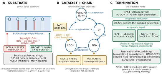
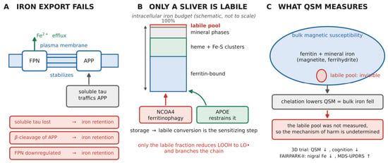
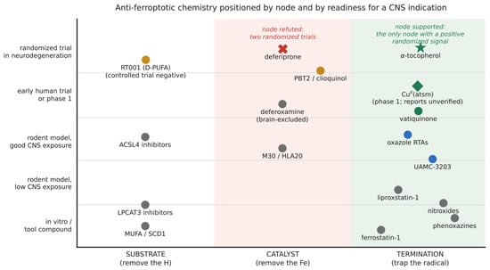
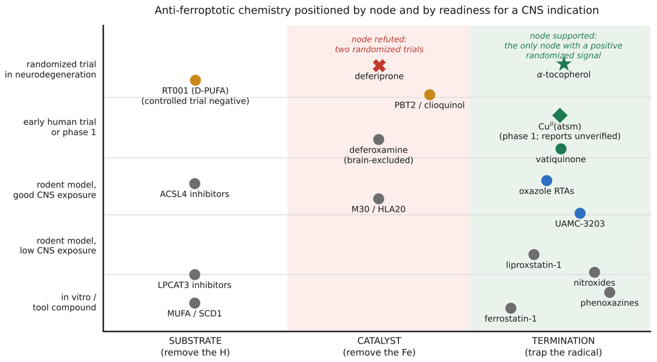

Disclaimer/Publisher’s Note: The statements, opinions and data contained in all publications are solely those of the individual author(s) and contributor(s) and not of MDPI and/or the editor(s). MDPI and/or the editor(s) disclaim responsibility for any injury to people or property resulting from any ideas, methods, instructions or products referred to in the content. |
© 2026 by the authors. Licensee MDPI, Basel, Switzerland. This article is an open access article distributed under the terms and conditions of the Creative Commons Attribution (CC BY) license.