


Abstract
Background: Cerebral artery dissection is a major cause of stroke in young and middle-aged adults. However, the prevalence of anxiety and depression among patients with cerebral artery dissection, their potential associations with cerebral artery dissection, and the underlying biological mechanisms remain poorly understood. This study aimed to describe anxiety and depression in patients with cerebral artery dissection and to explore potential bidirectional associations and candidate biological processes. Methods: Clinical prevalence and subgroup differences of anxiety and depression in cerebral artery dissection patients were assessed using clinical data. Mendelian randomization (MR) analyses were conducted to evaluate associations between anxiety, depression and cerebral artery dissection. Peripheral-blood RNA sequencing identified cerebral artery dissection-associated differentially expressed genes, which were integrated with anxiety-related genes from GeneCards for enrichment analysis of candidate shared biological processes. Results: Among 726 cerebral artery dissection patients, 315 (43.4%) had anxiety symptoms and 303 (41.7%) had depressive symptoms, with higher proportions in males, ever-smokers, and ever-drinkers. MR analyses provided preliminary evidence of a potential bidirectional association between anxiety and cerebral artery dissection, whereas no consistent evidence of an association was observed between depression and cerebral artery dissection. Integration of cerebral artery dissection-associated differentially expressed genes and anxiety-related genes identified 33 overlapping genes enriched in mitochondrial energy metabolism-related pathways, suggesting candidate systemic transcriptional signatures shared by anxiety and cerebral artery dissection. Conclusions: Anxiety and depression symptoms were frequently observed among patients with cerebral artery dissection. Genetic and transcriptomic analyses suggested potential links between anxiety and cerebral artery dissection, involving candidate mitochondrial energy metabolism-related processes that warrant validation.
1. Introduction
Cerebral artery dissection is a vital cause of ischemic stroke and subarachnoid hemorrhage in young and middle-aged adults, leading to substantial disability, high recurrence rates, and considerable socioeconomic burden [1,2,3]. Increasing evidence indicates that psychological factors may affect the susceptibility of cerebrovascular systems [4]. Anxiety has been associated with a higher risk of cerebrovascular disease, whereas depression has been linked to cerebrovascular events [5,6]. Epidemiological investigations in various populations show that mood and anxiety disorders increase the risk of cerebrovascular diseases [7,8]. However, cerebral artery dissection may represent a distinct clinical context, as it can occur unexpectedly in relatively young individuals and often presents with headache, neck pain, or acute neurological symptoms. Whether these clinical characteristics are associated with psychological symptoms in patients with cerebral artery dissection remains unclear. Existing studies are mostly indirect, and our clinical experience suggests that anxiety and depression are prevalent among cerebral artery dissection patients. Clinical observation and research evidence highlight the need for systematic, multidimensional investigation into how anxiety and depression intersect with cerebral artery dissection.
Cerebral artery dissection may also contribute to anxiety and depression. Existing data show that mood disorders are commonly found in patients with cerebrovascular diseases [9]. According to a meta-analysis, 31% of stroke survivors experience post-stroke depressive symptoms and 39% have post-stroke anxiety symptoms. The joint occurrence rate of depression and anxiety in young adults is 25% [9]. Stroke survivors also face an increased risk of suicide, with an estimated 3–4 deaths per 1000 individuals occurring within the first five years [10]. Besides stroke, other cerebrovascular disorders such as cerebral amyloid angiopathy and cerebral small vessel disease are linked to emotional and psychological disturbances [11,12]. Older adults suffering from small vessel disease often suffer from late-life depression, and meta-analytical studies have indicated small vessel disease as a risk factor for incident depression [12,13,14]. Despite these findings, research on the potential impact of cerebral artery dissection on anxiety and depression remains limited.
Given the high clinical burden of cerebral artery dissection and the recent but uncertain evidence regarding the association between anxiety, depression and cerebrovascular diseases, a thorough investigation is required to elucidate the connection between anxiety, depression, and cerebral artery dissection. Therefore, this study aims to characterize the clinical patterns of anxiety and depression among patients with cerebral artery dissection, to examine their potential bidirectional relationships, and to explore transcriptomic alterations that may reveal shared biological pathways. By integrating clinical data, genetic inference, and transcriptomic profiling, this study seeks to provide a multidimensional understanding of how anxiety and depression intersect with cerebral artery dissection, thus providing new understandings of the disease mechanisms and potential therapeutic approaches.
2. Methods
2.1. Study Design
This study adopted a multidimensional framework to investigate the relationships between anxiety, depression and cerebral artery dissection. We hypothesized that anxiety and depression may be clinically relevant among patients with cerebral artery dissection and that exploratory genetic and transcriptomic analyses could generate hypotheses regarding potential links with the disease. To provide complementary evidence from different analytical perspectives, the study comprised three analytical components. Tier 1 characterized the occurrence and distribution of anxiety and depressive symptoms among patients with cerebral artery dissection based on clinical data from a retrospective hospital cohort. Tier 2 used publicly available genome-wide association study (GWAS) summary statistics to investigate potential bidirectional genetic associations between anxiety, depression, and cerebral artery dissection using Mendelian randomization (MR). Tier 3 performed exploratory transcriptomic analyses to identify candidate biological pathways potentially related to cerebral artery dissection and anxiety. These three components were designed to address complementary aspects of the research question rather than to constitute a directly integrated analysis.
2.2. Clinical Data Collection of Anxiety and Depression in Cerebral Artery Dissection
Patients diagnosed with cerebral artery dissection between 2015 and 2025 at the Department of Neurology, Tongji Hospital, Huazhong University of Science and Technology, were included in this study. Cerebral artery dissection was diagnosed based on digital subtraction angiography, and the clinical cohort included patients with both intracranial and cervical artery dissection. Given the retrospective nature of the cohort, which was established from clinical records collected over approximately 10 years, detailed information on the anatomical distribution and other clinical characteristics of dissection was not systematically available for all patients. Anxiety and depressive symptoms were assessed by outpatient clinicians using the Hamilton Anxiety Rating Scale (HAMA) and Hamilton Depression Rating Scale (HAMD), respectively. Anxiety and depression were defined as HAMA scores ≥ 14 and HAMD scores ≥ 17, respectively. The assessments were performed during outpatient visits when cerebral artery dissection was suspected, and thus were conducted in the clinical evaluation period surrounding the diagnosis of cerebral artery dissection. The scales were administered by outpatient clinicians with relevant clinical experience. Assessment results were retrospectively extracted from the patients’ medical records. Patients with missing clinical information or those unable to provide informed consent were excluded. In addition, gender, age, smoking status, and drinking status were collected.
2.3. Bidirectional Mendelian Randomization Analysis Between Anxiety, Depression and Cerebral Artery Dissection
A bi-directional MR design was applied, using two-sample MR approaches and GWAS summary-level datasets to evaluate the association and directionality between anxiety, depression, and cerebral artery dissection. The analysis consisted of two stages: the first assessed whether anxiety and depression exert effects on cerebral artery dissection, and the second examined whether cerebral artery dissection influences anxiety and depression. All exposure and outcome datasets were derived from GWAS, restricted to individuals of European ancestry, and obtained from the Integrative Epidemiology Unit (IEU) GWAS database (https://opengwas.io/, accessed 9 September 2025). GWAS summary statistics for anxiety (ukb-d-20544_15) and depression (ukb-d-20544_11) were sourced from the UK Biobank, involving 117,751 individuals and 117,782 individuals, respectively, each with 13,571,547 SNPs [15]. Cerebral artery dissection summary statistics were obtained from FinnGen (finn-b-I9_DISCER_EXNONE), including 218,792 participants and 16,380,466 SNPs [16]. All datasets were publicly available and had received ethical approval from their respective institutions. The GWAS summary statistics for anxiety and depression were obtained from the UK Biobank, while those for cerebral artery dissection were obtained from FinnGen. Using different GWAS resources reduced the potential for sample overlap. However, individual-level overlap could not be verified from the publicly available summary statistics. Potential sample overlap may introduce bias into two-sample MR estimates and represents an additional source of uncertainty.
Instrumental variables (IVs) were selected based on MR assumptions. First, SNPs associated with the exposure at p < 1 × 10⁻⁵ were extracted, and SNPs directly associated with the outcome were excluded. This threshold has been used in previous MR studies when genome-wide significant variants are insufficient [17]. Second, independent variants were identified via clumping using the European 1000 Genomes Phase 3 reference panel [17]. Third, palindromic SNPs with intermediate allele frequencies were removed. Variants showing evidence of pleiotropy were excluded. Heterogeneity among instruments was assessed. The robustness of the results was evaluated through leave-one-out sensitivity analysis, which involved sequentially excluding each SNP.
2.4. Integrative Transcriptomic Analyses
Peripheral blood samples were collected from cerebral artery dissection patients who consented to participate in the study, as well as from healthy controls. Total RNA was extracted from the designated biological samples using TRIzol Reagent (Thermo Fisher, Waltham, MA, USA, cat. no. 15596026) according to the manufacturer’s protocol. Genomic DNA contamination was removed by DNase I treatment (New England Biolabs, Ipswich, MA, USA, cat. no. M0303L). RNA purity was assessed by a NanoDropTM OneC spectrophotometer (Thermo Fisher), and RNA integrity was evaluated with the LabChip GX Touch system (Revvity, Waltham, MA, USA). RNA concentrations were measured using a Qubit 3.0 fluorometer with the QubiTM RNA Broad Range Assay Kit (Thermo Fisher, cat. no. Q10210).
For library construction, stranded total RNA-seq libraries were prepared using the Ribo-off rRNA Depletion Kit (Vazyme, Nanjing, China, cat. no. N406 for human/mouse/rat, N417 for bacteria, and N409 for plants) to remove rRNA, followed by the KCTM Digital Stranded mRNA Library Prep Kit (Wuhan SeqHealth Co., Ltd., Wuhan, China) according to the manufacturer’s instructions. This kit incorporates a 12-base unique molecular identifier (UID) to reduce duplication bias and sequencing errors. Library preparation included PCR enrichment of fragments ranging from 200 to 500 bp. Final libraries were quantified and sequenced on the DNBSEQ-T7 platform (MGI Tech Co., Ltd., Shenzhen, China) using paired-end 150 bp reads.
For sequencing quality control, raw sequencing data were processed using fastp (v0.23.2). Reads containing adapter sequences were removed; reads shorter than 50 bp were discarded; reads with more than 40% low-quality bases were removed; and reads containing more than five undetermined bases were excluded. UMI identification was performed using umikit (v1.0). Reads were considered UMI-containing when paired UMI anchors were identified at both ends, and reads shorter than 15 nt after UMI extraction were discarded before downstream alignment.
After quality control and filtering of raw sequencing data, processed reads were aligned to the human reference genome (GRCh38). Gene-level quantification was performed using featureCounts (v2.0.2), followed by differential expression analysis between patients with cerebral artery dissection and healthy controls. Genes meeting the predefined criteria for differential expression were subjected to functional enrichment analysis.
2.5. Statistical Analyses
Categorical variables were summarized as counts and percentages, and differences between groups were compared using the chi-square test. Continuous variables were presented as mean ± standard deviation, with between-group differences assessed using the t-test. Multivariable logistic regression analyses were performed to examine the independent associations of sex, age, smoking status, and drinking status with anxiety and depression. Odds ratios (ORs) and 95% confidence intervals (CIs) were calculated for each variable, with all four variables entered simultaneously into the corresponding models.
For the bidirectional MR analysis, instrument strength was evaluated using the F statistic. An F-statistic > 10 was used as the conventional threshold to indicate adequate instrument strength and to minimize the potential bias arising from weak instrumental variables [18]. The inverse-variance weighted (IVW) method was applied as the primary analytical approach due to its higher statistical efficiency under valid MR assumptions, while MR-Egger, weighted median, simple, and weighted mode estimators served as complementary methods. Cochran’s Q test was used to assess heterogeneity, and horizontal pleiotropy was evaluated using the MR-Egger intercept.
Differential expression analysis was performed using thresholds of |log2FC| > 1 and adjusted p < 0.05. Functional enrichment analyses, including GO and KEGG pathways, were conducted on the identified gene sets. Adjusted p values for differential expression and enrichment analysis were calculated using the Bonferroni correction.
All statistical tests were two-sided, with p < 0.05 considered statistically significant. Statistical analyses and visualization were conducted using R software (v4.5.1). For differential-expression analysis, the normalized expression data were analyzed using the limma package.
3. Results
3.1. Clinical Observation of Anxiety Symptoms Among Patients with Cerebral Artery Dissection
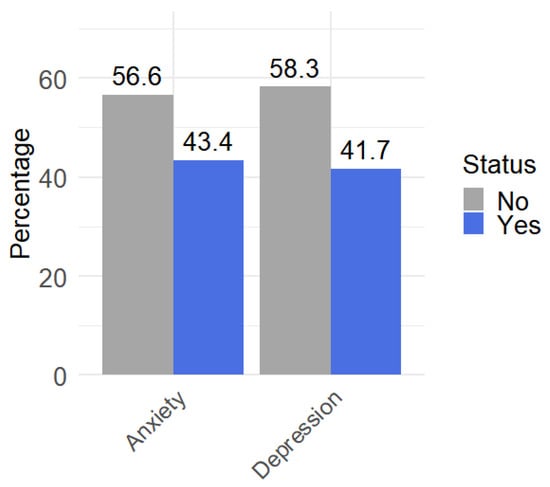
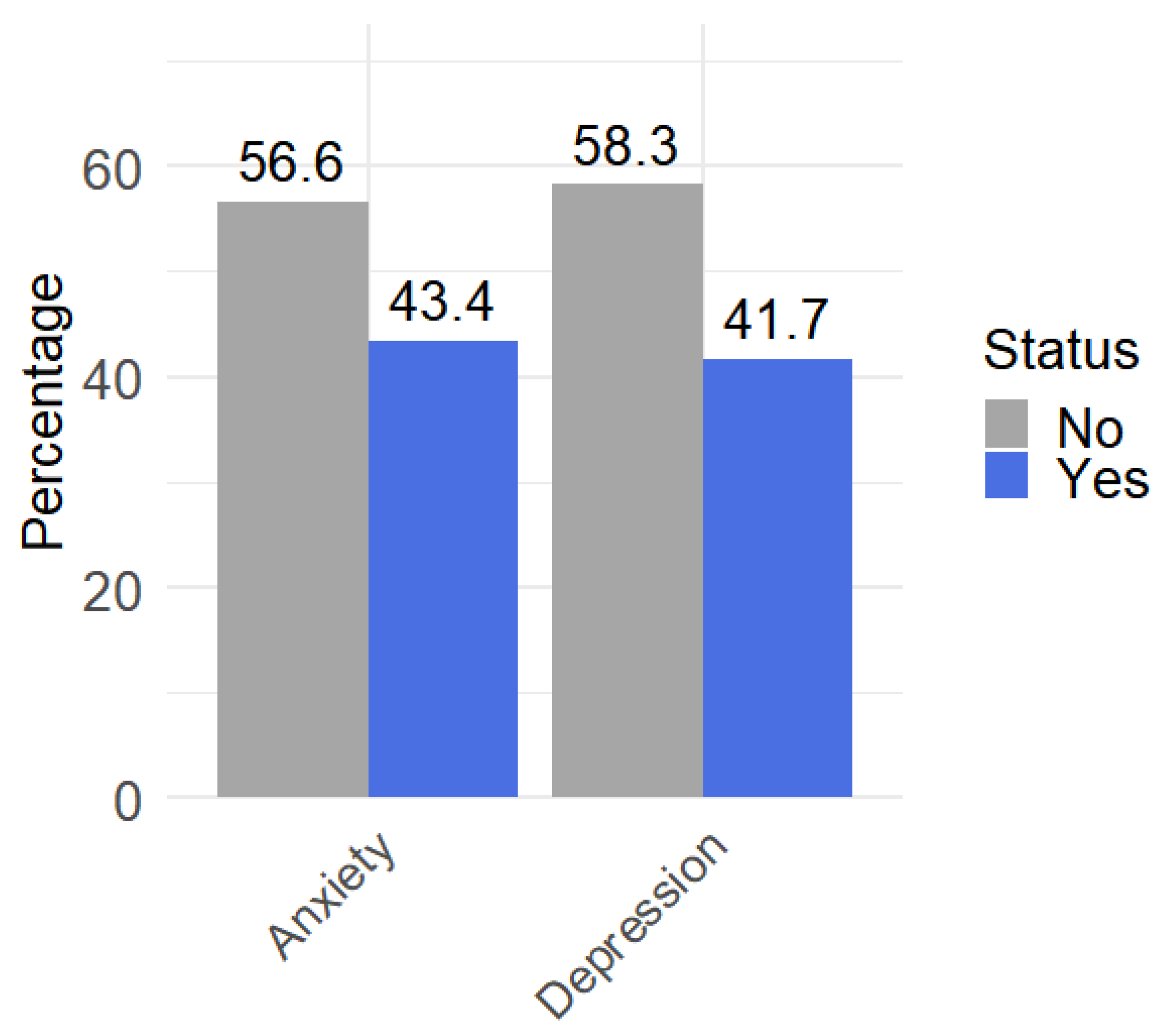
A total of 729 patients with cerebral artery dissection were admitted to the hospital between 2015 and 2025. After excluding 3 individuals with missing smoking or drinking information, 726 patients were included in the clinical observation (Table S1). As shown in Figure 1, 315 (43.4%) had anxiety symptoms and 303 (41.7%) had depressive symptoms.
Figure 1.
Prevalence of anxiety and depression among patients with cerebral artery dissection.
Descriptive analyses showed distribution patterns (Figures S1 and S2). Patients who were male, older than 60 years, ever-smokers, or ever-drinkers exhibited higher proportions of anxiety or depression compared with their counterparts.
Clinical observations were compared between patients with and without anxiety or depression (Table 1). ORs were estimated using chi-square tests. Female sex, age ≤ 60 years, non-smoking status, and non-drinking status were used as reference categories. In the anxiety group, the proportion of males was significantly higher than that of females, and both ever-smokers and ever-drinkers were more frequent than never-smokers and never-drinkers. Age did not differ significantly between groups. Similar patterns were observed for depression, with gender, smoking, and drinking showing clear differences, whereas age remained comparable.
Table 1.
Differences in clinical observations between cerebral artery dissection patients with and without anxiety and depression.
In multivariable logistic regression analyses, drinking status was independently associated with anxiety (OR, 1.59; 95% CI, 1.02–2.47; p = 0.040) and depression (OR, 1.58; 95% CI, 1.02–2.46; p = 0.042), whereas sex, age, and smoking status were not significantly associated with either outcome (Supplementary Table S2).
3.2. Bidirectional Relationships Between Anxiety, Depression and Cerebral Artery Dissection
The frequent occurrence of anxiety and depression symptoms in this cerebral artery dissection population motivated the subsequent exploratory genetic analyses. In the first stage, anxiety and depression were treated as exposures and cerebral artery dissection as the outcome. In the second stage, the direction was reversed. After instrument selection, we retained SNPs associated with each exposure, all with F-statistics > 10. Detailed information on the selected SNPs is provided in Supplementary Tables S3, S4, S7 and S8.
In the first stage, the IVW analysis suggested genetically predicted liability to anxiety was positively associated with cerebral artery dissection (beta = 8.56, p = 0.029; Table 2 and Figure S3). Beta represents the change in the log-odds of the binary outcome per unit increase in the genetically predicted exposure. However, the MR-Egger, weighted median, simple mode, and weighted mode estimates were not statistically significant, although their effect estimates were directionally positive (Table S5). No method indicated an association between depression and cerebral artery dissection. Heterogeneity and pleiotropy tests revealed no significant violations (Table S6), and leave-one-out analysis did not identify a single influential variant (Figure S4).
Table 2.
Bidirectional associations between anxiety, depression and cerebral artery dissection estimated using the inverse-variance weighted method.
In the second stage, the IVW analysis suggested genetically predicted liability to cerebral artery dissection was positively associated with anxiety (beta = 0.001, p = 0.023; Table 2 and Figure S5). The weighted median analysis also supported this association, whereas the MR-Egger and mode-based estimates were not statistically significant (Table S9). No association was observed between cerebral artery dissection and depression. No evidence of heterogeneity or pleiotropy was detected (Table S10), and leave-one-out analyses did not identify a single influential variant (Figure S6).
3.3. Exploratory Transcriptomic Analyses of Cerebral Artery Dissection and Anxiety-Related Genes
Building on the former findings, we next performed an exploratory transcriptomic analysis to characterize peripheral blood gene expression patterns associated with cerebral artery dissection. Peripheral blood samples were collected from 11 patients with cerebral artery dissection and 11 controls (Table S11).
Differential expression analysis (Figure S7A) identified 320 significantly differentially expressed genes (DEGs; adjusted p < 0.05 and |log2FC| > 1). Functional enrichment analysis of these DEGs identified amyotrophic lateral sclerosis as the most significantly enriched KEGG pathway. The leading Gene Ontology (GO) terms were responses to aerobic respiration in the biological process category, respiratory chain complex in the cellular component category, and active monoatomic ion transmembrane transporter activity in the molecular function category (Figure S7B,C). Collectively, these findings suggest that altered mitochondrial energy metabolism and ion-transport-related processes may be associated with cerebral artery dissection.
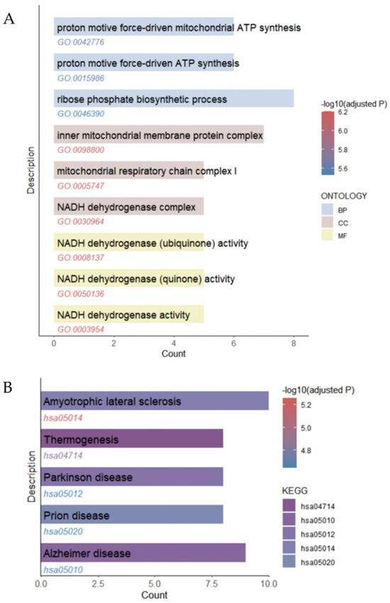
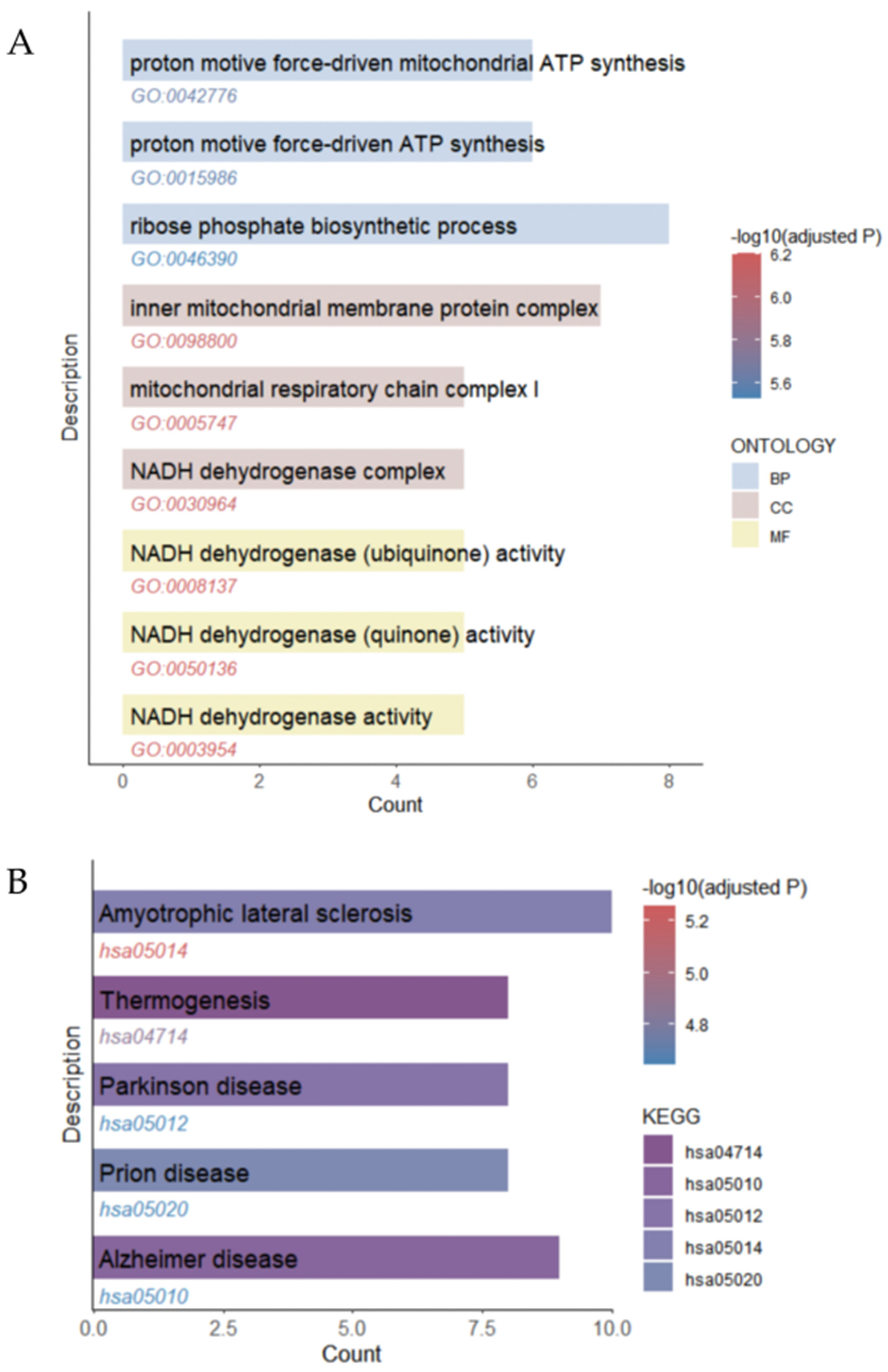
A search of GeneCards identified 8063 cerebral artery dissection-related genes with a relevance score ≥ 1. Intersecting these genes with the 320 DEGs yielded 114 cerebral artery dissection-associated DEGs. We then retrieved 4227 anxiety-related genes from GeneCards using the same relevance-score threshold. The intersection between the 114 cerebral artery dissection-associated DEGs and the anxiety-related gene set comprised 33 genes. Enrichment analysis of these 33 overlapping genes identified amyotrophic lateral sclerosis as the most significantly enriched KEGG pathway. The leading GO terms were proton motive force-driven mitochondrial ATP synthesis in the biological process category, inner mitochondrial membrane protein complex in the cellular component category, and NADH dehydrogenase (ubiquinone) activity in the molecular function category (Figure 2). These shared pathways suggest molecular links between cerebral artery dissection pathophysiology and anxiety-related biological processes. A relatively inclusive relevance-score threshold was selected to capture a broader range of potentially relevant candidate genes for exploratory analyses, given the limited and heterogeneous biological evidence available for cerebral artery dissection and anxiety.
Figure 2.
Functional enrichment of overlapping cerebral artery dissection-associated differentially expressed genes and anxiety-related genes. (A) GO enrichment analysis. (B) KEGG pathway enrichment analysis. GO, Gene Ontology; KEGG, Kyoto Encyclopedia of Genomes.
4. Discussion
This multidimensional pilot study reveals several insights into the relationship between anxiety, depression, and cerebral artery dissection. Clinically, anxiety and depressive symptoms were frequently observed among patients with cerebral artery dissection, with higher proportions of these symptoms observed among males, smokers, and drinkers. MR analyses provided preliminary evidence of a potential bidirectional association between anxiety and cerebral artery dissection, whereas no link was detected for depression in either direction. Exploratory transcriptomic analyses identified 33 cerebral artery dissection-associated differentially expressed genes that overlapped with anxiety-related genes. These genes were enriched in pathways related to mitochondrial energy metabolism, including proton motive force-driven ATP synthesis and NADH dehydrogenase activity, suggesting candidate biological processes potentially linking cerebral artery dissection and anxiety that require further validation.
In our clinical observation, anxiety and depressive symptoms were frequently observed among patients with cerebral artery dissection, based on the predefined HAMA and HAMD cut-off scores. This finding suggests that anxiety and depressive symptoms may be relatively common among patients with cerebral artery dissection, although the observed proportion should not be directly compared with prevalence estimates from the general population because of differences in assessment methods and population characteristics [19]. We observed several demographic and lifestyle differences between cerebral artery dissection patients with and without anxiety and depression. Male patients showed markedly higher proportions of anxiety and depression compared with females, which contrasts with trends in the general population where women typically display higher rates [20]. This reversal may reflect sex-specific differences in psychosocial stress responses that could be linked to cerebral artery dissection. In addition, ever-smokers and ever-drinkers were more likely to exhibit anxiety or depression than those without such histories. Smoking and alcohol use may not only contribute to vascular risk but also interact with psychological distress, amplifying susceptibility to anxiety and depression in cerebral artery dissection [21,22]. Residual confounding from unmeasured clinical and vascular factors cannot be excluded, and these clinical associations should not be interpreted as evidence of independent causal effects of smoking, drinking, or demographic factors on anxiety or depression.
Our MR analyses provided evidence of potential bidirectional associations between anxiety and cerebral artery dissection, aligning with existing epidemiological and biological evidence. Anxiety has been recognized as an independent risk factor for cerebrovascular disease, even among individuals without depression, and confers a comparable increase in vascular risk regardless of depressive status [23]. Several reasons may explain how anxiety elevates the likelihood of cerebral artery dissection. Increased sympathetic nervous system activity causes temporary rises in blood pressure and enhances heart rate variability and vascular shear stress, which may result in endothelial damage and arterial wall sensitivity [24,25,26]. Anxiety has also been associated with systemic inflammation, oxidative stress, and dysregulated autonomic tone—pathophysiological processes that may further predispose individuals to arterial dissection [27,28,29]. Moreover, cerebral artery dissection may also cause anxiety via both biological and psychosocial mechanisms. Cerebrovascular injury triggers inflammatory responses and alters cerebral perfusion—changes that have been associated with mood disorders in different cerebrovascular diseases [30,31]. Patients with structural cerebrovascular abnormalities, including intracranial aneurysms and small-vessel disease, usually have higher anxiety levels, indicating a common neurovascular-emotional basis [32,33]. Furthermore, the severe stress caused by a possible life-threatening vascular event, doubt about its recurrence and functional restrictions may all increase psychological distress [34]. These findings provided evidence of associations between anxiety and cerebral artery dissection in both directions. The markedly different beta coefficients observed for the two directions should not be interpreted as indicating substantially different strengths of association, as the estimates were derived from different exposure-outcome configurations and phenotype-specific effect scales. Although the MR analyses were based on predefined hypotheses, multiple MR directions and sensitivity analyses were performed without formal adjustment for multiple testing. The anxiety-cerebral artery dissection association was not consistently supported across all MR estimators and may be method-dependent. Therefore, the nominally significant findings should be interpreted cautiously as suggestive and exploratory and require confirmation in independent studies. In the clinical cohort, anxiety and depressive symptoms were frequently observed among patients with cerebral artery dissection. These findings describe the psychological symptom burden observed during the clinical evaluation period but do not establish a causal relationship between either anxiety or depression and cerebral artery dissection. In contrast, the MR analyses evaluated anxiety and depression separately and provided suggestive evidence of a potential bidirectional association between anxiety and cerebral artery dissection, whereas no consistent evidence of an association was observed between depression and cerebral artery dissection. The difference between the clinical observations and MR findings for depression may reflect the distinct questions addressed by these approaches. Depressive symptoms assessed around the time of diagnosis may be influenced by disease-related stress, diagnostic uncertainty, or other clinical and psychosocial factors that are not necessarily captured by genetic instruments for depression.
In this exploratory transcriptomic analysis, KEGG enrichment of cerebral artery dissection-associated differentially expressed genes identified the amyotrophic lateral sclerosis (ALS) pathway as the most significantly enriched pathway. This finding should not be interpreted as evidence of an ALS-specific pathological process in patients with cerebral artery dissection. Rather, the ALS pathway includes genes involved in mitochondrial dysfunction, oxidative stress, and neuronal cell injury, and its enrichment in peripheral blood may reflect broader systemic cellular stress or alterations in mitochondrial function [35,36]. Given the small transcriptomic sample size and the use of peripheral blood, however, this finding may also be influenced by the composition of the differentially expressed gene set and pathway annotation and therefore requires independent validation.
We next examined the genes shared between cerebral artery dissection-associated differentially expressed genes and anxiety-related genes. Enrichment analysis of these overlapping genes highlighted mitochondrial energy metabolism as a potential biological theme. The enriched terms included proton motive force-driven mitochondrial ATP synthesis, inner mitochondrial membrane protein complex, and NADH dehydrogenase (ubiquinone) activity, all of which are closely related to oxidative phosphorylation and cellular bioenergetics. Altered mitochondrial function, energy metabolism, and redox signaling have been implicated in anxiety-related neurobiological processes [37,38]. Our findings therefore identify mitochondrial processes as candidate systemic transcriptional signatures linking cerebral artery dissection and anxiety, providing hypothesis-generating evidence for potential shared biological mechanisms. Although several enriched KEGG pathways were annotated with neurodegenerative disease terms, these pathways largely reflect shared mitochondrial respiratory-chain and energy metabolism-related genes rather than disease-specific mechanisms. In addition, the GeneCards relevance-score threshold may influence the composition of the gene sets and, consequently, the genes identified through the sequential overlap analysis. Alternative relevance-score thresholds may yield different sets of overlapping genes and enriched biological pathways. Therefore, the 33 overlapping genes identified in this study should be regarded as exploratory candidate genes rather than a definitive set of genes linking cerebral artery dissection and anxiety.
The clinical, Mendelian randomization, and transcriptomic findings provide complementary evidence supporting a potential relationship between anxiety and cerebral artery dissection. The clinical association and MR findings suggest that anxiety may contribute to cerebral artery dissection, while the transcriptomic analysis identified mitochondrial energy metabolism-related pathways as potential systemic biological signatures shared by anxiety and cerebral artery dissection. Although these findings do not establish a direct molecular mechanism within cerebral artery tissue, they raise the possibility that systemic alterations in mitochondrial function may contribute to biological processes relevant to vascular vulnerability. These findings are hypothesis-generating and warrant further investigation in vascular tissues and experimental studies.
The results mentioned above should be considered in light of several limitations. Firstly, the transcriptomic sample size was small, which limited statistical power. In addition, the cerebral artery dissection and control groups showed imbalances in several baseline characteristics, and the differential expression analysis was not adjusted for potential covariates. These imbalances may have introduced residual confounding. Although 320 DEGs were identified based on the predefined criteria, these findings should be regarded as unadjusted exploratory and hypothesis-generating associations. Furthermore, the transcriptomic cohort was an independent sample set and was not derived from the 726-patient clinical cohort. Therefore, the identified molecular signatures should not be considered directly generalizable to the larger population. The transcriptomic analysis was based on peripheral blood rather than cerebral artery tissue. Although peripheral blood may capture systemic biological responses, its transcriptional profile may differ substantially from that of the affected vascular tissue. Therefore, the identified signatures cannot be assumed to directly reflect tissue-specific molecular alterations at the site of dissection. Medication information was also unavailable for the transcriptomic participants, precluding evaluation of potential medication-related effects on gene expression. Larger, adequately powered studies incorporating vascular tissues, where feasible, independent cohorts, and functional experiments are needed to further evaluate the tissue specificity and biological relevance of these findings. Secondly, the clinical cohort was retrospective and derived from a single center, which may have introduced selection bias and limited the generalizability of the clinical findings. Since all patients were diagnosed by digital subtraction angiography, the cohort may represent clinically recognized cases and potentially underrepresent less clinically apparent cases. Detailed information on anatomical distribution, presenting clinical manifestations, ischemic stroke or subarachnoid hemorrhage, neurological severity, recurrence status, vascular risk factors, and treatment-related factors was not systematically available, particularly in earlier medical records, limiting further stratified analyses. The absence of a healthy group prevented us from determining whether the observed psychological symptoms were specifically associated with cerebral artery dissection rather than acute hospitalization, neurological illness, or disease-related stress. Furthermore, psychological assessments were performed during the clinical evaluation period surrounding diagnosis and may have been influenced by acute disease-related stress and diagnostic uncertainty. In addition, detailed information regarding assessor training, standardized administration procedures, blinding to other clinical information, and inter-rater reliability of the HAMA and HAMD assessments was not documented. Therefore, potential variability in the administration and assessment of these scales cannot be fully excluded. Thirdly, the GWAS data lacked detailed phenotypic information, preventing stratified MR analyses that could provide more precise inferences and better account for potential sources of heterogeneity. In addition, differences in ancestry between the clinical cohort and the predominantly European GWAS datasets, as well as the relatively liberal SNP selection threshold, may introduce additional uncertainty into the MR findings. Therefore, the MR results should be interpreted cautiously and require confirmation in future studies. Finally, the three analytical components were based on heterogeneous study populations and data sources. Therefore, the findings across these components cannot be directly compared or considered mutually confirmatory. Differences in ancestry, sample characteristics, clinical context, environmental exposures, and measurement approaches may contribute to heterogeneity across the observed findings. Accordingly, the results should be interpreted as complementary, hypothesis-generating evidence. Future studies integrating clinically characterized cohorts with ancestry-matched genetic data and adequately powered molecular profiling are warranted to further evaluate the relationships identified across these analytical components.
5. Conclusions
Anxiety and depressive symptoms were frequently observed among patients with cerebral artery dissection. MR analyses provided suggestive evidence of a possible bidirectional association between anxiety and cerebral artery dissection, whereas no consistent evidence of an association was observed between depression and cerebral artery dissection. Exploratory transcriptomic analyses identified mitochondrial energy metabolism-related pathways as candidate biological signatures potentially linking cerebral artery dissection and anxiety, which require further validation in larger, prospective, and independent studies.
Supplementary Materials
The following supporting information can be downloaded at https://www.mdpi.com/article/10.3390/neurolint18110200/s1: Table S1. Characteristics of Chinese patients with cerebral artery dissection; Figure S1. Prevalence of anxiety among cerebral artery dissection patients across different subgroups; Figure S2. Prevalence of depression among cerebral artery dissection patients across different subgroups; Table S2. Multivariable logistic regression analyses for associations of sex, age, smoking, and drinking status with anxiety and depression; Table S3. Characteristics of instrumental variables used in the Mendelian randomization analysis of anxiety in relation to cerebral artery dissection; Table S4. Characteristics of instrumental variables used in the Mendelian randomization analysis of depression in relation to cerebral artery dissection; Table S5. Associations of anxiety and depression with cerebral artery dissection estimated using MR-Egger, weighted median, simple mode, and weighted mode methods; Figure S3. Scatter plots illustrating Mendelian randomization estimates for the associations of anxiety and depression with cerebral artery dissection; Table S6. Heterogeneity and pleiotropy tests for the Mendelian randomization analyses of anxiety and depression in relation to cerebral artery dissection; Figure S4. Leave-one-out sensitivity analyses for the Mendelian randomization analyses of anxiety and depression in relation to cerebral artery dissection; Table S7. Characteristics of instrumental variables used in the Mendelian randomization analysis of cerebral artery dissection in relation to anxiety; Table S8. Characteristics of instrumental variables used in the Mendelian randomization analysis of cerebral artery dissection in relation to depression; Table S9. Associations of cerebral artery dissection with anxiety and depression estimated using MR-Egger, weighted median, simple mode, and weighted mode methods; Figure S5. Scatter plots illustrating Mendelian randomization estimates for the associations of cerebral artery dissection with anxiety and depression; Table S10. Heterogeneity and pleiotropy tests for the Mendelian randomization analyses of cerebral artery dissection in relation to anxiety and depression; Figure S6. Leave-one-out sensitivity analyses for the Mendelian randomization analyses of cerebral artery dissection in relation to anxiety and depression; Table S11. Characteristics of cerebral artery dissection patients and controls with blood samples; Figure S7. Volcano plot of cerebral artery dissection differentially expressed genes and functional enrichment analysis.
Author Contributions
Methodology, W.Z.; formal analysis, W.Z.; validation, J.G.; data curation, J.G.; investigation, Y.W.; writing—original draft, W.Z.; writing—review and editing, F.W.; funding acquisition, F.W. All authors have read and agreed to the published version of the manuscript.
Funding
This study was approved by the Key Research and Development Program of Hubei Province (Grant No. 2020BCA089) and the China Postdoctoral Science Foundation (No.2025M780712).
Institutional Review Board Statement
The study was conducted in accordance with the Declaration of Helsinki and approved by the Medical Ethics Committee of Tongji Hospital, Tongji Medical College, Huazhong University of Science and Technology (No. TJ-IRB202407041. Approval Date: 1 July 2024).
Informed Consent Statement
Written informed consent was obtained from all subjects involved in the study.
Data Availability Statement
Data access is restricted for ethical and privacy reasons. The raw data supporting the conclusions of this article will be made available by the authors on request.
Acknowledgments
The authors would like to thank all the individuals, research staff, and students who participated in this study.
Conflicts of Interest
The authors declare no conflicts of interest.
References
- Sikkema, T.; Uyttenboogaart, M.; Eshghi, O.; De Keyser, J.; Brouns, R.; van Dijk, J.M.C.; Luijckx, G.J. Intracranial artery dissection. Eur. J. Neurol. 2014, 21, 820–826. [Google Scholar] [CrossRef] [Scilit] [PubMed]
- Stéphanie Debette, D.L. Cervical-artery dissections: Predisposing factors, diagnosis, and outcome. Lancet Neurol. 2009, 8, 668–678. [Google Scholar] [CrossRef] [Scilit] [PubMed]
- Griffin, K.J.; Harmsen, W.S.; Mandrekar, J.; Brown, R.D.; Keser, Z. Epidemiology of Spontaneous Cervical Artery Dissection: Population-Based Study. Stroke 2024, 55, 670–677. [Google Scholar] [CrossRef] [Scilit] [PubMed]
- Wolkowitz, O.M.; Epel, E.S.; Reus, V.I.; Mellon, S.H. Depression gets old fast: Do stress and depression accelerate cell aging? Depress. Anxiety 2010, 27, 327–338, Correction in Depress. Anxiety 2010, 27, 693. [Google Scholar] [CrossRef] [Scilit] [PubMed]
- Emdin, C.A.; Odutayo, A.; Wong, C.X.; Tran, J.; Hsiao, A.J.; Hunn, B.H.M. Meta-Analysis of Anxiety as a Risk Factor for Cardiovascular Disease. Am. J. Cardiol. 2016, 118, 511–519. [Google Scholar] [CrossRef] [Scilit] [PubMed]
- Yu, S.; Arima, H.; Bertmar, C.; Hirakawa, Y.; Priglinger, M.; Evans, K.; Krause, M. Depression but not anxiety predicts recurrent cerebrovascular events. Acta Neurol. Scand. 2016, 134, 29–34. [Google Scholar] [CrossRef] [Scilit] [PubMed]
- Fiedorowicz, J.G.; He, J.; Merikangas, K.R. The association between mood and anxiety disorders with vascular diseases and risk factors in a nationally representative sample. J. Psychosom. Res. 2011, 70, 145–154. [Google Scholar] [CrossRef] [Scilit] [PubMed]
- Ellins, E.A.; Shipley, M.J.; Rees, D.A.; Kemp, A.; Deanfield, J.E.; Brunner, E.J.; Halcox, J.P. Associations of depression-anxiety and dyslipidaemia with subclinical carotid arterial disease: Findings from the Whitehall II Study. Eur. J. Prev. Cardiol. 2019, 27, 800–807. [Google Scholar] [CrossRef] [Scilit] [PubMed]
- Ignacio, K.H.D.; Muir, R.T.; Diestro, J.D.B.; Singh, N.; Yu, M.H.L.L.; Omari, O.E.; Abdalrahman, R.; Barker-Collo, S.L.; Hackett, M.L.; Dukelow, S.P.; et al. Prevalence of depression and anxiety symptoms after stroke in young adults: A systematic review and meta-analysis. J. Stroke Cerebrovasc. Dis. 2024, 33, 107732. [Google Scholar] [CrossRef] [Scilit] [PubMed]
- Chun, H.Y.Y.; Ford, A.; Kutlubaev, M.A.; Almeida, O.P.; Mead, G.E. Depression, Anxiety, and Suicide After Stroke: A Narrative Review of the Best Available Evidence. Stroke 2022, 53, 1402–1410. [Google Scholar] [CrossRef] [Scilit] [PubMed]
- Kaushik, K.; Waslam, N.G.; van der Zwet, R.G.J.; Voigt, S.; van Dort, R.; van Zwet, E.W.; Terwindt, G.M.; van Etten, E.S.; Wermer, M.J.H. Quality of life, depression and anxiety in cerebral amyloid angiopathy: A cross-sectional study. Eur. J. Neurol. 2024, 31, e16476. [Google Scholar] [CrossRef] [Scilit] [PubMed]
- Fang, Y.; Qin, T.; Liu, W.; Ran, L.; Yang, Y.; Huang, H.; Pan, D.; Wang, M. Cerebral Small-Vessel Disease and Risk of Incidence of Depression: A Meta-Analysis of Longitudinal Cohort Studies. J. Am. Heart Assoc. 2020, 9, e016512. [Google Scholar] [CrossRef] [Scilit] [PubMed]
- Chen, Y.; Kong, Q.; Wang, Z.; Li, J.; Luo, Y.; Zhang, Y.; Yu, Z.; Huang, H.; Luo, X. Correlation between cerebral small vessel disease and late-life depression: Insights from neuroimaging studies. Brain Res. 2025, 1868, 150001. [Google Scholar] [CrossRef] [Scilit] [PubMed]
- He, Z.; Tan, W.; Ma, H.; Shuai, Y.; Shan, Z.; Zhai, J.; Qiu, Y.; Zeng, H.; Chen, X.; Wang, S.; et al. Prevalence and factors associated with depression and anxiety among older adults: A large-scale cross-sectional study in China. J. Affect. Disord. 2024, 346, 135–143. [Google Scholar] [CrossRef] [Scilit] [PubMed]
- Lab, N. UK Biobank GWAS Results 2018. Available online: http://www.nealelab.is/uk-biobank (accessed on 30 September 2025).
- Kurki, M.I.; Karjalainen, J.; Palta, P.; Sipilä, T.P.; Kristiansson, K.; Donner, K.M.; Reeve, M.P.; Laivuori, H.; Aavikko, M.; Kaunisto, M.A.; et al. FinnGen provides genetic insights from a well-phenotyped isolated population. Nature 2023, 613, 508–518, Correction in Nature 2023, 615, E19. [Google Scholar] [CrossRef] [Scilit] [PubMed]
- Xiao, G.; He, Q.; Liu, L.; Zhang, T.; Zhou, M.; Li, X.; Chen, Y.; Chen, Y.; Qin, C. Causality of genetically determined metabolites on anxiety disorders: A two-sample Mendelian randomization study. J. Transl. Med. 2022, 20, 475. [Google Scholar] [CrossRef] [Scilit] [PubMed]
- Pierce, B.L.; Ahsan, H.; VanderWeele, T.J. Power and instrument strength requirements for Mendelian randomization studies using multiple genetic variants. Int. J. Epidemiol. 2011, 40, 740–752. [Google Scholar] [CrossRef] [Scilit] [PubMed]
- Global Burden of Disease 2025. Available online: https://vizhub.healthdata.org/gbd-results/ (accessed on 12 October 2025).
- Hartman, C.A.; Larsson, H.; Vos, M.; Bellato, A.; Libutzki, B.; Solberg, B.S.; Chen, Q.; Du Rietz, E.; Mostert, J.C.; Kittel-Schneider, S.; et al. Anxiety, mood, and substance use disorders in adult men and women with and without attention-deficit/hyperactivity disorder: A substantive and methodological overview. Neurosci. Biobehav. Rev. 2023, 151, 105209. [Google Scholar] [CrossRef] [Scilit] [PubMed]
- Yuan, S.; Damrauer, S.M.; Larsson, S.C. Alcohol, Liver Disease, and Peripheral Arterial Disease: Epidemiology, Mechanisms, and Clinical Implications. Arterioscler. Thromb. Vasc. Biol. 2025, 45, 1493–1504. [Google Scholar] [CrossRef] [Scilit] [PubMed]
- Yang, X.; Cheng, S.; Li, C.; Pan, C.; Liu, L.; Meng, P.; Chen, Y.; Zhang, J.; Zhang, Z.; Zhang, H.; et al. Evaluating the interaction between 3′aQTL and alcohol consumption/smoking on anxiety and depression: 3′aQTL-by-environment interaction study in UK Biobank cohort. J. Affect. Disord. 2023, 338, 518–525. [Google Scholar] [CrossRef] [Scilit] [PubMed]
- Karlsen, H.R.; Saksvik-Lehouillier, I.; Stone, K.L.; Schernhammer, E.; Yaffe, K.; Langvik, E. Anxiety as a risk factor for cardiovascular disease independent of depression: A prospective examination of community-dwelling men (the MrOS study). Psychol. Health 2020, 36, 148–163. [Google Scholar] [CrossRef] [Scilit] [PubMed]
- Bigalke, J.A.; Durocher, J.J.; Greenlund, I.M.; Keller-Ross, M.; Carter, J.R. Blood pressure and muscle sympathetic nerve activity are associated with trait anxiety in humans. Am. J. Physiol.-Heart Circ. Physiol. 2023, 324, H494–H503. [Google Scholar] [CrossRef] [Scilit] [PubMed]
- Cheng, Y.C.; Su, M.I.; Liu, C.W.; Huang, Y.C.; Huang, W.L. Heart rate variability in patients with anxiety disorders: A systematic review and meta-analysis. Psychiatry Clin. Neurosci. 2022, 76, 292–302. [Google Scholar] [CrossRef] [Scilit] [PubMed]
- Cullen, T.; Steward, C.J.; Menzies, C.; Pugh, C.J.A.; Douglas Thake, C. The effect of underwater massage during hot water immersion on acute cardiovascular and mood responses. J. Therm. Biol. 2024, 121, 103858. [Google Scholar] [CrossRef] [Scilit] [PubMed]
- Zhou, Y.; Zhang, C.; Li, J.; Zheng, Y.; Xiao, S. Systemic inflammation mediates the association between dietary inflammation index and incident anxiety and depression in UK Biobank. J. Affect. Disord. 2025, 381, 205–214. [Google Scholar] [CrossRef] [Scilit] [PubMed]
- Dang, R.; Wang, M.; Li, X.; Wang, H.; Liu, L.; Wu, Q.; Zhao, J.; Ji, P.; Zhong, L.; Licinio, J.; et al. Edaravone ameliorates depressive and anxiety-like behaviors via Sirt1/Nrf2/HO-1/Gpx4 pathway. J. Neuroinflamm. 2022, 19, 41. [Google Scholar] [CrossRef] [Scilit] [PubMed]
- Vinkers, C.H.; Kuzminskaite, E.; Lamers, F.; Giltay, E.J.; Penninx, B.W.J.H. An integrated approach to understand biological stress system dysregulation across depressive and anxiety disorders. J. Affect. Disord. 2021, 283, 139–146. [Google Scholar] [CrossRef] [Scilit] [PubMed]
- Liu, X.; Hao, J.; Yao, E.; Cao, J.; Zheng, X.; Yao, D.; Zhang, C.; Li, J.; Pan, D.; Luo, X.; et al. Polyunsaturated fatty acid supplement alleviates depression-incident cognitive dysfunction by protecting the cerebrovascular and glymphatic systems. Brain Behav. Immun. 2020, 89, 357–370. [Google Scholar] [CrossRef] [Scilit] [PubMed]
- Yue, K.-C.; Zhu, Y.-Y.; Sun, J.-W.; Wu, X.-T.; Liu, W.-J.; Shi, H.-F. Imaging characteristics of brain microstructure and cerebral perfusion in Crohn’s disease patients with anxiety: A prospective comparative study. World J. Gastroenterol. 2025, 31, 99014. [Google Scholar] [CrossRef] [Scilit] [PubMed]
- Lim, Y.C.; Lee, E.; Song, J. Depression or Anxiety According to Management Modalities in Patients With Unruptured Intracranial Aneurysms. Stroke 2022, 53, 3662–3670. [Google Scholar] [CrossRef] [Scilit] [PubMed]
- Clancy, U.; Gilmartin, D.; Jochems, A.C.C.; Knox, L.; Doubal, F.N.; Wardlaw, J.M. Neuropsychiatric symptoms associated with cerebral small vessel disease: A systematic review and meta-analysis. Lancet Psychiatry 2021, 8, 225–236. [Google Scholar] [CrossRef] [Scilit] [PubMed]
- Princip, M.; Ledermann, K.; von Känel, R. Posttraumatic Stress Disorder as a Consequence of Acute Cardiovascular Disease. Curr. Cardiol. Rep. 2023, 25, 455–465. [Google Scholar] [CrossRef] [Scilit] [PubMed]
- Mattioli, A.V.; Coppi, F.; Manenti, A.; Farinetti, A. Subclinical Vascular Damage: Current Insights and Future Potential. Vasc. Health Risk Manag. 2021, 17, 729–738. [Google Scholar] [CrossRef] [Scilit] [PubMed]
- Selleri, V.; Bazo, M.; Sinigaglia, G.; Zanini, G.; Micheloni, G.; Coppi, F.; D’Alisera, R.; Savino, G.; Nasi, M.; Pinti, M. Cumulative increases in circulating mtDNA as a potential biomarker of brain injury in rugby union: A pilot study. J. Sci. Med. Sport 2026, 29, 1075–1085. [Google Scholar] [CrossRef] [Scilit] [PubMed]
- Alessandro, C.; Carmen, S. The Emerging Role of Brain Mitochondria in Fear and Anxiety. New Discov. Brain Sci. Fear Anxiety 2024, 73, 33–54. [Google Scholar] [CrossRef] [Scilit] [PubMed]
- Coronelli, M.M.; Coppi, F.; Mattioli, A.V. Inflammation, atherosclerosis and hypertension: The impact of depression and stress on their complex relationship. Future Cardiol. 2024, 20, 27–33. [Google Scholar] [CrossRef] [Scilit] [PubMed]

Disclaimer/Publisher’s Note: The statements, opinions and data contained in all publications are solely those of the individual author(s) and contributor(s) and not of MDPI and/or the editor(s). MDPI and/or the editor(s) disclaim responsibility for any injury to people or property resulting from any ideas, methods, instructions or products referred to in the content. |
© 2026 by the authors. Licensee MDPI, Basel, Switzerland. This article is an open access article distributed under the terms and conditions of the Creative Commons Attribution (CC BY) license.