


Abstract
Background: Pediatric dilated cardiomyopathy (DCM) is a genetically heterogeneous disorder with limited population-specific genetic data in Middle Eastern cohorts. Methods: Twenty-three pediatric DCM patients from a predominantly Kurdish–Arab consanguineous population underwent comprehensive clinical evaluation and targeted next-generation sequencing (NGS) using a 152-gene cardiomyopathy panel. Variants were classified according to ACMG/AMP guidelines. Results: Targeted NGS identified 163 rare variants; 24 (14.7%) were pathogenic/likely pathogenic (P/LP), 76 (46.6%) were variants of uncertain significance (VUS), and 63 (38.7%) were benign/likely benign. The overall diagnostic yield was 78.3% (18/23). The SCN5A c.1921delC (p.Gln641ArgfsTer3) frameshift variant was the most frequently detected P/LP variant, present in 69.6% of patients. No significant genotype–phenotype correlations were observed for SCN5A c.1921delC regarding age of onset, left ventricular ejection fraction, or valvular/structural abnormalities. Parental consanguinity was high (66.7%), and disease onset occurred early (mean 2.0 ± 3.27 years). Conclusions: This is the first genetic evaluation of a pediatric cohort with DCM from a mixed Kurdish–Arab population. A high diagnostic yield of DCM was found, particularly in consanguineous families, including an SCN5A recurrent variant. The study highlights the utility of targeted NGS in a consanguineous population. Further related studies are required in this region.
1. Introduction
Dilated cardiomyopathy (DCM) is a heterogeneous myocardial disorder characterized by left ventricular or biventricular dilation and systolic dysfunction in the absence of abnormal loading conditions or coronary artery disease [1]. It is the most prevalent form of cardiomyopathy in children and a leading indication for pediatric heart transplantation [1]. The clinical spectrum ranges from asymptomatic left ventricular dilation to progressive heart failure, arrhythmias, and sudden cardiac death [2]. Pediatric dilated cardiomyopathy (DCM) is a severe and genetically heterogeneous disorder of the myocardium. The clinical entity displays marked variability in age at presentation, clinical course, and underlying genetic defects. Recently, some large-scale sequencing studies in pediatric DCM have detected an increased frequency of pathogenic mutations in genes encoding components of the cytoskeleton, ion channels, and sarcomeric proteins. Pediatric DCM, in contrast to the adult form, is not only recessive but also has an overrepresentation of de novo mutations [3,4,5].
Genetic factors are a major cause of pediatric dilated cardiomyopathy. In 30–50% of cases, a pathogenic genetic change within genes that encode sarcomeric, cytoskeletal, ion-channel, or nuclear-envelope proteins can be identified. The contribution of genetic variants to pediatric genetic cardiomyopathy has been established in several cohorts and in large-scale sequencing studies [3,4,6]. Many cases of pediatric cardiomyopathies are caused by autosomal recessive inheritance and homozygosity for rare pathogenic variants. This is particularly true in consanguineous populations [4,5,7].
Pediatric dilated cardiomyopathy (DCM) has recently been redefined as a heterogeneous group of diseases with a high diagnostic yield and a predominantly genetic basis that can act in a dominant or recessive fashion. By examining inbred individuals and a founder population, a high number of homozygous variants have been identified in previous studies, several of which are novel DCM genes that cause early-onset cardiomyopathy [3,5,6,8]. Importantly, a large population-based study of DCM in pediatric populations from Finnish and other European populations revealed a population-specific genetic architecture and a high number of recurrent founder variants [4,9]. More recently, additional genomic studies have identified additional recessive forms of DCM, and as such, a comprehensive next-generation sequencing (NGS) approach is warranted for the gene identification of pediatric cases of DCM [5,7].
Despite the recent advances in genetic testing of cardiomyopathies in children, the information on the genetic architecture of the disease in different populations is still incomplete. In particular, pediatric cardiomyopathies in the Middle Eastern population and in consanguineous families have not been fully studied yet. There is emerging evidence that in these groups, there is an overrepresentation of rare recessive mutations as well as founder events [3,10,11].
In this study, we present the first comprehensive clinical and genetic characterization of pediatric DCM in a Kurdish–Arab cohort. Using a targeted 152-gene NGS panel, we evaluated variant distribution, diagnostic yield, and genotype–phenotype correlations, with a particular focus on the recurrent SCN5A c.1921delC variant. Our findings highlight the genetic burden of pediatric DCM in consanguineous populations and provide actionable insights for genetic counseling, cascade screening, and population-specific diagnostic algorithms.
2. Materials and Methods
2.1. Study Population and Ethical Approval
Twenty-three pediatric patients (≤18 years) clinically diagnosed with DCM were enrolled from a tertiary pediatric cardiology center in Sulaymaniyah, Iraq. DCM diagnosis was established according to Pediatric Cardiomyopathy Registry (PCMR) echocardiographic criteria: left ventricular end-diastolic dimension Z-score ≥ 2 and ejection fraction < 55%, excluding congenital heart disease or hemodynamic stress [12]. Parental consanguinity was defined as marriage between first or second cousins. Written informed consent was obtained from all parents/guardians.
The study only used DNA samples from affected probands; no systematic parental genetic testing was performed. The study was approved by the Ethics Committee of the University of Sulaimani (Approval No. 18) and conducted in accordance with the Declaration of Helsinki.
2.2. DNA Extraction and Targeted NGS
Peripheral venous blood (5–10 mL) was collected in EDTA tubes. Genomic DNA was extracted using the ReliaPrep™ DNA Miniprep Kit (Promega, Madison, WI, USA). A custom-designed targeted NGS panel covering 152 cardiomyopathy-associated genes (3894 amplicons; 739.6 kb target region; see Supplementary Table S3) was amplified and sequenced on the Ion PGM™ System (Thermo Fisher Scientific (Waltham, MA, USA)). The selected genes are a targeted gene panel for established genes associated with Cardiomyopathy. This is a subset of the genes known to cause pediatric dilated cardiomyopathy at the time of this panel’s design; not all genes of interest for pediatric dilated cardiomyopathy have been included.
2.3. Variant Analysis and Classification
Raw sequencing data were processed using an Ion Reporter and Torrent Variant Caller. Variants were annotated and prioritized using Franklin™ by Genoox. Only rare variants (population allele frequency < 1% in gnomAD) were retained. All Variants were inspected using Integrated Genome Viewer (IGV) for characteristics of reads supporting the variant, including quality, depth of coverage, and strand bias. In this study, the variants were not validated by alternative methods (e.g., Sanger sequencing). Classification followed the ACMG/AMP guidelines [13], categorizing variants as pathogenic (P), likely pathogenic (LP), variant of uncertain significance (VUS), or benign/likely benign (B/LB). P/LP variants are summarized in Supplementary Table S4 and VUS in Supplementary Table S5. A technical assessment for the SCN5A c.1921delC variant was conducted using read depth, allelic balance, strand bias, and visual assessment in Integrative Genomics Viewer (IGV). The deletion is present on both the forward and reverse strands of DNA and is not supported by read mapping to alternate locations. Although the deletion has not been confirmed by Sanger sequencing or quantitative real-time PCR, the variant has been validated by other NGS metrics that measure high quality, such as adequate depth of coverage and other bioinformatics metrics.
2.4. Clinical Data Collection
Demographic, family, and clinical data were extracted from electronic medical records. Echocardiographic parameters, including left ventricular ejection fraction (LVEF), valvular regurgitation severity, and atrial/pulmonary artery dimensions, were standardized as Z-scores or categorical grades. Patients who normalized LVEF (>50%) under standard heart failure therapy were classified as “recovered DCM” for descriptive purposes. The family history data were collected by clinical interviews, using a structured format. All probands were born to clinically unaffected parents at the time of evaluation by the researcher. However, systematic genetic testing of the parents was not performed.
2.5. Statistical Analysis
Data were analyzed using GraphPad Prism 10 and SPSS v25. Continuous variables are expressed as mean ± standard deviation, and categorical variables as counts and percentages. Genotype–phenotype associations for SCN5A c.1921delC were evaluated using Chi-square, Fisher’s exact, and Mann–Whitney U tests. A p-value < 0.05 was considered statistically significant.
3. Results
3.1. Clinical and Demographic Characteristics
The cohort comprised 23 patients (60.9% male, 39.1% female), with 91.3% Kurdish and 8.7% of Arab ethnicity. Parental consanguinity was reported in 66.7% of families, and 21.7% had a positive family history of cardiomyopathy. Mean age at disease onset was 2.00 ± 3.27 years (range: 0.03–12.0), and mean age at enrollment was 7.19 ± 5.36 years. Dyspnea (100%) and fatigue (95.7%) were the predominant presenting symptoms. Echocardiography revealed a mean LVEF of 47.26 ± 17.89%, with 39.1% exhibiting mild and 26.1% moderate left ventricular dysfunction. Atrial dilation (82.6%) and pulmonary artery dilation (77.3%) were common, alongside mild mitral and tricuspid regurgitation (Table 1). None of the probands were born to clinically affected parents at the time of their evaluation. No formal cardiac evaluation or genetic testing of the parents of the probands was performed as part of this study.
Table 1.
Descriptive clinical and echocardiographic characteristics of the pediatric DCM cohort (n = 23).
3.2. Variant Detection and Classification
Targeted NGS identified 163 rare variants across the 152-gene panel. According to ACMG/AMP criteria, 24 (14.7%) were classified as P/LP, 76 (46.6%) as VUS, and 63 (38.7%) as B/LB (Table 2). The diagnostic yield (proband with ≥1 P/LP variant) was 78.3% (18/23). The P/LP and VUS variants detected in this study are listed in Supplementary Tables S4 and S5.
Table 2.
Distribution of identified variants by ACMG/AMP classification.
3.3. Pathogenic and Likely Pathogenic Variants
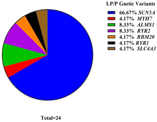
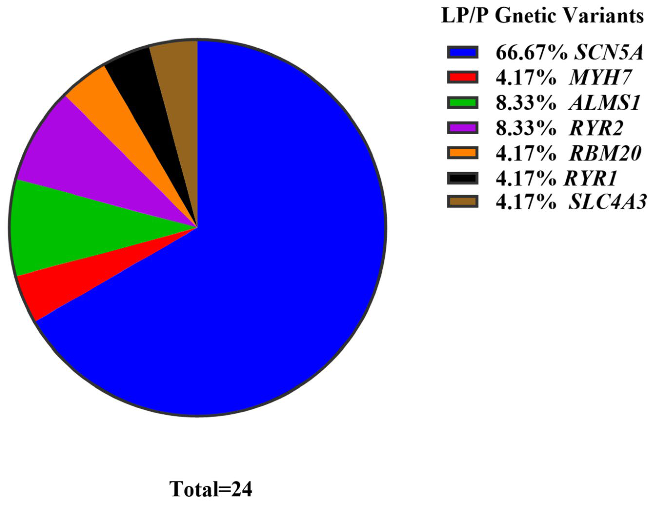
The SCN5A c.1921delC (p. Gln641ArgfsTer3) frameshift variant was the most prevalent P/LP variant, detected in 69.6% of patients. Strong sequencing data identified the SCN5A c.1921delC variant with sufficient coverage depth and a balanced number of reads for both alleles. The variant is a frameshift (p.Gln641ArgfsTer3) leading to a stop codon in the protein sequence. The stop codon is very likely to be removed by nonsense-mediated mRNA decay, resulting in a loss of function of the Nav1.5 sodium channel protein. Additional P/LP variants were identified in ALMS1, RYR2, MYH7, RBM20, RYR1 and SLC4A3 (Table 3). Several of these variants were found in patients who were also positive for the recurrent SCN5A c.1921delC variant; additional DCM cases could not be unambiguously attributed to these variants. The distribution of pathogenic and likely pathogenic variants across genes is shown in Figure 1.
Table 3.
Pediatric DCM cases and their pathogenic/likely pathogenic variants (ACMG classification; putative disease-associated variants).
Figure 1.
The distribution of pathogenic and likely pathogenic variants showed SCN5A as the leading gene at 66.67% of the LP/P variants, followed by ALMS1 and RYR2 at 8.33% each, and MYH7, RBM20, RYR1, and SLC4A3 at 4.17%.
3.4. Genotype–Phenotype Associations for SCN5A c.1921delC
Carriers and non-carriers of SCN5A c.1921delC did not differ significantly in age of onset, current age, LVEF, sex distribution, consanguinity status, LV dysfunction grade, or valvular/structural abnormalities (all p > 0.05; Table 4). This suggests the variant acts as a susceptibility or disease-modifying factor rather than a primary determinant of structural severity or functional decline.
Table 4.
Genotype–phenotype correlation analysis for SCN5A c.1921delC carriers vs. non-carriers.
4. Discussion
This study provides the first genetic and clinical characterization of pediatric DCM in a Kurdish–Arab cohort, demonstrating a remarkably high diagnostic yield (78.3%) driven by targeted NGS in a highly consanguineous population. The predominance of early-onset disease (mean 2.0 years) and high consanguinity rate (66.7%) align with established patterns of recessive and founder-driven cardiomyopathy in Middle Eastern cohorts [4,14]. In this cohort with 66.7% parental consanguinity, all probands were born to clinically unaffected parents. The probands’ parents may be carriers of the disorder and express it with reduced penetrance. However, without the parents’ genetic testing, alternative modes of inheritance and the segregation of the disorder within families cannot be confirmed. Lack of systematic cascade screening of relatives of index cases prevents evaluation of segregation and of penetrance of identified variants in families. A systematic familial genetic testing in future studies is highly recommended.
There has been a recent upsurge of research into the genetic architecture of pediatric dilated cardiomyopathy (DCM). Significantly, the proportion of pathogenic variants identified in cases of pediatric DCM is greater than that for adult DCM, and many of these cases of pediatric DCM will have an autosomal recessive pattern of disease, particularly in consanguineous families [3,8]. Information regarding genetic drift and founder effects has been determined for cardiomyopathy in the Finnish founder population [6,9]. Lastly, there is new information regarding novel genes causing recessive forms of DCM in the pediatric population [4,5,7]. These findings contrast with the use of established panels of genes for the dominant, adult-onset forms of DCM and suggest that a more comprehensive approach, such as whole-exome or whole-genome sequencing, is optimal for the diagnosis of a child with DCM. This would particularly be the case in a pediatric population or in a consanguineous family. The diagnostic yield of this study has to be interpreted with caution, as not all identified variants can be proven to be the cause of the DCM phenotype. Variants in genes primarily associated with arrhythmia syndromes or even with syndromic heart diseases were identified. In all cases, however, segregation data were not available to confirm causality.
The recurrent SCN5A c.1921delC frameshift variant, identified in 69.6% of patients, represents the most striking finding. SCN5A encodes the cardiac sodium channel Nav1.5 and is classically associated with arrhythmia syndromes (Brugada, long QT3, and conduction disease) [15]. Although no orthogonal validation by Sanger sequencing or other confirmatory tests (e.g., TaqMan genotyping) could be performed because of missing samples and funds, quality metrics of NGS data strongly support the presence of this variant. Independent molecular confirmation, however, is lacking. Emerging evidence, however, links SCN5A truncating variants to arrhythmogenic DCM and cardiomyopathy–arrhythmia overlap phenotypes [14,16]. The lack of significant genotype–phenotype correlation in our cohort suggests this variant may confer susceptibility to myocardial electrical remodeling rather than direct structural damage, consistent with reports of SCN5A-associated DCM demonstrating medical reversibility and variable penetrance [17].
The SCN5A c.1921delC variant, which is found with an unusually high frequency in our patient cohort, is a population-specific enrichment and presumably a founder-associated allele in the Kurdish–Arab population. Unfortunately, however, no segregation analysis and no parental genotyping data are available to assess the inheritance pattern and penetrance of this variant. Hence, a putative founder effect cannot be confirmed by the present data and will have to be investigated in future family-based studies by haplotype construction and analysis of additional patient cohorts from the respective region. A technical artifact for SCN5A c.1921delC cannot be entirely ruled out because this finding has not been validated by other means, such as Sanger sequencing or an alternative genotyping test.
In addition to previously reported disease-associated variants, we identified variants in ALMS1, RYR2, MYH7, RBM20, RYR1, and SLC4A3. However, in the individuals in whom they were found, several of these variants could not be unambiguously implicated in the individual’s DCM phenotype as they co-occurred with the recurrent SCN5A c.1921delC variant. ALMS1 variants are linked to Alström syndrome and early-onset cardiomyopathy [18], while RYR2 and MYH7 mutations typically present with early severe phenotypes and arrhythmic risk [19]. RBM20 truncating variants are increasingly recognized as drivers of progressive DCM with high arrhythmic burden [13].
The high VUS rate (46.6%) is consistent with global pediatric cardiomyopathy cohorts and underscores the need for familial segregation studies, functional validation, and longitudinal variant reclassification [20,21]. Some of the genes in the panel are associated with familial arrhythmia conditions (e.g., primarily RYR2- or SLC4A3-related conditions) and are not tested for in the diagnosis of primary structural cardiomyopathy. Similarly, the ALMS1-associated cardiomyopathy is part of Alström syndrome, which is caused by biallelic pathogenic variants in ALMS1, and a genotype–phenotype explanation for such variants would be highly exploratory and would require clinical and family validation [22,23].
There are several limitations to this study. The gene panel was designed to investigate the autosomal dominant forms of cardiomyopathy, and it may not capture all the genes involved in the autosomal recessive forms or even novel genes, particularly in a highly consanguineous pediatric population. Additionally, several established genes for pediatric DCM, including TTN, DMD, TAFAZZIN (TAZ), TNNT2, and TNNC1, were not included on the custom panel as they were outside of the design of the available panel at the time of study initiation. Omission of these genes likely reduced the sensitivity of the assay for a diagnosis of DCM. The parents of the children studied were not genotyped, and segregation analysis was performed to confirm the mode of inheritance and segregation of identified variants. Functional studies were not performed to confirm the pathogenic nature of identified variants, and no attempts were made to validate the variants using orthogonal methods such as Sanger sequencing. Confirmation of identified variants was based on the quality of the NGS run and bioinformatic analysis of the reads that covered the identified variants. Some of the genes in the gene panel are associated with channelopathy and with syndromes that can also be associated with cardiomyopathy; thus, it is essential to interpret any identified variants in these genes with caution. This study had a small number of patients and was a single-center study; thus, the results would not be expected to apply to other populations.
5. Conclusions
Pediatric DCM in the Kurdish–Arab population is characterized by early onset of disease, a high degree of consanguinity in families, and a strong genetic basis for disease in individual families. Targeted NGS in this patient group achieved a diagnostic yield of 78.3% and identified the SCN5A c.1921delC variant in a substantial number of cases. The SCN5A c.1921delC variant is a recurrent and uncertain variant for which the clinical significance has not been confirmed, as no parental genotyping was performed and no segregation analysis and/or orthogonal validation was performed. This study highlights the promise of population-based genetic testing in pediatric cases of cardiomyopathy and points to areas that require further research to clarify the clinical significance of such variants in this group of patients.
Supplementary Materials
The following supporting information can be downloaded at https://www.mdpi.com/article/10.3390/cardiogenetics16030015/s1. Table S1: Detailed clinical and demographic characteristics of the cohort. Table S2: Standardized echocardiographic Z-scores and valvular parameters. Table S3: Targeted NGS panel gene list and genomic coordinates. Table S4: Pathogenic and likely pathogenic variants with ACMG evidence codes. Table S5: Variants of uncertain significance (VUS) with in silico predictions. Figure S1: NGS quality control metrics (coverage depth, uniformity, and mapping quality).
Author Contributions
Conceptualization, H.M.F. and D.A.M.; methodology, H.M.F.; software and bioinformatics, H.M.F.; validation, D.A.M. and A.F.S.; formal analysis, H.M.F.; investigation, H.M.F. and A.F.S.; resources, A.F.S.; data curation, H.M.F.; writing—original draft preparation, H.M.F.; writing—review and editing, D.A.M. and A.F.S.; visualization, H.M.F.; supervision, D.A.M.; project administration, H.M.F. and A.F.S. All authors have read and agreed to the published version of the manuscript.
Funding
This research received no external funding.
Institutional Review Board Statement
The study was conducted in accordance with the Declaration of Helsinki and approved by the Ethics Committee of the University of Sulaimani, Kurdistan Region, Iraq (approval code: 18, approval date:11 January 2026).
Informed Consent Statement
Informed consent was obtained from all parents or legal guardians of the minor participants involved in the study.
Data Availability Statement
The raw sequencing data and clinical datasets are not publicly available due to patient privacy regulations and institutional policies. De-identified data and variant lists are available from the corresponding author upon reasonable request and subject to ethical approval.
Acknowledgments
We thank the Pediatric Cardiology team at Anwar Shekha Hospital for patient recruitment and clinical assessment, and Pharmagene Laboratory for technical assistance with NGS library preparation and sequencing. We are grateful to the patients and their families for their participation and consent.
Conflicts of Interest
The authors declare no conflicts of interest.
References
- Chao, T.; Ge, Y.; Sun, J.; Wang, C. Research landscape of genetics in dilated cardiomyopathy: Insight from a bibliometric analysis. Front. Cardiovasc. Med. 2024, 11, 1362551. [Google Scholar] [CrossRef] [Scilit] [PubMed]
- Yang, Q.; Berkman, A.M.; Ezekian, J.E.; Rosamilia, M.; Rosenfeld, J.A.; Liu, P.; Landstrom, A.P. Determining the Likelihood of Disease Pathogenicity Among Incidentally Identified Genetic Variants in Rare Dilated Cardiomyopathy-Associated Genes. J. Am. Heart Assoc. 2022, 11, e025257. [Google Scholar] [CrossRef] [Scilit] [PubMed]
- Al-Hassnan, Z.N.; Almesned, A.; Tulbah, S.; Alakhfash, A.; Alhadeq, F.; Alruwaili, N.; Alkorashy, M.; Alhashem, A.; Alrashdan, A.; Faqeih, E.; et al. Categorized genetic analysis in childhood-onset cardiomyopathy. Circ. Genom. Precis. Med. 2020, 13, 504–514. [Google Scholar] [CrossRef] [Scilit] [PubMed]
- Maurer, C.; Boleti, O.; Najarzadeh Torbati, P.; Norouzi, F.; Fowler, A.N.R.; Minaee, S.; Salih, K.H.; Taherpour, M.; Birjandi, H.; Alizadeh, B.; et al. Genetic insights from consanguineous cardiomyopathy families. Genes 2023, 14, 182. [Google Scholar] [CrossRef] [Scilit] [PubMed]
- Koskenvuo, J.W.; Saarinen, I.; Ahonen, S.; Tommiska, J.; Weckström, S.; Seppälä, E.H.; Tuupanen, S.; Kangas-Kontio, T.; Schleit, J.; Heliö, K.; et al. Biallelic loss-of-function in NRAP is a cause of recessive dilated cardiomyopathy. PLoS ONE 2021, 16, e0245681. [Google Scholar] [CrossRef] [Scilit] [PubMed]
- Vasilescu, C.; Ojala, T.H.; Brilhante, V.; Ojanen, S.; Hinterding, H.M.; Palin, E.; Alastalo, T.-P.; Koskenvuo, J.; Hiippala, A.; Jokinen, E.; et al. Genetic basis of severe childhood-onset cardiomyopathies. J. Am. Coll. Cardiol. 2018, 72, 2324–2338. [Google Scholar] [CrossRef] [Scilit] [PubMed]
- Lipov, A.; Jurgens, S.J.; Mazzarotto, F.; Allouba, M.; Pirruccello, J.P.; Aguib, Y.; Gennarelli, M.; Yacoub, M.H.; Ellinor, P.T.; Bezzina, C.R.; et al. Exploring the complex spectrum of dominance and recessiveness in genetic cardiomyopathies. Nat. Cardiovasc. Res. 2023, 2, 1078–1094. [Google Scholar] [CrossRef] [Scilit] [PubMed]
- Ware, S.M.; Wilkinson, J.D.; Tariq, M.; Schubert, J.A.; Sridhar, A.; Colan, S.D.; Shi, L.; Canter, C.E.; Hsu, D.T.; Webber, S.A.; et al. Genetic causes of cardiomyopathy in children: First results from the Pediatric Cardiomyopathy Genes Study. J. Am. Heart Assoc. 2021, 10, e017731. [Google Scholar] [CrossRef] [Scilit] [PubMed]
- Akinrinade, O.; Ollila, L.; Vattulainen, S.; Tallila, J.; Gentile, M.; Salmenperä, P.; Koillinen, H.; Kaartinen, M.; Nieminen, M.S.; Myllykangas, S.; et al. Genetics and genotype–phenotype correlations in Finnish patients with dilated cardiomyopathy. Eur. Heart J. 2015, 36, 2327–2337. [Google Scholar] [CrossRef] [Scilit] [PubMed]
- Al Zubaidi, A.; Al-Shamsi, A. A novel missense heterozygous mutation in NKX2-5 gene in a family with congenital septal defects and cardiomyopathy: Case series and literature review. Pediatr. Genet. 2024, 13, 308–314. [Google Scholar] [CrossRef] [Scilit] [PubMed]
- Haddad, S.; Salloum, E.; Silan, A.; Kalecioğlu, G.; Abdulnour, M.; Haddad, S.; Alasmar, D.; Alayash, M.; Ghaleb, A.N. Mitochondrial complex I deficiency in a 4-year-old boy due to compound heterozygous NDUFV1 mutation: A case report of a new pathogenic variant. Oxf. Med. Case Rep. 2025, 2025, omae166. [Google Scholar] [CrossRef] [Scilit] [PubMed]
- Dai, Y.; Wang, Y.; Fan, Y.; Han, B. Genotype-phenotype insights of pediatric dilated cardiomyopathy. Front. Pediatr. 2025, 13, 1505830. [Google Scholar] [CrossRef] [Scilit] [PubMed]
- Richards, S.; Aziz, N.; Bale, S.; Bick, D.; Das, S.; Gastier-Foster, J.; Grody, W.W.; Hegde, M.; Lyon, E.; Spector, E.; et al. Standards and guidelines for the interpretation of sequence variants: A joint consensus recommendation of the American College of Medical Genetics and Genomics and the Association for Molecular Pathology. Genet. Med. 2015, 17, 405–424. [Google Scholar] [CrossRef] [Scilit] [PubMed]
- Temaj, G.; Nuhii, N.; Sayer, J.A. The impact of consanguinity on human health and disease with an emphasis on rare diseases. J. Rare Dis. 2022, 1, 2. [Google Scholar] [CrossRef] [Scilit]
- Isezuo, K.O.; Sani, U.M.; Waziri, U.M.; Garba, B.I.; Jibrin, B.; Asma’u Adamu, F.B.J. Prevalence, clinical profile and outcome of children with cardiomyopathy. Sahel Med. J. 2022, 25, 104–108. [Google Scholar] [CrossRef] [Scilit] [PubMed]
- Hawi, Z.A.; Pugsley, K.; Namipashaki, A.; Samarrai, W.; Hawi, Z. A review of consanguinity in the Iraqi population: A call to action to prevent a future health catastrophe. Ibn Al-Haitham J. Pure Appl. Sci. 2024, 37, 64–70. [Google Scholar]
- Parker, L.E.; Landstrom, A.P. The clinical utility of pediatric cardiomyopathy genetic testing: From diagnosis to a precision medicine-based approach to care. Prog. Pediatr. Cardiol. 2021, 62, 101413. [Google Scholar] [CrossRef] [Scilit] [PubMed]
- Van der Meulen, M.H.; Herkert, J.C.; den Boer, S.L.; du Marchie Sarvaas, G.J.; Blom, N.A.; ten Harkel, A.D.J.; Breur, H.; Rammeloo, L.A.J.; Tanke, R.B.; Marcelis, C.; et al. Genetic Evaluation of a Nation-Wide Dutch Pediatric DCM Cohort: The Use of Genetic Testing in Risk Stratification. Circ. Genom. Precis. Med. 2022, 15, e002981. [Google Scholar] [CrossRef] [Scilit] [PubMed]
- Grenier, M.A.; Osganian, S.K.; Cox, G.F.; Towbin, J.A.; Colan, S.D.; Lurie, P.R.; Sleeper, L.A.; Orav, E.J.; Lipshultz, S.E. Design and implementation of the North American Pediatric Cardiomyopathy Registry. Am. Heart J. 2000, 139, S86–S95. [Google Scholar] [CrossRef] [Scilit] [PubMed]
- Towbin, J.A.; Lowe, A.M.; Colan, S.D.; Sleeper, L.A.; Orav, E.J.; Clunie, S.; Messere, J.; Cox, G.F.; Lurie, P.R.; Hsu, D.; et al. Incidence, causes, and outcomes of dilated cardiomyopathy in children. JAMA 2006, 296, 1867–1876. [Google Scholar] [CrossRef] [Scilit] [PubMed]
- Huggins, G.S.; Kinnamon, D.D.; Haas, G.J.; Jordan, E.; Hofmeyer, M.; Kransdorf, E.; Ewald, G.A.; Morris, A.A.; Owens, A.; Lowes, B.; et al. Prevalence and Cumulative Risk of Familial Idiopathic Dilated Cardiomyopathy. JAMA 2022, 327, 454–463. [Google Scholar] [CrossRef] [Scilit] [PubMed]
- Mallavarapu, A.; Taksande, A. Dilated Cardiomyopathy in Children: Early Detection and Treatment. Cureus 2022, 14, e31111. [Google Scholar] [CrossRef] [Scilit] [PubMed]
- Elshazali, O.H.; Abdalla, E. Paediatric dilated cardiomyopathy in Khartoum state, Sudan: A prospective study. BMJ Paediatr. Open 2021, 5, e000970. [Google Scholar] [CrossRef] [Scilit] [PubMed]

Disclaimer/Publisher’s Note: The statements, opinions and data contained in all publications are solely those of the individual author(s) and contributor(s) and not of MDPI and/or the editor(s). MDPI and/or the editor(s) disclaim responsibility for any injury to people or property resulting from any ideas, methods, instructions or products referred to in the content. |
© 2026 by the authors. Licensee MDPI, Basel, Switzerland. This article is an open access article distributed under the terms and conditions of the Creative Commons Attribution (CC BY) license.