


Abstract
Implantable devices have undergone enormous development in the past several decades and more results are expected as there are still unanswered questions. In manufacturing, a relatively new technology called three-dimensional (3D) printing has become increasingly involved, from which hot-melt extrusion (HME) coupled with fused deposition modeling (FDM) is one of the most researched methods in producing implantable drug delivery systems (IDDS). The HME process is used to produce polymer filaments that are the carriers of the applied drugs, while FDM creates the implant itself from the filaments, based on the computer-aided designs. The availability of several polymers like polycaprolactone, polylactic acid, or thermoplastic polyurethane, etc., allows the incorporation of many active pharmaceutical ingredients while digital designs offer numerous variabilities in designs, formulations, and applications of 3D-printed IDDS. This allows more specific and detailed modifications in the end product which can forecast the possibility of personalized treatments and therapies. In this review, our research group gathered together different HME-FDM-printed IDDS to provide examples about the diverse applicability of 3D printing. These products, just like any other device and medicine in the medical field, must be characterized and evaluated properly. The methods and technology that are needed already exist and can be repeatedly used in the characterization of IDDSs; we also discuss these methods based on the available publications. In conclusion, every condition is given to make research and manufacture personalized 3D-printed IDDSs possible, however more research work and proof of safe usage are crucial to make these devices applicable in everyday medical treatments.
1. Introduction
Implantable devices have gone through enormous development throughout the decades since the 1930s, when R. Deanesly and A.S. Parkes made the first implantable drug delivery system [1,2]. The primary objectives of implantable drug delivery devices are to maintain the concentration of the drug within the therapeutic interval for a longer period of time, avoiding multiple administration, improving patient adherence, while usually avoiding first-pass metabolism and enhancing comfort of the host [3,4,5]. As the basis for an implantable carrier system, several composition variations in polymers and hydrogels were developed to achieve the desired effect for well-defined treatments with many active pharmaceutical ingredients (APIs) [6]. The appearance of commercially available implants made sustained, predictable and continuous drug delivery possible, not just systemically, but even to targeted tissues, such as tumors [7]. Another benefit of using polymers is that some can degrade within the body to non-toxic monomers; this way, they can be eliminated by metabolism and there is no need for another invasive procedure to remove the implant [3]. Many forms and materials are compatible with the human body when it comes to implantable drug delivery devices or drug delivery systems, even containing electronic parts such as the implantable microchip-based human parathyroid hormone drug delivery device; however, non-metallic materials are dominating in this field when it comes to drug delivery systems [5].
Among the main disadvantages, the removal of the implant after use should be mentioned [8]. Sometimes it is considered more traumatic to remove the implant than the insertion itself. In addition, non-biodegradable polymers can accumulate in the tissue. Therefore, the evaluation of risks and benefits must be made during polymer selection [9]. The possible risk of implant contamination and disrupted drug delivery due to tissue damage are also not neglectable factors when balancing the advantages and disadvantages of implants. Therefore, individual cases might demand time-consuming consultations between other groups of professionals [10].
There are various methods of manufacturing polymer implants mentioning several techniques like compression, HME, or, as for porous polymer implants, particulate leaching, gas foaming, electrospinning, thermally induced phase separation, cryogelation, or freeze-drying [11,12]. The most commonly used, compression, has the advantage that it does not require heat nor solvent for manufacturing, but unluckily, the API dissolution can be quiet fast and irregular in shape and might cause inappropriate dissolution [9]. HME itself is a popular technique as it is a solvent-free, continuous, and scalable technique, but the API stability problems must be kept in mind [13]. Other newer techniques also face some advantages and some limitations [12]. Despite several manufacturing options, almost all of them are missing an important factor, personalization. A still rapidly developing method, additive manufacturing or 3D printing, could be a solution for this issue [14]. Three-dimensional printing technologies have many variations that use different kinds of materials, techniques, and geometries [15]. In the past several years, the most common additive manufacturing method in the pharmaceutical 3D printing research field is HME- FDM [16,17,18].
The characterization of these IDDSs is just as important as the manufacturing. Different characterization techniques are available for this purpose, such as physico-chemical characterization including powder X-ray diffraction (PXRD) or Fourier transform infrared spectroscopy (FTIR). Thermal characterization methods include thermogravimetric analysis (TG/TGA) and differential scanning calorimetry (DSC), while microscopic characterization methods like scanning electron microscopy (SEM), micro-computed tomography (micro-CT or μCT), X-ray computed tomography (XRCT), hot-stage microscopy (HSM), or polarized light microscopy (PLM) also provide valuable information about chemical stability, and behavior of materials during exposure to high temperatures. Some also enable the visualization of the integrity of the product or intermediate products [19].
In vivo and in vitro tests are also compulsory to determine the biocompatibility, potential cytotoxicity, biodegradability host response behavior, drug-dissolution profiles, and release rates of such devices [20].
In this study, the main principles of HME-FDM are gathered together on commonly used polymers. In a separate chapter, the HME-FDM manufacturing of implants is described in detail. The most important information is listed regarding the subtypes of the implants. In a separate chapter, the evaluation and the characterization are summarized. Then, some translational, regulations, limitations, and future prospectives are mentioned for better understanding.
2. Materials and Methods
This study was conducted based on a structured literature search to ensure transparency and reproducibility. Studies were identified through searches in the PubMed database and Google Scholar. Google Scholar was additionally used to retrieve peer-reviewed articles indexed in major scientific publishers, including Elsevier (ScienceDirect), Springer Nature, and MDPI. The search strategy employed combinations of keywords such as “HME”, “FDM”, “implantable drug delivery system”, “polymers”, “personalized medicine”, “analytical testing”, “controlled drug-release”, “3D printing”, “biocompatibility, “biodegradability”, “quality by design”, and “sterilization”. Although the search was mainly limited to articles published between 2016 and 2026, some works before 2016 were included. After removal of duplicates, studies were screened based on titles and abstracts for relevance. Full-text articles were then assessed for eligibility according to predefined inclusion and exclusion criteria. The inclusion criteria comprised HME-FDM-related processes, materials, software, and intermediate- and end-product characterization methods focusing on 3D-printed implantable drug delivery devices. Exclusion criteria included non-English publications, conference abstracts, reviews without original data, and studies lacking relevant biological outcomes. Although the study selection process followed PRISMA-inspired principles, this review remains narrative in design. Therefore, a formal systematic quality assessment and a PRISMA flow diagram were not performed. For illustration purposes, PTC Inc. Onshape (Version 1.207). PTC Inc.: Boston, MA, USA, 2025. Available online: onshape.com (accessed on 26 November 2025) and CraftWare (version 1.21.1, CraftUnique, Budapest, Hungary) were used.
3. Hot-Melt Extrusion (HME)
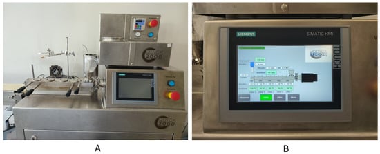
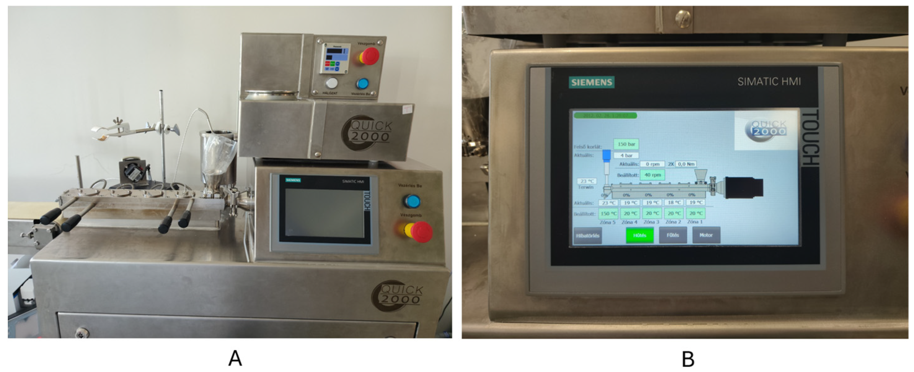
Hot-melt extrusion is a diverse technique which can also be used for API containing filament manufacturing coupled with FDM 3D printing. The preferred method of filament manufacturing is using a co-rotated twin-screw hot-melt extruder (Figure 1) [21,22]. The design of the twin-screw within the screw chamber makes it possible to mix and convey the added polymer and other excipients with the API into a homogeneous mixture while the applied heat and shear stress helps to melt and soften substances inside the twin-screw chamber [23]. The operating temperature, which can be set on the control panel of HME (Figure 1), must be in accordance with the selected polymer’s softening temperature range, otherwise the mixture might be clogged or burned before leaving the extruder’s nozzle [24,25]. After the homogeneous mixture leaves the nozzle, usually a conveyor belt drives the filament away from the nozzle and solidifies. The filament thickness is determined by the nozzle diameter and the conveying speed, which is synchronized with the screw speed, depending on the rheological properties of the semi-solid mixture. After solidification, it can be wound onto a spool for easier use and storage [22,26]. The viscoelastic properties of a polymer also influence the diameter of the filament due to the “die swell” behavior which means that the filament diameter will increase as it leaves the extruder [27].
Figure 1.
Twin-screw hot-melt extruder (A) and its control panel (B).
4. Model Designing and Printing Process with FDM Printer
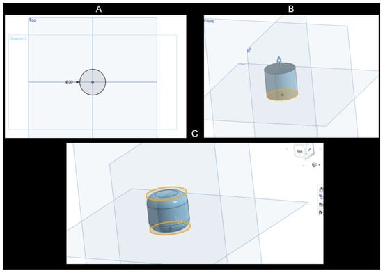
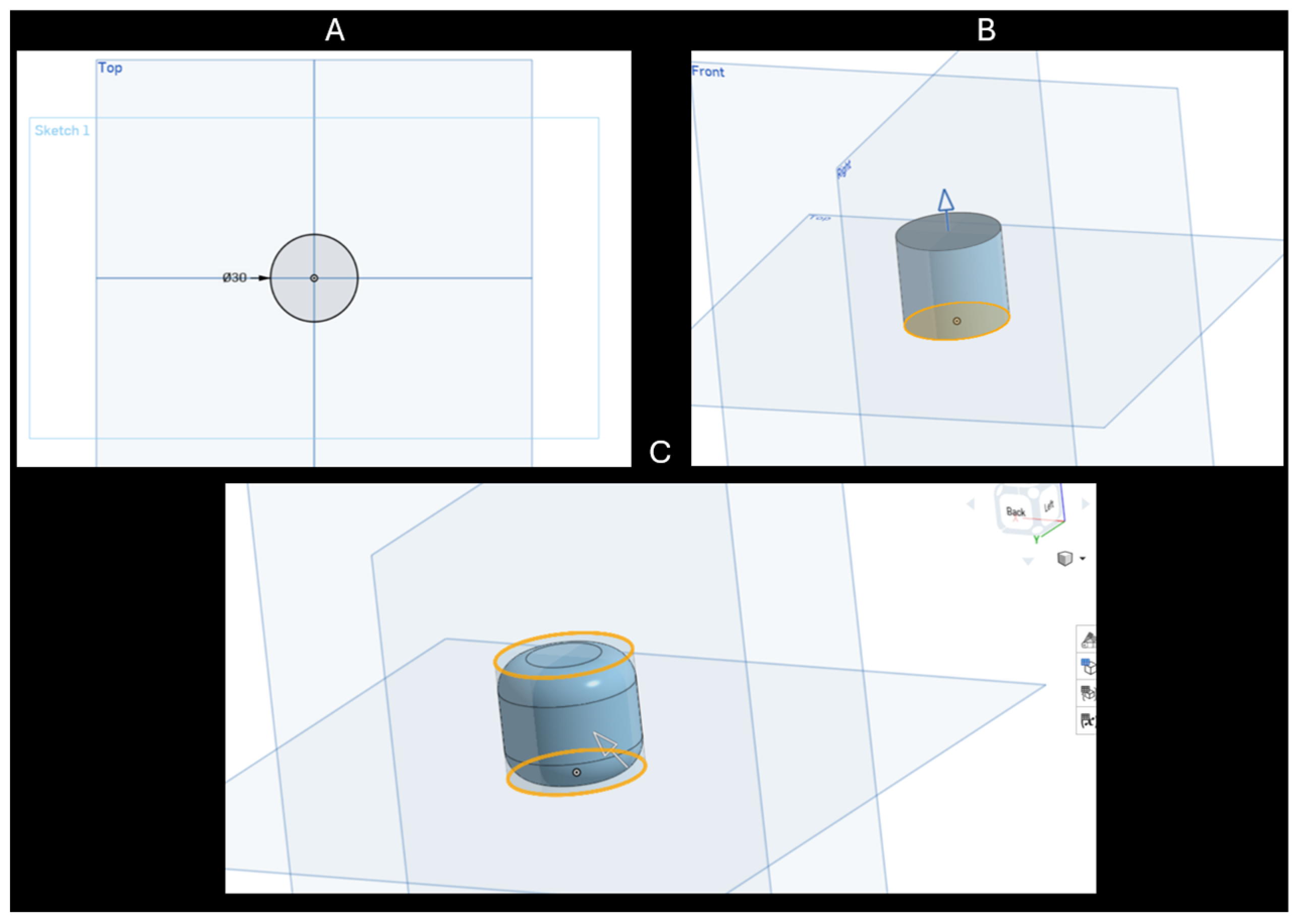
The very first step of manufacturing a 3D-printed implant is the designing of a Computer-Aided Design (CAD) model. The CADs are made in a modeling program (Figure 2), where the digital form of the implant can be modified until the desired size and shape is formed. There are several programs that are freeware or open source like FreeCAD, SketchUP, Onshape (Figure 2), Tinkercad, OpensCAD, or RepoCAD, as examples [28]. Then, these files should be converted into a Standard Tesselation Language (STL) file, which can be recognized by a slicer program [29,30].
Figure 2.
Modeling steps with Onshape online software. (A) Sketching in 2D. (B) Extruding sketch into a 3D digital object. (C) Modified shape of the digital object.
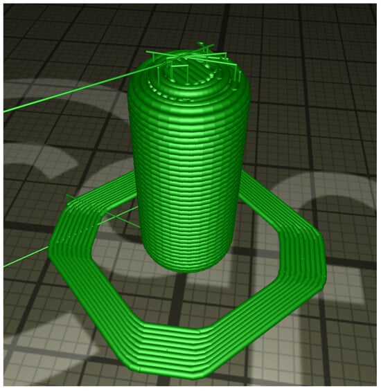
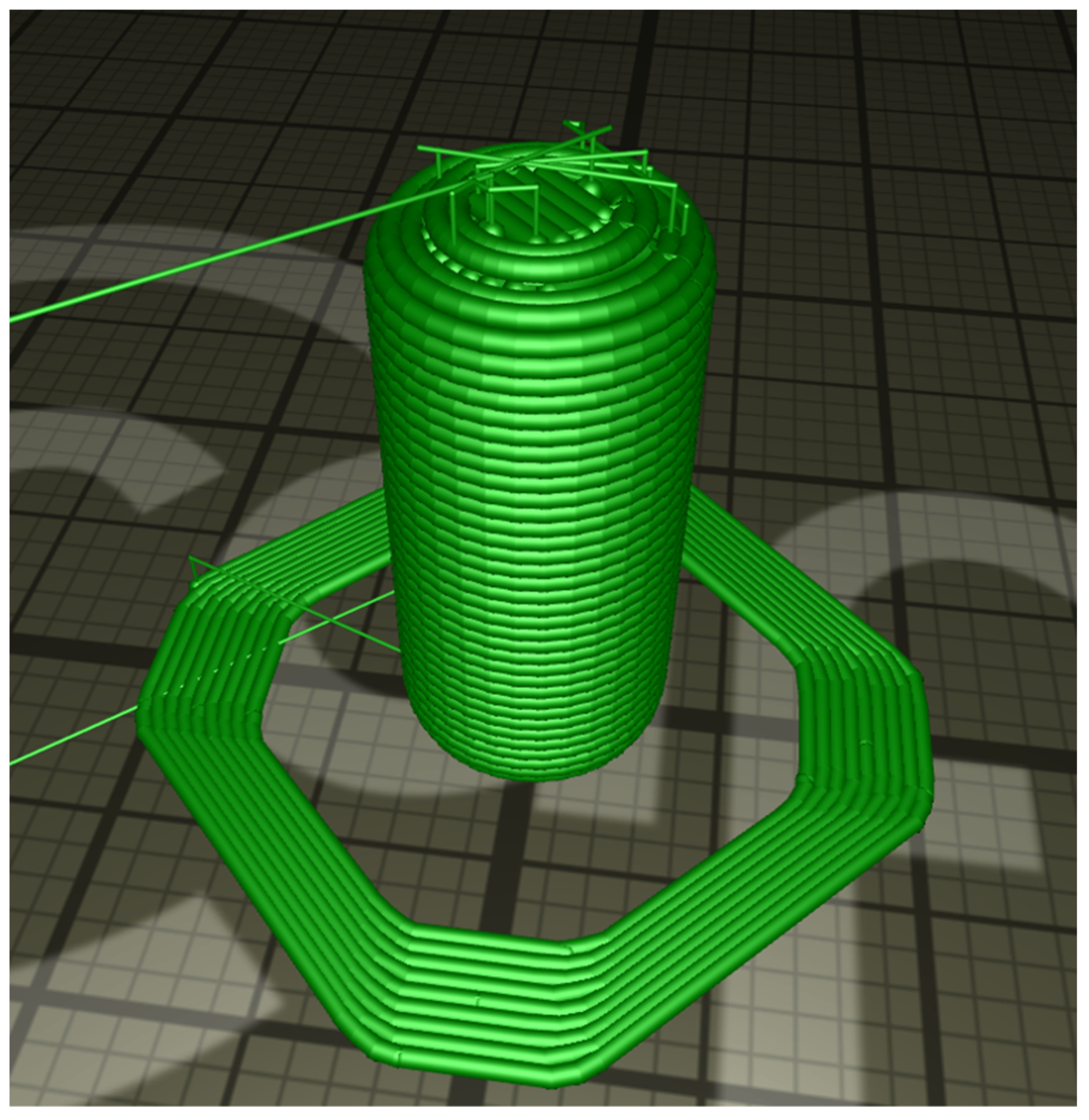
In the slicer program, STL files are converted into G-codes (Figure 3), which contain the printing parameters mentioning operating temperature, layer height, distance between two layers, printer head movement speed, and other attributes, depending on the printer’s properties [29,30].
Figure 3.
G-code of an object converted from .stl format in CraftWare slicer program.
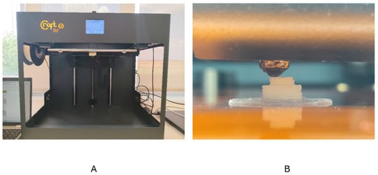
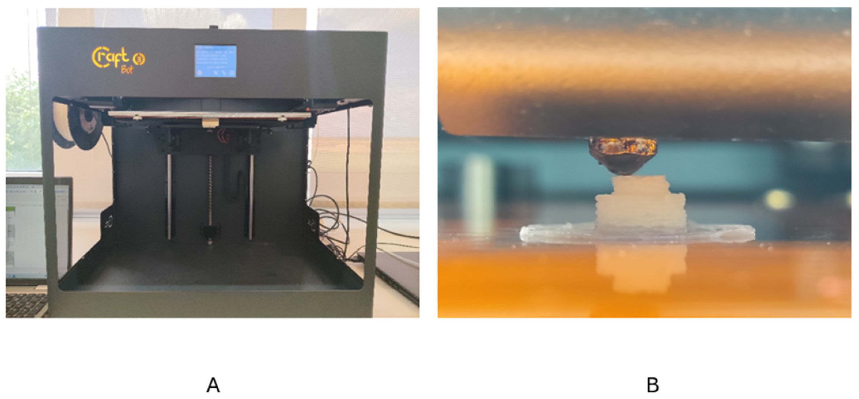
Fused deposition modeling (Figure 4) uses thermoplastic polymer filaments as printing materials. A gear wheel drives the filament to the printer head’s heated nozzle, where the filament melts into a semi-solid state, and passes through to the printer bed, while the printer’s head moves horizontally, following the route determined by G-code to make a layer. Once the layer is completed, the bed or the printer head moves diagonally to get in position in order to apply a new layer. Optimally, the melted polymer solidifies in seconds after reaching the printer bed or the lower layer which is necessary for a stable base and smoother structure [31,32].
Figure 4.
CraftBot FDM printer (A) and the printer head or nozzle (B).
Critical printing parameters such as printer head diameter, layer height, infill density, infill pattern, printer head temperature, platform temperature, printing speed, and filament diameter highly influence the quality of the final object, which can be changed to achieve the desired result [27,33,34].
5. Polymers of HME-FDM-Printed Implants
Not all polymers are suitable for hot-melt extrusion or fused deposition modeling. Polymers that are used in HME and FDM processes, must have thermoplastic or thermo-softening properties to ensure smooth operation and avoid damaging the machines [35,36]. For pharmaceutical usage, especially in the case of implants, thermoplasticity is not enough for a polymer, it must be biocompatible, otherwise inflammation and immune rejection has a higher possibility to occur [37]. It is not obligatory to use only one kind of polymer at once. What is more, sometimes it can be advantageous if the polymers are mixed to improve each other’s stiffness and brittleness, or to achieve the desired effect and drug-release profile [38]. In this section, the HME-FDM-specific polymers used for implant manufacturing are described from the chosen articles. Some important parameters like molecular weight, viscosity, glass transition temperature (Tg), melting temperature (Tm), decomposition temperature (Td), or solubility were collected if they were mentioned in the article as they may represent an important critical material attribute (CMA); this can be found in Table 1. However, the authors found that biodegradability was mentioned, but the physical parameters were only partially or not mentioned in the chosen articles. This makes the reproducibility difficult. One critical aspect might be the different physical parameters resulting from the varying molecular weights of polymers, like in case of polycaprolactone (PCL), where the thermal degradation temperatures altered from the molecular weight PCL (Mw= 10,000) degrades at 309 °C, PCL (Mw = 45,000) at 332 °C and PCL (Mw = 80,000) at 336 °C [39].
Table 1.
Biocompatible polymers used in HME-FDM for implant manufacturing. The parameters, solubility, and biodegrability statements are based on the revised articles.
6. Manufacturing of HME-FDM-Printed Implants
In this section, the manufactured implants are sub-grouped into different sections. The classification of devices proved to be a challenge since some of these IDDSs combine functionality and pharmaceutical purposes; therefore, the authors attempted to follow the FDA’s regulatory framework [48].
6.1. Subcutaneous Implant
Pediatric treatments often demand specific dosages considering age or even body weight. Three-dimensional printing gives the opportunity to personalize the size, shape, and, consequently, the dosage according to the patient’s needs. Daihom et al. formulated a gabapentin (GABA)-loaded long-acting medicinal implant for the treatment of pediatric neuropathy. They composed four formulations with GABA, using PCL and PEG. The GABA/PCL ratios were 10/80 w/w%, 20/70 w/w%, 30/60 w/w%, and 50/40 w/w%. For sustained release, PCL has already shown its effectiveness in different medical applications [49,50]. Each formulation contained 10 w/w% PEG3350 [43], which, in filaments, acts as a plasticizer and increases hydrophilicity [51]. In this research, their main objective was to develop an implant which has the potential to increase bioavailability as localized drug delivery and to reduce dosing to meet the patient’s need. Morphology and solid-state characterization, texture analysis, energy dispersive- X-ray (EDX) spectroscopy, and in vitro tests were also made for over 25 days. However, their main focus was to determine the effect of drug loading, infill densities, and surface area differences on the release profile, and to confirm the reliability of MeltPreps (melt preparations) in in vitro studies to accelerate development. In vitro drug release tests showed that higher drug loading resulted in higher initial release rates, increasing the availability of the drug; however, the higher drug-loaded implant’s release rate began to slow down with time compared to the lower loadings. The authors concluded that 3D printing was efficient in creating implants with the possibility of personalization due to the customizable release profiles [43].
Stewart et al. designed ibuprofen sodium-, ibuprofen acid-, and methylene blue-containing implants to compare each model drug’s behavior in the drug release profiles with differently designed implant structures with built-in drug release “windows”. These served as the surfaces where the drug could be liberated from the implant. They used PVA and PLA polymers to print hollow implant structures, then directly filled them with the model compounds. The combination of two types of PCL were also used as a coating material to obtain sustained drug release profiles. Their results showed that prolonged and sustained drug release can be acquired by a PCL coating, prolonging drug release up to 300 days while the surface area of drug release “windows” significantly influenced the drug release profiles. These results revealed that the design and the solubility of different APIs highly influence the drug-release rates, while the application of specific coatings placed on strategical sites of the implants provide the possibility of personalizing the release profiles according to the patient’s needs [44].
6.2. Drug-Eluting Implants
The use of drug-eluting implants can also be categorized based on the therapeutic purpose.
6.2.1. Drug-Eluting Implant for Tissue Regeneration
For tissue regeneration monoclonal antibodies are already in focus [52]. However the effectiveness of such biological API highly depends on chemical and physical stability [53]. HME and FDM processes use relatively high temperatures during operation; therefore, thermosensitive APIs are rarely used directly in HME and FDM techniques. However, some polymers have the thermo–rheological properties to be potentially suitable for low-temperature operations that are also safe in terms of heat-sensitive APIs [54]. Carlier et al. used PLGA as a carrier for monoclonal antibody (mAb)-loaded implants. Filaments were made by HME, where PLGA, spray-dried mAb and PEG 2000 were blended previously. PEG 2000 was added as a plasticizer to decrease the glass transition temperature. Homogeneous dispersions were successfully reached both in filaments and in 3D-printed IDDS. Trehalose and L-leucine association (TRE-LEU) were used to protect and stabilize mAb against thermal degradation and, as a result, no fragmentation was observed, while dissolution tests showed a sustained-release profile [45].
6.2.2. Drug-Eluting Surgical Implant
As a surgical implant, on-demand production of such devices may be necessary in urgent cases with customized dosage. A research group used an FDM technique with four different polymers, PLA, antibacterial PLA, PETG, and PMMA and diclofenac-sodium as a model drug for each, to print implantable samples. In this case, diclofenac-sodium was not embedded or extruded with the polymers, but rather they printed the bottom half of the hollow structure, filled it with diclofenac-sodium salt, put a pre-printed cover on top, then printed the top half upon that. Since the active pharmaceutical ingredient was not necessarily exposed to high heat, this method might be suitable for heat-sensitive APIs. In this early article, the focus was still on how to prevent the active ingredient from potential degradation through the FDM process, which the authors were able to provide, even though the API dissolution was within 24 h [42].
6.2.3. Drug-Eluting Implant for Sustained Blood Concentration
Yang et al. reported that a PCL matrix releases the embedded APIs at slow rates, so they proposed mixing PCL with chitosan to improve drug release. As a model drug, they used ibuprofen and extruded their own filament by HME, then successfully applied the filament to their FDM printer. Chitosan acted as a plasticizer in the filament and reduced the complex viscosity. In vitro results showed that the drug content crucially influenced the drug release. When the ibuprofen content was increased from 4 wt% to 20 wt%, the overall drug release also increased from 28% to 100%. Drug release was influenced by the chitosan too. When they changed from 4 wt% to 20 wt% chitosan, the release of the drug also increased from 55% to 100%. The reason for the changes in both cases was the formation of more release channels. As they concluded their results, the proper amount of chitosan could form enough release channels for the ideal performance, while acting as a plasticizer and improving the printability of the PCL filament [41].
6.3. Intrauterin Implant
Uterine cancer is one of the most common cancers affecting women. Varan et al. proposed using paclitaxel and carboplatin separately in two different filaments for 3D-printed implantable intra-uterine device (IUD). They encapsulated both drugs in different cyclodextrin derivatives with different ratios, where they found out the methyl-β cyclodextrin (MβCD) was the most efficient in both cases. The main structure of their filament was polycaprolactone. Differential scanning calorimetry (DSC) results showed that the different formulations’ melting peaks were close to 60 °C, similar to pure PCL. This indicates a possibly safe material choice for filament manufacturing, avoiding high temperatures when thermosensitive drugs and drug complexes are used. Drug release tests also confirmed the prolonged drug release in case of paclitaxel–cyclodextrin complex, where in 10 days, only 15% of the total drug amount was released. This is suitable for prolonged release profiles while, in case of carboplatin–cyclodextrin complex, the overall release was faster. In the case of paclitaxel, carboplatin was released independently from the PCL degradation rate. These interesting results suggest that the choice of drugs in anti-cancer therapies is far more significant, considering drug release profiles in personalized treatment strategies [39].
6.4. Scaffold Implant
Mei et al., in an effort to avoid these side effects and directly target the tumor cells, designed an anti-tumor implantable porous scaffold, using PLA and methotrexate (MTX) which were extruded into a filament. The targeting of tumor cells was based on the possibility of using the patient’s computed tomography (CT) and magnetic resonance imaging (MRI) to design and personalize the 3D model according to the tumor’s structure. An FDM printer was used to print the scaffolds, and these scaffolds were placed on tumors inside different mice groups. In comparison to the injection- and implant-treated animals, the mice that were treated with injections had a lower organ coefficient on the spleen in opposition to the scaffold implant-treated animals, while the scaffolds efficiently inhibited the growth of the tumor. In vitro test results also showed that MTX was released along with PLA degradation and the growth of tumor cells was successfully and significantly inhibited after 24 h. After 120 h, a 95% inhibition rate of tumor cells was measured, while for the growth of normal cells, the effect was negligible [46].
6.5. Drug-Eluting Functional Implants
6.5.1. Drug-Eluting Cardiovascular Prosthesis
In relation to cardiovascular diseases, many types of implants have been designed such as stents, grafts, and valvular prosthesis. The implantation of these devices has the risk of developing infections, or, in some cases, blood clots can form after operations. Martin et al. printed a vascular graft from TPU and an antibiotic, rifampicin, was embedded within it as a prophylactic agent. The same methodology was used with another drug, dipyridamole, which is an antiplatelet drug to prevent cardiovascular device-related thrombosis. In vitro release tests were performed with the two separate drug-loaded implantable devices. The rifampicin-containing device showed that it was capable of sustained release for more than 30 days, which might predict a potential and successful prevention from infections after surgery in the time range when most of the infections occur. In the case of the dipyridamole-containing device, sustained release was observed for 55 days; however, the test result predicted a longer period for the sustained release profile. Their conclusion was that it is possible to produce filaments with HME and FDM to print a cardiovascular device that can possibly prevent infections and blood clots forming after surgery [40].
6.5.2. Drug-Eluting Fixation Implant
Implantable devices in orthopedic surgery are commonly used for multiple purposes. A research group fabricated FDM-printed patient-specific fixation implants which allow local drug delivery. Gentamicin and methotrexate were impregnated into the PLA implants. The devices successfully showed both antibacterial and chemotherapeutic effects; however, they negatively influenced the overall strength of the implants [47].
6.6. Formulation Strategy
Following revision of the chosen articles, the authors concluded that for the development of IDDSs, several drug-loading techniques have been found that allow incorporation of thermo-sensitive APIs, but some of these require specific circumstances before, during, or after the procedure. While the main focus is on polymer types and formulations, no article has discussed API behavior comprehensively with these formulations. The above-mentioned studies focusing on solving 3D-printed IDDS-related issues from the aspect of chosen API, the formulation strategy, and the innovative approach can be found in Table 2.
Table 2.
IDDSs and innovative approach in their research.
Regarding the reviewed articles, there would be great opportunity for implant manufacturing and a great demand from several aspects as these techniques do not allow the possibility of personalization. The technology is already available and has been extensively used in the last decade, but some aspects are still missing for better quality and reproducibility.
For a better understanding regarding our research group’s recent work examining quality by design (QbD) possibilities for better quality outcomes in HME-FDM, see Table 3. The critical process attributes (CPPs) were collected for both HME and FDM processes regarding the collected articles and the provided data. In the case of HME, the extrusion temperature, extruder screw speed, and die diameter was revised. In the case of FDM, the layer height, infill density and pattern, print head diameter and temperature, platform temperature, and the printing speed were collected [27].
Table 3.
In the case of the revised articles, some CPPs were collected for HME-FDM manufacturing.
7. Characterization and Evaluation
Evaluating a filament and the finished 3D-printed implant is crucial for determining the safe and effective applicability of the final product. Structural integrity, biocompatibility, chemical stability, and stable drug-release profiles are demanded in all cases, and these can be examined using several methods applied to IDDSs.
The characterization is sub-grouped regarding the system of Repka et al. [19].
7.1. Physico-Chemical Characterization
The physico-chemical characterization is made possible by using powder X-ray diffraction (PXRD), Fourier transform infrared spectroscopy (FTIR), NIR, or nuclear magnetic resonance spectroscopy (NMR).
7.1.1. PXRD
The scattering of X-rays from atoms in a crystalline material creates diffraction patterns that reveal atomic arrangements in samples. The result is a diffractogram with diffraction patterns which consist of peaks [55]. This characterization technique can determine if the API is in an amorphous or crystalline form in the polymer. By varying humidity and temperature values, it also provides information about the stability of the final product [19]. In the case of the polycaprolactone–chitosan-based implant, the crystallization temperatures of PCL and ibuprofen were determined with this method. It was also confirmed that, after the extrusion and printing process, PCL retained its semi-crystalline state and ibuprofen remained in crystalline form. It also provided valuable information about composite filaments, revealing that the diffraction peaks of the polymer decreased as the drug-loading of the filament increased [41].
7.1.2. FTIR Spectroscopy
FTIR is used to identify molecules or molecular groups by their vibrational properties [56]. In terms of 3D-printed implants, it helps to identify possible chemical changes that occur during the manufacturing process and chemical interactions between component materials [57,58,59]. Although chemical changes between substances are unwanted in most of the cases, Yang et al. mentioned that FTIR analysis revealed that hydrogen bonds were formed between the hydroxyl group of PCL and the hydroxyl group of ibuprofen which improved the interfacial compatibility, while other chemical bonds were not formed [41]. Varan et al. compared blank, drug-loaded PCL filaments, and PCL granulates to determine if the extrusion process or drug-loading affected the melting peaks through the process; however, the similar melting peaks suggest that neither the process nor the drug-loading influenced the chemical structure significantly enough to change the melting properties of the filament [39].
7.2. Thermal Characterization
Thermal characterization is important in the manufacturing process, and is widely used for pre-, intermediate, and final analytical testing. It plays an important role as the HME-FDM process applies high heat on the components, which may result in thermal degradation. Thermal characterization techniques are also effective in the determination of glass-transition temperatures and melting temperatures [54]. It is crucial to perform these experiments as HME-FDM can expose drugs and excipients to heat and shear must be kept in mind in the case of peptides, antibiotics, and thermolabile small molecules.
7.2.1. Thermogravimetric Analysis (TG/TGA)
Thermogravimetric analysis is used to investigate thermal decomposition and to assess relative thermal stabilities [60]. In the case of 3D-printed implants, it provides valuable information about the different thermal degradation behaviors of pure polymer, polymer–polymer, polymer–excipient, and drug-loaded polymer mixtures [41,61,62]. For this analysis, a small sample, usually less than 10 mg, is more than enough. From the results, it is possible to determine the safe operation temperatures for filament extrusion and 3D printing, thereby avoiding critical API loss and other unwanted thermochemical reactions and instabilities [45,57,58]. For the cardiovascular prosthesis, TGA was sufficient in the determination of rifampicin stability at the extrusion temperatures, while the analysis also revealed that the rifampicin-containing TPU filaments showed higher degradation temperatures compared to pure TPU [40].
7.2.2. Differential Scanning Calorimetry (DSC)
Differential scanning calorimetry is a thermal analysis technique that is also applicable to various kinds of materials such as pharmaceuticals including implants, and polymers also used for 3D printing [63]. With DSC, polymer raw materials, filaments, and even polymer–excipient–drug mixtures can be monitored to describe the glass transition temperatures and changes as other materials are blended together. This both helps to determine the necessary operation temperatures during heat transfer processes, and also if the components dissolve into each other at such temperatures or stay in a crystalline form [43,45,59,64]. In most cases, TG and DSC analysis are evaluated together for a more complex and precise result [41,58].
7.3. Microscopic Characterization
Microscopic characterization is usually performed by scanning electron microscopy (SEM), micro-computed tomography (micro-CT or μCT), X-ray-computed tomography (XRCT), hot-stage microscopy (HSM), or polarized light microscopy (PLM).
7.3.1. SEM
Scanning electron microscopy provides a surface evaluation method for larger (200 mm or in some cases even larger) samples with up to 1,000,000× magnification and 1 nm resolution opportunity [65]. The usage of SEM helps to characterize the structural integrity and morphology of implants, revealing possible defects or changes that occur during or after printing, and also after going through several tests such as in vitro release studies [41,64]. SEM images are able to show possible drug aggregates within the extruded filaments or printed implants, from which we can also obtain valuable information about homogeneity which can affect the drug release profile [57,59]. The other crucial factor that must be examined is proper layer adhesion, which needs to be intact in order to ensure durability, mechanical robustness, tissue integration, and biocompatibility [43].
7.3.2. MicroCT
MicroCT has the ability to visualize and quantify porosity or defects in IDDS. This density measurement method allows the detection of inclusions, unconsolidated powder, and open porosity at the surface with visualization and part mass density. Part density is usually measured by the Archimedes method; however, in additive manufacturing, the rough surfaces which trap gas, infiltrated water through the pores, and the unconsolidated powder can conceal the presence of porosity in the density measurement. Rough surfaces also influence the microCT measurements; however, proper determination of the sample edge and regular calibration of the instrument can reduce the error margin [66].
7.3.3. Hot-Stage Microscopy (HSM)
HSM provides information about thermal events visually, which helps describe the polymer–API interaction in a molten state, such as the dissolution and recrystallization effects of temperature [19].
7.3.4. Polarized Light Microscopy (PLM)
PLM enables the observation and the visualization of state transitions by applying heat. With an optical microscope to which a camera is attached, video documentation can be conducted saving time, money, and other resources without repeating the experiment [35].
7.4. Pharmacopoeial Testing
Regarding the pharmacopoeial testing of 3D-printed IDDSs, in vivo and in vitro tests are well-described for conventional manufacturing of these systems. For this reason, the authors only focus on the most important aspects [20].
7.4.1. In Vitro Dissolution Testing
In vitro drug release tests help to determine drug release profiles of IDDS, where the release medium imitates the tissue environment. Samples are taken over time to predict the implant’s drug release kinetic behavior once it is implanted [67]. Significant differences were shown between polymers in drug release in different polymer–drug pairings, which provides information to select the proper polymer–drug combinations, considering the advantages and disadvantages of formulations depending on the future use [38,42,45,67]. Studies have shown that the solubility of the drug in the release medium also affects the release kinetics. How the drug behaves at different amounts of drug loadings must be taken into account to avoid burst effects and overdosing as the aim of implants is to ensure a well-defined, sustained, and prolonged release profile [44,68]. In the case of 3D-printed IDDSs, it is not only the change in polymer–drug ratio and the type of the polymer influences drug release, but also implant design is important, as surface area and structural modules can be changed with different shape designs. This could be a possible method for personalized release profiles [62]. In vitro dissolution testing is an important question and can provide a good prediction of the quality of the manufactured product. In Table 4, the authors collected the used API or model compound, the used polymer, the dissolution test circumstances, and the results for a better understanding of where we stand right now in implant manufacturing by HME-FDM.
Table 4.
Summaries of the used API or model compound, the used polymers, and the dissolution test circumstances and results of the reviewed articles.
7.4.2. Shelf Life
The shelf life of different products still has not been determined or tested among the mentioned IDDSs, or if sterilization methods, where applied, were effective enough to store these devices for a longer period of time. There is also the question of how much storage time would be ideal for such devices [39,40,41,42,43,44,45,46].
7.5. Biocompatibility
The biocompatibility measurement of implantable devices is essential. The implantation of a foreign object triggers a host response, including an immune response in the body. Host response includes inflammation at the implant site, tissue remodeling, and proliferation. During an immune response, the innate and adaptive immune systems are the definitive mediators, where the innate immune response is responsible for the nonspecific inflammatory response. The innate immune system also triggers the adaptive immune response where specific antigens are involved. These antigens may react to dissolving or degrading implant components, causing rejection. To avoid a host response, the testing of such devices involves device design, in vitro study of cytotoxicity, in vivo study of device performance using animal models, genotoxicity and cellular response to the materials or devices, and the final translation to the clinical testing [69]. For biocompatibility and cytotoxicity testing, several assays such as LDH test or real-time electronic sensing assay (RT-CES) may be used; however, the most widely used method is the rapid colorimetric 3-(4,5-dimethylthyazol-2-yl)-2,5-diphenyl-2H-tetrazolium bromide dye (MTT) cytotoxicity test, where the parameters are determined by the International Organization for Standardization (ISO) including exposure period, type of positive control, and the number of cells in the dishes before and after exposure to the material [42,70].
The surgical implant’s MTT assay proved that the different samples including PLA, antibacterial PLA, PETG, and PMMA are cytocompatible even after extrusion and printing [42]. The cardiovascular graft containing rifampicin was tested with a different method, by culturing HUVEGs onto the grafts after sterilization. Samples were observed for 72 h, and the results showed that the cells attached to the surface of the sample and only a few cell deaths were observed. A monolayer of HUVEGs also appeared on the surface and the graft samples proved to be non-cytotoxic [40].
7.5.1. In Vivo Tests
In vivo tests are often conducted on animals such as mice. These tests usually last for a couple of months to ensure reliable results. They are necessary to establish biocompatibility and to investigate further the polymer degradation mechanism or the tissue reaction by explantation of the implant and removing the surrounding tissues at the end of the experiment for quantitative and qualitative testing. Valuable information about tissue–material interactions such as cell response, tissue formation behavior, vascularization of newly formed tissues, and host response behavior can be evaluated before human clinical trial phases [71].
7.5.2. Sterilization
Just to mention the sterilization methods, HME-FDM 3D-printed implants still need more thorough study to examine possibly applicable processes to meet the determined demands and quality requirements of implants. This is important as they may affect the polymer molecular weight, drug potency, impurity profile, crystallinity, mechanical properties, surface wettability, residual moisture, and release kinetics [72]. However, some efforts to test different techniques, such as γ-sterilization, have been used by some groups; the method was conducted according to the proper ISO determined parameters [73]. PLA and heat-treatable PLA (PLA with crystallization compound) went through autoclave sterilization in case of an FDM-printed orthopedic cutting guide. Although this procedure was not applied to an implant, it did not cause any substantial negative effect on the dimensional functionality. Theoretically, in the case of PLA implants where a post-print drug loading process is applied, this might be a strategy worth considering for further investigation [74]. Other research groups performed several sterilization methods on different polymers, including PLA and PETG. The applied sterilization methods such as autoclave (with higher and lower heat exposure), ethylene oxide, and hydrogen peroxide plasma proved to be efficient in destroying microorganisms. High-temperature autoclave sterilization significantly affected the structure of the polymer, while in lower temperature environments, less deformation occurred. Gas sterilization eliminates deformation issues; however, it could have a significant impact on chemical structure [72]. In another study, they used 70% (υ/υ) ethanol for microbial reduction in which the implant was immersed for 12 h in a laminar air flow cabinet; however, the possible effects of this decontamination were not examined further in detail [42]. It will be crucial to place greater emphasis on the possible sterilization of HME-FDM manufactured implants.
7.6. Contact Angle Measurement
Wettability determines crucial biological and chemical processes. The contact angle is defined as the angle between the tangent and the three phase point and the solid surface. The lower the contact angle, the better the wetting properties of the solid object as hydrophilicity increases [75]. Surfaces with contact angles < 90° are considered hydrophilic, while contact angles > 90° are considered hydrophobic [76]. The implantable devices drug delivery rates are also highly determined by surface wettability, which is also determined by polymer structures [77]. Arany et al. compared the contact angle values of PLA and antibacterial PLA filaments, where the contact angle value of antibacterial PLA was nearly three times smaller than PLA, which was probably caused by the antibacterial nanoparticles [42]. M’Bengue et al. and other studies showed that some sterilization methods like gamma sterilization could influence contact angle values as after irradiation of TPU. Here, hydrophobicity decreased, which indicates that after sterilization, contact angle measurements should be repeated considering the significance of wettability of implants [73,78].
8. Translation and Regulation
There are already some opportunities for 3D printing technology and its industrial application, like the Triastek™ Melt extrusion deposition = MED® manufacturing, which is extrusion-based, allows continuous manufacturing, and is FDA approved [79]. In the author’s opinion, in the case of HME-FDM, the main concept would be to manufacture personalized medications, not only in dose but in design.
The regulatory environment is in development by agencies around the world to ensure patient safety while allowing innovative approaches [80]. The most advanced regulatory approach currently is Framework for Regulatory Advanced Manufacturing Evaluation (FRAME) which is under development by the FDA. FRAME’s main focus is on artificial intelligence, end-to-end continuous manufacturing, distributed manufacturing, and distributed manufacturing at non-traditional host sites [79]. HME is considered as a continuous manufacturing method, while 3D printing including FDM is a distributed manufacturing method [79,81]. This direction is forward-looking and will hopefully contribute to the regulatory acceptance of HME-FDM products.
9. Limitations
As with every technology, HME-FDM 3D printing has its own limitations. Concerns have been raised by professionals in the healthcare industry about medication safety and quality aspects mentioning stability, quality control, dose accuracy, shelf life and the identifications of drug products [82]. Other mentioned limitations include relatively low productivity, high costs, and issues about on-demand production and delivery [83]. However, the major limitation of the technique is the relatively low printing efficiency. Others have also raised issues about surface quality and mechanical strength of the final object [84,85].
There are some limitations, but we must keep in mind that every technology has at least some. In the author’s opinion, 3D printing, such as HME-FDM, including the QbD principles and the ability to manufacture reproducible and determined quality products, could become an effective tool for personalized drug product manufacturing. Personalized drug products, including drug-loaded implants, will be much more widespread in general healthcare, considering the need to vary dosage based on age and weight, and the possibility for 3D printing to influence release profiles and dosage by customized shapes and size [86].
10. Conclusions
Implantable drug delivery systems have demonstrated their significance in long-term treatments and therapies; however, it can be challenging to achieve the personalization of dosages and anatomical compatibility through traditional manufacturing processes. Three-dimensional printing could possibly provide an easier solution to these difficulties, considering the results research achieved to date, as well as polymers, APIs, characterization, and evaluation methods that are already used in the pharmaceutical industry. Numerous research groups are also working on the possible application of 3D printers and computer-aided programs that are still improving, but there are still some gaps between the research and the final applicable product. HME-FDM can be a potential manufacturing process for IDDSs, where the applicable materials, including some thermoplastic polymers, have the background for the use of medical purposes. In general, the characterization and evaluation methods are ready for the commercially available IDDSs, but some personalization could be included, and the quality and reproducibility should be provided in harmony with the QbD guidelines. The regulations and manufacturing standards for 3D-printed drug delivery systems are still under development.
Overall, 3D-printed IDDSs have the potential to provide another kind of safe, more patient-friendly, and personalized medication form in various treatments and therapies for both children and adults, but more research should be performed to achieve this goal.
Author Contributions
Conceptualization, P.A. and B.V.; software, B.V.; investigation, B.V.; resources, I.B.; writing—original draft preparation B.V. and P.A.; visualization, B.V.; supervision, P.A.; project administration I.B. and P.A.; writing—review and editing, P.A., Z.U. and P.F. All authors have read and agreed to the published version of the manuscript.
Funding
Project no. TKP2021EGA18 has been implemented with the support provided by the Ministry of Culture and Innovation of Hungary from the National Research, Development and Innovation Fund, financed under the TKP2021EGA funding scheme. Researcher Petra Arany was awarded a fellowship by the Hungarian Academy of Sciences to support researchers raising children.
Institutional Review Board Statement
Not applicable.
Informed Consent Statement
Not applicable.
Data Availability Statement
No new data were created or analyzed in this study. Data sharing is not applicable to this article.
Acknowledgments
During the preparation of this manuscript, the authors used [ChatGPT, GPT 5.5] for the purposes of [sorting polymers in alphabetical order in Table 1]. The authors have reviewed and edited the output and take full responsibility for the content of this publication.
Conflicts of Interest
The authors declare no conflicts of interest. The funders had no role in the design of the study; in the collection, analyses, or interpretation of data; in the writing of the manuscript; or in the decision to publish the results.
Abbreviations
The following abbreviations are used in this manuscript:
| 3D | Three-Dimensional |
| API | Active Pharmaceutical Ingredient |
| CAD | Computer-Aided Design |
| CMA | Critical material attribute |
| CPPs | Critical process attributes |
| CS | Chitosan |
| CT | Computed Tomography |
| DSC | Differential Scanning Calorimetry |
| EDX | Energy dispersive X-ray |
| FDM | Fused Deposition Modeling |
| FTIR | Fourier Transform Infrared Spectroscopy |
| FRAME | Framework for Regulatory Advanced Manufacturing Evaluation |
| GABA | Gabapentin |
| HME | Hot-Melt Extrusion |
| IDDS | Implantable Drug Delivery System |
| ISO | International Organization for Standardization |
| IUD | Intra Uterine Device |
| MeltPrep | Melt Preparation |
| MicroCT | Microcomputed tomography |
| MRI | Magnetic Resonance Imaging |
| MTX | Methotrexate |
| MβCD | Methylβ cyclodextrin |
| NIR | Nuclear magnetic resonance spectroscopy |
| PBS | Phosphate-buffered saline |
| PC | Polycarbonate |
| PCL | Polycaprolactone |
| PEG | Polyethylene glycol |
| PETG | Polyethylene terephthalate glycol |
| PLA | Polylactic acid |
| PLGA | Poly lacticcoglycolic acid |
| PMMA | Poly (methyl methacrylate) |
| PRXD | Powder X-ray diffraction |
| PVA | Polyvinyl alcohol |
| QbD | Quality by Design |
| SEM | Scanning Electron Microscopy |
| STL | Standard Tessellation Language |
| Td | Decomposition temperature |
| Tg | Glass transition temperature |
| TG/TGA | Thermogravimetric Analysis |
| Tm | Melting temperature |
| TPU | Thermoplastic polyurethane |
| TRELEU | Trehalose and Leucine association |
| RT-CES | Real time electronic sensing assay |
| MTT | 3-(4,5-dimethylthiazol-2-yl)-2,5-diphenyl-2H-tetrazolium bromide dye |
References
- Deanesly, R.; Parkes, A.S. Factors Influencing the Effectiveness of Administered Hormones. Proc. R. Soc. Lond. Ser. B—Biol. Sci. 1937, 124, 279–298. [Google Scholar] [CrossRef] [Scilit]
- Deanesly, R.; Parkes, A.S. Further Experiments on the Administration of Hormones by the Subcutaneous Implantation of Tablets. Lancet 1938, 232, 606–609. [Google Scholar] [CrossRef] [Scilit]
- Quarterman, J.C.; Geary, S.M.; Salem, A.K. Evolution of Drug-Eluting Biomedical Implants for Sustained Drug Delivery. Eur. J. Pharm. Biopharm. 2021, 159, 21–35. [Google Scholar] [CrossRef] [Scilit] [PubMed]
- Pons-Faudoa, F.P.; Ballerini, A.; Sakamoto, J.; Grattoni, A. Advanced Implantable Drug Delivery Technologies: Transforming the Clinical Landscape of Therapeutics for Chronic Diseases. Biomed. Microdevices 2019, 21, 47. [Google Scholar] [CrossRef] [Scilit] [PubMed]
- Bazaka, K.; Jacob, M. Implantable Devices: Issues and Challenges. Electronics 2012, 2, 1–34. [Google Scholar] [CrossRef] [Scilit]
- Park, K. Controlled Drug Delivery Systems: Past Forward and Future Back. J. Control. Release 2014, 190, 3–8. [Google Scholar] [CrossRef] [Scilit] [PubMed]
- Fung, L.K.; Saltzman, W.M. Polymeric Implants for Cancer Chemotherapy. Adv. Drug Deliv. Rev. 1997, 26, 209–230. [Google Scholar] [CrossRef] [Scilit] [PubMed]
- Dash, A.; Cudworth, G. Therapeutic Applications of Implantable Drug Delivery Systems. J. Pharmacol. Toxicol. Methods 1998, 40, 1–12. [Google Scholar] [CrossRef] [Scilit] [PubMed]
- Stewart, S.A.; Domínguez-Robles, J.; Donnelly, R.F.; Larrañeta, E. Implantable Polymeric Drug Delivery Devices: Classification, Manufacture, Materials, and Clinical Applications. Polymers 2018, 10, 1379. [Google Scholar] [CrossRef] [Scilit] [PubMed]
- Bakhrushina, E.O.; Demina, N.B. Implants as Targeted Drug Delivery Systems (Review). Pharm. Chem. J. 2022, 56, 396–402. [Google Scholar] [CrossRef] [Scilit]
- Fialho, S.L.; Silva Cunha, A.D. Manufacturing Techniques of Biodegradable Implants Intended for Intraocular Application. Drug Deliv. 2005, 12, 109–116. [Google Scholar] [CrossRef] [Scilit] [PubMed]
- Kovylin, R.S.; Aleynik, D.Y.; Fedushkin, I.L. Modern Porous Polymer Implants: Synthesis, Properties, and Application. Polym. Sci. Ser. C 2021, 63, 29–46. [Google Scholar] [CrossRef] [Scilit]
- Zheng, Y.; Pokorski, J.K. Hot Melt Extrusion: An Emerging Manufacturing Method for Slow and Sustained Protein Delivery. WIREs Nanomed. Nanobiotechnol. 2021, 13, e1712. [Google Scholar] [CrossRef] [Scilit] [PubMed]
- Wang, Y.; Sun, L.; Mei, Z.; Zhang, F.; He, M.; Fletcher, C.; Wang, F.; Yang, J.; Bi, D.; Jiang, Y.; et al. 3D Printed Biodegradable Implants as an Individualized Drug Delivery System for Local Chemotherapy of Osteosarcoma. Mater. Des. 2020, 186, 108336. [Google Scholar] [CrossRef] [Scilit]
- Arefin, A.M.E.; Khatri, N.R.; Kulkarni, N.; Egan, P.F. Polymer 3D Printing Review: Materials, Process, and Design Strategies for Medical Applications. Polymers 2021, 13, 1499. [Google Scholar] [CrossRef] [Scilit] [PubMed]
- Monteil, M.; Sanchez-Ballester, N.M.; Aubert, A.; Gimello, O.; Begu, S.; Soulairol, I. HME Coupled with FDM 3D Printing of a Customized Oral Solid Form to Treat Pediatric Epilepsy. Int. J. Pharm. 2025, 673, 125345. [Google Scholar] [CrossRef] [Scilit] [PubMed]
- Patil, R.; Bule, P.; Chella, N. Exploration of Conventional and FDM-Mediated 3D Printed Tablets Fabricated Using HME-Based Filaments for pH-Dependent Drug Delivery. AAPS PharmSciTech 2025, 26, 96. [Google Scholar] [CrossRef] [Scilit] [PubMed]
- Alhijjaj, M.; Belton, P.; Qi, S. An Investigation into the Use of Polymer Blends to Improve the Printability of and Regulate Drug Release from Pharmaceutical Solid Dispersions Prepared via Fused Deposition Modeling (FDM) 3D Printing. Eur. J. Pharm. Biopharm. 2016, 108, 111–125. [Google Scholar] [CrossRef] [Scilit] [PubMed]
- Repka, M.A.; Bandari, S.; Kallakunta, V.R.; Vo, A.Q.; McFall, H.; Pimparade, M.B.; Bhagurkar, A.M. Melt Extrusion with Poorly Soluble Drugs—An Integrated Review. Int. J. Pharm. 2018, 535, 68–85. [Google Scholar] [CrossRef] [Scilit] [PubMed]
- Schuh, J.C.L.; Funk, K.A. Compilation of International Standards and Regulatory Guidance Documents for Evaluation of Biomaterials, Medical Devices, and 3-D Printed and Regenerative Medicine Products. Toxicol. Pathol. 2019, 47, 344–357. [Google Scholar] [CrossRef] [Scilit] [PubMed]
- Chaudhari, V.S.; Malakar, T.K.; Murty, U.S.; Banerjee, S. Extruded Filaments Derived 3D Printed Medicated Skin Patch to Mitigate Destructive Pulmonary Tuberculosis: Design to Delivery. Expert Opin. Drug Deliv. 2021, 18, 301–313. [Google Scholar] [CrossRef] [Scilit] [PubMed]
- Arany, P.; Vámosi, B.; Bácskay, I.; Fehér, P. Hot-Melt Extrusion—Filament Manufacturing Coupled with Fused Deposition Modeling for 3D Printed Pharmaceuticals -a Brief Review. Remediis 2025, 1, 17–28. [Google Scholar] [CrossRef] [Scilit]
- Genina, N.; Boetker, J.P.; Colombo, S.; Harmankaya, N.; Rantanen, J.; Bohr, A. Anti-Tuberculosis Drug Combination for Controlled Oral Delivery Using 3D Printed Compartmental Dosage Forms: From Drug Product Design to in Vivo Testing. J. Control. Release 2017, 268, 40–48. [Google Scholar] [CrossRef] [Scilit] [PubMed]
- Zhao, X.; Wei, W.; Niu, R.; Li, Q.; Hu, C.; Jiang, S. 3D Printed Intragastric Floating and Sustained-Release Tablets with Air Chambers. J. Pharm. Sci. 2022, 111, 116–123. [Google Scholar] [CrossRef] [Scilit] [PubMed]
- Reddy Dumpa, N.; Bandari, S.; Repka, M.A. Novel Gastroretentive Floating Pulsatile Drug Delivery System Produced via Hot-Melt Extrusion and Fused Deposition Modeling 3D Printing. Pharmaceutics 2020, 12, 52. [Google Scholar] [CrossRef] [Scilit] [PubMed]
- Holländer, J.; Genina, N.; Jukarainen, H.; Khajeheian, M.; Rosling, A.; Mäkilä, E.; Sandler, N. Three-Dimensional Printed PCL-Based Implantable Prototypes of Medical Devices for Controlled Drug Delivery. J. Pharm. Sci. 2016, 105, 2665–2676. [Google Scholar] [CrossRef] [Scilit] [PubMed]
- Arany, P.; Papp, Á.; Nemes, D.; Fehér, P.; Ujhelyi, Z.; Bácskay, I. Quality by Design Approach for Hot-Melt Extrusion Coupled Fused Deposition Modeling (HME-FDM) 3D Printing: A Systematic Review. Pharmaceutics 2026, 18, 569. [Google Scholar] [CrossRef] [Scilit] [PubMed]
- Junk, S.; Kuen, C. Review of Open Source and Freeware CAD Systems for Use with 3D-Printing. Procedia CIRP 2016, 50, 430–435. [Google Scholar] [CrossRef] [Scilit]
- Mohammed, A.A.; Algahtani, M.S.; Ahmad, M.Z.; Ahmad, J.; Kotta, S. 3D Printing in Medicine: Technology Overview and Drug Delivery Applications. Ann. 3D Print. Med. 2021, 4, 100037. [Google Scholar] [CrossRef] [Scilit]
- Jamróz, W.; Szafraniec, J.; Kurek, M.; Jachowicz, R. 3D Printing in Pharmaceutical and Medical Applications—Recent Achievements and Challenges. Pharm. Res. 2018, 35, 176. [Google Scholar] [CrossRef] [Scilit] [PubMed]
- Penumakala, P.K.; Santo, J.; Thomas, A. A Critical Review on the Fused Deposition Modeling of Thermoplastic Polymer Composites. Compos. Part B Eng. 2020, 201, 108336. [Google Scholar] [CrossRef] [Scilit]
- Cailleaux, S.; Sanchez-Ballester, N.M.; Gueche, Y.A.; Bataille, B.; Soulairol, I. Fused Deposition Modeling (FDM), the New Asset for the Production of Tailored Medicines. J. Control. Release 2021, 330, 821–841. [Google Scholar] [CrossRef] [Scilit] [PubMed]
- Rajan, K.; Samykano, M.; Kadirgama, K.; Harun, W.S.W.; Rahman, M.M. Fused Deposition Modeling: Process, Materials, Parameters, Properties, and Applications. Int. J. Adv. Manuf. Technol. 2022, 120, 1531–1570. [Google Scholar] [CrossRef] [Scilit]
- Kristiawan, R.B.; Imaduddin, F.; Ariawan, D.; Ubaidillah; Arifin, Z. A Review on the Fused Deposition Modeling (FDM) 3D Printing: Filament Processing, Materials, and Printing Parameters. Open Eng. 2021, 11, 639–649. [Google Scholar] [CrossRef] [Scilit]
- Pereira, G.G.; Figueiredo, S.; Fernandes, A.I.; Pinto, J.F. Polymer Selection for Hot-Melt Extrusion Coupled to Fused Deposition Modelling in Pharmaceutics. Pharmaceutics 2020, 12, 795. [Google Scholar] [CrossRef] [Scilit] [PubMed]
- Ilyés, K.; Kovács, N.K.; Balogh, A.; Borbás, E.; Farkas, B.; Casian, T.; Marosi, G.; Tomuță, I.; Nagy, Z.K. The Applicability of Pharmaceutical Polymeric Blends for the Fused Deposition Modelling (FDM) 3D Technique: Material Considerations–Printability–Process Modulation, with Consecutive Effects on in Vitro Release, Stability and Degradation. Eur. J. Pharm. Sci. 2019, 129, 110–123. [Google Scholar] [CrossRef] [Scilit] [PubMed]
- Al-Zyoud, W.; Haddadin, D.; Hasan, S.A.; Jaradat, H.; Kanoun, O. Biocompatibility Testing for Implants: A Novel Tool for Selection and Characterization. Materials 2023, 16, 6881. [Google Scholar] [CrossRef] [Scilit] [PubMed]
- Muehlenfeld, C.; Duffy, P.; Yang, F.; Zermeño Pérez, D.; El-Saleh, F.; Durig, T. Excipients in Pharmaceutical Additive Manufacturing: A Comprehensive Exploration of Polymeric Material Selection for Enhanced 3D Printing. Pharmaceutics 2024, 16, 317. [Google Scholar] [CrossRef] [Scilit] [PubMed]
- Varan, C.; Aksüt, D.; Şen, M.; Bilensoy, E. Design and Characterization of Carboplatin and Paclitaxel Loaded PCL Filaments for 3D Printed Controlled Release Intrauterine Implants. Pharmaceutics 2023, 15, 1154. [Google Scholar] [CrossRef] [Scilit] [PubMed]
- Martin, N.K.; Domínguez-Robles, J.; Stewart, S.A.; Cornelius, V.A.; Anjani, Q.K.; Utomo, E.; García-Romero, I.; Donnelly, R.F.; Margariti, A.; Lamprou, D.A.; et al. Fused Deposition Modelling for the Development of Drug Loaded Cardiovascular Prosthesis. Int. J. Pharm. 2021, 595, 120243. [Google Scholar] [CrossRef] [Scilit] [PubMed]
- Yang, Y.; Wu, H.; Fu, Q.; Xie, X.; Song, Y.; Xu, M.; Li, J. 3D-Printed Polycaprolactone-Chitosan Based Drug Delivery Implants for Personalized Administration. Mater. Des. 2022, 214, 110394. [Google Scholar] [CrossRef] [Scilit]
- Arany, P.; Papp, I.; Zichar, M.; Csontos, M.; Elek, J.; Regdon, G.; Budai, I.; Béres, M.; Gesztelyi, R.; Fehér, P.; et al. In Vitro Tests of FDM 3D-Printed Diclofenac Sodium-Containing Implants. Molecules 2020, 25, 5889. [Google Scholar] [CrossRef] [Scilit] [PubMed]
- Daihom, B.A.; Abdelhakk, H.M.; Maniruzzaman, M. 3D-Printed Gabapentin-Loaded Implants for Sustained Release: Leveraging 3D Printing and Hot Melt Extrusion (HME) for Customizable Drug Delivery. AAPS PharmSciTech 2025, 26, 224. [Google Scholar] [CrossRef] [Scilit] [PubMed]
- Stewart, S.; Domínguez-Robles, J.; McIlorum, V.; Mancuso, E.; Lamprou, D.; Donnelly, R.; Larrañeta, E. Development of a Biodegradable Subcutaneous Implant for Prolonged Drug Delivery Using 3D Printing. Pharmaceutics 2020, 12, 105. [Google Scholar] [CrossRef] [Scilit] [PubMed]
- Carlier, E.; Marquette, S.; Peerboom, C.; Amighi, K.; Goole, J. Development of mAb-Loaded 3D-Printed (FDM) Implantable Devices Based on PLGA. Int. J. Pharm. 2021, 597, 120337. [Google Scholar] [CrossRef] [Scilit] [PubMed]
- Mei, Y.; He, C.; Gao, C.; Zhu, P.; Lu, G.; Li, H. 3D-Printed Degradable Anti-Tumor Scaffolds for Controllable Drug Delivery. Int. J. Bioprint. 2024, 7, 418. [Google Scholar] [CrossRef] [Scilit] [PubMed]
- Tappa, K.; Jammalamadaka, U.; Weisman, J.A.; Ballard, D.H.; Wolford, D.D.; Pascual-Garrido, C.; Wolford, L.M.; Woodard, P.K.; Mills, D.K. 3D Printing Custom Bioactive and Absorbable Surgical Screws, Pins, and Bone Plates for Localized Drug Delivery. J. Funct. Biomater. 2019, 10, 17. [Google Scholar] [CrossRef] [Scilit] [PubMed]
- Al-Jawadi, S.; Capasso, P.; Sharma, M. The Road to Market Implantable Drug Delivery Systems: A Review on US FDA’s Regulatory Framework and Quality Control Requirements. Pharm. Dev. Technol. 2018, 23, 953–963. [Google Scholar] [CrossRef] [Scilit] [PubMed]
- Arunkumar, P.; Dougherty, J.A.; Weist, J.; Kumar, N.; Angelos, M.G.; Powell, H.M.; Khan, M. Sustained Release of Basic Fibroblast Growth Factor (bFGF) Encapsulated Polycaprolactone (PCL) Microspheres Promote Angiogenesis In Vivo. Nanomaterials 2019, 9, 1037. [Google Scholar] [CrossRef] [Scilit] [PubMed]
- Winkins, S.; Kamath, M.; Dhanasekaran, M.; Ahmed, S. Polycaprolactone Scaffold Engineered for Sustained Release of Resveratrol: Therapeutic Enhancement in Bone Tissue Engineering. Int. J. Nanomed. 2013, 9, 183–195. [Google Scholar] [CrossRef] [Scilit] [PubMed]
- Kumar, R.; Alex, Y.; Nayak, B.; Mohanty, S. Effect of Poly (Ethylene Glycol) on 3D Printed PLA/PEG Blend: A Study of Physical, Mechanical Characterization and Printability Assessment. J. Mech. Behav. Biomed. Mater. 2023, 141, 105813. [Google Scholar] [CrossRef] [Scilit] [PubMed]
- Khojasteh, A.; Hosseinpour, S.; Dehghan, M.M.; Mashhadiabbas, F.; Rezai Rad, M.; Ansari, S.; Farzad Mohajeri, S.; Zadeh, H.H. Antibody-Mediated Osseous Regeneration for Bone Tissue Engineering in Canine Segmental Defects. BioMed. Res. Int. 2018, 2018, 9508721. [Google Scholar] [CrossRef] [Scilit] [PubMed]
- Laptoš, T.; Omersel, J. The Importance of Handling High-Value Biologicals: Physico-Chemical Instability and Immunogenicity of Monoclonal Antibodies (Review). Exp. Ther. Med. 2018, 15, 3161–3168. [Google Scholar] [CrossRef] [Scilit] [PubMed]
- Abdella, S.; Youssef, S.H.; Afinjuomo, F.; Song, Y.; Fouladian, P.; Upton, R.; Garg, S. 3D Printing of Thermo-Sensitive Drugs. Pharmaceutics 2021, 13, 1524. [Google Scholar] [CrossRef] [Scilit] [PubMed]
- Chauhan, A. Powder XRD Technique and Its Applications in Science and Technology. J. Anal. Bioanal. Tech. 2014, 5, 212. [Google Scholar] [CrossRef]
- Berthomieu, C.; Hienerwadel, R. Fourier Transform Infrared (FTIR) Spectroscopy. Photosynth. Res. 2009, 101, 157–170. [Google Scholar] [CrossRef] [Scilit] [PubMed]
- Domínguez-Robles, J.; Mancinelli, C.; Mancuso, E.; García-Romero, I.; Gilmore, B.F.; Casettari, L.; Larrañeta, E.; Lamprou, D.A. 3D Printing of Drug-Loaded Thermoplastic Polyurethane Meshes: A Potential Material for Soft Tissue Reinforcement in Vaginal Surgery. Pharmaceutics 2020, 12, 63. [Google Scholar] [CrossRef] [Scilit] [PubMed]
- Picco, C.J.; Domínguez-Robles, J.; Utomo, E.; Paredes, A.J.; Volpe-Zanutto, F.; Malinova, D.; Donnelly, R.F.; Larrañeta, E. 3D-Printed Implantable Devices with Biodegradable Rate-Controlling Membrane for Sustained Delivery of Hydrophobic Drugs. Drug Deliv. 2022, 29, 1038–1048. [Google Scholar] [CrossRef] [Scilit] [PubMed]
- Ferrari, G.; Da Silva, L.S.; Batistella, T.; Mannes, S.; Silveira, J.P.V.; Piva, C.P.; Kanis, L.A.; Salmoria, G.V. 3D Printing of Intrauterine Device Loaded with Silver Sulfadiazine to the Treatment of Fungal Diseases. Int. J. Adv. Manuf. Technol. 2025, 137, 105–116. [Google Scholar] [CrossRef] [Scilit]
- Flynn, J.H.; Wall, L.A. General Treatment of the Thermogravimetry of Polymers. J. Res. Natl. Bur. Stand. Sect. Phys. Chem. 1966, 70A, 487–523. [Google Scholar] [CrossRef] [Scilit] [PubMed]
- Farmer, Z.-L.; Utomo, E.; Domínguez-Robles, J.; Mancinelli, C.; Mathew, E.; Larrañeta, E.; Lamprou, D.A. 3D Printed Estradiol-Eluting Urogynecological Mesh Implants: Influence of Material and Mesh Geometry on Their Mechanical Properties. Int. J. Pharm. 2021, 593, 120145. [Google Scholar] [CrossRef] [Scilit] [PubMed]
- Brandl, B.; Zeiringer, S.; Loidl, L.; Palanisamy, A.; Heupl, S.; Katschnig, M.; Nguyen, T.; Roblegg, E.; Spoerk, M.; Eder, S. Dual-Extruder 3D-Printing of Biodegradable Subcutaneous Implants for Controlled Drug Delivery. Addit. Manuf. 2025, 110, 104928. [Google Scholar] [CrossRef] [Scilit]
- Verdonck, E.; Schaap, K.; Thomas, L.C. A Discussion of the Principles and Applications of Modulated Temperature DSC (MTDSC). Int. J. Pharm. 1999, 192, 3–20. [Google Scholar] [CrossRef] [Scilit] [PubMed]
- Bassand, C.; Benabed, L.; Charlon, S.; Verin, J.; Freitag, J.; Siepmann, F.; Soulestin, J.; Siepmann, J. 3D Printed PLGA Implants: APF DDM vs. FDM. J. Control. Release 2023, 353, 864–874. [Google Scholar] [CrossRef] [Scilit] [PubMed]
- Vernon-Parry, K.D. Scanning Electron Microscopy: An Introduction. III-Vs Rev. 2000, 13, 40–44. [Google Scholar] [CrossRef] [Scilit]
- Du Plessis, A.; Sperling, P.; Beerlink, A.; Tshabalala, L.; Hoosain, S.; Mathe, N.; Le Roux, S.G. Standard Method for microCT-Based Additive Manufacturing Quality Control 2: Density Measurement. MethodsX 2018, 5, 1117–1123. [Google Scholar] [CrossRef] [Scilit] [PubMed]
- Kempin, W.; Franz, C.; Koster, L.-C.; Schneider, F.; Bogdahn, M.; Weitschies, W.; Seidlitz, A. Assessment of Different Polymers and Drug Loads for Fused Deposition Modeling of Drug Loaded Implants. Eur. J. Pharm. Biopharm. 2017, 115, 84–93. [Google Scholar] [CrossRef] [Scilit] [PubMed]
- Genina, N.; Holländer, J.; Jukarainen, H.; Mäkilä, E.; Salonen, J.; Sandler, N. Ethylene Vinyl Acetate (EVA) as a New Drug Carrier for 3D Printed Medical Drug Delivery Devices. Eur. J. Pharm. Sci. 2016, 90, 53–63. [Google Scholar] [CrossRef] [Scilit] [PubMed]
- Li, C.; Guo, C.; Fitzpatrick, V.; Ibrahim, A.; Zwierstra, M.J.; Hanna, P.; Lechtig, A.; Nazarian, A.; Lin, S.J.; Kaplan, D.L. Design of Biodegradable, Implantable Devices towards Clinical Translation. Nat. Rev. Mater. 2019, 5, 61–81. [Google Scholar] [CrossRef] [Scilit]
- Arany, P.; Róka, E.; Mollet, L.; Coleman, A.W.; Perret, F.; Kim, B.; Kovács, R.; Kazsoki, A.; Zelkó, R.; Gesztelyi, R.; et al. Fused Deposition Modeling 3D Printing: Test Platforms for Evaluating Post-Fabrication Chemical Modifications and In-Vitro Biological Properties. Pharmaceutics 2019, 11, 277. [Google Scholar] [CrossRef] [Scilit] [PubMed]
- Seyednejad, H.; Gawlitta, D.; Kuiper, R.V.; De Bruin, A.; Van Nostrum, C.F.; Vermonden, T.; Dhert, W.J.A.; Hennink, W.E. In Vivo Biocompatibility and Biodegradation of 3D-Printed Porous Scaffolds Based on a Hydroxyl-Functionalized Poly(ε-Caprolactone). Biomaterials 2012, 33, 4309–4318. [Google Scholar] [CrossRef] [Scilit] [PubMed]
- Rynio, P.; Galant, K.; Wójcik, Ł.; Grygorcewicz, B.; Kazimierczak, A.; Falkowski, A.; Gutowski, P.; Dołęgowska, B.; Kawa, M. Effects of Sterilization Methods on Different 3D Printable Materials for Templates of Physician-Modified Aortic Stent Grafts Used in Vascular Surgery—A Preliminary Study. Int. J. Mol. Sci. 2022, 23, 3539. [Google Scholar] [CrossRef] [Scilit] [PubMed]
- M’Bengue, M.-S.; Mesnard, T.; Chai, F.; Maton, M.; Gaucher, V.; Tabary, N.; García-Fernandez, M.-J.; Sobocinski, J.; Martel, B.; Blanchemain, N. Evaluation of a Medical Grade Thermoplastic Polyurethane for the Manufacture of an Implantable Medical Device: The Impact of FDM 3D-Printing and Gamma Sterilization. Pharmaceutics 2023, 15, 456. [Google Scholar] [CrossRef] [Scilit] [PubMed]
- Frizziero, L.; Santi, G.M.; Leon-Cardenas, C.; Ferretti, P.; Sali, M.; Gianese, F.; Crescentini, N.; Donnici, G.; Liverani, A.; Trisolino, G.; et al. Heat Sterilization Effects on Polymeric, FDM-Optimized Orthopedic Cutting Guide for Surgical Procedures. J. Funct. Biomater. 2021, 12, 63. [Google Scholar] [CrossRef] [Scilit] [PubMed]
- Huhtamäki, T.; Tian, X.; Korhonen, J.T.; Ras, R.H.A. Surface-Wetting Characterization Using Contact-Angle Measurements. Nat. Protoc. 2018, 13, 1521–1538. [Google Scholar] [CrossRef] [Scilit] [PubMed]
- Zhao, T.; Jiang, L. Contact Angle Measurement of Natural Materials. Colloids Surf. B Biointerfaces 2018, 161, 324–330. [Google Scholar] [CrossRef] [Scilit] [PubMed]
- Paun, I.A.; Moldovan, A.; Luculescu, C.R.; Dinescu, M. Biocompatible Polymeric Implants for Controlled Drug Delivery Produced by MAPLE. Appl. Surf. Sci. 2011, 257, 10780–10788. [Google Scholar] [CrossRef] [Scilit]
- Wetzel, C.; Schönfelder, J.; Schwarz, W.; Funk, R.H.W. Surface Modification of Polyurethane and Silicone for Therapeutic Medical Technics by Means of Electron Beam. Surf. Coat. Technol. 2010, 205, 1618–1623. [Google Scholar] [CrossRef] [Scilit]
- Gioumouxouzis, C.I.; Eleftheriadis, G.K.; Kyriakidis, A.S.; Karavasili, C. Translation of Pharmaceutical 3D Printing to Clinical Point-of-Care and Industrial Manufacturing. Drug Deliv. Transl. Res. 2026. [Google Scholar] [CrossRef] [Scilit] [PubMed]
- Prasanthi Nori, L.; Manikiran, S.S. An Outlook on Regulatory Aspects of 3D Printing in Pharmaceutical and Medical Sectors. Curr. Trends Pharm. Pharm. Chem. 2022, 4, 98–108. [Google Scholar] [CrossRef] [Scilit]
- Maniruzzaman, M.; Nokhodchi, A. Continuous Manufacturing via Hot-Melt Extrusion and Scale up: Regulatory Matters. Drug Discov. Today 2017, 22, 340–351. [Google Scholar] [CrossRef] [Scilit] [PubMed]
- Rautamo, M.; Kvarnström, K.; Sivén, M.; Airaksinen, M.; Lahdenne, P.; Sandler, N. Benefits and Prerequisites Associated with the Adoption of Oral 3D-Printed Medicines for Pediatric Patients: A Focus Group Study among Healthcare Professionals. Pharmaceutics 2020, 12, 229. [Google Scholar] [CrossRef] [Scilit] [PubMed]
- Raijada, D.; Wac, K.; Greisen, E.; Rantanen, J.; Genina, N. Integration of Personalized Drug Delivery Systems into Digital Health. Adv. Drug Deliv. Rev. 2021, 176, 113857. [Google Scholar] [CrossRef] [Scilit] [PubMed]
- Jamróz, W.; Kurek, M.; Czech, A.; Szafraniec, J.; Gawlak, K.; Jachowicz, R. 3D Printing of Tablets Containing Amorphous Aripiprazole by Filaments Co-Extrusion. Eur. J. Pharm. Biopharm. 2018, 131, 44–47. [Google Scholar] [CrossRef] [Scilit] [PubMed]
- Cui, M.; Pan, H.; Fang, D.; Qiao, S.; Wang, S.; Pan, W. Fabrication of High Drug Loading Levetiracetam Tablets Using Semi-Solid Extrusion 3D Printing. J. Drug Deliv. Sci. Technol. 2020, 57, 101683. [Google Scholar] [CrossRef] [Scilit]
- Borthakur, P.P.; Das, A.; Sahariah, J.J.; Pramanik, P.; Baruah, E.; Pathak, K. Revolutionizing Patient Care: 3D Printing for Customized Medical Devices and Therapeutics. Biomed. Mater. Devices 2026, 4, 1275–1302. [Google Scholar] [CrossRef] [Scilit]

Disclaimer/Publisher’s Note: The statements, opinions and data contained in all publications are solely those of the individual author(s) and contributor(s) and not of MDPI and/or the editor(s). MDPI and/or the editor(s) disclaim responsibility for any injury to people or property resulting from any ideas, methods, instructions or products referred to in the content. |
© 2026 by the authors. Licensee MDPI, Basel, Switzerland. This article is an open access article distributed under the terms and conditions of the Creative Commons Attribution (CC BY) license.