


Abstract
Magnetic fields (MFs) represent an emerging modality in cancer therapy, encompassing static, low-frequency, pulsed, and nanoparticle-mediated alternating fields. These interventions have demonstrated the capacity to modulate proliferation, apoptosis, ferroptosis, migration, and epithelial-to-mesenchymal transition (EMT) in tumor cells, often through reactive oxygen species (ROS) modulation, ion channel regulation, membrane receptor dynamics, and lysosomal membrane permeabilization. Magnetic nanoparticle hyperthermia (MHT) has reached clinical application, showing promising outcomes in glioblastoma and prostate cancer, while pulsed electromagnetic fields (PEMFs) and magneto-mechanical approaches are under preclinical investigation. The mechanistic diversity of MFs allows synergistic combination with chemotherapy, radiotherapy, and immunotherapy. However, parameter sensitivity, field standardization, and long-term safety remain challenges. Here, we review mechanistic insights, signaling pathways, and experimental and clinical evidence for MF-based cancer therapies, highlighting translational potential and the need for rigorous optimization to realize clinical efficacy.
1. Introduction
In contemporary daily life, humans are continuously exposed to magnetic fields (MFs) generated by electricity distribution networks, domestic appliances, communication systems, and transportation infrastructure [1]. While such everyday exposures are generally low in intensity, their potential influence on human health—particularly cancer risk—remains a topic of debate. At the same time, magnetic fields have emerged as engineered tools in oncology, capable of modulating tumor cell behavior when applied under controlled conditions. This duality—ubiquitous environmental exposure versus deliberate therapeutic application—underscores the importance of understanding how MFs interact with biological systems [2].
Magnetic fields used in cancer research and clinical settings can be broadly categorized into several types. Static magnetic fields (SMFs) extend from weak background exposures in the microtesla-to-millitesla range to strong laboratory-generated fields exceeding several tesla [3]. Extremely low-frequency electromagnetic fields (ELF-EMFs), typically oscillating at 50/60 Hz, represent the most common form of environmental exposure [4]. Pulsed electromagnetic fields (PEMFs), already employed clinically to promote bone repair, deliver time-varying stimulation with tunable parameters [5]. Finally, alternating fields applied to magnetic nanoparticles (MNPs) form the basis of magnetic hyperthermia, a therapeutic strategy in which localized heating selectively damages tumor tissue, often enhancing the effects of chemotherapy or radiotherapy [6].
At the cellular and molecular level, MFs have been shown to regulate a wide array of cancer-related processes. Depending on field characteristics and cell type, they can modulate proliferation, arrest cell-cycle progression, and trigger apoptosis or ferroptosis, frequently through pathways linked to oxidative stress and mitochondrial dysfunction [7]. They may also influence autophagy, alter reactive oxygen species (ROS) homeostasis, and perturb calcium signaling, membrane potential, and cytoskeletal organization, thereby affecting cell migration and epithelial-to-mesenchymal transition (EMT)-like behavior. However, these effects are highly parameter-dependent—varying with intensity, frequency, waveform, and duration—and are therefore often inconsistent across studies [8].
From a public health standpoint, the omnipresence of ELF-EMFs raises valid concerns regarding long-term cancer risk, though epidemiological studies have thus far produced inconclusive results [9]. Most laboratory data suggest that effects at environmental levels are mild and reversible, though subtle contributions to tumorigenesis over long timescales cannot be excluded. By contrast, translational research has embraced controlled magnetic fields as therapeutic agents. Among these, MNP-mediated hyperthermia represents the most advanced approach, with clinical trials demonstrating promising tumor regression through localized heating while sparing surrounding tissues [10]. Moreover, emerging evidence indicates that magnetic fields may potentiate conventional modalities, including chemotherapy, radiotherapy, and immunotherapy, thereby positioning MFs as promising adjuvant strategies [11].
Magnetic fields can interact with tumors either indirectly via engineered transducers (magnetic nanoparticles that convert fields into heat or force) or directly when low-frequency fields perturb electrophysiology and stress pathways [12]. MHT has progressed furthest clinically, with intratumoral iron-oxide nanoparticle instillation followed by AMF exposure showing feasibility and meaningful survival signals in recurrent glioblastoma when combined with stereotactic radiotherapy, and interstitial thermotherapy explored in localized prostate cancer [13,14,15].
A separate translational thread is magnetic drug targeting (MDT), in which chemotherapeutics or carriers are steered to tumor vasculature by external magnets [16]. The first-in-human phase I feasibility study used intra-arterial mitoxantrone with magnetic targeting and reported selective localization with preliminary antitumor activity [17,18].
On the cell-biology side, robust studies in breast and cervical cancer lines document PEMF-mediated modulation of viability and drug response and magneto-mechanical lysosomal disruption capable of rapidly inducing apoptosis—mechanisms complementary to, and in some cases orthogonal to, thermal cytotoxicity [14,19,20].
Static Magnetic Fields (SMF): Emerging evidence shows that moderate to strong SMFs (ranging from ~150 mT to several tesla) can modulate cancer cell behavior through non-thermal, magnetomechanical or orientation-dependent mechanisms. For instance, exposure to ~150 mT SMF in 4T1 breast cancer cells accelerated proliferation but inhibited migration and telomerase activity, implicating biomechanical orientation effects and transcriptional regulation [21]. Similarly, high-intensity SMFs directly altered the orientation of EGFR kinase domains, leading to reduced proliferation in EGFR-overexpressing cells—supporting a direct physical influence on protein conformation [22].
Pulsed Electromagnetic Fields (PEMF): Low-frequency PEMFs (<100 Hz, mT-level) typically exert magnetomechanical effects on ion channels and signaling pathways, without generating significant heat. In breast cancer cell lines (e.g., MCF-7, MDA-MB-231), 8 Hz PEMFs at ~0.011 T reduced proliferation and induced senescence selectively in cancer cells while sparing normal fibroblasts, illustrating a non-thermal, selective cytotoxic effect [23]. In vivo, PEMF exposure enhanced CD8+ T-cell cytotoxicity and inhibited cervical cancer progression via the SMAD3/p38/NF-κB axis, further highlighting immune–mechanical crosstalk [24].
Alternating Magnetic Fields (AMF) for Magnetic Hyperthermia (MHT): AMFs in the ~100 kHz range engage magnetothermal mechanisms through nanoparticle heating, directly inducing tumor cell death via apoptosis, necrosis, or enhanced immunogenicity. Magnetic nanoparticle hyperthermia is well characterized in oncology, and has shown strong potential for spatially localized thermal ablation and synergy with other therapies [11,25].
Despite progress, the biological and clinical implications of magnetic fields remain scattered, and a systematic classification is missing. To address this gap, this review categorizes magnetic field applications in cancer therapy according to three principal types: static magnetic fields (SMF), pulsed electromagnetic fields (PEMF), and alternating magnetic fields (AMF). Within this framework, we distinguish two major mechanisms of action: (i) magnetomechanical (MM) effects, where mechanical forces generated by magnetic nanoparticles or field gradients perturb cellular processes, and (ii) magnetothermal (MT) effects, where alternating fields induce localized heating for hyperthermia-based therapies (Figure 1). This classification provides a systematic foundation to link biological outcomes with field parameters and application scenarios. Specifically, SMFs influence cell proliferation and cytoskeletal organization, PEMFs modulate apoptosis and immune activation via non-thermal mechanisms, and AMFs primarily engage magnetothermal effects for hyperthermia. This framework underpins the structure of the present review.
Figure 1.
Schema of Magnetomachanical effects and Magnetothermal effects [26,27,28,29,30,31,32,33,34,35].
This review aims to systematically evaluate the biological effects of magnetic fields and magnetic nanoparticles on cancer cells, with a focus on parameter-dependent outcomes and translational potential. By linking biophysical principles to biological responses, we highlight opportunities and challenges for integrating magnetic modalities into future therapeutic strategies.
2. Biological Effects of Magnetic Fields on Cancer Cells
Static Magnetic Fields (SMFs) show nuanced effects on proliferation and migration. For example, exposure to 1 T SMF reduced viable cell numbers in several solid human cancer cell lines—particularly at high cell densities—likely through modulation of the EGFR–Akt–mTOR survival pathway [36]. Another study found that moderate SMFs (~150 mT) applied to 4T1 breast cancer cells inhibited migration and telomerase activity, while increasing proliferation—a potential trade-off involving telomere regulation [21]. Additionally, SMFs directly affected the orientation of EGFR kinase domains in an intensity-dependent manner (up to 9 T), thereby inhibiting proliferation in EGFR-overexpressing cancer cells—supporting a physically oriented, non-thermal mechanism (Figure 2) [22].
Pulsed Electromagnetic Fields (PEMFs) primarily modulate calcium signaling and apoptosis. In mesenchymal stem cells, PEMF stimulated proliferation and differentiation through enhanced intracellular Ca2+ transients and activation of MAPK signaling pathways [37]. In MCF-7 breast cancer cells, PEMFs (2.5 mT at 70 Hz for repeated exposures) heightened etoposide-induced cell death via increased ROS generation, DNA damage response, and caspase-dependent apoptosis [38].
Alternating Magnetic Fields (AMFs), particularly in the context of magnetic hyperthermia, drive oxidative stress–mediated tumor cytotoxicity. Human hepatocarcinoma cells exposed to AMF (around 186 kHz) in the presence of Fe3O4 nanoparticles showed significant increases in oxidative DNA damage markers (8-oxo-dG) in a dose- and time-dependent manner [39]. Similarly, oral cancer cells treated with cisplatin and ferucarbotran (SPIONs) under AMF (~308 kHz) exhibited pronounced hyperthermia (~42.5 °C) and enhanced apoptosis compared to cisplatin alone [40].
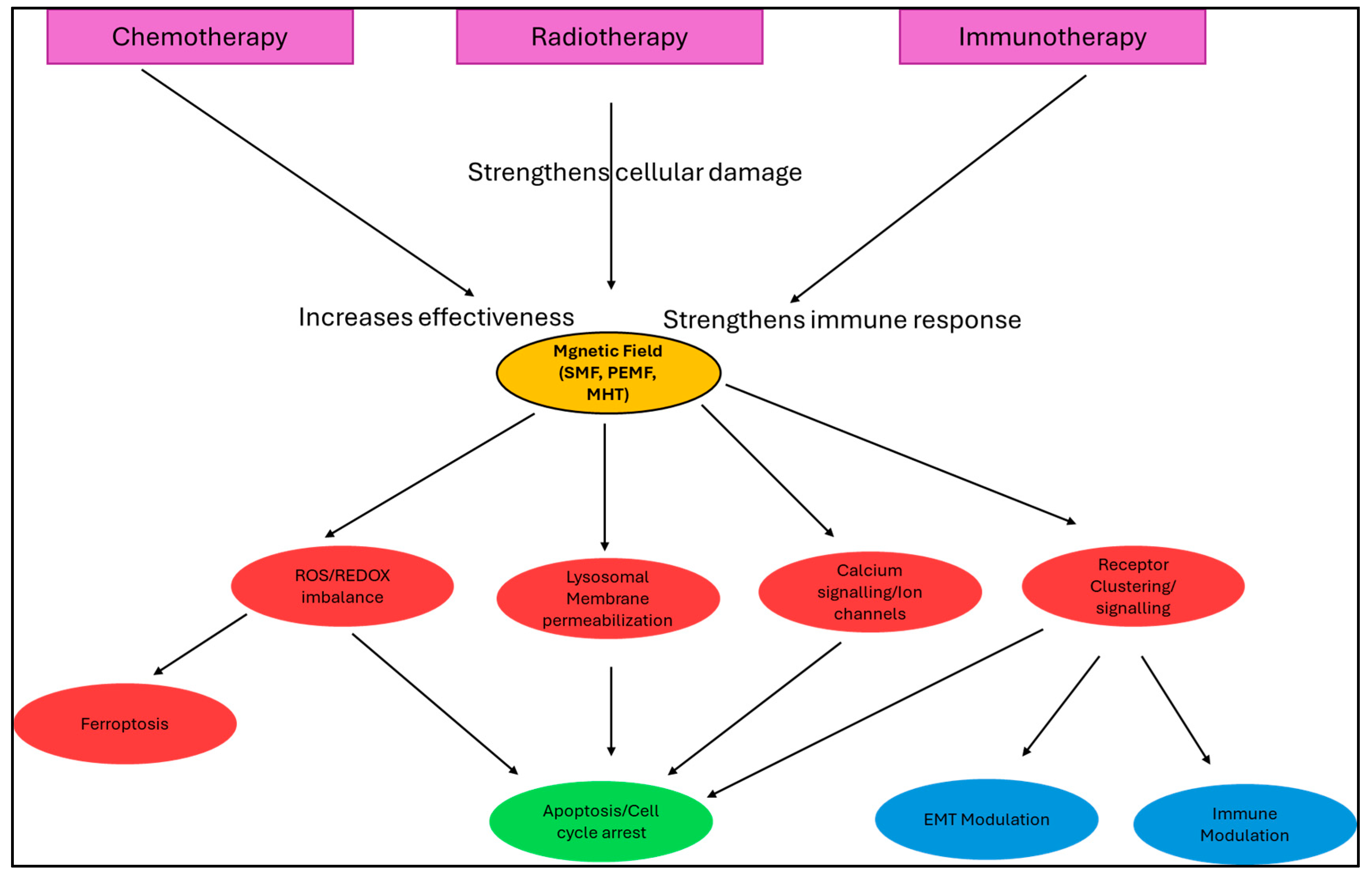
Figure 2.
An overview of the mechanisms by which magnetic fields influence cancer cells and their potential integration with existing cancer therapies [20,41,42,43,44,45,46,47,48].
2.1. Proliferation and Cell-Cycle Regulation
Many primary studies report that magnetic-field effects on proliferation are parameter- and cell-type dependent. Moderate-to-strong static magnetic fields (SMFs) have been shown to alter proliferative signalling: exposure to 1 T SMF modulated the EGFR–Akt–mTOR axis and reduced proliferation in several cancer cell lines under specific conditions, an effect that depended on cell density and receptor expression [22]. Conversely, moderate SMFs (~150 mT) accelerated proliferation yet suppressed migration and telomerase activity in murine 4T1 breast cancer cells, illustrating how the same nominal field can produce divergent outcomes depending on biological context [21]. Together these studies underscore that claims about “SMF increases or decreases proliferation” must be qualified by field strength, exposure regimen, and cell type [22]. In MCF-7 cells, exposure to 0.1–1 mT ELF-MF increased proliferation via ERK activation [49,50]. In contrast, stronger fields promoted apoptosis, underscoring safety concerns.
2.2. Apoptosis, Autophagy and Ferroptosis
Apoptosis and autophagy are frequent readouts in EMF studies and often reflect non-thermal (PEMF/SMF) or nanoparticle-mediated thermal/mechanical mechanisms. Pulsed electromagnetic fields (PEMFs) can potentiate chemotherapeutic apoptosis: for example, PEMF co-exposure enhanced etoposide-induced ROS accumulation, DNA damage and caspase-dependent apoptosis in MCF-7 cells [38]. Separately, magnetically actuated nanoparticles under alternating magnetic fields (AMF/MHT) elicit thermal and microthermal stresses that activate apoptosis or autophagy depending on dose, particle loading and thermal history [51,52]. Importantly, emerging work shows that magnetic strategies can also trigger ferroptosis-like death via MF-boosted Fenton chemistry or nanoparticle iron release, providing an orthogonal cell-death route particularly relevant when iron-containing carriers are used [53].
2.3. Oxidative Stress (ROS) and Redox Homeostasis
Modulation of reactive oxygen species (ROS) is a recurrent mechanistic thread linking diverse MF exposures to downstream outcomes. PEMF and SMF regimens have been reported to increase intracellular ROS in cancer cells, amplifying DNA damage and apoptotic signalling in supportive contexts, while other exposure paradigms elicit adaptive antioxidant responses instead—again highlighting parameter sensitivity [38]. In nanoparticle-assisted AMF treatments, localized heating and/or nanoparticle degradation further elevate labile iron and ROS production, which can potentiate lipid peroxidation and ferroptotic pathways when combined with disrupted antioxidant defence [51,53].
Recent studies show that MF-induced calcium influx via voltage-gated and mechanosensitive channels exacerbates mitochondrial depolarization, thereby accelerating ROS generation [54]. ROS can directly trigger lysosomal membrane permeabilization (LMP) through lipid peroxidation, linking calcium signalling, oxidative stress, and lysosomal instability into a single interconnected cascade [55].
2.4. Calcium Signalling, Ion Channels and Membrane Perturbation
Several primary studies directly connect low-frequency EMFs and PEMFs to changes in intracellular Ca2+ dynamics and ion-channel function. ELF/PEMF exposures have been shown to increase cytosolic Ca2+ transients and activate Ca2+-sensitive signalling (FAK/Rho GTPases, MAPKs) that regulate migration, differentiation and cell fate in multiple cell types [56,57]. Mechanistically, these calcium effects may arise from modulation of TRP channels, voltage-gated channels, or altered membrane receptor clustering; in cancer cells, such perturbations can shift the balance between proliferation, apoptosis and motility depending on exposure parameters [56].
Magneto-mechanical forces on membranes may activate Piezo1/TRPV channels, causing Ca2+ influx [58,59]. Moreover, disruption of integrin clustering by mechanical perturbation alters downstream signalling, providing a mechanistic link between LMP, ion fluxes, and receptor signalling [60,61].
2.5. Migration, Invasion, and EMT Modulation
Magnetic fields can alter cytoskeletal organization and adhesive behaviour, producing anti-metastatic phenotypes in some models. For example, rotating low-frequency magnetic fields (0.1–0.4 T at ~4.2 Hz) directly destabilized F-actin and reduced migration, invasion and metastatic colonization of breast cancer cells in vitro and in vivo, consistent with a magnetomechanical mode of action [62]. Likewise, moderate SMF exposure (≈150 mT) suppressed migration and telomerase function in 4T1 cells, despite concurrent proliferative effects, illustrating that migration/EMT readouts frequently diverge from proliferation endpoints under the same MF regimen [21]. These studies suggest magnetic interventions may be particularly useful to target motility and metastatic processes, but emphasize the need to couple EMT/migration assays with detailed parameter reporting [62].
3. Mechanistic Models of Magnetic Field Action in Cancer Research
Building upon the basic interactions between magnetic fields and cellular components described above, the following section discusses their impact on cell-cycle regulation and survival pathways. Several mechanistic models have been proposed to explain how magnetic fields (MFs) exert their effects on cancer cells. One prominent hypothesis involves reactive oxygen species (ROS)-mediated damage. MFs, particularly when combined with iron or magnetic nanoparticles (MNPs), can enhance ROS production, leading to oxidative damage of DNA and proteins. This in turn can trigger apoptosis, ferroptosis, or cellular senescence [63,64]. Another mechanism is based on magneto-mechanical effects, where oscillating magnetic fields acting on MNPs generate physical forces that stress cellular membranes or intracellular organelles, disrupt the cytoskeleton, and ultimately alter cell viability or motility [65]. A third proposed mechanism involves ion-channel modulation and calcium signaling, as magnetic fields may influence the gating of voltage-gated or T-type calcium channels. Such alterations can propagate downstream effects on signal transduction and transcriptional regulation [50]. Finally, the most well-understood mechanism is the thermal effect. Alternating magnetic fields cause MNPs to generate localized heating, producing hyperthermia (≥41 °C), which leads to protein denaturation, membrane disruption, and cell death [66]. Importantly, magnetic hyperthermia also exerts immunomodulatory effects and represents the clearest, dose-dependent, and physically characterized mechanism, one that has already advanced into clinical application and trials.
High-frequency AMFs (typically in the 100–300 kHz range) are optimized for magnetothermal effects in magnetic hyperthermia (Table 1). This frequency range balances safety with heating efficiency, as reflected in specific absorption rates (SAR) and the Rosensweig power equation [67]. For instance, some studies report substantial heat generation in Fe3O4 nanoparticle suspensions when exposed to 100 kHz fields, especially at field amplitudes of a few millitesla [67,68].
Table 1.
Summary of key magnetic field types, their primary physical parameters, and associated biological or mechanistic outcomes. AMF (alternating magnetic field, MHT: magnetic hyperthermia therapy).
Strong field gradients (greater than 10 T/m) substantially enhance MM forces, such as nanoparticle displacement or membrane stress. In diabetic mouse models, highly non-uniform SMFs with gradients > 10 T/m induced increased ROS and apoptosis, while uniform SMFs did not elicit such effects [69]. Theoretical frameworks similarly suggest that gradients may alter transmembrane potentials or ion-channel function via mechanical stress [65].
The choice of waveform, frequency, and duty cycle can bias biological outcomes. For example, PEMFs at 8 Hz and 11 mT selectively induced apoptosis in breast cancer cells while sparing fibroblasts [23]. Low-frequency PEMFs (1 mT) reduced ROS and inflammatory cytokine levels, whereas higher frequencies (6–7 mT) increased ROS, DNA damage, and apoptosis, illustrating parameter-dependent effects on cell fate [70].
3.1. ROS-Mediated Damage
One of the most frequently proposed models of magnetic field (MF) action in cancer biology involves the modulation of reactive oxygen species (ROS) [71]. Exposure to pulsed electromagnetic fields (PEMFs), extremely low-frequency electromagnetic fields (ELF-EMFs), or magnetic nanoparticle-mediated alternating magnetic fields has been shown to increase intracellular ROS production [72]. This elevation in ROS can overwhelm the antioxidant defense systems of cancer cells, which are often already under oxidative stress due to rapid proliferation and metabolic reprogramming [73]. The resulting oxidative damage to DNA, lipids, and proteins can trigger apoptosis, ferroptosis, or permanent cell-cycle arrest. Importantly, because malignant cells are more vulnerable to redox imbalance than healthy cells, this mechanism suggests a potential therapeutic window for selective cancer targeting. At the same time, excess ROS can also promote mutations and genomic instability, raising the possibility that magnetic field exposure might have dual outcomes depending on the context and duration of exposure. Whether ROS drives apoptosis or tumorigenesis depends on dose, duration, and repair capacity. Transient bursts promote cell death, whereas chronic sub-lethal ROS foster mutagenesis and adaptive resistance [74,75]. Thus, understanding how MF exposure balances between cytotoxicity and tumor-promoting mutagenesis is crucial for its translation into cancer therapy [76,77,78].
3.2. Ferroptosis and Redox Integration
Magnetic fields (MFs) influence the redox state of cancer cells by altering radical pair lifetimes and modifying mitochondrial electron flow [73]. When iron is present—either as endogenous labile Fe2+ or in the form of superparamagnetic iron oxide nanoparticles (SPIONs)—Fenton chemistry intensifies, amplifying lipid peroxidation and pushing cells toward ferroptosis [79]. The ferroptosis threshold is tightly regulated by factors such as GPX4, FTH1, SLC7A11, ACSL4, and intracellular iron pools. In this context, MF exposure can create parameter regimes where reactive oxygen species (ROS) accumulate without sufficient NRF2-driven antioxidant compensation, thereby sensitizing tumors to chemotherapy, radiotherapy, or magnetic hyperthermia (MHT) [80,81,82]. Conversely, robust NRF2 or HO-1 induction may blunt cytotoxicity and promote resistance. Practical biomarkers for monitoring these processes include oxidative DNA lesions (8-oxo-dG), lipid peroxidation products (4-HNE, MDA), mitochondrial membrane potential (Δψm), glutathione ratios (GSH/GSSG), BODIPY-C11 oxidation, and measures of transferrin receptor or labile iron pool levels [71,83,84]. These readouts can help stratify tumor types most vulnerable to MF-driven ferroptosis.
3.3. Lysosomal Membrane Permeabilization (LMP) via Magneto-Mechanics
A unique aspect of SPION-based therapies is their preferential accumulation within endo-lysosomal compartments [85]. Under oscillating or alternating magnetic fields, these nanoparticles can undergo mechanical torque or translation, physically stressing lysosomal membranes [20]. This local mechanical disruption triggers lysosomal membrane permeabilization (LMP), releasing cathepsins into the cytosol and activating apoptotic cascades independent of bulk temperature rise [85]. This mechanism explains observations of rapid apoptosis even when average tissue heating is minimal, distinguishing mechanical MF effects from pure thermal damage [85]. LMP also synergizes with DNA-damaging therapies, providing a complementary cell-death pathway [86]. Experimental tracking can rely on galectin-3 puncta formation, loss of LysoTracker fluorescence, detection of cytosolic cathepsin B/L, and pharmacological rescue using lysosomal inhibitors such as bafilomycin A1 [87,88]. Together, these approaches underscore LMP as a distinct and therapeutically exploitable mechanism of MF-induced cytotoxicity.
3.4. Membrane Receptor Modulation and Signaling Dynamics
Another line of evidence suggests that magnetic fields may directly influence the organization and activity of membrane proteins, particularly receptors involved in survival and proliferation pathways [89]. Cancer cells rely heavily on receptor tyrosine kinases (RTKs), integrins, and G-protein-coupled receptors (GPCRs) to sense their microenvironment and sustain malignant signaling [90]. Magnetic fields, through physical effects on lipid bilayer fluidity or via the orientation of charged residues, have been reported to modulate receptor clustering and downstream signaling cascades [91]. For example, PEMFs have been associated with altered activity of EGFR and integrin β1, leading to disruptions in focal adhesion signaling and PI3K/AKT pathway activation [92,93,94]. Such effects can impair cell adhesion, migration, and metastatic potential, while sensitizing tumor cells to conventional therapies. However, the same mechanisms may also induce adaptive responses, including compensatory activation of alternative receptors or pathways, which could contribute to treatment resistance. This dual potential highlights the importance of dissecting receptor-specific effects when integrating MF-based interventions into oncology.
3.5. Calcium Signaling and Ion Channel Regulation
Magnetic fields are also hypothesized to affect the function of ion channels, particularly those regulating calcium flux across cellular membranes [95,96]. Calcium is a master regulator of diverse cellular processes, including proliferation, apoptosis, and migration [97]. Evidence suggests that ELF-EMFs and PEMFs can alter intracellular calcium dynamics by modulating voltage-gated calcium channels, transient receptor potential (TRP) channels, or store-operated calcium entry (SOCE) mechanisms [56,98,99]. In cancer cells, where calcium homeostasis is often dysregulated to favor survival and invasion, MF-induced perturbations can tip the balance toward cell death or differentiation [100]. For instance, enhanced calcium influx may activate pro-apoptotic calpain or caspase pathways, while disrupting cytoskeletal organization critical for invasion [101]. Conversely, under certain conditions, increased calcium signaling might promote proliferation and EMT, underscoring the context-dependent nature of MF effects [102]. Thus, calcium-mediated pathways represent both an opportunity and a caution in leveraging MF exposure, demanding precise control over exposure parameters to maximize therapeutic benefit.
3.6. Immune Modulation and Tumor Microenvironment Interactions
Beyond direct effects on tumor cells, magnetic fields may also influence the tumor microenvironment (TME) and immune surveillance [2]. Tumor-associated macrophages, dendritic cells, and T lymphocytes have all been shown to respond to EMFs through altered cytokine secretion and activation states [103,104,105]. For instance, exposure to PEMFs can increase pro-inflammatory cytokines such as TNF-α and IL-6, enhancing anti-tumor immune responses, while also modulating the phagocytic activity of macrophages [106,107,108]. Similarly, magnetic nanoparticle-based hyperthermia has been reported to induce immunogenic cell death, releasing damage-associated molecular patterns (DAMPs) that activate dendritic cells and prime cytotoxic T cells [109]. This suggests that MFs may synergize with immune checkpoint inhibitors or adoptive T-cell therapies by reshaping the TME into a more immunostimulatory state. Nonetheless, inappropriate or chronic magnetic field exposure may drive macrophage polarization toward an M2-like, tumor-promoting state and potentiate immunosuppressive signaling within the tumor microenvironment, thereby counteracting anti-tumor immunity [110]. Therefore, immune modulation represents one of the most promising—yet complex—dimensions of MF-based cancer therapy, offering opportunities for combination strategies with immunotherapy.
Exposure of macrophages or macrophage co-cultures to pulsed electromagnetic fields or low-frequency magnetic fields has been shown to modulate polarization toward anti- or pro-inflammatory phenotypes depending on the MF parameters. For example, Vinhas et al. (2020) showed that a PEMF exposure in co-culture of IL-1β-primed tendon cells with THP-1-derived macrophages increased expression of the anti-inflammatory marker IL-10, upregulated M2-associated genes (like MRC-1) and downregulated pro-inflammatory markers such as NOS-2 and TNF-α [111].
On the other hand, in “Innate Immune Regulation Under Magnetic Fields with Possible Mechanisms and Therapeutic Applications” (2021), various studies are summarized in which low-frequency alternating magnetic fields (LF-MFs) increase pro-inflammatory cytokines (e.g., IL-1β, IL-6, TNF-α) in RAW264.7 or bone marrow-derived macrophages, depending on field strength and frequency. Moderate or high gradient magnetic fields (HGMFs), by contrast, tend to bias macrophage polarization toward the anti-inflammatory M2 phenotype, often associated with increased expression of IL-10 and other “repair/regulatory” markers [112].
These findings suggest that subtle differences in MF amplitude, frequency, exposure duration, and/or gradient can determine whether macrophages adopt tumor-promoting (M2-like) or tumor-suppressive (M1-like) roles. Mechanistically, changes in signaling pathways like STAT6 upregulation and suppression of STAT1 have been implicated in the M2 polarization under certain MF conditions [112].
4. Experimental and In Vitro Evidence
Experimental evidence from in vitro models further supports the potential of MFs in oncology. For example, pulsed electromagnetic fields (PEMFs) have been shown to potentiate chemotherapy. In MCF-7 breast cancer cells, PEMF exposure enhanced etoposide-induced cell death through a mechanism involving ROS accumulation, DNA damage, and activation of caspase-dependent apoptotic pathways [23,38]. Another consistent line of evidence comes from magnetic nanoparticle–mediated hyperthermia, where iron oxide nanoparticles exposed to alternating magnetic fields reliably induce cell death through localized heating. Numerous studies have demonstrated that this approach not only kills cancer cells directly but also sensitizes them to radiotherapy and chemotherapy, while imaging-guided systems further improve spatial precision and safety [25]. In contrast, the effects of static magnetic fields (SMFs) appear more variable and highly intensity-dependent (Table 2). Some studies report that specific SMF intensities can inhibit proliferation and suppress epithelial-to-mesenchymal transition (EMT), while others observe negligible or even stimulatory effects. High-field exposures in the tesla range tend to produce distinct responses compared to lower-intensity exposures, highlighting the complexity of MF–cell interactions [113,114].
Table 2.
Summary of magnetic field-based modalities in cancer therapy. The table lists the typical frequency or field strength, delivery methods, primary mechanisms of action, and current clinical status of each approach. Abbreviations: MHT, magnetic nanoparticle hyperthermia; AMF, alternating magnetic field; PEMF, pulsed electromagnetic fields; SMF, static magnetic fields.
5. Translational Applications
Among translational applications, magnetic hyperthermia (MHT) remains the most mature and clinically advanced [11]. Preclinical and clinical studies have shown that MNPs activated by alternating magnetic fields can induce local tumor regression, with applications currently being explored in breast, prostate, and brain cancers, among others. Advances in imaging-guided MHT and the development of multifunctional nanoparticles for combined therapeutic and diagnostic (“theranostic”) purposes are further expanding the scope of this approach [25]. In addition, PEMF and low-frequency electromagnetic devices are under active investigation. Several experimental systems are being designed to selectively inhibit tumor growth or to enhance tumor sensitivity to conventional drugs [119]. Early preclinical studies have produced encouraging results; however, successful clinical translation will require careful standardization of exposure parameters, improved understanding of mechanisms, and comprehensive safety evaluation [120].
On the biophysical side, several well-established logical tests can be applied to distinguish between magnetomechanical (MM) and magnetothermal (MT) effects. The following measures provide a systematic approach for resolving this distinction.
First, according to the principle of macro-scale heat generation, if bulk suspension temperature measurements—obtained Via calorimetry or thermal imaging—show a significant rise, MT effects are likely responsible for the observed outcomes [121]. Conversely, if cell death occurs without a corresponding bulk temperature increase, this suggests MM contributions (e.g., localized mechanisms or surface heating).
Following the evaluation of macro thermal changes, the particle immobilization (gelation) test can be employed. When the same particles are immobilized in a viscous gel, disappearance of the AC-field-induced effect indicates a predominant MM (Brownian) contribution. In contrast, persistence of thermal elevation under these conditions reveals a dominant MT (Néel) mechanism [122]. Because this strategy has been consistently applied in numerous studies, it is now considered a standardized and essential diagnostic test [123,124,125,126].
Thirdly, the test for AC susceptibility () measurement can be performed in conjunction with frequency scanning. In AC magnetic susceptibility (, ) measurements, the loss component typically exhibits a peak frequency () that is related to the relaxation time. In the event that the peak frequency is in close proximity to the applied frequency and the relationship is observed, this result is indicative of an MT (LRT) effect [127,128,129].
Furthermore, the fourth test is examined using low-frequency (Hz–kHz) application and imaging (live confocal). This test, conducted at low frequencies (10–100 Hz) and corresponding magnetic field strengths, suggests a high probability of MM effect in response to observed intracellular aggregate/lysosome movement/rupture. Also, Néel relaxation is ineffective because energy loss at these frequencies is negligible. However, when intracellular lysosomal movement, membrane deformation or rupture is observed, energy transfer occurs not thermally but through direct magnetomechanical (MM) interactions [123,130,131,132].
Immobilization and temperature-sensitive biomarkers (e.g., ROS generation vs. mechanical fragments) have been proposed in the literature as a fifth diagnostic test for distinguishing between MT and MM effects. Predominance of thermal stress markers—such as heat shock proteins (HSPs) or a generalized heat response—indicates an MT-driven mechanism. In contrast, evidence of oxidative stress, lysosomal leakage, or mechanical membrane rupture supports the involvement of MM processes [133,134,135].
The field dependence test (sixth test) is generally scalable with for both MM and MT effects. However, aggregation, magnetic interactions, and non-linear behaviour exhibit divergent H thresholds in the MM case. Consequently, simultaneous frequency/field scanning and immobilisation testing are mandatory [19,136,137,138,139].
5.1. Magnetic Hyperthermia Therapy (MHT)
Magnetic hyperthermia therapy (MHT) exploits the ability of superparamagnetic iron oxide nanoparticles (SPIONs) to generate localized heat when exposed to an alternating magnetic field (AMF). This effect arises from Néel and Brownian relaxation losses, which lead to the dissipation of thermal energy in the tumor microenvironment, thereby inducing apoptosis, necrosis, or sensitization to chemotherapy and radiotherapy [140,141,142]. Clinical translation of MHT has been most successful in glioblastoma multiforme (GBM), where the FDA-approved NanoTherm® therapy demonstrated improved survival when used in combination with radiotherapy [141,143]. Prostate cancer trials have similarly shown promising safety and efficacy. Advances in theranostic nanoparticle design now allow SPIONs to serve dual functions: acting as contrast agents for magnetic resonance imaging (MRI) while simultaneously functioning as localized heat sources. Moreover, integration with image-guided delivery systems enhances spatial precision and minimizes off-target effects, making MHT a powerful adjunct to conventional cancer therapies [85]. These clinical and preclinical successes highlight the importance of understanding the physical principles underlying MHT and optimizing experimental parameters [140].
The physical concept underlying MHT studies are based on Rosensweig power calculations [129]. In small-angle () and linear response assumptions, the average power per unit volume (suspension) is given by,
Here, is the permeability of the vacuum, the expression ,
is the effective relaxation time (below), and is the low-frequency sensitivity. For independent SPIONs, is , and (: volume fraction, : single particle volume) is given; this Equation (2) reduce to
From Equation (3) it is observable that relaxation mechanisms operate in two modes: while it is defined as Brownian relaxation caused by metal nanoparticles rotating around their own axis as first mode. Second mode is known Néel relaxation, which occurs when the external magnetic field effect is lost, and magnetic moment/internal spin motion [144]. Néel relaxation is defined as,
Here, is called characteristic relaxation time [145,146] and takes a value in the . Brownian relaxation time is calculated from,
Overall, final relaxation time can be calculated,
using these two relaxation times. These calculations allow researchers to predict and optimize the effects of experimental parameters, such as particle size, field strength, and frequency, on MHT efficacy. Ultimately, this approach ensures the effective and safe application of MHT in both clinical and preclinical settings [147,148]. The relaxation times derived from the calculations are utilised in the particle immobilisation test, which is then presented in comparison with Néel and Brownian.
5.2. Magnetic Drug Targeting (MDT)
Magnetic drug targeting (MDT) represents another application of magnetic nanoparticles, where drug-loaded SPIONs are guided to tumor sites using external magnetic fields. Early feasibility studies demonstrated that mitoxantrone, an anthracenedione chemotherapeutic, could be magnetically localized in experimental tumor models, thereby increasing intratumoral accumulation while reducing systemic toxicity [149]. This approach has since been expanded to a variety of drug–nanoparticle conjugates, with evidence showing enhanced therapeutic indices and reduced off-target cytotoxicity [150]. Despite these encouraging results, the primary limitation of MDT remains the depth and precision of magnetic field penetration in human tissues, restricting its practical application largely to superficial or organ-confined tumors. For typical permanent magnet arrays, clinically achievable targeting depths are limited to ~2–3 cm [151]. Advanced superconducting or Halbach array designs may extend this to ~5 cm, but remain technically challenging. Ongoing research on implantable magnetic field-generating devices and advanced magnet arrays shows potential to overcome these limitations and improve clinical translatability [152].
From a physical perspective, MDT is conceptualised based on classical magnetic force ,
calculations for magnetic targeting (the magnetic force exerted on a nanoparticle) as linear regime (non-saturation) . Here, is called susceptibility difference between in-phase and out off-phase . In the presence of magnetic saturation, and Equation (7) simplifies to,
Equation (8) stokes drag is also calculated using the force, , when there is flow towards the target. The condition for the capture process, which is required to ensure target adhesion, is [153,154]. These equations provide a quantitative framework for designing and optimizing MDT experiments, enabling precise targeting of therapeutic nanoparticles while minimizing off-target distribution.
Overall, these physical principles provide a quantitative foundation for optimizing MDT strategies. By carefully selecting nanoparticle properties, magnetic field strengths, and exposure geometries, researchers can maximize intratumoral accumulation while minimizing systemic dispersion. Such optimization is crucial for translating MDT from preclinical models to human patients, particularly for tumors located at challenging depths or within organs. Continued advances in nanoparticle engineering, field-guiding devices, and real-time imaging modalities promise to enhance the precision, safety, and therapeutic efficacy of MDT, potentially establishing it as a versatile complement to conventional cancer treatments.
5.3. Magneto-Mechanical Approaches
Beyond thermal and targeting strategies, magneto-mechanical therapy leverages the torque and oscillatory forces exerted by magnetic nanoparticles under an alternating or rotating magnetic field. These mechanical forces can induce cell membrane rupture, disrupt cytoskeletal architecture, and trigger apoptotic pathways, representing a distinct cytotoxic mechanism independent of hyperthermia [141]. Preclinical studies have demonstrated that oscillating magnetic fields acting on anisotropic nanoparticles, such as nanorods or nanodiscs, can selectively kill cancer cells while sparing healthy counterparts [140,141]. Although still in the experimental stage, these approaches are particularly appealing because they offer an orthogonal strategy to conventional thermal methods and may be especially useful for overcoming resistance to hyperthermia or chemotherapeutic treatments.
The fundamental equation of torque is utilised in these magneto-mechanical calculations, which is defined as classically based on the magnetic moment . In order to solve for the rotational friction coefficient (assumed sphere, ) in a viscous medium, as outlined in , it is first necessary to assume that the magnetic field B required to synchronously drive the rotational at the maximum stable angular velocity is calculated and then used as [42,155,156,157]. These calculations provide a quantitative basis for designing magneto-mechanical therapies, allowing researchers to predict the required field strengths and frequencies to achieve effective cytotoxic mechanical forces.
5.4. Pulsed Electromagnetic Field (PEMF) Devices and Low-Frequency Applications
In addition to nanoparticle-mediated approaches, pulsed electromagnetic fields (PEMFs) and low-frequency electromagnetic fields have been investigated for their direct biological effects on tumor cells. Preclinical studies have reported that exposure to PEMFs can reduce cancer cell proliferation, promote apoptosis, and sensitize tumor cells to chemotherapeutic agents such as doxorubicin and cisplatin [158,159]. These effects are thought to be mediated through modulation of reactive oxygen species (ROS), intracellular calcium signaling, and p53 activation (Table 3), (Figure 3) (see Supplementary Materials). Despite encouraging laboratory data, translation into standardized clinical protocols remains limited by the lack of consensus on optimal exposure parameters, including frequency, intensity, and treatment duration [160]. The development of precise dose–response maps and mechanistic biomarkers will be crucial for advancing PEMF therapies into mainstream oncology.
Figure 3.
Magnetic Field Effects on Cancer Cell Lines. The dot bubble graph summarizes the observed effects of magnetic field exposure on various biological modalities across different cancer cell lines. X-axis represents the cancer cell lines (MCF-7, HeLa, U87, GL261). Y-axis represents the biological modalities [38,161,162,163,164,165]. ↑: upregulation, ↓: downregulation.
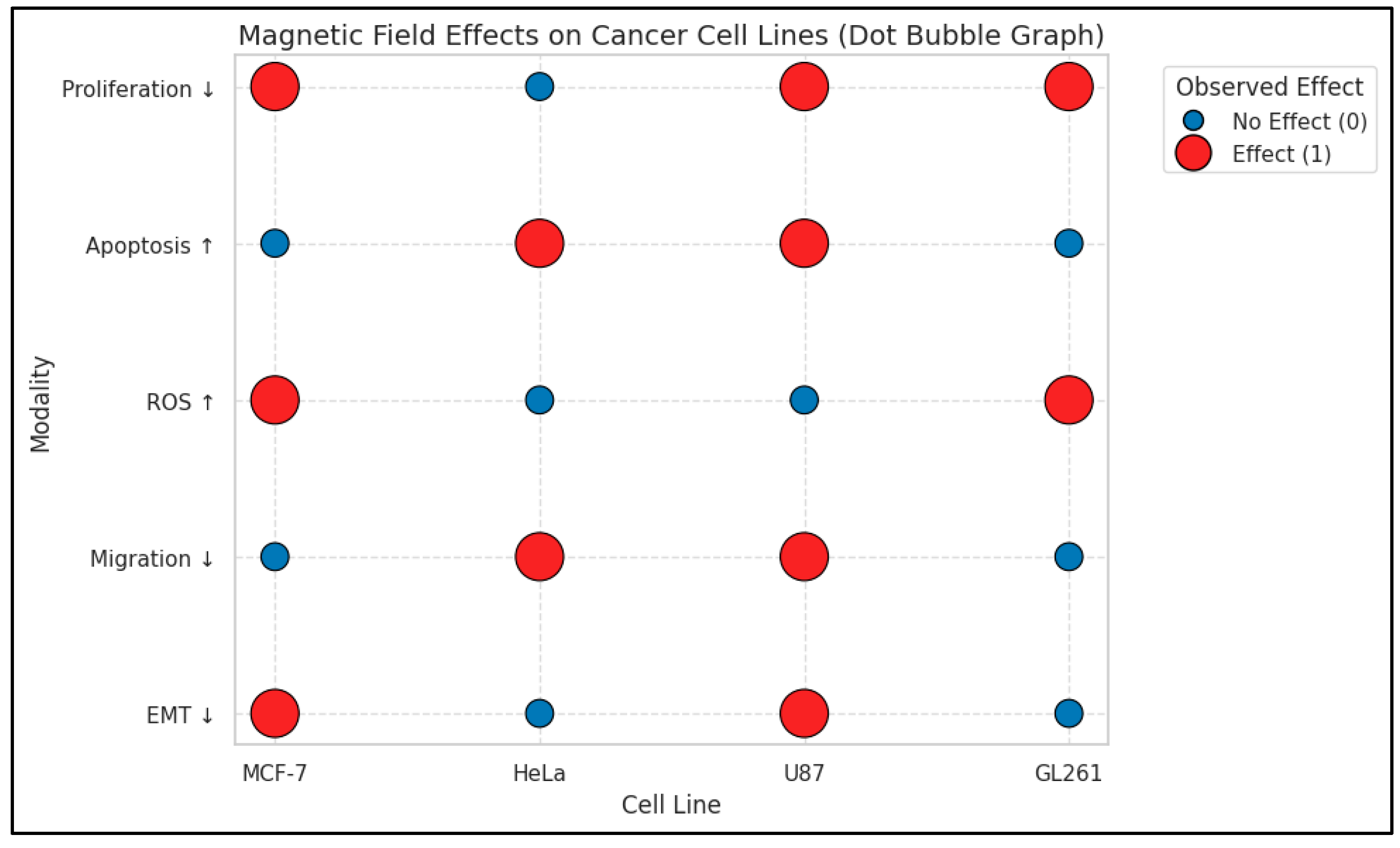
Table 3.
In vitro studies of magnetic field effects on cancer cells.
6. Clinical Translation and Trials
6.1. Recurrent Glioblastoma
The largest prospective experience comes from intratumoral iron-oxide nanoparticle thermotherapy combined with fractionated stereotactic radiotherapy. In a two-center, single-arm study of 66 patients (of whom 59 had recurrent glioblastoma), median OS from first recurrence was 13.4 months (95% CI 10.6–16.2), with moderate side effects and no serious complications reported. A prior feasibility study established procedural safety, temperature monitoring, and local control signals [13].
Ongoing work aims to optimize applicators and field delivery and to integrate MHT with modern chemoradiation and immunotherapy. Selected updates and reviews summarize device advances and combination strategies [140,169].
6.2. Prostate Cancer
Interstitial thermotherapy for prostate cancer has been explored in phase I settings, demonstrating feasibility, patient-specific 3-D thermal planning, and the need to manage discomfort at higher field strengths; early studies also integrated brachytherapy [14].
6.3. Human Feasibility of Magnetic Drug Targeting
The first-in-human MDT trial administered intra-arterial mitoxantrone with magnetic targeting.
In advanced solid tumors, demonstrating selective localization and feasibility; this remains the most often cited human MDT feasibility evidence [170] (Table 4).
Table 4.
Key clinical studies using magnetic fields in cancer therapy.
Magnetic nanoparticle hyperthermia is a cancer treatment where tiny iron particles are placed into a tumor and then heated by an alternating magnetic field. The heat can damage cancer cells or make them more sensitive to treatments like radiotherapy.
It has been tested mainly in glioblastoma (a type of brain cancer) and prostate cancer. Early studies showed it is safe and can improve survival compared with older data. Larger registries and clinical trials are still running to confirm how well and how safely it works (Table 4).
7. Limitations
While MHT has reproducible human data in glioblastoma and prostate feasibility settings, larger randomized trials are limited. Standardization of field parameters, nanoparticle dosing, and reporting will be key for cross-study comparisons. The requirement for high-field gradient magnets, RF coils, and cooling systems significantly raises costs. Device miniaturization and energy efficiency remain barriers for clinical adoption [174]. For PEMF, parameter sensitivity (frequency, amplitude, duty cycle) and tumor-specific biology likely explain divergent results; robust dose–response mapping and biomarker-guided designs (e.g., DDR competence, lysosomal stability) are priorities. Magneto-mechanical strategies are compelling but remain preclinical; translation will hinge on targeting, field penetration, and safety [175].
Long-term or high-intensity exposures may have off-target effects; while magnetic hyperthermia targets tumors, systemic MNP biodistribution, long-term iron handling, and heating of non-target tissue require careful preclinical toxicology [25]. Regulatory assessment must consider both electromagnetic exposure and nanoparticle safety.
From the perspective of classical electromagnetic physics, in the context of alternating magnetic fields (AMFs), the generation of eddy currents within conductive tissues arises directly from Faraday’s law. The tissue’s electrical conductivity (σ), together with the frequency (f) and amplitude (H) of the magnetic field, plays a critical role in determining the strength of these currents. This interaction induces thermal effects, resulting in localized temperature elevation within the targeted tissue regions.
At elevated frequencies or large field amplitudes, the thermal consequences for the patient can become significant, potentially causing tissue damage, pain, burns, or even systemic effects [176]. Heat diffusion in this context is constrained by factors such as blood perfusion and the intrinsic thermal properties of tissues—specifically, electrical conductivity, specific heat capacity, and thermal conductivity.
From a safety standpoint, “non-critical” heating thresholds are applied, commonly defined as maintaining local tissue temperatures at 42–43 °C for a controlled duration and ensuring that increases in core body temperature remain within safe limits [122,177,178].
The Brezovich (Atkinson–Brezovich) limit, derived from experimental studies by Atkinson and Brezovich, specifies that H·f < 4.85 × 10⁸ A·m−1·s−1 [179,180]. In certain cases, this limit has been extended slightly to H·f < 5 × 10⁸ A·m−1·s−1 for regions of small volume or diameter. This guideline serves two primary purposes: (1) minimizing patient discomfort, burns, and systemic heat load during prolonged exposures; and (2) preventing both local and systemic tissue injury [176].
However, it is important to note that this limit is idealized, derived primarily from experiments on healthy individuals and designed for large-diameter body regions. In more localized applications—such as intracranial treatments or small tumor sites—higher H·f values may be tolerable if both the affected tissue volume and body region diameter are sufficiently small [181,182].
8. Future Directions
Magnetic field-based cancer therapies offer considerable promise, yet significant challenges remain before their widespread clinical adoption. Standardization of exposure parameters—including frequency, intensity, waveform, and duration—is critical to achieve reproducible and predictable biological effects across tumor types. Integrating magnetic field modalities with chemotherapy, radiotherapy, or immunotherapy may enhance therapeutic efficacy, but requires careful mechanistic understanding to prevent adaptive resistance. Advances in nanoparticle design and targeted delivery are needed to maximize tumor specificity, enable simultaneous imaging and therapy, and overcome limitations of tissue penetration. Elucidating mechanisms such as ROS modulation, lysosomal membrane permeabilization, receptor clustering, and calcium signaling will help identify predictive biomarkers (Potential predictive biomarkers include ROS scavenging enzymes (SOD2, GPX4) [183,184], lysosomal cathepsins, mechanosensitive ion channels (Piezo1, TRPV4 [185,186]), and macrophage polarization markers (CD163/CD206 vs. CD80/CD86) [187,188]) and responsive tumor subtypes. Additionally, optimizing magnetic field-induced immune modulation to enhance antitumor immunity while avoiding immunosuppressive polarization represents a promising avenue for combination with immunotherapies. Comprehensive safety evaluation—including nanoparticle biodistribution, off-target heating, and long-term effects—is essential, and regulatory frameworks must consider both electromagnetic exposure and nanoparticle safety. For example, Yarjanli et al. reported iron accumulation and oxidative stress in brain tissue 6 months post-SPION exposure [189]. Surface coatings (dextran, PEG) reduce but do not eliminate persistence. Beyond conventional superparamagnetic iron oxide nanoparticles (SPIONs), alternative iron-oxide-based nanoparticle classes present unique opportunities and challenges for in vivo biomedical applications. Natural magnetosomes—magnetite (Fe3O4) crystals produced by magnetotactic bacteria—exhibit stable single-domain magnetic behavior and very high specific absorption rates, enabling more efficient magnetic hyperthermia than many synthetic SPIONs of comparable size [190]. Their native lipid bilayer membrane confers excellent biodegradability and low cytotoxicity, but reproducibility and large-scale production remain difficult [191]. To address these hurdles, biomimetic iron oxide nanoparticles have been synthesized to mimic magnetosome size and composition (~40–60 nm), offering tunable surface chemistry and scalable fabrication strategies. For example, the use of magnetosome-associated proteins such as MamC or Mms6 enables precise control of particle morphology and magnetic properties [192], while polymersome-based approaches yield magnetosome-like vesicles with comparable intrinsic loss power for hyperthermia [193]. SPIONs, though the most widely studied and commercially available, often show lower heating efficiency and may aggregate in vivo, necessitating careful coating or clustering strategies to improve performance and biocompatibility [194]. A critical comparison of these nanoparticle classes underscores the need for standardized evaluation of magnetic behavior, immune compatibility, and long-term biodistribution to optimize their clinical translation. Finally, large, randomized clinical trials with standardized protocols and biomarker-guided patient stratification are needed to validate efficacy, reproducibility, and translational potential, ultimately enabling magnetic field-based interventions to become robust, clinically actionable cancer therapies.
Supplementary Materials
The following supporting information can be downloaded at: https://www.mdpi.com/article/10.3390/pharmaceutics18060742/s1, File S1: Dot bubble graph generation, Data source and preparation.
Author Contributions
A.B.B.E. and S.B.S.; writing the original draft, review & editing. All authors have read and agreed to the published version of the manuscript.
Funding
This study was supported by the Recep Tayyip Erdoğan University Development Foundation (Grant No: 02025011017760).
Conflicts of Interest
The authors declare no conflict of interest.
References
- Maffei, M.E. Magnetic Fields and Cancer: Epidemiology, Cellular Biology, and Theranostics. Int. J. Mol. Sci. 2022, 23, 1339. [Google Scholar] [CrossRef] [Scilit]
- Zhang, G.; Liu, X.; Liu, Y.; Zhang, S.; Yu, T.; Chai, X.; Yu, T.; Chai, X.; He, J.; Yin, D.; et al. The effect of magnetic fields on tumor occurrence and progression: Recent advances. Prog. Biophys. Mol. Biol. 2023, 179, 38–50. [Google Scholar] [CrossRef] [Scilit]
- Zhang, B.; Yuan, X.; Lv, H.; Che, J.; Wang, S.; Shang, P. Biophysical mechanisms underlying the effects of static magnetic fields on biological systems. Prog. Biophys. Mol. Biol. 2023, 177, 14–23. [Google Scholar] [CrossRef] [Scilit]
- Tang, W.; He, D.; Li, X.; Feng, Y.; Xu, Y.; Hu, J.; Xu, W.; Xue, L. Extremely low-frequency electromagnetic fields facilitate proliferation and functional differentiation in spinal neural stem cells. Sci. Rep. 2025, 15, 29291. [Google Scholar] [CrossRef] [Scilit]
- Hu, H.; Yang, W.; Zeng, Q.; Chen, W.; Zhu, Y.; Liu, W.; Wang, S.; Wang, B.; Shao, Z.; Zhang, Y. Promising application of Pulsed Electromagnetic Fields (PEMFs) in musculoskeletal disorders. Biomed. Pharmacother. 2020, 131, 110767. [Google Scholar] [CrossRef] [Scilit]
- Fatima, H.; Charinpanitkul, T.; Kim, K.S. Fundamentals to Apply Magnetic Nanoparticles for Hyperthermia Therapy. Nanomaterials 2021, 11, 1203. [Google Scholar] [CrossRef] [Scilit]
- Tota, M.; Jonderko, L.; Witek, J.; Novickij, V.; Kulbacka, J. Cellular and Molecular Effects of Magnetic Fields. Int. J. Mol. Sci. 2024, 25, 8973. [Google Scholar] [CrossRef] [Scilit] [PubMed]
- Chen, Y.; Hong, L.; Zeng, Y.; Shen, Y.; Zeng, Q. Power frequency magnetic fields induced reactive oxygen species-related autophagy in mouse embryonic fibroblasts. Int. J. Biochem. Cell Biol. 2014, 57, 108–114. [Google Scholar] [CrossRef] [Scilit] [PubMed]
- Habash, M.; Gogna, P.; Krewski, D.; Habash, R.W.Y. Scoping Review of the Potential Health Effects of Exposure to Extremely Low-Frequency Electric and Magnetic Fields. Crit. Rev. Biomed. Eng. 2019, 47, 323–347. [Google Scholar] [CrossRef] [Scilit]
- Ahmed, S.; Rajak, B.L.; Gogoi, M.; Sarma, H.D. Chapter 16—Magnetic nanoparticles mediated cancer hyperthermia. In Smart Healthcare for Disease Diagnosis and Prevention; Paul, S., Bhatia, D., Eds.; Academic Press: Cambridge, MA, USA, 2020; pp. 153–173. [Google Scholar] [CrossRef] [Scilit]
- Xiong, L.; Liang, B.; Yu, K. Magnetic hyperthermia in oncology: Nanomaterials-driven combinatorial strategies for synergistic therapeutic gains. Mater. Today Bio 2025, 33, 102070. [Google Scholar] [CrossRef] [Scilit] [PubMed]
- Sun, J.; Tong, Y.; Jia, Y.; Jia, X.; Wang, H.; Chen, Y.; Wu, J.; Jin, W.; Ma, Z.; Cao, K.; et al. Effects of extremely low frequency electromagnetic fields on the tumor cell inhibition and the possible mechanism. Sci. Rep. 2023, 13, 6989. [Google Scholar] [CrossRef] [Scilit]
- Maier-Hauff, K.; Ulrich, F.; Nestler, D.; Niehoff, H.; Wust, P.; Thiesen, B.; Orawa, H.; Budach, V.; Jordan, A. Efficacy and safety of intratumoral thermotherapy using magnetic iron-oxide nanoparticles combined with external beam radiotherapy on patients with recurrent glioblastoma multiforme. J. Neuro-Oncol. 2011, 103, 317–324. [Google Scholar] [CrossRef] [Scilit]
- Johannsen, M.; Thiesen, B.; Wust, P.; Jordan, A. Magnetic nanoparticle hyperthermia for prostate cancer. Int. J. Hyperth. 2010, 26, 790–795. [Google Scholar] [CrossRef] [Scilit]
- Johannsen, M.; Gneveckow, U.; Taymoorian, K.; Cho, C.H.; Thiesen, B.; Scholz, R.; Waldofner, N.; Loening, S.A.; Wust, P.; Jordan, A. Thermal therapy of prostate cancer using magnetic nanoparticles. Actas Urol. Esp. 2007, 31, 660–667. [Google Scholar] [CrossRef] [Scilit]
- Van Durme, R.; Crevecoeur, G.; Dupre, L.; Coene, A. Improved magnetic drug targeting with maximized magnetic forces and limited particle spreading. Med. Phys. 2023, 50, 1715–1727. [Google Scholar] [CrossRef] [Scilit] [PubMed]
- Ellis, J.A.; Cooke, J.; Singh-Moon, R.P.; Wang, M.; Bruce, J.N.; Emala, C.W.; Bigio, I.J.; Joshi, S. Safety, feasibility, and optimization of intra-arterial mitoxantrone delivery to gliomas. J. Neuro-Oncol. 2016, 130, 449–454. [Google Scholar] [CrossRef] [Scilit] [PubMed]
- Janko, C.; Durr, S.; Munoz, L.E.; Lyer, S.; Chaurio, R.; Tietze, R.; Lohneysen, S.; Schorn, C.; Herrmann, M.; Alexiou, C. Magnetic drug targeting reduces the chemotherapeutic burden on circulating leukocytes. Int. J. Mol. Sci. 2013, 14, 7341–7355. [Google Scholar] [CrossRef] [Scilit] [PubMed]
- Zhang, E.; Kircher, M.F.; Koch, M.; Eliasson, L.; Goldberg, S.N.; Renström, E. Dynamic Magnetic Fields Remote-Control Apoptosis via Nanoparticle Rotation. ACS Nano 2014, 8, 3192–3201. [Google Scholar] [CrossRef] [Scilit]
- Mi, Y.; Wang, J.; Tang, S.; Ma, C.; Zheng, W.; Chen, J. Decaying Oscillating Pulsed Magnetic Field Induces Lysosome-Dependent Cell Death in A375 Melanoma via Magneto-Mechanical Force. Magnetochemistry 2025, 11, 33. [Google Scholar] [CrossRef] [Scilit]
- Fan, Z.; Hu, P.; Xiang, L.; Liu, Y.; He, R.; Lu, T. A Static Magnetic Field Inhibits the Migration and Telomerase Function of Mouse Breast Cancer Cells. Biomed Res. Int. 2020, 2020, 7472618. [Google Scholar] [CrossRef] [Scilit]
- Zhang, L.; Wang, J.; Wang, H.; Wang, W.; Li, Z.; Liu, J.; Yang, X.; Ji, X.; Luo, Y.; Hu, C.; et al. Moderate and strong static magnetic fields directly affect EGFR kinase domain orientation to inhibit cancer cell proliferation. Oncotarget 2016, 7, 41527–41539. [Google Scholar] [CrossRef] [Scilit]
- Pantelis, P.; Theocharous, G.; Veroutis, D.; Vagena, I.-A.; Polyzou, A.; Thanos, D.-F.; Kyrodimos, E.; Kotsinas, A.; Evangelou, K.; Lagopati, N.; et al. Pulsed Electromagnetic Fields (PEMFs) Trigger Cell Death and Senescence in Cancer Cells. Int. J. Mol. Sci. 2024, 25, 2473. [Google Scholar] [CrossRef] [Scilit] [PubMed]
- Jin, K.; Zhao, D.; Zhou, J.; Zhang, X.; Wang, Y.; Wu, Z. Pulsed electromagnetic fields inhibit IL-37 to alleviate CD8(+) T cell dysfunction and suppress cervical cancer progression. Apoptosis 2024, 29, 2108–2127. [Google Scholar] [CrossRef] [Scilit]
- Szwed, M.; Marczak, A. Application of Nanoparticles for Magnetic Hyperthermia for Cancer Treatment—The Current State of Knowledge. Cancers 2024, 16, 1156. [Google Scholar] [CrossRef] [Scilit]
- Tsong, T.Y. Electroporation of cell membranes. Biophys. J. 1991, 60, 297–306. [Google Scholar] [CrossRef] [Scilit]
- Sözer, E.B.; Pocetti, C.F.; Vernier, P.T. Transport of charged small molecules after electropermeabilization—Drift and diffusion. BMC Biophys. 2018, 11, 4. [Google Scholar] [CrossRef] [Scilit]
- Mir, L.M.; Moller, P.H.; André, F.; Gehl, J. Electric pulse-mediated gene delivery to various animal tissues. Adv. Genet. 2005, 54, 83–114. [Google Scholar] [CrossRef] [Scilit] [PubMed]
- Kanthou, C.; Kranjc, S.; Sersa, G.; Tozer, G.; Zupanic, A.; Cemazar, M. The endothelial cytoskeleton as a target of electroporation-based therapies. Mol. Cancer Ther. 2006, 5, 3145–3152. [Google Scholar] [CrossRef] [Scilit]
- Cemazar, M. Effects of electroporation of mammalian cells on cytoskeleton and intercellular connections. In Handbook of Electroporation; Miklavcic, D., Ed.; Springer International Publishing: Cham, Switzerland, 2016; pp. 1–15. [Google Scholar] [CrossRef] [Scilit]
- Carr, L.; Bardet, S.M.; Burke, R.C.; Arnaud-Cormos, D.; Leveque, P.; O’Connor, R.P. Calcium-independent disruption of microtubule dynamics by nanosecond pulsed electric fields in U87 human glioblastoma cells. Sci. Rep. 2017, 7, 41267. [Google Scholar] [CrossRef] [Scilit] [PubMed]
- Kim, H.B.; Lee, S.; Chung, J.H.; Kim, S.N.; Sung, C.K.; Baik, K.Y. Effects of Actin Cytoskeleton Disruption on Electroporation In Vitro. Appl. Biochem. Biotechnol. 2020, 191, 1545–1561. [Google Scholar] [CrossRef] [Scilit]
- Rosazza, C.; Escoffre, J.M.; Zumbusch, A.; Rols, M.P. The actin cytoskeleton has an active role in the electrotransfer of plasmid DNA in mammalian cells. Mol. Ther. 2011, 19, 913–921. [Google Scholar] [CrossRef] [Scilit]
- Titushkin, I.; Cho, M. Regulation of Cell Cytoskeleton and Membrane Mechanics by Electric Field: Role of Linker Proteins. Biophys. J. 2009, 96, 717–728. [Google Scholar] [CrossRef] [Scilit]
- Graybill, P.M.; Davalos, R.V. Cytoskeletal Disruption after Electroporation and Its Significance to Pulsed Electric Field Therapies. Cancers 2020, 12, 1132. [Google Scholar] [CrossRef] [Scilit] [PubMed]
- Zhang, L.; Ji, X.; Yang, X.; Zhang, X. Cell type- and density-dependent effect of 1 T static magnetic field on cell proliferation. Oncotarget 2017, 8, 13126–13141. [Google Scholar] [CrossRef] [Scilit] [PubMed]
- Tong, J.; Sun, L.; Zhu, B.; Fan, Y.; Ma, X.; Yu, L.; Zhang, J. Pulsed electromagnetic fields promote the proliferation and differentiation of osteoblasts by reinforcing intracellular calcium transients. Bioelectromagnetics 2017, 38, 541–549. [Google Scholar] [CrossRef] [Scilit]
- Woo, S.H.; Kim, B.; Kim, S.H.; Jung, B.C.; Lee, Y.; Kim, Y.S. Pulsed electromagnetic field potentiates etoposide-induced MCF-7 cell death. BMB Rep. 2022, 55, 148–153. [Google Scholar] [CrossRef] [Scilit] [PubMed]
- Cellai, F.; Munnia, A.; Viti, J.; Doumett, S.; Ravagli, C.; Ceni, E.; Mello, T.; Polvani, S.; Giese, R.W.; Baldi, G.; et al. Magnetic Hyperthermia and Oxidative Damage to DNA of Human Hepatocarcinoma Cells. Int. J. Mol. Sci. 2017, 18, 939. [Google Scholar] [CrossRef] [Scilit]
- Sato, I.; Umemura, M.; Mitsudo, K.; Kioi, M.; Nakashima, H.; Iwai, T.; Feng, X.; Oda, K.; Miyajima, A.; Makino, A.; et al. Hyperthermia generated with ferucarbotran (Resovist®) in an alternating magnetic field enhances cisplatin-induced apoptosis of cultured human oral cancer cells. J. Physiol. Sci. 2014, 64, 177–183. [Google Scholar] [CrossRef] [Scilit]
- Kanber, M.; Umerah, O.; Brindley, S.; Zhang, X.; Brown, J.M.; Reynolds, L.; Beltran-Huarac, J. Magneto-Mechanical Actuation Induces Endothelial Permeability. ACS Biomater. Sci. Eng. 2023, 9, 6902–6914. [Google Scholar] [CrossRef] [Scilit]
- Goršak, T.; Jovičić, E.J.; Tratnjek, L.; Križaj, I.; Sepulveda, B.; Nogues, J.; Kreft, M.E.; Petan, T.; Kralj, S.; Makovec, D. The efficient magneto-mechanical actuation of cancer cells using a very low concentration of non-interacting ferrimagnetic hexaferrite nanoplatelets. J. Colloid Interface Sci. 2024, 657, 778–787. [Google Scholar] [CrossRef] [Scilit]
- Xu, X.; Xu, S.; Wan, J.; Wang, D.; Pang, X.; Gao, Y.; Ni, N.; Chen, D.; Sun, X. Disturbing cytoskeleton by engineered nanomaterials for enhanced cancer therapeutics. Bioact. Mater. 2023, 29, 50–71. [Google Scholar] [CrossRef] [Scilit]
- Wu, J.; Ning, P.; Gao, R.; Feng, Q.; Shen, Y.; Zhang, Y.; Li, Y.; Xu, C.; Qin, Y.; Plaza, G.R.; et al. Programmable ROS-Mediated Cancer Therapy via Magneto-Inductions. Adv. Sci. 2020, 7, 1902933. [Google Scholar] [CrossRef] [Scilit] [PubMed]
- Nikitin, A.A.; Ivanova, A.V.; Semkina, A.S.; Lazareva, P.A.; Abakumov, M.A. Magneto-Mechanical Approach in Biomedicine: Benefits, Challenges, and Future Perspectives. Int. J. Mol. Sci. 2022, 23, 11134. [Google Scholar] [CrossRef] [Scilit]
- Wu, X.; Zhang, H. Therapeutic strategies of iron-based nanomaterials for cancer therapy. Biomed. Mater. 2021, 16, 032003. [Google Scholar] [CrossRef] [Scilit] [PubMed]
- Garanina, A.S.; Efremova, M.V.; Machulkin, A.E.; Lyubin, E.V.; Vorobyeva, N.S.; Zhironkina, O.A.; Strelkova, O.S.; Kireev, I.I.; Alieva, I.B.; Uzbekov, R.E.; et al. Bifunctional Magnetite–Gold Nanoparticles for Magneto-Mechanical Actuation and Cancer Cell Destruction. Magnetochemistry 2022, 8, 185. [Google Scholar] [CrossRef] [Scilit]
- Naud, C.; Thébault, C.; Carrière, M.; Hou, Y.; Morel, R.; Berger, F.; Diény, B.; Joisten, H. Cancer treatment by magneto-mechanical effect of particles, a review. Nanoscale Adv. 2020, 2, 3632–3655. [Google Scholar] [CrossRef] [Scilit]
- Xu, A.; Wang, Q.; Lin, T. Low-Frequency Magnetic Fields (LF-MFs) Inhibit Proliferation by Triggering Apoptosis and Altering Cell Cycle Distribution in Breast Cancer Cells. Int. J. Mol. Sci. 2020, 21, 2952. [Google Scholar] [CrossRef] [Scilit]
- Barati, M.; Darvishi, B.; Javidi, M.A.; Mohammadian, A.; Shariatpanahi, S.P.; Eisavand, M.R.; Madjid Ansari, A. Cellular stress response to extremely low-frequency electromagnetic fields (ELF-EMF): An explanation for controversial effects of ELF-EMF on apoptosis. Cell Prolif. 2021, 54, e13154. [Google Scholar] [CrossRef] [Scilit]
- Lao, L.L.; Ramanujan, R.V. Magnetic and hydrogel composite materials for hyperthermia applications. J. Mater. Sci. Mater. Med. 2004, 15, 1061–1064. [Google Scholar] [CrossRef] [Scilit] [PubMed]
- Vilas-Boas, V.; Carvalho, F.; Espiña, B. Magnetic Hyperthermia for Cancer Treatment: Main Parameters Affecting the Outcome of In Vitro and In Vivo Studies. Molecules 2020, 25, 2874. [Google Scholar] [CrossRef] [Scilit]
- Yu, B.; Choi, B.; Li, W.; Kim, D.-H. Magnetic field boosted ferroptosis-like cell death and responsive MRI using hybrid vesicles for cancer immunotherapy. Nat. Commun. 2020, 11, 3637. [Google Scholar] [CrossRef] [Scilit]
- Franco-Obregón, A. Harmonizing Magnetic Mitohormetic Regenerative Strategies: Developmental Implications of a Calcium-Mitochondrial Axis Invoked by Magnetic Field Exposure. Bioengineering 2023, 10, 1176. [Google Scholar] [CrossRef] [Scilit]
- Aits, S.; Jäättelä, M. Lysosomal cell death at a glance. J. Cell Sci. 2013, 126, 1905–1912. [Google Scholar] [CrossRef] [Scilit]
- Ma, T.; Ding, Q.; Liu, C.; Wu, H. Electromagnetic fields regulate calcium-mediated cell fate of stem cells: Osteogenesis, chondrogenesis and apoptosis. Stem Cell Res. Ther. 2023, 14, 133. [Google Scholar] [CrossRef] [Scilit]
- Zhang, Y.; Yan, J.; Xu, H.; Yang, Y.; Li, W.; Wu, H.; Liu, C. Extremely low frequency electromagnetic fields promote mesenchymal stem cell migration by increasing intracellular Ca2+ and activating the FAK/Rho GTPases signaling pathways in vitro. Stem Cell Res. Ther. 2018, 9, 143. [Google Scholar] [CrossRef] [Scilit]
- Borbiro, I.; Badheka, D.; Rohacs, T. Activation of TRPV1 channels inhibits mechanosensitive Piezo channel activity by depleting membrane phosphoinositides. Sci. Signal. 2015, 8, ra15. [Google Scholar] [CrossRef] [Scilit] [PubMed]
- Verkest, C.; Lechner, S.G. Advances and recent insights into the gating mechanisms of the mechanically activated ion channels PIEZO1 and PIEZO2. Curr. Opin. Physiol. 2023, 31, 100625. [Google Scholar] [CrossRef] [Scilit]
- Chen, J.; Fabry, B.; Schiffrin, E.L.; Wang, N. Twisting integrin receptors increases endothelin-1 gene expression in endothelial cells. Am. J. Physiol. Cell Physiol. 2001, 280, C1475–C1484. [Google Scholar] [CrossRef] [Scilit]
- Wang, N.; Butler, J.P.; Ingber, D.E. Mechanotransduction across the cell surface and through the cytoskeleton. Science 1993, 260, 1124–1127. [Google Scholar] [CrossRef] [Scilit] [PubMed]
- Ji, X.; Tian, X.; Feng, S.; Zhang, L.; Wang, J.; Guo, R.; Zhu, Y.; Yu, X.; Zhang, Y.; Du, H.; et al. Intermittent F-actin Perturbations by Magnetic Fields Inhibit Breast Cancer Metastasis. Research 2023, 6, 0080. [Google Scholar] [CrossRef] [Scilit] [PubMed]
- Falone, S.; Santini, S., Jr.; Cordone, V.; Di Emidio, G.; Tatone, C.; Cacchio, M.; Amicarelli, F. Extremely Low-Frequency Magnetic Fields and Redox-Responsive Pathways Linked to Cancer Drug Resistance: Insights from Co-Exposure-Based In Vitro Studies. Front. Public Health 2018, 6, 33. [Google Scholar] [CrossRef] [Scilit]
- Yuan, L.Q.; Wang, C.; Lu, D.F.; Zhao, X.D.; Tan, L.H.; Chen, X. Induction of apoptosis and ferroptosis by a tumor suppressing magnetic field through ROS-mediated DNA damage. Aging 2020, 12, 3662–3681. [Google Scholar] [CrossRef] [Scilit]
- Zablotskii, V.; Polyakova, T.; Lunov, O.; Dejneka, A. How a High-Gradient Magnetic Field Could Affect Cell Life. Sci. Rep. 2016, 6, 37407. [Google Scholar] [CrossRef] [Scilit]
- Myrovali, E.; Papadopoulos, K.; Charalampous, G.; Kesapidou, P.; Vourlias, G.; Kehagias, T.; Angelakeris, M.; Wiedwald, U. Toward the Separation of Different Heating Mechanisms in Magnetic Particle Hyperthermia. ACS Omega 2023, 8, 12955–12967. [Google Scholar] [CrossRef] [Scilit]
- Wu, K.; Wang, J.-P. Magnetic hyperthermia performance of magnetite nanoparticle assemblies under different driving fields. AIP Adv. 2017, 7, 056327. [Google Scholar] [CrossRef] [Scilit]
- Lacroix, L.M.; Carrey, J.; Respaud, M. A frequency-adjustable electromagnet for hyperthermia measurements on magnetic nanoparticles. Rev. Sci. Instrum. 2008, 79, 093909. [Google Scholar] [CrossRef] [Scilit] [PubMed]
- Yu, B.; Song, C.; Feng, C.L.; Zhang, J.; Wang, Y.; Zhu, Y.M.; Zhang, L.; Ji, X.M.; Tian, X.F.; Cheng, G.F.; et al. Effects of gradient high-field static magnetic fields on diabetic mice. Zool. Res. 2023, 44, 249–258. [Google Scholar] [CrossRef] [Scilit] [PubMed]
- John Helmy, M.V. An integrative review of pulsed electromagnetic field therapy (PEMF) and wound healing. Wound Pract. Res. 2024, 32, 58–65. [Google Scholar] [CrossRef] [Scilit]
- Egg, M.; Kietzmann, T. Little strokes fell big oaks: The use of weak magnetic fields and reactive oxygen species to fight cancer. Redox Biol. 2025, 79, 103483. [Google Scholar] [CrossRef] [Scilit]
- Hambarde, S.; Manalo, J.M.; Baskin, D.S.; Sharpe, M.A.; Helekar, S.A. Spinning magnetic field patterns that cause oncolysis by oxidative stress in glioma cells. Sci. Rep. 2023, 13, 19264. [Google Scholar] [CrossRef] [Scilit]
- Sharpe, M.A.; Baskin, D.S.; Pichumani, K.; Ijare, O.B.; Helekar, S.A. Rotating Magnetic Fields Inhibit Mitochondrial Respiration, Promote Oxidative Stress and Produce Loss of Mitochondrial Integrity in Cancer Cells. Front. Oncol. 2021, 11, 768758. [Google Scholar] [CrossRef] [Scilit]
- Chen, D.; Guo, Z.; Yao, L.; Sun, Y.; Dian, Y.; Zhao, D.; Ke, Y.; Zeng, F.; Zhang, C.; Deng, G.; et al. Targeting oxidative stress-mediated regulated cell death as a vulnerability in cancer. Redox Biol. 2025, 84, 103686. [Google Scholar] [CrossRef] [Scilit] [PubMed]
- Wang, M.; Xiao, Y.; Miao, J.; Zhang, X.; Liu, M.; Zhu, L.; Liu, H.; Shen, X.; Wang, J.; Xie, B.; et al. Oxidative Stress and Inflammation: Drivers of Tumorigenesis and Therapeutic Opportunities. Antioxidants 2025, 14, 735. [Google Scholar] [CrossRef] [Scilit]
- Usselman, R.J.; Hill, I.; Singel, D.J.; Martino, C.F. Spin biochemistry modulates reactive oxygen species (ROS) production by radio frequency magnetic fields. PLoS ONE 2014, 9, e93065. [Google Scholar] [CrossRef] [Scilit] [PubMed]
- Usselman, R.J.; Chavarriaga, C.; Castello, P.R.; Procopio, M.; Ritz, T.; Dratz, E.A.; Singel, D.J.; Martino, C.F. The Quantum Biology of Reactive Oxygen Species Partitioning Impacts Cellular Bioenergetics. Sci. Rep. 2016, 6, 38543. [Google Scholar] [CrossRef] [Scilit]
- Austvold, C.K.; Keable, S.M.; Procopio, M.; Usselman, R.J. Quantitative measurements of reactive oxygen species partitioning in electron transfer flavoenzyme magnetic field sensing. Front. Physiol. 2024, 15, 1348395. [Google Scholar] [CrossRef] [Scilit]
- Yu, S.; Zhang, H.; Zhang, S.; Zhong, M.; Fan, H. Ferrite Nanoparticles-Based Reactive Oxygen Species-Mediated Cancer Therapy. Front. Chem. 2021, 9, 651053. [Google Scholar] [CrossRef] [Scilit] [PubMed]
- Xin, Z.; Xiang, Y.; Wang, Q.; Bai, X.; Meng, D.; Wu, J.; Sun, K.; Zhang, L.; Qiang, R.; Liu, W.; et al. Regulation of Iron Metabolism in Ferroptosis: From Mechanism Research to Clinical Translation. J. Pharm. Anal. 2025, 101304. [Google Scholar] [CrossRef] [Scilit]
- Song, X.; Long, D. Nrf2 and Ferroptosis: A New Research Direction for Neurodegenerative Diseases. Front. Neurosci. 2020, 14, 267. [Google Scholar] [CrossRef] [Scilit]
- Vangijzegem, T.; Lecomte, V.; Ternad, I.; Van Leuven, L.; Muller, R.N.; Stanicki, D.; Laurent, S. Superparamagnetic Iron Oxide Nanoparticles (SPION): From Fundamentals to State-of-the-Art Innovative Applications for Cancer Therapy. Pharmaceutics 2023, 15, 236. [Google Scholar] [CrossRef] [Scilit]
- Li, J.; Cao, F.; Yin, H.-l.; Huang, Z.-j.; Lin, Z.-t.; Mao, N.; Sun, B.; Wang, G. Ferroptosis: Past, present and future. Cell Death Dis. 2020, 11, 88. [Google Scholar] [CrossRef] [Scilit]
- Yang, L.; Tang, H.; Wang, J.; Xu, D.; Xuan, R.; Xie, S.; Xu, P.; Li, X. O-GlcNAcylation attenuates ischemia-reperfusion-induced pulmonary epithelial cell ferroptosis via the Nrf2/G6PDH pathway. BMC Biol. 2025, 23, 32. [Google Scholar] [CrossRef] [Scilit]
- Chen, Y.; Hou, S. Recent progress in the effect of magnetic iron oxide nanoparticles on cells and extracellular vesicles. Cell Death Discov. 2023, 9, 195. [Google Scholar] [CrossRef] [Scilit]
- Yamashita, G.; Takano, N.; Kazama, H.; Tsukahara, K.; Miyazawa, K. p53 regulates lysosomal membrane permeabilization as well as cytoprotective autophagy in response to DNA-damaging drugs. Cell Death Discov. 2022, 8, 502. [Google Scholar] [CrossRef] [Scilit]
- Aits, S.; Kricker, J.; Liu, B.; Ellegaard, A.M.; Hamalisto, S.; Tvingsholm, S.; Corcelle-Termeau, E.; Hogh, S.; Farkas, T.; Holm Jonassen, A.; et al. Sensitive detection of lysosomal membrane permeabilization by lysosomal galectin puncta assay. Autophagy 2015, 11, 1408–1424. [Google Scholar] [CrossRef] [Scilit]
- Hook, V.; Podvin, S.; Yoon, M.C.; Phan, V.V.; Florio, J.; Spencer, B.; Mosier, C.; Cheng, A.; Ahuett, S.; Almaliti, J.; et al. Neutral pH-Selective Inhibition of Cytosolic Cathepsin B: A Novel Drug Targeting Strategy for Traumatic Brain Injury and Alzheimer’s Disease. ACS Chem. Biol. 2025, 20, 1841–1848. [Google Scholar] [CrossRef] [Scilit]
- Gorobets, O.; Gorobets, S.; Sharai, I.; Polyakova, T.; Zablotskii, V. Interaction of magnetic fields with biogenic magnetic nanoparticles on cell membranes: Physiological consequences for organisms in health and disease. Bioelectrochemistry 2023, 151, 108390. [Google Scholar] [CrossRef] [Scilit] [PubMed]
- Saraon, P.; Pathmanathan, S.; Snider, J.; Lyakisheva, A.; Wong, V.; Stagljar, I. Receptor tyrosine kinases and cancer: Oncogenic mechanisms and therapeutic approaches. Oncogene 2021, 40, 4079–4093. [Google Scholar] [CrossRef] [Scilit] [PubMed]
- Pöyry, S.; Vattulainen, I. Role of charged lipids in membrane structures—Insight given by simulations. Biochim. Biophys. Acta (BBA) Biomembr. 2016, 1858, 2322–2333. [Google Scholar] [CrossRef] [Scilit] [PubMed]
- Rao, T.C.; Patel, M.; Beggs, R.R.; Ma, R.; Salaita, K.; Mattheyses, A.L. Activated EGFR-αVβ3 interactions directly drive integrin-mediated cell mechanics. Biophys. J. 2023, 122, 267a. [Google Scholar] [CrossRef] [Scilit]
- Sollazzo, V.; Palmieri, A.; Pezzetti, F.; Massari, L.; Carinci, F. Effects of pulsed electromagnetic fields on human osteoblastlike cells (MG-63): A pilot study. Clin. Orthop. Relat. Res. 2010, 468, 2260–2277. [Google Scholar] [CrossRef] [Scilit]
- Varani, K.; Vincenzi, F.; Pasquini, S.; Blo, I.; Salati, S.; Cadossi, M.; De Mattei, M. Pulsed Electromagnetic Field Stimulation in Osteogenesis and Chondrogenesis: Signaling Pathways and Therapeutic Implications. Int. J. Mol. Sci. 2021, 22, 809. [Google Scholar] [CrossRef] [Scilit]
- Wu, H.; Li, C.; Masood, M.; Zhang, Z.; González-Almela, E.; Castells-Garcia, A.; Zou, G.; Xu, X.; Wang, L.; Zhao, G.; et al. Static Magnetic Fields Regulate T-Type Calcium Ion Channels and Mediate Mesenchymal Stem Cells Proliferation. Cells 2022, 11, 2460. [Google Scholar] [CrossRef] [Scilit] [PubMed]
- Zablotskii, V.; Polyakova, T.; Dejneka, A. Exploring Ion Channel Magnetic Pharmacology: Are Magnetic Cues a Viable Alternative to Ion Channel Drugs? Bioessays 2025, 47, e202400200. [Google Scholar] [CrossRef] [Scilit] [PubMed]
- Patergnani, S.; Danese, A.; Bouhamida, E.; Aguiari, G.; Previati, M.; Pinton, P.; Giorgi, C. Various Aspects of Calcium Signaling in the Regulation of Apoptosis, Autophagy, Cell Proliferation, and Cancer. Int. J. Mol. Sci. 2020, 21, 8323. [Google Scholar] [CrossRef] [Scilit] [PubMed]
- Pall, M.L. Electromagnetic fields act via activation of voltage-gated calcium channels to produce beneficial or adverse effects. J. Cell. Mol. Med. 2013, 17, 958–965. [Google Scholar] [CrossRef] [Scilit]
- Cui, Y.; Liu, X.; Yang, T.; Mei, Y.-A.; Hu, C. Exposure to extremely low-frequency electromagnetic fields inhibits T-type calcium channels via AA/LTE4 signaling pathway. Cell Calcium 2014, 55, 48–58. [Google Scholar] [CrossRef] [Scilit]
- Marchi, S.; Pinton, P. Alterations of calcium homeostasis in cancer cells. Curr. Opin. Pharmacol. 2016, 29, 1–6. [Google Scholar] [CrossRef] [Scilit]
- Moon, D.O. Calcium’s Role in Orchestrating Cancer Apoptosis: Mitochondrial-Centric Perspective. Int. J. Mol. Sci. 2023, 24, 8982. [Google Scholar] [CrossRef] [Scilit]
- Jones, C.A.; Hazlehurst, L.A. Role of Calcium Homeostasis in Modulating EMT in Cancer. Biomedicines 2021, 9, 1200. [Google Scholar] [CrossRef] [Scilit]
- Zhu, X.; Liu, Y.; Cao, X.; Liu, H.; Sun, A.; Shen, H.; Zhao, J.; Li, R.; Wu, L.; Fang, Z.; et al. Moderate static magnetic fields enhance antitumor CD8+ T cell function by promoting mitochondrial respiration. Sci. Rep. 2020, 10, 14519. [Google Scholar] [CrossRef] [Scilit]
- Han, J.; Dong, L.; Wu, M.; Ma, F. Dynamic polarization of tumor-associated macrophages and their interaction with intratumoral T cells in an inflamed tumor microenvironment: From mechanistic insights to therapeutic opportunities. Front. Immunol. 2023, 14, 1160340. [Google Scholar] [CrossRef] [Scilit] [PubMed]
- Huang, L.; Liu, Z.; Wu, C.; Lin, J.; Liu, N. Magnetic nanoparticles enhance the cellular immune response of dendritic cell tumor vaccines by realizing the cytoplasmic delivery of tumor antigens. Bioeng. Transl. Med. 2023, 8, e10400. [Google Scholar] [CrossRef] [Scilit]
- Ross, C.L.; Zhou, Y.; McCall, C.E.; Soker, S.; Criswell, T.L. The Use of Pulsed Electromagnetic Field to Modulate Inflammation and Improve Tissue Regeneration: A Review. Bioelectricity 2019, 1, 247–259. [Google Scholar] [CrossRef] [Scilit]
- Yoo, S.-H.; Han, E.; Hong, J.-E.; Hong, J.; Jang, H.-N.; Kim, S.-M.; Eom, M.; Lee, Y.; Rhee, K.-J. Effect of Pulsed Electromagnetic Field (PEMF) on Pressure Ulcer in BALB/c and C57BL/6 Mice. Appl. Sci. 2025, 15, 9071. [Google Scholar] [CrossRef] [Scilit]
- Merighi, S.; Gessi, S.; Bencivenni, S.; Battistello, E.; Vincenzi, F.; Setti, S.; Cadossi, M.; Borea, P.A.; Cadossi, R.; Varani, K. Signaling pathways involved in anti-inflammatory effects of Pulsed Electromagnetic Field in microglial cells. Cytokine 2020, 125, 154777. [Google Scholar] [CrossRef] [Scilit] [PubMed]
- Jiao, W.; Dai, L.; Yan, B.; Lyu, Y.; Fan, H.; Liu, X. Heating up the immune battle: Magnetic hyperthermia against cancer. Fundam. Res. 2024, in press. [CrossRef] [Scilit]
- Zhang, W.; Wang, M.; Ji, C.; Liu, X.; Gu, B.; Dong, T. Macrophage polarization in the tumor microenvironment: Emerging roles and therapeutic potentials. Biomed. Pharmacother. 2024, 177, 116930. [Google Scholar] [CrossRef] [Scilit]
- Vinhas, A.; Almeida, A.F.; Gonçalves, A.I.; Rodrigues, M.T.; Gomes, M.E. Magnetic Stimulation Drives Macrophage Polarization in Cell to-Cell Communication with IL-1β Primed Tendon Cells. Int. J. Mol. Sci. 2020, 21, 5441. [Google Scholar] [CrossRef] [Scilit]
- Lei, H.; Pan, Y.; Wu, R.; Lv, Y. Innate Immune Regulation Under Magnetic Fields with Possible Mechanisms and Therapeutic Applications. Front. Immunol. 2020, 11, 582772. [Google Scholar] [CrossRef] [Scilit] [PubMed]
- Xu, A.; Wang, Q.; Lv, X.; Lin, T. Progressive Study on the Non-thermal Effects of Magnetic Field Therapy in Oncology. Front Oncol. 2021, 11, 638146. [Google Scholar] [CrossRef] [Scilit]
- Sun, Z.; Zhao, W.; Fei, X.; He, B.; Shi, L.; Zhang, Z.; Cai, S. Static magnetic field inhibits epithelial mesenchymal transition and metastasis of glioma. Sci. Rep. 2025, 15, 12430. [Google Scholar] [CrossRef] [Scilit] [PubMed]
- Wang, Z.; Hao, F.; Ding, C.; Yang, Z.; Shang, P. Effects of static magnetic field on cell biomechanical property and membrane ultrastructure. Bioelectromagnetics 2014, 35, 251–261. [Google Scholar] [CrossRef] [Scilit] [PubMed]
- Li, X.; Zhang, M.; Bai, L.; Bai, W.; Xu, W.; Zhu, H. Effects of 50 Hz pulsed electromagnetic fields on the growth and cell cycle arrest of mesenchymal stem cells: An in vitro study. Electromagn. Biol. Med. 2012, 31, 356–364. [Google Scholar] [CrossRef] [Scilit]
- Wu, S.; Yu, Q.; Lai, A.; Tian, J. Pulsed electromagnetic field induces Ca2+-dependent osteoblastogenesis in C3H10T1/2 mesenchymal cells through the Wnt-Ca2+/Wnt-β-catenin signaling pathway. Biochem. Biophys. Res. Commun. 2018, 503, 715–721. [Google Scholar] [CrossRef] [Scilit]
- Villanueva, A.; de la Presa, P.; Alonso, J.; Rueda, T.; Martínez, A.; Crespo, P.; Morales, M.d.P.; Gonzalez-Fernandez, M.; Valdés, J.; Rivero, G. Hyperthermia HeLa Cell Treatment with Silica-Coated Manganese Oxide Nanoparticles. J. Phys. Chem. C 2010, 114, 1976–1981. [Google Scholar] [CrossRef] [Scilit]
- Montoya, S.; Soong, D.; Nguyen, N.; Affer, M.; Munamarty, S.P.; Taylor, J. Targeted Therapies in Cancer: To Be or Not to Be, Selective. Biomedicines 2021, 9, 1591. [Google Scholar] [CrossRef] [Scilit]
- Zhang, X.; Yan, Z.; Huang, L.; Yu, X.; Huang, R. A new tumor-treating device OM-100 with low-frequency magnetic fields inhibits proliferation and metastasis in liver cancer. BMC Cancer 2024, 24, 1383. [Google Scholar] [CrossRef] [Scilit]
- Wildeboer, R.R.; Southern, P.; Pankhurst, Q.A. On the reliable measurement of specific absorption rates and intrinsic loss parameters in magnetic hyperthermia materials. J. Phys. D Appl. Phys. 2014, 47, 495003. [Google Scholar] [CrossRef] [Scilit]
- Dutz, S.; Hergt, R. Magnetic nanoparticle heating and heat transfer on a microscale: Basic principles, realities and physical limitations of hyperthermia for tumour therapy. Int. J. Hyperth. 2013, 29, 790–800. [Google Scholar] [CrossRef] [Scilit]
- Master, A.M.; Williams, P.N.; Pothayee, N.; Pothayee, N.; Zhang, R.; Vishwasrao, H.M.; Golovin, Y.I.; Riffle, J.S.; Sokolsky, M.; Kabanov, A.V. Remote Actuation of Magnetic Nanoparticles for Cancer Cell Selective Treatment Through Cytoskeletal Disruption. Sci. Rep. 2016, 6, 33560. [Google Scholar] [CrossRef] [Scilit] [PubMed]
- Soukup, D.; Moise, S.; Cespedes, E.; Dobson, J.; Telling, N.D. In situ measurement of magnetization relaxation of internalized nanoparticles in live cells. ACS Nano 2015, 9, 231–240. [Google Scholar] [CrossRef] [Scilit] [PubMed]
- Cabrera, D.; Coene, A.; Leliaert, J.; Artes-Ibanez, E.J.; Dupre, L.; Telling, N.D.; Teran, F.J. Dynamical Magnetic Response of Iron Oxide Nanoparticles Inside Live Cells. ACS Nano 2018, 12, 2741–2752. [Google Scholar] [CrossRef] [Scilit]
- Castellanos-Rubio, I.; Rodrigo, I.; Olazagoitia-Garmendia, A.; Arriortua, O.; Gil de Muro, I.; Garitaonandia, J.S.; Bilbao, J.R.; Fdez-Gubieda, M.L.; Plazaola, F.; Orue, I.; et al. Highly Reproducible Hyperthermia Response in Water, Agar, and Cellular Environment by Discretely PEGylated Magnetite Nanoparticles. ACS Appl. Mater. Interfaces 2020, 12, 27917–27929. [Google Scholar] [CrossRef] [Scilit]
- Carrey, J.; Mehdaoui, B.; Respaud, M. Simple models for dynamic hysteresis loop calculations of magnetic single-domain nanoparticles: Application to magnetic hyperthermia optimization. J. Appl. Phys. 2011, 109, 083921. [Google Scholar] [CrossRef] [Scilit]
- Wysokiński, M.M. Mechanism for transitions between ferromagnetic and antiferromagnetic orders in d-electron metallic magnets. Sci. Rep. 2019, 9, 19461. [Google Scholar] [CrossRef] [Scilit]
- Rosensweig, R.E. Heating magnetic fluid with alternating magnetic field. J. Magn. Magn. Mater. 2002, 252, 370–374. [Google Scholar] [CrossRef] [Scilit]
- Ball, P. Material witness: Bringing crystals to life. Nat. Mater. 2012, 11, 840. [Google Scholar] [CrossRef] [Scilit] [PubMed]
- Urban, J.J. Prospects for thermoelectricity in quantum dot hybrid arrays. Nat. Nanotechnol. 2015, 10, 997–1001. [Google Scholar] [CrossRef] [Scilit]
- Du, X.; Skachko, I.; Barker, A.; Andrei, E.Y. Approaching ballistic transport in suspended graphene. Nat. Nanotechnol. 2008, 3, 491–495. [Google Scholar] [CrossRef] [Scilit]
- Kang, A.; Seo, H.I.; Chung, B.G.; Lee, S.H. Concave microwell array-mediated three-dimensional tumor model for screening anticancer drug-loaded nanoparticles. Nanomedicine 2015, 11, 1153–1161. [Google Scholar] [CrossRef] [Scilit]
- Espinosa, A.; Di Corato, R.; Kolosnjaj-Tabi, J.; Flaud, P.; Pellegrino, T.; Wilhelm, C. Duality of Iron Oxide Nanoparticles in Cancer Therapy: Amplification of Heating Efficiency by Magnetic Hyperthermia and Photothermal Bimodal Treatment. ACS Nano 2016, 10, 2436–2446. [Google Scholar] [CrossRef] [Scilit]
- Domenech, M.; Marrero-Berrios, I.; Torres-Lugo, M.; Rinaldi, C. Lysosomal Membrane Permeabilization by Targeted Magnetic Nanoparticles in Alternating Magnetic Fields. ACS Nano 2013, 7, 5091–5101. [Google Scholar] [CrossRef] [Scilit]
- You, Y.-Z.; Hong, C.-Y.; Pan, C.-Y. Directly growing ionic polymers on multi-walled carbon nanotubes via surface RAFT polymerization. Nanotechnology 2006, 17, 2350. [Google Scholar] [CrossRef] [Scilit]
- Kerroum, M.A.A.; Iacovita, C.; Baaziz, W.; Ihiawakrim, D.; Rogez, G.; Benaissa, M.; Lucaciu, C.M.; Ersen, O. Quantitative Analysis of the Specific Absorption Rate Dependence on the Magnetic Field Strength in ZnxFe3−xO4 Nanoparticles. Int. J. Mol. Sci. 2020, 21, 7775. [Google Scholar] [CrossRef] [Scilit]
- Muela, A.; Muñoz, D.; Martín-Rodríguez, R.; Orue, I.; Garaio, E.; Abad Díaz de Cerio, A.; Alonso, J.; García, J.Á.; Fdez-Gubieda, M.L. Optimal Parameters for Hyperthermia Treatment Using Biomineralized Magnetite Nanoparticles: Theoretical and Experimental Approach. J. Phys. Chem. C 2016, 120, 24437–24448. [Google Scholar] [CrossRef] [Scilit]
- Niculaes, D.; Lak, A.; Anyfantis, G.C.; Marras, S.; Laslett, O.; Avugadda, S.K.; Cassani, M.; Serantes, D.; Hovorka, O.; Chantrell, R.; et al. Asymmetric Assembling of Iron Oxide Nanocubes for Improving Magnetic Hyperthermia Performance. ACS Nano 2017, 11, 12121–12133. [Google Scholar] [CrossRef] [Scilit] [PubMed]
- Rodriguez, B.; Rivera, D.; Zhang, J.Y.; Brown, C.; Young, T.; Williams, T.; Huq, S.; Mattioli, M.; Bouras, A.; Hadjpanayis, C.G. Magnetic Hyperthermia Therapy for High-Grade Glioma: A State-of-the-Art Review. Pharmaceuticals 2024, 17, 300. [Google Scholar] [CrossRef] [Scilit] [PubMed]
- Grauer, O.; Jaber, M.; Hess, K.; Weckesser, M.; Schwindt, W.; Maring, S.; Wölfer, J.; Stummer, W. Combined intracavitary thermotherapy with iron oxide nanoparticles and radiotherapy as local treatment modality in recurrent glioblastoma patients. J. Neuro-Oncol. 2019, 141, 83–94. [Google Scholar] [CrossRef] [Scilit]
- Liu, X.; Zhang, Y.; Wang, Y.; Zhu, W.; Li, G.; Ma, X.; Zhang, Y.; Chen, S.; Tiwari, S.; Shi, K.; et al. Comprehensive understanding of magnetic hyperthermia for improving antitumor therapeutic efficacy. Theranostics 2020, 10, 3793–3815. [Google Scholar] [CrossRef] [Scilit] [PubMed]
- Grzegorzewski, J.; Michalak, M.; Wołoszczuk, M.; Bulicz, M.; Majchrzak-Celińska, A. Nanotherapy of Glioblastoma—Where Hope Grows. Int. J. Mol. Sci. 2025, 26, 1814. [Google Scholar] [CrossRef] [Scilit]
- Farkaš, B.; de Leeuw, N.H. A Perspective on Modelling Metallic Magnetic Nanoparticles in Biomedicine: From Monometals to Nanoalloys and Ligand-Protected Particles. Materials 2021, 14, 3611. [Google Scholar] [CrossRef] [Scilit]
- Deissler, R.J.; Wu, Y.; Martens, M.A. Dependence of Brownian and Néel relaxation times on magnetic field strength. Med. Phys. 2014, 41, 012301. [Google Scholar] [CrossRef] [Scilit] [PubMed]
- Carrey, J.; Mehdaoui, B.; Respaud, M. Publisher’s Note: “Simple models for dynamic hysteresis loop calculations of magnetic single-domain nanoparticles: Application to magnetic hyperthermia optimization” [J. Appl. Phys. 109, 083921 (2011)]. J. Appl. Phys. 2011, 110, 039902. [Google Scholar] [CrossRef] [Scilit]
- Hergt, R.; Dutz, S. Magnetic particle hyperthermia—Biophysical limitations of a visionary tumour therapy. J. Magn. Magn. Mater. 2007, 311, 187–192. [Google Scholar] [CrossRef] [Scilit]
- Miaskowski, A.; Sawicki, B. Magnetic Fluid Hyperthermia Modeling Based on Phantom Measurements and Realistic Breast Model. IEEE Trans. Biomed. Eng. 2013, 60, 1806–1813. [Google Scholar] [CrossRef] [Scilit]
- Alexiou, C.; Jurgons, R.; Schmid, R.J.; Bergemann, C.; Henke, J.; Erhardt, W.; Huenges, E.; Parak, F. Magnetic drug targeting—Biodistribution of the magnetic carrier and the chemotherapeutic agent mitoxantrone after locoregional cancer treatment. J. Drug Target 2003, 11, 139–149. [Google Scholar] [CrossRef] [Scilit]
- Alexiou, C.; Arnold, W.; Klein, R.J.; Parak, F.G.; Hulin, P.; Bergemann, C.; Erhardt, W.; Wagenpfeil, S.; Lübbe, A.S. Locoregional Cancer Treatment with Magnetic Drug Targeting1. Cancer Res. 2000, 60, 6641–6648. [Google Scholar]
- Shapiro, B. Towards dynamic control of magnetic fields to focus magnetic carriers to targets deep inside the body. J. Magn. Magn. Mater. 2009, 321, 1594–1599. [Google Scholar] [CrossRef] [Scilit]
- Hornung, A.; Poettler, M.; Friedrich, R.P.; Weigel, B.; Duerr, S.; Zaloga, J.; Cicha, I.; Alexiou, C.; Janko, C. Toxicity of Mitoxantrone-loaded Superparamagnetic Iron Oxide Nanoparticles in a HT-29 Tumour Spheroid Model. Anticancer. Res. 2016, 36, 3093–3101. [Google Scholar]
- Furlani, E.P. Analysis of particle transport in a magnetophoretic microsystem. J. Appl. Phys. 2006, 99, 024912. [Google Scholar] [CrossRef] [Scilit]
- Furlani, E.P. Magnetic Biotransport: Analysis and Applications. Materials 2010, 3, 2412–2446. [Google Scholar] [CrossRef] [Scilit]
- Lopez, S.; Hallali, N.; Lalatonne, Y.; Hillion, A.; Antunes, J.C.; Serhan, N.; Clerc, P.; Fourmy, D.; Motte, L.; Carrey, J.; et al. Magneto-mechanical destruction of cancer-associated fibroblasts using ultra-small iron oxide nanoparticles and low frequency rotating magnetic fields. Nanoscale Adv. 2022, 4, 421–436. [Google Scholar] [CrossRef] [Scilit] [PubMed]
- Wang, X.; Gong, Z.; Wang, T.; Law, J.; Chen, X.; Wanggou, S.; Wang, J.; Ying, B.; Francisco, M.; Dong, W.; et al. Mechanical nanosurgery of chemoresistant glioblastoma using magnetically controlled carbon nanotubes. Sci. Adv. 2023, 9, eade5321. [Google Scholar] [CrossRef] [Scilit]
- Wong, D.W.; Gan, W.L.; Liu, N.; Lew, W.S. Magneto-actuated cell apoptosis by biaxial pulsed magnetic field. Sci. Rep. 2017, 7, 10919. [Google Scholar] [CrossRef] [Scilit]
- Muramatsu, Y.; Matsui, T.; Deie, M.; Sato, K. Pulsed Electromagnetic Field Stimulation Promotes Anti-Cell Proliferative Activity in Doxorubicin-treated Mouse Osteosarcoma Cells. In Vivo 2017, 31, 61–68. [Google Scholar] [CrossRef] [Scilit]
- Chen, M.Y.; Li, J.; Zhang, N.; Waldorff, E.I.; Ryaby, J.T.; Fedor, P.; Jia, Y.; Wang, Y. In Vitro and in Vivo Study of the Effect of Osteogenic Pulsed Electromagnetic Fields on Breast and Lung Cancer Cells. Technol. Cancer Res. Treat. 2022, 21, 15330338221124658. [Google Scholar] [CrossRef] [Scilit] [PubMed]
- Funk, R.H.; Monsees, T.; Ozkucur, N. Electromagnetic effects—From cell biology to medicine. Prog. Histochem. Cytochem. 2009, 43, 177–264. [Google Scholar] [CrossRef] [Scilit] [PubMed]
- Noh, J.I.; Mun, S.K.; Lim, E.H.; Kim, H.; Chang, D.J.; Hur, J.S.; Yee, S.T. Induction of Apoptosis in MDA-MB-231 Cells Treated with the Methanol Extract of Lichen Physconia hokkaidensis. J. Fungi 2021, 7, 188. [Google Scholar] [CrossRef] [Scilit]
- Zafari, J.; Rastegar-Pouyani, N.; Javani Jouni, F.; Najjar, N.; Azarshin, S.Z.; Jafarzadeh, E.; Abdolmaleki, P.; Hoseini Shirazi, F. Static magnetic field reduces cisplatin resistance via increasing apoptosis pathways and genotoxicity in cancer cell lines. Sci. Rep. 2024, 14, 5792. [Google Scholar] [CrossRef] [Scilit]
- Manescu, V.; Antoniac, I.; Paltanea, G.; Nemoianu, I.V.; Mohan, A.G.; Antoniac, A.; Rau, J.V.; Laptoiu, S.A.; Mihai, P.; Gavrila, H.; et al. Magnetic Hyperthermia in Glioblastoma Multiforme Treatment. Int. J. Mol. Sci. 2024, 25, 10065. [Google Scholar] [CrossRef] [Scilit]
- Luo, Y.; Ji, X.; Liu, J.; Li, Z.; Wang, W.; Chen, W.; Wang, J.; Liu, Q.; Zhang, X. Moderate intensity static magnetic fields affect mitotic spindles and increase the antitumor efficacy of 5-FU and Taxol. Bioelectrochemistry 2016, 109, 31–40. [Google Scholar] [CrossRef] [Scilit]
- Tsiapla, A.-R.; Uzunova, V.; Oreshkova, T.; Angelakeris, M.; Samaras, T.; Kalogirou, O.; Tzoneva, R. Cell Behavioral Changes after the Application of Magneto-Mechanical Activation to Normal and Cancer Cells. Magnetochemistry 2022, 8, 21. [Google Scholar] [CrossRef] [Scilit]
- Ediriweera, M.K.; Tennekoon, K.H.; Samarakoon, S.R.; Thabrew, I.; Silva, E.D.d. Cytotoxic and Apoptotic Effects of the Bark of Two Common Mango (Mangifera indica) Varieties from Sri Lanka on Breast and Ovarian Cancer Cells. J. Pharm. Res. Int. 2016, 10, 1–7. [Google Scholar] [CrossRef] [Scilit]
- Qiu, B.; Sun, X.; Zhang, D.; Wang, Y.; Tao, J.; Ou, S. TRAIL and Paclitaxel Synergize to Kill U87 Cells and U87-Derived Stem-Like Cells in Vitro. Int. J. Mol. Sci. 2012, 13, 9142–9156. [Google Scholar] [CrossRef] [Scilit]
- Falahi, F.; Akbari-Birgani, S.; Mortazavi, Y.; Johari, B. Caspase-9 suppresses metastatic behavior of MDA-MB-231 cells in an adaptive organoid model. Sci. Rep. 2024, 14, 15116. [Google Scholar] [CrossRef] [Scilit]
- Saha, N.; Kuehne, A.; Millward, J.M.; Eigentler, T.W.; Starke, L.; Waiczies, S.; Niendorf, T. Advanced Radio Frequency Applicators for Thermal Magnetic Resonance Theranostics of Brain Tumors. Cancers 2023, 15, 2303. [Google Scholar] [CrossRef] [Scilit] [PubMed]
- Alexiou, C.; Schmid, R.; Jurgons, R.; Bergemann, C.; Arnold, W.; Parak, F.G. Targeted tumor therapy with “magnetic drug targeting”: Therapeutic efficacy of ferrofluid bound mitoxantrone. In Ferrofluids: Magnetically Controllable Fluids and Their Applications; Odenbach, S., Ed.; Springer: Berlin/Heidelberg, Germany, 2002; pp. 233–251. [Google Scholar] [CrossRef] [Scilit]
- Stummer, W. Performance and Safety of thermotherapy with nanoparticles (NanoTherm® Therapy System) as an adjuvant therapy to the Standard of Care treatment of patients with recurrent glioblastoma. 2024. Available online: https://drks.de/search/en/trial/DRKS00023339 (accessed on 12 June 2025).
- Michalak, S. NanoTherm in Adjuvant Therapy of Glioblastoma Multiforme (ANCHIALE). 2024. Available online: https://www.clinicaltrials.gov/study/NCT06271421 (accessed on 17 September 2025).
- Johannsen, M.; Gneveckow, U.; Thiesen, B.; Taymoorian, K.; Cho, C.H.; Waldofner, N.; Scholz, R.; Jordan, A.; Loening, S.A.; Wust, P. Thermotherapy of prostate cancer using magnetic nanoparticles: Feasibility, imaging, and three-dimensional temperature distribution. Eur. Urol. 2007, 52, 1653–1661. [Google Scholar] [CrossRef] [Scilit] [PubMed]
- Thiesen, B.; Jordan, A. Clinical applications of magnetic nanoparticles for hyperthermia. Int. J. Hyperth. 2008, 24, 467–474. [Google Scholar] [CrossRef] [Scilit]
- Lukacsi, S.; Munkacsy, G.; Gyorffy, B. Harnessing Hyperthermia: Molecular, Cellular, and Immunological Insights for Enhanced Anticancer Therapies. Integr. Cancer Ther. 2024, 23, 15347354241242094. [Google Scholar] [CrossRef] [Scilit] [PubMed]
- Rouni, M.A.; Shalev, B.; Tsanidis, G.; Markakis, I.; Kraus, S.; Rukenstein, P.; Suchi, D.; Shalev, O.; Samaras, T. A Validated Methodological Approach to Prove the Safety of Clinical Electromagnetic Induction Systems in Magnetic Hyperthermia. Cancers 2024, 16, 621. [Google Scholar] [CrossRef] [Scilit]
- Stigliano, R.V.; Shubitidze, F.; Petryk, J.D.; Shoshiashvili, L.; Petryk, A.A.; Hoopes, P.J. Mitigation of eddy current heating during magnetic nanoparticle hyperthermia therapy. Int. J. Hyperth. 2016, 32, 735–748. [Google Scholar] [CrossRef] [Scilit]
- Ziegelberger, G.; Croft, R.; Feychting, M.; Green, A.C.; Hirata, A.; D’Inzeo, G.; Jokela, K.; Loughran, S.; Marino, C.; Miller, S.; et al. Guidelines for Limiting Exposure to Electromagnetic Fields (100 kHz to 300 GHz). Health Phys. 2020, 118, 483–524. [Google Scholar] [CrossRef] [Scilit]
- Atkinson, W.J.; Brezovich, I.A.; Chakraborty, D.P. Usable frequencies in hyperthermia with thermal seeds. IEEE Trans. Biomed. Eng. 1984, 31, 70–75. [Google Scholar] [CrossRef] [Scilit]
- Obaidat, I.M.; Narayanaswamy, V.; Alaabed, S.; Sambasivam, S.; Muralee Gopi, C.V.V. Principles of Magnetic Hyperthermia: A Focus on Using Multifunctional Hybrid Magnetic Nanoparticles. Magnetochemistry 2019, 5, 67. [Google Scholar] [CrossRef] [Scilit]
- Herrero de la Parte, B.; Rodrigo, I.; Gutiérrez-Basoa, J.; Iturrizaga Correcher, S.; Mar Medina, C.; Echevarría-Uraga, J.J.; Garcia, J.A.; Plazaola, F.; García-Alonso, I. Proposal of New Safety Limits for In Vivo Experiments of Magnetic Hyperthermia Antitumor Therapy. Cancers 2022, 14, 3084. [Google Scholar] [CrossRef] [Scilit]
- Mahmoudi, K.; Bouras, A.; Bozec, D.; Ivkov, R.; Hadjipanayis, C. Magnetic hyperthermia therapy for the treatment of glioblastoma: A review of the therapy’s history, efficacy and application in humans. Int. J. Hyperth. 2018, 34, 1316–1328. [Google Scholar] [CrossRef] [Scilit] [PubMed]
- Jomova, K.; Alomar, S.Y.; Alwasel, S.H.; Nepovimova, E.; Kuca, K.; Valko, M. Several lines of antioxidant defense against oxidative stress: Antioxidant enzymes, nanomaterials with multiple enzyme-mimicking activities, and low-molecular-weight antioxidants. Arch. Toxicol. 2024, 98, 1323–1367. [Google Scholar] [CrossRef] [Scilit]
- Costa, I.; Barbosa, D.J.; Benfeito, S.; Silva, V.; Chavarria, D.; Borges, F.; Remião, F.; Silva, R. Molecular mechanisms of ferroptosis and their involvement in brain diseases. Pharmacol. Ther. 2023, 244, 108373. [Google Scholar] [CrossRef] [Scilit]
- Steinecker-Frohnwieser, B.; Lohberger, B.; Toegel, S.; Windhager, R.; Glanz, V.; Kratschmann, C.; Leithner, A.; Weigl, L. Activation of the Mechanosensitive Ion Channels Piezo1 and TRPV4 in Primary Human Healthy and Osteoarthritic Chondrocytes Exhibits Ion Channel Crosstalk and Modulates Gene Expression. Int. J. Mol. Sci. 2023, 24, 7868. [Google Scholar] [CrossRef] [Scilit]
- Rocio Servin-Vences, M.; Moroni, M.; Lewin, G.R.; Poole, K. Direct measurement of TRPV4 and PIEZO1 activity reveals multiple mechanotransduction pathways in chondrocytes. eLife 2017, 6, e21074. [Google Scholar] [CrossRef] [Scilit]
- Nielsen, M.C.; Hvidbjerg Gantzel, R.; Clària, J.; Trebicka, J.; Møller, H.J.; Grønbæk, H. Macrophage Activation Markers, CD163 and CD206, in Acute-on-Chronic Liver Failure. Cells 2020, 9, 1175. [Google Scholar] [CrossRef] [Scilit]
- Fang, C.; Cheung, M.Y.; Chan, R.C.; Poon, I.K.; Lee, C.; To, C.C.; Tsang, J.Y.; Li, J.; Tse, G.M. Prognostic Significance of CD163+ and/or CD206+ Tumor-Associated Macrophages Is Linked to Their Spatial Distribution and Tumor-Infiltrating Lymphocytes in Breast Cancer. Cancers 2024, 16, 2147. [Google Scholar] [CrossRef] [Scilit]
- Yarjanli, Z.; Ghaedi, K.; Esmaeili, A.; Rahgozar, S.; Zarrabi, A. Iron oxide nanoparticles may damage to the neural tissue through iron accumulation, oxidative stress, and protein aggregation. BMC Neurosci. 2017, 18, 51. [Google Scholar] [CrossRef] [Scilit]
- Villanueva, D.G.; Gubieda, A.; Gandarias, L.; Abad Díaz de Cerio, A.; Orue, I.; Ángel García, J.; de Cos, D.; Alonso, J.; Fdez-Gubieda, M.L. Heating Efficiency of Different Magnetotactic Bacterial Species: Influence of Magnetosome Morphology and Chain Arrangement. ACS Appl. Mater. Interfaces 2024, 16, 67216–67224. [Google Scholar] [CrossRef] [Scilit]
- Ren, G.; Zhou, X.; Long, R.; Xie, M.; Kankala, R.K.; Wang, S.; Zhang, Y.S.; Liu, Y. Biomedical applications of magnetosomes: State of the art and perspectives. Bioact. Mater. 2023, 28, 27–49. [Google Scholar] [CrossRef] [Scilit] [PubMed]
- Peigneux, A.; Jabalera, Y.; Vivas, M.A.F.; Casares, S.; Azuaga, A.I.; Jimenez-Lopez, C. Tuning properties of biomimetic magnetic nanoparticles by combining magnetosome associated proteins. Sci. Rep. 2019, 9, 8804. [Google Scholar] [CrossRef] [Scilit]
- Bain, J.; Legge, C.J.; Beattie, D.L.; Sahota, A.; Dirks, C.; Lovett, J.R.; Staniland, S.S. A biomimetic magnetosome: Formation of iron oxide within carboxylic acid terminated polymersomes. Nanoscale 2019, 11, 11617–11625. [Google Scholar] [CrossRef] [Scilit] [PubMed]
- Albarqi, H.A.; Wong, L.H.; Schumann, C.; Sabei, F.Y.; Korzun, T.; Li, X.; Hansen, M.N.; Dhagat, P.; Moses, A.S.; Taratula, O.; et al. Biocompatible Nanoclusters with High Heating Efficiency for Systemically Delivered Magnetic Hyperthermia. ACS Nano 2019, 13, 6383–6395. [Google Scholar] [CrossRef] [Scilit] [PubMed]
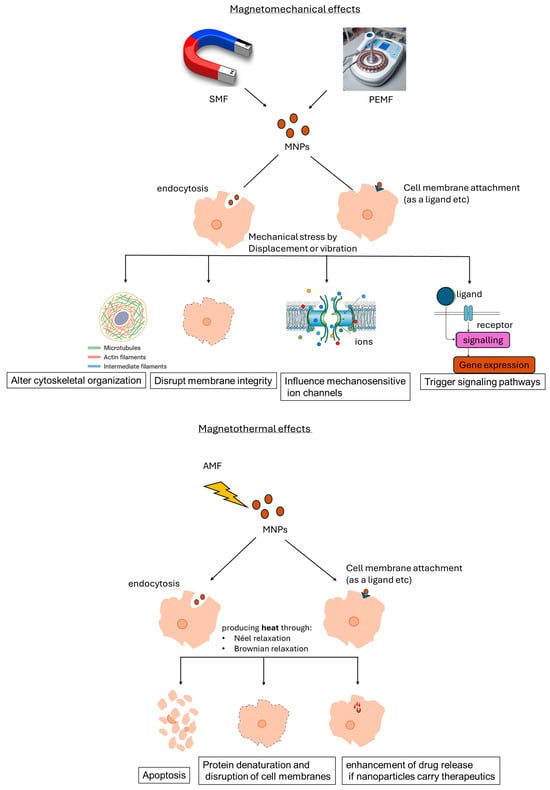
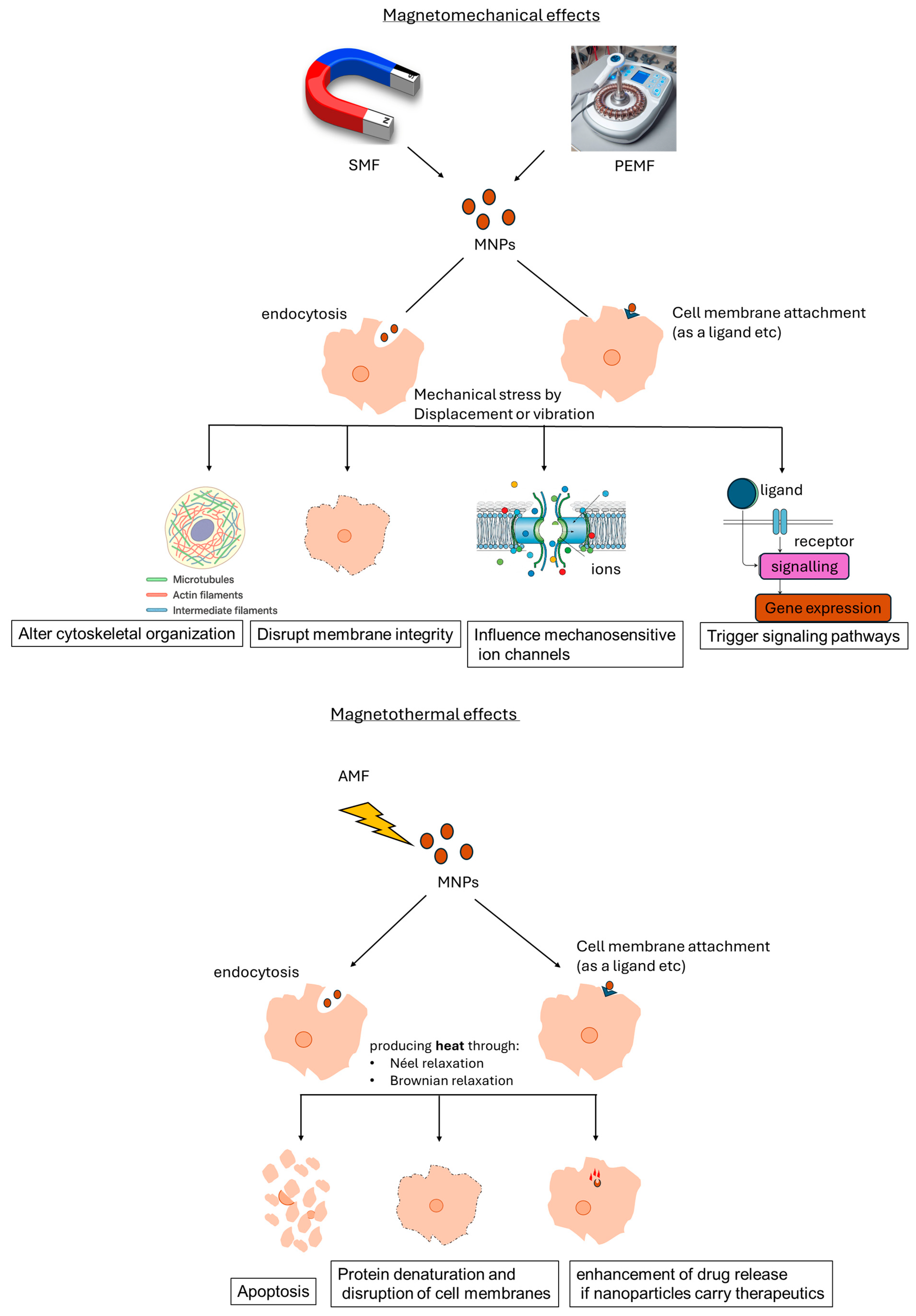

Disclaimer/Publisher’s Note: The statements, opinions and data contained in all publications are solely those of the individual author(s) and contributor(s) and not of MDPI and/or the editor(s). MDPI and/or the editor(s) disclaim responsibility for any injury to people or property resulting from any ideas, methods, instructions or products referred to in the content. |
© 2026 by the authors. Licensee MDPI, Basel, Switzerland. This article is an open access article distributed under the terms and conditions of the Creative Commons Attribution (CC BY) license.