


Abstract
Background: The clinical landscape for antibody–drug conjugates (ADCs) is currently experiencing an unprecedented expansion, with more than 20 agents approved to date and hundreds presently under clinical evaluation, underscoring their growing impact in precision oncology. By combining the cytotoxic potency of chemotherapy with the selectivity of monoclonal antibodies, ADCs have redefined targeted cancer therapy. Nevertheless, challenges related to toxicity, resistance, and suboptimal drug delivery continue to limit their full clinical potential. Objectives: This review provides a comprehensive description of currently approved ADCs, with a particular focus on their pharmacotherapeutic properties, mechanisms of action, therapeutic indications, and safety profiles. By integrating currently available clinical data and pharmacological properties, it is possible to identify key translational gaps between ADC design and their real-world performance. This article also evaluates how the structural components contribute to both efficacy and toxicity of ADCs, offering a framework for rational molecular optimizations. Conclusions: Beyond the current oncology-centric paradigm, this review highlights the imminent pivot toward non-oncology applications, including targeted therapies for autoimmune, infectious, and neurodegenerative diseases. Importantly, this article highlights emerging innovations shaping the next generation of ADCs, including bispecific antibodies, novel cytotoxic payloads with improved therapeutic indices, and advanced linker technologies enabling more precise payload release. Despite current limitations, ongoing advances in ADC development, along with a rapidly expanding clinical pipeline, position these drugs in a dynamic therapeutic class with the potential to transform multiple complex diseases and improve the quality of life of patients who have them.
1. Introduction
Cancer remains one of the most devastating diseases worldwide, not only because of its high mortality and morbidity, but also due to the emotional and financial burdens it places on patients, families, and healthcare systems. Although major advances have been made in cancer research, current treatments are mostly ineffective, leading to poor prognosis and disease management. In addition, they are frequently associated with significant undesirable effects that further reduce the quality of life of patients and entail high costs due to drug pricing and the need for repeated administrations [1,2]. Over the years, new cancer therapies have been developed to address the limitations of existing treatments. For example, targeted therapies with antibody–drug conjugates (ADCs) emerged as an innovative way of delivering highly cytotoxic agents, also known as payloads, by taking advantage of the selectivity of certain antibodies to specific receptors that are overly expressed on the surface of cancer cells when compared to their healthy counterparts [3,4]. Although this approach to targeted therapies was first conceptualized in the 60s, based on the “magic bullet” concept of Paul Ehrlich, it was only during the year 2000 that the first ADC (gemtuzumab ozogamicin) obtained its market approval for clinical use [5]. Structurally, ADCs are composed of three main components [6]: (i) an antibody moiety responsible for directing the ADC to the targeted cells, promoting the internalization of the ADC upon interacting with its complementary cell surface receptor or, if not internalized, stimulating the activation of the immune system via antibody-dependent cell-mediated cytotoxicity (ADCC), antibody-dependent cellular phagocytosis (ADCP), and complement-dependent cytotoxicity (CDC); (ii) a certain number of payloads (commonly between two to eight) that induce cell death through their specific mechanisms of action (e.g., they can act as mitotic inhibitors, topoisomerase I inhibitors, or damaging the genetic material of cancer cells); and (iii) a certain number of linkers (equal to the number of payloads) responsible for maintaining the payloads covalently bound to the antibody moiety and for promoting their rapid release whenever the original ADC is exposed to certain chemical or enzymatic stimuli that are frequently found within tumoral microenvironments or inside cancer cells.
The concept of using ADCs to treat some types of cancer seems very promising, as patients can be submitted to highly effective treatments, like the ones used in conventional chemotherapy, while minimizing the risk of adverse side effects, since ADCs use antibody moieties that are very selective to a tumor-associated antigens (TAA), like the ones used in immunotherapies with monoclonal antibodies (mAbs) or, ideally, to a tumor-specific antigen (TSA) [7]. However, the collected evidence has shown that ADCs still exhibit some limitations that need to be addressed in order to optimize their efficacy/effectiveness and safety profiles [8]. Considering the aforementioned information, this review aims to provide a summarized overview of ADC pharmacotherapy, outline the current limitations restricting their optimal use, explore novel strategies addressing these challenges, and examine emerging therapeutic applications. To achieve these objectives, the methodology adopted in this article involved a bibliographic review of the available scientific literature on topics related to ADC-based targeted therapies. The information required to elaborate this review was mostly obtained from a few searchable bibliographic online databases (like PubMed® and ScienceDirect®), books, e-books, and other kinds of documents published by international health societies and regulatory agencies. During the research phase, some of the terms inserted in the query box included “Antibody-Drug Conjugate*”, “Mechanism of Action[MeSH Terms]”, “Pharmacotherapy[MeSH Terms]”, “Pharmacovigilance[MeSH Terms]”, “Adverse Drug Reaction[MeSH Terms]”, “Limitation* OR Challenge*”, and “Future Perspective* OR Future Prospect*”.
2. Pharmacotherapy of ADCs
ADCs are a specialized group of biopharmaceutical products that belong to the family of targeted therapeutic agents for the treatment of cancer. As previously mentioned, they were introduced into the clinical setting to overcome the toxicity issues associated with the use of conventional chemotherapy, as well as the susceptibility for the development of drug resistance of immunotherapy with mAbs, by combining the two most favorable attributes of each of these types of cancer treatments [6]. For instance, brentuximab vedotin, an ADC used to treat stage III/IV Hodgkin’s lymphoma obtained a 2-year modified progression-free survival (PFS) of 82.1%, when compared to 77.2% with standard adriamicyn, bleomycin, vinblastine, and dacarbazine, while having a hazard ratio (HR) of 0.77 when compared to the same standard-of-care treatment [9,10]. For non-hematologic malignancies, trastuzumab emtansine, for example, extended the median overall survival (OS) to 30.9 months compared to 25.1 months with standard-of-care treatment and an HR of 0.68 in patients with human epidermal growth factor receptor 2 (HER2)-positive metastatic breast cancer [10,11]. In terms of biological activity, ADCs can exert multiple mechanisms of action. They can act similarly to mAbs by either inducing the activation of the immune system (via ADCC, ADCP, or CDC) or by blocking the signaling transduction pathways of some surface receptors involved in the proliferation of malignant cells [12]. Moreover, ADCs also possess a mechanism of action that is unique to this class of targeted therapies [13]: (i) first, the antibody moiety binds to overexpressed or specific receptors that are present on the surface of targeted cells; (ii) the interaction creates signals in the cell to internalize the newly formed ADC–antigen complex via endocytosis; (iii) the resulting endosome then fuses with a lysosome, forming an endolysosome where the internalized ADC is going to be digested, releasing the payloads from the antibody moiety through a specific release mechanism that is correlated with the type of linker used. For instance, while acid-sensitive cleavable linkers depend on the presence of lower pH values within the endolysosome to release the payloads, glutathione-sensitive disulfide linkers rely on the presence of intracellular reducing agents, such as glutathione (whose levels are typically elevated in malignant cells), to promote the release of these cytotoxic agents. Linkers can also be classified as enzymatically cleavable linkers (if they release the payloads in the presence of specific hydrolytic enzymes, particularly those that are found inside the endolysosomal lumen) or as non-cleavable linkers (if the payload release relies on the complete degradation of the antibody moiety); (iv) once released from the digested ADC, payloads leave the endolysosome and reach their specific intracellular target where they will exert their unique mechanisms of action and induce the death of targeted cells; and (v) due to their small, apolar, and uncharged molecular structure, some payloads can passively diffuse across cellular membranes, enabling them to enter the cytoplasm of adjacent tumor cells and produce a bystander killing effect, ensuring that multiple cancer cells are effectively killed, even in highly heterogeneous tumors and/or with low rates of internalization [14]. Figure 1 depicts a schematic representation of the multiple mechanisms of action that current ADCs can exert on tumoral cells. However, it does not account for several factors that can substantially impact their clinical effectiveness in real-world settings. For instance, while the figure suggests an equal (1:1) distribution between the primary and alternative mechanisms of action, in reality, only about 1–2% of administered ADCs reach, and are internalized by, the targeted tumor cells [3]. This limited uptake can be attributed to variability among tumor cells, even within the same cancer type, in terms of target antigen expression, as well as endocytic and intracellular trafficking pathways, all of which can directly affect the therapeutic response [15]. Furthermore, heterogeneity in ADC-related properties, such as the drug-to-antibody ratio (DAR, the average number of payloads per antibody moiety), can contribute to differences in clinical efficacy, as ADCs with higher DARs generally show greater therapeutic activity but also carry an increased risk of adverse effects [16].
Figure 1.
Schematic representation of the multiple mechanisms of action that antibody–drug conjugates (ADCs) can exert on tumoral cells. In addition to their main mechanism of action (the one enumerated in the figure), ADCs can also exert alternative mechanisms when they are not internalized by the targeted cell. Legend: 1—The antibody moiety of the ADC interacts with its complementary surface receptor; 2—The interaction created signals in the cell to internalize the formed complex via endocytosis; 3—The generated endosome fuses with a lysosome to promote the degradation of the internalized ADC and release of the payloads; 4—The released payloads leave the endolysosome and reach the cell cytosol where they will exert their mechanism of action on their intracellular targets or enter the nucleus, if the intracellular target is present inside this organelle; and 5—Due to the cytotoxicity of the payloads, the targeted cell undergoes cell death. When using relatively small, uncharged, and apolar payload molecules, it is sometimes observed that free payloads can easily diffuse to other adjacent cells and exert their cytotoxic mechanism of action on them, ensuring that multiple cancer cells are effectively killed, even in tumors with low internalization rates and/or heterogeneous levels of expression of the targeted surface receptor. Abbreviation: Fc, Fragment Crystallizable. Created in BioRender. Augusto, A. (2026) https://BioRender.com/hztmi6y, (accessed on 6 February 2026).
The introduction of ADCs into the drug market was initially hindered by their structural complexity and the limited clinical benefit observed in early studies [17]. However, their clinical implementation has accelerated in recent years, partly driven by the need for improved therapeutic responses in cancers where mAb-based immunotherapies have shown limited effectiveness [18]. By the end of 2025, there was a total of twenty-one ADCs approved globally for clinical use. Of these, twenty retained their marketing authorizations at that time, while only moxetumomab pasudotox had been withdrawn from the market due to commercial reasons unrelated to its safety or efficacy. According to their therapeutic applications, ADCs can be divided into those used in hematologic malignancies or those used in non-hematologic malignancies [6]. Table 1 summarizes the most important characteristics regarding molecular composition and the main pharmacotherapeutic indications of the previously mentioned twenty-one ADCs ever approved in at least one of the major drug markets by the end of 2025. Data were extracted from both regulatory documents and the published literature. When discrepancies were identified between these two sources of information, priority was given to the most authoritative source, i.e., the regulatory documents. However, when relevant data were not available in regulatory documentation, information was supplemented from the scientific literature. Potential off-label uses were not considered. Table 2, on the other hand, summarizes the most relevant undesirable effects, drug interactions, effects on fertility, pregnancy, and lactation, alongside the most important precautions and contraindications of the ADCs previously mentioned in Table 1. The data were extracted based on the same search strategy as the one used to collect the data presented in Table 1.
Table 1.
Molecular composition and main pharmacotherapeutic indications of most clinically relevant antibody–drug conjugates worldwide. Abbreviations: AcBut, 4-(4-Acetylphenoxy)butanoic acid; Ala, Alanine; BCMA, B-Cell Maturation Antigen; CD, Cluster of Differentiation; Cit, Citrulline; c-Met, Cellular Mesenchymal-Epithelial Transition factor; DAR, Drug-to-Antibody Ratio; DM1, Mertansine; DM4, Ravtansine; EGFR, Epidermal Growth Factor Receptor; Frα, Folate Receptor alpha; Gly, Glycine; HER2, Human Epidermal growth factor Receptor 2; HR, Hormone Receptor; IgG1, Immunoglobulin G1; IgG4, Immunoglobulin G4; INN, International Non-proprietary Name; Mal, Maleimide; Mc, Maleimidocaproyl; MCC, 4-(N-maleimidomethyl)cyclohexane-1-carboxylate; MMAE, Monomethyl Auristatin E; MMAF, Monomethyl Auristatin F; NSCLC, Non-Small Cell Lung Cancer; PABC, para-Aminobenzyl Carbamate; PD-1, Programmed Death protein 1; PD-L1, Programmed Death-Ligand 1; PEG, Polyethylene Glycol; Ph+, Chromosome Philadelphia-positive; Phe, Phenylalanine; TF, Tissue Factor; TNBC, Triple-Negative Breast Cancer; Trop2, Trophoblast Cell-Surface Antigen 2; and Val, Valine.
Table 2.
Undesirable effects, drug interactions, influence on fertility, pregnancy and lactation, as well as the key precautions and contraindications of the most clinically relevant antibody–drug conjugates worldwide. Abbreviations: CYP3A4, Cytochrome P450 3A4; CYP3A5, Cytochrome P450 3A5; G-CSF, Granulocyte colony-stimulating factor; HUS, Hemolytic uremic syndrome; ILD, Interstitial lung disease; INN, International non-proprietary name; N/A, Not applicable; SJS, Stevens–Johnson syndrome; TLS, Tumor lysis syndrome; UGT1A1, Uridine diphosphate-glucuronosyl transferase; and VOD/SOS, Veno-occlusive disease/Sinusoidal obstruction syndrome.
3. Current Limitations
Although the introduction of ADCs into the clinical setting has brought several promising therapeutic advantages, particularly in relapsed, refractory, and metastatic cancers, there are still some drawbacks that need to be addressed in order to design and develop new ADCs with optimized chemical and biological properties. Some of these limitations include: (i) toxicity; (ii) susceptibility to the development of drug resistances; (iii) complex pharmacokinetic profiles; and (iv) stability issues.
Even though ADCs were conceptualized with the intent of overcoming the toxicity issues associated with the use of conventional chemotherapy (which was achieved, to a certain extent), they still retain a suboptimal safety profile, mainly attributed to the emergence of off-target toxicities related to the untimely release of payloads into the bloodstream instead of at the actual site of the tumor(s) [102]. This phenomenon is typically associated with the use of cleavable linkers that are susceptible to release of the payloads when they are exposed to certain chemical or enzymatic stimuli that are exacerbated within tumoral microenvironments and inside cancer cells but are also present in normal physiological conditions, although to a lower extent [103]. Other causes of off-target toxicity may involve the excessive bystander killing effect and the non-specific uptake of these drugs by healthy cells. For instance, the temporary withdrawal of belantamab mafodotin, an ADC bearing a non-cleavable linker, was partially associated with the excessive uptake of this drug by the corneal epithelial cells and due to the incapacity of the charged amino acid–linker–payload complex that is released from the antibody moiety to diffuse out of these cells, resulting in the emergence of frequent episodes of ocular toxicity [104]. It is worth mentioning that on-target toxicity issues may also occur with this type of targeted therapy, because until now, every antibody moiety used in currently market-approved ADCs can only target TAAs instead of TSAs [6]. In addition to the toxicity issues correlated with the cytotoxicity of the payloads used in this kind of targeted therapy, the risk of unwanted immunogenicity is another issue that is observed with the use of biologic drugs, including ADCs [105].
A major limitation associated with the use of targeted therapies is their susceptibility to drug resistances developed by cancer cells. Although ADCs were (in part) introduced into the market to overcome resistances that rapidly emerged with other treatments, such as mAb-based immunotherapies, clinical evidence indicates that resistance to ADCs can also occur [106]: (i) antigen–antibody-mediated drug resistances are a group of resistances that negatively impair the effectiveness of antibody-based therapies by reducing the expression of targeted antigens on cancer cells, increasing receptor degradation, altering receptor structure, and/or by decreasing antigen–antibody affinity [106,107,108]; (ii) upon binding the antibody moiety to its corresponding receptor, ADCs should be internalized into the targeted cell via receptor-mediated endocytosis. However, in this step, tumoral cells may develop mechanisms of decreased internalization efficiency and/or induce alterations in the endosomal transport pathways of ADCs, preventing them from exerting their expected efficacy [106,107,109]; (iii) lysosomal degradation can also contribute to drug resistance by blocking the release of payloads from ADCs and preventing their exit from the endolysosomal lumen. For example, elevated endolysosomal pH can hinder ADCs with non-cleavable and acid-labile linkers from releasing their cytotoxic agents and impede the diffusion of weakly acidic payloads into the cytosol [106]. Conversely, lowered pH may prevent hydrophobic weak-base payloads from reaching the cytosol [110]. Moreover, reduced expression of the lysosomal membrane protein solute carrier family 46 member 3 (SLC46A3) has been associated with decreased payload release from ADCs bearing non-cleavable linkers such as those derived from maytansine and SG3376 [106,111]; (iv) even if the payloads are released from the ADC and from the endolysosome, it is still possible for the targeted cell to develop resistances to the payloads themselves, similar to what is observed with the use of conventional chemotherapy. Genetic mutations, decreased expression rates of susceptible intracellular targets, increased expression rates of resistant isoforms, post-translational modifications to the targets, and upregulation of the adenosine triphosphate (ATP)-binding cassette efflux transporters are examples of payload-related drug resistance mechanisms that may arise with the use of ADCs [109,112]; (v) tumor cells can increase survival by exploiting redundancy in receptor tyrosine kinase signaling, altering cell-cycle dynamics (particularly relevant for cell-cycle dependent payloads), upregulating cytoprotective proteins, and downregulating pro-apoptotic proteins, thereby reducing the efficacy of ADCs and other cancer therapies [106,107,113]; and (vi) in addition to the resistance mechanisms that can emerge from individual cells, the tumoral microenvironment where these cells reside may also be involved in further drug resistance, as they possess immunosuppressive properties and have poor accessibility that can hinder the penetration of antibody-based therapies because of the large molecular size of these drugs and due to the elevated interstitial fluid pressure created within tumoral microenvironments, which neutralizes the pressure gradient needed for drugs to leave the bloodstream and reach the tissues [114].
In addition to the primary limitations, ADCs exhibit a complex pharmacokinetic profile that makes it difficult to predict their behavior inside the body and, consequently, adjust the dosing to each individual patient. Unlike other drugs, the pharmacokinetic profile of ADCs requires measuring not only the concentration of intact ADCs (antibody moieties and conjugated payloads) but also non-conjugated antibody moieties, unconjugated payloads, and their metabolites in the bloodstream to predict therapeutic efficacy and toxicity risk [115]. Moreover, ADCs face an increased likelihood of aggregation, even more than mAb therapies, since the hydrophobic nature of the attached payloads might facilitate the exposure of aggregation-prone regions and induce the formation of strong inter-molecular interactions with other protein-based drugs [116]. The formed aggregates not only prevent the sequestered molecules from binding to their intended targets and exerting their expected mechanism of action; however, they can also trigger unwanted immune reactions and increase the risk of off-target payload release into the bloodstream, reducing efficacy and raising safety concerns at the same time with the use of ADCs [116].
4. Novel Approaches to the Concept of ADC and New Therapeutic Applications
The introduction of ADCs revolutionized targeted therapies for cancer by leveraging the selectivity of mAbs to deliver potent cytotoxic agents directly to cancer cells, offering therapeutic advantages over other treatments. However, clinical use has revealed limitations that must be addressed to enhance the efficacy and safety of future ADCs. Key proposed improvements for next-generation ADC design and development include the following aspects [117]: (i) better tumor penetration capacity; (ii) improved microenvironment targeting activity, instead of just targeting individual tumoral cells; (iii) flexibility to comprise novel payloads with new mechanisms of action and innovative approaches that promote enhanced activity against tumoral cells, while causing minimal damages to healthy cells; (iv) adjustable linker chemistry and its conjugation techniques, which should be improved to prevent the premature discharge of payloads into the bloodstream, whilst facilitating their release inside cancer cells or within tumoral microenvironments; (v) reduced risk of unwanted immunogenicity and on-target toxicity caused by the antibody moiety; and (vi) enhanced internalization trafficking and capacity to target multiple and, preferably, highly conserved cellular structures within tumoral cells.
As was previously mentioned, one of the main limitations with the use of large biomolecules in the treatment of solid tumors is their accessibility, especially to deeper cells within the tumoral microenvironments. Instead of using a fully assembled antibody moiety, it is possible to enhance the penetration capacity of ADCs by using smaller molecules or antibody derivatives that can still interact with similar selectivity to their complementary targets, when compared to their fully assembled counterparts [13]. Furthermore, using smaller molecules or antibody fragments can significantly reduce the production cost and complexity of these types of drugs, as they could be produced in microbial expression systems instead of using mammalian expression systems, allowing for the fragments to be produced more quickly and at higher yield rates [118]. In addition, smaller molecules (e.g., aptamers, peptides, and other small-molecule linkers) or antibody fragments can substantially decrease the potential risk of unwanted immunogenicity, as they possess fewer domains recognizable by the immune system [118,119]. However, smaller formats can also exhibit altered pharmacokinetics, including reduced circulation half-life; the absence of a fragment crystallizable (Fc) region in these fragments prevents the activation of immune effector functions (e.g., ADCC, ADCP, and CDC) [120,121]. Examples of some novel alternatives to ADCs using antibody fragments and smaller molecules include the fragment antigen-binding (Fab)-drug conjugate (a type of ADC comprising just one of two Fab regions of an antibody, instead of using the entire molecular structure of the antibody), the single-chain variable fragment (scFv)–drug conjugate (a conjugate-based drug incorporating a fragment containing the variable region of one heavy and one light chain connected by a peptide linker with 15–20 amino acids), the nanobody–drug conjugate (a small conjugated drug that uses the variable domain of a single antibody heavy chain instead of the entire antibody), and the aptamer–drug conjugate (which, instead of using an antibody derivative, uses a single-stranded oligonucleotide that folds on its own to acquire a unique three-dimensional structure, enabling it to bind to specific targets with high affinity and specificity) [122,123]. Another approach is the use of heavy-chain ADCs, where the antibody moiety is modified to lack the two light chains and the first constant domain of each heavy chain. While this novel alternative shows limited improvement in penetration capacity, compared to other drug conjugation strategies, heavy-chain ADCs are unique in their ability to activate the immune system, given the presence of the Fc region [124]. Figure 2 shows a schematic representation of the novel approaches that have been considered to overcome the poor penetration capacity of ADCs in solid tumors.
Figure 2.
Schematic representation of the novel approaches designed to overcome the limitations associated with the poor penetration capacity of market approved antibody–drug conjugates to the tumoral microenvironment of solid tumors. Abbreviations: CH1, Constant domain 1 of the heavy chain; CH2, Constant domain 2 of the heavy chain; CH3, Constant domain 3 of the heavy chain; CL, Constant domain of the light chain; Fab, Fragment antigen-binding; scFv, Single-chain variable fragment; VH, Variable domain of the heavy chain; and VL, Variable domain of the light chain. Created in BioRender. Augusto, A. (2026) https://BioRender.com/bl37z7p, (accessed on 14 February 2026).
Tumoral microenvironments are known to be enriched with various types of proteases, such as matrix metalloproteases and serine proteases, that can be used to enhance the selectivity of antibody moieties that target TAAs [125]. One such strategy involves protecting the antigen-binding sites of the antibody moiety with small masking peptides that are covalently bound to the antibody on each antigen-binding site through a cleavable linker sensitive to these proteases [126]. Upon reaching the protease-enriched medium of tumoral microenvironments, the protease-sensitive linkers are cleaved, and the masking peptides are released from the antibody, restoring the availability of the antigen-binding sites. This allows probody–drug conjugates (Figure 3) to remain inactive in circulation, preventing them from interacting with the membrane receptors of healthy cells, ensuring that the probody is only going to be bioactivated near tumoral cells [126].
Figure 3.
Schematic representation of some of the strategies that are being considered for the development of new antibody–drug conjugates with improved efficacy, better capacity to overcome drug resistance, and higher safety profiles. Abbreviations: ADC, Antibody–drug conjugate. Created in BioRender. Augusto, A. (2026) https://BioRender.com/31ofzwe, (accessed on 14 February 2026).
Future improvements to antibody moieties in ADCs should focus on enhancing the penetration of large biomolecules, while optimizing the overall therapeutic efficacy and safety of this component. For instance, by using bispecific antibodies (Figure 3) it is possible to overcome some safety concerns related to on-target toxicity, as they can target two distinct TAAs that, ideally, are not present simultaneously on healthy cells but coexist on tumor cells, enabling selective internalization or activation of the immune system only when both antigen-binding sites interact with their respective targets [127]. Antibody moieties that interact with highly conserved structures that are less likely to undergo molecular alteration have a lower probability to fall prey to the development of drug resistance [128]. Engineering the Fc region can also be considered to enhance Fc-mediated effectors functions and to optimize the structure of the antibody moiety, so the latter can act as an independent drug. Therefore, the original ADC becomes less dependent on the cytotoxicity of the small fraction of payloads that are internalized to exert an effective therapeutic response [129]. Beyond what was previously stated, the use of human-derived antibodies should be taken into account to reduce the risk of unwanted immunogenicity associated with the use of non-human antibodies [117].
Most adverse drug reactions associated with ADCs arise from the on- and off-target cytotoxicity of payloads to healthy cells. On-target toxicity is primarily due to antibody moieties binding to TAAs on healthy cell membranes, while off-target toxicity is often linked to linker instability, leading to premature payload release into the bloodstream. Additionally, payload chemistry plays a significant role [103]. Excessive hydrophobicity can increase aggregation, immunogenicity, and unwanted diffusion to healthy cells, while high hydrophilicity may prevent effective bystander killing of receptor-negative cancer cells [103]. In addition, novel payloads need to overcome drug resistance [106]. Some of the most innovative developments proposed for new payloads include the use of multiple payloads with different intracellular mechanisms of action and the exploitation of new types of payloads, such as, for example, immune stimulator compounds used as agonists of stimulator of interferon genes (STING) and toll-like receptors (TLRs), two crucial receptors involved in the activation of the innate immune system [130]. The use of antisense oligonucleotides (short sequences of single-stranded deoxyribonucleic acid (DNA) or ribonucleic acid (RNA) designed to bind to specific messenger RNA (mRNA) sequences in order to alter the mRNA involved in the pathogenesis of the disease and modulate protein expression through several distinct biological mechanisms) have also been considered, as well as the use of radionuclides and protein degraders such as proteolysis targeting chimeras (PROTACs, a type of heterobifunctional molecules containing a protein-of-interest binding domain connected to an E3 ubiquitin ligand by a linker. PROTACs target specific proteins to be degraded by taking advantage of the ubiquitin–protease system to bring together the target protein and an E3 ligase) [131,132,133]. Figure 3 provides a schematic representation of some of these novel approaches to the development of future payloads.
Finally, the linker chemistry is another critical factor involved in both the safety and efficacy of ADCs. As mentioned earlier, premature off-target release, caused by poor linker stability, is a major factor in ADC toxicity. However, the aggregation phenomena that is associated with decreased serum stability is closely correlated with the conjugation technique used to bind the payloads to the antibody moiety [134]. For instance, while low DARs are linked to reduced therapeutic outcomes, ADCs with high DARs tend to exhibit stability issues, potentially resulting in the premature release of conjugated payloads, increasing the risk of off-target toxicity [6]. To address the limitations of current linkers, it is important to improve their stability in solution and ensure a rapid payload release only when they are exposed to unique chemical or enzymatic triggers solely found inside cancer cells and within their microenvironments. Additionally, innovative controlled homogenous conjugation techniques can help to produce more uniform molecules with better DAR values and a more predictable pharmacokinetic behavior, while maintaining the antibody’s integrity [135]. Examples of such techniques include the engineered amino acid approaches (which use engineered cysteines and non-natural amino acids) (Figure 4), enzyme-mediated approaches (with transglutaminase, formylglycine-generating enzyme, sortase A, glycotransferases, and endoglycosidase), and linker-based approaches (with dibromopyridazinediones, dibromomaleimides and other enhanced maleimides, with hydrophilic linkers, and/or with bis-alkylating linkers) [136]. Figure 4 presents a simplified diagram of the reactional steps that need to be conducted in order to produce new ADCs with homogeneous DARs through engineered amino acid approaches. While enzymatic and linker-based conjugation strategies are increasingly prominent in industrial pipelines, site-specific conjugation techniques with engineered cysteine and non-natural amino acid are presented here as representative models due to their conceptual clarity.
Figure 4.
Emerging site-specific ADC conjugation techniques with engineered cysteines (on the left) and non-natural amino acids (on the right). Engineered cysteines conjugation technique: (1) A number (n) of single nucleotide replacements are made in the heavy- and/or light-chain genes that encode the antibody moiety to promote synthesis of a modified antibody with 2n strategically introduced cysteine residues at predetermined positions. Upon being synthesized, the antibody chains spontaneously assemble the antibody structure through the formation of disulfide bonds between the free thiol groups of natural cysteines present within the molecular structure of the antibody chains. During this process, engineered cysteines also form disulfide bonds with other free thiol groups (–SSR); (2) In order to make it possible for 2n payloads conjugate with the antibody moiety, the engineered cysteines must to be reduced and the natural ones need to be maintained as oxidized; (3) Then, the desired payload carrying a reactive maleimide group is added into the reactional medium to promote the conjugation of the ADC components; (4) The newly formed ADCs are expected to possess a DAR equal to two times the number of nucleotide replacements that led to the substitution of a natural amino acid with an engineered cysteine. Non-natural amino acid conjugation technique: (1) A nucleotide from an encoding triplet is replaced with another one that leads the formation of an amber stop codon (UAG, i.e., uracil–adenine–guanine); (2) An orthogonal transfer RNA (tRNA) specific for the amber stop codon is submitted to the reaction catalyzed by an engineered aminoacyl-tRNA synthetase, which selectively charges the tRNA with a desired non-natural amino acid (nnAA); (3) Upon the formation of the modified antibody, the engineered nnAAs become readily available to react with the specialized and highly reactive groups of desired payloads; (4) The newly formed ADCs, similar to what is observed with the conjugation technique with engineered cysteines, possess a DAR equal to 2n. Legend: Cys, Cysteine; DAR, Drug-to-antibody ratio; n, Number of nucleotide replacements; nnAA, Non-natural amino acid; RSS or SSR, Disulfide bond between the engineered cysteine and other molecules with free thiol groups; SH, Free thiol group; tRNA, Transfer ribonucleic acid. Please note that, for schematic purposes, it was considered that the conjugation site of engineered cysteines and non-natural amino acid conjugation processes was within the heavy and light chain, respectively. However, it could be the other way around. It will depend on the gene where the nucleotide replacement was made. Adapted from [137,138,139]. Created in BioRender. Augusto, A. (2026) https://BioRender.com/zumgxye, (accessed on 5 April 2026).
Given the success of ADCs, researchers have already proposed expanding their therapeutic applications [6]. While most ADCs are in clinical trials for other stages and cancer types, a few ADC candidates are also being explored for non-oncological conditions such as inflammatory diseases and infectious diseases (especially, against Staphylococcus aureus infectious) [140]. Although not yet in clinical trials, the use of ADCs in neurodegenerative diseases has also been proposed and is under development [141]. Notwithstanding the fact that all the non-oncological clinical trials form only around 1% of the currently running clinical trials involving ADCs, the potential use of this type of targeted therapy in the future for some diseases beyond cancer is bound to mark one of the biggest advancements in the history of targeted therapies [141]. Figure 5 illustrates the unique mechanism of action proposed for antibody–antibiotic conjugates, the ADC-derivative approach that uses antibiotics as payloads instead of cytotoxic agents.
Figure 5.
Proposed mechanism of action of antibody–antibiotic conjugates, a new therapeutic approach that facilitates the activation of the immune system against severe bacterial infections that failed previous treatments with unconjugated antibiotics. Legend: 1—The antibody–antibiotic conjugate interacts with a targeted antigen present on the surface of the bacterium through its specific antibody moiety. Upon interaction, phagocytic cells recognize the exposed Fc region of the antibody moiety through their complementary Fc receptor present on the surface of effector cells. Following the recognition, the phagocytic engulfs the opsonized bacterium (opsonization is not shown in the figure); 2—The newly formed phagocytic vesicle fuses with a lysosome to promote the degradation of the pathogen; 3—During the degradation step, proteolytic enzymes also digest the antibody moiety, allowing for the conjugated antibiotics to be released; and 4—The released antibiotics aid the immune system eliminate the pathogens by exerting their specific mechanism of action on the targeted bacterium. Abbreviations: FcR, Fragment crystallizable receptor. Adapted from [142]. Created in BioRender. Augusto, A. (2026) https://BioRender.com/q32cuub, (accessed on 19 February 2026).
5. ADCs in Clinical Trials
Table 3 shows examples of ADCs in phase I–III clinical trials identified in the Clinicaltrials.gov database, indicating targets, payload, condition/disease, and other important observations. Several targets for ADCs are highlighted such as human epidermal growth factor receptor 2 (HER2), trophoblast cell surface antigen 2 (TROP2), human epidermal growth factor receptor 3 (HER3), and claudin 18.2 (CLDN18.2).
Table 3.
Names and other important characteristics of antibody–drug conjugates currently in clinical trials. Abbreviations: CDH3, Cadherin-3; CDH17, Cadherin-17; CEACAM5, Carcinoembryonic antigen-related cell adhesion molecule 5; CLDN6, Claudin 6; CLDN18.2, Claudin 18.2; c-Met, Mesenchymal–epithelial transition factor; DLK1, Delta-like 1 homolog; DLL3, Delta-like ligand 3; DNA, Deoxyribonucleic acid; EGFR, Epidermal growth factor receptor; FAP-alpha, Fibroblast activation protein alpha; FGFR2b, Fibroblast growth factor receptor 2b; FOLR1, Folate receptor alpha; GPC3, Glypican-3; HER2, Human epidermal growth factor receptor 2; HER3, Human epidermal growth factor receptor 3; MUC16, Mucin-16; PARP, Poly ADP-ribose polymerase; PD-L1, Programmed death-ligand 1; PSMA, Prostate-specific membrane antigen; ROR1, Receptor tyrosine kinase like orphan receptor 1; ROR2, Receptor tyrosine kinase-like orphan receptor 2; TF, Tissue factor; and TROP2, Trophoblast cell surface antigen 2.
6. Conclusions
The history of ADCs is a remarkable example of how precision pharmacotherapy has evolved since the day Paul Ehrlich proposed the concept of “magic bullets”. Although ADCs faced a challenging beginning, they are now believed to have changed the paradigm regarding targeted therapies, a type of treatment that takes advantage of the presence of cell surface receptors to promote the selective delivery of potent cytotoxic agents inside the cytoplasm of cancer cells, while at the same time minimizing unwanted damage to healthy cells.
The introduction of ADCs into the therapeutic drug arsenal available against oncological diseases (hematologic and non-hematologic) has led to significant improvements in the treatment of some types of cancer, specifically in advanced or metastatic stages. However, their clinical use still faces some limitations and challenges that need to be overcome to optimize their therapeutic potential. Several improvements to the target selection and the characteristics of ADC components have been proposed; however, further investigation is still needed. The overall great success achieved with the introduction of ADCs into the drug market has sparked significant interest regarding the expansion of their therapeutic applications to new diseases beyond cancer, such as inflammatory, infectious, and neurodegenerative diseases.
Author Contributions
Conceptualization, A.A., M.L.S.C. and J.C.; methodology, A.A., M.L.S.C. and J.C.; software, A.A., M.L.S.C. and J.C.; validation, M.L.S.C. and J.C.; formal analysis, M.L.S.C. and J.C.; investigation, A.A., M.L.S.C. and J.C.; resources, A.A., M.L.S.C. and J.C.; data curation, M.L.S.C. and J.C.; writing—original draft preparation, A.A.; writing—review and editing, M.L.S.C. and J.C.; visualization, A.A., M.L.S.C. and J.C.; supervision, M.L.S.C. and J.C.; project administration, A.A., M.L.S.C. and J.C. All authors have read and agreed to the published version of the manuscript.
Funding
This research received no external funding.
Institutional Review Board Statement
Not applicable.
Informed Consent Statement
Not applicable.
Data Availability Statement
No new data were created or analyzed in this study. Data sharing is not applicable to this article.
Conflicts of Interest
The authors declare no conflicts of interest.
Abbreviations
The following abbreviations are used in this manuscript:
| ADCC | Antibody-dependent cell-mediated cytotoxicity |
| ADCP | Antibody-dependent cellular phagocytosis |
| ADCs | Antibody–drug conjugates |
| ATP | Adenosine triphosphate |
| CDC | Complement-dependent cytotoxicity |
| CLDN18.2 | Claudin 18.2 |
| DAR | Drug-to-antibody ratios |
| DNA | Deoxyribonucleic acid |
| Fab | Fragment antigen-binding |
| Fc | Fragment crystallizable |
| HER2 | Human epidermal growth factor receptor 2 |
| HER3 | Human epidermal growth factor receptor 3 |
| mAbs | Monoclonal antibodies |
| mRNA | Messenger ribonucleic acid |
| PROTACs | Proteolysis targeting chimeras |
| RNA | Ribonucleic acid |
| scFv | Single-chain variable fragment |
| STING | Stimulator of interferon genes |
| TAA | Tumor-associated antigen |
| TLRs | Toll-like receptors |
| TROP2 | Trophoblast cell surface antigen 2 |
| TSA | Tumor-specific antigen |
References
- Conceição, J.; Adeoye, O.; Cabral Marques, H.; Concheiro, A.; Alvarez Lorenzo, C.; Sousa Lobo, J.M. Carbamazepine bilayer tablets combining hydrophilic and hydrophobic cyclodextrins as a quick/slow biphasic release system. J. Drug Deliv. Sci. Technol. 2020, 57, 101611. [Google Scholar] [CrossRef] [Scilit]
- Kim, Y.; Park, E.J.; Na, D.H. Antibody–drug conjugates for targeted anticancer drug delivery. J. Pharm. Investig. 2016, 46, 341–349. [Google Scholar] [CrossRef] [Scilit]
- Fong, J.; Phuna, Z.; Chong, D.; Heryanto, C.; Low, Y.; Oh, K.; Lee, Y.; Ng, A.; In, L.; Teo, M. Advancements in antibody-drug conjugates as cancer therapeutics. J. Natl. Cancer Cent. 2025, 5, 362–378. [Google Scholar] [CrossRef] [Scilit] [PubMed]
- Alradwan, I.A.; Alnefaie, M.K.; Al Fayez, N.; Aodah, A.H.; Majrashi, M.A.; Alturki, M.; Fallatah, M.M.; Almughem, F.A.; Tawfik, E.A.; Alshehri, A.A. Strategic and Chemical Advances in Antibody–Drug Conjugates. Pharmaceutics 2025, 17, 1164. [Google Scholar] [CrossRef] [Scilit] [PubMed]
- Chis, A.; Dobrea, C.; Arseniu, A.; Frum, A.; Rus, L.; Cormos, G.; Georgescu, C.; Morgovan, C.; Butuca, A.; Gligor, F.; et al. Antibody-Drug Conjugates—Evolution and Perspectives. Int. J. Mol. Sci. 2024, 25, 6969. [Google Scholar] [CrossRef] [Scilit]
- Wang, R.; Hu, B.; Pan, Z.; Mo, C.; Zhao, X.; Liu, G.; Hou, P.; Cui, Q.; Xu, Z.; Wang, W.; et al. Antibody-Drug Conjugates (ADCs): Current and future biopharmaceuticals. J. Hematol. Oncol. 2025, 18, 51. [Google Scholar] [CrossRef] [Scilit] [PubMed]
- Chen, B.; Zheng, X.; Wu, J.; Chen, G.; Yu, J.; Xu, Y.; Wu, W.; Tse, G.; To, K.; Kang, W. Antibody–drug conjugates in cancer therapy: Current landscape, challenges, and future directions. Mol. Cancer 2025, 24, 279. [Google Scholar] [CrossRef] [Scilit] [PubMed]
- Zhou, X.; Han, Y.; Fang, Y.; Ma, P.; Zhou, J.; Jiang, Y.; Xing, S.; Tang, Q.; Hou, Y.; Wang, S.; et al. Antibody-drug conjugates: Current challenges and innovative solutions for precision cancer therapy. Med 2025, 6, 100849. [Google Scholar] [CrossRef] [Scilit] [PubMed]
- Connors, J.; Jurczak, W.; Straus, D.; Ansell, S.; Kim, W.; Gallamini, A.; Younes, A.; Alekseev, S.; Illés, Á.; Picardi, M.; et al. Brentuximab Vedotin with Chemotherapy for Stage III or IV Hodgkin’s Lymphoma. N. Engl. J. Med. 2018, 378, 331–344. [Google Scholar] [CrossRef] [Scilit]
- Mansinho, A.; Albuquerque, J.; Barigazzi, C.; Calvo, E. Antibody-drug conjugates in cancer treatment: From molecular design to clinical implementation. Lancet Reg. Health Eur. 2026, 101634. [Google Scholar] [CrossRef] [Scilit]
- Verma, S.; Miles, D.; Gianni, L.; Krop, I.; Welslau, M.; Baselga, J.; Pegram, M.; Oh, D.; Diéras, V.; Guardino, E.; et al. Trastuzumab emtansine for HER2-positive advanced breast cancer. N. Engl. J. Med. 2012, 367, 1783–1791. [Google Scholar] [CrossRef] [Scilit] [PubMed]
- Zhou, M.; Huang, Z.; Ma, Z.; Chen, J.; Lin, S.; Yang, X.; Gong, Q.; Braunstein, Z.; Wei, Y.; Rao, X.; et al. The next frontier in antibody-drug conjugates: Challenges and opportunities in cancer and autoimmune therapy. Cancer Drug Resist. 2025, 8, 34. [Google Scholar] [CrossRef] [Scilit]
- Fu, Z.; Li, S.; Han, S.; Shi, C.; Zhang, Y. Antibody drug conjugate: The “biological missile” for targeted cancer therapy. Signal Transduct. Target. Ther. 2022, 7, 93. [Google Scholar] [CrossRef] [Scilit] [PubMed]
- Wang, Y.; Cheng, X.; Li, X.; Chen, W.; Zhang, X.; Liu, Y. Bystander effect in antibody-drug conjugates: Navigating the fine line in tumor heterogeneity. Crit. Rev. Oncol. Hematol. 2025, 216, 104979. [Google Scholar] [CrossRef] [Scilit] [PubMed]
- Metrangolo, V.; Engelholm, L. Antibody-Drug Conjugates: The Dynamic Evolution from Conventional to Next-Generation Constructs. Cancers 2024, 16, 447. [Google Scholar] [CrossRef] [Scilit] [PubMed]
- Al Meslamani, A.; Bostanudin, M. Antibody-drug conjugates in oncology: Insights into Current challenges. Expert Opin. Biol. Ther. 2023, 23, 1035–1038. [Google Scholar] [CrossRef] [Scilit] [PubMed]
- Noorkhajavi, G.; Banakholdi, A.; Torabi, A.; Zoghi, A.; Iranijam, E.; Safarzadeh, E. Recent clinical advances in nonconjugated antibodies and antibody-drug conjugates for colorectal cancer treatment. Cancer Cell Int. 2025, 25, 395. [Google Scholar] [CrossRef] [Scilit]
- Justiz-Vaillant, A. Antibody–Drug Conjugates (ADCs) and Their Journey to Autoimmune Disease Immunotherapy. Med. Sci. Forum 2025, 40, 2. [Google Scholar]
- Lonial, S.; Lee, H.; Badros, A.; Trudel, S.; Nooka, A.; Chari, A.; Abdallah, A.; Callander, N.; Lendvai, N.; Sborov, D.; et al. Belantamab mafodotin for relapsed or refractory multiple myeloma (DREAMM-2): A two-arm, randomised, open-label, phase 2 study. Lancet Oncol. 2020, 21, 207–221. [Google Scholar] [CrossRef] [Scilit]
- Mukhopadhyay, P.; Abdullah, H.A.; Opalinska, J.B.; Paka, P.; Richards, E.; Weisel, K.; Trudel, S.; Mateos, M.-V.; Dimopoulos, M.A.; Lonial, S. The clinical journey of belantamab mafodotin in relapsed or refractory multiple myeloma: Lessons in drug development. Blood Cancer J. 2025, 15, 15. [Google Scholar] [CrossRef] [Scilit]
- Sureda, A.; García-Sanz, R.; Domingo-Domenech, E.; Capote, F.J.; Gutiérrez, A.; Rodriguez, A.; Aguiar, D.; Giraldo, P.; Infante, M.S.; López-Jiménez, J.; et al. Retreatment with Brentuximab Vedotin in Patients with Relapsed/Refractory CD30+ Malignancies: A Retrospective Medical Chart Review Study in Spain-The BELIEVE Study. Cancers 2025, 17, 1137. [Google Scholar] [CrossRef] [Scilit] [PubMed]
- Lambert, J.; Pautas, C.; Terré, C.; Raffoux, E.; Turlure, P.; Caillot, D.; Legrand, O.; Thomas, X.; Gardin, C.; Gogat-Marchant, K.; et al. Gemtuzumab ozogamicin for de novo acute myeloid leukemia: Final efficacy and safety updates from the open-label, phase III ALFA-0701 trial. Haematologica 2019, 104, 113–119. [Google Scholar] [CrossRef] [Scilit] [PubMed]
- Ronnacker, J.; Muller, P.J.; Mikesch, J.-H.; Zukunft, S.; Weinbergerová, B.; Šrámek, J.; Valka, J.; Novak, J.; Zak, P.; Szotkowski, T.; et al. Gemtuzumab ozogamicin in first-line treatment of CBF-AML: Insights from a retrospective multi-center analysis. Leukemia 2025, 39, 2174–2180. [Google Scholar] [CrossRef] [Scilit]
- Kantarjian, H.; DeAngelo, D.; Stelljes, M.; Liedtke, M.; Stock, W.; Gökbuget, N.; O’Brien, S.; Jabbour, E.; Wang, T.; White, J.; et al. Inotuzumab ozogamicin versus standard of care in relapsed or refractory acute lymphoblastic leukemia: Final report and long-term survival follow-up from the randomized, phase 3 INO-VATE study. Cancer 2019, 125, 2474–2487. [Google Scholar] [CrossRef] [Scilit]
- Lao, C.; Wen, J.; Wen, Y. Efficacy and safety of inotuzumab ozogamicin and its combination therapies in acute lymphoblastic leukemia: A systematic review and meta-analysis. Front. Oncol. 2025, 15, 1613777. [Google Scholar] [CrossRef] [Scilit] [PubMed]
- Caimi, P.; Ai, W.; Alderuccio, J.; Ardeshna, K.; Hamadani, M.; Hess, B.; Kahl, B.; Radford, J.; Solh, M.; Stathis, A.; et al. Loncastuximab tesirine in relapsed or refractory diffuse large B-cell lymphoma (LOTIS-2): A multicentre, open-label, single-arm, phase 2 trial. Lancet Oncol. 2021, 22, 790–800. [Google Scholar] [CrossRef] [Scilit] [PubMed]
- Juárez-Salcedo, L.M.; Nimkar, S.; Corazón, A.M.; Dalia, S. Loncastuximab Tesirine in the Treatment of Relapsed or Refractory Diffuse Large B-Cell Lymphoma. Int. J. Mol. Sci. 2024, 25, 7580. [Google Scholar] [CrossRef] [Scilit]
- Kuruvilla, D.; Chia, Y.; Balic, K.; Yao, N.; Kreitman, R.; Pastan, I.; Li, X.; Standifer, N.; Liang, M.; Tseng, C.; et al. Population pharmacokinetics, efficacy, and safety of moxetumomab pasudotox in patients with relapsed or refractory hairy cell leukaemia. Br. J. Clin. Pharmacol. 2020, 86, 1367–1376. [Google Scholar] [CrossRef] [Scilit]
- Yurkiewicz, I.R.; Coutre, S.; Ghesquieres, H.; Pastan, I.; Kreitman, R.J. Moxetumomab pasudotox as re-treatment for heavily-pretreated relapsed hairy cell leukemia. Leuk. Lymphoma 2021, 62, 2812–2814. [Google Scholar] [CrossRef] [Scilit]
- Sehn, L.; Herrera, A.; Flowers, C.; Kamdar, M.; McMillan, A.; Hertzberg, M.; Assouline, S.; Kim, T.; Kim, W.; Ozcan, M.; et al. Polatuzumab Vedotin in Relapsed or Refractory Diffuse Large B-Cell Lymphoma. J. Clin. Oncol. 2020, 38, 155–165. [Google Scholar] [CrossRef] [Scilit] [PubMed]
- Tilly, H.; Morschhauser, F.; Sehn, L.H.; Friedberg, J.W.; Trněný, M.; Sharman, J.P.; Herbaux, C.; Burke, J.M.; Matasar, M.; Rai, S.; et al. Polatuzumab Vedotin in Previously Untreated Diffuse Large B-Cell Lymphoma. N. Engl. J. Med. 2022, 386, 351–363. [Google Scholar] [CrossRef] [Scilit] [PubMed]
- Blair, H. Becotatug Vedotin: First Approval. Drugs 2026, 86, 573–579. [Google Scholar] [CrossRef] [Scilit] [PubMed]
- Han, F.; Lv, X.; Zhou, Y.-J.; He, Q.; Guo, Y.; Xiang, Y.-Q.; Shu, X.-L.; Pan, S.-M.; Qu, S.-H.; Zhang, P.; et al. Becotatug vedotin, MRG003, in previously treated recurrent or metastatic nasopharyngeal carcinoma: A multicenter, single-arm, phase IIa trial. Med 2026, 7, 101029. [Google Scholar] [CrossRef] [Scilit] [PubMed]
- Okamoto, I.; Hasegawa, O.; Kushihashi, Y.; Masubuchi, T.; Tokashiki, K.; Tsukahara, K. Real-World Effectiveness and Safety of Photoimmunotherapy for Head and Neck Cancer: A Multicenter Retrospective Study. Cancers 2025, 17, 2671. [Google Scholar] [CrossRef] [Scilit]
- Okamoto, I. Photoimmunotherapy for head and neck cancer: A systematic review. Auris Nasus Larynx 2025, 52, 186–194. [Google Scholar] [CrossRef] [Scilit]
- Ahn, M.; Tanaka, K.; Paz-Ares, L.; Cornelissen, R.; Girard, N.; Pons-Tostivint, E.; Vicente Baz, D.; Sugawara, S.; Cobo, M.; Pérol, M.; et al. Datopotamab Deruxtecan Versus Docetaxel for Previously Treated Advanced or Metastatic Non-Small Cell Lung Cancer: The Randomized, Open-Label Phase III TROPION-Lung01 Study. J. Clin. Oncol. 2025, 43, 260–272. [Google Scholar] [CrossRef] [Scilit]
- Huang, J.; Huang, T.; Guo, J.; Hu, K.; Zhou, H. The efficacy and safety of datopotamab deruxtecan (Dato-DXd) in advanced solid tumors: A systematic review and meta-analysis. Eur. J. Med. Res. 2025, 30, 1265. [Google Scholar] [CrossRef] [Scilit]
- Shi, F.; Liu, Y.; Zhou, X.; Shen, P.; Xue, R.; Zhang, M. Disitamab vedotin: A novel antibody-drug conjugates for cancer therapy. Drug Deliv. 2022, 29, 1335–1344. [Google Scholar] [CrossRef] [Scilit]
- Ye, J.; Li, Y.; Zhang, M.; Liao, X.; Wang, Q.; Chen, Z.; Tu, X.; Tan, P.; Zhang, P.; Zeng, H.; et al. Efficacy of disitamab vedotin-based therapy in HER2-negative and HER2-low locally advanced or metastatic urothelial carcinoma: A systematic review and meta-analysis. Urol. Oncol. 2026, 111034. [Google Scholar] [CrossRef] [Scilit]
- Powles, T.; Rosenberg, J.; Sonpavde, G.; Loriot, Y.; Durán, I.; Lee, J.; Matsubara, N.; Vulsteke, C.; Castellano, D.; Wu, C.; et al. Enfortumab Vedotin in Previously Treated Advanced Urothelial Carcinoma. N. Engl. J. Med. 2021, 384, 1125–1135. [Google Scholar] [CrossRef] [Scilit] [PubMed]
- Powles, T.; Valderrama, B.P.; Gupta, S.; Bedke, J.; Kikuchi, E.; Hoffman-Censits, J.; Iyer, G.; Vulsteke, C.; Park, S.H.; Shin, S.J.; et al. Enfortumab Vedotin and Pembrolizumab in Untreated Advanced Urothelial Cancer. N. Engl. J. Med. 2024, 390, 875–888. [Google Scholar] [CrossRef] [Scilit] [PubMed]
- Moore, K.; Angelergues, A.; Konecny, G.; García, Y.; Banerjee, S.; Lorusso, D.; Lee, J.; Moroney, J.; Colombo, N.; Roszak, A.; et al. Mirvetuximab Soravtansine in FRα-Positive, Platinum-Resistant Ovarian Cancer. N. Engl. J. Med. 2023, 389, 2162–2174. [Google Scholar] [CrossRef] [Scilit] [PubMed]
- Kokori, E.; Olatunji, G.; Komolafe, R.; Abraham, I.C.; Ukoaka, B.; Samuel, O.; Ayodeji, A.; Ogunbowale, I.; Ezenwoba, C.; Aderinto, N. Mirvetuximab soravtansine: A breakthrough in targeted therapy for platinum-resistant ovarian cancer. Medicine 2024, 103, e38132. [Google Scholar] [CrossRef] [Scilit]
- Bardia, A.; Hurvitz, S.; Tolaney, S.; Loirat, D.; Punie, K.; Oliveira, M.; Brufsky, A.; Sardesai, S.; Kalinsky, K.; Zelnak, A.; et al. Sacituzumab Govitecan in Metastatic Triple-Negative Breast Cancer. N. Engl. J. Med. 2021, 384, 1529–1541. [Google Scholar] [CrossRef] [Scilit] [PubMed]
- Jiang, L.; Dai, Y.; Li, M.; Zhao, Z. The efficacy and safety of sacituzumab govitecan in the treatment of breast cancer: A systemic review and meta-analysis of emerging clinical data. Front. Immunol. 2025, 16, 1683594. [Google Scholar] [CrossRef] [Scilit]
- Tolaney, S.; Cardillo, T.; Chou, C.; Dornan, C.; Faris, M. The Mode of Action and Clinical Outcomes of Sacituzumab Govitecan in Solid Tumors. Clin. Cancer Res. 2025, 31, 1390–1399. [Google Scholar] [CrossRef] [Scilit]
- Sofianidi, A.A.; Papatheodoridi, A.; Dimitrakakis, C.; Marinopoulos, S.; Michalaki, V.; Zagouri, F. Sacituzumab tirumotecan (sac-TMT/MK-2870/SKB264): A novel antibody–drug conjugate in breast cancer. Oncol. Rev. 2026, 20, 1781533. [Google Scholar] [CrossRef] [Scilit]
- Camidge, D.; Bar, J.; Horinouchi, H.; Goldman, J.; Moiseenko, F.; Filippova, E.; Cicin, I.; Ciuleanu, T.; Daaboul, N.; Liu, C.; et al. Telisotuzumab Vedotin Monotherapy in Patients with Previously Treated c-Met Protein-Overexpressing Advanced Nonsquamous EGFR-Wildtype Non-Small Cell Lung Cancer in the Phase II LUMINOSITY Trial. J. Clin. Oncol. 2024, 42, 3000–3011. [Google Scholar] [CrossRef] [Scilit]
- Khan, M.A.H.; Ahmad, W.; Ehsan, H.; Iqbal, M.; Rayyan, S.M.; Mehrpoor, A.J. Emrelis (telisotuzumab vedotin-tllv): Targeting C-MET in non-small cell lung cancer. Ann. Med. Surg. 2025, 87, 7870–7871. [Google Scholar] [CrossRef] [Scilit]
- Hong, D.; Concin, N.; Vergote, I.; de Bono, J.; Slomovitz, B.; Drew, Y.; Arkenau, H.; Machiels, J.; Spicer, J.; Jones, R.; et al. Tisotumab Vedotin in Previously Treated Recurrent or Metastatic Cervical Cancer. Clin. Cancer Res. 2020, 26, 1220–1228. [Google Scholar] [CrossRef] [Scilit] [PubMed]
- Vergote, I.; González-Martín, A.; Fujiwara, K.; Kalbacher, E.; Bagaméri, A.; Ghamande, S.; Lee, J.-Y.; Banerjee, S.; Maluf, F.C.; Lorusso, D.; et al. Tisotumab Vedotin as Second- or Third-Line Therapy for Recurrent Cervical Cancer. N. Engl. J. Med. 2024, 391, 44–55. [Google Scholar] [CrossRef] [Scilit]
- Hu, X.; Zhang, J.; Ouyang, Q.; Zhang, Q.; Li, H.; Wang, X.; Wang, Y.; Yin, Y.; Wang, S.; Gu, Y.; et al. LBA24 Trastuzumab botidotin vs trastuzumab emtansine (T-DM1) in HER2-positive unresectable or metastatic breast cancer: Results from a randomized phase III study. Ann. Oncol. 2025, 36, S1569. [Google Scholar] [CrossRef] [Scilit]
- Cerci, B.; Saatci, O.; Basik, M.; Sahin, O. Mechanisms of resistance to antibody-drug conjugates in breast cancer. Drug Resist. Updat. 2026, 85, 101353. [Google Scholar] [CrossRef] [Scilit]
- Cortés, J.; Kim, S.; Chung, W.; Im, S.; Park, Y.; Hegg, R.; Kim, M.; Tseng, L.; Petry, V.; Chung, C.; et al. Trastuzumab Deruxtecan versus Trastuzumab Emtansine for Breast Cancer. N. Engl. J. Med. 2022, 386, 1143–1154. [Google Scholar] [CrossRef] [Scilit]
- Quaquarini, E.; Luelli, F.; Lasagna, A.; Rizzo, G.; Perrone, L.; Figini, S.; Achille, R.; Pedrazzoli, P. Trastuzumab–Deruxtecan for the Treatment of Metastatic Breast Cancer Patients: Data from Real World Studies. Cancers 2025, 17, 3505. [Google Scholar] [CrossRef] [Scilit] [PubMed]
- Diéras, V.; Miles, D.; Verma, S.; Pegram, M.; Welslau, M.; Baselga, J.; Krop, I.; Blackwell, K.; Hoersch, S.; Xu, J.; et al. Trastuzumab emtansine versus capecitabine plus lapatinib in patients with previously treated HER2-positive advanced breast cancer (EMILIA): A descriptive analysis of final overall survival results from a randomised, open-label, phase 3 trial. Lancet Oncol. 2017, 18, 732–742. [Google Scholar] [CrossRef] [Scilit]
- Chandrakanth, M.V.; Agarwala, V.; Manna, R.; Sharma, A.; Roy, M.; Dasgupta, A.; Mandal, K.; Sen, N.; Sardar, P.K.; Sadhya, A.; et al. Real-World Evaluation of a Trastuzumab Emtansine Biosimilar (Ujvira®) in Human Epidermal Growth Factor Receptor 2 (HER2)-Positive Metastatic Breast Cancer. Cureus 2026, 18, e103700. [Google Scholar] [CrossRef] [Scilit] [PubMed]
- Li, Z.; Wang, Y.; Sun, Y.; Wang, L.; Li, X.; Sun, L.; He, Z.; Yang, H.; Wang, Y.; Wang, Q.; et al. Trastuzumab rezetecan, a HER2-directed antibody-drug conjugate, in patients with advanced HER2-mutant non-small-cell lung cancer (HORIZON-Lung): Phase 2 results from a multicentre, single-arm study. Lancet Oncol. 2025, 26, 437–446. [Google Scholar] [CrossRef] [Scilit] [PubMed]
- Hoy, S.M. Trastuzumab Rezetecan: First Approval. Drugs 2026, 86, 111–120. [Google Scholar] [CrossRef] [Scilit]
- Wahab, A.; Rafae, A.; Mushtaq, K.; Masood, A.; Ehsan, H.; Khakwani, M.; Khan, A. Ocular Toxicity of Belantamab Mafodotin, an Oncological Perspective of Management in Relapsed and Refractory Multiple Myeloma. Front. Oncol. 2021, 11, 678634. [Google Scholar] [CrossRef] [Scilit]
- Sunami, K.; Fujisaki, T.; Funaki, T.; Ichii, M.; Ito, S.; Matsumoto, M.; Oshima, K.-I.; Suzuki, K.; Takakuwa, T. Practical guidance on the clinical management of ocular adverse events associated with belantamab mafodotin in patients with relapsed/refractory multiple myeloma: Recommendations from a Japanese expert panel. Jpn. J. Clin. Oncol. 2025, 55, 1349–1356. [Google Scholar] [CrossRef] [Scilit]
- Tan, F.; Li, Y.; Xia, H.; Yin, W. Signal mining and analysis of adverse events of Brentuximab Vedotin base on FAERS and JADER databases. PLoS ONE 2025, 20, e0322378. [Google Scholar] [CrossRef] [Scilit]
- Gao, S.; Zhang, M.; Wu, K.; Zhu, J.; He, Z.; Li, J.; Chen, C.; Qiu, K.; Yu, X.; Wu, J. Risk of adverse events in lymphoma patients treated with brentuximab vedotin: A systematic review and meta-analysis. Expert Opin. Drug Saf. 2020, 19, 617–623. [Google Scholar] [CrossRef] [Scilit]
- Liu, X.; Wu, F.; Li, Z.; Li, C.; Li, X.; Yuan, Y.; Sun, X.; Du, Y.; Du, X.; Wang, S.; et al. Safety assessment of gemtuzumab ozogamicin: Real-world adverse event analysis based on the FDA Adverse Event Reporting System. Front. Med. 2025, 12, 1643780. [Google Scholar] [CrossRef] [Scilit] [PubMed]
- Cortes, J.E.; Lima, M.; Dombret, H.; Estey, E.H.; Giralt, S.A.; Montesinos, P.; Röllig, C.; Venditti, A.; Wang, E.S. Prevention, recognition, and management of adverse events associated with gemtuzumab ozogamicin use in acute myeloid leukemia. J. Hematol. Oncol. 2020, 13, 137. [Google Scholar] [CrossRef] [Scilit]
- Tran, V.; Srinivasa, N.; Tatum, C.; Reed, D.; Keng, M. Inotuzumab Ozogamicin in Clinical Practice: An Overview of Efficacy, Safety, and Real-World Applications. Curr. Hematol. Malig. Rep. 2026, 21, 5. [Google Scholar] [CrossRef] [Scilit]
- Ogrodnik, P.; Anderson, E.; Gloude, N.; Schiff, D. Efficacy and Toxicity of Inotuzumab Ozogamicin for Treatment of Relapsed/Refractory B-Cell Acute Lymphoblastic Leukemia in Pediatric and Young Adult Patients After CD19-Chimeric Antigen Receptor T-Cell Therapy. Pediatr. Blood Cancer 2025, 72, e32013. [Google Scholar] [CrossRef] [Scilit]
- Epperla, N.; Olszewski, A.J.; Ayers, E.C.; Ahmed, S. Management of Adverse Reactions to Loncastuximab in Patients with Relapsed or Refractory Diffuse Large B-Cell Lymphoma. Hematol. Oncol. 2025, 43, e70128. [Google Scholar] [CrossRef] [Scilit] [PubMed]
- Derenzini, E.; Gibb, A.; Kwiatek, M.; Strati, P. Loncastuximab tesirine in relapsed/refractory diffuse large B-cell lymphoma: Drug profile and expert opinion on the prevention and management of adverse events. Leuk. Lymphoma 2025, 66, 1990–2002. [Google Scholar] [CrossRef] [Scilit] [PubMed]
- Kreitman, R.J.; Dearden, C.; Zinzani, P.L.; Delgado, J.; Robak, T.; Coutre, P.D.; Gjertsen, B.T.; Troussard, X.; Roboz, G.J.; Karlin, L.; et al. Moxetumomab pasudotox in heavily pre-treated patients with relapsed/refractory hairy cell leukemia (HCL): Long-term follow-up from the pivotal trial. J. Hematol. Oncol. 2021, 14, 35. [Google Scholar] [CrossRef] [Scilit] [PubMed]
- Kang, C. Moxetumomab Pasudotox in Hairy Cell Leukaemia: A Profile of Its Use. Clin. Drug Investig. 2021, 41, 829–834. [Google Scholar] [CrossRef] [Scilit]
- Liu, D.; Mao, W.; Hu, B.; Li, X.; Zhao, Q.; Zhang, L.; Hu, J. A real-world pharmacovigilance study of polatuzumab vedotin based on the FDA adverse event reporting system (FAERS). Front. Pharmacol. 2024, 15, 1405023. [Google Scholar] [CrossRef] [Scilit]
- Xu, Z.; Huang, D.; Liu, Q.; Liu, S.; Liu, J.; Wang, H.; Shen, Z. A real-world pharmacovigilance analysis of the FDA adverse event reporting system events for polatuzumab vedotin. Expert. Opin. Drug Saf. 2025, 24, 49–57. [Google Scholar] [CrossRef] [Scilit] [PubMed]
- Qiu, M.-Z.; Zhang, Y.; Guo, Y.; Guo, W.; Nian, W.; Liao, W.; Xu, Z.; Zhang, W.; Zhao, H.-Y.; Wei, X.; et al. Evaluation of Safety of Treatment with Anti–Epidermal Growth Factor Receptor Antibody Drug Conjugate MRG003 in Patients with Advanced Solid Tumors. JAMA Oncol. 2022, 8, 1042–1046. [Google Scholar] [CrossRef] [Scilit]
- Abraham, F.F.; Julian, R. Emerging and Investigational Systemic Therapies in Recurrent/Metastatic Head and Neck Cancer After Progression on Immunotherapy. Cancers 2025, 17, 3817. [Google Scholar] [CrossRef] [Scilit]
- Okamoto, I.; Okada, T.; Tokashiki, K.; Tsukahara, K. Quality-of-Life Evaluation of Patients with Unresectable Locally Advanced or Locally Recurrent Head and Neck Carcinoma Treated with Head and Neck Photoimmunotherapy. Cancers 2022, 14, 4413. [Google Scholar] [CrossRef] [Scilit]
- Takuma, S.; Karayama, M.; Suzuki, R.; Miyashita, K.; Inoue, Y.; Yasui, H.; Hozumi, H.; Suzuki, Y.; Furuhashi, K.; Enomoto, N.; et al. Severe drug-induced interstitial lung disease after photoimmunotherapy with cetuximab–sarotalocan sodium in a patient with oropharyngeal cancer: A case report. Respir. Med. Case Rep. 2025, 57, 102260. [Google Scholar] [CrossRef] [Scilit] [PubMed]
- Lisberg, A.; Huppert, L.A.; Halmos, B.; Ledezma, B.; Soto-Romano, V.; Traina, T.A. Datopotamab deruxtecan–associated select adverse events: Clinical practices and institutional protocols on prophylaxis, monitoring, and management. Oncologist 2025, 30, oyaf225. [Google Scholar] [CrossRef] [Scilit] [PubMed]
- Gadaleta-Caldarola, G.; Lanotte, L.; Infusino, S.; Gadaleta-Caldarola, A.; Schipilliti, F.M.; Citrigno, C.; Petrarota, C.; Cusmai, A.; Rizzo, A. Safety evaluation of Datopotamab deruxtecan for triple-negative breast cancer: A meta-analysis. Cancer Treat. Res. Commun. 2023, 37, 100775. [Google Scholar] [CrossRef] [Scilit] [PubMed]
- He, S.; Wang, C.; Li, X.; Li, C.; Li, D.; Zhang, B.; Wan, J.; Yue, Z.; Wu, G.; Zhang, S.; et al. Real-world efficacy and safety of disitamab vedotin monotherapy or in combination with PD-1 inhibitors in locally advanced or metastatic upper tract urothelial carcinoma: A multicenter retrospective study. Front. Immunol. 2026, 16, 1699538. [Google Scholar] [CrossRef] [Scilit]
- Zhou, T.; Liu, H.; Shen, Y.; Shao, S. Real-world evaluation of the efficacy and safety of disitamab vedotin (RC48) in urothelial carcinoma. BMC Urol. 2025, 25, 276. [Google Scholar] [CrossRef] [Scilit] [PubMed]
- Nizam, A.; Nguyen, C.B.; Li, J.; Zabor, E.C.; Msaouel, P.; Jiang, C.Y.; Alhalabi, O.; Oh, E.; Davidsohn, M.P.; Epstein, I.B.; et al. Treatment-Related Adverse Events and Associated Outcomes in Patients with Advanced Urothelial Carcinoma Treated with Enfortumab Vedotin: Analysis of the UNITE Study. Cancer Med. 2025, 14, e71284. [Google Scholar] [CrossRef] [Scilit] [PubMed]
- Dobry, A.S.; Virgen, C.A.; Hosking, A.-M.; Mar, N.; Doan, L.; Lee, B.; Smith, J. Cutaneous reactions with enfortumab vedotin: A case series and review of the literature. JAAD Case Rep. 2021, 14, 7–9. [Google Scholar] [CrossRef] [Scilit]
- Lang, S.M.; Vingopoulos, F.; Beshar, I.; Somasegar, S.; Adams, E.V.; Sasse, S.A.; Ghezelayagh, T.; McClung, E.C.; Karam, A.; Dorigo, O.; et al. Early experience and assessment of real-world toxicities with mirvetuximab soravtansine in a heavily pretreated patient cohort with ovarian cancer. Gynecol. Oncol. Rep. 2025, 59, 101738. [Google Scholar] [CrossRef] [Scilit] [PubMed]
- Haidar, A.J.; Chen, A.; Chen, J.L.; Al-Hashimi, S.; Ghaffari, R.S.; Fung, S.; Silverstein, J.; Konecny, G.E.; Tsui, E. Ocular Toxic Effects During Mirvetuximab Soravtansine Therapy. JAMA Oncol. 2026, 12, 317–319. [Google Scholar] [CrossRef] [Scilit] [PubMed]
- Gui, X.; Zhao, J.; Ding, L.; Chai, J.; Lai, H.; Cai, Y.; Luo, S.; Zeng, Y.; Wu, W.; Chen, H.; et al. Assessing real-world safety concerns of Sacituzumab govitecan: A disproportionality analysis using spontaneous reports in the FDA adverse event reporting system. Front. Oncol. 2023, 13, 1276976. [Google Scholar] [CrossRef] [Scilit] [PubMed]
- Tolaney, S.M.; Sonpavde, G.P.; Tarantino, P.; Lustberg, M.B.; Rugo, H.S. Clinical perspective on management of key adverse events with sacituzumab govitecan. Oncologist 2025, 30, oyaf311. [Google Scholar] [CrossRef] [Scilit]
- Zhu, Y.; Jiang, S.; Shi, Y.; Wang, S.; Yuan, F.; Zhou, F.; Jiang, K.; Zhang, X.; Seneviratne, L.; Yu, G.; et al. Sacituzumab tirumotecan in participants with advanced or metastatic urothelial carcinoma and disease progression after chemotherapy and immune checkpoint inhibitors. Ann. Oncol. 2026, 37, 378–387. [Google Scholar] [CrossRef] [Scilit]
- Yin, Y.; Fan, Y.; Ouyang, Q.; Song, L.; Wang, X.; Li, W.; Li, M.; Yan, X.; Wang, S.; Sun, T.; et al. Sacituzumab tirumotecan in previously treated metastatic triple-negative breast cancer: A randomized phase 3 trial. Nat. Med. 2025, 31, 1969–1975. [Google Scholar] [CrossRef] [Scilit]
- Babel, H.; Brunsdon, P.; Engelhardt, B.; Schmitt, V.; Ratajczak, C.; Mensing, S.; Menon, R.M.; Parikh, A. Population Pharmacokinetics and Exposure-Response Analyses for Telisotuzumab Vedotin in Patients with c-Met Protein Overexpressing Tumors. CPT Pharmacomet. Syst. Pharmacol. 2026, 15, e70219. [Google Scholar] [CrossRef] [Scilit]
- Zhao, C.; Lu, D.; Gao, J. Telisotuzumab vedotin: The first-in-class c-Met-targeted antibody-drug conjugate granted FDA accelerated approval for treatment of non-squamous non-small cell lung cancer (NSCLC). Drug Discov. Ther. 2025, 19, 275–276. [Google Scholar] [CrossRef] [Scilit]
- Arn, C.R.; Halla, K.J.; Gill, S. Tisotumab Vedotin Safety and Tolerability in Clinical Practice: Managing Adverse Events. J. Adv. Pract. Oncol. 2023, 14, 139–152. [Google Scholar] [PubMed]
- Chess, I.; Mercier, A.M.; Gross, S.; Blank, S.V.; Hayes, M.P. Tisotumab vedotin–associated Stevens–Johnson syndrome in a patient with recurrent vulvar cancer: Vulnerability and antibody drug conjugate toxicity considerations. Gynecol. Oncol. Rep. 2026, 64, 102065. [Google Scholar] [CrossRef] [Scilit] [PubMed]
- Liu, X.; Deng, J.; Zhang, R.; Xing, J.; Wu, Y.; Chen, W.; Liang, B.; Xing, D.; Xu, J.; Zhang, M. The clinical development of antibody-drug conjugates for non-small cell lung cancer therapy. Front. Immunol. 2023, 14, 1335252. [Google Scholar] [CrossRef] [Scilit] [PubMed]
- Evangelou, C. Recent Advances in the Treatment of Early-Stage HER2-Positive Breast Cancer: Updates from ESMO 2025. Oncol. Am. Med. J. 2025, 2, 2–12. [Google Scholar] [CrossRef] [Scilit]
- Bianchini, G.; Fabi, A.; Guarneri, V.; Arpino, G.; Laurentis, M.D.; Zambelli, A.; Zamagni, C.; Biganzoli, L.; Puglisi, F.; Santini, D. Management of trastuzumab deruxtecan-related adverse events in breast cancer: Italian expert panel recommendations. Future Oncol. 2025, 21, 2565–2573. [Google Scholar] [CrossRef] [Scilit] [PubMed]
- Pathak, N.; Iorio, M.D.; Gimenez, D.M.; Berner-Wygoda, Y.; Savill, J.; Aljuhani, A.; Mittal, A.; Kumar, V.; Amir, E. Adverse effect of trastuzumab deruxtecan in solid tumours: A systematic review and meta-analysis. Crit. Rev. Oncol. Hematol. 2025, 213, 104787. [Google Scholar] [CrossRef] [Scilit]
- Liu, F.; Yin, G.; Xue, S.; Rehman, F.U.L.; Liao, D.; Pan, Y. Adverse Event Profile Differences between Trastuzumab Emtansine and Trastuzumab Deruxtecan: A Real-world, Pharmacovigilance Study. J. Cancer 2023, 14, 3275–3284. [Google Scholar] [CrossRef] [Scilit]
- Kim, H.J.; Yoon, J.-H.; Park, Y.H. Long-term hepatobiliary disorder associated with trastuzumab emtansine pharmacovigilance study using the FDA Adverse Event Reporting System database. Sci. Rep. 2024, 14, 19587. [Google Scholar] [CrossRef] [Scilit]
- Zhang, T.; Xu, J.; Yin, J.; Gao, Y.; Zheng, H.; Fu, B.; Sun, J.; Xu, Z.; Tu, S.; Mao, Y.; et al. SHR-A1811, a novel anti-HER2 antibody–drug conjugate with optimal drug-to-antibody ratio, efficient tumor killing potency, and favorable safety profiles. PLoS ONE 2025, 20, e0326691. [Google Scholar] [CrossRef] [Scilit]
- Papakonstantinou, A.; Foukakis, T. Neoadjuvant treatment of HER2-positive breast cancer: Has the era of antibody-drug conjugates arrived? Ann. Oncol. 2025, 36, 603–605. [Google Scholar] [CrossRef] [Scilit]
- Nguyen, T.; Bordeau, B.; Balthasar, J. Mechanisms of ADC Toxicity and Strategies to Increase ADC Tolerability. Cancers 2023, 15, 713. [Google Scholar] [CrossRef] [Scilit]
- Cheng, Y.; Lu, J.; Zhang, C.; Yan, W.; Zhu, P.; Qin, Q.; Gong, L. Overview of antibody-drug conjugates nonclinical and clinical toxicities and related contributing factors. Antib. Ther. 2025, 8, 124–144. [Google Scholar] [CrossRef] [Scilit]
- Tan, H.; Morcillo, M.; Lopez, J.; Minchom, A.; Sharp, A.; Paschalis, A.; Silva-Fortes, G.; Raobaikady, B.; Banerji, U. Treatment-related adverse events of antibody drug-conjugates in clinical trials. J. Hematol. Oncol. 2025, 18, 71. [Google Scholar] [CrossRef] [Scilit] [PubMed]
- Grudzinska-Goebel, J.; Benstein, K.; Bloem, K.; Cowan, K.; Gorovits, B.; Jadhav, M.; Janssen, M.; Jawa, V.; Kiessling, A.; Kramer, D.; et al. Immunogenicity risk assessment for tailored mitigation and monitoring of biotherapeutics during development: Recommendations from the European Immunogenicity Platform. Front. Immunol. 2025, 16, 1581153. [Google Scholar] [CrossRef] [Scilit]
- Li, S.; Zhao, X.; Fu, K.; Zhu, S.; Pan, C.; Yang, C.; Wang, F.; To, K.; Fu, L. Resistance to antibody–drug conjugates: A review. Acta Pharm. Sin. B 2024, 15, 737–756. [Google Scholar] [CrossRef] [Scilit] [PubMed]
- Khoury, R.; Saleh, K.; Khalife, N.; Saleh, M.; Chahine, C.; Ibrahim, R.; Lecesne, A. Mechanisms of Resistance to Antibody-Drug Conjugates. Int. J. Mol. Sci. 2023, 24, 9674. [Google Scholar] [CrossRef] [Scilit]
- Murase, Y.; Nanjo, S.; Ueda, T.; Liu, Y.; Nomura, S.; Arai, S.; Terada, N.; Koba, H.; Tambo, Y.; Yano, S. Mechanisms of resistance to antibody-drug conjugates in cancers. Respir. Investig. 2025, 63, 693–698. [Google Scholar] [CrossRef] [Scilit] [PubMed]
- Abelman, R.; Wu, B.; Spring, L.; Ellisen, L.; Bardia, A. Mechanisms of Resistance to Antibody–Drug Conjugates. Cancers 2023, 15, 1278. [Google Scholar] [CrossRef] [Scilit] [PubMed]
- Yousef, M.; Le, T.; Zuo, J.; Park, C.; Chacra, N.; Davies, N.; Löbenberg, R. Sub-cellular sequestration of alkaline drugs in lysosomes: New insights for pharmaceutical development of lysosomal fluid. Res. Pharm. Sci. 2022, 18, 1–15. [Google Scholar] [CrossRef] [Scilit]
- Kinneer, K.; Meekin, J.; Tiberghien, A.; Tai, Y.; Phipps, S.; Kiefer, C.; Rebelatto, M.; Dimasi, N.; Moriarty, A.; Papadopoulos, K.; et al. SLC46A3 as a Potential Predictive Biomarker for Antibody-Drug Conjugates Bearing Noncleavable Linked Maytansinoid and Pyrrolobenzodiazepine Warheads. Clin. Cancer Res. 2018, 24, 6570–6582. [Google Scholar] [CrossRef] [Scilit]
- Abelman, R.; Wu, B.; Barnes, H.; Medford, A.; Norden, B.; Putur, A.; Bitman, E.; Thant, W.; Liu, T.; Weipert, C.; et al. TOP1 Mutations and Cross-Resistance to Antibody-Drug Conjugates in Patients with Metastatic Breast Cancer. Clin. Cancer Res. 2025, 31, 1966–1974. [Google Scholar] [CrossRef] [Scilit] [PubMed]
- High, P.; Guernsey, C.; Subramanian, S.; Jacob, J.; Carmon, K. The Evolving Paradigm of Antibody–Drug Conjugates Targeting the ErbB/HER Family of Receptor Tyrosine Kinases. Pharmaceutics 2024, 16, 890. [Google Scholar] [CrossRef] [Scilit]
- Zhang, T.; Jia, Y.; Yu, Y.; Zhang, B.; Xu, F.; Guo, H. Targeting the tumor biophysical microenvironment to reduce resistance to immunotherapy. Adv. Drug Deliv. Rev. 2022, 186, 114319. [Google Scholar] [CrossRef] [Scilit]
- Géraud, A.; Gougis, P.; de Nonneville, A.; Beaufils, M.; Bertucci, F.; Billon, E.; Brisou, G.; Gravis, G.; Greillier, L.; Guerin, M.; et al. Pharmacology and pharmacokinetics of antibody-drug conjugates, where do we stand? Cancer Treat. Rev. 2025, 135, 102922. [Google Scholar] [CrossRef] [Scilit] [PubMed]
- Mckertish, C.; Kayser, V. Advances and Limitations of Antibody Drug Conjugates for Cancer. Biomedicines 2021, 9, 872. [Google Scholar] [CrossRef] [Scilit] [PubMed]
- Khongorzul, P.; Ling, C.; Khan, F.; Ihsan, A.; Zhang, J. Antibody-Drug Conjugates: A Comprehensive Review. Mol. Cancer Res. 2020, 18, 3–19. [Google Scholar] [CrossRef] [Scilit] [PubMed]
- Bates, A.; Power, C. David vs. Goliath: The Structure, Function, and Clinical Prospects of Antibody Fragments. Antibodies 2019, 8, 28. [Google Scholar] [CrossRef] [Scilit]
- Wang, T.; Li, M.; Wei, R.; Wang, X.; Lin, Z.; Chen, J.; Wu, X. Small Molecule-Drug Conjugates Emerge as a New Promising Approach for Cancer Treatment. Mol. Pharm. 2024, 21, 1038–1055. [Google Scholar] [CrossRef] [Scilit]
- Chen, W.; Yuan, Y.; Jiang, X. Antibody and antibody fragments for cancer immunotherapy. J. Control. Release 2020, 328, 395–406. [Google Scholar] [CrossRef] [Scilit]
- Riccardi, F.; Dal Bo, M.; Macor, P.; Toffoli, G. A comprehensive overview on antibody-drug conjugates: From the conceptualization to cancer therapy. Front. Pharmacol. 2023, 14, 1274088. [Google Scholar] [CrossRef] [Scilit]
- Alibakhshi, A.; Kahaki, F.; Ahangarzadeh, S.; Yaghoobi, H.; Yarian, F.; Arezumand, R.; Ranjbari, J.; Mokhtarzadeh, A.; de la Guardia, M. Targeted cancer therapy through antibody fragments-decorated nanomedicines. J. Control Release 2017, 268, 323–334. [Google Scholar] [CrossRef] [Scilit]
- Kalinovsky, D.; Kholodenko, I.; Kibardin, A.; Doronin, I.; Svirshchevskaya, E.; Ryazantsev, D.; Konovalova, M.; Rozov, F.; Larin, S.; Deyev, S.; et al. Minibody-Based and scFv-Based Antibody Fragment-Drug Conjugates Selectively Eliminate GD2-Positive Tumor Cells. Int. J. Mol. Sci. 2023, 24, 1239. [Google Scholar] [CrossRef] [Scilit] [PubMed]
- Ma, X.; Wang, M.; Ying, T.; Wu, Y. Reforming solid tumor treatment: The emerging potential of smaller format antibody-drug conjugate. Antib. Ther. 2024, 7, 114–122. [Google Scholar] [CrossRef] [Scilit]
- Solomonov, I.; Kollet, O.; Sagi, I. Extracellular matrix and proteolysis: Mechanisms driving irreversible changes and shaping cell behavior. FEBS J. 2025. [Google Scholar] [CrossRef] [Scilit] [PubMed]
- Lucchi, R.; Bentanachs, J.; Oller-Salvia, B. The Masking Game: Design of Activatable Antibodies and Mimetics for Selective Therapeutics and Cell Control. ACS Cent. Sci. 2021, 7, 724–738. [Google Scholar] [CrossRef] [Scilit] [PubMed]
- Luo, M.; Wang, X.; Yu, G.; Ji, J.; Li, L.; Song, F. Development of a bispecific antibody-drug conjugate targeting EpCAM and CLDN3 for the treatment of multiple solid tumors. Exp. Hematol. Oncol. 2025, 14, 33. [Google Scholar] [CrossRef] [Scilit]
- VanBlargan, L.; Adams, L.; Liu, Z.; Chen, R.; Gilchuk, P.; Raju, S.; Smith, B.; Zhao, H.; Case, J.; Winkler, E.; et al. A potently neutralizing SARS-CoV-2 antibody inhibits variants of concern by utilizing unique binding residues in a highly conserved epitope. Immunity 2021, 54, 2399–2416.E6. [Google Scholar] [CrossRef] [Scilit]
- Abdeldaim, D.; Schindowski, K. Fc-Engineered Therapeutic Antibodies: Recent Advances and Future Directions. Pharmaceutics 2023, 15, 2402. [Google Scholar] [CrossRef] [Scilit]
- Conilh, L.; Sadilkova, L.; Viricel, W.; Dumontet, C. Payload diversification: A key step in the development of antibody-drug conjugates. J. Hematol. Oncol. 2023, 16, 3. [Google Scholar] [CrossRef] [Scilit]
- Grairi, M.; Le Borgne, M. Antibody-drug conjugates: Prospects for the next generation. Drug Discov. Today 2024, 29, 104241. [Google Scholar] [CrossRef] [Scilit] [PubMed]
- Dhuri, K.; Bechtold, C.; Quijano, E.; Pham, H.; Gupta, A.; Vikram, A.; Bahal, R. Antisense Oligonucleotides: An Emerging Area in Drug Discovery and Development. J. Clin. Med. 2020, 9, 2004. [Google Scholar] [CrossRef] [Scilit] [PubMed]
- Sincere, N.; Anand, K.; Ashique, S.; Yang, J.; You, C. PROTACs: Emerging Targeted Protein Degradation Approaches for Advanced Druggable Strategies. Molecules 2023, 28, 4014. [Google Scholar] [CrossRef] [Scilit]
- Aoyama, M.; Tada, M.; Yokoo, H.; Ito, T.; Misawa, T.; Demizu, Y.; Ishii-Watabe, A. Linker and Conjugation Site Synergy in Antibody–Drug Conjugates: Impacts on Biological Activity. Bioconjug. Chem. 2024, 35, 1568–1576. [Google Scholar] [CrossRef] [Scilit] [PubMed]
- Su, Z.; Xiao, D.; Xie, F.; Liu, L.; Wang, Y.; Fan, S.; Zhou, X.; Li, S. Antibody–drug conjugates: Recent advances in linker chemistry. Acta Pharm. Sin. B 2021, 11, 3889–3907. [Google Scholar] [CrossRef] [Scilit]
- Jackson, D. Processes for Constructing Homogeneous Antibody Drug Conjugates. Org. Process Res. Dev. 2016, 20, 852–866. [Google Scholar] [CrossRef] [Scilit]
- Fan, Q.; Chen, H.; Wei, G.; Wei, D.; Wang, Z.; Zhang, L.; Wang, J.; Zhu, M. A review of conjugation technologies for antibody drug conjugates. Antib. Ther. 2025, 8, 157–170. [Google Scholar] [CrossRef] [Scilit]
- Blake-Hedges, J.; Groff, D.; Foo, W.; Hanson, J.; Castillo, E.; Wen, M.; Cheung, D.; Masikat, M.R.; Lu, J.; Park, Y.; et al. Production of antibodies and antibody fragments containing non-natural amino acids in Escherichia coli. MAbs 2024, 16, 2316872. [Google Scholar] [CrossRef] [Scilit] [PubMed]
- Matsuda, Y.; Chang, J.; Mendelsohn, B. Advanced Antibody-Drug Conjugates Design: Innovation in Linker Chemistry and Site-Specific Conjugation Technologies. ChemBioChem 2025, 26, e202500305. [Google Scholar] [CrossRef] [Scilit]
- Pal, L.; Bule, P.; Khan, W.; Chella, N. An Overview of the Development and Preclinical Evaluation of Antibody–Drug Conjugates for Non-Oncological Applications. Pharmaceutics 2023, 15, 1807. [Google Scholar] [CrossRef] [Scilit] [PubMed]
- Sasso, J.; Tenchov, R.; Bird, R.; Iyer, K.; Ralhan, K.; Rodriguez, Y.; Zhou, Q. The Evolving Landscape of Antibody-Drug Conjugates: In Depth Analysis of Recent Research Progress. Bioconjug. Chem. 2023, 34, 1951–2000. [Google Scholar] [CrossRef] [Scilit] [PubMed]
- Cavaco, M.; Castanho, M.; Neves, V. The Use of Antibody-Antibiotic Conjugates to Fight Bacterial Infections. Front. Microbiol. 2022, 13, 835677. [Google Scholar] [CrossRef] [Scilit] [PubMed]
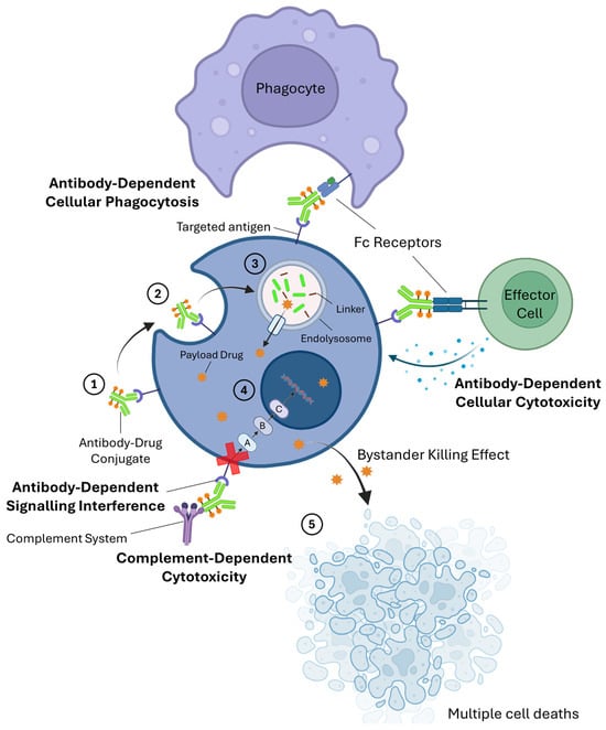
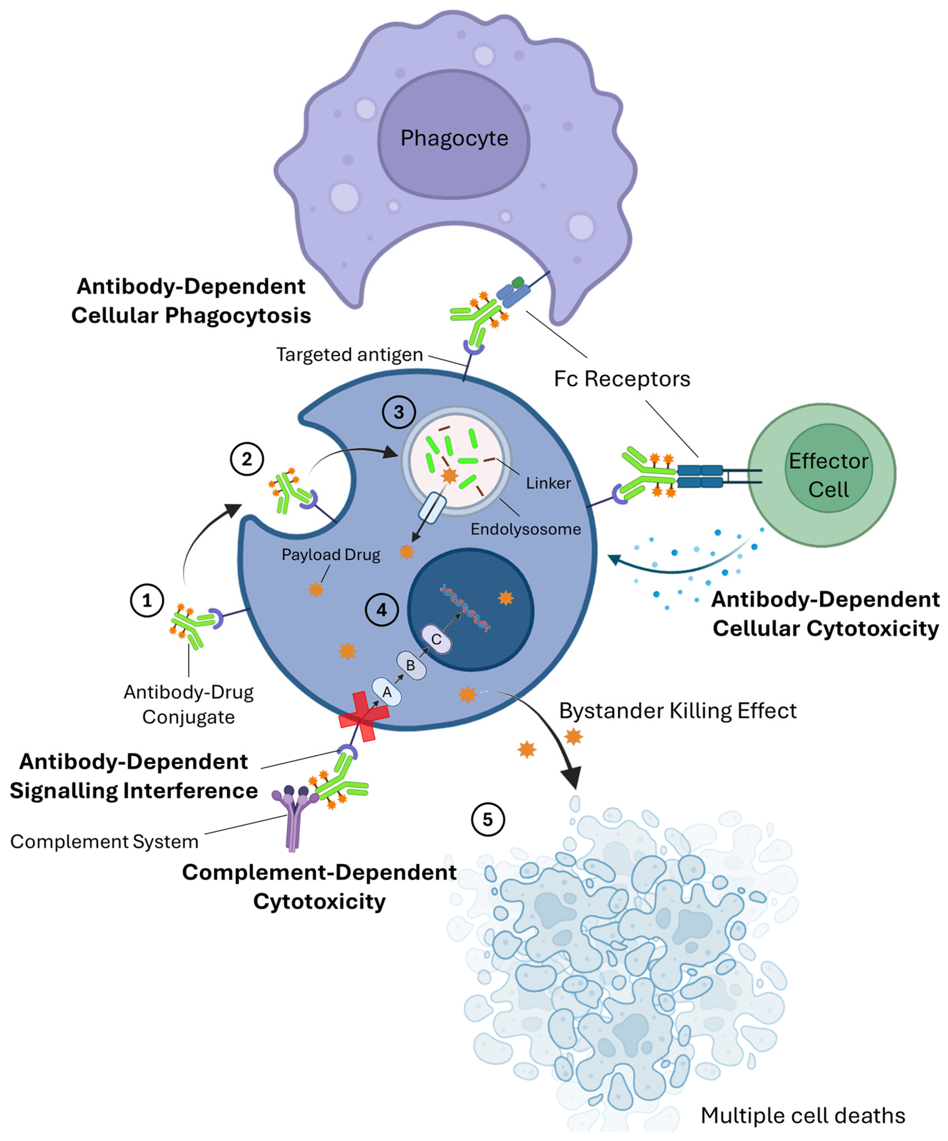
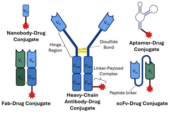
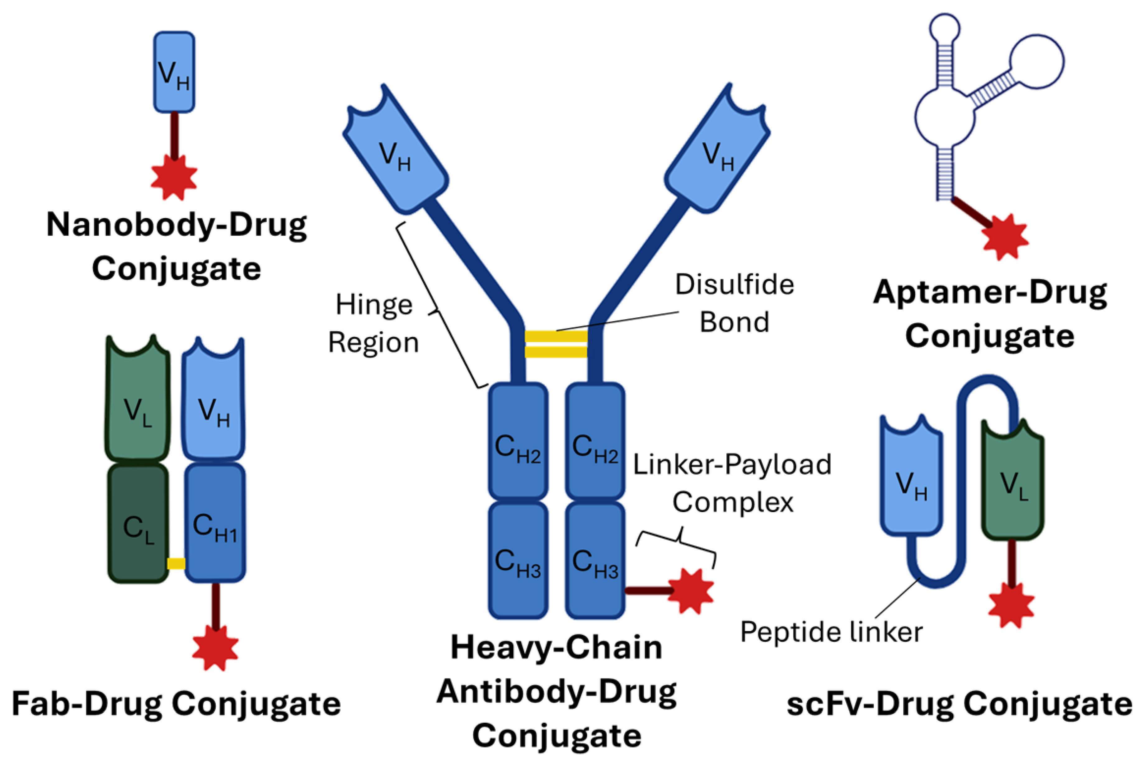
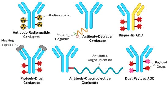
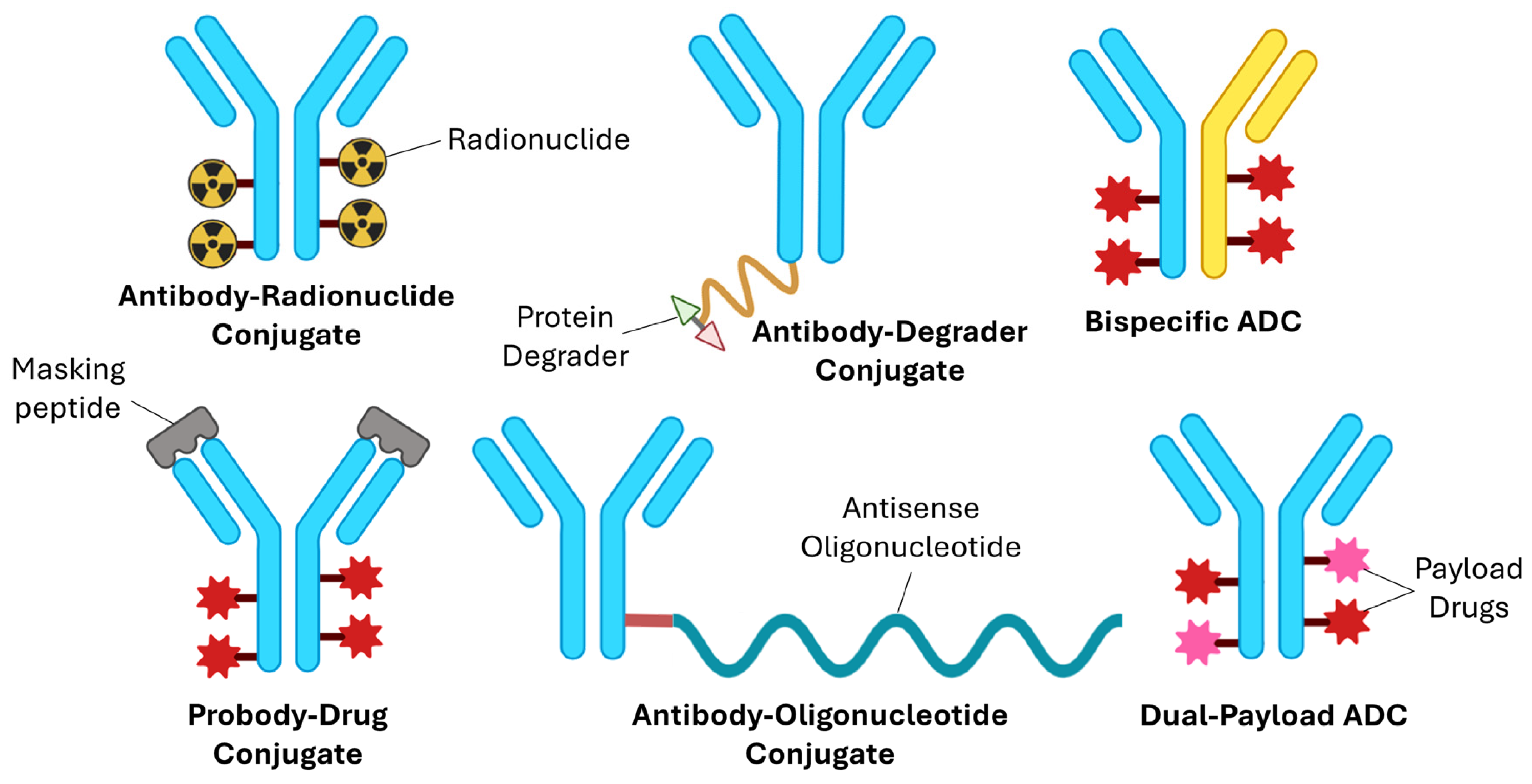
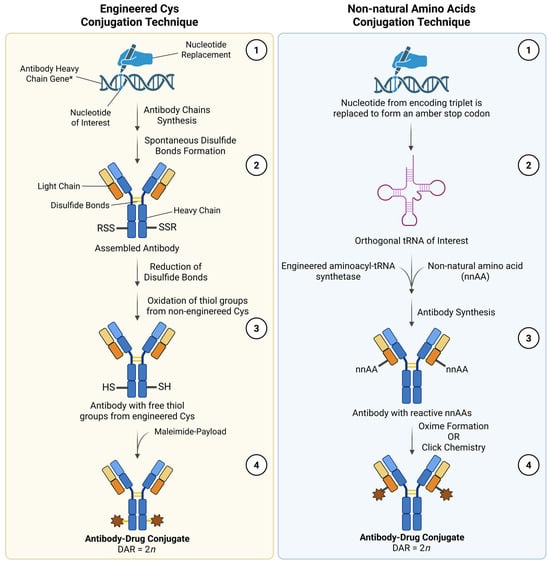
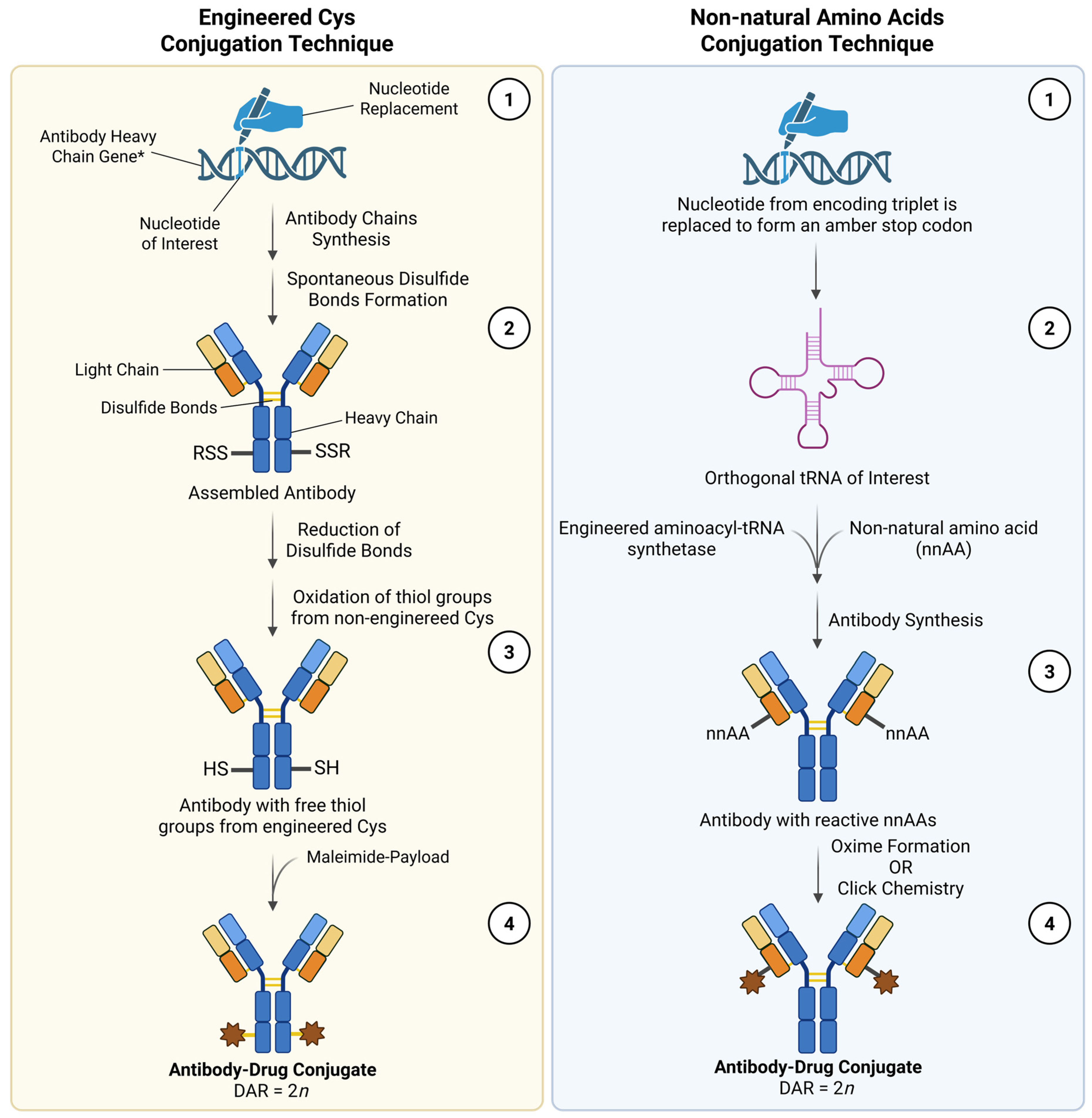
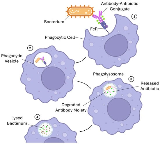
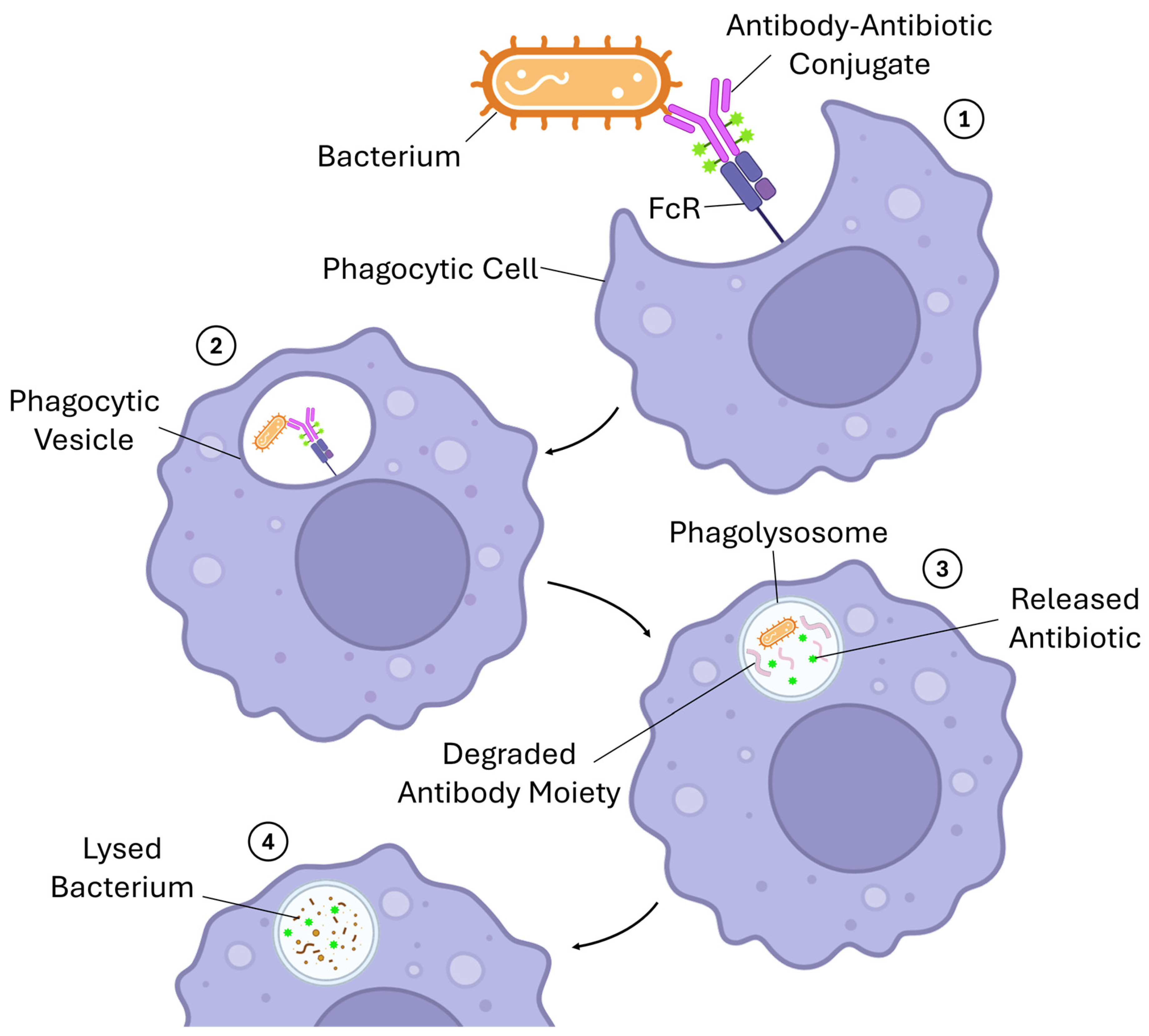

Disclaimer/Publisher’s Note: The statements, opinions and data contained in all publications are solely those of the individual author(s) and contributor(s) and not of MDPI and/or the editor(s). MDPI and/or the editor(s) disclaim responsibility for any injury to people or property resulting from any ideas, methods, instructions or products referred to in the content. |
© 2026 by the authors. Licensee MDPI, Basel, Switzerland. This article is an open access article distributed under the terms and conditions of the Creative Commons Attribution (CC BY) license.