Antibody–Drug Conjugates: Pharmacotherapeutic Properties and Future Perspectives




Abstract
1. Introduction
2. Pharmacotherapy of ADCs
3. Current Limitations
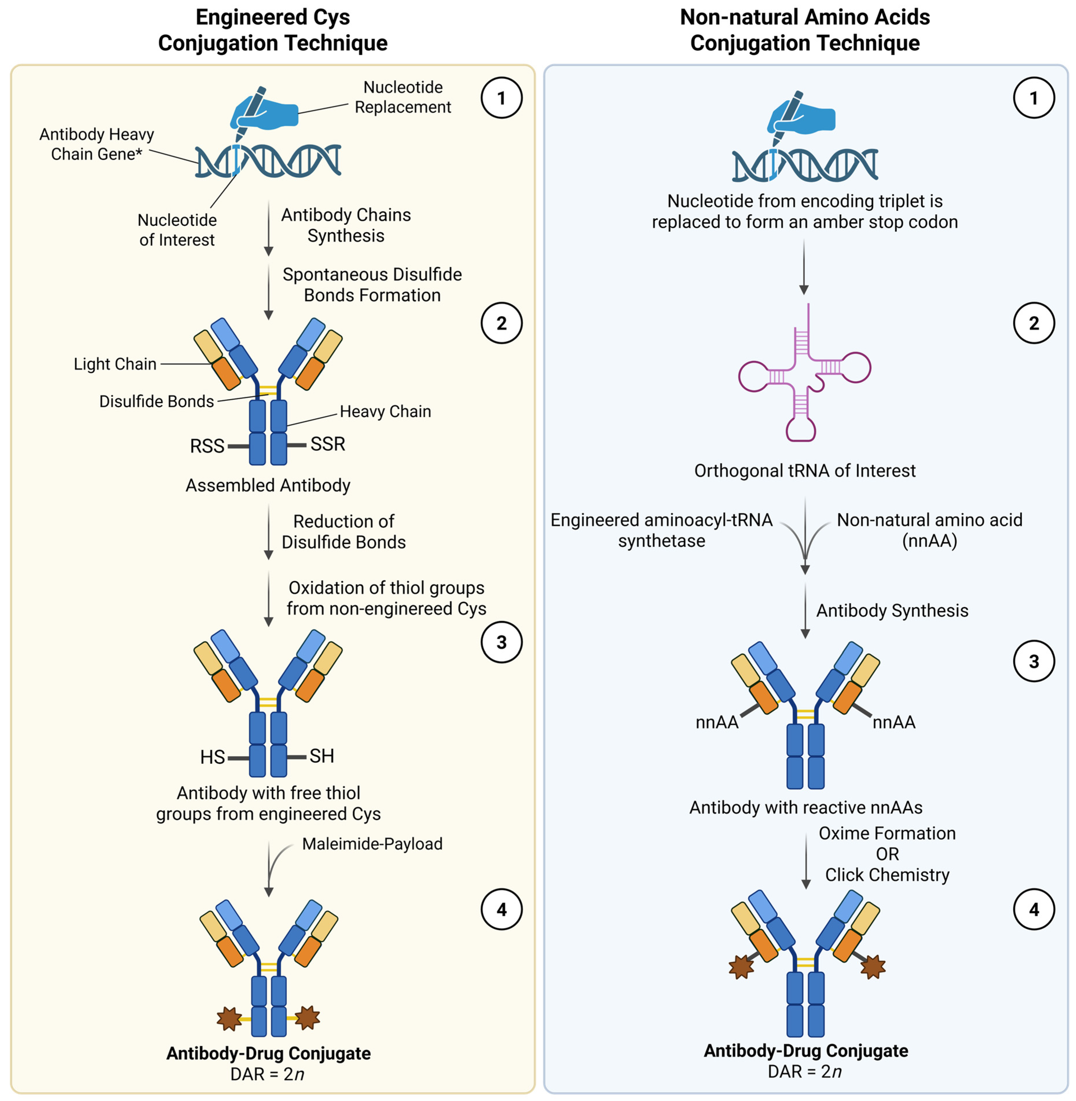
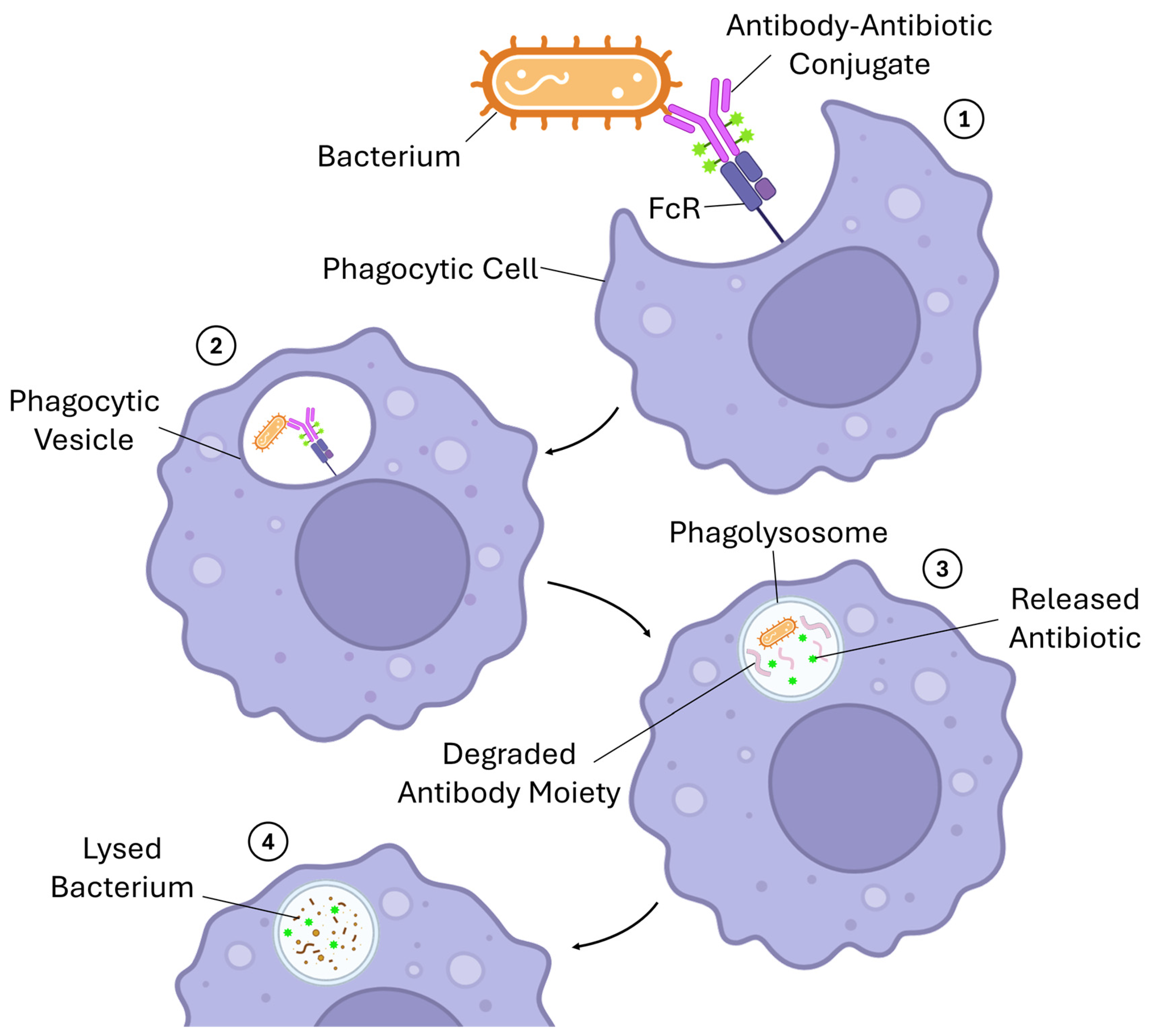
4. Novel Approaches to the Concept of ADC and New Therapeutic Applications
5. ADCs in Clinical Trials
6. Conclusions
Author Contributions
Funding
Institutional Review Board Statement
Informed Consent Statement
Data Availability Statement
Conflicts of Interest
Abbreviations
| ADCC | Antibody-dependent cell-mediated cytotoxicity |
| ADCP | Antibody-dependent cellular phagocytosis |
| ADCs | Antibody–drug conjugates |
| ATP | Adenosine triphosphate |
| CDC | Complement-dependent cytotoxicity |
| CLDN18.2 | Claudin 18.2 |
| DAR | Drug-to-antibody ratios |
| DNA | Deoxyribonucleic acid |
| Fab | Fragment antigen-binding |
| Fc | Fragment crystallizable |
| HER2 | Human epidermal growth factor receptor 2 |
| HER3 | Human epidermal growth factor receptor 3 |
| mAbs | Monoclonal antibodies |
| mRNA | Messenger ribonucleic acid |
| PROTACs | Proteolysis targeting chimeras |
| RNA | Ribonucleic acid |
| scFv | Single-chain variable fragment |
| STING | Stimulator of interferon genes |
| TAA | Tumor-associated antigen |
| TLRs | Toll-like receptors |
| TROP2 | Trophoblast cell surface antigen 2 |
| TSA | Tumor-specific antigen |
References
- Conceição, J.; Adeoye, O.; Cabral Marques, H.; Concheiro, A.; Alvarez Lorenzo, C.; Sousa Lobo, J.M. Carbamazepine bilayer tablets combining hydrophilic and hydrophobic cyclodextrins as a quick/slow biphasic release system. J. Drug Deliv. Sci. Technol. 2020, 57, 101611. [Google Scholar] [CrossRef]
- Kim, Y.; Park, E.J.; Na, D.H. Antibody–drug conjugates for targeted anticancer drug delivery. J. Pharm. Investig. 2016, 46, 341–349. [Google Scholar] [CrossRef]
- Fong, J.; Phuna, Z.; Chong, D.; Heryanto, C.; Low, Y.; Oh, K.; Lee, Y.; Ng, A.; In, L.; Teo, M. Advancements in antibody-drug conjugates as cancer therapeutics. J. Natl. Cancer Cent. 2025, 5, 362–378. [Google Scholar] [CrossRef] [PubMed]
- Alradwan, I.A.; Alnefaie, M.K.; Al Fayez, N.; Aodah, A.H.; Majrashi, M.A.; Alturki, M.; Fallatah, M.M.; Almughem, F.A.; Tawfik, E.A.; Alshehri, A.A. Strategic and Chemical Advances in Antibody–Drug Conjugates. Pharmaceutics 2025, 17, 1164. [Google Scholar] [CrossRef] [PubMed]
- Chis, A.; Dobrea, C.; Arseniu, A.; Frum, A.; Rus, L.; Cormos, G.; Georgescu, C.; Morgovan, C.; Butuca, A.; Gligor, F.; et al. Antibody-Drug Conjugates—Evolution and Perspectives. Int. J. Mol. Sci. 2024, 25, 6969. [Google Scholar] [CrossRef]
- Wang, R.; Hu, B.; Pan, Z.; Mo, C.; Zhao, X.; Liu, G.; Hou, P.; Cui, Q.; Xu, Z.; Wang, W.; et al. Antibody-Drug Conjugates (ADCs): Current and future biopharmaceuticals. J. Hematol. Oncol. 2025, 18, 51. [Google Scholar] [CrossRef] [PubMed]
- Chen, B.; Zheng, X.; Wu, J.; Chen, G.; Yu, J.; Xu, Y.; Wu, W.; Tse, G.; To, K.; Kang, W. Antibody–drug conjugates in cancer therapy: Current landscape, challenges, and future directions. Mol. Cancer 2025, 24, 279. [Google Scholar] [CrossRef] [PubMed]
- Zhou, X.; Han, Y.; Fang, Y.; Ma, P.; Zhou, J.; Jiang, Y.; Xing, S.; Tang, Q.; Hou, Y.; Wang, S.; et al. Antibody-drug conjugates: Current challenges and innovative solutions for precision cancer therapy. Med 2025, 6, 100849. [Google Scholar] [CrossRef] [PubMed]
- Connors, J.; Jurczak, W.; Straus, D.; Ansell, S.; Kim, W.; Gallamini, A.; Younes, A.; Alekseev, S.; Illés, Á.; Picardi, M.; et al. Brentuximab Vedotin with Chemotherapy for Stage III or IV Hodgkin’s Lymphoma. N. Engl. J. Med. 2018, 378, 331–344. [Google Scholar] [CrossRef]
- Mansinho, A.; Albuquerque, J.; Barigazzi, C.; Calvo, E. Antibody-drug conjugates in cancer treatment: From molecular design to clinical implementation. Lancet Reg. Health Eur. 2026, 101634. [Google Scholar] [CrossRef]
- Verma, S.; Miles, D.; Gianni, L.; Krop, I.; Welslau, M.; Baselga, J.; Pegram, M.; Oh, D.; Diéras, V.; Guardino, E.; et al. Trastuzumab emtansine for HER2-positive advanced breast cancer. N. Engl. J. Med. 2012, 367, 1783–1791. [Google Scholar] [CrossRef] [PubMed]
- Zhou, M.; Huang, Z.; Ma, Z.; Chen, J.; Lin, S.; Yang, X.; Gong, Q.; Braunstein, Z.; Wei, Y.; Rao, X.; et al. The next frontier in antibody-drug conjugates: Challenges and opportunities in cancer and autoimmune therapy. Cancer Drug Resist. 2025, 8, 34. [Google Scholar] [CrossRef]
- Fu, Z.; Li, S.; Han, S.; Shi, C.; Zhang, Y. Antibody drug conjugate: The “biological missile” for targeted cancer therapy. Signal Transduct. Target. Ther. 2022, 7, 93. [Google Scholar] [CrossRef] [PubMed]
- Wang, Y.; Cheng, X.; Li, X.; Chen, W.; Zhang, X.; Liu, Y. Bystander effect in antibody-drug conjugates: Navigating the fine line in tumor heterogeneity. Crit. Rev. Oncol. Hematol. 2025, 216, 104979. [Google Scholar] [CrossRef] [PubMed]
- Metrangolo, V.; Engelholm, L. Antibody-Drug Conjugates: The Dynamic Evolution from Conventional to Next-Generation Constructs. Cancers 2024, 16, 447. [Google Scholar] [CrossRef] [PubMed]
- Al Meslamani, A.; Bostanudin, M. Antibody-drug conjugates in oncology: Insights into Current challenges. Expert Opin. Biol. Ther. 2023, 23, 1035–1038. [Google Scholar] [CrossRef] [PubMed]
- Noorkhajavi, G.; Banakholdi, A.; Torabi, A.; Zoghi, A.; Iranijam, E.; Safarzadeh, E. Recent clinical advances in nonconjugated antibodies and antibody-drug conjugates for colorectal cancer treatment. Cancer Cell Int. 2025, 25, 395. [Google Scholar] [CrossRef]
- Justiz-Vaillant, A. Antibody–Drug Conjugates (ADCs) and Their Journey to Autoimmune Disease Immunotherapy. Med. Sci. Forum 2025, 40, 2. [Google Scholar]
- Lonial, S.; Lee, H.; Badros, A.; Trudel, S.; Nooka, A.; Chari, A.; Abdallah, A.; Callander, N.; Lendvai, N.; Sborov, D.; et al. Belantamab mafodotin for relapsed or refractory multiple myeloma (DREAMM-2): A two-arm, randomised, open-label, phase 2 study. Lancet Oncol. 2020, 21, 207–221. [Google Scholar] [CrossRef]
- Mukhopadhyay, P.; Abdullah, H.A.; Opalinska, J.B.; Paka, P.; Richards, E.; Weisel, K.; Trudel, S.; Mateos, M.-V.; Dimopoulos, M.A.; Lonial, S. The clinical journey of belantamab mafodotin in relapsed or refractory multiple myeloma: Lessons in drug development. Blood Cancer J. 2025, 15, 15. [Google Scholar] [CrossRef]
- Sureda, A.; García-Sanz, R.; Domingo-Domenech, E.; Capote, F.J.; Gutiérrez, A.; Rodriguez, A.; Aguiar, D.; Giraldo, P.; Infante, M.S.; López-Jiménez, J.; et al. Retreatment with Brentuximab Vedotin in Patients with Relapsed/Refractory CD30+ Malignancies: A Retrospective Medical Chart Review Study in Spain-The BELIEVE Study. Cancers 2025, 17, 1137. [Google Scholar] [CrossRef] [PubMed]
- Lambert, J.; Pautas, C.; Terré, C.; Raffoux, E.; Turlure, P.; Caillot, D.; Legrand, O.; Thomas, X.; Gardin, C.; Gogat-Marchant, K.; et al. Gemtuzumab ozogamicin for de novo acute myeloid leukemia: Final efficacy and safety updates from the open-label, phase III ALFA-0701 trial. Haematologica 2019, 104, 113–119. [Google Scholar] [CrossRef] [PubMed]
- Ronnacker, J.; Muller, P.J.; Mikesch, J.-H.; Zukunft, S.; Weinbergerová, B.; Šrámek, J.; Valka, J.; Novak, J.; Zak, P.; Szotkowski, T.; et al. Gemtuzumab ozogamicin in first-line treatment of CBF-AML: Insights from a retrospective multi-center analysis. Leukemia 2025, 39, 2174–2180. [Google Scholar] [CrossRef]
- Kantarjian, H.; DeAngelo, D.; Stelljes, M.; Liedtke, M.; Stock, W.; Gökbuget, N.; O’Brien, S.; Jabbour, E.; Wang, T.; White, J.; et al. Inotuzumab ozogamicin versus standard of care in relapsed or refractory acute lymphoblastic leukemia: Final report and long-term survival follow-up from the randomized, phase 3 INO-VATE study. Cancer 2019, 125, 2474–2487. [Google Scholar] [CrossRef]
- Lao, C.; Wen, J.; Wen, Y. Efficacy and safety of inotuzumab ozogamicin and its combination therapies in acute lymphoblastic leukemia: A systematic review and meta-analysis. Front. Oncol. 2025, 15, 1613777. [Google Scholar] [CrossRef] [PubMed]
- Caimi, P.; Ai, W.; Alderuccio, J.; Ardeshna, K.; Hamadani, M.; Hess, B.; Kahl, B.; Radford, J.; Solh, M.; Stathis, A.; et al. Loncastuximab tesirine in relapsed or refractory diffuse large B-cell lymphoma (LOTIS-2): A multicentre, open-label, single-arm, phase 2 trial. Lancet Oncol. 2021, 22, 790–800. [Google Scholar] [CrossRef] [PubMed]
- Juárez-Salcedo, L.M.; Nimkar, S.; Corazón, A.M.; Dalia, S. Loncastuximab Tesirine in the Treatment of Relapsed or Refractory Diffuse Large B-Cell Lymphoma. Int. J. Mol. Sci. 2024, 25, 7580. [Google Scholar] [CrossRef]
- Kuruvilla, D.; Chia, Y.; Balic, K.; Yao, N.; Kreitman, R.; Pastan, I.; Li, X.; Standifer, N.; Liang, M.; Tseng, C.; et al. Population pharmacokinetics, efficacy, and safety of moxetumomab pasudotox in patients with relapsed or refractory hairy cell leukaemia. Br. J. Clin. Pharmacol. 2020, 86, 1367–1376. [Google Scholar] [CrossRef]
- Yurkiewicz, I.R.; Coutre, S.; Ghesquieres, H.; Pastan, I.; Kreitman, R.J. Moxetumomab pasudotox as re-treatment for heavily-pretreated relapsed hairy cell leukemia. Leuk. Lymphoma 2021, 62, 2812–2814. [Google Scholar] [CrossRef]
- Sehn, L.; Herrera, A.; Flowers, C.; Kamdar, M.; McMillan, A.; Hertzberg, M.; Assouline, S.; Kim, T.; Kim, W.; Ozcan, M.; et al. Polatuzumab Vedotin in Relapsed or Refractory Diffuse Large B-Cell Lymphoma. J. Clin. Oncol. 2020, 38, 155–165. [Google Scholar] [CrossRef] [PubMed]
- Tilly, H.; Morschhauser, F.; Sehn, L.H.; Friedberg, J.W.; Trněný, M.; Sharman, J.P.; Herbaux, C.; Burke, J.M.; Matasar, M.; Rai, S.; et al. Polatuzumab Vedotin in Previously Untreated Diffuse Large B-Cell Lymphoma. N. Engl. J. Med. 2022, 386, 351–363. [Google Scholar] [CrossRef] [PubMed]
- Blair, H. Becotatug Vedotin: First Approval. Drugs 2026, 86, 573–579. [Google Scholar] [CrossRef] [PubMed]
- Han, F.; Lv, X.; Zhou, Y.-J.; He, Q.; Guo, Y.; Xiang, Y.-Q.; Shu, X.-L.; Pan, S.-M.; Qu, S.-H.; Zhang, P.; et al. Becotatug vedotin, MRG003, in previously treated recurrent or metastatic nasopharyngeal carcinoma: A multicenter, single-arm, phase IIa trial. Med 2026, 7, 101029. [Google Scholar] [CrossRef] [PubMed]
- Okamoto, I.; Hasegawa, O.; Kushihashi, Y.; Masubuchi, T.; Tokashiki, K.; Tsukahara, K. Real-World Effectiveness and Safety of Photoimmunotherapy for Head and Neck Cancer: A Multicenter Retrospective Study. Cancers 2025, 17, 2671. [Google Scholar] [CrossRef]
- Okamoto, I. Photoimmunotherapy for head and neck cancer: A systematic review. Auris Nasus Larynx 2025, 52, 186–194. [Google Scholar] [CrossRef]
- Ahn, M.; Tanaka, K.; Paz-Ares, L.; Cornelissen, R.; Girard, N.; Pons-Tostivint, E.; Vicente Baz, D.; Sugawara, S.; Cobo, M.; Pérol, M.; et al. Datopotamab Deruxtecan Versus Docetaxel for Previously Treated Advanced or Metastatic Non-Small Cell Lung Cancer: The Randomized, Open-Label Phase III TROPION-Lung01 Study. J. Clin. Oncol. 2025, 43, 260–272. [Google Scholar] [CrossRef]
- Huang, J.; Huang, T.; Guo, J.; Hu, K.; Zhou, H. The efficacy and safety of datopotamab deruxtecan (Dato-DXd) in advanced solid tumors: A systematic review and meta-analysis. Eur. J. Med. Res. 2025, 30, 1265. [Google Scholar] [CrossRef]
- Shi, F.; Liu, Y.; Zhou, X.; Shen, P.; Xue, R.; Zhang, M. Disitamab vedotin: A novel antibody-drug conjugates for cancer therapy. Drug Deliv. 2022, 29, 1335–1344. [Google Scholar] [CrossRef]
- Ye, J.; Li, Y.; Zhang, M.; Liao, X.; Wang, Q.; Chen, Z.; Tu, X.; Tan, P.; Zhang, P.; Zeng, H.; et al. Efficacy of disitamab vedotin-based therapy in HER2-negative and HER2-low locally advanced or metastatic urothelial carcinoma: A systematic review and meta-analysis. Urol. Oncol. 2026, 111034. [Google Scholar] [CrossRef]
- Powles, T.; Rosenberg, J.; Sonpavde, G.; Loriot, Y.; Durán, I.; Lee, J.; Matsubara, N.; Vulsteke, C.; Castellano, D.; Wu, C.; et al. Enfortumab Vedotin in Previously Treated Advanced Urothelial Carcinoma. N. Engl. J. Med. 2021, 384, 1125–1135. [Google Scholar] [CrossRef] [PubMed]
- Powles, T.; Valderrama, B.P.; Gupta, S.; Bedke, J.; Kikuchi, E.; Hoffman-Censits, J.; Iyer, G.; Vulsteke, C.; Park, S.H.; Shin, S.J.; et al. Enfortumab Vedotin and Pembrolizumab in Untreated Advanced Urothelial Cancer. N. Engl. J. Med. 2024, 390, 875–888. [Google Scholar] [CrossRef] [PubMed]
- Moore, K.; Angelergues, A.; Konecny, G.; García, Y.; Banerjee, S.; Lorusso, D.; Lee, J.; Moroney, J.; Colombo, N.; Roszak, A.; et al. Mirvetuximab Soravtansine in FRα-Positive, Platinum-Resistant Ovarian Cancer. N. Engl. J. Med. 2023, 389, 2162–2174. [Google Scholar] [CrossRef] [PubMed]
- Kokori, E.; Olatunji, G.; Komolafe, R.; Abraham, I.C.; Ukoaka, B.; Samuel, O.; Ayodeji, A.; Ogunbowale, I.; Ezenwoba, C.; Aderinto, N. Mirvetuximab soravtansine: A breakthrough in targeted therapy for platinum-resistant ovarian cancer. Medicine 2024, 103, e38132. [Google Scholar] [CrossRef]
- Bardia, A.; Hurvitz, S.; Tolaney, S.; Loirat, D.; Punie, K.; Oliveira, M.; Brufsky, A.; Sardesai, S.; Kalinsky, K.; Zelnak, A.; et al. Sacituzumab Govitecan in Metastatic Triple-Negative Breast Cancer. N. Engl. J. Med. 2021, 384, 1529–1541. [Google Scholar] [CrossRef] [PubMed]
- Jiang, L.; Dai, Y.; Li, M.; Zhao, Z. The efficacy and safety of sacituzumab govitecan in the treatment of breast cancer: A systemic review and meta-analysis of emerging clinical data. Front. Immunol. 2025, 16, 1683594. [Google Scholar] [CrossRef]
- Tolaney, S.; Cardillo, T.; Chou, C.; Dornan, C.; Faris, M. The Mode of Action and Clinical Outcomes of Sacituzumab Govitecan in Solid Tumors. Clin. Cancer Res. 2025, 31, 1390–1399. [Google Scholar] [CrossRef]
- Sofianidi, A.A.; Papatheodoridi, A.; Dimitrakakis, C.; Marinopoulos, S.; Michalaki, V.; Zagouri, F. Sacituzumab tirumotecan (sac-TMT/MK-2870/SKB264): A novel antibody–drug conjugate in breast cancer. Oncol. Rev. 2026, 20, 1781533. [Google Scholar] [CrossRef]
- Camidge, D.; Bar, J.; Horinouchi, H.; Goldman, J.; Moiseenko, F.; Filippova, E.; Cicin, I.; Ciuleanu, T.; Daaboul, N.; Liu, C.; et al. Telisotuzumab Vedotin Monotherapy in Patients with Previously Treated c-Met Protein-Overexpressing Advanced Nonsquamous EGFR-Wildtype Non-Small Cell Lung Cancer in the Phase II LUMINOSITY Trial. J. Clin. Oncol. 2024, 42, 3000–3011. [Google Scholar] [CrossRef]
- Khan, M.A.H.; Ahmad, W.; Ehsan, H.; Iqbal, M.; Rayyan, S.M.; Mehrpoor, A.J. Emrelis (telisotuzumab vedotin-tllv): Targeting C-MET in non-small cell lung cancer. Ann. Med. Surg. 2025, 87, 7870–7871. [Google Scholar] [CrossRef]
- Hong, D.; Concin, N.; Vergote, I.; de Bono, J.; Slomovitz, B.; Drew, Y.; Arkenau, H.; Machiels, J.; Spicer, J.; Jones, R.; et al. Tisotumab Vedotin in Previously Treated Recurrent or Metastatic Cervical Cancer. Clin. Cancer Res. 2020, 26, 1220–1228. [Google Scholar] [CrossRef] [PubMed]
- Vergote, I.; González-Martín, A.; Fujiwara, K.; Kalbacher, E.; Bagaméri, A.; Ghamande, S.; Lee, J.-Y.; Banerjee, S.; Maluf, F.C.; Lorusso, D.; et al. Tisotumab Vedotin as Second- or Third-Line Therapy for Recurrent Cervical Cancer. N. Engl. J. Med. 2024, 391, 44–55. [Google Scholar] [CrossRef]
- Hu, X.; Zhang, J.; Ouyang, Q.; Zhang, Q.; Li, H.; Wang, X.; Wang, Y.; Yin, Y.; Wang, S.; Gu, Y.; et al. LBA24 Trastuzumab botidotin vs trastuzumab emtansine (T-DM1) in HER2-positive unresectable or metastatic breast cancer: Results from a randomized phase III study. Ann. Oncol. 2025, 36, S1569. [Google Scholar] [CrossRef]
- Cerci, B.; Saatci, O.; Basik, M.; Sahin, O. Mechanisms of resistance to antibody-drug conjugates in breast cancer. Drug Resist. Updat. 2026, 85, 101353. [Google Scholar] [CrossRef]
- Cortés, J.; Kim, S.; Chung, W.; Im, S.; Park, Y.; Hegg, R.; Kim, M.; Tseng, L.; Petry, V.; Chung, C.; et al. Trastuzumab Deruxtecan versus Trastuzumab Emtansine for Breast Cancer. N. Engl. J. Med. 2022, 386, 1143–1154. [Google Scholar] [CrossRef]
- Quaquarini, E.; Luelli, F.; Lasagna, A.; Rizzo, G.; Perrone, L.; Figini, S.; Achille, R.; Pedrazzoli, P. Trastuzumab–Deruxtecan for the Treatment of Metastatic Breast Cancer Patients: Data from Real World Studies. Cancers 2025, 17, 3505. [Google Scholar] [CrossRef] [PubMed]
- Diéras, V.; Miles, D.; Verma, S.; Pegram, M.; Welslau, M.; Baselga, J.; Krop, I.; Blackwell, K.; Hoersch, S.; Xu, J.; et al. Trastuzumab emtansine versus capecitabine plus lapatinib in patients with previously treated HER2-positive advanced breast cancer (EMILIA): A descriptive analysis of final overall survival results from a randomised, open-label, phase 3 trial. Lancet Oncol. 2017, 18, 732–742. [Google Scholar] [CrossRef]
- Chandrakanth, M.V.; Agarwala, V.; Manna, R.; Sharma, A.; Roy, M.; Dasgupta, A.; Mandal, K.; Sen, N.; Sardar, P.K.; Sadhya, A.; et al. Real-World Evaluation of a Trastuzumab Emtansine Biosimilar (Ujvira®) in Human Epidermal Growth Factor Receptor 2 (HER2)-Positive Metastatic Breast Cancer. Cureus 2026, 18, e103700. [Google Scholar] [CrossRef] [PubMed]
- Li, Z.; Wang, Y.; Sun, Y.; Wang, L.; Li, X.; Sun, L.; He, Z.; Yang, H.; Wang, Y.; Wang, Q.; et al. Trastuzumab rezetecan, a HER2-directed antibody-drug conjugate, in patients with advanced HER2-mutant non-small-cell lung cancer (HORIZON-Lung): Phase 2 results from a multicentre, single-arm study. Lancet Oncol. 2025, 26, 437–446. [Google Scholar] [CrossRef] [PubMed]
- Hoy, S.M. Trastuzumab Rezetecan: First Approval. Drugs 2026, 86, 111–120. [Google Scholar] [CrossRef]
- Wahab, A.; Rafae, A.; Mushtaq, K.; Masood, A.; Ehsan, H.; Khakwani, M.; Khan, A. Ocular Toxicity of Belantamab Mafodotin, an Oncological Perspective of Management in Relapsed and Refractory Multiple Myeloma. Front. Oncol. 2021, 11, 678634. [Google Scholar] [CrossRef]
- Sunami, K.; Fujisaki, T.; Funaki, T.; Ichii, M.; Ito, S.; Matsumoto, M.; Oshima, K.-I.; Suzuki, K.; Takakuwa, T. Practical guidance on the clinical management of ocular adverse events associated with belantamab mafodotin in patients with relapsed/refractory multiple myeloma: Recommendations from a Japanese expert panel. Jpn. J. Clin. Oncol. 2025, 55, 1349–1356. [Google Scholar] [CrossRef]
- Tan, F.; Li, Y.; Xia, H.; Yin, W. Signal mining and analysis of adverse events of Brentuximab Vedotin base on FAERS and JADER databases. PLoS ONE 2025, 20, e0322378. [Google Scholar] [CrossRef]
- Gao, S.; Zhang, M.; Wu, K.; Zhu, J.; He, Z.; Li, J.; Chen, C.; Qiu, K.; Yu, X.; Wu, J. Risk of adverse events in lymphoma patients treated with brentuximab vedotin: A systematic review and meta-analysis. Expert Opin. Drug Saf. 2020, 19, 617–623. [Google Scholar] [CrossRef]
- Liu, X.; Wu, F.; Li, Z.; Li, C.; Li, X.; Yuan, Y.; Sun, X.; Du, Y.; Du, X.; Wang, S.; et al. Safety assessment of gemtuzumab ozogamicin: Real-world adverse event analysis based on the FDA Adverse Event Reporting System. Front. Med. 2025, 12, 1643780. [Google Scholar] [CrossRef] [PubMed]
- Cortes, J.E.; Lima, M.; Dombret, H.; Estey, E.H.; Giralt, S.A.; Montesinos, P.; Röllig, C.; Venditti, A.; Wang, E.S. Prevention, recognition, and management of adverse events associated with gemtuzumab ozogamicin use in acute myeloid leukemia. J. Hematol. Oncol. 2020, 13, 137. [Google Scholar] [CrossRef]
- Tran, V.; Srinivasa, N.; Tatum, C.; Reed, D.; Keng, M. Inotuzumab Ozogamicin in Clinical Practice: An Overview of Efficacy, Safety, and Real-World Applications. Curr. Hematol. Malig. Rep. 2026, 21, 5. [Google Scholar] [CrossRef]
- Ogrodnik, P.; Anderson, E.; Gloude, N.; Schiff, D. Efficacy and Toxicity of Inotuzumab Ozogamicin for Treatment of Relapsed/Refractory B-Cell Acute Lymphoblastic Leukemia in Pediatric and Young Adult Patients After CD19-Chimeric Antigen Receptor T-Cell Therapy. Pediatr. Blood Cancer 2025, 72, e32013. [Google Scholar] [CrossRef]
- Epperla, N.; Olszewski, A.J.; Ayers, E.C.; Ahmed, S. Management of Adverse Reactions to Loncastuximab in Patients with Relapsed or Refractory Diffuse Large B-Cell Lymphoma. Hematol. Oncol. 2025, 43, e70128. [Google Scholar] [CrossRef] [PubMed]
- Derenzini, E.; Gibb, A.; Kwiatek, M.; Strati, P. Loncastuximab tesirine in relapsed/refractory diffuse large B-cell lymphoma: Drug profile and expert opinion on the prevention and management of adverse events. Leuk. Lymphoma 2025, 66, 1990–2002. [Google Scholar] [CrossRef] [PubMed]
- Kreitman, R.J.; Dearden, C.; Zinzani, P.L.; Delgado, J.; Robak, T.; Coutre, P.D.; Gjertsen, B.T.; Troussard, X.; Roboz, G.J.; Karlin, L.; et al. Moxetumomab pasudotox in heavily pre-treated patients with relapsed/refractory hairy cell leukemia (HCL): Long-term follow-up from the pivotal trial. J. Hematol. Oncol. 2021, 14, 35. [Google Scholar] [CrossRef] [PubMed]
- Kang, C. Moxetumomab Pasudotox in Hairy Cell Leukaemia: A Profile of Its Use. Clin. Drug Investig. 2021, 41, 829–834. [Google Scholar] [CrossRef]
- Liu, D.; Mao, W.; Hu, B.; Li, X.; Zhao, Q.; Zhang, L.; Hu, J. A real-world pharmacovigilance study of polatuzumab vedotin based on the FDA adverse event reporting system (FAERS). Front. Pharmacol. 2024, 15, 1405023. [Google Scholar] [CrossRef]
- Xu, Z.; Huang, D.; Liu, Q.; Liu, S.; Liu, J.; Wang, H.; Shen, Z. A real-world pharmacovigilance analysis of the FDA adverse event reporting system events for polatuzumab vedotin. Expert. Opin. Drug Saf. 2025, 24, 49–57. [Google Scholar] [CrossRef] [PubMed]
- Qiu, M.-Z.; Zhang, Y.; Guo, Y.; Guo, W.; Nian, W.; Liao, W.; Xu, Z.; Zhang, W.; Zhao, H.-Y.; Wei, X.; et al. Evaluation of Safety of Treatment with Anti–Epidermal Growth Factor Receptor Antibody Drug Conjugate MRG003 in Patients with Advanced Solid Tumors. JAMA Oncol. 2022, 8, 1042–1046. [Google Scholar] [CrossRef]
- Abraham, F.F.; Julian, R. Emerging and Investigational Systemic Therapies in Recurrent/Metastatic Head and Neck Cancer After Progression on Immunotherapy. Cancers 2025, 17, 3817. [Google Scholar] [CrossRef]
- Okamoto, I.; Okada, T.; Tokashiki, K.; Tsukahara, K. Quality-of-Life Evaluation of Patients with Unresectable Locally Advanced or Locally Recurrent Head and Neck Carcinoma Treated with Head and Neck Photoimmunotherapy. Cancers 2022, 14, 4413. [Google Scholar] [CrossRef]
- Takuma, S.; Karayama, M.; Suzuki, R.; Miyashita, K.; Inoue, Y.; Yasui, H.; Hozumi, H.; Suzuki, Y.; Furuhashi, K.; Enomoto, N.; et al. Severe drug-induced interstitial lung disease after photoimmunotherapy with cetuximab–sarotalocan sodium in a patient with oropharyngeal cancer: A case report. Respir. Med. Case Rep. 2025, 57, 102260. [Google Scholar] [CrossRef] [PubMed]
- Lisberg, A.; Huppert, L.A.; Halmos, B.; Ledezma, B.; Soto-Romano, V.; Traina, T.A. Datopotamab deruxtecan–associated select adverse events: Clinical practices and institutional protocols on prophylaxis, monitoring, and management. Oncologist 2025, 30, oyaf225. [Google Scholar] [CrossRef] [PubMed]
- Gadaleta-Caldarola, G.; Lanotte, L.; Infusino, S.; Gadaleta-Caldarola, A.; Schipilliti, F.M.; Citrigno, C.; Petrarota, C.; Cusmai, A.; Rizzo, A. Safety evaluation of Datopotamab deruxtecan for triple-negative breast cancer: A meta-analysis. Cancer Treat. Res. Commun. 2023, 37, 100775. [Google Scholar] [CrossRef] [PubMed]
- He, S.; Wang, C.; Li, X.; Li, C.; Li, D.; Zhang, B.; Wan, J.; Yue, Z.; Wu, G.; Zhang, S.; et al. Real-world efficacy and safety of disitamab vedotin monotherapy or in combination with PD-1 inhibitors in locally advanced or metastatic upper tract urothelial carcinoma: A multicenter retrospective study. Front. Immunol. 2026, 16, 1699538. [Google Scholar] [CrossRef]
- Zhou, T.; Liu, H.; Shen, Y.; Shao, S. Real-world evaluation of the efficacy and safety of disitamab vedotin (RC48) in urothelial carcinoma. BMC Urol. 2025, 25, 276. [Google Scholar] [CrossRef] [PubMed]
- Nizam, A.; Nguyen, C.B.; Li, J.; Zabor, E.C.; Msaouel, P.; Jiang, C.Y.; Alhalabi, O.; Oh, E.; Davidsohn, M.P.; Epstein, I.B.; et al. Treatment-Related Adverse Events and Associated Outcomes in Patients with Advanced Urothelial Carcinoma Treated with Enfortumab Vedotin: Analysis of the UNITE Study. Cancer Med. 2025, 14, e71284. [Google Scholar] [CrossRef] [PubMed]
- Dobry, A.S.; Virgen, C.A.; Hosking, A.-M.; Mar, N.; Doan, L.; Lee, B.; Smith, J. Cutaneous reactions with enfortumab vedotin: A case series and review of the literature. JAAD Case Rep. 2021, 14, 7–9. [Google Scholar] [CrossRef]
- Lang, S.M.; Vingopoulos, F.; Beshar, I.; Somasegar, S.; Adams, E.V.; Sasse, S.A.; Ghezelayagh, T.; McClung, E.C.; Karam, A.; Dorigo, O.; et al. Early experience and assessment of real-world toxicities with mirvetuximab soravtansine in a heavily pretreated patient cohort with ovarian cancer. Gynecol. Oncol. Rep. 2025, 59, 101738. [Google Scholar] [CrossRef] [PubMed]
- Haidar, A.J.; Chen, A.; Chen, J.L.; Al-Hashimi, S.; Ghaffari, R.S.; Fung, S.; Silverstein, J.; Konecny, G.E.; Tsui, E. Ocular Toxic Effects During Mirvetuximab Soravtansine Therapy. JAMA Oncol. 2026, 12, 317–319. [Google Scholar] [CrossRef] [PubMed]
- Gui, X.; Zhao, J.; Ding, L.; Chai, J.; Lai, H.; Cai, Y.; Luo, S.; Zeng, Y.; Wu, W.; Chen, H.; et al. Assessing real-world safety concerns of Sacituzumab govitecan: A disproportionality analysis using spontaneous reports in the FDA adverse event reporting system. Front. Oncol. 2023, 13, 1276976. [Google Scholar] [CrossRef] [PubMed]
- Tolaney, S.M.; Sonpavde, G.P.; Tarantino, P.; Lustberg, M.B.; Rugo, H.S. Clinical perspective on management of key adverse events with sacituzumab govitecan. Oncologist 2025, 30, oyaf311. [Google Scholar] [CrossRef]
- Zhu, Y.; Jiang, S.; Shi, Y.; Wang, S.; Yuan, F.; Zhou, F.; Jiang, K.; Zhang, X.; Seneviratne, L.; Yu, G.; et al. Sacituzumab tirumotecan in participants with advanced or metastatic urothelial carcinoma and disease progression after chemotherapy and immune checkpoint inhibitors. Ann. Oncol. 2026, 37, 378–387. [Google Scholar] [CrossRef]
- Yin, Y.; Fan, Y.; Ouyang, Q.; Song, L.; Wang, X.; Li, W.; Li, M.; Yan, X.; Wang, S.; Sun, T.; et al. Sacituzumab tirumotecan in previously treated metastatic triple-negative breast cancer: A randomized phase 3 trial. Nat. Med. 2025, 31, 1969–1975. [Google Scholar] [CrossRef]
- Babel, H.; Brunsdon, P.; Engelhardt, B.; Schmitt, V.; Ratajczak, C.; Mensing, S.; Menon, R.M.; Parikh, A. Population Pharmacokinetics and Exposure-Response Analyses for Telisotuzumab Vedotin in Patients with c-Met Protein Overexpressing Tumors. CPT Pharmacomet. Syst. Pharmacol. 2026, 15, e70219. [Google Scholar] [CrossRef]
- Zhao, C.; Lu, D.; Gao, J. Telisotuzumab vedotin: The first-in-class c-Met-targeted antibody-drug conjugate granted FDA accelerated approval for treatment of non-squamous non-small cell lung cancer (NSCLC). Drug Discov. Ther. 2025, 19, 275–276. [Google Scholar] [CrossRef]
- Arn, C.R.; Halla, K.J.; Gill, S. Tisotumab Vedotin Safety and Tolerability in Clinical Practice: Managing Adverse Events. J. Adv. Pract. Oncol. 2023, 14, 139–152. [Google Scholar] [PubMed]
- Chess, I.; Mercier, A.M.; Gross, S.; Blank, S.V.; Hayes, M.P. Tisotumab vedotin–associated Stevens–Johnson syndrome in a patient with recurrent vulvar cancer: Vulnerability and antibody drug conjugate toxicity considerations. Gynecol. Oncol. Rep. 2026, 64, 102065. [Google Scholar] [CrossRef] [PubMed]
- Liu, X.; Deng, J.; Zhang, R.; Xing, J.; Wu, Y.; Chen, W.; Liang, B.; Xing, D.; Xu, J.; Zhang, M. The clinical development of antibody-drug conjugates for non-small cell lung cancer therapy. Front. Immunol. 2023, 14, 1335252. [Google Scholar] [CrossRef] [PubMed]
- Evangelou, C. Recent Advances in the Treatment of Early-Stage HER2-Positive Breast Cancer: Updates from ESMO 2025. Oncol. Am. Med. J. 2025, 2, 2–12. [Google Scholar] [CrossRef]
- Bianchini, G.; Fabi, A.; Guarneri, V.; Arpino, G.; Laurentis, M.D.; Zambelli, A.; Zamagni, C.; Biganzoli, L.; Puglisi, F.; Santini, D. Management of trastuzumab deruxtecan-related adverse events in breast cancer: Italian expert panel recommendations. Future Oncol. 2025, 21, 2565–2573. [Google Scholar] [CrossRef] [PubMed]
- Pathak, N.; Iorio, M.D.; Gimenez, D.M.; Berner-Wygoda, Y.; Savill, J.; Aljuhani, A.; Mittal, A.; Kumar, V.; Amir, E. Adverse effect of trastuzumab deruxtecan in solid tumours: A systematic review and meta-analysis. Crit. Rev. Oncol. Hematol. 2025, 213, 104787. [Google Scholar] [CrossRef]
- Liu, F.; Yin, G.; Xue, S.; Rehman, F.U.L.; Liao, D.; Pan, Y. Adverse Event Profile Differences between Trastuzumab Emtansine and Trastuzumab Deruxtecan: A Real-world, Pharmacovigilance Study. J. Cancer 2023, 14, 3275–3284. [Google Scholar] [CrossRef]
- Kim, H.J.; Yoon, J.-H.; Park, Y.H. Long-term hepatobiliary disorder associated with trastuzumab emtansine pharmacovigilance study using the FDA Adverse Event Reporting System database. Sci. Rep. 2024, 14, 19587. [Google Scholar] [CrossRef]
- Zhang, T.; Xu, J.; Yin, J.; Gao, Y.; Zheng, H.; Fu, B.; Sun, J.; Xu, Z.; Tu, S.; Mao, Y.; et al. SHR-A1811, a novel anti-HER2 antibody–drug conjugate with optimal drug-to-antibody ratio, efficient tumor killing potency, and favorable safety profiles. PLoS ONE 2025, 20, e0326691. [Google Scholar] [CrossRef]
- Papakonstantinou, A.; Foukakis, T. Neoadjuvant treatment of HER2-positive breast cancer: Has the era of antibody-drug conjugates arrived? Ann. Oncol. 2025, 36, 603–605. [Google Scholar] [CrossRef]
- Nguyen, T.; Bordeau, B.; Balthasar, J. Mechanisms of ADC Toxicity and Strategies to Increase ADC Tolerability. Cancers 2023, 15, 713. [Google Scholar] [CrossRef]
- Cheng, Y.; Lu, J.; Zhang, C.; Yan, W.; Zhu, P.; Qin, Q.; Gong, L. Overview of antibody-drug conjugates nonclinical and clinical toxicities and related contributing factors. Antib. Ther. 2025, 8, 124–144. [Google Scholar] [CrossRef]
- Tan, H.; Morcillo, M.; Lopez, J.; Minchom, A.; Sharp, A.; Paschalis, A.; Silva-Fortes, G.; Raobaikady, B.; Banerji, U. Treatment-related adverse events of antibody drug-conjugates in clinical trials. J. Hematol. Oncol. 2025, 18, 71. [Google Scholar] [CrossRef] [PubMed]
- Grudzinska-Goebel, J.; Benstein, K.; Bloem, K.; Cowan, K.; Gorovits, B.; Jadhav, M.; Janssen, M.; Jawa, V.; Kiessling, A.; Kramer, D.; et al. Immunogenicity risk assessment for tailored mitigation and monitoring of biotherapeutics during development: Recommendations from the European Immunogenicity Platform. Front. Immunol. 2025, 16, 1581153. [Google Scholar] [CrossRef]
- Li, S.; Zhao, X.; Fu, K.; Zhu, S.; Pan, C.; Yang, C.; Wang, F.; To, K.; Fu, L. Resistance to antibody–drug conjugates: A review. Acta Pharm. Sin. B 2024, 15, 737–756. [Google Scholar] [CrossRef] [PubMed]
- Khoury, R.; Saleh, K.; Khalife, N.; Saleh, M.; Chahine, C.; Ibrahim, R.; Lecesne, A. Mechanisms of Resistance to Antibody-Drug Conjugates. Int. J. Mol. Sci. 2023, 24, 9674. [Google Scholar] [CrossRef]
- Murase, Y.; Nanjo, S.; Ueda, T.; Liu, Y.; Nomura, S.; Arai, S.; Terada, N.; Koba, H.; Tambo, Y.; Yano, S. Mechanisms of resistance to antibody-drug conjugates in cancers. Respir. Investig. 2025, 63, 693–698. [Google Scholar] [CrossRef] [PubMed]
- Abelman, R.; Wu, B.; Spring, L.; Ellisen, L.; Bardia, A. Mechanisms of Resistance to Antibody–Drug Conjugates. Cancers 2023, 15, 1278. [Google Scholar] [CrossRef] [PubMed]
- Yousef, M.; Le, T.; Zuo, J.; Park, C.; Chacra, N.; Davies, N.; Löbenberg, R. Sub-cellular sequestration of alkaline drugs in lysosomes: New insights for pharmaceutical development of lysosomal fluid. Res. Pharm. Sci. 2022, 18, 1–15. [Google Scholar] [CrossRef]
- Kinneer, K.; Meekin, J.; Tiberghien, A.; Tai, Y.; Phipps, S.; Kiefer, C.; Rebelatto, M.; Dimasi, N.; Moriarty, A.; Papadopoulos, K.; et al. SLC46A3 as a Potential Predictive Biomarker for Antibody-Drug Conjugates Bearing Noncleavable Linked Maytansinoid and Pyrrolobenzodiazepine Warheads. Clin. Cancer Res. 2018, 24, 6570–6582. [Google Scholar] [CrossRef]
- Abelman, R.; Wu, B.; Barnes, H.; Medford, A.; Norden, B.; Putur, A.; Bitman, E.; Thant, W.; Liu, T.; Weipert, C.; et al. TOP1 Mutations and Cross-Resistance to Antibody-Drug Conjugates in Patients with Metastatic Breast Cancer. Clin. Cancer Res. 2025, 31, 1966–1974. [Google Scholar] [CrossRef] [PubMed]
- High, P.; Guernsey, C.; Subramanian, S.; Jacob, J.; Carmon, K. The Evolving Paradigm of Antibody–Drug Conjugates Targeting the ErbB/HER Family of Receptor Tyrosine Kinases. Pharmaceutics 2024, 16, 890. [Google Scholar] [CrossRef]
- Zhang, T.; Jia, Y.; Yu, Y.; Zhang, B.; Xu, F.; Guo, H. Targeting the tumor biophysical microenvironment to reduce resistance to immunotherapy. Adv. Drug Deliv. Rev. 2022, 186, 114319. [Google Scholar] [CrossRef]
- Géraud, A.; Gougis, P.; de Nonneville, A.; Beaufils, M.; Bertucci, F.; Billon, E.; Brisou, G.; Gravis, G.; Greillier, L.; Guerin, M.; et al. Pharmacology and pharmacokinetics of antibody-drug conjugates, where do we stand? Cancer Treat. Rev. 2025, 135, 102922. [Google Scholar] [CrossRef] [PubMed]
- Mckertish, C.; Kayser, V. Advances and Limitations of Antibody Drug Conjugates for Cancer. Biomedicines 2021, 9, 872. [Google Scholar] [CrossRef] [PubMed]
- Khongorzul, P.; Ling, C.; Khan, F.; Ihsan, A.; Zhang, J. Antibody-Drug Conjugates: A Comprehensive Review. Mol. Cancer Res. 2020, 18, 3–19. [Google Scholar] [CrossRef] [PubMed]
- Bates, A.; Power, C. David vs. Goliath: The Structure, Function, and Clinical Prospects of Antibody Fragments. Antibodies 2019, 8, 28. [Google Scholar] [CrossRef]
- Wang, T.; Li, M.; Wei, R.; Wang, X.; Lin, Z.; Chen, J.; Wu, X. Small Molecule-Drug Conjugates Emerge as a New Promising Approach for Cancer Treatment. Mol. Pharm. 2024, 21, 1038–1055. [Google Scholar] [CrossRef]
- Chen, W.; Yuan, Y.; Jiang, X. Antibody and antibody fragments for cancer immunotherapy. J. Control. Release 2020, 328, 395–406. [Google Scholar] [CrossRef]
- Riccardi, F.; Dal Bo, M.; Macor, P.; Toffoli, G. A comprehensive overview on antibody-drug conjugates: From the conceptualization to cancer therapy. Front. Pharmacol. 2023, 14, 1274088. [Google Scholar] [CrossRef]
- Alibakhshi, A.; Kahaki, F.; Ahangarzadeh, S.; Yaghoobi, H.; Yarian, F.; Arezumand, R.; Ranjbari, J.; Mokhtarzadeh, A.; de la Guardia, M. Targeted cancer therapy through antibody fragments-decorated nanomedicines. J. Control Release 2017, 268, 323–334. [Google Scholar] [CrossRef]
- Kalinovsky, D.; Kholodenko, I.; Kibardin, A.; Doronin, I.; Svirshchevskaya, E.; Ryazantsev, D.; Konovalova, M.; Rozov, F.; Larin, S.; Deyev, S.; et al. Minibody-Based and scFv-Based Antibody Fragment-Drug Conjugates Selectively Eliminate GD2-Positive Tumor Cells. Int. J. Mol. Sci. 2023, 24, 1239. [Google Scholar] [CrossRef] [PubMed]
- Ma, X.; Wang, M.; Ying, T.; Wu, Y. Reforming solid tumor treatment: The emerging potential of smaller format antibody-drug conjugate. Antib. Ther. 2024, 7, 114–122. [Google Scholar] [CrossRef]
- Solomonov, I.; Kollet, O.; Sagi, I. Extracellular matrix and proteolysis: Mechanisms driving irreversible changes and shaping cell behavior. FEBS J. 2025. [Google Scholar] [CrossRef] [PubMed]
- Lucchi, R.; Bentanachs, J.; Oller-Salvia, B. The Masking Game: Design of Activatable Antibodies and Mimetics for Selective Therapeutics and Cell Control. ACS Cent. Sci. 2021, 7, 724–738. [Google Scholar] [CrossRef] [PubMed]
- Luo, M.; Wang, X.; Yu, G.; Ji, J.; Li, L.; Song, F. Development of a bispecific antibody-drug conjugate targeting EpCAM and CLDN3 for the treatment of multiple solid tumors. Exp. Hematol. Oncol. 2025, 14, 33. [Google Scholar] [CrossRef]
- VanBlargan, L.; Adams, L.; Liu, Z.; Chen, R.; Gilchuk, P.; Raju, S.; Smith, B.; Zhao, H.; Case, J.; Winkler, E.; et al. A potently neutralizing SARS-CoV-2 antibody inhibits variants of concern by utilizing unique binding residues in a highly conserved epitope. Immunity 2021, 54, 2399–2416.E6. [Google Scholar] [CrossRef]
- Abdeldaim, D.; Schindowski, K. Fc-Engineered Therapeutic Antibodies: Recent Advances and Future Directions. Pharmaceutics 2023, 15, 2402. [Google Scholar] [CrossRef]
- Conilh, L.; Sadilkova, L.; Viricel, W.; Dumontet, C. Payload diversification: A key step in the development of antibody-drug conjugates. J. Hematol. Oncol. 2023, 16, 3. [Google Scholar] [CrossRef]
- Grairi, M.; Le Borgne, M. Antibody-drug conjugates: Prospects for the next generation. Drug Discov. Today 2024, 29, 104241. [Google Scholar] [CrossRef] [PubMed]
- Dhuri, K.; Bechtold, C.; Quijano, E.; Pham, H.; Gupta, A.; Vikram, A.; Bahal, R. Antisense Oligonucleotides: An Emerging Area in Drug Discovery and Development. J. Clin. Med. 2020, 9, 2004. [Google Scholar] [CrossRef] [PubMed]
- Sincere, N.; Anand, K.; Ashique, S.; Yang, J.; You, C. PROTACs: Emerging Targeted Protein Degradation Approaches for Advanced Druggable Strategies. Molecules 2023, 28, 4014. [Google Scholar] [CrossRef]
- Aoyama, M.; Tada, M.; Yokoo, H.; Ito, T.; Misawa, T.; Demizu, Y.; Ishii-Watabe, A. Linker and Conjugation Site Synergy in Antibody–Drug Conjugates: Impacts on Biological Activity. Bioconjug. Chem. 2024, 35, 1568–1576. [Google Scholar] [CrossRef] [PubMed]
- Su, Z.; Xiao, D.; Xie, F.; Liu, L.; Wang, Y.; Fan, S.; Zhou, X.; Li, S. Antibody–drug conjugates: Recent advances in linker chemistry. Acta Pharm. Sin. B 2021, 11, 3889–3907. [Google Scholar] [CrossRef]
- Jackson, D. Processes for Constructing Homogeneous Antibody Drug Conjugates. Org. Process Res. Dev. 2016, 20, 852–866. [Google Scholar] [CrossRef]
- Fan, Q.; Chen, H.; Wei, G.; Wei, D.; Wang, Z.; Zhang, L.; Wang, J.; Zhu, M. A review of conjugation technologies for antibody drug conjugates. Antib. Ther. 2025, 8, 157–170. [Google Scholar] [CrossRef]
- Blake-Hedges, J.; Groff, D.; Foo, W.; Hanson, J.; Castillo, E.; Wen, M.; Cheung, D.; Masikat, M.R.; Lu, J.; Park, Y.; et al. Production of antibodies and antibody fragments containing non-natural amino acids in Escherichia coli. MAbs 2024, 16, 2316872. [Google Scholar] [CrossRef] [PubMed]
- Matsuda, Y.; Chang, J.; Mendelsohn, B. Advanced Antibody-Drug Conjugates Design: Innovation in Linker Chemistry and Site-Specific Conjugation Technologies. ChemBioChem 2025, 26, e202500305. [Google Scholar] [CrossRef]
- Pal, L.; Bule, P.; Khan, W.; Chella, N. An Overview of the Development and Preclinical Evaluation of Antibody–Drug Conjugates for Non-Oncological Applications. Pharmaceutics 2023, 15, 1807. [Google Scholar] [CrossRef] [PubMed]
- Sasso, J.; Tenchov, R.; Bird, R.; Iyer, K.; Ralhan, K.; Rodriguez, Y.; Zhou, Q. The Evolving Landscape of Antibody-Drug Conjugates: In Depth Analysis of Recent Research Progress. Bioconjug. Chem. 2023, 34, 1951–2000. [Google Scholar] [CrossRef] [PubMed]
- Cavaco, M.; Castanho, M.; Neves, V. The Use of Antibody-Antibiotic Conjugates to Fight Bacterial Infections. Front. Microbiol. 2022, 13, 835677. [Google Scholar] [CrossRef] [PubMed]



| INN | Antibody Moiety | Linker | Payload | DAR | Therapeutic Indication(s) | Reference |
|---|---|---|---|---|---|---|
| Antibody–Drug Conjugates used in Hematologic Malignancies | ||||||
| Belantamab mafodotin | Anti-BCMA IgG1 | Maleimido-Caproyl (non-cleavable) | MMAF (Antimitotic Agent) | 4 | Adult patients with relapsed or refractory multiple myeloma (combination therapy) | [19,20] |
| Brentuximab vedotin | Anti-CD30 IgG1 | Mc-Val-Cit-PABC (protease-sensitive) | MMAE (Antimitotic Agent) | 4 | First-line treatment of adult patients with stage III/IV CD30+ Hodgkin lymphoma (combination therapy). It can also be used in adult patients with systemic anaplastic large cell lymphoma (in combination) or with CD30+ cutaneous T-cell lymphoma (monotherapy) | [9,21] |
| Gemtuzumab ozogamicin | Anti-CD33 IgG4 | AcBut (acid, and glutathione-sensitive) | N-Acetyl-Calicheamicin γ1 I (DNA Damaging Agent) | 2–3 | Patients aged 15 years or above, with previously untreated de novo CD33+ core binding factor acute myeloid leukemia (combination therapy) | [22,23] |
| Inotuzumab ozogamicin | Anti-CD22 IgG4 | AcBut (acid and glutathione-sensitive) | N-Acetyl-Calicheamicin γ1 I (DNA Damaging Agent) | 6 | Adult patients with relapsed or refractory CD22+ (first line) and Ph+ (second line) B cell precursor acute lymphoblastic leukemia (monotherapy) | [24,25] |
| Loncastuximab tesirine | Anti-CD19 IgG1 | Mal-PEG8-Val-Ala-PABC (protease-sensitive) | SG3199 (DNA Damaging Agent) | 2.3 | Adult patients with relapsed or refractory diffuse large B-cell lymphoma and high-grade B-cell lymphoma (monotherapy) | [26,27] |
| Moxetumomab pasudotox (Not authorised) | Anti-CD22 IgG1 | Mc-Val-Cit-PABC (protease-sensitive) | Truncated form of Pseudomonas exotoxin A (Protein Synthesis Inhibitor) | 1 | Adult patients with relapsed or refractory hairy cell leukemia after receiving at least two prior systemic therapies (monotherapy) | [28,29] |
| Polatuzumab vedotin | Anti-CD79b IgG1 | Mc-Val-Cit-PABC (protease-sensitive) | MMAE (Antimitotic Agent) | 3–4 | Adult patients with previously untreated diffuse B-cell lymphoma or with relapsed/refractory diffuse B-cell lymphoma who cannot undergo transplantation (combination therapy) | [30,31] |
| Antibody–Drug Conjugates used in Non-Hematologic Malignancies | ||||||
| Becotatug vedotin | Anti-EGFR IgG1 | Mc-Val-Cit-PABC (protease-sensitive) | MMAE (Antimitotic Agent) | 3.8 | Adult patients with recurrent or metastatic nasopharyngeal carcinoma | [32,33] |
| Cetuximab sarotalocan | Anti-EGFR IgG1 | Linear alkyl/alkoxy linker (non-cleavable) | IRDye 700DX (Near-Infrared Photosensitizer) | Between 1.3–3.8 | Adult patients with unresectable locally recurrent head and neck squamous cell carcinoma | [34,35] |
| Datopotamab deruxtecan | Anti-Trop2 IgG1 | Mc-Gly-Gly-Phe-Gly (protease-sensitive) | DXd (Topoisomerase I Inhibitor) | 4 | Adult patients with unresectable or metastatic HR+, HER2− breast cancer who have received other treatments (monotherapy) | [36,37] |
| Disitamab vedotin | Anti-HER2 IgG1 | Mc-Val-Cit-PABC (protease-sensitive) | MMAE (Antimitotic Agent) | 4 | Adult patients with HER2-overexpressed locally advanced or metastatic gastric cancer or urothelial carcinoma. It can also be used for HER2+ and for HER2-low-expressing metastatic breast cancer with liver metastases (monotherapy) | [38,39] |
| Enfortumab vedotin | Anti-Nectin-4 IgG1 | Mc-Val-Cit-PABC (protease-sensitive) | MMAE (Antimitotic Agent) | 3.8 | First-line treatment for adult patients with advanced or metastatic urothelial carcinoma who are eligible for platinum-containing chemotherapy (combination therapy). It can also be used in adult patients with locally advanced or metastatic urothelial cancer who have received prior platinum-containing chemotherapy and a PD-1 or a PD-L1 inhibitor (monotherapy) | [40,41] |
| Mirvetuximab soravtansine | Anti-FRα IgG1 | Sulfo-TBA (glutathione-sensitive) | DM4 (Antimitotic Agent) | 3.4 | Alternative treatment of adult patients with FRα+, platinum-resistant high-grade serous epithelial ovarian, fallopian tube, or primary peritoneal cancer (monotherapy) | [42,43] |
| Sacituzumab govitecan | Anti-Trop2 IgG1 | CL2A (acid-sensitive) | SN-38 (Topoisomerase I Inhibitor) | 7.6 | Adult patients with unresectable or metastatic TNBC or HR+, and HER2- breast cancer (monotherapy) | [44,45] |
| Sacituzumab tirumotecan | Anti-Trop2 IgG1 | Pyrimidine-CL2A-carbonate (acid-sensitive) | KL610023 (Topoisomerase I Inhibitor) | 7.4 | Adult patients with unresectable locally advanced or metastatic TNBC and metastatic HR+/HER2- breast cancer (monotherapy). It can also be used in EGFR-mutant locally advanced or metastatic non-squamous NSCLC following tyrosine kinase inhibitors and platinum-based chemotherapy (monotherapy) | [46,47] |
| Telisotuzumab vedotin | c-Met-directed IgG1 | Mc-Val-Cit-PABC (protease-sensitive) | MMAE (Antimitotic Agent) | 3.1 | Adult patients with locally advanced or metastatic, NSCLC with high c-Met protein overexpression (monotherapy) | [48,49] |
| Tisotumab vedotin | Anti-TF IgG1 | Mc-Val-Cit-PABC (protease-sensitive) | MMAE (Antimitotic Agent) | 3–4 | Adult patients with recurrent or metastatic cervical cancer with disease progression on or after systemic therapy (monotherapy) | [50,51] |
| Trastuzumab botidotin | Anti-HER2 IgG1 | K-lock-Val-Cit-PABC (protease-sensitive) | Duostatin 5 (Antimitotic Agent) | 2 | Adult patients with unresectable or metastatic HER2+ breast cancer who have received one or more prior anti-HER2 therapies (monotherapy) | [52,53] |
| Trastuzumab deruxtecan | Anti-HER2 IgG1 | Mc-Gly-Gly-Phe-Gly (protease-sensitive) | DXd (Topoisomerase I Inhibitor) | 8 | Second-line treatment for adult patients with HER2+ metastatic breast cancer with stable or undetectable brain metastases after disease progression or with active brain metastases who cannot undergo local intervention (monotherapy). It can also be used in adult patients with advanced HER2+ non-small cell lung cancer or with advanced HER2+ gastric cancer or gastroesophageal junction adenocarcinoma (monotherapy) | [54,55] |
| Trastuzumab emtansine | Anti-HER2 IgG1 | MCC (non-cleavable) | DM1 (Antimitotic Agent) | 3.5 | Adjuvant treatment of adult patients with HER2+ early breast cancer with residual invasive disease (monotherapy). It can also be used as a second- or third-line alternative to HER2+ metastatic breast cancer in adult patients with non-existent or stabilized brain metastases (monotherapy) | [56,57] |
| Trastuzumab rezetecan | Anti-HER2 IgG1 | Mc-Gly-Gly-Phe-Gly (protease-sensitive) | SHR9265 (Topoisomerase I Inhibitor) | 5.7 | Adult patients with HER2-mutant NSCLC (monotherapy) | [58,59] |
| INN | Undesirable Effects | Drug Interactions | Fertility, Pregnancy, and Lactation | Special Precautions and Contraindications | Reference |
|---|---|---|---|---|---|
| Antibody–Drug Conjugates used in Hematologic Malignancies | |||||
| Belantamab mafodotin | Ocular toxicity, neutropenia, anemia, diarrhea, neuropathies, pneumonia, pyrexia and reactivation of Hepatitis B | N/A | Limited data. Usually, not recommended for use during pregnancy neither during breastfeeding. May cause reproductive and embryo–fetal toxicity | Regular ophthalmologic monitoring recommended. Patients should be advised to avoid driving or operating heavy machinery, when visual acuity is affected | [60,61] |
| Brentuximab vedotin | Pulmonary toxicity, progressive multifocal leukoencephalopathy, pancreatitis, serious and opportunistic infections, TLS, peripheral neuropathy, hematological toxicities, severe cutaneous adverse reactions, gastrointestinal complications, hepatotoxicity and hyperglycemia | Inhibitors and inducers of CYP3A4 | Limited data. Usually, not recommended for use during pregnancy neither during breastfeeding. May cause reproductive toxicity | Combined use with bleomycin is contraindicated, due to increased risk of pulmonary toxicity | [62,63] |
| Gemtuzumab ozogamicin | Hepatotoxicity (including VOD/SOS), hemorrhage, risk of infection, TLS, myelosuppression, pyrexia and tachycardia | Minor interactions. Not clinically significant | Limited data. Usually, not recommended for use during pregnancy neither during breastfeeding. May cause reproductive toxicity | Due to elevated risk of infections and hemorrhagic reactions, patients should undergo complete blood count prior to each administration | [64,65] |
| Inotuzumab ozogamicin | Hepatotoxicity (especially, VOD/SOS), myelosuppression, QT interval prolongation, TLS and increased amylase and lipase | Drugs that increase QT interval | Limited data. Usually, not recommended for use during pregnancy neither during breastfeeding | Contraindicated use on patients with prior VOD/SOS or serious ongoing hepatic diseases. Concomitant use with other drugs that prolong QT interval should be carefully assessed, due to the increased risk for torsade de pointes | [66,67] |
| Loncastuximab tesirine | Effusion and oedema, myelosuppression, risk of infections, photosensitivity and cutaneous reactions | Minor interactions. Not clinically significant | Testicular and embryo–fetal toxicity. General use is not recommended | Serious effusion, edema, serious or severe myelosuppression, fatal and serious infections, and serious cutaneous reactions have been reported. Complete blood cell counts should be monitored prior to each dose | [68,69] |
| Moxetumomab pasudotox (Not authorised) | HUS, CLS, hypoalbuminemia, nausea, edema, infusion related reactions, increased transaminases and/or blood creatinine | N/A | Maternal and embryo–fetal toxicity when administered in pregnant woman | Contraindicated use in patients with pre-existing severe renal impairment (creatinine clearance ≤ 29 mL/min) | [70,71] |
| Polatuzumab vedotin | Myelosuppression, risk of infections, TLS, hepatotoxicity, peripheral neuropathy, and progressive multifocal leukoencephalopathy | Inhibitors and inducers of CYP3A4 | Testicular and embryo–fetal toxicity. General use is not recommended | Neutropenia, severe and severe febrile neutropenia, severe infections, and peripheral neuropathy have been reported. Blood cell counts should be determined before each dose | [72,73] |
| Antibody–Drug Conjugates used in Non-Hematologic Malignancies | |||||
| Becotatug vedotin | Myelosuppression, elevated transaminases, skin rash, hair loss, itching, decreased sensation, decreased appetite, myalgia, weight loss, weakness, constipation, intestinal obstruction, peripheral neuropathy, and pneumonia | Inhibitors and inducers of CYP3A4 | Limited data. Usually, not recommended for use during pregnancy neither during breastfeeding. May cause reproductive toxicity | Patients should be monitored for the emergence or worsening of myelosuppression signs and symptoms, especially for febrile neutropenia | [74,75] |
| Cetuximab sarotalocan | Arterial and tumor hemorrhage, swollen tongue, laryngeal oedema, infusion reaction, photosensitivity, severe skin disorders, fatigue, dysphagia, constipation, hyponatremia, and tumor pain | Limited data. May interact with photosensitizing agents and drugs that cause hypomagnesaemia | Limited data. May induce miscarriage or embryonic death. Usually, not recommended during pregnancy | Contraindicated in patients with tumor invasion in the carotid artery. Only patients considered eligible may receive this therapy | [76,77] |
| Datopotamab deruxtecan | Interstitial lung disease or pneumonitis, keratitis and stomatitis | Minor interactions. Not clinically significant | Causes embryo–fetal toxicity. General use is not recommended | Patients may need to consider undergoing preventive treatment prior to the infusion to prevent the occurrence of infusion related reactions | [78,79] |
| Disitamab vedotin | Gastrointestinal issues, fever, fatigue, peripheral sensory neuropathy (including hypoesthesia), hematologic toxicity (including neutropenia and leukopenia), increased aminotransferases and conjugated blood bilirubin | Inhibitors and inducers of CYP3A4 | Embryo–fetal toxicity and impair fertility. General use is not recommended | Patients should be monitored for the emergence of hematological abnormalities, peripheral neuropathy, or liver disfunction | [80,81] |
| Enfortumab vedotin | Skin reactions, pneumonitis or interstitial lung disease, hyperglycemia, severe infections, peripheral neuropathy and ocular disorders | Inhibitors and inducers of CYP3A4 | Embryo–fetal toxicity. General use is not recommended. May cause testicular toxicity | Patients should be monitored for eye conditions | [82,83] |
| Mirvetuximab soravtansine | Ocular disorders, pneumonitis, peripheral neuropathy, nausea and vomiting | Inhibitors and inducers of CYP3A4 | Embryo–fetal toxicity. General use is not recommended | Patients may need to consider undergoing preventive treatment prior to the infusion to prevent the occurrence of nausea and vomiting and/or infusion related reactions | [84,85] |
| Sacituzumab govitecan | Neutropenia, severe diarrhea, nausea and vomiting and life-threatening hypersensitivity reactions | Inhibitors and inducers of UGT1A1 | Embryo–fetal toxicity. General use is not recommended | Loperamide should be used to reduce the severity of diarrhea. Patients may need to consider undergoing preventive treatment prior to the infusion to prevent the occurrence of nausea and vomiting and/or hypersensitivity side effects. Genetic variants of UGT1A1 have an increased risk of higher exposure to SN-38 | [86,87] |
| Sacituzumab tirumotecan | Neutropenia, anemia, leukopenia, diarrhea, nausea, and fatigue | Inhibitors and inducers of UGT1A1 | Teratogenicity and/or embryo–fetal lethality. It might cause serious adverse reactions in a breastfed child. Generally, use is not recommended | Treatment should be withheld for absolute neutrophil count below 1500/mm3 or neutropenic fever. Administration of G-CSF for secondary prophylaxis may be considered. Patients with diarrhea should be monitored and, if needed, treated for fluid and electrolyte imbalances | [88,89] |
| Telisotuzumab vedotin | Peripheral neuropathy, ILD/pneumonitis, ocular surface disorders, and infusion-related reactions | Inhibitors and inducers of CYP3A4 | Embryo–fetal toxicity and impair fertility. General use is not recommended | Patients should be monitored for the appearance or worsening of signs and symptoms associated with the undesirable effects | [90,91] |
| Tisotumab vedotin | Ocular toxicity, peripheral neuropathy and severe cutaneous adverse reactions (including SJS) | Inhibitors and inducers of CYP3A4 | Embryo–fetal toxicity. General use is not recommended | Patients should be monitored for the appearance or worsening of ocular signs and symptoms | [92,93] |
| Trastuzumab botidotin | Corneal disorders, dry eye, and blurred vision | Limited data but may be sensitive to CYP3A4 inhibitors and inducers | Limited data. General use is not recommended | Patients should be monitored for the appearance or worsening of ocular signs and symptoms | [94,95] |
| Trastuzumab deruxtecan | Pneumonitis or ILD, neutropenia and left ventricular dysfunction | Minor interactions. Not clinically significant | Embryo–fetal toxicity. General use is not recommended | Patients should be monitored with complete blood counts and for signs and symptoms of ILD/pneumonitis | [96,97] |
| Trastuzumab emtansine | Thrombocytopenia, hemorrhage, hepatotoxicity, neurotoxicity, left ventricular dysfunction and pulmonary toxicity | Inhibitors and inducers of CYP3A4 and CYP3A5 | Limited data. Usually not recommended for use during pregnancy neither during breastfeeding | Monitoring of platelet count and liver function is recommended. Patients should be carefully observed for the appearance of hypersensitivity/allergic reactions | [98,99] |
| Trastuzumab rezetecan | Leukopenia (especially, neutropenia), anemia, thrombocytopenia, and ILD | Minor interactions. Not clinically significant | Carries significant risks to fetal development. A negative pregnancy test is required within 7 days before starting the treatment. Use is generally not recommended | Requires close monitoring for serious treatment-related adverse events, especially for ILD/pneumonitis and hematologic toxicity | [100,101] |
| ADC | Target | Payload | Condition/Disease | Other Observations | Clinical Trial Phase |
|---|---|---|---|---|---|
| AGX101 | TM4SF1 | Microtubule inhibitor | Advanced solid tumors | Pancreatic adenocarcinoma, triple-negative breast cancer, gastrointestinal cancers can be therapeutic indications | I |
| ALX2004 | EGFR | Topoisomerase I inhibitor | Advanced or metastatic solid tumors | Non-small cell lung cancer, colorectal cancer, and head and neck squamous cell carcinoma are the main therapeutic indications | I |
| ARX517 | PSMA | Microtubule inhibitor | Metastatic castration-resistant prostate cancer | High serum stability with significant antitumor activity | I/II |
| AZD0901 | CLDN18.2 | Microtubule inhibitor | Advanced solid tumors | Advanced gastric or gastroesophageal junction adenocarcinoma can be therapeutic indications | II |
| AZD4512 | CD22 | Topoisomerase I inhibitor | Acute lymphoblastic leukemia | Its key advantages include high efficacy, a unique mechanism to overcome resistance, and a well-tolerated safety profile | I/II |
| BAT8008 | TROP2 | Topoisomerase I inhibitor | Advanced solid tumors | It includes breast cancer as a therapeutic indication | I |
| BAY 3547926 | GPC3 | Radioactive isotope | Advanced hepatocellular carcinoma | The radioactive isotope emits high-energy alpha particles to induce lethal DNA double-strand breaks in tumor cells | I |
| BC3195 | CDH3 | Microtubule inhibitor | Advanced or metastatic solid tumors | Non-small cell lung cancer, breast cancer, and esophageal cancer can be therapeutic indications | I |
| BG-C137 | FGFR2b | Topoisomerase I inhibitor | Advanced solid tumors | Gastric and breast cancer can be therapeutic indications | I |
| BNT329 | CA19-9 | Topoisomerase I inhibitor | Advanced solid tumors | The CA19-9 is present in cancers of the pancreas, bladder, and ovary | I/II |
| CAB-ROR2-ADC | ROR2 | Microtubule inhibitor | Advanced solid tumors | Lung, breast, and head and neck cancers can be therapeutic indications | II |
| CRB-701 | Nectin-4 | Microtubule inhibitor | Advanced solid tumors | Improved stability and reduced payload release in plasma | I/II |
| DS-7300a | B7-H3 (also known as CD276) | Topoisomerase I inhibitor | Advanced esophageal cancer | Advanced-stage small cell lung cancer, non-small cell lung cancer, and metastatic castration-resistant prostate cancer studies are underway | II |
| FOR46 (also known as FG-3246) | CD46 | Microtubule inhibitor | Metastatic castration-resistant prostate cancer | Beyond direct cytotoxicity, it has been shown to induce immune-priming effects, such as increasing effector CD8+ T cells | II |
| GSK5733584 (also known as HS-20089) | B7-H4 | Topoisomerase I inhibitor | Endometrial cancer | Ovarian and breast cancers can be other therapeutic indications | III |
| GSK5764227 (also known as HS-20093) | B7-H3 | Topoisomerase I inhibitor | Relapsed small cell lung cancer | Relapsed/refractory osteosarcoma can be other therapeutic indication | III |
| HLX43 | PD-L1 | Topoisomerase I inhibitor | Advanced solid tumors | Non-small cell lung cancer, cervical cancer, and esophageal squamous cell carcinoma can be therapeutic indications | I |
| HMBD-501 | HER3 | Topoisomerase I inhibitor | Advanced-stage, relapsed and/or refractory HER3-expressing solid tumors | It has a stable conjugation profile with increased hydrophilicity | I/II |
| HS-20110 | CDH17 | Topoisomerase I inhibitor | Advanced colorectal cancer | Can be used in other solid tumors | I/II |
| HWK-016-101 | MUC16 | Topoisomerase I inhibitor | Advanced or metastatic solid tumors | Ovarian and endometrial cancer are the main therapeutic indications | I |
| IMGN632 | CD123 | DNA alkylating agent | Acute myeloid leukemia and other CD123-positive hematologic malignancies | Its advantages stem from its unique payload, high selectivity for cancer cells over normal cells, and potent synergistic effects in combination treatments | I/II |
| JS212 | EGFR/HER3 | Topoisomerase I inhibitor | Metastatic colorectal cancer | Bispecific ADC | II |
| LY4170156 | FOLR1 | Topoisomerase I inhibitor | Ovarian cancer | In addition, it is being studied for peritoneal and fallopian tube cancers | III |
| M3554 | GD2 | Topoisomerase I inhibitor | Advanced solid tumors | Neuroblastoma, sarcoma, and glioma can be therapeutic indications | I |
| M9140 | CEACAM5 | Topoisomerase I inhibitor | Colorectal cancer | Absence of specific toxicities, high stability, and targeted release | I |
| MHB088C | B7-H3 | Topoisomerase I inhibitor | Advanced solid tumors | It is designed to have superior potency compared to other B7-H3 ADCs, with high internalization rates and high stability | I/II |
| MRG004A | TF | Microtubule inhibitor | Metastatic or unresectable solid tumors | Cervical cancer, pancreatic cancer, and triple-negative breast cancer can be therapeutic indications | I/II |
| MRG006A | GPC3 | Topoisomerase I inhibitor | Advanced solid tumors | Hepatocellular carcinoma can be a therapeutic indication | I/II |
| NN3201 | c-Kit | Microtubule inhibitor | Advanced and/or metastatic solid tumors | Therapeutic indications can be gastrointestinal stromal tumors and small cell lung cancer | I |
| OBI-902 | TROP2 | Topoisomerase I inhibitor | Advanced solid tumors | Orphan drug status is awarded to cholangiocarcinoma | I/II |
| OBI-992 | TROP2 | Topoisomerase I inhibitor | Advanced solid tumors | High stability, bystander effect, and synergy (e.g., combined with PARP inhibitors) | I/II |
| OBT076 (also known as MEN1309) | CD205 | Microtubule inhibitor | Recurrent and/or metastatic CD205+ solid tumors | CD205-positive malignancies include pancreatic, bladder, triple-negative breast cancer, and non-Hodgkin lymphoma | I |
| OMTX705 | FAP-alpha | Microtubule inhibitor | Advanced/metastatic pancreatic adenocarcinoma | A strategy for cancer treatment, including tumors resistant to immunotherapy | I |
| QLS5132 | CLDN6 | Topoisomerase I inhibitor | Advanced solid tumors | Ovarian and non-small cell lung cancer can be therapeutic indications | I |
| REGN5093-M114 | c-Met | Microtubule inhibitor | Advanced non-small cell lung cancer | Biparatopic ADC, acting through binding to two different and non-overlapping epitopes on the MET receptor | I/II |
| SAR3419 | CD19 | Microtubule inhibitor | B-cell malignancies | Primarily studied for B-cell non-Hodgkin lymphoma and acute lymphoblastic leukemia | I/II |
| SHR-A1811 | HER2 | Topoisomerase I inhibitor | Locally advanced/metastatic HER2 positive breast cancer | In combination with pyrotinib | II |
| SKB315 | CLDN18.2 | Topoisomerase I inhibitor | Advanced solid tumors | Specific use in gastric/gastroesophageal junction cancer | I |
| STI-6129 | CD38 | Microtubule inhibitor | Relapsed/refractory multiple myeloma | Amyloid light-chain amyloidosis can be a therapeutic indication | I/II |
| SYS6010 (also known as CPO301) | EGFR | Topoisomerase I inhibitor | Advanced or metastatic esophageal squamous cell carcinoma | Non-small cell lung cancer can be other therapeutic indication | II/III |
| TORL-4–500 | DLK1 | Microtubule inhibitor | Advanced or metastatic solid tumors | Adrenocortical carcinoma can be a therapeutic option | I |
| TQB2101 | ROR1 | Topoisomerase I inhibitor | Advanced hematologic malignancies | Advanced solid tumors are a potential therapeutic option | I |
| TQB2102 | HER2 | Topoisomerase I inhibitor | Recurrent/metastatic advanced gynecological tumors | Advanced or metastatic non-small cell lung cancer with HER2 gene abnormality can be other therapeutic indication | II |
| TQB6411 | EGFR/c-Met | Topoisomerase I inhibitor | Advanced malignant tumors, including non-small cell lung cancer | Bispecific ADC | I |
| ZL-1310 | DLL3 | Topoisomerase I inhibitor | Relapsed small cell lung cancer | Promising antitumor activity, including in brain metastases | III |
| ZW251 | GPC3 | Topoisomerase I inhibitor | Advanced solid tumors | Hepatocellular carcinoma can be a therapeutic indication | I |
Disclaimer/Publisher’s Note: The statements, opinions and data contained in all publications are solely those of the individual author(s) and contributor(s) and not of MDPI and/or the editor(s). MDPI and/or the editor(s) disclaim responsibility for any injury to people or property resulting from any ideas, methods, instructions or products referred to in the content. |
© 2026 by the authors. Licensee MDPI, Basel, Switzerland. This article is an open access article distributed under the terms and conditions of the Creative Commons Attribution (CC BY) license.
Share and Cite
Augusto, A.; Cristiano, M.L.S.; Conceição, J. Antibody–Drug Conjugates: Pharmacotherapeutic Properties and Future Perspectives. Pharmaceutics 2026, 18, 468. https://doi.org/10.3390/pharmaceutics18040468
Augusto A, Cristiano MLS, Conceição J. Antibody–Drug Conjugates: Pharmacotherapeutic Properties and Future Perspectives. Pharmaceutics. 2026; 18(4):468. https://doi.org/10.3390/pharmaceutics18040468
Chicago/Turabian StyleAugusto, André, Maria L. S. Cristiano, and Jaime Conceição. 2026. "Antibody–Drug Conjugates: Pharmacotherapeutic Properties and Future Perspectives" Pharmaceutics 18, no. 4: 468. https://doi.org/10.3390/pharmaceutics18040468
APA StyleAugusto, A., Cristiano, M. L. S., & Conceição, J. (2026). Antibody–Drug Conjugates: Pharmacotherapeutic Properties and Future Perspectives. Pharmaceutics, 18(4), 468. https://doi.org/10.3390/pharmaceutics18040468