


Abstract
Background: Acute exacerbation of idiopathic pulmonary fibrosis (AE-IPF), frequently triggered by infections, is characterized by acute inflammation occurring on top of progressive fibrosis, resulting in high mortality rates and a lack of effective treatments. This study aimed to disrupt the “infection–inflammation–fibrosis” cascade through a chitosan-coated liposomal formulation that co-loads pirfenidone (PFD) and azithromycin (AZM) for nebulized inhalation (CS-PFD/AZM-Lip). Methods: The formulation utilized a dipalmitoylphosphatidylcholine (DPPC)/cholesterol lipid backbone that closely resembles the endogenous pulmonary surfactant. The liposomal surface was electrostatically coated with chitosan to enhance mucoadhesion, antibacterial synergy, and aerosolization stability. We characterized particle size, polydispersity index (PDI), zeta potential, and encapsulation efficiency, alongside stability assessments during nebulization and storage at 4 °C. In vitro release was evaluated in bronchoalveolar lavage fluid from bleomycin/lipopolysaccharide-challenged mice, simulating the pathological microenvironment. Cellular uptake and antifibrotic effects were examined in TGF-β1-activated fibroblasts, while efficacy and safety were tested in a bleomycin- and lipopolysaccharide-induced AE-IPF mouse model. Results: CS-PFD/AZM-Lip demonstrated a uniform size of 138.83 ± 0.31 nm (PDI 0.251 ± 0.009), a zeta potential of +23.67 ± 1.27 mV, and encapsulation efficiencies of 83.7% (PFD) and 86.0% (AZM), all of which remained unchanged following nebulization, with an 8-week stability at 4 °C. Both drugs exhibited enhanced release in the simulated pathological microenvironment. The formulation was efficiently internalized by activated fibroblasts, significantly suppressing migration, myofibroblast differentiation, and collagen production at both transcriptional and protein levels. In AE-IPF mice, nebulized CS-PFD/AZM-Lip attenuated pulmonary inflammation and collagen deposition compared to oral free drugs, improved lung function and survival, and showed no detectable toxicity in major organs or in hematological, serum biochemical, and hemolysis assessments. Conclusions: This study successfully synthesized the CS-PFD/AZM-Lip nano-liposome for nebulized inhalation. By integrating clinically established therapeutic regimens with an inhalable nanocarrier platform, CS-PFD/AZM-Lip presents a highly promising novel strategy for the treatment of AE-IPF.
1. Introduction
Idiopathic pulmonary fibrosis (IPF) is a chronic, progressive interstitial lung disease of unknown etiology, characterized by abnormal deposition of extracellular matrix and irreversible destruction of alveolar architecture. This leads to pulmonary dysfunction and respiratory failure, with a median survival of only 3 to 5 years [1,2,3]. Throughout the progression of IPF, some patients experience acute deterioration of respiratory function, referred to as acute exacerbation of IPF (AE-IPF), which has a 30-day mortality rate as high as 30 to 50%, making it the most fatal complication associated with IPF. Currently, effective therapeutic approaches for AE-IPF are lacking [4,5,6]. Notably, more than half of acute exacerbation events are closely linked to respiratory viral or bacterial infections. These pathogens drive lung tissue into a detrimental cycle of “infection—inflammation—fibrosis” by inducing apoptosis of type II alveolar epithelial cells, activating the TGF-β/SMAD3 signaling pathway, and triggering an uncontrolled inflammatory cascade [5,7]. Consequently, a single anti-infective or anti-fibrotic therapy is unlikely to disrupt this pathological chain, underscoring the urgent need for a combined therapeutic strategy targeting the triad of “infection control—inflammation suppression—fibrosis intervention.”
Among the existing anti-fibrotic drugs, pirfenidone exhibits both anti-fibrotic and anti-inflammatory activities. It can inhibit the maturation of TGF-β by blocking furin and downregulating NF-κB inflammatory signaling [8]. In models of influenza virus infection, Pirfenidone (PFD) has been shown to reduce pulmonary viral load, highlighting its unique advantage of combining anti-inflammatory effects with a reduction in pathogen burden during infections. This mechanism aligns more closely with infection-triggered AE-IPF than nintedanib [9]. Azithromycin, a macrolide antibiotic, not only provides broad-spectrum coverage against both typical and atypical respiratory pathogens but also has well-established immunomodulatory effects. It inhibits the nuclear translocation of NF-κB [10], promotes macrophage polarization toward the M2 anti-inflammatory phenotype [11], and suppresses myofibroblast differentiation by facilitating the proteasomal degradation of NOX4 [12]. Additionally, Azithromycin (AZM) enhances the antiviral innate immune response of bronchial epithelial cells and exhibits a selectively enhanced antifibrotic effect on lung fibroblasts derived from IPF patients [13,14]. Retrospective clinical studies indicate that AZM can reduce the 60-day mortality rate of patients with AE-IPF from 70% to 26% [7], and its prophylactic administration significantly lowers the hospitalization risk for IPF patients [15]. Importantly, these two drugs create a mechanistically complementary dual blockade in the NF-κB pathway—where AZM inhibits IκB-α degradation and PFD inhibits NF-κB transcriptional activity—and in the TGF-β pathway, with PFD inhibiting upstream maturation and AZM attenuating downstream Reactive Oxygen Species (ROS)-dependent fibroblast activation [8,10,12]. Preliminary clinical proof-of-concept for the combination of these two drugs has also been documented in case reports [16].
Currently, both PFD and AZM are primarily administered orally, which presents limitations such as delayed onset of action, hepatic first-pass effect, and gastrointestinal adverse reactions. These limitations are particularly pronounced during the acute phase of AE-IPF, where urgent intervention is required [17]. In this context, nebulized inhalation has emerged as a highly promising alternative strategy for treating respiratory diseases [18,19]. Nebulized inhalation allows for the direct delivery of drugs to pulmonary lesions, increasing local drug concentration, reducing systemic exposure, and achieving rapid onset of action [20,21]. However, several physiological barriers, including mucociliary clearance, alveolar macrophage phagocytosis, and the low efficiency of deep lung deposition, hinder the ability of free drugs to reach effective therapeutic concentrations in fibrotic lesions [22].
To address these unmet clinical needs, this study has developed a chitosan-coated PFD/AZM co-loaded liposome pulmonary aerosol delivery platform (CS-PFD/AZM-Lip). Unlike previous inhaled nanoformulations primarily targeting chronic IPF [18,19,23], this platform is uniquely designed to address the pathological progression of AE-IPF—specifically, the sequence of “infection trigger–inflammatory amplification–fibrotic progression.” Utilizing Dipalmitoylphosphatidylcholine (DPPC)/cholesterol, which is homologous to endogenous pulmonary surfactant, as the lipid framework, this platform takes advantage of the compartmentalization characteristics of the liposomal bilayer and inner aqueous phase to achieve stable co-loading of two drugs with markedly different physicochemical properties [24,25]. Additionally, electrostatic coating with chitosan imparts mucoadhesiveness, intrinsic antibacterial activity, and aerosol shear stability to the formulation [26,27,28,29,30]. This study aims to provide a novel precision therapeutic strategy characterized by “co-localization, co-delivery, and synergistic intervention” for AE-IPF.
2. Materials and Methods
2.1. Materials
Pirfenidone and chitosan (degree of deacetylation ≥ 95%) were sourced from Aladdin Biochemical Technology Co., Ltd. (Shanghai, China), while azithromycin was obtained from MedChemExpress (MCE, Monmouth Junction, NJ, USA). 1,2-Dipalmitoyl-sn-glycero-3-phosphocholine (DPPC) and cholesterol, along with 1,2-dipalmitoyl-sn-glycero-3-phosphoethanolamine-N-(lissamine rhodamine B sulfonyl) (16:0 Liss Rhod PE), were procured from Avanti Polar Lipids, Inc. (Alabaster, AL, USA). Chloroform and glacial acetic acid, both of analytical grade, were also acquired from Aladdin Biochemical Technology Co., Ltd. (Shanghai, China). Bleomycin (BLM) was purchased from Shanghai Maokang Biotechnology Co., Ltd. (Shanghai, China). Lipopolysaccharide (LPS) and recombinant mouse TGF-β1 were sourced from Beijing Solarbio Science & Technology Co., Ltd. (Beijing, China). The mouse type I collagen (Col1) antibody was obtained from Proteintech Group (Wuhan, China), while the alpha-smooth muscle actin (α-SMA) antibody was purchased from ABclonal Biotechnology Co., Ltd. (Wuhan, China). Interleukin-6 (IL-6) and interleukin-1 beta (IL-1β) antibodies were obtained from Beijing Bioss Biotechnology Co., Ltd. (Beijing, China) and Abcam plc (Cambridge, UK), respectively. Enzyme-linked immunosorbent assay (ELISA) kits for α-SMA, Col1, IL-1β, TGF-β1, and hydroxyproline (Hyp) were obtained from Shanghai Amonino Biotechnology Co., Ltd. (Shanghai, China). RNA extraction kits and reverse transcription kits were sourced from TOYOBO (Osaka, Japan), and SYBR Green qPCR kits were acquired from Xinbei (Shanghai, China) Biotechnology Co., Ltd. PCR primers were synthesized by Sangon Biotech (Shanghai, China) Co., Ltd. The NIH-3T3 mouse embryonic fibroblast cell line was obtained from the Cell Bank of the Chinese Academy of Sciences (Shanghai, China). Specific pathogen-free (SPF) male C57BL/6 mice (6–8 weeks old, weighing 20–25 g) were purchased from Beijing Vital River Laboratory Animal Technology Co., Ltd. (Beijing, China). and housed in the barrier environment of the Laboratory Animal Center of Harbin Medical University, with free access to food and water. All animal experimental protocols received approval from the Experimental Animal Ethics Committee at Harbin Medical University, and all experimental procedures strictly adhered to the Guide for the Care and Use of Laboratory Animals.
2.2. Construction of CS-PFD/AZM-Lip
The co-loaded liposomes were synthesized using the thin-film dispersion–extrusion method, while the chitosan-coated liposomes (CS-PFD/AZM-Lip) were produced through electrostatic adsorption. Initially, 25 mg of DPPC, 1.8 mg of cholesterol, 1.5 mg of PFD, and 2 mg of AZM were accurately weighed and fully dissolved in chloroform. The organic solvent was subsequently removed by rotary evaporation under reduced pressure at 40 °C, resulting in a uniform lipid film formed on the inner wall of the test tube. This film was then placed in a vacuum drying oven overnight to ensure the complete removal of the organic solvent. Following this, 1 mL of water for injection was added, and the thin film was hydrated using water bath sonication, followed by probe sonication (200 W, operating for 3 s with 5 s intervals, totaling 5 min) to achieve a uniform dispersion. The resulting dispersion was cyclically extruded 11 times through a 200 nm polycarbonate membrane to produce co-loaded liposomes with a consistent particle size. Chitosan was pre-dissolved in a 1% (v/v) aqueous acetic acid solution (final concentration of 1 mg/mL) and mixed with the liposome suspension at a volume ratio of 1:1. This mixture was then subjected to magnetic stirring at room temperature for 2 h, allowing the positively charged chitosan to coat the liposome surfaces via electrostatic interaction. Subsequently, the suspension was transferred into a dialysis bag and dialyzed for 2 h to eliminate free drug and residual acetic acid, resulting in the formation of chitosan-coated co-loaded liposomes, which were stored at 4 °C for future use. For the fluorescent liposomes utilized in cell uptake experiments, 16:0 Liss Rhod PE (0.5%, w/w) was incorporated during the film formation, and the remaining steps were conducted as previously described.
In the aforementioned ratio of CS-PFD/AZM-Lip, drawing from our prior research [31,32], we established that the optimal molar ratio of DPPC to Chol (DPPC/Chol) is approximately 7.3:1, effectively modulating membrane fluidity and stability. A solubility gradient investigation revealed that the maximum soluble concentration of chitosan in a 1% (v/v) aqueous acetic acid solution is 1 mg/mL; concentrations exceeding this threshold resulted in the formation of fine suspended particles. Consequently, 1 mg/mL was selected as the working concentration for the coating solution. The liposome suspension was mixed with the chitosan solution at a 1:1 (v/v) ratio, resulting in a CS/lipid (w/w) ratio of approximately 0.037. This ratio is within the effective coating range reported in the literature and is expected to significantly enhance nebulization stability and drug loading [26]. After determining the ratio of CS Lip, we optimized the loading ratio of the dual drugs PFD and AZM. Given that this study targets fibrotic diseases, the administered dose of PFD was optimized first. With other formulation parameters held constant, PFD doses of 1.0, 1.5, 2.0, 2.5, and 3.0 mg were evaluated to assess the particle size, polydispersity index (PDI), and encapsulation efficiency of liposomes at different doses. The results are presented in Table S1. No significant differences in particle size and PDI were observed among the various dosage groups. At a PFD dosage of 2.0 mg, the encapsulation efficiency was approximately 82.7%; however, when the dosage increased to 2.5 mg, the encapsulation efficiency decreased to about 73.6%, falling below 80%. The prepared CS-PFD-Lip exhibited turbidity and a milky white appearance, with visible precipitation after standing for 24 h. Consequently, 2.0 mg was selected as the baseline dosage of PFD. Following this, the AZM dosage was further optimized, with AZM dosages set at 1.0, 1.5, 2.0, 2.5, and 3.0 mg to evaluate the particle size, PDI, and dual-drug encapsulation efficiency of the dual-drug-loaded liposomes, as shown in Table S1. When the AZM dosage reached 2.5 mg, its encapsulation efficiency fell below 80%, leading to the selection of 2.0 mg as the AZM dosage. However, under these conditions, the encapsulation efficiency of PFD also dropped below 80%. To further balance the encapsulation efficiency of both drugs, the PFD dosage was adjusted to 1.5 mg, and the encapsulation efficiencies of both drugs were re-evaluated. The results indicated that with a PFD dosage of 1.5 mg and an AZM dosage of 2.0 mg, the encapsulation efficiencies of both drugs exceeded 80%, and the system showed no significant turbidity. In summary, this drug combination was established as the final formulation ratio of CS-PFD/AZM-Lip.
2.3. Characterization
Particle size, Zeta potential, and polydispersity index (PDI) were assessed for the CS-PFD/AZM-Lip formulation, which was diluted with deionized water at a ratio of 1:1000 (v/v) to achieve a final liposome concentration of 0.1% (v/v) in a total volume of 3 mL. The hydrated particle size, Zeta potential, and PDI were measured using a Zetasizer Nano ZS90 (Malvern Instruments) at room temperature, with three parallel measurements conducted for each group. For Transmission Electron Microscopy (TEM) analysis, CS-PFD/AZM-Lip was diluted five-fold with deionized water prior to observation. A 10 μL aliquot of the diluted sample was placed onto a carbon film copper grid, allowed to stand for five minutes, and excess liquid was removed using filter paper. The sample was then negatively stained with 2% phosphotungstic acid for one minute, air-dried at room temperature, and subsequently imaged using a Hitachi HT7800 transmission electron microscope (TEM; Hitachi High-Tech Corporation, Tokyo, Japan).
2.4. In Vitro Stability Study
The CS-PFD/AZM-Lip samples were stored at 4 °C in the dark. Samples were collected at weeks 0, 1, 2, 4, 6, and 8 to measure particle size and PDI, thereby evaluating their long-term storage stability.
2.5. Stability Evaluation Before and After Nebulization
A vibrating mesh nebulizer, specifically the Aerogen Pro (Aerogen, Galway, Ireland), was employed to nebulize the samples, and the resulting nebulized condensate was collected. To assess the physical stability of the formulation throughout the nebulization delivery process, we compared the particle size, zeta potential, PDI, and encapsulation efficiency both before and after nebulization.
2.6. In Vitro Drug Release
The in vitro drug release behavior of CS-PFD/AZM-Lip was evaluated using the dialysis cup method. In this procedure, 1 mL of CS-PFD/AZM-Lip suspension was placed in a dialysis cup, which was subsequently immersed in an EP tube containing 3.8 mL of various release media, including bronchoalveolar lavage fluid (BALF) from healthy mice, BALF from bleomycin-induced pulmonary fibrosis mice, and BALF from a mouse model of bleomycin combined with LPS-induced acute exacerbation of pulmonary fibrosis. The release experiment was performed in a constant-temperature shaking incubator set at 37 °C and 50 rpm. At predetermined time intervals (1, 2, 4, 8, 12, 24, 48, and 60 h), 200 μL samples were collected, and an equal volume of fresh release medium was added. The concentrations of pirfenidone and AZM were quantified using high-performance liquid chromatography (HPLC; Waters e2695, Waters Corporation, Milford, MA, USA), and the cumulative release rates were calculated.
2.7. Cell Culture and Cytotoxicity Assessment
NIH-3T3 cells were cultured in Dulbecco’s modified Eagle medium (DMEM; high glucose) supplemented with 10% fetal bovine serum (FBS) and 1% penicillin–streptomycin, and maintained in a 37 °C incubator with 5% CO2. Cells were passaged every 2 to 3 days. The cytotoxicity of CS-PFD/AZM-Lip against NIH-3T3 cells was assessed using the 3-(4,5-dimethylthiazol-2-yl)-2,5-diphenyl-2H-tetrazolium bromide (MTT) assay. Cells were seeded in 96-well plates at a density of 5 × 10³ cells per well. After 24 h of culture, media containing serially diluted CS-PFD/AZM-Lip at various concentrations were added separately. The concentration gradient, calculated based on PFD in the formulation, was 2.43, 4.86, 9.71, 19.43, 38.86, 77.72, 155.44, and 310.88 μg/mL, corresponding to AZM concentrations of 3.26, 6.52, 13.03, 26.06, 52.12, 104.25, 208.50, and 417.00 μg/mL, respectively. Each concentration was tested in 5 replicate wells. Additionally, a co-treatment group with TGF-β1 (10 ng/mL) was established to evaluate cytotoxicity under a fibrosis model. After continued cultivation for 24 h, 20 μL of MTT solution (5 mg/mL) was added to each well. Following an additional 4 h of incubation, the supernatant was discarded, and 150 μL of DMSO was added to each well to dissolve the formazan crystals. The absorbance was measured at a wavelength of 490 nm using a microplate reader, and cell viability was calculated.
2.8. Cellular Uptake Assay
Cellular uptake experiments were conducted using 16:0 Liss Rhod PE-labeled CS-PFD/AZM-Lip. NIH-3T3 cells, including those induced by TGF-β1, were seeded in confocal dishes and incubated with the labeled CS-PFD/AZM-Lip for 4 h. Following three washes with cold PBS, the cells were fixed in 4% paraformaldehyde, and the nuclei were stained with DAPI for 10 min. Fluorescence microscopy was employed for observation and imaging.
2.9. Wound Healing Assay
The scratch wound healing assay was utilized to assess the impact of CS-PFD/AZM-Lip on the migration of fibroblasts induced by TGF-β1. Fibroblasts in the logarithmic growth phase were seeded into 6-well plates at a density of 2 × 10⁵ cells per well and cultured until they reached approximately 90% confluence. Subsequently, the cells were serum-starved in serum-free medium for 12 h to eliminate any interference from cell proliferation during the scratch closure process. A sterile 200 μL pipette tip was then employed to create a linear scratch across the center of each well, followed by gentle washing with PBS three times to remove any detached cells and debris. The cells were further cultured in serum-free medium containing the corresponding drugs, which were divided into the following groups: control (blank control group, without TGF-β1 or any drug), TGF-β1 (model group, 10 ng/mL), free PFD+AZM (TGF-β1 + free PFD and AZM mixed solution, with drug concentrations consistent with those in the liposomal formulation), and CS-PFD/AZM-Lip (TGF-β1 + CS-PFD/AZM-Lip, with drug loading equimolar to that of the free drug group). At 0 h and 24 h post-scratch, images of fixed fields in each well were captured using an inverted microscope. The scratch area was measured with ImageJ software (version 1.45, National Institutes of Health, Bethesda, MD, USA), and the healing rate for each group was calculated using the formula:
wound healing rate (%) = (scratch area at 0 h − scratch area at 24 h)/scratch area at 0 h × 100%
Three replicate wells were established for each group, and the experiment was independently repeated three times.
2.10. Immunofluorescence Staining
NIH-3T3 cells were seeded into confocal culture dishes at a density of 2 × 10⁴ cells per well and cultured for 12 h. Following this, the cells were treated according to the specified groupings and cultured for an additional 48 h. The culture medium was then removed, and the cells were washed three times with PBS. Subsequently, they were fixed with 4% paraformaldehyde for 15 min, permeabilized with 0.5% Triton X-100 for 10 min, and blocked with 5% BSA for 30 min. The cells were divided into two groups and incubated separately with α-SMA rabbit monoclonal antibody (1:200 dilution) and Col1 rabbit monoclonal antibody (1:200 dilution) overnight at 4 °C. The following day, after washing three times with PBS, both groups were incubated with FITC-labeled goat anti-rabbit fluorescent secondary antibody (1:500 dilution) for 1 h at room temperature in the dark. The nuclei were stained with DAPI for 5 min, and the samples were mounted with anti-fade mounting medium before being observed and imaged under a laser scanning confocal microscope (AXR-NSPARC; Nikon Corporation, Tokyo, Japan).
2.11. Enzyme-Linked Immunosorbent Assay (ELISA)
Culture supernatants from each group of cells, cell lysates, and tissue homogenate supernatants were collected. The contents of Col1, α-SMA, hydroxyproline (Hyp), IL-1β, and transforming growth factor beta 1 (TGF-β1) were quantified according to the manufacturer’s instructions. Absorbance values were measured at 450 nm using a microplate reader (Synergy HTX, Agilent Technologies, Santa Clara, CA, USA), and the concentrations of the target proteins in each sample were calculated based on the standard curve.
2.12. Real-Time Quantitative PCR (qRT-PCR)
Total RNA was extracted from cells or lung tissue using the TRIzol method, followed by reverse transcription into complementary DNA (cDNA) with a dedicated reverse transcription kit. Amplification reactions were subsequently performed using cDNA as the template on a real-time quantitative PCR instrument, utilizing the SYBR Green qPCR kit. The relative mRNA expression levels of Col1a1, ACTA2 (α-SMA), TGF-β1, IL-6, and IL-1β were calculated using β-actin as the internal reference gene, employing the 2−ΔΔCt method.
2.13. Establishment of Animal Models and Drug Administration
The AE-IPF mouse model was induced using a combination of BLM and LPS. Following one week of adaptive feeding, SPF male C57BL/6 mice underwent intratracheal instillation of BLM (2 U/kg, 50 μL, prepared in normal saline at a concentration of 1 U/mL) on day 0 to establish the pulmonary fibrosis model. This was followed by an intratracheal instillation of LPS (0.5 mg/kg, 50 μL, 0.25 mg/mL) on day 14 to induce acute exacerbation. The healthy control group received an equivalent volume of normal saline via the same route. On day 16, the modeled mice were randomly divided into three groups (n = 10) using a random number table: the model group (BLM+LPS), the free drug oral administration group (free PFD+AZM (p.o.)), and the CS-PFD/AZM-Lip nebulized inhalation group (CS-PFD/AZM-Lip (inh.)), with a healthy control group (control) also established. Treatment was administered from days 16 to 20. In the free PFD+AZM oral group, PFD was administered at a dose of 100 mg/kg and AZM at 130 mg/kg, maintaining a ratio of approximately 3:4 (w/w), consistent with the formulation prescription. The dosage of PFD at 100 mg/kg/d is commonly used in the bleomycin mouse model [24,33], while the dosage of AZM at 130 mg/kg/d approximates the mouse equivalent of the clinical dose of 500 mg/d, converted based on body surface area (approximately 100 mg/kg/d). This falls within the safe range for short-term oral administration in mice, as reported in the literature [12,34]. The two drugs were formulated in sterile saline into a mixed suspension containing PFD at 10 mg/mL and AZM at 13 mg/mL, which was administered by gavage at a volume of 10 mL/kg once daily for five consecutive days. For a 25 g mouse, each dose delivered 2.5 mg of PFD and 3.25 mg of AZM, with a total dosing volume of 250 µL. The CS-PFD/AZM-Lip group received treatment via nebulized inhalation at nominal doses (PFD 5 mg/kg, AZM 6.7 mg/kg, based on the actual drug loading concentration of the formulation). Each 25 g mouse received 200 µL of the formulation, supplemented with sterile saline to a total volume of 1 mL, and nebulized using a mouse-specific nebulization device for 15 min until complete nebulization, once daily for five consecutive days. It is important to note that the actual pulmonary deposition was lower than the nominal administered dose. The control and model groups received nebulization with an equal volume of sterile saline to balance operational stress. On day 21, following pulmonary function testing, all surviving mice were euthanized for histopathological and molecular analyses; survival was recorded daily throughout the study.
2.14. Small Animal Pulmonary Function Testing
Twenty four hours after the last dose, mice were anesthetized with Zoletil 50 (5 mg/kg, i.m.), tracheally intubated, and connected to a small-animal pulmonary function system for the measurement of lung function parameters, including functional residual capacity (FRC), forced vital capacity (FVC), inspiratory capacity (IC), tidal volume (TV), and vital capacity (VC).
2.15. Analysis of Inflammatory Cells in BALF
Following pulmonary function testing, bronchoalveolar lavage was conducted using an endotracheal tube and pre-cooled PBS, with 0.5 mL instilled per wash and repeated for three cycles. The lavage fluid was collected, and cells were pelleted by centrifugation at 4 °C. The cell pellet was then resuspended, and total inflammatory cell counts were determined using a hemocytometer. The supernatant of the BALF was filtered through a 0.22 μm membrane and subsequently utilized for in vitro drug release experiments.
2.16. Histopathological Analysis
Lung tissue samples were fixed in 4% paraformaldehyde, embedded in paraffin, and subsequently sectioned. The sections underwent hematoxylin–eosin (H&E) staining and Masson’s trichrome staining to assess lung tissue structure and collagen deposition. Additional sections were processed for immunofluorescence staining of Col1 and α-SMA, along with immunohistochemical analysis of IL-6 and IL-1β.
2.17. Safety Evaluation
Additional healthy male C57BL/6 mice were randomly assigned to four groups (n = 3 each): a control group and three CS-PFD/AZM-Lip groups receiving low, medium, and high doses (nebulized therapeutic doses of 1×, 5×, and 10× the standard dose of CS-PFD/AZM-Lip). Nebulized inhalation was administered once daily for five consecutive days. Whole blood, serum, and various organ tissues were collected 24 h after the final administration for safety assessment. Histological examinations of major organs, including the heart, liver, spleen, lung, kidney, and intestinal tissues, were conducted post-dosing, with pathological changes observed through H&E staining. For hematology and blood biochemical analysis, mouse whole blood was collected, and a fully automated hematology analyzer was utilized to measure routine blood parameters, including white blood cells (WBC), red blood cells (RBC), hemoglobin (HGB), and platelets (PLT). After serum separation, a fully automated biochemical analyzer was employed to assess liver function and systemic metabolic indicators, such as alanine aminotransferase (ALT), aspartate aminotransferase (AST) and cholinesterase (CHE). In the hemolysis assay, mouse whole blood was collected to prepare a 2% (v/v) red blood cell suspension, which was then incubated with varying concentrations of CS-PFD/AZM-Lip. Distilled water served as the positive control, while normal saline was used as the negative control. The samples were incubated at 37 °C for 2 h, followed by centrifugation to collect the supernatant. The absorbance was measured at 540 nm, and the hemolysis rate was calculated.
2.18. In Vivo Biodistribution
Model mice were subjected to aerosolized inhalation of fluorescently labeled CS-PFD/AZM-Lip. Six hours post-administration, major organs were harvested for ex vivo imaging, and fluorescence intensity was assessed using the IVIS imaging system (PerkinElmer, Waltham, MA, USA).
2.19. Statistical Analysis
All data are expressed as mean ± standard deviation (SD) and analyzed using GraphPad Prism version 9.0. Comparisons between two groups were conducted using the independent samples t-test, while comparisons among multiple groups were compared using one-way analysis of variance (ANOVA) followed by Tukey’s post hoc test. Differences were deemed statistically significant when p < 0.05 (* p < 0.05, ** p < 0.01, *** p < 0.001, **** p < 0.0001). In in vitro experiments, each group consisted of at least three independent replicates, whereas in vivo experiments included at least three subjects per group (n ≥ 3).
3. Results
3.1. Preparation and Characterization of CS-PFD/AZM-Lip
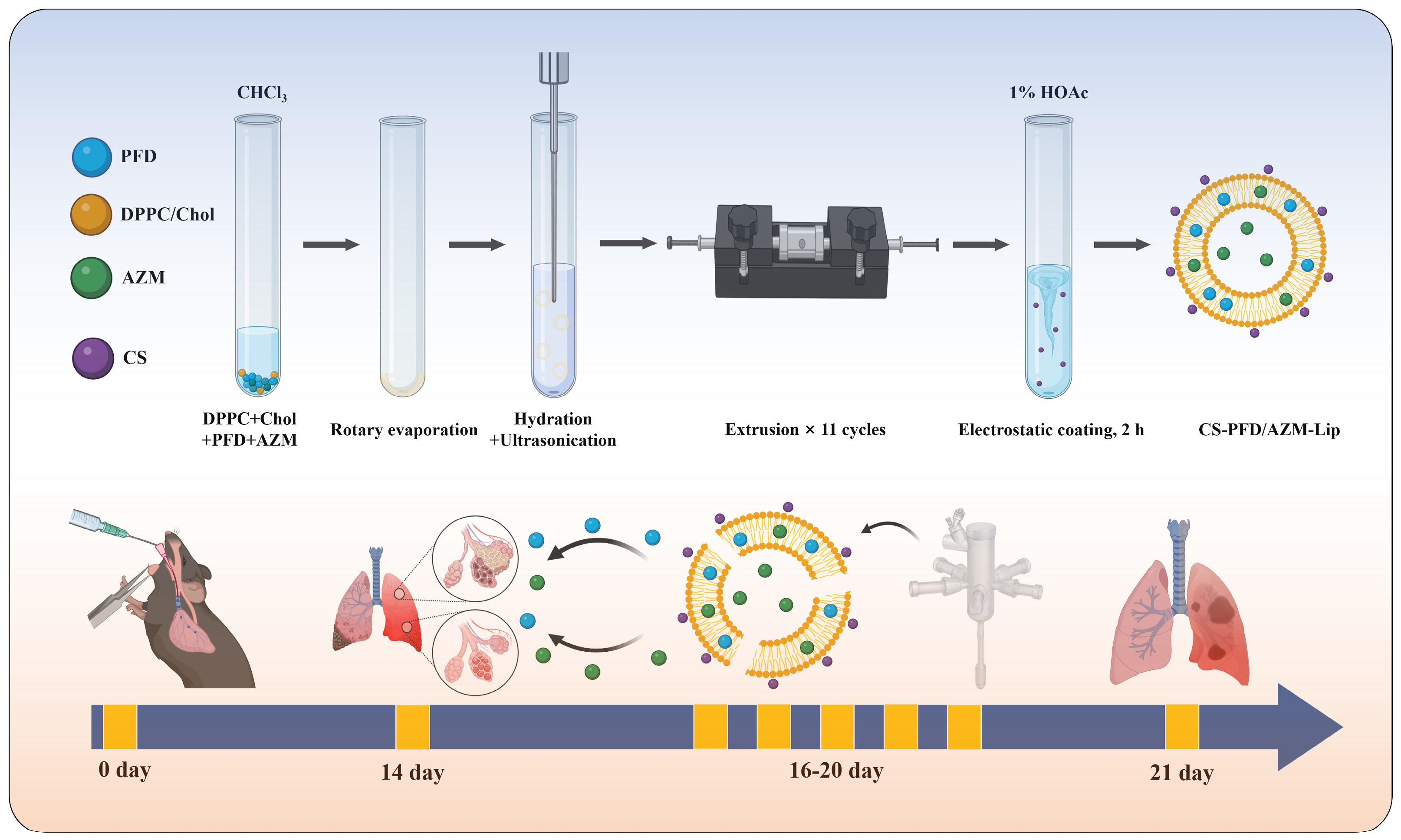
Based on the pathological features of the “infection–inflammation–fibrosis” cascade exacerbation in acute exacerbation of idiopathic pulmonary fibrosis (AE-IPF), this study designed a chitosan-coated, co-loaded liposome inhalation platform. DPPC was selected as the backbone phospholipid of the liposomes due to its dual role: it serves as the primary component of endogenous pulmonary surfactant, which confers excellent pulmonary biocompatibility, and it can self-assemble into uniform vesicles through conventional processes. This dual functionality balances pulmonary administration safety with preparation controllability. The DPPC/Chol molar ratio was set at 7.3:1, consistent with the formulation system previously established by our research group [31,32]. This ratio not only maintains good biocompatibility but also regulates the fluidity and stability of the lipid membrane, enhances the co-encapsulation efficiency of the two drugs, and reduces drug leakage during nebulization. The combination of DPPC and Chol forms the lipid bilayer backbone; the lipophilic PFD is embedded in the hydrophobic region of the bilayer, while the amphiphilic AZM is distributed at the membrane interface and within the inner aqueous phase, thereby achieving stable co-loading of both drugs within the same nanocarrier. The surface chitosan coating layer is formed through electrostatic adsorption, with a CS/total lipid mass ratio of approximately 3.7% (w/w). This dosage falls within the low-ratio coating range reported in the literature, which is favorable for maintaining liposome dispersion stability [26], and represents the optimal solubility of chitosan in a 1% acetic acid system. This coating layer simultaneously possesses triple functions: mucosal adhesion, cationic antibacterial activity, and nebulization stabilization, thereby enabling multi-node synergistic therapy for controlling infection triggers, suppressing inflammation, and intervening in fibrosis (Scheme 1).
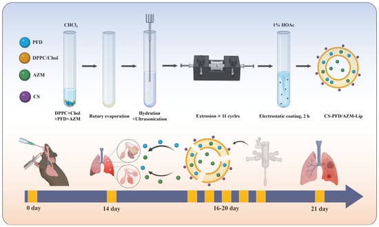
Scheme 1.
The preparation process and in vivo evaluation protocol of CS-PFD/AZM-Lip. The upper panel depicts the preparation of PFD/AZM co-loaded liposomes using the thin-film dispersion–extrusion method, followed by electrostatic adsorption coating with pulmonary function testing and tissue collection for evaluation performed on day 21. Created in BioRender. Kaiqi, Wang. (2026) https://BioRender.com/xxt8qyy.
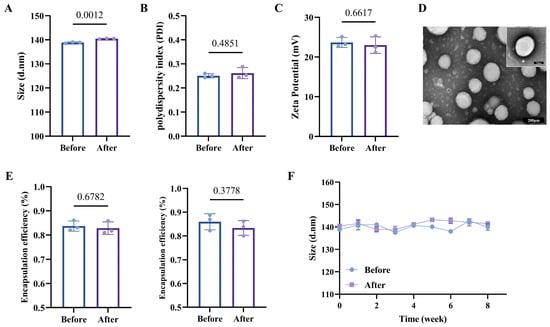
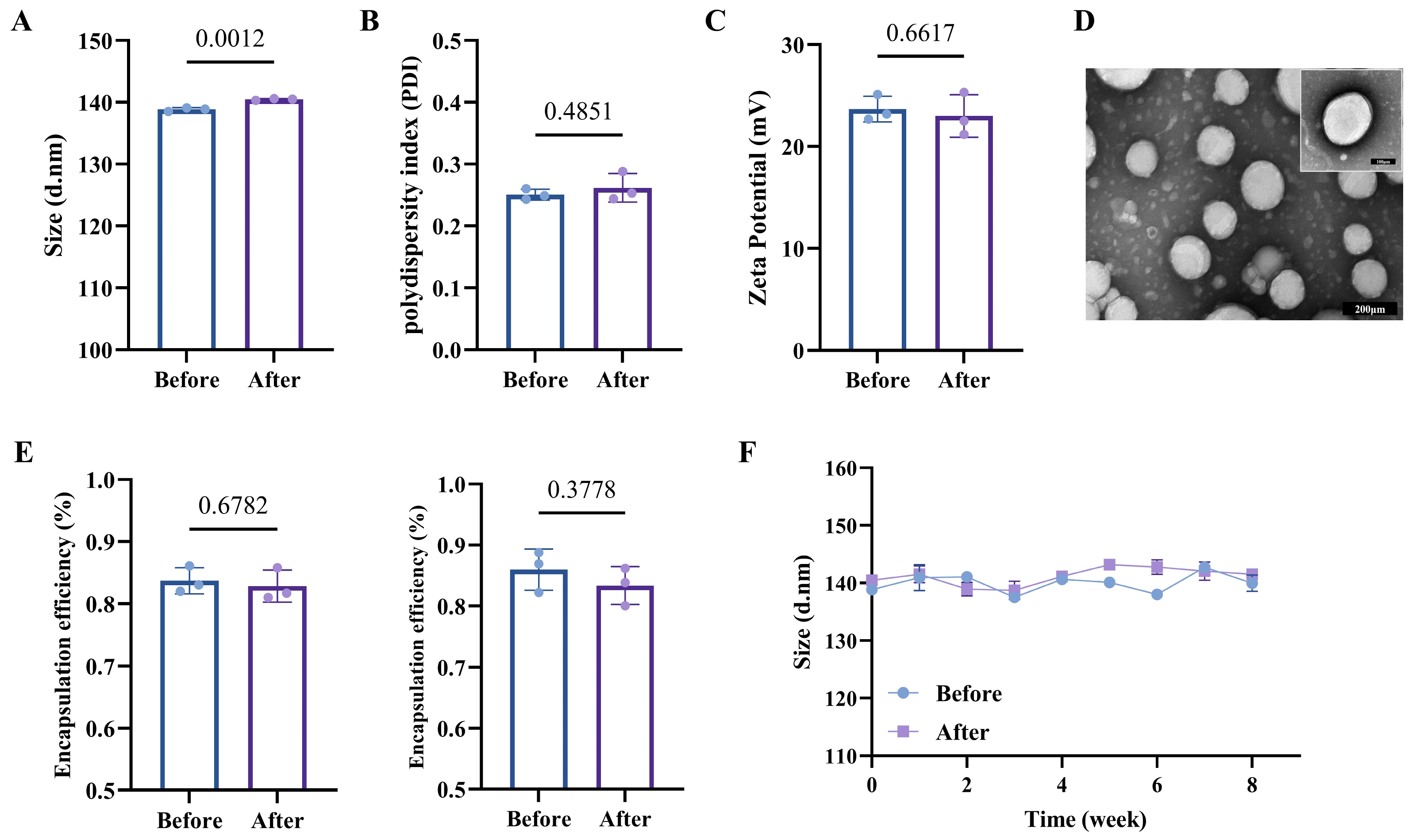
In this study, PFD/AZM coloaded liposomes were prepared using a thin-film dispersion method combined with ultrasonic disruption. Chitosan was subsequently coated onto the surface of the liposomes via electrostatic adsorption, resulting in the formation of CS-PFD/AZM-Lip. The chitosan molecular chain contains a significant number of positively charged amino groups, which can interact with the negatively charged liposomal membrane through electrostatic interactions, thereby forming a stable core–shell structure. Figure 1A,B illustrate the particle size of CS-PFD/AZM-Lip. Dynamic light scattering (DLS) results indicated that the average hydrodynamic diameter of CS-PFD/AZM-Lip was 138.83 ± 0.31 nm, with a PDI of 0.251 ± 0.009, suggesting a uniform particle size distribution and good monodispersity of the nanoparticles. Figure 1C and Figure S1 present the zeta potential measurement results. The zeta potential of PFD/AZM-Lip was negative; however, following chitosan coating, the potential of CS-PFD/AZM-Lip reversed to +23.67 ± 1.27 mV. This reversal phenomenon directly confirms the successful coating of chitosan on the liposome surface. TEM observations (Figure 1D) revealed that CS-PFD/AZM-Lip exhibited a regular spherical or quasi-spherical shape with uniform size. HPLC results (Figure 1E) demonstrated that the encapsulation efficiency of pirfenidone in CS-PFD/AZM-Lip was 83.7%, while that of azithromycin was 86.0%.
Figure 1.
Characterization and stability of CS-PFD/AZM-Lip. Panel (A) presents the hydrodynamic particle size measured before and after nebulization (n = 3). Panel (B) shows the polydispersity index (PDI) before and after nebulization (n = 3). Panel (C) depicts the zeta potential prior to and following nebulization (n = 3). Panel (D) displays the morphology as observed through transmission electron microscopy (TEM), with a scale bar of 100 nm. Panel (E) details the encapsulation efficiency of PFD and AZM before and after nebulization (n = 3). Lastly, panel (F) illustrates the changes in particle size over an 8-week storage period at 4 °C (n = 3). Data are presented as mean ± S.D.
An inhalable drug delivery platform capable of withstanding the shear forces generated during nebulization is essential for achieving effective pulmonary drug delivery. Accordingly, we investigated the physicochemical properties and stability of CS-PFD/AZM-Lip before and after nebulization. The results are presented in Figure 1A–C,E. After passing through a vibrating mesh nebulizer, the particle size of CS-PFD/AZM-Lip measured 140.47 ± 0.15 nm, with an increase of only approximately 1.6%. Although this difference was statistically significant, the magnitude of change was minimal and lacked practical pharmaceutical significance. The zeta potential was recorded at +23.00 ± 2.10 mV, and the PDI was 0.262 ± 0.023, demonstrating no significant changes compared to measurements taken prior to nebulization. After nebulization, the encapsulation efficiencies of PFD and AZM were 82.9% and 83.4%, respectively, representing decreases of only 0.8% and 2.6% relative to pre-nebulization values. Neither of these efficiencies exhibited a significant decrease compared to the values recorded before nebulization. These results suggest that the chitosan coating effectively enhances the structural stability of liposomes under the shear force of nebulization, allowing CS-PFD/AZM-Lip to demonstrate good nebulization tolerance and suitability for nebulized inhalation administration. The long-term stability results (Figure 1F and Figure S2) demonstrated that the CS-PFD/AZM-Lip formulation maintained a relatively stable particle size and polydispersity index (PDI) over 8 weeks of storage at 4 °C. Specifically, the particle size increased by only approximately 0.99%, and no significant increases in particle size or aggregation were observed. The encapsulation efficiency measurements taken at week 8 indicated that the encapsulation efficiencies for PFD and AZM remained at 81.5% and 83.2%, respectively, both exceeding 80%. This suggests that the formulation exhibited minimal drug leakage over the 8-week period at 4 °C, thereby demonstrating good storage stability.
Next, we evaluated the droplet size distribution of the CS-PFD/AZM-Lip nebulized aerosol and its in vivo pulmonary distribution following nebulized administration (Figure S3) to confirm the efficacy of pulmonary delivery. Laser diffraction measurements indicated that the volume median diameter (VMD, Xv50) of the nebulized droplets was 2.13 μm, with the volume fraction of droplets ≤5 μm reaching 95.5%. This finding suggests that the majority of the aerosol falls within the respirable particle size range of 1–5 μm, thereby meeting the particle size requirements for effective pulmonary delivery. Additionally, the number median diameter (Xn50) was measured at 0.567 μm, with 99.8% of droplets being ≤5 μm. Following nebulized inhalation of fluorescently labeled CS-PFD/AZM-Lip, ex vivo fluorescence imaging of major organs revealed that the signal was significantly enriched in lung tissue, while almost no signal was detected in the heart, liver, spleen, kidneys, and intestines, directly confirming the targeted pulmonary deposition of the formulation. These two lines of evidence corroborate each other and collectively support the effective pulmonary delivery of this inhalation platform.
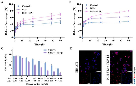
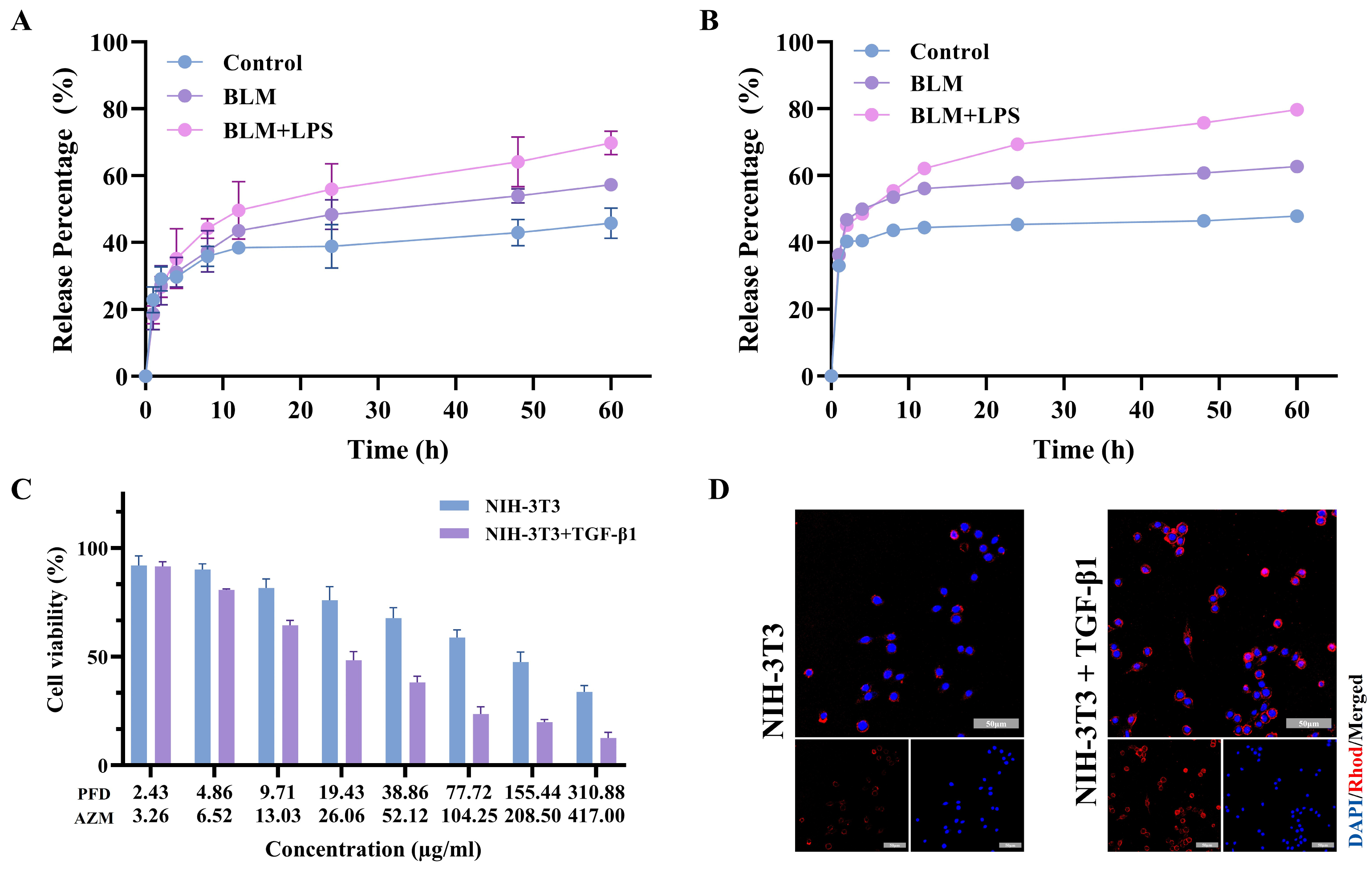
To evaluate the drug release behavior of CS-PFD/AZM-Lip in various pulmonary microenvironments, in vitro release experiments were performed using BALF from healthy mice (control), mice with BLM-induced pulmonary fibrosis (BLM), and mice with acute exacerbation induced by BLM and lipopolysaccharide (LPS) (BLM+LPS) as release media. The results are presented in Figure 2A,B. The cumulative release rates of both drugs demonstrated a gradient of BLM+LPS > BLM > control. At 60 h, the cumulative release rates of AZM were 45.8 ± 4.6% (control), 57.3 ± 1.4% (BLM), and 69.8 ± 3.5% (BLM+LPS), respectively; for PFD, the rates were 47.9 ± 0.9%, 62.7 ± 0.8%, and 79.8 ± 0.8%, respectively. The release profiles of the two drugs exhibited similar shapes, both displaying a biphasic release characteristic with a relatively rapid initial phase (0–8 h) followed by a slower release phase. These results indicate that CS-PFD/AZM-Lip has enhanced release characteristics responsive to the pathological microenvironment of fibrosis and acute exacerbation, which facilitates the synchronized synergistic release of the two drugs in the pulmonary lesion area.
Figure 2.
The in vitro release, cytotoxicity, and cellular uptake of CS-PFD/AZM-Lip. (A,B) The cumulative release curves of AZM and PFD in bronchoalveolar lavage fluid (BALF) from healthy mice (control), BLM-induced pulmonary fibrosis mice (BLM), and BLM+LPS-induced acute exacerbation mice (BLM+LPS) are shown, with n = 3 for each group. (C) The cell viability of NIH-3T3 cells treated with varying concentrations of CS-PFD/AZM-Lip for 24 h is assessed under both resting conditions and following activation with TGF-β1 (10 ng/mL), as measured by the MTT assay (n = 5). (D) Uptake images of CS-PFD/AZM-Lip, labeled with Rhod-DHPE, are presented after co-incubation with resting and TGF-β1-activated NIH-3T3 cells for 4 h (blue: DAPI-stained nuclei; red: Rhod-DHPE; scale bar: 50 μm). Data are presented as mean ± S.D.
3.2. Biocompatibility and Cellular Uptake
The cytotoxicity of CS-PFD/AZM-Lip against NIH-3T3 fibroblasts was assessed using the MTT assay. As illustrated in Figure 2C, when the concentrations of PFD were ≤2.43 μg/mL and those of AZM were ≤3.26 μg/mL, the viability of NIH-3T3 cells remained above 85%, regardless of co-treatment with TGF-β1. Notably, as the concentrations increased, the formulation exhibited a stronger inhibitory effect on TGF-β1-activated fibroblasts compared to quiescent cells, indicating a selective inhibitory tendency towards activated phenotype cells. Consequently, to mitigate the impact of significant cytotoxicity differences on subsequent efficacy evaluations and to ensure the effectiveness of CS-PFD/AZM-Lip against myofibroblasts, subsequent in vitro experiments were conducted using PFD at 4.86 μg/mL and AZM at 6.52 μg/mL. At the selected concentrations, the viability of both quiescent and TGF-β1-activated cells remained above 80% (Figure 2C), indicating that the subsequent anti-fibrotic effects were not attributable to cytotoxicity. This approach ensured NIH-3T3 cell viability while facilitating differential killing of TGF-β1-activated fibroblasts. To investigate the cellular uptake of CS-PFD/AZM-Lip, we employed 16:0 Liss Rhod PE-labeled liposomes for the uptake experiments. The results demonstrated (Figure 2D and Figure S4) that after 4 h of incubation, weak intracellular fluorescence was observed in quiescent NIH-3T3 cells, whereas the signal was markedly enhanced in TGF-β1-activated cells, indicating that CS-PFD/AZM-Lip can be efficiently internalized by myofibroblasts.
3.3. In Vitro Antifibrotic Efficacy
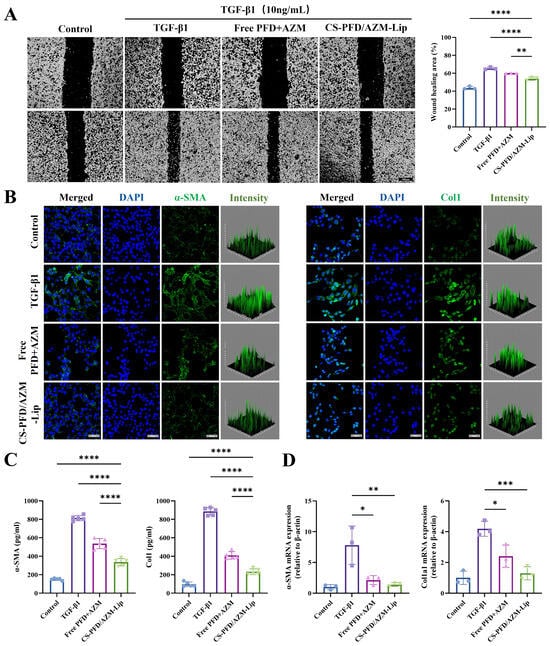
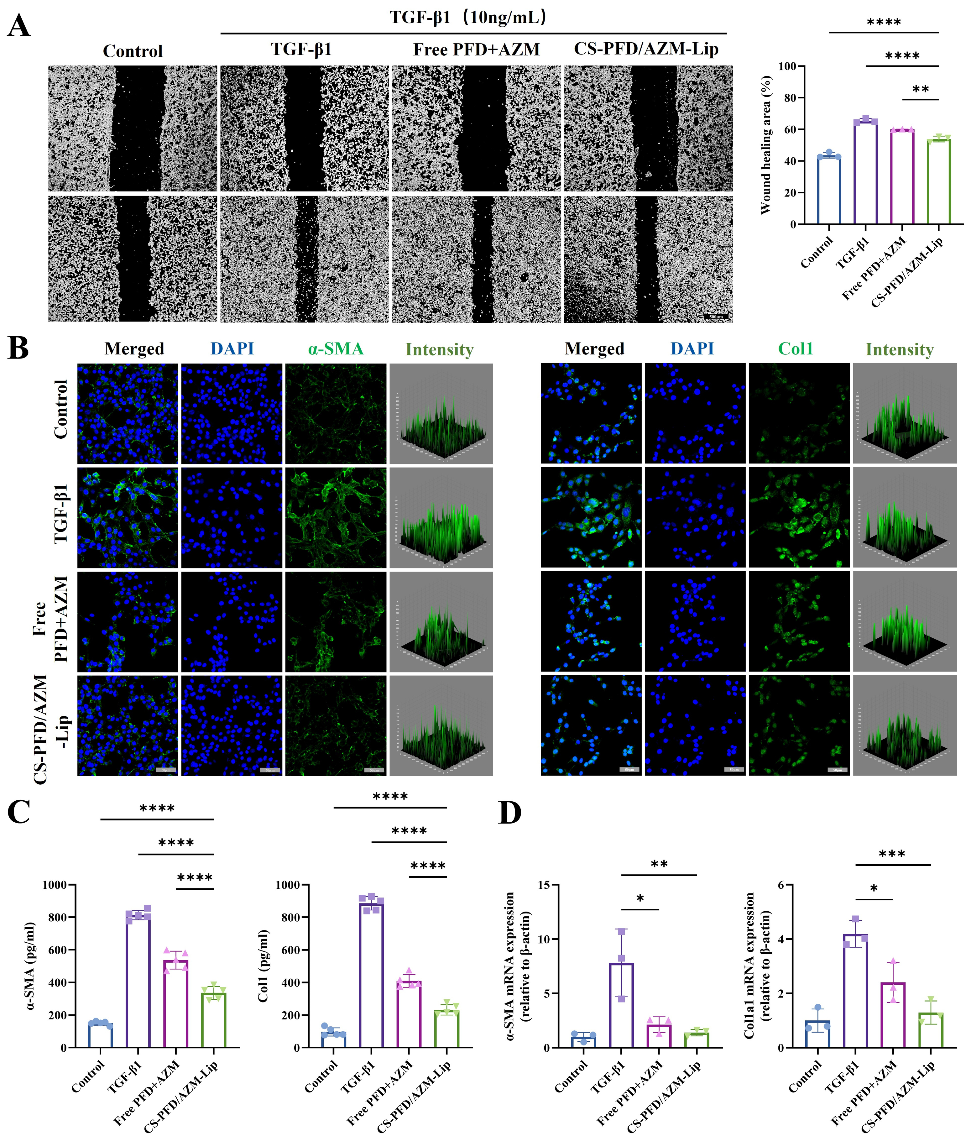
The effect of CS-PFD/AZM-Lip on TGF-β1-induced migration ability of NIH-3T3 cells was evaluated using a cell scratch assay. The results of this assay (Figure 3A) demonstrated that after 24 h of TGF-β1 stimulation, the scratch healing rate in the model group was significantly higher than that in the control group, indicating that TGF-β1 significantly promotes the migration ability of fibroblasts. Both the free drug combination group and the CS-PFD/AZM-Lip group markedly inhibited TGF-β1-induced cell migration, with the inhibitory effect of the Lip group being significantly greater than that of the free drug combination group. These findings suggest that nanocarriers enhance cellular uptake, thereby increasing the intracellular concentration of drugs and more effectively inhibiting the activation and migration of fibroblasts. The immunofluorescence staining results (Figure 3B and Figure S5) demonstrated that following TGF-β1 induction, the fluorescence intensities of intracellular α-SMA and Col1 were significantly increased. This indicates that TGF-β1 effectively induced the transdifferentiation of fibroblasts into myofibroblasts and enhanced collagen synthesis. Both the free drug combination group and the nanoparticle group exhibited a significant reduction in the fluorescence intensities of α-SMA and Col1, with the nanoparticle group showing a more pronounced inhibitory effect and restoring fluorescence intensities nearly to control levels. To quantitatively evaluate the in vitro antifibrotic effect of CS-PFD/AZM-Lip, we employed ELISA and qRT-PCR to measure the protein levels of α-SMA and Col1 in the cell supernatant and cell lysate, alongside the corresponding intracellular mRNA expression levels. The ELISA results (Figure 3C) aligned with the immunofluorescence findings: in the TGF-β1 model group, the levels of Col1 and α-SMA in cell supernatants were significantly elevated; conversely, in the CS-PFD/AZM-Lip group, these levels decreased by approximately 70% and 60%, respectively, compared to the model group, and were significantly lower than those in the free drug group. Additionally, qRT-PCR results (Figure 3D) further confirmed that CS-PFD/AZM-Lip significantly downregulated the mRNA expression of Col1a1 and ACTA2.
Figure 3.
The in vitro anti-fibrotic efficacy of CS-PFD/AZM-Lip. (A) presents representative images from the wound healing assay at 0 h and 24 h, alongside a quantitative analysis of the wound closure rate at 24 h (n = 3; scale bar: 500 μm). (B) Representative immunofluorescence staining images for α-SMA and Col1, as well as three-dimensional distribution maps of fluorescence intensity (blue: DAPI; green: FITC; scale bar: 50 μm). (C) A quantitative ELISA analysis of cellular Col1 and α-SMA protein levels (n = 3). (D) The relative expression levels of Col1a1 and Acta2 mRNA are reported, using Actb as an internal reference (n = 3). The experimental groups are categorized as follows: control (normal DMEM medium), TGF-β1 (10 ng/mL), free PFD+AZM (TGF-β1 + free drug, PFD 4.86 μg/mL + AZM 6.52 μg/mL), and CS-PFD/AZM-Lip (TGF-β1 + formulation of equal concentration). Data are presented as mean ± S.D. * p < 0.05, ** p < 0.01, *** p < 0.001, **** p < 0.0001.
3.4. In Vivo Anti-AE-IPF Efficacy
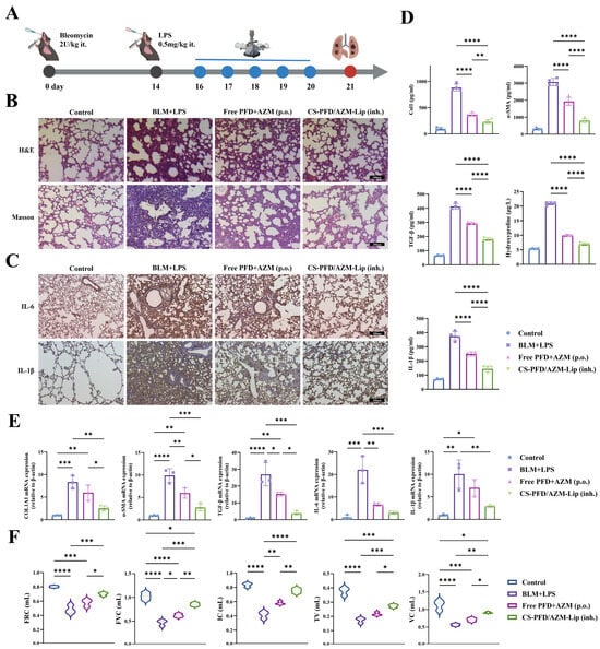
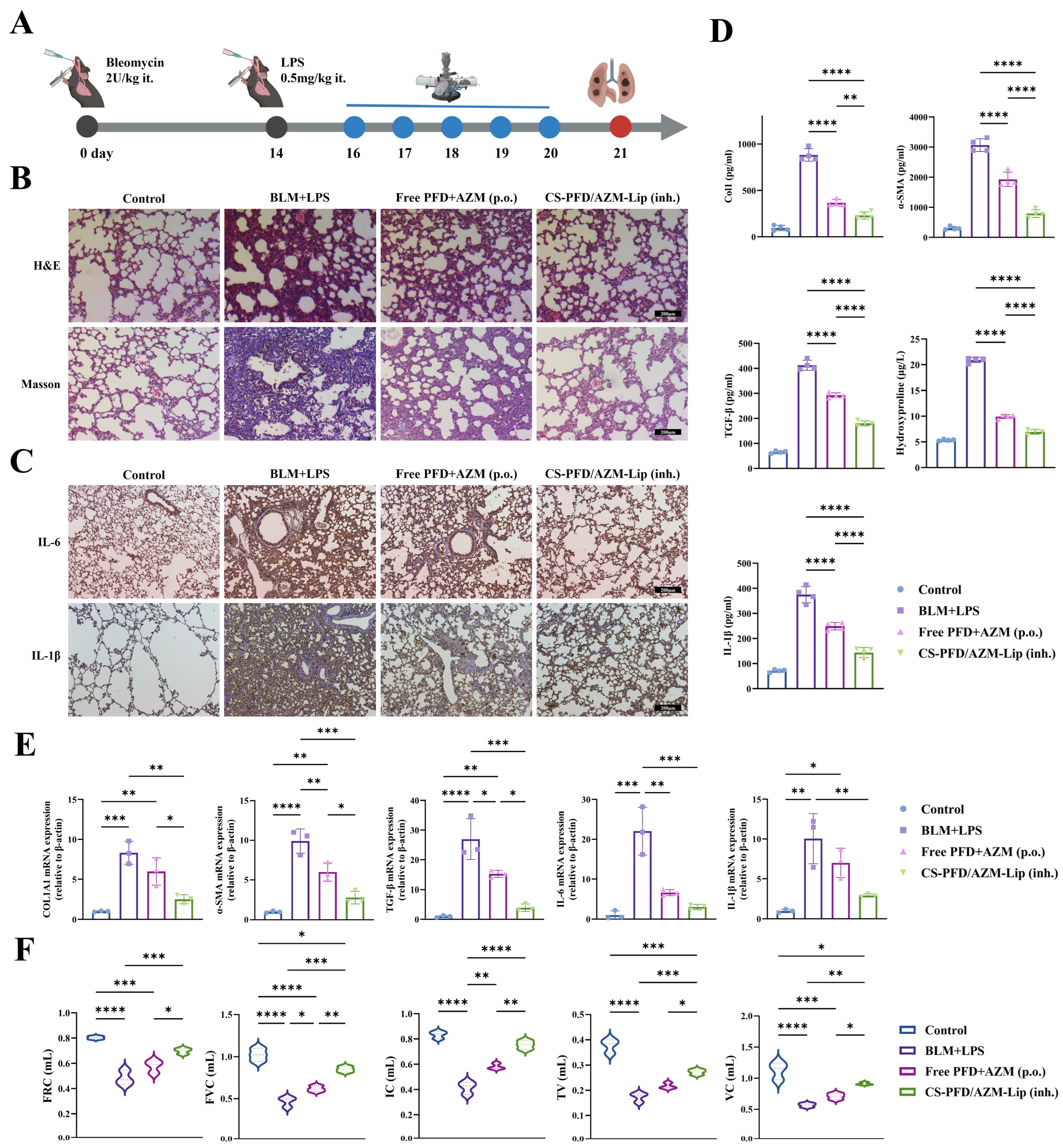
The in vivo efficacy was assessed using a mouse model of AE-IPF induced by sequential administration of BLM and LPS (Figure 4A). H&E staining (Figure 4B) revealed severe destruction of the alveolar structure, with markedly thickened alveolar septa, extensive infiltration of inflammatory cells, and notable consolidation areas within the lung parenchyma in the model group. Masson staining indicated significant areas of blue-stained collagen deposition in the pulmonary interstitium and extensive fibrosis of the alveolar septa. Pathological changes were alleviated in the free PFD + AZM oral group. In contrast, the CS-PFD/AZM-Lip nebulized inhalation group exhibited significant restoration of the alveolar structure, reduced inflammatory infiltration, decreased collagen deposition, and a notable attenuation of fibrosis, approaching the levels observed in the control group. Lung tissue immunofluorescence (Figure S6) demonstrated extensive and high expression of Col1 and α-SMA in the model group, while their expression was significantly reduced in the CS-PFD/AZM-Lip group, which outperformed the free PFD + AZM group. Immunohistochemistry results (Figure 4C) indicated markedly increased IL-6 and IL-1β expression, mainly localized to bronchial epithelial cells and infiltrating inflammatory cells. In the CS-PFD/AZM-Lip inhalation treatment group, the expression of IL-6 and IL-1β was significantly downregulated, further confirming the anti-inflammatory effect of this formulation.
Figure 4.
The in vivo anti-AE-IPF efficacy of CS-PFD/AZM-Lip. (A) A schematic timeline depicting the establishment of the model and the administration of the drug. (B) Representative images of H&E and Masson’s trichrome staining of lung tissues (scale bar: 200 μm). (C) Representative immunohistochemical images showing the presence of IL-6 and IL-1β in lung tissues (scale bar: 200 μm). (D) ELISA quantification of Col1, α-SMA, TGF-β, hydroxyproline, and IL-1β in lung tissue homogenates (n = 4). (E) Relative mRNA expression levels of Col1a1, Acta2, Tgfb1, Il6, and Il1b in lung tissues (n = 3). (F) Pulmonary function parameters, including functional residual capacity (FRC), forced vital capacity (FVC), inspiratory capacity (IC), tidal volume (TV), and vital capacity (VC) (n = 3). Data are presented as mean ± S.D. * p < 0.05, ** p < 0.01, *** p < 0.001, and **** p < 0.0001.
To further evaluate the in vivo anti-inflammatory and antifibrotic effects, we utilized ELISA to measure the protein expression levels of hydroxyproline, α-SMA, Col1, TGF-β1, and IL-1β in lung tissue homogenates. As illustrated in Figure 4D, the protein levels of hydroxyproline, α-SMA, Col1, TGF-β1, and IL-1β in the lung tissue of the BLM+LPS model group were significantly elevated compared to the control group. Each treatment group demonstrated varying degrees of downregulation of these indicators, with the CS-PFD/AZM-Lip inhalation group exhibiting the most pronounced effect, as all indicators were significantly lower than those observed in the free drug oral administration group. The qRT-PCR results (Figure 4E) corroborated the ELISA findings. Notably, aerosol inhalation of CS-PFD/AZM-Lip significantly reduced the mRNA expression of fibrosis- and inflammation-related genes, including Col1a1, ACTA2, TGF-β1, IL-6, and IL-1β, outperforming the free drug oral administration group. These findings further substantiate that aerosol inhalation of CS-PFD/AZM-Lip demonstrates remarkable dual anti-inflammatory and antifibrotic efficacy in vivo.
In addition, we examined the total cell count in BALF. The results demonstrated (Figure S7) a significant increase in total cell count in the BLM+LPS model group. All treatment groups notably reduced the total cell count in BALF, with the CS-PFD/AZM-Lip inhalation group exhibiting superior efficacy compared to the free drug oral administration group. This finding indicates that CS-PFD/AZM-Lip aerosol inhalation effectively alleviates inflammatory cell infiltration in the lungs of AE-IPF mice. Furthermore, changes in body weight, lung coefficient, lung wet/dry weight ratio, and survival across groups further corroborated the in vivo efficacy of CS-PFD/AZM-Lip (Figures S8–S11). To assess the ameliorative effect of CS-PFD/AZM-Lip nebulized inhalation on pulmonary function in AE-IPF model mice, we measured pulmonary function indicators, including functional residual capacity (FRC), forced vital capacity (FVC), inspiratory capacity (IC), tidal volume (TV), and vital capacity (VC). As illustrated in Figure 4F, all pulmonary function indicators significantly decreased in the BLM+LPS model group compared to the control group. However, both the free drug oral administration group and the Lip inhalation group demonstrated varying degrees of improvement in pulmonary function, with the Lip inhalation group exhibiting the most pronounced enhancement and marked amelioration of pulmonary function.
3.5. Safety Assessment
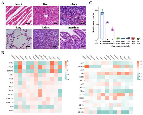
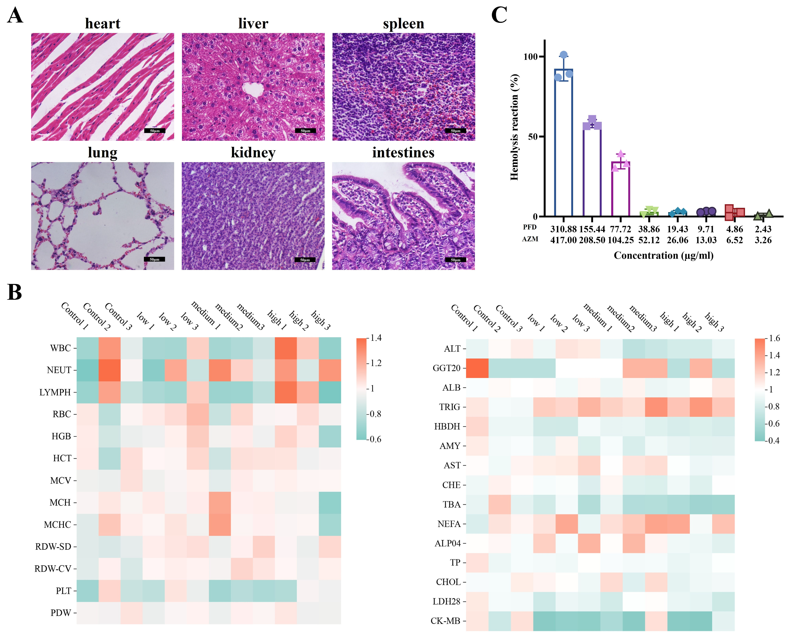
Healthy mice received nebulized inhalation of low, medium, and high doses of CS-PFD/AZM-Lip for five consecutive days. Twenty-four hours after the last administration, H&E staining of heart, liver, spleen, lung, kidney, and intestinal tissues from each group revealed no significant pathological changes (Figure 5A, high-dose group shown). The results of routine blood tests and biochemical analyses (Figure 5B) indicated that nebulized inhalation of low, medium, and high doses of CS-PFD/AZM-Lip did not result in significant differences in routine blood parameters, including WBC, RBC, HGB, and PLT, nor in serum liver and kidney function parameters such as ALT and AST when compared to the control group. These findings suggest that nebulized inhalation of CS-PFD/AZM-Lip demonstrates good blood compatibility and safety regarding liver and kidney function. Additionally, we assessed the blood compatibility of CS-PFD/AZM-Lip through a hemolysis assay. The results (Figure 5C) indicated that when the PFD concentration was less than 38.86 μg/mL and the AZM concentration was below 52.12 μg/mL, the hemolysis rates of CS-PFD/AZM-Lip remained below 5%, which meets the safety threshold, thus confirming good blood compatibility and the absence of significant hemolytic reactions.
Figure 5.
The safety evaluation of CS-PFD/AZM-Lip. (A) H&E staining revealing the histological features of the heart, liver, spleen, lung, kidney, and intestine in mice subjected to 5 days of continuous nebulization with the high-dose group (PFD 50 mg/kg + AZM 67 mg/kg), with a scale bar of 50 μm. (B) A heatmap illustrating the blood routine indicators (WBC, NEUT, LYMPH, RBC, HGB, HCT, MCV, MCH, MCHC, RDW-SD, RDW-CV, PLT, PDW) and blood biochemistry parameters (ALT, AST, etc.) across low-, medium-, and high-dose groups, normalized to the mean of the control group (n = 3). (C) The in vitro hemolysis rates of CS-PFD/AZM-Lip at varying concentrations, with distilled water serving as the positive control and normal saline as the negative control (n = 3). Data are presented as mean ± S.D.
4. Discussion
The core finding of this study is that chitosan-coated PFD/AZM coloaded liposomes can effectively achieve the pulmonary co-delivery of two drugs with complementary mechanisms of action, while maintaining both nebulization stability and storage stability. Additionally, these liposomes can simultaneously inhibit inflammatory and fibrotic processes at both the cellular and whole-animal levels. In contrast to previous inhaled nanoformulations primarily targeting chronic IPF, this study specifically addresses AE-IPF, a clinical context characterized by heightened acuity, criticality, and infection-related features. In this context, AZM serves as a foundation for anti-infection and immunomodulation, while PFD continuously intervenes in the TGF-β-driven fibrotic program. Furthermore, the chitosan coating enhances the stability of the liposomes during nebulization and at the pulmonary interface. Therefore, this platform represents not merely a simple combination of the two drugs, but rather an integration of materials science focused on the pathological chain of AE-IPF: “infection trigger–inflammatory amplification–fibrotic progression.”
The synergistic effects observed in this study are supported by a clear mechanistic framework and the existing literature. Within the NF-κB inflammatory pathway, azithromycin (AZM) inhibits p65 nuclear translocation by blocking IKKβ activity and reducing IκB-α degradation, a mechanism that has been directly validated through in vivo imaging studies of pulmonary inflammation [10]. Additionally, AZM suppresses STAT1 phosphorylation, which promotes macrophage polarization towards the M2 anti-inflammatory phenotype [11]. Conversely, pirfenidone (PFD) inhibits the maturation of TGF-β by blocking furin protease activity and diminishes the DNA-binding and transcriptional activity of NF-κB [8]. In the context of viral infections, PFD has been shown to mitigate TGF-β-induced inflammation and decrease pulmonary viral load [9]. Therefore, these two drugs establish a dual blockade at both upstream (nuclear translocation) and downstream (transcriptional activity) levels of the core inflammatory pathways. Regarding the TGF-β fibrotic pathway, AZM enhances the proteasomal degradation of NOX4 through STUB1-mediated ubiquitination, thereby attenuating TGF-β downstream reactive oxygen species (ROS)-dependent myofibroblast differentiation [12]. This action, combined with PFD’s upstream inhibition of TGF-β maturation, creates a synergistic pattern of ‘source reduction + effector attenuation.’ The mechanistic network outlined above accounts for the experimental observation that the co-loaded liposomes in this study outperformed equivalent doses of free drugs at both cellular and whole-animal levels.
The role of the chitosan coating extends beyond mere physical modification, embodying the design philosophy that “the carrier itself is a therapeutic component.” First, the positive charge of chitosan imparts mucoadhesive properties, which can prolong the retention time of nanoparticles in the airway and counteract rapid mucociliary clearance [35,36]. Second, chitosan interacts with negatively charged bacterial cell membranes, disrupting membrane integrity and exhibiting intrinsic broad-spectrum antibacterial activity that synergizes with the antibacterial action of AZM [27]. Third, the chitosan coating significantly enhances the structural stability of liposomes against nebulization shear forces. Zaru et al. confirmed that chitosan-coated liposomes demonstrated markedly superior stability after nebulization compared to uncoated liposomes [26]. The findings of this study, which observed no significant changes in particle size, zeta potential, and encapsulation efficiency before and after nebulization, align with the aforementioned reports. In vitro release experiments indicated that the cumulative release rate of CS-PFD/AZM-Lip in bronchoalveolar lavage fluid under pathological conditions (BLM+LPS) was significantly higher than that under physiological conditions, exhibiting a gradient release characteristic of “lesion microenvironment responsiveness.” This phenomenon is corroborated by the existing literature: in IPF patients, secretory phospholipase A2 (sPLA2) is abnormally distributed and exhibits elevated activity in the lungs, where its hydrolysis of phospholipid membranes can accelerate liposome degradation [37]. Similarly, stimulus-responsive PFD liposomes designed for IPF exploit the biochemical characteristics of the fibrotic microenvironment to achieve triggered release [33]. This responsive property facilitates enriched drug release at the lesion site while minimizing its impact on normal lung tissue [38].
The design strategies for inhaled nanocarriers in the treatment of IPF and respiratory infections have become increasingly defined in the existing literature. Zaru et al. demonstrated that chitosan coating significantly enhances the stability and mucus adhesion of liposomes after nebulization [26]. In the context of IPF treatment, Liu et al. constructed ROS-responsive inhaled liposomes loaded with dimethyl fumarate, which attenuated pulmonary fibrosis via the Nrf2 signaling pathway [38]. Wang et al. reported that inhaled lung surfactant biomimetic liposomes reversed IPF through a synergistic strategy [23]. Han et al. utilized inhaled nanoparticles co-loaded with verteporfin and pirfenidone to inhibit the formation of honeycomb cysts and interstitial remodeling [18]. Lee et al. designed inhaled microgels that evade alveolar macrophage phagocytosis to achieve dual-drug co-delivery of nintedanib and pirfenidone [19]. Dallal Bashi et al. developed an inhaled liposomal azithromycin dry powder for the treatment of chronic respiratory infections [25]. In comparison to the aforementioned studies, this research presents several distinct features: (i) previous platforms primarily targeted chronic IPF or simple infections, while this study uniquely addresses the acute exacerbation scenario of AE-IPF, wherein the “infection–inflammation–fibrosis” cascade coexists; (ii) for the first time, pirfenidone and azithromycin, which have a clinical basis for combined use, are co-loaded onto a single inhalable carrier to achieve synergistic antibacterial and anti-fibrotic effects [16]; (iii) chitosan coating provides mucus adhesion, intrinsic antibacterial activity, and aerosol stability, while also enabling pathological microenvironment-responsive release.
Despite its contributions, this study has several notable limitations. Firstly, the extrapolability of the animal model is constrained. Although the BLM+LPS sequential induction mouse model effectively replicates the key characteristic of acute exacerbation of idiopathic pulmonary fibrosis (AE-IPF)—specifically, “acute inflammation superimposed on a foundation of fibrosis”—the bleomycin model has inherent self-limiting properties. Furthermore, LPS stimulation fails to fully emulate the actual pathogenic infection context and irreversible disease progression observed in human AE-IPF. Both existing systematic reviews of AE-IPF animal models and the official report from the American Thoracic Society (ATS) have underscored the limited correlation between animal models of pulmonary fibrosis and the corresponding human conditions, highlighting the necessity for caution when translating preclinical findings to clinical settings [7,39]. Secondly, the representativeness of the cell model is inadequate. This study utilized the mouse embryo-derived NIH-3T3 fibroblast cell line; however, Krempaska et al. demonstrated that lung fibroblasts isolated from IPF patients exhibit a significantly stronger anti-fibrotic response to azithromycin (AZM) compared to normal control cells [14]. This finding suggests that validation using human primary cells or lung organoids may yield more accurate predictions of clinical outcomes. Thirdly, the depth of efficacy and safety assessments is limited. The animal sample size in this study was small (n = 3 in most experiments), and the observation period was restricted to 21 days, without evaluating the potential immunogenicity and pulmonary accumulation toxicity associated with the long-term repeated inhalation of chitosan-coated nanoparticles. These limitations will be systematically addressed in subsequent studies to further the clinical translation of this inhalation platform.
5. Conclusions
This study investigates the pathological features of the aggravated “infection–inflammation–fibrosis” cascade in acute exacerbation of idiopathic pulmonary fibrosis (AE-IPF) and successfully formulates a chitosan-coated pirfenidone/azithromycin co-loaded liposomal aerosol inhalation formulation (CS-PFD/AZM-Lip). The formulation demonstrates uniform particle size, good dispersibility, and high encapsulation efficiency, along with stability before and after nebulization and during 8 weeks of storage at 4 °C, thereby meeting the fundamental requirements for pulmonary inhaled nanoformulations to maintain structural integrity. In vitro studies reveal that CS-PFD/AZM-Lip exhibits enhanced responsive release in pathological bronchoalveolar lavage fluid (BALF), is efficiently taken up by TGF-β1-induced activated fibroblasts, and inhibits cell migration, myofibroblast differentiation, and extracellular matrix production at both transcriptional and protein levels. In the bleomycin and lipopolysaccharide (BLM+LPS)-induced AE-IPF mouse model, aerosol inhalation of this formulation alleviates pulmonary inflammation and collagen deposition more effectively than oral administration of the free drug, with no significant toxicity observed in major organs, hematology, serum biochemistry, or hemolysis tests. This study integrates drug combinations with a clinical rationale for combined use alongside inhalable nanocarrier design, transforming traditional combination therapy into a “co-localization, co-delivery, synergistic intervention” strategy targeting pulmonary lesions, thereby offering a novel approach with translational potential for the precise treatment of AE-IPF.
However, we remain acutely aware that this study has certain limitations. The animal model employed does not fully replicate the true infection background and irreversible disease progression observed in human AE-IPF. Additionally, the in vitro mechanistic validation relies solely on mouse-derived cell lines. Furthermore, systematic safety data regarding aerosol aerodynamic characterization, intrapulmonary pharmacokinetics, and long-term repeated dosing still need to be addressed. The limitations also include a restricted animal sample size and observation period, along with the absence of a head-to-head comparison with clinically standard treatment regimens. These shortcomings necessitate gradual refinement in future models that more closely align with clinical practice and a more comprehensive formulation quality evaluation system, thereby facilitating the clinical translation of this platform.
Supplementary Materials
The following supporting information can be downloaded at https://www.mdpi.com/article/10.3390/pharmaceutics18111435/s1, Table S1: Optimization of PFD and AZM Contents in CS-PFD/AZM-Lip. Figure S1: The zeta potential of PFD/AZM-Lip and CS-PFD/AZM-Lip (n = 3; data are presented as mean ± S.D.). Figure S2: Changes in PDI of CS-PFD/AZM-Lip during 8 weeks of storage at 4 °C and encapsulation efficiency after 8 weeks (n = 3; data are presented as mean ± S.D.). Figure S3: Measurement of aerosol droplet size distribution by laser diffraction (left: volume distribution; right: number distribution), and ex vivo tissue fluorescence imaging after nebulized inhalation of fluorescently labeled CS-PFD/AZM-Lip (n = 3; data are presented as mean ± S.D.). Figure S4: Quantitative analysis of uptake by NIH-3T3 and TGF-β1-induced NIH-3T3 cells (n = 3; data are presented as mean ± S.D.). Figure S5: Immunofluorescence quantitative analysis of α-SMA and Col1 (n = 3; data are presented as mean ± S.D.; * p < 0.05, ** p < 0.01, *** p < 0.001, **** p < 0.0001). Figure S6: Representative immunofluorescence staining images of Col1 and α-SMA in lung tissues from each group of mice and quantification of their fluorescence intensity (green: FITC; blue: DAPI; scale bar: 100 μm). Figure S7: The total inflammatory cell counts in the BALF of mice across each experimental group (n = 3). Statistical significance is indicated with *** p < 0.001 and **** p < 0.0001. Figure S8: The changes in body weight of mice across each group from day 0 to day 21 (n = 3; data are presented as mean ± S.D.). Figure S9: The Kaplan–Meier survival curves for the mice in each group over a 21-day period (n = 10); Figure S10: The lung coefficient (lung weight/body weight, mg/g) for mice in each group (n = 3; data are presented as mean ± S.D.; ** p < 0.01 and **** p < 0.0001). Figure S11: The wet/dry weight ratio of lung tissues among the various groups of mice (n = 3; data are presented as mean ± S.D.; * p < 0.05, ** p < 0.01, **** p < 0.0001).
Author Contributions
Conceptualization, K.W. and S.C.; methodology, S.C.; validation, Z.L., C.Z. and X.Z.; formal analysis, K.W.; resources, L.W.; data curation, K.W.; writing—original draft preparation, K.W.; writing—review and editing, K.W.; supervision, L.W.; project administration, L.W.; funding acquisition, L.W. All authors have read and agreed to the published version of the manuscript.
Funding
This research was funded by the HMU Marshal Initiative Fund, the Joint Guidance Project of the Natural Science Foundation of Heilongjiang Province (PL2025TO06), the Program for Young Talents of Basic Research in Heilongjiang Provincial Universities (YQJH2025128), the Capacity Enhancement Program for Science and Technology InnovationPlatforms in Heilongjiang Province (JD25D004), and the Heilongjiang Provincial Key Laboratory of Molecular Imaging.
Institutional Review Board Statement
All animal procedures were approved by the Experimental Animal Ethics Committee of Harbin Medical University, Heilongjiang, China (animal ethics application number: 2026-DWSYLLCZ-150; approval date: 8 May 2026) and were performed in accordance with the Guide for the Care and Use of Laboratory Animals published by the National Institutes of Health (NIH).
Data Availability Statement
Data are contained within the article.
Conflicts of Interest
The authors declare no conflicts of interest.
Abbreviations
The following abbreviations are used in this manuscript:
| AE-IPF | Acute exacerbation of idiopathic pulmonary fibrosis |
| ALT | Alanine aminotransferase |
| AST | Aspartate aminotransferase |
| AZM | Azithromycin |
| BALF | Bronchoalveolar lavage fluid |
| BLM | Bleomycin |
| CHE | Cholinesterase |
| Col1 | Type I collagen |
| DLS | Dynamic light scattering |
| DPPC | Dipalmitoylphosphatidylcholine |
| ELISA | Enzyme-linked immunosorbent assay |
| FRC | Functional residual capacity |
| FVC | Forced vital capacity |
| H&E | Hematoxylin and eosin |
| HGB | Hemoglobin |
| Hyp | Hydroxyproline |
| IC | Inspiratory capacity |
| IL-1β | Interleukin-1 beta |
| IL-6 | Interleukin-6 |
| IPF | Idiopathic pulmonary fibrosis |
| LPS | Lipopolysaccharide |
| NF-κB | Nuclear factor kappa-B |
| NIH-3T3 | Mouse embryonic fibroblast |
| NOX4 | NADPH oxidase 4 |
| PDI | Polydispersity index |
| PFD | Pirfenidone |
| PLT | Platelets |
| qRT-PCR | Real-time quantitative polymerase chain reaction |
| RBC | Red blood cells |
| ROS | Reactive oxygen species |
| TEM | Transmission electron microscopy |
| TGF-β1 | Transforming growth factor beta 1 |
| TV | Tidal volume |
| VC | Vital capacity |
| WBC | White blood cells |
| α-SMA | Alpha-smooth muscle actin |
References
- Lederer, D.J.; Martinez, F.J. Idiopathic Pulmonary Fibrosis. N. Engl. J. Med. 2018, 378, 1811–1823. [Google Scholar] [CrossRef] [Scilit] [PubMed]
- Richeldi, L.; Collard, H.R.; Jones, M.G. Idiopathic pulmonary fibrosis. Lancet 2017, 389, 1941–1952. [Google Scholar] [PubMed]
- Vernimmen, T. Treatments for idiopathic pulmonary fibrosis are on the horizon. Nature 2026, 649, S13–S15. [Google Scholar] [CrossRef] [Scilit] [PubMed]
- Kreuter, M.; Polke, M.; Walsh, S.L.F.; Krisam, J.; Collard, H.R.; Chaudhuri, N.; Avdeev, S.; Behr, J.; Calligaro, G.; Corte, T.; et al. Acute exacerbation of idiopathic pulmonary fibrosis: International survey and call for harmonisation. Eur. Respir. J. 2020, 55, 1901760. [Google Scholar] [CrossRef] [Scilit] [PubMed]
- Collard, H.R.; Ryerson, C.J.; Corte, T.J.; Jenkins, G.; Kondoh, Y.; Lederer, D.J.; Lee, J.S.; Maher, T.M.; Wells, A.U.; Antoniou, K.M.; et al. Acute Exacerbation of Idiopathic Pulmonary Fibrosis. An International Working Group Report. AM J. Respir. Crit. Care. Med. 2016, 194, 265–275. [Google Scholar] [CrossRef] [Scilit] [PubMed]
- Kawamura, K.; Ichikado, K.; Yasuda, Y.; Anan, K.; Suga, M. Azithromycin for idiopathic acute exacerbation of idiopathic pulmonary fibrosis: A retrospective single-center study. BMC Pulm. Med. 2017, 17, 94. [Google Scholar] [CrossRef] [Scilit] [PubMed]
- Ye, X.; Zhang, M.; Gu, H.; Liu, M.; Zhao, Y.; Shi, Y.; Wu, S.; Jiang, C.; Ye, X.; Zhu, H.; et al. Animal models of acute exacerbation of pulmonary fibrosis. Respir. Res. 2023, 24, 296. [Google Scholar] [CrossRef] [Scilit] [PubMed]
- Seifirad, S. Pirfenidone: A novel hypothetical treatment for COVID-19. Med. Hypotheses. 2020, 144, 110005. [Google Scholar] [CrossRef] [Scilit] [PubMed]
- Thomas, B.J.; Kan-o, K.; Gantier, M.P.; Simpson, I.; Chitty, J.G.; Lam, M.; Dousha, L.; Gottschalk, T.A.; Lawlor, K.E.; Tate, M.D.; et al. Pirfenidone Mitigates Transforming Growth Factor-beta-induced Inflammation after Viral Infection. AM J. Respir. Cell Mol. Biol. 2025, 73, 545–558. [Google Scholar] [CrossRef] [Scilit] [PubMed]
- Stellari, F.F.; Sala, A.; Donofrio, G.; Ruscitti, F.; Caruso, P.; Topini, T.M.; Francis, K.P.; Li, X.; Carnini, C.; Civelli, M.; et al. Azithromycin inhibits nuclear factor-kappaB activation during lung inflammation: An in vivo imaging study. Pharmacol. Res. Perspect. 2014, 2, e00058. [Google Scholar] [CrossRef] [Scilit] [PubMed]
- Haydar, D.; Cory, T.J.; Birket, S.E.; Murphy, B.S.; Pennypacker, K.R.; Sinai, A.P.; Feola, D.J. Azithromycin Polarizes Macrophages to an M2 Phenotype via Inhibition of the STAT1 and NF-kappaB Signaling Pathways. J. Immunol. 2019, 203, 1021–1030. [Google Scholar] [CrossRef] [Scilit] [PubMed]
- Tsubouchi, K.; Araya, J.; Minagawa, S.; Hara, H.; Ichikawa, A.; Saito, N.; Kadota, T.; Sato, N.; Yoshida, M.; Kurita, Y.; et al. Azithromycin attenuates myofibroblast differentiation and lung fibrosis development through proteasomal degradation of NOX4. Autophagy 2017, 13, 1420–1434. [Google Scholar] [CrossRef] [Scilit] [PubMed]
- Gielen, V.; Johnston, S.L.; Edwards, M.R. Azithromycin induces anti-viral responses in bronchial epithelial cells. Eur. Respir. J. 2010, 36, 646–654. [Google Scholar] [CrossRef] [Scilit] [PubMed]
- Krempaska, K.; Barnowski, S.; Gavini, J.; Hobi, N.; Ebener, S.; Simillion, C.; Stokes, A.; Schliep, R.; Knudsen, L.; Geiser, T.K.; et al. Azithromycin has enhanced effects on lung fibroblasts from idiopathic pulmonary fibrosis (IPF) patients compared to controls. Respir. Res. 2020, 21, 25. [Google Scholar] [CrossRef] [Scilit] [PubMed]
- Macaluso, C.; Maritano Furcada, J.; Alzaher, O.; Chaube, R.; Chua, F.; Wells, A.U.; Maher, T.M.; George, P.M.; Renzoni, E.A.; Molyneaux, P.L. The potential impact of azithromycin in idiopathic pulmonary fibrosis. Eur. Respir. J. 2019, 53, 1800628. [Google Scholar] [CrossRef] [Scilit] [PubMed]
- Saha, A.; Vaidya, P.J.; Chavhan, V.B.; Achlerkar, A.; Leuppi, J.D.; Chhajed, P.N. Combined pirfenidone, azithromycin and prednisolone in post-H1N1 ARDS pulmonary fibrosis. Sarcoidosis Vasc. Diffus. Lung Dis. 2018, 35, 85–90. [Google Scholar] [CrossRef] [Scilit] [PubMed]
- Bonella, F.; Spagnolo, P.; Ryerson, C. Current and Future Treatment Landscape for Idiopathic Pulmonary Fibrosis. Drugs 2023, 83, 1581–1593. [Google Scholar] [CrossRef] [Scilit] [PubMed]
- Han, M.M.; Tang, L.; Huang, B.; Li, X.-N.; Fang, Y.-F.; Qi, L.; Duan, B.-W.; Yao, Y.-T.; He, Y.-J.; Xing, L.; et al. Inhaled nanoparticles for treating idiopathic pulmonary fibrosis by inhibiting honeycomb cyst and alveoli interstitium remodeling. J. Control Release 2024, 366, 732–745. [Google Scholar] [CrossRef] [Scilit] [PubMed]
- Lee, W.T.; Lee, H.; Kim, J.; Jung, Y.; Choi, E.; Jeong, J.H.; Jeong, J.-H.; Lee, J.H.; Youn, Y.S. Alveolar macrophage phagocytosis-evading inhaled microgels incorporating nintedanib-PLGA nanoparticles and pirfenidone-liposomes for improved treatment of pulmonary fibrosis. Bioact. Mater. 2024, 33, 262–278. [Google Scholar] [CrossRef] [Scilit] [PubMed]
- Anderson, S.; Atkins, P.; Bäckman, P.; Cipolla, D.; Clark, A.; Daviskas, E.; Disse, B.; Entcheva-Dimitrov, P.; Fuller, R.; Gonda, I.; et al. Inhaled Medicines: Past, Present, and Future. Pharmacol. Rev. 2022, 74, 48–118. [Google Scholar] [CrossRef] [Scilit] [PubMed]
- Togami, K.; Maruta, Y.; Nanbu, M.; Tada, H.; Chono, S. Prolonged distribution of aerosolized PEGylated liposomes in the lungs of mice with bleomycin-induced pulmonary fibrosis. Drug Dev. Ind. Pharm. 2020, 46, 1873–1880. [Google Scholar] [CrossRef] [Scilit] [PubMed]
- Yue, P.; Zhou, W.; Huang, G.; Lei, F.; Chen, Y.; Ma, Z.; Chen, L.; Yang, M. Nanocrystals based pulmonary inhalation delivery system: Advance and challenge. Drug Deliv. 2022, 29, 637–651. [Google Scholar] [CrossRef] [Scilit] [PubMed]
- Wang, B.; Gao, Y.; Sun, L.; Xue, M.; Wang, M.; Zhang, Z.; Zhang, L.; Zhang, H. Inhaled pulmonary surfactant biomimetic liposomes for reversing idiopathic pulmonary fibrosis through synergistic therapeutic strategy. Biomaterials 2023, 303, 122404. [Google Scholar] [CrossRef] [Scilit] [PubMed]
- Meng, H.; Xu, Y. Pirfenidone-loaded liposomes for lung targeting: Preparation and in vitro/in vivo evaluation. Drug Des. Devel. Ther. 2015, 9, 3369–3376. [Google Scholar] [CrossRef] [Scilit] [PubMed]
- Dallal Bashi, Y.H.; Ali, A.; Al Ayoub, Y.; Assi, K.H.; Mairs, R.; McCarthy, H.O.; Tunney, M.M.; Kett, V.L. Inhaled dry powder liposomal azithromycin for treatment of chronic lower respiratory tract infection. Int. J. Pharm. 2024, 653, 123841. [Google Scholar] [CrossRef] [Scilit] [PubMed]
- Zaru, M.; Manca, M.L.; Fadda, A.M.; Antimisiaris, S.G. Chitosan-coated liposomes for delivery to lungs by nebulisation. Colloids Surf. B Biointerfaces 2009, 71, 88–95. [Google Scholar] [CrossRef] [Scilit] [PubMed]
- Wang, X.; Cheng, F.; Wang, X.; Feng, T.; Xia, S.; Zhang, X. Chitosan decoration improves the rapid and long-term antibacterial activities of cinnamaldehyde-loaded liposomes. Int. J. Biol. Macromol. 2021, 168, 59–66. [Google Scholar] [CrossRef] [Scilit] [PubMed]
- Moyá, M.L.; López-López, M.; Lebrón, J.A.; Ostos, F.J.; Pérez, D.; Camacho, V.; Beck, I.; Merino-Bohórquez, V.; Camean, M.; Madinabeitia, N.; et al. Preparation and Characterization of New Liposomes. Bactericidal Activity of Cefepime Encapsulated into Cationic Liposomes. Pharmaceutics 2019, 11, 69. [Google Scholar] [CrossRef] [Scilit] [PubMed]
- Zhang, J.-Q.; Wang, Y.; Ou, J.-G.; Feng, J.-W.; Gui, P.; Ma, D.; Wang, G.-H. Inflammation-responsive liposomes co-delivering tobramycin and nitric oxide for overcoming mucus barriers against MRSA pneumonia. Colloids Surf. B Biointerfaces 2026, 267, 115959. [Google Scholar] [CrossRef] [Scilit] [PubMed]
- Peng, J.; Wang, Q.; Guo, M.; Liu, C.; Chen, X.; Tao, L.; Zhang, K.; Shen, X. Development of Inhalable Chitosan-Coated Oxymatrine Liposomes to Alleviate RSV-Infected Mice. Int. J. Mol. Sci. 2022, 23, 15909. [Google Scholar] [CrossRef] [Scilit] [PubMed]
- Yang, J.; Li, Y.; Sun, J.; Zou, H.; Sun, Y.; Luo, J.; Xie, Q.; Rong, A.; Wang, H.; Li, X.; et al. An Osimertinib-Perfluorocarbon Nanoemulsion with Excellent Targeted Therapeutic Efficacy in Non-small Cell Lung Cancer: Achieving Intratracheal and Intravenous Administration. ACS Nano 2022, 16, 12590–12605. [Google Scholar] [CrossRef] [Scilit] [PubMed]
- Rong, A.; Wang, H.; Nie, C.; Han, Z.; Zhou, M.; Atinuke, O.O.; Wang, K.; Wang, X.; Liu, S.; Zhao, J.; et al. Glycerol-weighted chemical exchange saturation transfer nanoprobes allow (19)F(/1)H dual-modality magnetic resonance imaging-guided cancer radiotherapy. Nat. Commun. 2023, 14, 6644. [Google Scholar] [CrossRef] [Scilit] [PubMed]
- Han, M.; Song, Y.; Liu, S.; Lu, X.; Su, L.; Liu, M.; Zhu, X.; Sun, K.; Lu, Y.; Wang, A. Engineering of Stimulus-Responsive Pirfenidone Liposomes for Pulmonary Delivery During Treatment of Idiopathic Pulmonary Fibrosis. Front. Pharmacol. 2022, 13, 882678. [Google Scholar] [CrossRef] [Scilit] [PubMed]
- Solleti, V.S.; Alhariri, M.; Halwani, M.; Omri, A. Antimicrobial properties of liposomal azithromycin for Pseudomonas infections in cystic fibrosis patients. J. Antimicrob. Chemother. 2015, 70, 784–796. [Google Scholar] [CrossRef] [Scilit] [PubMed]
- Zacaron, T.M.; Silva, M.L.S.e.; Costa, M.P.; Silva, D.M.e.; Silva, A.C.; Apolônio, A.C.M.; Fabri, R.L.; Pittella, F.; Rocha, H.V.A.; Tavares, G.D. Advancements in Chitosan-Based Nanoparticles for Pulmonary Drug Delivery. Polymers 2023, 15, 3849. [Google Scholar] [CrossRef] [Scilit] [PubMed]
- Zhang, C.; D’Angelo, D.; Buttini, F.; Yang, M. Long-acting inhaled medicines: Present and future. Adv. Drug Deliv. Rev. 2024, 204, 115146. [Google Scholar] [CrossRef] [Scilit] [PubMed]
- Jaiswal, A.; Rehman, R.; Dutta, J.; Singh, S.; Ray, A.; Shridhar, M.; Jaisankar, J.; Bhatt, M.; Khandelwal, D.; Sahoo, B.; et al. Cellular Distribution of Secreted Phospholipase A2 in Lungs of IPF Patients and Its Inhibition in Bleomycin-Induced Pulmonary Fibrosis in Mice. Cells 2023, 12, 1044. [Google Scholar] [CrossRef] [Scilit] [PubMed]
- Liu, J.; Wu, Z.; Liu, Y.; Zhan, Z.; Yang, L.; Wang, C.; Jiang, Q.; Ran, H.; Li, P.; Wang, Z. ROS-responsive liposomes as an inhaled drug delivery nanoplatform for idiopathic pulmonary fibrosis treatment via Nrf2 signaling. J. Nanobiotechnol. 2022, 20, 213. [Google Scholar] [CrossRef] [Scilit] [PubMed]
- Jenkins, R.G.; Moore, B.B.; Chambers, R.C.; Eickelberg, O.; Königshoff, M.; Kolb, M.; Laurent, G.J.; Nanthakumar, C.B.; Olman, M.A.; Pardo, A.; et al. An Official American Thoracic Society Workshop Report: Use of Animal Models for the Preclinical Assessment of Potential Therapies for Pulmonary Fibrosis. AM J. Respir. Cell Mol. Biol. 2017, 56, 667–679. [Google Scholar] [CrossRef] [Scilit] [PubMed]
Disclaimer/Publisher’s Note: The statements, opinions and data contained in all publications are solely those of the individual author(s) and contributor(s) and not of MDPI and/or the editor(s). MDPI and/or the editor(s) disclaim responsibility for any injury to people or property resulting from any ideas, methods, instructions or products referred to in the content. |
© 2026 by the authors. Licensee MDPI, Basel, Switzerland. This article is an open access article distributed under the terms and conditions of the Creative Commons Attribution (CC BY) license.