


Abstract
Solid organ transplant (SOT) recipients are especially vulnerable to pharmacokinetic drug interactions because narrow-therapeutic-index immunosuppressants are used in the setting of polypharmacy, rapidly changing physiology, variable gastrointestinal function, and frequent anti-infective therapy. Interaction magnitude is rarely determined by the perpetrator drug alone; formulation, genotype, hematocrit, albumin, organ function, inflammation, and time after transplantation can materially change the observed exposure response. This narrative review integrates mechanistic and clinical evidence across desensitization, induction, maintenance immunosuppression, and rejection treatment. Particular attention is given to cytochrome P450 3A (CYP3A)- and transporter-mediated interactions, mycophenolic acid enterohepatic recirculation, formulation-dependent exposure, drug–drug–gene interactions, food and herbal products, physiological modifiers, interaction-informed therapeutic drug monitoring, and model-informed dosing. Quantitative transplant data are prioritized where available, with non-SOT evidence identified when it materially informs mechanism or management. A practical 6-D framework (Drug, Dose, Determinants, Drug interactions, Data, and Decision) is proposed to organize patient-specific assessment and move interaction management beyond binary alerts toward quantitative, concentration-guided precision dosing.
1. Introduction
Solid organ transplantation (SOT) has transformed the prognosis of patients with end-stage organ failure, but long-term graft preservation depends on pharmacotherapy that is both complex and continuously changing [1]. Maintenance immunosuppression is routinely combined with antimicrobial prophylaxis or treatment and therapies for cardiovascular, metabolic, gastrointestinal, neurologic, and other comorbidities. Polypharmacy is therefore intrinsic to transplant care; in a systematic review and meta-analysis of chronic kidney disease populations, its prevalence exceeded 80% and was highest among kidney transplant recipients and patients receiving dialysis [2].
This medication burden becomes clinically important because several cornerstone immunosuppressants have narrow therapeutic indices and marked inter- and intra-individual pharmacokinetic variability. Tacrolimus is the best-characterized example: its disposition depends on intestinal and hepatic cytochrome P450 (CYP)3A4/5 activity, P-glycoprotein-mediated transport, erythrocyte partitioning, and patient-specific determinants such as CYP3A genotype, hematocrit, body size, time after transplantation, and concomitant medications [3,4]. Consequently, the same perpetrator drug can produce substantially different exposure changes across recipients.
The magnitude of clinically important interactions can be striking. Voriconazole substantially reduces tacrolimus dose requirements, and discontinuation may require rapid dose escalation; in one heart transplant cohort, the tacrolimus dose ultimately required an average 215% increase within one month after voriconazole withdrawal [5]. Ritonavir-containing regimens can produce an even larger effect, with approximately 99% lower tacrolimus dose requirements reported in liver transplant recipients receiving lopinavir/ritonavir [6]. These examples show why identifying an inhibitor or inducer is only the first step: safe management also requires understanding magnitude, onset, offset, and reversibility.
Transplant pharmacokinetic interactions extend beyond CYP3A. Mycophenolic acid (MPA) undergoes glucuronidation, biliary secretion, and enterohepatic recirculation, creating susceptibility to interactions involving intestinal microbiota, biliary transport, gastrointestinal binding, renal dysfunction, and concomitant cyclosporine [7]. Food and nonprescription products introduce additional mechanisms; grapefruit inhibits intestinal CYP3A and increases tacrolimus exposure [8], while controlled data with extended-release MeltDose tacrolimus (LCPT) confirm that intestinal CYP3A remains relevant despite more distal drug release [9].
Genetic and physiological variability further complicate prediction. In kidney transplant recipients receiving voriconazole, the increase in dose-normalized tacrolimus predose concentration (C0) was 320.93% in CYP3A5 nonexpressers compared with 232.19% in expressers [10]. Hematocrit, albumin, diarrhea, inflammation, hepatic and renal function, formulation, and the early post-transplant state can likewise modify measured concentrations or true exposure. These factors make a fixed interaction estimate potentially misleading when applied across organs and clinical contexts.
Electronic interaction resources do not resolve this complexity reliably. In 52 kidney transplant recipients, four commonly used drug–drug interaction (DDI) databases collectively identified 127 potential interactions, yet only 11 (8.7%) were identified by all four and agreement was weak (κ 0.05–0.36) [11]. This discordance underscores the need for transplant-specific assessment of mechanism, quantitative evidence, patient susceptibility, and therapeutic drug monitoring rather than reliance on binary alerts.
This review therefore integrates molecular mechanisms with the phase of transplant care, the affected immunosuppressant, the perpetrator drug, formulation science, pharmacogenomics, dynamic physiology, therapeutic drug monitoring (TDM), and model-informed prediction. The objective is not to catalog every possible drug pair, but to explain why interaction magnitude differs between recipients and how mechanistic information can be translated into individualized management.
Review Scope and Evidence Synthesis
This narrative review was informed by a structured search of PubMed/MEDLINE and Google Scholar through August 2026. Search concepts included SOT, individual immunosuppressants, pharmacokinetics, drug–drug interactions, CYP enzymes, transporters, pharmacogenomics, TDM, formulation, population pharmacokinetics (population PK), and physiologically based pharmacokinetic (PBPK) modeling. Representative search combinations included (“solid organ transplantation” OR “kidney transplant*” OR “liver transplant*” OR “heart transplant*” OR “lung transplant*”) AND (“drug interaction*” OR “pharmacokinetic*” OR “CYP3A” OR “transporter*”), with additional targeted searches for individual immunosuppressants, formulations, pharmacogenomic determinants, therapeutic drug monitoring, population pharmacokinetics, and physiologically based pharmacokinetic modeling. Titles and abstracts were screened for relevance, followed by full-text review of potentially relevant studies and citation tracking of key articles. Priority was given to prospective pharmacokinetic studies and studies conducted in solid organ transplant recipients, followed by transplant observational pharmacokinetic studies, controlled studies in healthy participants, population pharmacokinetic or physiologically based pharmacokinetic analyses, systematic reviews, and mechanistic studies. Case reports and case series were retained when they provided clinically important quantitative information for uncommon interactions. Evidence from hematopoietic cell transplantation or non-transplant populations was used selectively when it established a mechanism or interaction magnitude not adequately characterized in solid organ transplantation. Approximately 719 potentially relevant publications were considered, of which approximately 175 were retained for the final synthesis. Because this is a narrative synthesis, no formal risk of bias assessment or meta-analysis was performed.
2. Mechanistic Basis of Pharmacokinetic Interactions in Solid Organ Transplantation
Pharmacokinetic interactions in solid organ transplantation arise from alterations in one or more components of drug absorption, distribution, metabolism, transport, and elimination. These processes are particularly important for immunosuppressive agents because relatively small changes in systemic exposure may necessitate clinically meaningful dose adjustments. Furthermore, several pharmacokinetic pathways operate simultaneously: intestinal metabolism and efflux may determine oral bioavailability, hepatic metabolism determines systemic clearance, blood cell and plasma protein binding influence measured concentrations, and biliary or renal transport may govern elimination and enterohepatic recirculation. Consequently, pharmacokinetic interactions in transplantation should be interpreted as disturbances of an interconnected disposition system rather than as isolated enzyme-mediated events [12,13,14].
The principal mechanisms underlying pharmacokinetic interactions in SOT and their clinical implications are summarized in Table 1.
Table 1.
Mechanistic determinants of pharmacokinetic interactions in solid organ transplantation.
2.1. Absorption and Gastrointestinal Determinants
Oral absorption represents an important source of pharmacokinetic variability for several transplant immunosuppressants. Tacrolimus has incomplete and highly variable oral bioavailability, historically estimated at approximately 25% in liver transplant recipients, reflecting extensive presystemic metabolism and intestinal efflux [12,15]. Both CYP3A enzymes and P-glycoprotein (P-gp), encoded by ATP-binding cassette subfamily B member 1 (ABCB1), are expressed in the intestinal epithelium and act as sequential barriers to tacrolimus absorption: CYP3A metabolizes drug within enterocytes, while P-gp transports intracellular drug back into the intestinal lumen [12,14]. Variation or pharmacological modulation of either pathway can therefore substantially alter the fraction of an orally administered tacrolimus dose reaching the systemic circulation.
Food is another important determinant of oral immunosuppressant exposure. Administration of tacrolimus with food reduces and delays absorption, with the magnitude depending on meal composition and timing [29,30]. In stable kidney transplant recipients receiving once-daily tacrolimus, administration with breakfast decreased both the area under the concentration–time curve from 0 to 24 h (AUC0–24) and C0 exposure compared with fasting administration; importantly, individual changes in AUC ranged from approximately −38% to +29%, demonstrating substantial heterogeneity in the food effect [30]. These observations provide the mechanistic rationale for consistent administration of tacrolimus in relation to meals and demonstrate how inconsistent food intake may mimic or modify a pharmacokinetic DDI.
Gastrointestinal motility influences the temporal pattern of drug absorption, although its effect on total exposure may differ according to the drug and formulation.
MPA illustrates a fundamentally different absorption and recycling pathway. After administration of mycophenolate mofetil, the parent compound is rapidly converted to MPA, which subsequently undergoes glucuronidation to mycophenolic acid glucuronide (MPAG). MPAG may be secreted into bile, hydrolyzed by intestinal microbiota back to MPA, and reabsorbed, producing enterohepatic recirculation and a characteristic secondary concentration peak approximately 6–12 h after administration. Enterohepatic recirculation has historically been estimated to contribute approximately 40% of total MPA AUC, although considerable interpatient variability exists [13]. Contemporary intensive pharmacokinetic data from 84 kidney transplant recipients further demonstrate this variability, with a 5.5-fold range in MPA AUC0–12, an 18.4-fold range in C0 values, and a 3.4-fold difference in the contribution of enterohepatic recirculation [17]. These properties make MPA particularly susceptible to interactions involving intestinal microbiota, bile secretion, gastrointestinal binding, and transporter activity rather than conventional CYP-mediated interactions alone.
2.2. Distribution, Blood Cell Partitioning, and Protein Binding
Distribution characteristics can substantially modify the relationship between measured drug concentrations and pharmacologically relevant exposure. Tacrolimus is extensively distributed into erythrocytes, producing whole blood concentrations substantially higher than corresponding plasma concentrations. In an early pharmacokinetic study of 16 liver transplant recipients, the maximum blood-to-plasma concentration ratio averaged 55.5 ± 26.8, with individual values ranging approximately from 13 to 114, demonstrating marked variability in erythrocyte partitioning [31]. Consequently, whole blood tacrolimus concentrations are strongly dependent on hematocrit, and changes in hematocrit may alter measured concentrations without proportionate changes in unbound or intracellular exposure.
Protein binding is particularly relevant to MPA. MPA is highly albumin-bound, whereas its major metabolite MPAG accumulates substantially in renal dysfunction and may compete with MPA for albumin-binding sites [13]. Hypoalbuminemia and renal impairment can therefore increase the unbound fraction of MPA while paradoxically reducing measured total MPA concentrations because an increased free fraction is available for clearance. Under these circumstances, a low total concentration does not necessarily imply inadequate pharmacologically active exposure [13,32]. This represents an important example of how physiological changes can alter the interpretation of an apparent drug interaction even when the administered dose and intrinsic metabolic pathway remain unchanged.
2.3. Drug-Metabolizing Enzymes
The cytochrome P450 3A family represents the dominant metabolic pathway underlying many clinically important transplant DDIs. Tacrolimus undergoes extensive metabolism through CYP3A4 and CYP3A5 in both the intestine and liver, and cyclosporine, sirolimus, and everolimus similarly depend substantially on CYP3A-mediated metabolism [12,14,20,21]. This shared pathway explains the marked susceptibility of these agents to CYP3A inhibitors and inducers and creates the potential for simultaneous changes in exposure when multiple CYP3A-dependent immunosuppressants are administered.
The relative contributions of intestinal and hepatic CYP3A are clinically important. Intestinal CYP3A activity contributes to first-pass extraction and therefore influences oral bioavailability, whereas hepatic CYP3A contributes predominantly to systemic clearance. In living-donor liver transplantation, both intestinal and hepatic CYP3A5 genotypes have been shown to influence tacrolimus disposition.
CYP3A activity is also dynamic rather than genetically fixed. Concomitant medications, corticosteroid exposure, inflammation, hepatic dysfunction, and recovery of transplanted liver function may alter functional enzyme activity over time. Population pharmacokinetic studies in liver transplantation have demonstrated relationships between tacrolimus apparent clearance and markers of graft or hepatic function, including bilirubin, international normalized ratio, postoperative time, and graft characteristics [23,24]. Consequently, an interaction of identical inhibitory potency may generate different concentration changes in the same recipient at different stages after transplantation.
Not all important transplant immunosuppressants depend on CYP pathways. MPA is primarily metabolized through uridine diphosphate-glucuronosyltransferase (UGT) enzymes to MPAG and other glucuronide metabolites [13]. This distinction is clinically important because drugs or physiological conditions affecting UGT activity, biliary secretion, or enterohepatic recycling may alter MPA exposure without affecting CYP activity. Mechanistic understanding of transplant DDIs therefore requires consideration of both oxidative and conjugative metabolic pathways.
2.4. Transporter-Mediated Pharmacokinetic Interactions
Drug transporters act together with metabolizing enzymes to determine intestinal absorption, hepatic uptake and excretion, renal handling, tissue distribution, and biliary elimination. P-gp is particularly important in transplantation because tacrolimus, cyclosporine, and sirolimus interact with this efflux system and because intestinal P-gp activity can substantially influence oral drug availability [14]. P-gp is expressed not only in the gastrointestinal tract but also at the blood–brain barrier, bile canalicular membrane, and renal tubular epithelium, allowing transporter modulation to influence several components of drug disposition simultaneously [14].
The close anatomical and functional relationship between intestinal P-gp and CYP3A is particularly relevant. A drug transported back into the intestinal lumen by P-gp may undergo repeated cycles of absorption and efflux, increasing opportunities for metabolism by enterocyte CYP3A. Consequently, simultaneous inhibition of CYP3A and P-gp can produce greater changes in exposure than inhibition of either pathway considered independently. This enzyme–transporter interplay contributes to the substantial interaction liability of calcineurin and mammalian target of rapamycin (mTOR) inhibitors [14,20].
Cyclosporine is especially important because it can function not only as a victim of DDIs but also as a potent interaction perpetrator. In addition to its interaction with CYP3A and P-gp, cyclosporine inhibits hepatic organic anion-transporting polypeptides (OATPs), multidrug resistance-associated protein 2 (MRP2), and breast cancer resistance protein (BCRP), although the clinical relevance varies according to transporter, substrate, and cyclosporine concentration [33,34]. PBPK analyses incorporating simultaneous inhibition of CYP enzymes, OATP, P-gp, BCRP, and MRP2 have successfully reproduced several clinically observed cyclosporine interactions, particularly with statins and other transporter substrates [33]. This illustrates why a purely CYP-centered framework is insufficient for interpreting pharmacokinetic interactions in transplant recipients.
Transporter-mediated processes are also central to MPA disposition. MPAG undergoes biliary transport, followed by intestinal deconjugation and reabsorption of MPA. MRP2, BCRP, OATP-family transporters, and related pathways have been implicated in the disposition of MPA and its metabolites [13,32]. The best-established clinical example is the interaction between cyclosporine and MPA, in which inhibition of biliary MPAG transport reduces enterohepatic recirculation and decreases MPA exposure. Thus, transporter inhibition may either increase or decrease systemic exposure depending on whether the transporter facilitates elimination, uptake, or recycling.
2.5. Elimination, Organ Dysfunction, and Dynamic Post-Transplant Physiology
The effect of renal or hepatic dysfunction on pharmacokinetic interactions depends on the disposition pathway of the affected drug. Tacrolimus is predominantly metabolized rather than renally eliminated; therefore, renal dysfunction has a limited direct effect on tacrolimus clearance. Nevertheless, renal impairment substantially alters the pharmacokinetics of many concomitant anti-infective, cardiovascular, and metabolic drugs and may indirectly modify DDI risk. Conversely, hepatic dysfunction can reduce CYP3A-mediated tacrolimus clearance, particularly in liver transplant recipients, where graft function and regeneration change dynamically during the early postoperative period [12,23,24].
For MPA, renal dysfunction primarily alters metabolite disposition and protein binding rather than directly reducing MPA metabolic clearance. MPAG is predominantly eliminated renally and therefore accumulates as renal function deteriorates. Increased MPAG concentrations can affect protein binding and enterohepatic recycling, thereby changing total and unbound MPA exposure [13]. Consequently, changes in renal function can alter the apparent magnitude of an interaction involving MPA even when the interacting medication itself does not directly affect UGT metabolism.
The early post-transplant period is characterized by simultaneous changes in organ function, hematocrit, plasma proteins, gastrointestinal motility, inflammatory status, corticosteroid dose, nutritional intake, and concomitant pharmacotherapy. These variables can alter intrinsic clearance, bioavailability, protein binding, transporter activity, and interpretation of measured concentrations.
3. Pharmacokinetic Interaction Landscape Across the Transplant Continuum
Pharmacokinetic interaction risk in solid organ transplantation is dynamic and varies according to the phase of transplant care. Before transplantation, desensitization may involve therapeutic plasma exchange, immunoadsorption, intravenous immunoglobulin (IVIG), monoclonal antibodies, immunoglobulin G (IgG)-degrading therapies, and other immune-modifying agents whose disposition differs substantially from that of conventional small-molecule immunosuppressants. During induction, long-circulating polyclonal and monoclonal antibodies may persist for days to weeks after administration, whereas maintenance immunosuppression introduces the greatest burden of classical enzyme- and transporter-mediated DDIs. Treatment of rejection can subsequently superimpose corticosteroids, lymphocyte-depleting antibodies, plasma exchange, IVIG, and emerging B-cell-, plasma-cell-, complement-, or cytokine-directed therapies onto an established maintenance regimen. Thus, the clinically relevant pharmacokinetic question changes across the transplant continuum from drug removal and treatment sequencing, to biologic persistence, to enzyme/transporter-mediated exposure changes, and finally to combinations of these mechanisms during rejection therapy [35,36,37,38].
The dominant interaction mechanisms across desensitization, induction, maintenance immunosuppression, and rejection treatment are summarized in Table 2.
Table 2.
Major pharmacokinetic interaction mechanisms by phase of transplantation.
3.1. Pre-Transplant Desensitization
Pre-transplant desensitization is used primarily in highly sensitized candidates when preformed donor-specific antibodies or ABO incompatibility limit access to transplantation. Conventional strategies have incorporated therapeutic plasma exchange or immunoadsorption to remove circulating antibodies, frequently in combination with IVIG and, depending on the protocol and immunological risk, B-cell- or plasma-cell-directed therapies such as rituximab or proteasome inhibitors [35,36]. More recently, strategies targeting complement, interleukin-6 (IL-6) signaling, cluster of differentiation 38 (CD38)-positive plasma cells, and direct IgG degradation have expanded the therapeutic landscape, although the strength of clinical evidence and regulatory status differ substantially among agents and indications [35,37]. From a pharmacokinetic perspective, this phase is distinctive because the intervention intended to remove pathogenic antibodies can simultaneously remove therapeutic proteins from the circulation.
IVIG is frequently incorporated into desensitization protocols either after antibody-removal procedures or as part of combined immunomodulatory strategies. Following intravenous administration, IgG initially distributes predominantly within the intravascular compartment before equilibrating with the extravascular space, and its prolonged persistence is largely attributable to neonatal Fc receptor (FcRn)-mediated protection from lysosomal degradation. In healthy and immunodeficient populations, terminal IgG half-life is generally measured in weeks rather than days, although substantial interindividual and product-related variability exists [51,52]. In the setting of therapeutic plasma exchange, the most relevant pharmacokinetic consideration is treatment sequencing. Plasma exchange directly removes circulating immunoglobulins from the intravascular compartment; therefore, IVIG administered immediately before an exchange session may be partially removed before redistribution into extravascular tissues. For this reason, IVIG is commonly administered after completion of a planned plasma-exchange session or series when the therapeutic objective is to restore or maintain circulating IgG exposure [52]. The magnitude of removal depends on the exchanged plasma volume, timing of IVIG administration, redistribution kinetics, and the number and frequency of exchange procedures, making fixed quantitative assumptions inappropriate across protocols.
The interaction between therapeutic plasma exchange and rituximab provides one of the clearest quantitative examples of a procedure–drug pharmacokinetic interaction. Rituximab is a large anti-cluster of differentiation 20 (CD20) monoclonal antibody with a predominantly intravascular distribution early after infusion, making the recently administered drug susceptible to removal during plasma exchange. In a population pharmacokinetic study of 20 patients with antibody-mediated kidney diseases, the first plasmapheresis session removed approximately 47–54% of the administered rituximab dose when performed 24–72 h after infusion. Plasma exchange performed 24 h after rituximab increased apparent rituximab clearance approximately 261-fold during the procedure and reduced total rituximab AUC by approximately 26% [38]. These data demonstrate that sequencing is pharmacokinetically important: administration of a therapeutic antibody immediately before plasma exchange can substantially reduce systemic exposure and potentially necessitate alteration of timing or cumulative dosing.
This principle is likely relevant to other therapeutic monoclonal antibodies, although the magnitude of extracorporeal removal cannot be assumed to be equivalent to that observed with rituximab. Removal during plasma exchange depends on molecular size, volume of distribution, protein binding, timing after administration, redistribution from extravascular compartments, and the exchanged plasma volume. Therefore, extrapolation between rituximab, anti-CD38 antibodies, complement-directed therapies, and cytokine-targeting monoclonal antibodies should be avoided in the absence of agent-specific data. The limited availability of transplant-specific pharmacokinetic studies for many newer biologics represents an important evidence gap.
Imlifidase introduces a mechanistically distinct form of desensitization. Rather than physically removing circulating antibody, imlifidase is an IgG-specific cysteine protease derived from Streptococcus pyogenes that cleaves human IgG and rapidly suppresses Fc-mediated antibody function [39,40]. In an early dose-escalation study in highly sensitized patients with chronic kidney disease, treatment reduced intact plasma IgG to <1% of baseline within 48 h, with concentrations remaining markedly suppressed for up to approximately 7 days [39]. In the international phase 2 Highdes study, 89.5% of highly sensitized recipients converted from a positive to a negative crossmatch within 24 h, enabling transplantation despite a median calculated panel-reactive antibody level of 99.83% [40]. Donor-specific antibodies commonly rebounded between approximately day 3 and 14, and biopsy-proven antibody-mediated rejection occurred in 38.9% of recipients, illustrating the narrow therapeutic window created between rapid IgG degradation and subsequent antibody reconstitution [40].
Imlifidase also creates an unusual drug–drug interaction problem because its substrate is not limited to pathogenic alloantibody. Therapeutic IgG antibodies administered while enzymatically active imlifidase remains present may also be cleaved, potentially reducing or abolishing their pharmacological activity. This is particularly relevant when induction or rescue therapies rely on IgG-based monoclonal or polyclonal antibodies. Reviews of clinical implementation have therefore emphasized careful sequencing of imlifidase with antibody-based induction therapies; importantly, susceptibility is not uniform across immunoglobulin preparations, and horse antithymocyte globulin has been described as resistant to imlifidase cleavage whereas several IgG-based therapeutic antibodies may be susceptible [41]. This interaction illustrates an important principle unique to transplant desensitization: one immunomodulatory intervention may directly alter the molecular integrity and exposure of another immunosuppressive therapy.
Proteasome inhibitors, complement inhibitors, IL-6/IL-6 receptor inhibitors, and anti-CD38 antibodies are also increasingly discussed in the desensitization or highly sensitized transplant literature, but their pharmacokinetic interaction evidence is considerably less mature than that for rituximab/plasma exchange or imlifidase [35,37]. Daratumumab, for example, has been used experimentally or as rescue therapy in antibody-mediated transplant settings, but much of the current clinical transplant literature consists of small series and case reports rather than dedicated pharmacokinetic DDI studies [53]. Accordingly, these agents should be differentiated as established, center-specific, rescue, or investigational approaches rather than presented as interchangeable components of standard desensitization.
3.2. Induction Immunosuppression
Induction immunosuppression creates a different pharmacokinetic environment. Commonly used agents include the interleukin-2 (IL-2) receptor antagonist basiliximab, rabbit antithymocyte globulin (rATG), corticosteroids, and, in selected centers and populations, alemtuzumab. Unlike calcineurin and mTOR inhibitors, these biologic agents are not primarily cleared through cytochrome P450 (CYP) pathways and therefore have relatively limited conventional CYP-mediated DDI liability. Their pharmacokinetic relevance instead derives from prolonged systemic persistence, target-mediated disposition, dose–exposure relationships, and potential interactions with procedures or subsequent antibody-based therapies.
Basiliximab is a chimeric monoclonal antibody directed against the α-chain of the IL-2 receptor, cluster of differentiation 25 (CD25). In liver transplant recipients, basiliximab demonstrated a mean clearance of approximately 55 mL/h, a volume of distribution of approximately 9.7 L, and a mean terminal half-life of 8.7 days. Receptor-saturating concentrations were maintained for approximately 38 ± 16 days after administration. Notably, large-volume postoperative ascites drainage was associated with greater basiliximab clearance and a shorter duration of receptor saturation, demonstrating that postoperative fluid losses can represent a clinically relevant nontraditional determinant of monoclonal-antibody exposure [42]. This is an instructive transplant-specific example in which surgical and physiological conditions, rather than a concomitant CYP inhibitor or inducer, alter drug disposition.
rATG has even more prolonged persistence. In renal allograft recipients receiving rATG, an early pharmacokinetic study estimated a mean elimination half-life of approximately 29.8 days, with individual values ranging from 14.3 to 45.0 days [43]. More contemporary kidney transplant data demonstrate that administration schedule can influence total rATG exposure even when cumulative dose is identical. In a randomized study of 17 renal transplant recipients receiving a total dose of 6 mg/kg, two different administration schedules produced significantly different dose-normalized AUC values at 3 months despite similar maximum concentrations and terminal half-lives [44]. These findings illustrate that for polyclonal antibody induction, dose fractionation and treatment schedule can modify systemic exposure and duration of pharmacological activity.
Alemtuzumab provides another example of prolonged and target-dependent disposition. Earlier reviews noted the absence of dedicated pharmacokinetic studies in kidney transplantation and cautioned against direct extrapolation from oncology because target-mediated clearance differs according to the burden of cluster of differentiation 52 (CD52)-positive cells. This evidence gap has since begun to narrow. A population pharmacokinetic study published in 2023 analyzed 362 concentrations from 61 adult kidney transplant recipients receiving two 15 mg subcutaneous doses and developed a transplant-specific model of alemtuzumab disposition [45]. The evolution of this evidence base illustrates why biologic pharmacokinetics should be characterized within transplant populations rather than assumed to follow PK parameters established in unrelated indications.
A practical consequence of the prolonged persistence of induction antibodies is that pharmacological activity may extend well beyond the dosing period. This is fundamentally different from many small-molecule DDIs: after a CYP inhibitor is discontinued, its effect generally resolves according to inhibitor clearance and enzyme recovery, whereas lymphocyte-depleting biologics may remain measurable or produce pharmacodynamic effects for weeks to months. Therefore, the relevant interaction during induction is often not a simultaneous concentration-based DDI but rather temporal overlap between induction exposure and subsequent maintenance or rescue immunosuppression.
3.3. Maintenance Immunosuppression
Maintenance immunosuppression is the phase in which conventional DDIs are most frequent. Tacrolimus, cyclosporine, sirolimus, and everolimus are highly susceptible to CYP3A/P-glycoprotein inhibition or induction, whereas MPA is vulnerable to disruption of enterohepatic recirculation [46,54,55]. Because these interactions are quantified in Section 4 and Section 5, the phase-specific point is that maintenance therapy establishes the pharmacokinetic baseline onto which later anti-infective, cardiovascular, neurologic, gastrointestinal, and oncologic therapies are superimposed.
3.4. Treatment of Rejection
Treatment of rejection may abruptly transform a relatively stable maintenance regimen into a high-intensity and pharmacokinetically complex treatment environment. Therapy varies according to organ type, rejection phenotype, severity, timing, and center practice. T-cell-mediated rejection is commonly treated with high-dose corticosteroids, with lymphocyte-depleting therapy such as rATG used for severe or steroid-resistant episodes. Antibody-mediated rejection may involve plasma exchange, IVIG, rituximab, and other B-cell-, plasma-cell-, complement-, or cytokine-directed strategies, although the evidence base for many interventions remains limited and heterogeneous [35].
From a pharmacokinetic perspective, high-dose corticosteroid therapy can alter the disposition of established maintenance immunosuppression. Corticosteroids interact with CYP3A- and P-gp-dependent pathways and have been associated with changes in tacrolimus, cyclosporine, sirolimus, and mycophenolate exposure [48]. Consequently, tacrolimus concentrations may change not only during corticosteroid escalation but also during subsequent tapering, requiring interpretation of concentration trends in relation to both rejection resolution and changing corticosteroid exposure.
Plasma exchange during antibody-mediated rejection recreates the procedure–drug interaction described in Section 3.1; therefore, sequencing plasma exchange with recently administered therapeutic antibodies remains pharmacokinetically important.
Emerging therapies introduce additional and sometimes unconventional interaction mechanisms. IL-6 blockade is of particular interest because systemic inflammation can suppress CYP expression, creating the theoretical possibility that IL-6 inhibition restores CYP activity and thereby lowers exposure to CYP substrates. This mechanism is well recognized in inflammatory disease but appears context dependent in transplantation. In a pharmacokinetic substudy of 20 kidney transplant recipients receiving clazakizumab for late antibody-mediated rejection, IL-6 blockade produced no clinically meaningful change in CYP2C19/CYP3A4 probe pharmacokinetics or dose-adjusted tacrolimus and cyclosporine C0 values in a population without elevated baseline systemic inflammatory markers [49]. This finding is important because it demonstrates that theoretically plausible cytokine–CYP interactions should not automatically be extrapolated to transplant recipients without direct evidence.
4. Immunosuppressant-Centered Pharmacokinetic Interactions
Maintenance immunosuppressants differ substantially in their susceptibility to pharmacokinetic interactions. Calcineurin inhibitors (CNIs) and mTOR inhibitors are predominantly affected by CYP3A- and P-glycoprotein-mediated inhibition and induction, whereas mycophenolic acid (MPA) is influenced primarily by absorption, glucuronidation, biliary transport, and enterohepatic recirculation. Azathioprine interactions occur through thiopurine metabolic pathways, particularly xanthine oxidase, thiopurine S-methyltransferase (TPMT), and nudix hydrolase 15 (NUDT15), while biologic immunosuppressants generally exhibit limited conventional CYP- or transporter-mediated interaction liability. These differences make an immunosuppressant-centered approach essential for understanding the mechanism, magnitude, and clinical significance of pharmacokinetic interactions [12,14,25,56].
The clinical consequences of these interactions extend beyond changes in measured drug exposure. Excessive immunosuppressant exposure may increase drug-related toxicity, whereas underexposure may compromise graft protection and contribute to rejection. Reported cases illustrate this risk: rifampin-associated induction in a kidney transplant recipient produced marked tacrolimus underexposure despite substantial dose escalation and was associated with acute rejection [57], while carbamazepine initiation in a heart transplant recipient reduced dose-normalized tacrolimus exposure by approximately 50% and increased dose requirements [58]. These observations emphasize that pharmacokinetic interactions can directly affect the safety and effectiveness of immunosuppression in SOT.
Table 3 provides a comparative overview of the major immunosuppressants, their principal interaction pathways, and the consequences most relevant to clinical management.
Table 3.
Immunosuppressant-centered pharmacokinetic interaction profiles.
4.1. Tacrolimus
Tacrolimus has the most extensively characterized interaction profile among transplant immunosuppressants. Oral tacrolimus undergoes substantial presystemic and systemic metabolism through CYP3A4 and CYP3A5 and is a substrate of P-glycoprotein. Its oral bioavailability is low and highly variable, and changes in intestinal CYP3A/P-gp activity may alter bioavailability independently of hepatic clearance. Consequently, both inhibition and induction can produce rapid and clinically important changes in systemic exposure [12].
CYP3A inhibition is the predominant mechanism responsible for tacrolimus overexposure. Azole antifungals, macrolides, calcium-channel blockers, and pharmacokinetic enhancers such as ritonavir and cobicistat can markedly reduce tacrolimus metabolism. The magnitude of inhibition varies substantially among perpetrators. In heart transplant recipients receiving voriconazole, tacrolimus dose requirements decreased by an average of approximately 67% during concomitant therapy. After voriconazole discontinuation, the tacrolimus dose ultimately required an average 215% increase within one month to maintain concentrations within the therapeutic target range [5].
Ritonavir produces an even more pronounced interaction. In liver transplant recipients receiving lopinavir/ritonavir, tacrolimus dose requirements decreased by approximately 99% to maintain therapeutic concentrations [6]. Such interactions demonstrate why simple percentage-based dose adjustments may be inadequate for potent CYP3A inhibitors; temporary withholding, markedly prolonged dosing intervals, and intensive therapeutic drug monitoring may be required depending on the perpetrator, baseline exposure, and clinical context.
Conversely, CYP3A and P-gp induction can result in profound tacrolimus underexposure. In a controlled pharmacokinetic study, rifampin increased tacrolimus clearance from 36.0 ± 8.1 to 52.8 ± 9.6 mL/h/kg and reduced oral bioavailability from 14.4% to 7.0%, consistent with simultaneous induction of intestinal and hepatic disposition pathways [59]. In a kidney transplant case report, rifampin was associated with a more than 10-fold increase in tacrolimus dose requirement to maintain target concentrations, with acute rejection occurring after tacrolimus C0 fell to 1.5 ng/mL [57].
The interaction susceptibility of tacrolimus is not determined by the perpetrator drug alone. CYP3A5 genotype, hematocrit, hepatic function, diarrhea, inflammation, corticosteroid exposure, food intake, formulation, and time after transplantation all modify tacrolimus disposition and may influence the magnitude of an observed interaction. CYP3A5 expression is particularly important because recipients with functional CYP3A5 activity possess greater baseline metabolic capacity, and emerging data support genotype-dependent differences in the magnitude of CYP3A inhibition. These patient-specific modifiers are considered separately in the pharmacogenomics and physiological-modifier sections.
Tacrolimus therefore represents a prototypical victim drug in transplant pharmacology. However, unlike cyclosporine, tacrolimus has substantially less inhibitory activity against hepatic uptake transporters such as OATP1B1 and does not reproduce the same magnitude of interaction with statins. This distinction is clinically important because calcineurin inhibitors should not be considered pharmacokinetically interchangeable when evaluating perpetrator potential. In particular, cyclosporine markedly increases systemic exposure to several statins, especially those dependent on hepatic uptake and/or CYP3A-mediated metabolism, by simultaneously inhibiting OATP1B1/1B3, P-glycoprotein, and CYP3A4. This results in clinically relevant increases in statin AUC and peak concentrations, with reported multi-fold elevations for agents such as simvastatin, atorvastatin, and pravastatin, thereby substantially increasing the risk of dose-dependent myotoxicity, including myalgia, myopathy, and rare cases of rhabdomyolysis. In contrast, tacrolimus exerts minimal inhibition of hepatic uptake transporters and produces only modest or clinically negligible changes in statin exposure in most settings, allowing more standard statin dosing with a lower requirement for preemptive dose reduction [78].
4.2. Cyclosporine
Cyclosporine shares several disposition pathways with tacrolimus but has a more complex role in DDIs because it functions both as a victim and a perpetrator. It is extensively metabolized by intestinal and hepatic CYP3A enzymes and transported by P-glycoprotein, making cyclosporine concentrations susceptible to CYP3A/P-gp inhibitors and inducers. At the same time, cyclosporine inhibits CYP3A, P-glycoprotein, OATP1B1/1B3, BCRP, and other transport pathways, enabling it to increase systemic exposure to numerous concomitant drugs [14,78].
The effect of CYP3A inhibition on cyclosporine is clinically well documented. Diltiazem, for example, inhibits cyclosporine metabolism and has historically been used as a cyclosporine-sparing agent. In nine renal transplant recipients receiving microemulsion cyclosporine, addition of diltiazem increased cyclosporine AUC by approximately 51% and maximum concentration (Cmax) by 34% [65]. In another study, diltiazem coadministration allowed an approximately 25.8% reduction in cyclosporine dose while maintaining comparable predose concentration (C0) and 2 h post-dose concentration (C2) [66]. Considerable interpatient variability, however, makes therapeutic drug monitoring necessary rather than relying on a fixed dose reduction.
Cyclosporine’s role as a perpetrator is particularly evident with statins. Exposure to several statins increases multiple-fold during cyclosporine treatment, including agents that are not predominantly metabolized by CYP3A4. This observation led to recognition that inhibition of hepatic uptake transporters, particularly OATP1B1, plays an important role in addition to CYP3A and P-gp inhibition [67,68]. In contrast, tacrolimus has considerably less effect on these transport systems and does not produce an equivalent increase in statin exposure, emphasizing a major mechanistic difference between the two calcineurin inhibitors [78].
Cyclosporine also alters the disposition of other immunosuppressants. Its inhibition of biliary transport reduces enterohepatic recirculation of MPA and lowers MPA systemic exposure, whereas inhibition of CYP3A/P-gp increases exposure to sirolimus and everolimus. Consequently, changing from cyclosporine to another calcineurin inhibitor can simultaneously alter the pharmacokinetics of multiple components of an immunosuppressive regimen even when their individual doses remain unchanged [25,47].
Therapeutic drug monitoring of cyclosporine is additionally complicated by formulation and absorption characteristics. Both C0 and C2 have been used clinically, with C2 serving as a better surrogate of early exposure in some populations. Interacting medications may alter absorption or clearance and thereby modify the relationship among dose, C0, C2, and AUC; concentration interpretation should therefore account for both formulation and the mechanism of the suspected interaction.
4.3. Sirolimus and Everolimus
Sirolimus and everolimus are structurally related mTOR inhibitors with important but nonidentical pharmacokinetic properties. Both undergo metabolism predominantly through CYP3A4/5 and are substrates of P-glycoprotein, making them susceptible to strong CYP3A/P-gp inhibitors and inducers. Their considerable interindividual variability and narrow concentration–toxicity relationship support routine therapeutic drug monitoring when used for transplant immunosuppression [69,70].
The interaction with calcineurin inhibitors is particularly important. Cyclosporine can increase sirolimus exposure through inhibition of CYP3A and P-glycoprotein. In kidney transplant recipients, a two-fold increase in morning cyclosporine AUC was associated with an approximately 70% increase in sirolimus AUC, while a five-fold increase in sirolimus exposure was associated with a smaller but measurable increase in cyclosporine AUC [47]. Mechanistic modeling has estimated a 3.9-fold increase in sirolimus AUC during cyclosporine coadministration, consistent with an approximately 3.3-fold increase observed in clinical pharmacokinetic data [46].
Everolimus exhibits similar susceptibility to CYP3A/P-gp interactions but has pharmacokinetic characteristics that distinguish it from sirolimus, including higher bioavailability and a shorter terminal half-life. Cyclosporine increases everolimus exposure, while everolimus does not appear to produce a comparable effect on cyclosporine pharmacokinetics [21,70]. These differences are important because interaction magnitude and dose-adjustment recommendations should not automatically be extrapolated from sirolimus to everolimus.
Strong CYP3A inhibitors, particularly azole antifungals, can markedly increase sirolimus and everolimus exposure, whereas potent inducers may produce clinically important underexposure. Because the magnitude varies by individual azole, mTOR inhibitor, and patient characteristics, preemptive dose adjustment should be accompanied by intensive concentration monitoring rather than relying solely on class-based interaction tables [54].
4.4. Mycophenolate
Mycophenolate has a fundamentally different interaction profile from calcineurin and mTOR inhibitors. Mycophenolate mofetil (MMF) and enteric-coated mycophenolate sodium are prodrugs or formulations delivering the active compound MPA, which is primarily metabolized by UGT enzymes to MPAG. MPAG undergoes biliary excretion, intestinal deconjugation, and reabsorption of MPA, generating clinically important enterohepatic recirculation. Consequently, interactions can arise through altered absorption, glucuronidation, biliary transport, intestinal microbiota, or disruption of enterohepatic recycling rather than CYP inhibition alone [13].
Cyclosporine represents the most important immunosuppressant-mediated interaction with MPA. By inhibiting biliary transport of MPAG, cyclosporine reduces enterohepatic recirculation and lowers MPA systemic exposure. MPA AUC during cyclosporine coadministration is approximately 40% lower than with tacrolimus, voclosporin, or absence of calcineurin-inhibitor cotreatment [25]. Consequently, discontinuation of cyclosporine can substantially increase MPA exposure even if the mycophenolate dose remains unchanged, potentially increasing gastrointestinal or hematologic toxicity.
Antibiotics can produce another clinically relevant interaction by disrupting intestinal bacterial β-glucuronidase activity. Reduced deconjugation of MPAG decreases regeneration and reabsorption of MPA and can markedly lower systemic exposure. A recent case series documented severe reductions in MPA exposure during oral antibiotic therapy and emphasized the risk of clinically important underexposure when therapeutic drug monitoring is infrequent [71].
Other absorption-mediated interactions occur with antacids and bile-acid sequestrants, while renal dysfunction, hypoalbuminemia, and accumulation of MPAG can change the relationship between total and unbound MPA concentrations. These interactions illustrate why C0 alone is an imperfect measure of MPA exposure and why AUC-based monitoring may be particularly valuable when a clinically important interaction is suspected.
4.5. Azathioprine
Azathioprine is a prodrug that is rapidly converted to 6-mercaptopurine and subsequently metabolized through competing anabolic and catabolic pathways. Xanthine oxidase and TPMT contribute to inactivation, whereas formation of thioguanine nucleotides produces the principal cytotoxic and immunosuppressive metabolites. NUDT15 further regulates thiopurine toxicity by hydrolyzing active thioguanine triphosphate metabolites and limiting their incorporation into DNA [56].
The interaction between azathioprine and xanthine oxidase inhibitors is among the most clinically important non-CYP interactions in transplant pharmacotherapy. Allopurinol reduces the catabolism of 6-mercaptopurine, shifting metabolism toward active thiopurine metabolites and markedly increasing the risk of myelosuppression unless the azathioprine dose is substantially reduced. This interaction has long been recognized in solid organ transplantation, where gout and hyperuricemia are common, particularly during cyclosporine therapy [75].
Febuxostat produces the same mechanistic concern and may result in severe toxicity. A U.S. Food and Drug Administration (FDA) pharmacovigilance and literature review identified 19 cases of myelosuppression in patients receiving febuxostat with azathioprine or mercaptopurine; 16 required hospitalization and 15 required blood-product administration, with a median of 1.6 months from initiation of the combination to recognition of toxicity [76]. This combination should therefore generally be avoided rather than managed empirically without a compelling indication and intensive monitoring.
Genetic variation substantially modifies susceptibility to azathioprine toxicity. Decreased- or no-function variants in TPMT and NUDT15 reduce thiopurine catabolism and are associated with severe myelosuppression when conventional doses are administered. The updated Clinical Pharmacogenetics Implementation Consortium (CPIC) guideline recommends genotype-guided initial dosing of azathioprine based on both TPMT and NUDT15 phenotypes, reinforcing the importance of considering drug–gene and drug–drug–gene interactions when azathioprine is used [56].
4.6. Corticosteroids
Corticosteroids occupy a unique position in transplant pharmacokinetic interactions because they can function both as substrates and modulators of CYP3A and P-glycoprotein. Their interaction potential is dose dependent and may change substantially during the early post-transplant period as corticosteroid doses are tapered [48,50].
A clinically relevant interaction with tacrolimus has been demonstrated in kidney transplantation. In 83 renal transplant recipients, increasing corticosteroid doses were associated with lower tacrolimus concentration-to-dose ratios and greater tacrolimus dose requirements to achieve comparable C0 values. The investigators concluded that corticosteroid-mediated induction of CYP3A and/or P-glycoprotein was the most likely explanation and noted that the interaction remained detectable even at relatively low corticosteroid doses [50].
The inverse situation occurs during corticosteroid tapering or withdrawal. Reduction in induction pressure on CYP3A/P-gp may increase exposure to tacrolimus or other susceptible immunosuppressants even when their dose remains unchanged. Consequently, an increase in calcineurin-inhibitor concentration during corticosteroid withdrawal should not automatically be attributed to worsening hepatic or renal function, adherence changes, or another newly introduced interacting medication. Reviews of the available transplant literature similarly support clinically relevant corticosteroid interactions with tacrolimus, cyclosporine, sirolimus, and potentially MPA, although effect size is heterogeneous [48].
4.7. Belatacept and Other Biologic Immunosuppressants
Belatacept differs fundamentally from small-molecule maintenance immunosuppressants. As a cytotoxic T-lymphocyte-associated protein 4 immunoglobulin (CTLA4-Ig) fusion protein, it is not dependent on CYP-mediated metabolism or membrane transporters involved in conventional DDIs. In adult kidney transplant recipients, its median elimination half-life is approximately 8–9 days, with concentration-dependent cluster of differentiation 86 (CD86) receptor occupancy. Predose receptor occupancy decreased from approximately 94% early after transplantation to 65% at 1 year, in parallel with decreasing C0 values [77].
Accordingly, belatacept has relatively low susceptibility to classical metabolic DDIs. Its pharmacokinetic considerations are more closely related to body size, dosing interval, biologic persistence, immunogenicity, and sequencing with other immunomodulatory therapies. Unlike calcineurin inhibitors, concentration-guided therapeutic drug monitoring is not routinely required.
5. Interacting Co-Medications in Transplant Recipients
The highest-impact antimicrobial interactions, including their direction and quantitative effects on immunosuppressant exposure, are summarized in Table 4.
Table 4.
High-impact antimicrobial interactions with transplant immunosuppressants.
Solid organ transplant recipients frequently require anti-infective, cardiovascular, neurologic, gastrointestinal, metabolic, and antineoplastic therapies that can modify immunosuppressant exposure. The pharmacokinetic consequences of these co-medications arise through several mechanisms, including inhibition or induction of CYP enzymes and transport proteins, alteration of gastrointestinal absorption, and disruption of MPA enterohepatic recirculation. Importantly, drugs within the same therapeutic class may differ substantially in interaction magnitude; therefore, interaction management should be based on the individual perpetrator, affected immunosuppressant, expected time course, and therapeutic drug monitoring (TDM), rather than therapeutic-class membership alone [54,95].
5.1. Antifungal Agents
Triazole antifungals are among the most consequential pharmacokinetic perpetrators in transplantation because calcineurin and mTOR inhibitors depend heavily on CYP3A and, to varying degrees, P-glycoprotein. The interaction is not a uniform class effect: voriconazole and posaconazole generally produce much larger changes than fluconazole or isavuconazole, and the observed magnitude is modified by genotype, baseline clearance, hematocrit, formulation, organ function, and antifungal exposure [54,60,95].
In 126 SOT recipients (95 lung and 31 kidney), voriconazole increased tacrolimus dose-corrected C0 values 5.0 ± 2.7-fold and posaconazole 4.4 ± 2.6-fold. Individual responses ranged from 1.0 to 20.2-fold with voriconazole and 0.9 to 18.0-fold with posaconazole [60]. CYP3A5 expressers had an approximately 43% smaller interaction effect than nonexpressers, while hematocrit, baseline predose concentration-to-dose ratio (C0/D), and age explained additional variability. These data make clear that empirical reductions are starting estimates rather than definitive dose instructions.
Posaconazole also affects cyclosporine and mTOR inhibitors. A systematic review of seven studies involving 215 transplant recipients reported tacrolimus dose reductions of approximately 75%, sirolimus reductions of 50–68%, and cyclosporine reductions of 14–49% during coadministration [79]. Most included studies were in hematopoietic cell transplantation, so these estimates should be applied to SOT as approximate initial guidance rather than universal algorithms.
Fluconazole produces a smaller but still clinically important effect. In kidney transplant recipients receiving 100–200 mg/day, dose-normalized tacrolimus AUC increased 2.08-fold and dose-normalized Cmax 2.24-fold, while apparent clearance decreased from 38.79 to 14.74 L/h [80]. The average tacrolimus dose fell from 10.7 to 5.7 mg/day. The interaction is therefore clinically meaningful despite being less potent than that of voriconazole or posaconazole.
Isavuconazole generally produces less sustained CYP3A inhibition. Among 55 SOT recipients, discontinuation was associated with a 16% reduction in weight-normalized tacrolimus concentration-to-dose ratio, with the greatest effect in liver recipients [81]. In 68 thoracic transplant recipients, de novo isavuconazole temporarily increased tacrolimus C0/D by approximately 110% at day 3, but values returned to within ±17% of baseline during weeks 2–9 [82].
Azole switching is especially dynamic. After transition from another azole to isavuconazole, tacrolimus C0/D fell by 53 ± 6%, cyclosporine C0/D by 45 ± 16%, and sirolimus C0/D by 73 ± 13% in the reported thoracic cohort [82]. Tacrolimus and cyclosporine doses ultimately required approximately two-fold increases as the stronger inhibition from the preceding azole resolved. Initiation, discontinuation, and switching should therefore all trigger intensified TDM.
Echinocandins have substantially less conventional CYP-mediated interaction liability. Micafungin did not meaningfully alter tacrolimus exposure in a controlled pharmacokinetic study in healthy volunteers [83], and anidulafungin produced a tacrolimus AUC ratio of 101.6% (90% confidence interval [CI], 92.77–111.22%), within conventional bioequivalence limits [84]. These agents generally do not justify empirical immunosuppressant dose changes solely on pharmacokinetic grounds.
Caspofungin has a less consistent profile. In 200 kidney transplant recipients, overall tacrolimus C0/D was not significantly different with caspofungin, but among CYP3A5*3/*3 recipients the C0/D ratio was significantly lower and the tacrolimus dose requirement was approximately 11.1% higher [85]. The effect is modest compared with triazoles and supports TDM rather than routine preemptive dose escalation.
Amphotericin B is different: its major interaction with CNIs is pharmacodynamic nephrotoxicity rather than CYP3A inhibition. In historical marrow-transplant data, 8 of 10 patients (80%) receiving cyclosporine plus amphotericin B developed at least a two-fold increase in serum creatinine, compared with 8 of 21 (38%) receiving cyclosporine alone [86]. Lipid formulations are less nephrotoxic than amphotericin B deoxycholate but do not eliminate renal and electrolyte risk; renal function, potassium, magnesium, volume status, and CNI concentrations should be monitored closely.
5.2. Antibacterial Agents
Antibacterial therapies produce pharmacokinetic interactions through several distinct and sometimes opposing mechanisms. Clarithromycin and erythromycin inhibit CYP3A and can increase exposure to calcineurin and mTOR inhibitors, whereas rifampin strongly induces CYP3A and P-glycoprotein and can markedly reduce their concentrations. Azithromycin has substantially weaker CYP3A inhibitory activity than clarithromycin or erythromycin and is therefore generally associated with a lower pharmacokinetic interaction burden, although concentration monitoring remains appropriate when clinically relevant changes are suspected.
Rifamycins are particularly important because induction involves multiple pathways and persists beyond the period of drug administration. Rifampin induces CYP3A, P-glycoprotein, and glucuronidation pathways and may therefore reduce exposure to several immunosuppressants simultaneously [59,87]. Rifabutin is a weaker CYP3A inducer than rifampin and may be considered when clinically appropriate, but it remains capable of reducing immunosuppressant exposure and should not be considered interaction-free [95].
Antibacterial agents also produce an important non-CYP interaction with MPA. In a prospective study of 64 tacrolimus-treated kidney transplant recipients, oral ciprofloxacin or amoxicillin/clavulanate reduced the 12 h predose MPA concentration to approximately 46% of baseline within 3 days of antibiotic initiation. Concentrations recovered rapidly after antibiotic discontinuation [73]. The proposed mechanism is suppression of intestinal bacterial β-glucuronidase activity, thereby reducing deconjugation of MPAG and interrupting MPA enterohepatic recirculation [71].
More recent clinical reports confirm that this interaction may produce profound reductions in MPA exposure with several orally administered antibiotics [71]. Because enterohepatic recirculation predominantly contributes to the later portion of the concentration–time profile, the fall in C0 may be proportionally greater than the reduction in total AUC; therefore, C0 values should be interpreted cautiously in this setting [71,73].
5.3. Antiviral Agents
Antiviral therapies include some of the strongest pharmacokinetic perpetrators encountered after transplantation. Ritonavir and cobicistat are potent CYP3A inhibitors, while contemporary cytomegalovirus (CMV) therapies such as letermovir and maribavir exhibit distinct enzyme- and transporter-mediated interaction profiles. Hepatitis C virus (HCV) direct-acting antivirals (DAAs) are similarly heterogeneous, with interaction liability determined by the individual components of the regimen rather than antiviral class alone [88,89,90,93].
Nirmatrelvir/ritonavir renewed the clinical importance of ritonavir-mediated CYP3A inhibition in SOT recipients. Because ritonavir produces rapid, potent, and relatively persistent CYP3A inhibition, calcineurin and mTOR inhibitor exposure may remain increased after the antiviral course has ended. Prospective pharmacokinetic data in kidney transplant recipients confirm substantial interpatient variability in both nirmatrelvir/ritonavir exposure and the subsequent recovery of tacrolimus pharmacokinetics, supporting individualized concentration-guided reintroduction of the calcineurin inhibitor rather than a fixed restart schedule [88].
Letermovir has a particularly relevant interaction profile in contemporary transplantation. Controlled pharmacokinetic studies in healthy participants demonstrated that letermovir increased the AUC of cyclosporine, tacrolimus, and sirolimus by approximately 1.7-, 2.4-, and 3.4-fold, respectively, whereas MPA exposure was not meaningfully changed [90]. Cyclosporine also increased letermovir exposure by approximately two-fold, confirming a clinically important bidirectional interaction [90].
The tacrolimus–letermovir interaction also has a delayed time course. In an observational cohort of allogeneic hematopoietic cell transplantation recipients, the tacrolimus concentration-to-dose ratio increased progressively after letermovir initiation, by approximately 22% on days 2–4, 47% on days 5–7, 66% on days 8–11, and 81% on days 12–14 [89]. This progressive increase demonstrates why an apparently acceptable tacrolimus concentration during the first several days does not exclude a later clinically important interaction.
The interaction may be further modified by concomitant azole therapy. Clinical data demonstrate substantially higher tacrolimus concentration-to-dose ratios when letermovir is combined with azole antifungals, emphasizing that interaction magnitude in transplant recipients may reflect three-drug networks rather than isolated pairwise DDIs [91]. PBPK modeling in kidney transplantation additionally predicts genotype-dependent effects, with letermovir increasing extended-release tacrolimus AUC approximately 2.33–2.53-fold in CYP3A5*1 carriers and 2.82–2.86-fold in CYP3A5*3/*3 recipients [96].
Maribavir also affects tacrolimus exposure. In a randomized, double-blind pharmacokinetic study involving 25 stable kidney transplant recipients, maribavir 400 mg twice daily increased tacrolimus Cmax by 38%, C0 by 57%, and AUC by 51%, while apparent oral tacrolimus clearance decreased by 34%. TDM was therefore recommended both when maribavir was initiated and when it was discontinued [92].
HCV direct-acting antiviral (DAA) regimens are pharmacokinetically heterogeneous, and protease inhibitor-containing regimens may involve clinically important CYP- and transporter-mediated interactions. Glecaprevir/pibrentasvir is particularly relevant with cyclosporine: in healthy subjects, cyclosporine 100 mg increased glecaprevir/pibrentasvir exposure by no more than 37%, whereas cyclosporine 400 mg increased glecaprevir AUC by 410% and pibrentasvir AUC by 93%; tacrolimus AUC increased by approximately 45% with glecaprevir/pibrentasvir [93]. In MAGELLAN-2, 100 liver or kidney transplant recipients treated with glecaprevir/pibrentasvir achieved a 98% sustained virologic response at 12 weeks (SVR12), with cyclosporine restricted to ≤100 mg/day [94]. This interaction is increasingly relevant as transplantation of organs from HCV-viremic donors expands.
5.4. Cardiovascular Therapies
Cardiovascular co-medications are frequent after SOT and may either alter immunosuppressant exposure or themselves become victims of CNI-mediated transporter inhibition. Calcium-channel blockers, antiarrhythmics, lipid-lowering therapies, and anticoagulants are particularly relevant.
Diltiazem inhibits CYP3A and P-glycoprotein and can increase tacrolimus exposure in a dose- and genotype-dependent manner. In a stepwise pharmacokinetic study, tacrolimus AUC increased by 26–67% even at diltiazem 20 mg/day in kidney transplant recipients, and increases of up to 177% were observed with 180 mg/day; the magnitude was substantially smaller in the liver transplant recipients studied [61]. These findings demonstrate pronounced interindividual and organ-specific variability [61].
CYP3A5 genotype further modifies this interaction. In kidney recipients, diltiazem produced larger increases in dose-adjusted tacrolimus C0, Cmax, and AUC among CYP3A5 expressers than nonexpressers, providing an example of a clinically relevant drug–drug–gene interaction [62]. A randomized trial using very-low-dose diltiazem, 30 mg twice daily, similarly demonstrated increased tacrolimus exposure during the first week after kidney transplantation, particularly among CYP3A5 expressers [63].
Verapamil similarly inhibits CYP3A/P-gp and may increase calcineurin- or mTOR-inhibitor exposure, although transplant-specific quantitative data are less extensive than for diltiazem [95]. Amiodarone can inhibit CYP3A and P-glycoprotein and has an exceptionally long terminal half-life; therefore, any interaction may persist well beyond dose reduction or discontinuation. However, the transplant evidence for amiodarone consists predominantly of case-level and observational data, and a fixed interaction magnitude should not be assumed [97].
Angiotensin-converting enzyme (ACE) inhibitors represent a distinct cardiovascular interaction during therapeutic plasma exchange (TPE). Contact of plasma with extracorporeal surfaces can activate the kallikrein–kinin system and increase bradykinin generation, while ACE inhibition reduces bradykinin degradation. This combination can precipitate flushing, marked hypotension, dyspnea, bradycardia, and, in severe cases, hemodynamic collapse during TPE, particularly when albumin is used as replacement fluid. In a retrospective series of 299 patients undergoing TPE, all 14 patients receiving an ACE inhibitor developed an atypical reaction, compared with 20 of 285 patients not receiving ACE inhibitors (p < 0.001); the authors recommended withholding ACE inhibitors for at least 24 h before plasma exchange [98]. Bradykinin concentrations have also been shown to be significantly higher during plasmapheresis in individuals receiving ACE inhibitors, supporting the proposed mechanism [99]. Although isolated severe reactions have also been reported with angiotensin receptor blockers, including losartan, the evidence is much less established than for ACE inhibitors [100].
Lipid-lowering therapy presents the reverse interaction direction. Cyclosporine inhibits OATP1B1/1B3, BCRP, P-glycoprotein, and CYP3A and can markedly increase statin exposure; tacrolimus produces substantially weaker inhibition of several of these pathways. Because this victim–perpetrator distinction was addressed in Section 4.2, the numerical statin interaction data are not repeated here.
Direct oral anticoagulants (DOACs) illustrate the difficulty of extrapolating healthy-volunteer interaction studies to transplant recipients. In healthy subjects, cyclosporine increased apixaban Cmax by approximately 43% and AUC by 20%, whereas tacrolimus modestly reduced apixaban exposure [101]. In contrast, a pharmacokinetic study in kidney and lung transplant recipients found apixaban AUC0–∞ approximately 2.24-fold higher with tacrolimus and 2.80-fold higher with cyclosporine than in healthy control subjects [102]. These findings suggest that transplant physiology, polypharmacy, and organ dysfunction can substantially modify the interaction observed under controlled healthy-volunteer conditions [101,102].
5.5. Neurologic and Psychiatric Therapies
Traditional enzyme-inducing antiseizure medications are among the most important neurologic perpetrators in transplantation. Phenytoin, carbamazepine, and phenobarbital induce CYP3A and can increase P-glycoprotein expression, thereby reducing exposure to calcineurin and mTOR inhibitors [95,103].
The carbamazepine–tacrolimus interaction has been described in a heart-transplant case report. Tacrolimus C0 values decreased within 7 days of carbamazepine initiation, and tacrolimus dose requirements increased approximately 1.3–1.4-fold. Eleven days after carbamazepine initiation, dose-normalized tacrolimus AUC0–12 was approximately 50% of the pre-carbamazepine value, while apparent oral clearance approximately doubled [58].
The potency of phenytoin induction is illustrated by its deliberate use to accelerate tacrolimus elimination during toxicity. In a case series of four SOT recipients with symptomatic tacrolimus toxicity and C0 values >30 ng/mL, administration of phenytoin 300–400 mg/day for 2–3 days was followed by reduction in tacrolimus concentrations to <15 ng/mL within 3 days [64]. Although this represents therapeutic use of an interaction rather than routine coadministration, it demonstrates the potential magnitude of CYP3A induction [64].
The offset of enzyme induction is clinically important. When carbamazepine, phenytoin, or phenobarbital is discontinued, CYP activity returns toward baseline gradually, creating a delayed risk of immunosuppressant overexposure if previously escalated doses are not reduced. When clinically appropriate, antiseizure medications with minimal CYP3A-inducing activity are therefore pharmacokinetically preferable in transplant recipients.
Valproic acid does not share the same potent CYP3A-inducing effect, but reductions in MPA concentrations have been described and may involve effects on glucuronidation [103]. The evidence is less robust than that supporting rifampin- or antibiotic-mediated reductions in MPA exposure, and the interaction should therefore be interpreted cautiously.
Psychiatric medications exhibit heterogeneous interaction profiles. Most selective serotonin reuptake inhibitors have relatively modest effects on CYP3A, whereas specific agents such as fluvoxamine have greater enzyme-inhibitory potential. Given the limited transplant-specific PK evidence, psychiatric drugs should be evaluated individually according to their CYP and transporter profile rather than considered as a uniform class [95].
5.6. Gastrointestinal Therapies
Gastrointestinal medications predominantly modify immunosuppressant pharmacokinetics through changes in gastric pH, complex formation or binding within the gastrointestinal tract, and interruption of enterohepatic recirculation. These effects are particularly relevant to mycophenolate and may depend on formulation [72,104,105].
The best-studied interaction involves proton-pump inhibitors (PPIs). A systematic review and meta-analysis of 12 studies involving 473 participants found that PPI coadministration was associated with a mean reduction in MPA C0 of 0.62 mg/L, reduction in Cmax of 4.71 mg/L, and prolongation of time to maximum concentration (Tmax) by approximately 0.30 h. However, no statistically significant effect was observed on dose-adjusted AUC0–12 or measured pharmacodynamic outcomes. Subgroup analyses suggested that the effect was primarily associated with mycophenolate mofetil rather than enteric-coated mycophenolate sodium [72].
Individual transplant studies demonstrate why the effect should not be generalized. In heart transplant recipients receiving mycophenolate mofetil, discontinuation of PPI therapy increased dose-adjusted abbreviated MPA AUC from approximately 45.2 to 65.2 mg·h/L/g, whereas a kidney transplant analysis found no significant difference in full MPA AUC at day 5 despite differences at individual sampling points [104,105]. These discrepant findings support the conclusion that PPI interactions are formulation-, population-, and sampling-method dependent rather than universally clinically significant.
Aluminum- and magnesium-containing antacids can reduce mycophenolate mofetil absorption, while cholestyramine reduces MPA exposure by interrupting enterohepatic recycling.
Cholestyramine is particularly relevant in liver transplant recipients with cholestatic diseases such as primary biliary cholangitis or primary sclerosing cholangitis, where it may be used for pruritus [106,107]. It is commonly administered at 4–16 g/day, and other oral drugs are generally separated by at least 1 h before or 4–6 h after dosing [107]. Interruption of enterohepatic recycling can reduce MPA exposure by approximately 40% [74], so AUC-based assessment may be more informative than an isolated C0 when concomitant therapy is unavoidable.
Prokinetic drugs and other agents that alter gastric motility may change the timing of tacrolimus absorption, but changes in Tmax do not necessarily produce corresponding changes in AUC. Accordingly, a change in absorption rate should not automatically be interpreted as a clinically important DDI without evidence of altered systemic exposure.
5.7. Metabolic and Endocrine Therapies
Compared with anti-infective agents, the direct pharmacokinetic interaction burden associated with contemporary glucose-lowering therapies is substantially lower. Nevertheless, their increasing use in SOT recipients and the possibility of transporter-mediated or gastrointestinal effects justify targeted consideration.
Cyclosporine can alter exposure to selected antidiabetic agents through inhibition of CYP3A and transport proteins. Pharmacokinetic reviews of post-transplant diabetes therapy have identified increased exposure to drugs including repaglinide and sitagliptin during cyclosporine therapy, whereas tacrolimus generally has substantially less perpetrator activity [108]. Because these interactions predominantly affect the glucose-lowering drug rather than the immunosuppressant, their principal clinical consequence is altered antidiabetic exposure rather than destabilization of immunosuppression.
Glucagon-like peptide-1 (GLP-1) receptor agonists raise a theoretical pharmacokinetic concern because delayed gastric emptying could modify absorption of orally administered tacrolimus. However, available transplant evidence has not demonstrated a consistent clinically significant effect. A retrospective study including kidney, liver, and heart transplant recipients found no significant change in tacrolimus concentrations during GLP-1 receptor agonist therapy over 12 months [109].
More recent SOT-specific data have directly examined tacrolimus dose-to-concentration ratios during GLP-1 receptor agonist use and have not identified a signal sufficiently strong to establish a clinically important pharmacokinetic interaction [110]. Thus, delayed gastric emptying remains mechanistically plausible, but the current clinical evidence supports monitoring rather than routine empirical tacrolimus dose adjustment [109,110].
Sodium-glucose cotransporter 2 (SGLT2) inhibitors similarly have no established major CYP3A-mediated interaction with calcineurin inhibitors. Available transplant studies have focused primarily on efficacy, renal effects, glycemic control, and safety rather than dedicated immunosuppressant pharmacokinetic assessment [108]. Accordingly, the absence of a recognized interaction should not be interpreted as evidence from robust formal DDI trials; this remains an area where prospective PK data are limited.
5.8. Oncology Therapies
Malignancy after transplantation may introduce a particularly complex set of pharmacokinetic interactions because many oral antineoplastic agents are substrates, inhibitors, or inducers of CYP3A and transport proteins. Tyrosine kinase inhibitors (TKIs) are especially relevant because CYP3A4 is involved in the disposition of many agents within this class, while P-glycoprotein and BCRP may contribute to absorption and elimination [111].
The interaction may be bidirectional. A CYP3A-inhibiting TKI may increase exposure to tacrolimus, cyclosporine, sirolimus, or everolimus, whereas cyclosporine may increase antineoplastic exposure by inhibiting CYP3A, OATP, P-glycoprotein, or BCRP. Conversely, an antineoplastic agent with enzyme-inducing activity may reduce immunosuppressant exposure. The magnitude of these interactions is highly drug-specific, and transplant-specific prospective PK data remain sparse [111].
Because the antineoplastic literature contains numerous agents with rapidly evolving indications and interaction profiles, an exhaustive catalog of individual TKIs would add considerable length while relying heavily on non-transplant data. A more defensible approach is to identify the metabolic and transporter profile of each antineoplastic agent at the time of prescribing, assess its expected direction of interaction with the patient’s immunosuppressive regimen, and intensify TDM during initiation, dose modification, and discontinuation [111].
Immune checkpoint inhibitors present a major clinical problem in SOT recipients, but the dominant interaction is pharmacodynamic because of activation of alloimmunity and risk of graft rejection, rather than pharmacokinetic [112]. They are therefore outside the primary scope of this pharmacokinetic review.
6. Food–Drug, Herb–Drug, and Supplement–Drug Interactions
Clinically relevant non-antimicrobial, food, herbal, and supplement interactions are summarized in Table 5.
Table 5.
Non-antimicrobial, food, herbal, and supplement interactions relevant to transplant pharmacotherapy.
Food, herbal products, and dietary supplements represent clinically important but often underrecognized modifiers of immunosuppressant pharmacokinetics. Their effects may arise through inhibition or induction of intestinal CYP enzymes and transporters, changes in gastrointestinal absorption, or modulation of metabolic pathways. Unlike prescription medications, these exposures may not be captured reliably during medication reconciliation, making targeted questioning particularly important in transplant recipients receiving narrow-therapeutic-index immunosuppressants [95,103].
Grapefruit is the best-characterized food interaction involving calcineurin inhibitors. Furanocoumarins contained in grapefruit inhibit intestinal CYP3A, thereby reducing presystemic metabolism of orally administered CYP3A substrates. In liver transplant recipients receiving tacrolimus, grapefruit juice increased tacrolimus bioavailability and systemic exposure, consistent with inhibition of intestinal rather than hepatic metabolism [8]. The interaction can persist beyond the period of juice consumption because mechanism-based intestinal CYP3A inhibition requires synthesis of new enzyme before activity returns to baseline [8,95].
St John’s wort produces the opposite pharmacokinetic effect. Hyperforin-containing preparations activate the pregnane X receptor, inducing CYP3A4 and P-glycoprotein and thereby increasing intestinal and hepatic clearance of susceptible drugs [113]. The interaction is particularly well-documented with cyclosporine. A systematic review identified 11 case reports and two case series describing reduced cyclosporine concentrations during St John’s wort exposure, with several cases complicated by transplant rejection [114]. In one kidney transplant recipient receiving St John’s wort 300 mg three times daily, mean cyclosporine concentrations decreased from approximately 210 ng/mL before exposure to 81 ng/mL during coadministration, despite escalation of the cyclosporine dose [115]. Acute rejection has also been reported in heart transplant recipients after St John’s wort-associated reductions in cyclosporine exposure [116].
The inducing effect of St John’s wort is not limited to cyclosporine. Pharmacokinetic studies and clinical reports demonstrate reduced exposure to tacrolimus and other CYP3A/P-glycoprotein substrates, and the magnitude of induction appears related to the hyperforin content of the herbal preparation [113,117]. Because commercial herbal products vary substantially in hyperforin concentration, the interaction may be difficult to predict quantitatively. Given the narrow therapeutic index of calcineurin and mTOR inhibitors and the potential for graft rejection, St John’s wort should generally be avoided in transplant recipients receiving CYP3A-dependent immunosuppression [113,114].
Cannabidiol (CBD) has emerged as another clinically important nonprescription exposure. CBD is metabolized through CYP3A4 and CYP2C19 and can inhibit CYP3A-mediated drug metabolism. The first clinically significant tacrolimus interaction was described in a case report of a patient receiving purified CBD for epilepsy, in whom dose-normalized tacrolimus concentrations increased approximately three-fold during CBD doses of 2000–2900 mg/day [118].
This interaction has now been confirmed prospectively. In a phase I crossover study involving 12 healthy participants, steady-state CBD at 5 mg/kg twice daily increased tacrolimus Cmax by 4.2-fold and AUC0–∞ by 3.1-fold compared with tacrolimus administered alone [119]. Tacrolimus half-life did not change significantly, suggesting that the interaction may involve presystemic CYP3A inhibition and potentially intestinal transport rather than systemic clearance alone [119]. In vitro data further demonstrate potent inhibition of tacrolimus metabolism by CBD and 7-hydroxy-CBD, with particularly strong inhibition of recombinant CYP3A5, suggesting that genotype may potentially modify the interaction magnitude [120].
Clinical experience in transplant recipients remains limited and somewhat heterogeneous. In a small cohort of seven kidney transplant recipients treated with CBD for chronic pain, tacrolimus concentrations were variable and dose reductions were required in selected patients, whereas cyclosporine concentrations remained stable [121]. These observations reinforce the need for intensified TDM when CBD is initiated, titrated, or discontinued rather than assuming a fixed interaction magnitude [122].
7. Formulation Science as a Determinant of Pharmacokinetic Interactions
Key formulation-dependent pharmacokinetic differences and their implications for interaction assessment are summarized in Table 6.
Table 6.
Formulation science as a modifier of pharmacokinetic interactions.
Pharmaceutical formulation can materially alter the absorption profile, oral bioavailability, peak concentration, gastrointestinal site of drug release, and relationship between dose and systemic exposure. These differences are particularly important for immunosuppressants because many clinically important interactions occur during intestinal absorption or first-pass metabolism. Consequently, interaction data generated with one formulation should not automatically be assumed to apply quantitatively to another formulation of the same active drug [123,124,125]. Formulation can modify interaction susceptibility by altering drug release site, dissolution, intestinal exposure, and bioavailability. Technologies such as prolonged-release systems, MeltDose delivery, microemulsions, and enteric coatings may also change the influence of food and gastrointestinal factors such as pH, motility, bile availability, and altered anatomy. However, baseline pharmacokinetic differences between formulations do not by themselves establish a formulation-dependent DDI; they only provide a mechanistic basis for why interaction magnitude may differ. Direct evidence requires comparative interaction data across formulations or clinical evidence demonstrating a formulation-specific effect.
7.1. Tacrolimus Formulations
Three principal oral tacrolimus formulations are used clinically: twice-daily immediate-release tacrolimus (IR-Tac), once-daily prolonged-release tacrolimus (PR-Tac), and once-daily MeltDose tacrolimus (LCPT). Although all deliver the same active compound, they differ substantially in the rate and site of drug release, relative bioavailability, Cmax, Tmax, peak-to-trough fluctuation, and dose required to achieve comparable systemic exposure [123,124,125,126].
The ASTCOFF study provided a direct head-to-head comparison of all three innovator formulations in 30 stable kidney transplant recipients using a randomized crossover design. At steady state, LCPT produced significantly greater exposure per milligram than either IR-Tac or ER/PR-Tac, together with a lower peak concentration, prolonged Tmax, and reduced intraday fluctuation. A conversion ratio of approximately 1:1:0.80 for IR-Tac:ER-Tac was used, reflecting the greater relative bioavailability of LCPT [123].
Earlier conversion data similarly demonstrated that stable kidney transplant recipients switched from IR-Tac to LCPT achieved comparable AUC at an average conversion ratio of approximately 0.71. LCPT produced a significantly lower Cmax, reduced maximum-to-minimum concentration ratio (Cmax/Cmin), less peak-to-trough fluctuation, and a longer Tmax than IR-Tac [124]. These differences are pharmacologically relevant because intestinal CYP3A and P-glycoprotein activity vary along the gastrointestinal tract; a formulation absorbed more distally may encounter a different presystemic metabolic environment than a rapidly absorbed proximal formulation.
LCPT also demonstrates greater relative bioavailability compared with conventional once-daily prolonged-release tacrolimus. In a randomized study of 75 de novo kidney transplant recipients, LCPT demonstrated approximately 30% greater relative bioavailability than PR-Tac, approximately 30% lower peak-to-trough fluctuation, a longer Tmax, and lower Cmax. By day 28, patients receiving LCPT required an approximately 40% lower total daily dose than those receiving PR-Tac to achieve comparable C0 values [125]. Real-world data involving 218 de novo kidney transplant recipients similarly demonstrated approximately 61% greater dose-normalized bioavailability with LCPT and approximately 30% lower total daily dose requirements compared with PR-Tac [126].
CYP3A5 genotype interacts with formulation-dependent absorption. The ASERTAA randomized crossover study in 50 African American kidney transplant recipients compared IR-Tac with LCPT and demonstrated that CYP3A5 expressers required higher tacrolimus doses and experienced higher peak concentrations with IR-Tac. LCPT attenuated the genotype-associated peak exposure, supporting the concept that moving tacrolimus absorption distally along the gastrointestinal tract may partially reduce the pharmacokinetic effect of high intestinal CYP3A5 activity [127].
These formulation differences raise an important DDI question: whether a perpetrator acting primarily through intestinal CYP3A produces the same quantitative interaction with IR-Tac, PR-Tac, and LCPT. Direct comparative interaction studies remain limited. However, the recent randomized grapefruit study in LCPT-treated kidney transplant recipients demonstrated a 28% increase in AUC0–24 and a 73% increase in Cmax, confirming that intestinal CYP3A remains clinically relevant despite distal absorption [9]. This study therefore provides direct evidence that LCPT remains susceptible to an intestinal CYP3A-mediated interaction; however, because grapefruit exposure was not compared directly across tacrolimus formulations, it does not establish that the magnitude of this interaction differs between LCPT, IR-Tac, and PR-Tac. The quantitative effect of strong inhibitors such as azoles, ritonavir, or letermovir across formulations remains incompletely characterized and represents an important knowledge gap.
Food effects also differ according to formulation and administration conditions. Tacrolimus exposure generally decreases when administered with food, particularly high-fat meals, and timing relative to food can alter the rate and extent of absorption. Because LCPT, PR-Tac, and IR-Tac have distinct dissolution and release profiles, consistency in administration relative to meals remains important even when C0 values are stable [29].
7.2. Cyclosporine Formulations
Cyclosporine provides one of the clearest historical examples of formulation-dependent pharmacokinetics in transplantation. The original conventional formulation, Sandimmune, exhibits variable and relatively bile-dependent absorption. The microemulsion formulation, Neoral, was designed to form a fine dispersion spontaneously within gastrointestinal fluid, thereby improving dissolution, absorption consistency, and systemic bioavailability [128,129,135].
In a randomized study involving 101 primary kidney transplant recipients, dose-normalized AUC was approximately 16–31% higher and Cmax approximately 32–42% higher with the microemulsion formulation than with conventional cyclosporine [128]. A separate multicenter crossover study demonstrated that switching stable kidney transplant recipients milligram-for-milligram from conventional cyclosporine to the microemulsion formulation increased AUC by approximately 30% and Cmax by approximately 59%, while intra-individual pharmacokinetic variability decreased substantially [129].
The formulation difference becomes even more pronounced when bile delivery is impaired. In liver transplant recipients with external biliary drainage, the bioavailability of the microemulsion formulation was approximately 6.5-fold greater than that of conventional cyclosporine in one randomized crossover study [135]. In another study of liver transplant recipients without enteric bile exposure, cyclosporine AUC was approximately 156% higher with the microemulsion formulation than with the conventional formulation [136]. These observations established that formulation can fundamentally change the dependence of oral drug absorption on gastrointestinal physiology.
Nevertheless, absorption of the microemulsion formulation is not completely independent of bile. Studies in early liver transplantation demonstrated that bile diversion and T-tube management continued to influence AUC, Cmax, and Tmax even with the microemulsion formulation [137]. Thus, formulation may reduce but not entirely eliminate physiological determinants of cyclosporine absorption.
The improved and more predictable early absorption of microemulsion cyclosporine also explains the historical development of C2 monitoring [138]. Because early post-dose exposure correlates more closely with systemic AUC for microemulsion cyclosporine than does C0 alone in selected populations, an interacting drug that alters absorption may affect C2 and AUC disproportionately to C0. Formulation and sampling strategy should therefore be considered together when interpreting cyclosporine interactions.
7.3. Mycophenolate Formulations
Mycophenolate is available principally as mycophenolate mofetil and enteric-coated mycophenolate sodium (EC-MPS). Both ultimately deliver MPA, but they differ in the location and timing of drug release. Mycophenolate mofetil undergoes rapid dissolution and conversion to MPA in the upper gastrointestinal tract, whereas EC-MPS is designed to resist dissolution in the stomach and release MPA predominantly at higher intestinal pH. These differences can alter Tmax, early concentration profiles, and susceptibility to interactions that modify gastric pH or upper gastrointestinal absorption [13,72,74].
The overall systemic exposure produced by equimolar doses of the two formulations is generally comparable under stable conditions. A meta-analysis of three pharmacokinetic studies involving 82 stable kidney transplant recipients demonstrated bioequivalence between EC-MPS and mycophenolate mofetil for MPA AUC and Cmax. The 90% confidence interval for the EC-MPS/mycophenolate mofetil ratio was 101.1–114.5% for MPA AUC and 83.0–112.7% for Cmax, supporting comparable total exposure despite differences in the temporal pattern of absorption [130].
In a randomized, open-label, four-period crossover study of 20 stable kidney transplant recipients receiving cyclosporine, pantoprazole 40 mg was evaluated during treatment with both mycophenolate mofetil and EC-MPS. Pantoprazole produced numerical changes in dose-adjusted MPA exposure, but differences in dose-adjusted AUC and Cmax were not statistically significant, and no effect on inosine monophosphate dehydrogenase activity was observed. For EC-MPS, concomitant pantoprazole actually shortened median Tmax from 3.0 to 2.0 h, illustrating that PPI effects on mycophenolate pharmacokinetics are not explained by a simple universal reduction in absorption [131].
Other transplant studies suggest greater formulation dependence. In a study of 88 kidney transplant recipients receiving concomitant omeprazole, MPA Cmax and AUC0–12 were significantly higher among the 27 patients receiving EC-MPS than among the 61 receiving mycophenolate mofetil. The investigators concluded that EC-MPS maintained more favorable MPA exposure than mycophenolate mofetil during PPI coadministration, although the nonrandomized design and differences between treatment groups limit causal interpretation [132]. These findings, together with broader meta-analytic evidence, suggest that the magnitude of a PPI–MPA interaction is dependent on formulation, PPI, transplant population, concomitant immunosuppression, and pharmacokinetic sampling strategy rather than representing a uniform class effect.
Gastrointestinal physiology can also interact with formulation. EC-MPS releases MPA more distally than mycophenolate mofetil and therefore produces a more delayed absorption profile. This distinction may be particularly relevant in patients with gastroparesis or altered gastrointestinal anatomy. In 15 simultaneous pancreas–kidney transplant recipients with severe gastroparesis, conversion from mycophenolate mofetil 500 mg twice daily to EC-MPS 360 mg twice daily did not significantly alter MPA AUC0–12 (40.13 ± 14.0 versus 38.24 ± 15.5 mg·h/L, respectively), despite substantial differences in gastrointestinal tolerability [139]. Similarly, EC-MPS has been proposed as a formulation whose distal release may be advantageous when delayed gastric emptying alters the absorption profile of mycophenolate mofetil, although prospective comparative pharmacokinetic data remain limited [140].
The delayed and variable absorption of EC-MPS also has important implications for therapeutic drug monitoring. Limited-sampling strategies developed for mycophenolate mofetil cannot necessarily be transferred directly to EC-MPS. In a study comparing MPA monitoring in kidney transplant recipients, pharmacokinetic profiles were considered uninterpretable in 34.5% of EC-MPS assessments compared with only 1.8% of mycophenolate mofetil assessments, largely because delayed absorption resulted in predose concentrations exceeding concentrations measured at 1–3 h after administration [133]. Dedicated limited-sampling equations have therefore been developed specifically for EC-MPS rather than extrapolating models validated with mycophenolate mofetil [134].
From a clinical perspective, formulation switching during an active pharmacokinetic interaction deserves particular caution. Although 720 mg EC-MPS is approximately equimolar to 1000 mg mycophenolate mofetil, switching can substantially alter the early concentration–time profile without necessarily changing overall AUC [130]. Therefore, when formulation conversion coincides with initiation or discontinuation of a PPI, antibiotic, antacid, bile-acid sequestrant, or another gastrointestinal modifier, attribution of a subsequent concentration change to either the formulation or the interacting therapy alone may be difficult. Where exposure is clinically uncertain, AUC-based assessment is more informative than reliance on an isolated C0.
8. Pharmacogenomics and Drug–Drug–Gene Interactions
Genetic variation can influence both baseline immunosuppressant disposition and the magnitude of a pharmacokinetic interaction. In transplantation, this is most clearly established for tacrolimus, where CYP3A5 genotype modifies dose requirements and can alter the quantitative effect of CYP3A inhibitors. Pharmacogenomics therefore provides an additional layer beyond conventional perpetrator–victim classification: the same interacting drug may produce different exposure changes in patients with different metabolic phenotypes. This concept is increasingly described as a drug–drug–gene interaction (DDGI) and is particularly relevant when narrow-therapeutic-index immunosuppressants are combined with strong inhibitors or inducers.
8.1. CYP3A5 and Tacrolimus
CYP3A5 is the most clinically established pharmacogenetic determinant of tacrolimus disposition. The common loss-of-function allele CYP3A5*3 results in markedly reduced or absent CYP3A5 expression, whereas carriers of at least one functional CYP3A5*1 allele express CYP3A5 and generally require higher tacrolimus doses to achieve the same target concentration. The CPIC guideline therefore recommends an initial tacrolimus dose approximately 1.5–2 times higher than the standard starting dose in CYP3A5 expressers, without exceeding 0.3 mg/kg/day, followed by therapeutic drug monitoring. No genotype-based dose increase is recommended for CYP3A5 nonexpressers [141]. Importantly, genotype-guided initial dosing and genotype-informed management of an established DDI represent different levels of clinical maturity. CYP3A5-guided tacrolimus starting-dose recommendations are supported by established pharmacogenetic guidance and are intended to improve initial dose selection before concentration data are available. In contrast, using CYP3A5 or other genotypes to prospectively modify the magnitude of dose adjustment for a specific inhibitor or inducer remains an emerging application. Current drug–drug–gene interaction data demonstrate that genotype can modify interaction magnitude, but they do not yet support standardized genotype-specific DDI dose-adjustment algorithms; therapeutic drug monitoring therefore remains central to management.
The influence of CYP3A5 extends beyond baseline tacrolimus dose requirement. Genotype can also modify the magnitude of DDIs. This has been demonstrated particularly clearly with voriconazole. In Chinese kidney transplant recipients, voriconazole increased dose-normalized tacrolimus C0 values by 232.19% in CYP3A5 expressers and 320.93% in CYP3A5 nonexpressers. The increase was therefore approximately 1.38-fold greater in nonexpressers, and during combined treatment tacrolimus C0/D was approximately 90% higher in CYP3A5*3/*3 recipients than in CYP3A5 expressers.
The genotype effect is not restricted to azole antifungals. Diltiazem provides another clinically relevant drug–drug–gene interaction. In kidney transplant recipients, diltiazem increased tacrolimus dose-adjusted C0, Cmax, and AUC0–12 by 48.7%, 31.7%, and 38.2%, respectively, in CYP3A5 expressers, compared with only 3.7%, 17.2%, and 18.5% in nonexpressers. A prospective genotype-guided dosing strategy subsequently improved attainment of target tacrolimus exposure among CYP3A5 expressers receiving diltiazem [62].
Liver transplantation introduces an additional layer because both donor and recipient genotypes may contribute to tacrolimus metabolism. The recipient intestinal genotype remains relevant to presystemic metabolism, while the donor liver genotype determines hepatic CYP3A expression after transplantation. Recent data in 139 liver transplant recipients receiving or not receiving voriconazole demonstrated that donor and recipient CYP3A4/5 genotypes jointly influenced tacrolimus C0/D during voriconazole therapy. Donor CYP3A5 nonexpresser status was associated with approximately 34% higher tacrolimus C0/D, while combined donor–recipient CYP3A4 genotype also influenced exposure [142]. This donor–recipient distinction is unique to organ transplantation and supports a more sophisticated interpretation of DDGIs after liver transplantation.
8.2. CYP3A4 and Transporter Genetics
Although CYP3A5 explains a substantial proportion of tacrolimus dose variability, CYP3A4 genetic variation provides additional information, particularly among CYP3A5 nonexpressers. The reduced-function CYP3A4*22 allele is associated with lower CYP3A4 expression and decreased tacrolimus dose requirements. In kidney transplant recipients, combining CYP3A4*22 and CYP3A5*3 genotypes improved discrimination of tacrolimus dose requirement compared with CYP3A5 alone, supporting the concept of a composite CYP3A metabolic phenotype [143].
The clinical importance of CYP3A4 variation may become greater during DDIs because strong inhibitors can produce different proportional effects depending on baseline metabolic capacity. Recent liver transplant evidence supports this concept: donor and recipient CYP3A4*1G genotype modified tacrolimus exposure during voriconazole coadministration, and recipients with particular donor–recipient genotype combinations demonstrated substantially higher tacrolimus C0/D [142]. However, these findings remain less mature than those for CYP3A5 and require replication before routine genotype-guided DDI management can be recommended.
Transporter genetics have been investigated extensively, particularly polymorphisms in ABCB1, which encodes P-glycoprotein. Because tacrolimus is a P-glycoprotein substrate, ABCB1 variants are mechanistically plausible determinants of intestinal absorption and intracellular exposure. However, clinical associations have been inconsistent. A systematic review and meta-analysis found no consistent effect of the commonly studied ABCB1 3435C>T variant on tacrolimus dose-adjusted C0s, except for a modest effect during the first post-transplant month [144].
8.3. TPMT and NUDT15
Azathioprine provides a different model of pharmacogenomics because genetic variation primarily modifies susceptibility to toxicity rather than conventional systemic drug exposure. Azathioprine is converted to 6-mercaptopurine and subsequently metabolized through competing pathways. TPMT methylates thiopurine metabolites, whereas NUDT15 hydrolyzes active thioguanine triphosphate metabolites and limits their incorporation into DNA. Decreased- or no-function alleles in either gene therefore increase exposure of hematopoietic cells to active thiopurine metabolites and markedly increase the risk of severe myelosuppression at conventional doses [56].
The updated 2025 CPIC guideline provides genotype-guided starting-dose recommendations for azathioprine based on both TPMT and NUDT15 phenotype. Patients with normal activity for both enzymes can generally receive conventional starting doses, whereas intermediate metabolizers require dose reduction and poor metabolizers require major dose reduction or consideration of an alternative therapy depending on indication. The guideline also specifically addresses patients with decreased function in both TPMT and NUDT15, in whom thiopurine tolerance may be particularly limited [56].
This pharmacogenetic framework becomes especially important when azathioprine is combined with a metabolic inhibitor. Xanthine oxidase inhibitors such as allopurinol or febuxostat decrease thiopurine catabolism and shift metabolism toward active thioguanine nucleotide formation. In a patient with reduced TPMT or NUDT15 activity, the combination of an inherited reduction in metabolic capacity and pharmacologic inhibition of a competing elimination pathway may substantially amplify myelotoxicity. This represents a clinically relevant drug–drug–gene interaction, even though the underlying mechanism differs from the CYP3A-based tacrolimus examples.
9. Disease, Organ Function, and Physiological Modifiers of Pharmacokinetic Interactions
Pharmacokinetic interactions in solid organ transplantation are not determined by the perpetrator drug alone. Dynamic physiological conditions can alter absorption, distribution, metabolic capacity, protein binding, and elimination, thereby modifying the magnitude or clinical interpretation of a DDI. Gastrointestinal dysfunction, hepatic and renal impairment, inflammation, hematocrit, hypoalbuminemia, critical illness, transplant type, and time after transplantation are particularly relevant because they may change rapidly during the early post-transplant period. In this setting, the observed exposure change reflects the combined effects of the interacting drug and the recipient’s evolving physiological phenotype rather than a fixed perpetrator–victim relationship.
The major patient-specific factors that can amplify, attenuate, or complicate interpretation of a pharmacokinetic interaction are summarized in Table 7.
Table 7.
Patient-specific determinants that modify interaction magnitude or interpretation.
9.1. Gastrointestinal Conditions
Diarrhea can produce a substantial increase in tacrolimus exposure. In a mechanistic study of kidney transplant recipients with persistent diarrhea, dose-normalized tacrolimus Cmax increased from 6 ± 2 to 14 ± 3 ng/mL/mg, while dose-normalized AUC increased from 42 ± 6 to 94 ± 18 ng·h/mL/mg, representing more than a two-fold increase in systemic exposure. Intestinal P-glycoprotein activity decreased by approximately 50%, whereas hepatic CYP3A4 and P-glycoprotein activity were not significantly altered. Tacrolimus Tmax and terminal half-life were also unchanged, supporting increased intestinal bioavailability rather than reduced systemic elimination as the principal mechanism [16].
Gastroparesis produces a different pattern. In 50 stable kidney transplant recipients, gastric emptying half-time correlated significantly with tacrolimus Tmax (r2 = 0.30, p < 0.0001). Recipients with delayed gastric emptying reached Cmax significantly later (2.0 ± 1.0 vs. 1.48 ± 0.68 h), while total tacrolimus exposure was not significantly different. Thus, delayed gastric emptying primarily modifies the timing rather than the extent of tacrolimus absorption [149]. Postoperative ileus and gastroparesis are particularly relevant in pancreas transplantation, where oral or enteral tacrolimus administration may be difficult during the immediate postoperative period. Sublingual tacrolimus has therefore been used as a temporary strategy to avoid unreliable gastrointestinal absorption in selected pancreas transplant recipients [150].
Gastrointestinal surgery can alter immunosuppressant disposition even more substantially. In a pilot pharmacokinetic study of patients with end-stage renal disease or transplantation after Roux-en-Y gastric bypass, tacrolimus, sirolimus, and MPA demonstrated substantial pharmacokinetic variability compared with historical non-bypass populations, suggesting that altered anatomy can change the dose required to achieve comparable systemic exposure [151].
Sleeve gastrectomy may produce a different effect from bypass surgery. In a prospective pharmacokinetic study after laparoscopic sleeve gastrectomy, significant changes were observed in the pharmacokinetics of immediate-release tacrolimus, extended-release tacrolimus, MMF, and EC-MPS, demonstrating that even primarily restrictive bariatric procedures can modify immunosuppressant exposure [152]. Long-term observational data in 34 SOT recipients undergoing sleeve gastrectomy or Roux-en-Y gastric bypass likewise demonstrated changes in immunosuppressant dose requirements and C0 values, reinforcing the importance of post-surgical TDM [153].
9.2. Hepatic and Renal Dysfunction
Hepatic function is a major determinant of exposure to CYP3A-dependent immunosuppressants. Tacrolimus, cyclosporine, sirolimus, and everolimus rely substantially on hepatic metabolism and biliary elimination; impaired hepatic metabolic capacity can therefore reduce clearance and amplify exposure increases produced by CYP3A inhibitors. Conversely, improving graft function after liver transplantation may increase clearance over time, reducing exposure even when drug doses and concomitant medications remain unchanged.
The early post-liver transplant period is particularly complex because graft ischemia–reperfusion injury, cholestasis, inflammation, corticosteroid exposure, intestinal dysfunction, and evolving hepatic CYP3A activity occur simultaneously. Consequently, an apparent tacrolimus DDI during the first postoperative days may partly reflect rapidly changing graft metabolic capacity rather than the perpetrator drug alone.
Renal dysfunction has substantially different effects depending on the immunosuppressant. Tacrolimus and cyclosporine are not primarily eliminated unchanged by the kidney, so impaired glomerular filtration does not directly produce major parent-drug accumulation. Nevertheless, renal dysfunction can indirectly change tacrolimus interpretation through anemia, hypoalbuminemia, acid–base disturbances, critical illness, and altered co-medication exposure.
Renal impairment has a much greater impact on MPA disposition. The inactive glucuronide metabolite MPAG is renally eliminated and accumulates when renal function declines. In 23 renal transplant recipients, the unbound MPA fraction was more than doubled in patients with chronic renal insufficiency compared with recipients with preserved renal function (5.8 ± 2.7% vs. 2.5 ± 0.4%), while unbound MPA AUC was approximately doubled [146].
This effect results from both uremia and competition between accumulated MPAG and MPA for albumin-binding sites. Clinical PK studies consistently show that renal insufficiency decreases MPA protein binding and increases the pharmacologically active unbound fraction [154]. In an extreme pancreas-transplant case with renal failure, total MPA AUC remained within a conventional range while the free fraction reached 13.8% and unbound MPA exposure was markedly elevated, coinciding with leukopenia [155].
Population pharmacokinetic modeling in 88 renal transplant recipients further quantified these relationships. Reducing creatinine clearance from 60 to 25 mL/min increased the modeled unbound fraction from approximately 2.7% to 3.5%, while decreasing albumin from 40 to 30 g/L increased it from approximately 2.6% to 3.5%. Increasing MPAG concentrations from 50 to 150 mg/L produced a similar rise in unbound fraction [156].
9.3. Hematocrit, Protein Binding, and Inflammation
Tacrolimus is highly partitioned into erythrocytes, making hematocrit a major determinant of the relationship between whole-blood concentration and pharmacologically active exposure. In 30 heart and lung transplant recipients studied immediately after transplantation, plasma tacrolimus concentrations were <1% of whole-blood concentrations, and erythrocyte-binding capacity was directly proportional to hematocrit [18].
This creates an important interpretive problem. A decrease in hematocrit can lower measured whole-blood tacrolimus concentrations even when unbound plasma exposure is unchanged or increased. Increasing the tacrolimus dose solely to restore a whole-blood C0 target may therefore inadvertently increase active exposure. Conversely, hematocrit recovery may increase whole-blood tacrolimus concentrations without a corresponding increase in unbound drug.
Protein binding similarly affects MPA interpretation. Hypoalbuminemia increases the unbound MPA fraction, and the conventional total MPA concentration may become misleading. In 42 renal transplant recipients studied early after transplantation, the percentage of free MPA increased significantly as albumin declined. An albumin concentration of approximately 31 g/L identified patients with an elevated free fraction ≥3%, with a sensitivity of 75% and specificity of 80% [19].
Early post-transplant data further demonstrate that unbound exposure may correlate more closely with toxicity than total exposure. In 42 de novo kidney transplant recipients, free MPA AUC was significantly higher among patients who developed leukopenia, thrombocytopenia, or infectious complications, whereas total MPA exposure did not show the same relationship [147]. These findings support consideration of unbound MPA when renal dysfunction and hypoalbuminemia coexist.
Inflammation adds another dynamic layer because pro-inflammatory cytokines can downregulate drug-metabolizing enzymes, particularly CYP3A. The resulting phenoconversion means that actual metabolic capacity may temporarily differ from that predicted by genotype. This phenomenon has been demonstrated clinically with tacrolimus. In 248 liver transplant recipients contributing 1573 paired tacrolimus concentration/dose and C-reactive protein (CRP) measurements, tacrolimus Cmin/dose increased significantly with CRP. Patients in the highest CRP quartile had a markedly greater risk of tacrolimus overexposure [28].
9.4. Transplant-Specific Factors
Thoracic organ recipients often have particularly unstable early tacrolimus pharmacokinetics. Heart and lung recipients may experience vasopressor exposure, extracorporeal support, gastrointestinal hypoperfusion, systemic inflammation, anemia, and major volume shifts, all of which can alter absorption, distribution, and interpretation of whole-blood tacrolimus concentrations. Direct measurement of whole-blood, total plasma, and unbound plasma tacrolimus in thoracic recipients demonstrated large interindividual variability and emphasized the importance of erythrocyte binding in the early postoperative period [18].
Liver transplantation introduces the additional complexity that the transplanted organ is itself the principal site of systemic tacrolimus metabolism. Recipient intestinal CYP3A activity governs presystemic metabolism, whereas the donor liver determines hepatic CYP3A metabolic capacity. Consequently, donor genotype, recipient genotype, graft function, and time after transplantation can all influence the apparent magnitude of a CYP3A-mediated DDI. These donor–recipient pharmacogenomic effects were discussed in Section 8.
Kidney transplantation presents a different dynamic. Recovery of renal function progressively decreases MPAG accumulation, modifies MPA protein binding, and is accompanied by rising hematocrit and normalization of albumin. MPA clearance is also strongly time dependent. A population pharmacokinetic meta-analysis demonstrated a structural decline in MPA clearance with increasing time after renal transplantation, attributable in part to changing renal function, albumin concentration, and concomitant cyclosporine exposure [148].
Early after kidney transplantation, MPA pharmacokinetic variability is particularly large. In one prospective study, free and total MPA/MPAG exposure varied 12- to 18-fold among cyclosporine-treated patients and approximately four- to five-fold among tacrolimus-treated recipients. Renal impairment was associated with the highest free MPA fractions observed in the cohort [147]. These dynamic changes mean that the effect of an antibiotic, PPI, cyclosporine withdrawal, or another MPA-modifying intervention may differ substantially between the first postoperative week and stable long-term transplantation.
Time after transplantation should therefore be considered a pharmacokinetic modifier in its own right. Drug absorption, hematocrit, albumin, renal function, hepatic function, corticosteroid dose, inflammatory burden, concomitant anti-infective therapy, and metabolic enzyme activity all change during the first weeks to months. As a result, a DDI characterized early after transplantation cannot automatically be assumed to have the same magnitude in a stable recipient months or years later.
10. Interaction-Informed Therapeutic Drug Monitoring
Table 8 summarizes practical TDM and model-informed strategies for managing interaction-related changes in drug exposure.
Table 8.
Interaction-informed therapeutic drug monitoring and precision-dosing strategies.
TDM is the principal clinical tool for detecting and managing exposure changes caused by pharmacokinetic interactions in solid organ transplantation. However, interaction-informed TDM differs from routine maintenance monitoring because the clinically relevant question is not simply whether a measured concentration lies within a predefined target range, but how exposure changes after initiation, discontinuation, or dose modification of an interacting therapy. The onset and offset of enzyme inhibition or induction, formulation, sampling time, hematocrit, organ function, intra-patient variability, and analytical method can all influence interpretation. Contemporary consensus recommendations therefore support integrating measured concentrations with pharmacokinetic context rather than interpreting a single C0 value in isolation [157,163]. Importantly, therapeutic targets are context dependent and should be interpreted according to transplant type, time after transplantation, concomitant immunosuppressive regimen, and the monitoring method used.
10.1. Calcineurin and mTOR Inhibitors
Tacrolimus TDM is conventionally based on whole-blood predose concentration (C0), which remains the most practical surrogate of systemic exposure in clinical transplantation. Nevertheless, the second international consensus report on tacrolimus TDM emphasizes that C0 does not capture all clinically relevant variability and should be interpreted in the context of formulation, pharmacogenetics, adherence, interacting medications, hematocrit, organ function, and time after transplantation [157].
Interaction-informed monitoring requires particular attention to time course. Direct CYP3A inhibition can alter tacrolimus exposure within several doses, whereas enzyme induction generally develops more gradually because increased enzyme expression is required. Similarly, the interaction may persist after the perpetrator is discontinued. Mechanism-based CYP3A inhibitors such as ritonavir can continue to suppress metabolic activity after the last dose, whereas enzyme induction from rifampin or enzyme-inducing anticonvulsants may also persist during the period of enzyme turnover. Consequently, one concentration obtained immediately after starting or stopping an interacting medication may underestimate the eventual magnitude of the interaction.
The letermovir–tacrolimus interaction illustrates this delayed time course, with concentration-to-dose ratios continuing to rise over several days after initiation [89]. Thus, apparently acceptable tacrolimus concentrations during the first several days of an interaction do not necessarily indicate that a new steady state has been reached.
Serial measurements are therefore preferable to a single concentration when a strong inhibitor or inducer is initiated or withdrawn. The optimal monitoring interval depends on the perpetrator, but concentrations should generally be obtained early enough to detect a clinically important change and repeated until exposure has stabilized. This principle is especially important for narrow-therapeutic-index calcineurin and mTOR inhibitors, where both overexposure and underexposure can have significant clinical consequences [157].
AUC-based tacrolimus monitoring may provide additional information when C0 and total exposure become discordant. In kidney transplant recipients, Bayesian forecasting using C0 plus one additional sample obtained 2–4 h after dosing correlated strongly with full AUC0–12, with an r2 of approximately 0.94, and reduced the 95% prediction interval by approximately 50% compared with C0 alone [158]. A separate population PK/Bayesian approach using concentrations at 0, 1, and 3 h estimated tacrolimus AUC0–12 with a mean prediction bias of approximately 2% and relative root mean square error of approximately 11% [159]. These approaches may be particularly useful when interacting medications alter absorption, peak exposure, or the relationship between C0 and AUC.
Cyclosporine monitoring differs because both C0 and the 2 h post-dose concentration (C2) have been used clinically. C2 more closely reflects early absorption and correlates better with AUC than C0 in selected recipients receiving microemulsion cyclosporine [160]. This distinction becomes relevant during formulation switching, gastrointestinal dysfunction, or interactions affecting intestinal CYP3A or P-glycoprotein, because the early concentration profile may change disproportionately relative to C0.
Sirolimus and everolimus are also generally managed using whole-blood C0 values. Everolimus C0 correlates sufficiently with systemic exposure to support C0-guided dosing, with consensus targets commonly around 3–8 ng/mL when used with reduced-exposure calcineurin inhibitors and approximately 6–10 ng/mL in calcineurin-inhibitor-free regimens, although targets vary according to transplant type and treatment strategy [161,162]. Samples should be obtained consistently near the end of the dosing interval, particularly when interacting medications are initiated or discontinued [161]. For everolimus, commonly used target ranges depend on transplant type, time after transplantation, concomitant calcineurin-inhibitor exposure, and treatment strategy; therefore, numerical targets should be interpreted according to the specific regimen and clinical context rather than applied uniformly across SOT populations.
Interaction-informed TDM should also account for intra-patient variability (IPV). High tacrolimus IPV has been associated with nonadherence, CYP3A5 genotype, formulation, concomitant medications, gastrointestinal disturbances, and changing physiology. A systematic review of 44 observational studies linked high tacrolimus IPV with acute rejection, development of donor-specific antibodies, graft loss, and mortality, although definitions and thresholds varied substantially [171]. A later systematic review and meta-analysis incorporating 15 randomized trials, 34 cohort studies, and 20 self-controlled studies identified CYP3A5 expression, nonadherence, and selected concomitant medications as factors associated with greater tacrolimus IPV [172].
10.2. Mycophenolic Acid
MPA monitoring requires a different approach because predose concentration correlates less reliably with total exposure and because enterohepatic recirculation produces a secondary concentration peak several hours after dosing. Consensus recommendations therefore favor AUC-based assessment when individualized MPA exposure measurement is clinically indicated [163].
For kidney transplantation, the most commonly recommended MPA AUC0–12 target is approximately 30–60 mg·h/L [163]. Exposure below approximately 30 mg·h/L has been associated with increased risk of rejection, whereas exposure above approximately 60 mg·h/L has been associated with a greater risk of hematologic and gastrointestinal toxicity [163]. The evidence is strongest in kidney transplantation, while targets in heart, lung, and liver transplantation are less firmly established.
AUC monitoring becomes particularly valuable when an interaction alters enterohepatic recirculation. Antibiotics that suppress intestinal bacterial β-glucuronidase activity can reduce regeneration of MPA from MPAG and disproportionately affect the later portion of the concentration–time curve. Cyclosporine similarly decreases MPA enterohepatic recirculation by altering biliary transport. In these settings, an isolated C0 may not accurately represent the magnitude of the change in total exposure [71,163].
Limited-sampling strategies can reduce the practical burden of full 12 h pharmacokinetic profiling. Regression-based and Bayesian approaches using two to four concentrations have been developed for both MMF and EC-MPS. However, the sampling strategy must be appropriate for the formulation because EC-MPS exhibits delayed and more variable absorption than MMF. Limited-sampling equations developed for MMF should therefore not automatically be extrapolated to EC-MPS [133,134]. Despite these advantages, routine AUC-based monitoring has important practical limitations. Full pharmacokinetic profiling is resource intensive and requires multiple accurately timed samples, while limited-sampling or Bayesian approaches depend on validated population- and formulation-specific models, reliable sampling times, appropriate software, and local laboratory support. Their performance may also be less predictable during rapidly changing physiology or an evolving DDI, when the underlying pharmacokinetic parameters differ from those represented in the original model. These considerations currently limit routine implementation and support selective use of AUC-based monitoring when C0 values are insufficient to characterize exposure.
Interaction-informed MPA monitoring should also account for renal function and protein binding. Renal dysfunction can produce MPAG accumulation and increase the unbound fraction of MPA, while hypoalbuminemia may further increase pharmacologically active unbound exposure. Consequently, a conventional total MPA AUC within the target range may coexist with excessive unbound exposure in patients with severe renal impairment or low serum albumin. Measurement or estimation of unbound MPA may therefore be useful when unexplained toxicity occurs despite apparently acceptable total concentrations [19,147].
10.3. Analytical and Pre-Analytical Considerations
Before an unexpected drug concentration is attributed to a pharmacokinetic interaction, analytical and pre-analytical sources of variability should be excluded. Tacrolimus and cyclosporine are particularly susceptible to method-dependent differences because immunoassays can cross-react with metabolites, whereas liquid chromatography–tandem mass spectrometry (LC-MS/MS) provides greater analytical specificity for the parent drug [164,165].
In one comparison of four commercial tacrolimus immunoassays with LC-MS/MS, mean assay bias ranged from approximately −6.7% to +12.3%. Cross-reactivity with tacrolimus metabolites was substantial for some assays; one platform demonstrated approximately 81% cross-reactivity with 31-O-demethyl-tacrolimus and 78% with 15-O-demethyl-tacrolimus [164]. Such metabolite interference may become more important during hepatic or renal dysfunction, when metabolite accumulation can alter the relationship between immunoassay and LC-MS/MS results.
A separate comparison of the Abbott Architect tacrolimus immunoassay with LC-MS/MS found that immunoassay results were on average approximately 18.5% higher, with individual discrepancies reaching approximately 37% [165]. Thus, an apparent change in tacrolimus exposure after a DDI may partly reflect a change in assay methodology, particularly if laboratory platforms are changed during follow-up.
Sampling-time errors are equally important. A tacrolimus concentration obtained several hours before the next scheduled dose is not a true C0 and may substantially overestimate C0. Conversely, a sample obtained after the scheduled dose or after an excessively prolonged dosing interval may underestimate expected exposure. For cyclosporine C2 monitoring, even relatively small deviations from the intended 2 h collection time may alter interpretation because concentrations change rapidly around the absorption phase [157,160].
Pre-analytical documentation should therefore include the exact time of the preceding immunosuppressant dose, sampling time, formulation, food intake, timing of the interacting medication, and any recent dose or formulation change. Without these data, an unexpected concentration should not automatically be interpreted as evidence of a DDI.
11. Population Pharmacokinetics, PBPK, and Model-Informed DDI Prediction
Empirical dose-adjustment rules are often inadequate for transplant pharmacotherapy because the magnitude of a pharmacokinetic interaction varies substantially between recipients. Population pharmacokinetic (popPK), Bayesian, and physiologically based pharmacokinetic (PBPK) approaches provide complementary quantitative tools that can incorporate genotype, hematocrit, body size, organ function, formulation, post-transplant time, and interacting medications. These methods are particularly relevant to narrow-therapeutic-index immunosuppressants, where a fixed percentage dose reduction may not adequately account for simultaneous drug, gene, and physiological determinants of exposure [166].
11.1. Population PK and Bayesian Dosing
Population PK models quantify typical pharmacokinetic parameters, interindividual variability, and residual unexplained variability while identifying patient characteristics that influence drug disposition. Tacrolimus has been the most extensively modeled immunosuppressant. A 2016 review identified 56 tacrolimus population PK studies and 14 Bayesian AUC-estimation studies, with CYP3A5 genotype, hematocrit, body weight, postoperative day, and hepatic function among the most frequently identified determinants of apparent clearance [173]. A later systematic review identified 69 adult tacrolimus population PK studies, of which approximately 55% involved kidney and 30% liver transplant recipients. CYP3A5 genotype, post-transplant time, and hematocrit explained variability in clearance in approximately 41%, 30%, and 29% of studies, respectively [166].
Despite the importance of DDIs in tacrolimus therapy, interaction covariates have historically been underrepresented in population models. In the systematic review of adult transplant models, only approximately 17% explicitly evaluated the effects of DDIs on tacrolimus pharmacokinetics, and only 35% performed external model validation [166]. This limitation is important because models developed without strong inhibitors, inducers, or formulation changes may perform poorly when applied to precisely the patients in whom model-informed dosing would be most clinically useful.
More recent analyses continue to demonstrate the dominant role of genetic and physiological covariates. A recent review of 68 tacrolimus population PK models reported CYP3A5 genotype as a retained clearance covariate in 88%, followed by body size and hematocrit in 29% each and time after transplantation in 28%. Across the reviewed population PK models, the model-derived median apparent tacrolimus clearance was approximately 1.64-fold higher in CYP3A5 normal/intermediate metabolizers than in poor metabolizers [174]. These data reinforce the concept that genotype and changing physiology should be incorporated into DDI prediction rather than considered separately.
Bayesian forecasting extends population modeling by combining prior population information with measured concentrations from an individual recipient. Patient-specific pharmacokinetic parameters are progressively updated as new concentrations become available, allowing the model to adapt to a changing interaction state. This is particularly attractive during initiation or withdrawal of an inhibitor or inducer, when clearance may change progressively over several days.
Clinical evidence supports this approach. In a prospective randomized trial of 90 kidney transplant recipients, tacrolimus dosing using a population PK Bayesian model was compared with conventional manufacturer-guided dosing. In total, 54.8% of patients receiving model-guided dosing reached the target tacrolimus concentration at the first steady state compared with 20.8% in the conventional-dosing group. Model-guided recipients also reached target concentrations faster, with a median of 5 days versus 10 days, demonstrated lower intra-patient variability, and required fewer dose modifications during the first 90 days [167].
The importance of individual Bayesian updating was further demonstrated in a 2026 external evaluation of published tacrolimus models in kidney transplantation. A priori predictions based on demographic and clinical covariates alone showed limited accuracy, whereas performance improved substantially after individual concentration data were incorporated. Several published models achieved mean bias below 10% and mean absolute percentage error below 30% after Bayesian updating, supporting the feasibility of implementing validated existing models rather than developing a new model for every transplant center [168].
11.2. Physiologically Based Pharmacokinetic Modeling
PBPK modeling provides a more mechanistic framework than conventional population PK because it explicitly represents physiological compartments, organ blood flows, gastrointestinal absorption, enzyme and transporter abundance, protein binding, and drug-specific physicochemical properties. This allows the contributions of intestinal and hepatic CYP3A4/5, transporter activity, genotype, formulation, and perpetrator characteristics to be modeled separately. PBPK is therefore particularly suited to predicting drug–drug, food–drug, and drug–drug–gene interactions when direct clinical studies are unavailable or impractical [169].
A whole-body tacrolimus PBPK model illustrates this potential. The model was developed from 37 whole-blood concentration–time profiles representing 911 individuals and incorporated intravenous, immediate-release, and extended-release tacrolimus administration. It was subsequently used to predict food effects and CYP3A-mediated interactions with voriconazole, itraconazole, and rifampicin, including genotype-dependent drug–drug–gene interactions [169]. This mechanistic structure makes it possible to estimate how intestinal and hepatic CYP3A4 and CYP3A5 contribute separately to a particular interaction.
PBPK modeling also offers important advantages in liver transplantation because donor and recipient determinants can be represented independently. In a minimal PBPK model of living-donor liver transplantation, tacrolimus clearance was approximately 1.35-fold higher when the donor liver carried a functional CYP3A5*1 allele than when the donor liver was CYP3A5*3/*3. Recipient intestinal CYP3A5 expression also influenced bioavailability, which was approximately 0.7-fold in CYP3A5 expressers compared with nonexpressers [22]. The model therefore separated donor hepatic metabolism from recipient intestinal presystemic metabolism, a distinction that is difficult to achieve using conventional clinical PK analyses.
The letermovir–tacrolimus interaction provides a contemporary transplant-specific example of PBPK-based DDI prediction. In a virtual Japanese kidney transplant population, letermovir was predicted to increase extended-release tacrolimus AUC approximately 2.33–2.53-fold in CYP3A5*1 carriers and 2.82–2.86-fold in CYP3A5*3/*3 recipients. Cmax increased approximately 1.5-fold, and the model suggested tacrolimus dose reductions of approximately 57–60% in CYP3A5 expressers and 65% in nonexpressers [96].
Nevertheless, PBPK predictions should not be treated as equivalent to observed clinical exposure. Their reliability depends on accurate enzyme and transporter parameters, appropriate virtual population characteristics, and external validation. In transplantation, additional complexity arises from rapidly changing hematocrit, organ function, inflammation, gastrointestinal physiology, and concomitant therapy. PBPK should therefore complement rather than replace prospective TDM in recipients receiving narrow-therapeutic-index immunosuppressants [22,96,169].
11.3. Clinical Decision Support
The major clinical opportunity for model-informed approaches is integration into transplant-specific decision-support systems. Conventional electronic DDI systems generally provide binary warnings based on the presence or absence of an interacting drug. Such systems cannot readily account for genotype, formulation, hematocrit, inflammatory status, organ function, measured concentrations, or the recipient’s prior response to the same perpetrator. A model-informed system could instead generate a patient-specific prediction of exposure change and revise that prediction as new TDM data become available.
This concept has now been evaluated prospectively. In a randomized clinical study of 293 de novo kidney transplant recipients, a fully automated tacrolimus model-informed precision dosing application embedded within the electronic patient record was compared with physician-directed dosing during the first 14 days after transplantation. By day 8, 52.2% of recipients managed with model-informed dosing achieved three consecutive in-target tacrolimus concentrations compared with 35.7% receiving physician-guided dosing, corresponding to a hazard ratio of 1.64. The mean proportion of concentrations within the target range was also greater with model-informed dosing (0.38 ± 0.14 vs. 0.28 ± 0.14; p < 0.001) [170].
The principal limitations are model transportability, data quality, laboratory interoperability, electronic-health-record integration, and automation bias. Recent external evaluations demonstrate that tacrolimus population models may perform poorly when applied a priori to new populations but improve substantially after Bayesian updating with individual concentration data [168]. Accordingly, clinical decision support should combine validated models with real-time TDM and clinician oversight, rather than replacing pharmacological expertise. Importantly, current clinical evidence for model-informed tacrolimus dosing primarily demonstrates improved and faster attainment of target concentrations [167]. Whether these improvements translate into reductions in acute rejection, nephrotoxicity, graft loss, or DDI-related clinical events has not yet been established.
12. A Practical Pharmacokinetic Framework for Interaction Management
The preceding sections demonstrate that pharmacokinetic interactions in solid organ transplantation are rarely explained by a single perpetrator–victim relationship. The observed change in immunosuppressant exposure may simultaneously reflect formulation, genotype, organ function, hematocrit, gastrointestinal physiology, inflammation, and the timing of an interacting medication. A concise structured approach is therefore useful for translating mechanistic pharmacokinetic evidence into patient-specific management. The proposed 6-D framework “Drug, Dose, Determinants, Drug interactions, Data, and Decision” integrates these factors into a reproducible sequence for evaluating newly introduced or discontinued interacting therapies, unexplained concentration changes, formulation switches, and rapidly changing physiological conditions. The proposed 6-D framework and its sequential application to patient-specific management of pharmacokinetic interactions are illustrated in Figure 1.
Figure 1.
The 6-D Framework for Patient-Specific Management of Pharmacokinetic Drug Interactions in Solid Organ Transplantation.
The 6-D Framework
Drug. Identify the immunosuppressant, formulation, route, therapeutic index, and principal pharmacokinetic pathways. Tacrolimus and mTOR inhibitors require particular attention to CYP3A4/5 and P-glycoprotein, whereas MPA interactions are more strongly influenced by absorption, glucuronidation, biliary transport, and enterohepatic recirculation. Formulation should be considered explicitly because immediate-release, prolonged-release, and MeltDose tacrolimus differ in absorption profile and relative bioavailability, while MMF and EC-MPS differ in release characteristics.
Dose. Confirm the dose actually administered, dosing interval, route, timing relative to food, missed or delayed doses, and any recent formulation conversion. The exact timing of the preceding dose should also be verified before interpreting C0. Dose changes made in anticipation of an interaction should be documented because subsequent concentrations reflect both the perpetrator effect and the prior dose adjustment.
Determinants. Assess patient-specific variables that can modify drug disposition or the apparent magnitude of an interaction. These include CYP3A5 genotype, hematocrit, serum albumin, hepatic and renal function, gastrointestinal dysfunction, inflammation, transplant type, and time after transplantation. Persistent diarrhea can more than double dose-normalized tacrolimus exposure, renal impairment can increase the unbound MPA fraction more than two-fold, and systemic inflammation can increase the risk of tacrolimus overexposure through suppression of CYP3A activity. These determinants should therefore be considered before attributing a concentration change exclusively to an interacting medication.
Drug interactions. Define the mechanism rather than relying only on interaction severity labels. Relevant mechanisms include CYP inhibition or induction, transporter inhibition or induction, altered UGT activity, disruption of enterohepatic recirculation, altered gastrointestinal absorption, food- or supplement-mediated effects, extracorporeal drug removal, and biologic degradation. The expected direction, magnitude, onset, and offset of the interaction should be estimated from available clinical or pharmacokinetic data. For example, letermovir increases exposure to tacrolimus, cyclosporine, and sirolimus to different degrees, while cyclosporine reciprocally increases letermovir exposure, illustrating that some transplant DDIs are bidirectional.
Data. Interpret measured concentrations in context. Determine whether the result represents a correctly timed C0, C2, limited-sampling estimate, or AUC; review the assay method, recent concentration trajectory, intra-patient variability, organ function, hematocrit or albumin, and evidence of toxicity or underimmunosuppression. Serial measurements are often more informative than a single value. Analytical methodology should also be considered because tacrolimus immunoassays may yield higher concentrations than LC-MS/MS because of metabolite cross-reactivity.
Decision. Select the intervention that best preserves therapeutic exposure while minimizing toxicity. Options include adjusting the immunosuppressant dose, withholding one or more doses, selecting a less-interacting co-medication, separating administration times, changing formulation, modifying the sequence of plasma exchange and biologic therapy, intensifying TDM, obtaining an AUC estimate, measuring unbound concentrations, or applying Bayesian/model-informed dosing. Importantly, management should account for both the onset and offset of the interaction. Dose reductions made during enzyme inhibition must be reversed as inhibition resolves, while dose escalation during induction must be reduced as enzyme activity returns toward baseline.
The framework is most useful when several determinants coexist. A CYP3A5 nonexpresser receiving tacrolimus who starts voriconazole during diarrhea and systemic inflammation may experience a much larger exposure increase than would be predicted from the azole interaction alone. In such settings, an empirical dose change should be viewed as an initial estimate and revised promptly from serial concentrations. The 6-D framework is proposed as a conceptual structure for organizing pharmacokinetic information and supporting clinical reasoning; it has not been prospectively validated as a prediction algorithm, dosing model, or clinical decision-support tool.
The 6-D sequence is deliberately simple: identify the Drug and formulation; confirm the Dose and administration conditions; assess patient-specific Determinants; define the relevant Drug interaction mechanism and time course; interpret the available Data; and make a Decision that preserves therapeutic exposure. The framework organizes existing pharmacological tools rather than replacing TDM or clinical judgment. In practice, the 6-D framework can be applied as a structured reassessment whenever an interacting therapy is initiated, discontinued, or switched; when an unexpected immunosuppressant concentration is detected; or when a major change in formulation or patient physiology occurs. The sequence is intended to prevent premature attribution of an exposure change to a single interacting drug by requiring confirmation of the administered drug and dose, assessment of patient-specific determinants, characterization of the interaction mechanism and time course, and interpretation of measured pharmacokinetic data before a management decision is made. Its potential advantage over conventional DDI systems is that it moves beyond binary interaction alerts or severity categories by incorporating formulation, genotype, physiology, sampling quality, and interaction onset and offset. Similarly, it complements precision-dosing approaches, which primarily estimate individualized exposure or dose requirements, by placing model-derived recommendations within the broader clinical and pharmacokinetic context. Thus, the framework is intended to integrate existing DDI resources, TDM, and model-informed dosing rather than replace them.
13. Knowledge Gaps, Future Directions, and Conclusions
Despite a large transplant pharmacokinetic literature, the evidence base remains uneven. Many clinically important recommendations still rely on retrospective single-center studies, healthy-volunteer interaction trials, case reports, or isolated drug pairs. Prospective comparisons across transplanted organs, formulations, and multidrug regimens are uncommon, even though interaction magnitude may differ substantially between a stable kidney recipient and an early liver, heart, or lung recipient with rapidly changing physiology.
Three areas deserve particular attention. First, formulation-specific interactions remain poorly characterized: data generated with immediate-release tacrolimus or MMF should not automatically be extrapolated quantitatively to LCPT, prolonged-release tacrolimus, or EC-MPS. Second, drug–drug–gene studies should move beyond baseline genotype effects to examine how CYP3A5 and other determinants interact with inhibitors, inducers, inflammation, and organ dysfunction. Third, real-world multicenter registries are needed to characterize multidrug interaction networks that are unlikely to be studied in conventional pairwise trials.
TDM will remain central, but the field should make greater use of AUC estimation, limited-sampling strategies, microsampling, and Bayesian forecasting when C0 values do not adequately describe exposure. Population PK and PBPK models are particularly promising when they are externally validated and updated with the recipient’s measured concentrations rather than used as stand-alone predictions.
The practical implication is straightforward: empirical percentage dose adjustments are useful for initiating management of a well-characterized interaction, but the final dose should be determined by the patient’s observed pharmacokinetic response. Starting, stopping, or switching a strong perpetrator should be treated as a dynamic process with an onset and an offset, not as a one-time medication-list event.
The proposed 6-D framework (Drug, Dose, Determinants, Drug interactions, Data, and Decision) provides a structured way to translate this principle into practice. Its value lies in forcing the clinician to consider formulation, actual dose administration, patient physiology, interaction mechanism, and data quality before attributing an unexpected concentration to a single cause.
In conclusion, clinically important pharmacokinetic interactions in SOT arise from the intersection of drug properties, formulation, metabolic enzymes and transporters, genotype, physiology, and therapeutic context. Precision management therefore requires more than identifying an interacting pair. The goal is to anticipate the likely direction and magnitude of exposure change, measure the actual response, and adapt therapy as the recipient’s pharmacokinetic state evolves. Because much of the quantitative pharmacokinetic evidence has been generated in kidney transplant recipients, often under relatively stable clinical conditions, the magnitude of reported interactions should not be assumed to transfer directly to liver, heart, or lung transplantation, particularly during the early post-transplant period when physiology and drug disposition are rapidly evolving. Integrating mechanistic pharmacology with TDM and validated model-informed dosing offers the most rational path to maintaining effective immunosuppression while minimizing toxicity and underexposure.
Funding
This research received no external funding.
Institutional Review Board Statement
Not applicable.
Informed Consent Statement
Not applicable.
Data Availability Statement
No new data were created or analyzed in this study. Data sharing is not applicable to this article.
Conflicts of Interest
The author declares no conflicts of interest.
References
- Black, C.K.; Termanini, K.M.; Aguirre, O.; Hawksworth, J.S.; Sosin, M. Solid organ transplantation in the 21st century. Ann. Transl. Med. 2018, 6, 409. [Google Scholar] [CrossRef] [Scilit] [PubMed]
- Oosting, I.J.; Colombijn, J.M.T.; Kaasenbrood, L.; Liabeuf, S.; Laville, S.M.; Hooft, L.; Bots, M.L.; Verhaar, M.C.; Vernooij, R.W. Polypharmacy in Patients with CKD: A Systematic Review and Meta-Analysis. Kidney360 2024, 5, 841–850. [Google Scholar] [CrossRef] [Scilit] [PubMed]
- Campagne, O.; Mager, D.E.; Brazeau, D.; Venuto, R.C.; Tornatore, K.M. Tacrolimus Population Pharmacokinetics and Multiple CYP3A5 Genotypes in Black and White Renal Transplant Recipients. J. Clin. Pharmacol. 2018, 58, 1184–1195. [Google Scholar] [CrossRef] [Scilit] [PubMed]
- Cong, Z.X.; Ng, C.M.; Barrett, J.S.; Luo, A.-J.; Zhang, B.-K.; Deng, C.-H.; Xi, L.-Y.; Cheng, K.; Ming, Y.-Z.; Yang, G.-P.; et al. Effects of CYP3A4 and CYP3A5 polymorphisms on tacrolimus pharmacokinetics in Chinese adult renal transplant recipients: A population pharmacokinetic analysis. Pharmacogenet Genom. 2013, 23, 251–261. [Google Scholar] [CrossRef] [Scilit] [PubMed]
- Chheda, J.J.; Tarleton, A.; Eidem, J.H. Targeted Aspergillus Prophylaxis With Voriconazole in Heart Transplant Patients: A Focus on the Interaction With Tacrolimus. J. Pharm. Technol. 2019, 35, 164–171. [Google Scholar] [CrossRef] [Scilit] [PubMed]
- Teicher, E.; Vincent, I.; Bonhomme-Faivre, L.; Abbara, C.; Barrail, A.; Boissonnas, A.; Duclos-Vallée, J.-C.; Taburet, A.-M.; Samuel, D.; Vittecoq, D. Effect of highly active antiretroviral therapy on tacrolimus pharmacokinetics in hepatitis C virus and HIV co-infected liver transplant recipients in the ANRS HC-08 study. Clin. Pharmacokinet. 2007, 46, 941–952. [Google Scholar] [CrossRef] [Scilit] [PubMed]
- Kuypers, D.R.; Ekberg, H.; Grinyó, J.; Nashan, B.; Vincenti, F.; Snell, P.; Mamelok, R.D.; Bouw, R.M. Mycophenolic acid exposure after administration of mycophenolate mofetil in the presence and absence of cyclosporin in renal transplant recipients. Clin. Pharmacokinet. 2009, 48, 329–341. [Google Scholar] [CrossRef] [Scilit] [PubMed]
- Liu, C.; Shang, Y.F.; Zhang, X.F.; Zhang, X.-G.; Wang, B.; Wu, Z.; Liu, X.-M.; Yu, L.; Ma, F.; Lv, Y. Co-administration of grapefruit juice increases bioavailability of tacrolimus in liver transplant patients: A prospective study. Eur. J. Clin. Pharmacol. 2009, 65, 881–885. [Google Scholar] [CrossRef] [Scilit] [PubMed]
- Alabi, A.; Kerr, J.S.; Shah, M.; Khan, A.; Ma, J.D.; Suhandynata, R.T.; Momper, J.D.; Tsunoda, S.M. Assessment of the Intestinal CYP3A Contribution to Drug Interactions with Extended-Release Tacrolimus (LCPT) Using Grapefruit Juice. Clin. Pharmacol. Drug Dev. 2026, 15, e70016. [Google Scholar] [CrossRef] [Scilit] [PubMed]
- Zhang, Y.; Du, Y.; Ren, S.; Li, Y.; Zhang, X.; Cao, X.; Liu, F.; Zong, H.; Li, Y. CYP3A5 Genotype-Dependent Drug-Drug Interaction Between Tacrolimus and Voriconazole in Chinese Kidney Transplant Patients. Ann. Pharmacother. 2024, 58, 605–613. [Google Scholar] [CrossRef] [Scilit] [PubMed]
- Pehlivanli, A.; Eren-Sadioglu, R.; Aktar, M.; Eyupoglu, S.; Sengul, S.; Keven, K.; Erturk, S.; Basgut, B.; Ozcelikay, A.T. Potential drug-drug interactions of immunosuppressants in kidney transplant recipients: Comparison of drug interaction resources. Int. J. Clin. Pharm. 2022, 44, 651–662. [Google Scholar] [CrossRef] [Scilit] [PubMed]
- Staatz, C.E.; Tett, S.E. Clinical pharmacokinetics and pharmacodynamics of tacrolimus in solid organ transplantation. Clin. Pharmacokinet. 2004, 43, 623–653. [Google Scholar] [CrossRef] [Scilit] [PubMed]
- Staatz, C.E.; Tett, S.E. Clinical pharmacokinetics and pharmacodynamics of mycophenolate in solid organ transplant recipients. Clin. Pharmacokinet. 2007, 46, 13–58. [Google Scholar] [CrossRef] [Scilit] [PubMed]
- Lo, A.; Burckart, G.J. P-glycoprotein and drug therapy in organ transplantation. J. Clin. Pharmacol. 1999, 39, 995–1005. [Google Scholar] [CrossRef] [Scilit] [PubMed]
- Jusko, W.J.; Piekoszewski, W.; Klintmalm, G.B.; Shaefer, M.S.; Hebert, M.F.; Piergies, A.A.; Lee, C.C.; Schechter, P.; Mekki, Q.A. Pharmacokinetics of tacrolimus in liver transplant patients. Clin. Pharmacol. Ther. 1995, 57, 281–290. [Google Scholar] [CrossRef] [Scilit] [PubMed]
- Lemahieu, W.; Maes, B.; Verbeke, K.; Rutgeerts, P.; Geboes, K.; Vanrenterghem, Y. Cytochrome P450 3A4 and P-glycoprotein activity and assimilation of tacrolimus in transplant patients with persistent diarrhea. Am. J. Transpl. Off. J. Am. Soc. Transpl. Am. Soc. Transpl. Surg. 2005, 5, 1383–1391. [Google Scholar] [CrossRef] [Scilit] [PubMed]
- Mohamed, M.E.; Saqr, A.; Onyeaghala, G.; Remmel, R.P.; Staley, C.; Dorr, C.R.; Teigen, L.; Guan, W.; Vo, D.; El-Rifai, R.; et al. A Mycophenolate Pharmacokinetic Study with New Insights into Enterohepatic Recirculation in Kidney Transplant Recipients. Clin. Pharmacokinet. 2026, 65, 479–495. [Google Scholar] [CrossRef] [Scilit] [PubMed]
- Sikma, M.A.; Van Maarseveen, E.M.; Hunault, C.C.; Moreno, J.M.; Van de Graaf, E.A.; Kirkels, J.H.; Verhaar, M.C.; Grutters, J.C.; Kesecioglu, J.; De Lange, D.W.; et al. Unbound Plasma, Total Plasma, and Whole-Blood Tacrolimus Pharmacokinetics Early After Thoracic Organ Transplantation. Clin. Pharmacokinet. 2020, 59, 771–780. [Google Scholar] [CrossRef] [Scilit] [PubMed]
- Atcheson, B.A.; Taylor, P.J.; Kirkpatrick, C.M.J.; Duffull, S.B.; Mudge, D.W.; Pillans, P.I.; Johnson, D.W.; Tett, S.E. Free mycophenolic acid should be monitored in renal transplant recipients with hypoalbuminemia. Ther. Drug Monit. 2004, 26, 284–286. [Google Scholar] [CrossRef] [Scilit] [PubMed]
- Sádaba, B.; Campanero, M.A.; Quetglas, E.G.; Azanza, J.R. Clinical relevance of sirolimus drug interactions in transplant patients. Transpl. Proc. 2004, 36, 3226–3228. [Google Scholar] [CrossRef] [Scilit] [PubMed]
- Kirchner, G.I.; Meier-Wiedenbach, I.; Manns, M.P. Clinical pharmacokinetics of everolimus. Clin. Pharmacokinet. 2004, 43, 83–95. [Google Scholar] [CrossRef] [Scilit] [PubMed]
- Itohara, K.; Yano, I.; Tsuzuki, T.; Uesugi, M.; Nakagawa, S.; Yonezawa, A.; Okajima, H.; Kaido, T.; Uemoto, S.; Matsubara, K. A Minimal Physiologically-Based Pharmacokinetic Model for Tacrolimus in Living-Donor Liver Transplantation: Perspectives Related to Liver Regeneration and the cytochrome P450 3A5 (CYP3A5) Genotype. CPT Pharmacomet. Syst. Pharmacol. 2019, 8, 587–595. [Google Scholar] [CrossRef] [Scilit] [PubMed]
- Fukatsu, S.; Yano, I.; Igarashi, T.; Hashida, T.; Takayanagi, K.; Saito, H.; Uemoto, S.; Kiuchi, T.; Tanaka, K.; Inui, K.; et al. Population pharmacokinetics of tacrolimus in adult recipients receiving living-donor liver transplantation. Eur. J. Clin. Pharmacol. 2001, 57, 479–484. [Google Scholar] [CrossRef] [Scilit] [PubMed]
- Lee, J.Y.; Hahn, H.J.; Son, I.J.; Suh, K.S.; Yi, N.J.; Oh, J.M.; Shin, W.G. Factors affecting the apparent clearance of tacrolimus in Korean adult liver transplant recipients. Pharmacotherapy 2006, 26, 1069–1077. [Google Scholar] [CrossRef] [Scilit] [PubMed]
- van Gelder, T. How cyclosporine reduces mycophenolic acid exposure by 40% while other calcineurin inhibitors do not. Kidney Int. 2021, 100, 1185–1189. [Google Scholar] [CrossRef] [Scilit] [PubMed]
- van Gelder, T.; Klupp, J.; Barten, M.J.; Christians, U.; Morris, R.E. Comparison of the effects of tacrolimus and cyclosporine on the pharmacokinetics of mycophenolic acid. Ther. Drug Monit. 2001, 23, 119–128. [Google Scholar] [CrossRef] [Scilit] [PubMed]
- Sikma, M.A.; Hunault, C.C.; Huitema, A.D.R.; De Lange, D.W.; Van Maarseveen, E.M. Clinical Pharmacokinetics and Impact of Hematocrit on Monitoring and Dosing of Tacrolimus Early After Heart and Lung Transplantation. Clin. Pharmacokinet. 2020, 59, 403–408. [Google Scholar] [CrossRef] [Scilit] [PubMed]
- Chavant, A.; Fonrose, X.; Gautier-Veyret, E.; Hilleret, M.N.; Roustit, M.; Stanke-Labesque, F. Variability of Tacrolimus Trough Concentration in Liver Transplant Patients: Which Role of Inflammation? Pharmaceutics 2021, 13, 1960. [Google Scholar] [CrossRef] [Scilit] [PubMed]
- Bekersky, I.; Dressler, D.; Mekki, Q.A. Effect of low- and high-fat meals on tacrolimus absorption following 5 mg single oral doses to healthy human subjects. J. Clin. Pharmacol. 2001, 41, 176–182. [Google Scholar] [CrossRef] [Scilit] [PubMed]
- Stifft, F.; Undre, N.; van Hooff, J.P.; Christiaans, M.H.L. Effect of Breakfast on the Exposure of the Once-Daily Tacrolimus Formulation in Stable Kidney Transplant Recipients. Ther. Drug Monit. 2016, 38, 456–462. [Google Scholar] [CrossRef] [Scilit] [PubMed]
- Venkataramanan, R.; Swaminathan, A.; Prasad, T.; Jain, A.; Zuckerman, S.; Warty, V.; McMichael, J.; Lever, J.; Burckart, G.; Starzl, T. Clinical pharmacokinetics of tacrolimus. Clin. Pharmacokinet. 1995, 29, 404–430. [Google Scholar] [CrossRef] [Scilit] [PubMed]
- Kiang, T.K.L.; Ensom, M.H.H. Population Pharmacokinetics of Mycophenolic Acid: An Update. Clin. Pharmacokinet. 2018, 57, 547–558. [Google Scholar] [CrossRef] [Scilit] [PubMed]
- Yang, Y.; Li, P.; Zhang, Z.; Wang, Z.; Liu, L.; Liu, X. Prediction of Cyclosporin-Mediated Drug Interaction Using Physiologically Based Pharmacokinetic Model Characterizing Interplay of Drug Transporters and Enzymes. Int. J. Mol. Sci. 2020, 21, 7023. [Google Scholar] [CrossRef] [Scilit] [PubMed]
- Xia, C.Q.; Liu, N.; Miwa, G.T.; Gan, L.S. Interactions of cyclosporin a with breast cancer resistance protein. Drug Metab. Dispos. Biol. Fate Chem. 2007, 35, 576–582. [Google Scholar] [CrossRef] [Scilit] [PubMed]
- Wan, S.S.; Ying, T.D.; Wyburn, K.; Roberts, D.M.; Wyld, M.; Chadban, S.J. The Treatment of Antibody-Mediated Rejection in Kidney Transplantation: An Updated Systematic Review and Meta-Analysis. Transplantation 2018, 102, 557–568. [Google Scholar] [CrossRef] [Scilit] [PubMed]
- Montgomery, R.A.; Tatapudi, V.S.; Leffell, M.S.; Zachary, A.A. HLA in transplantation. Nat. Rev. Nephrol. 2018, 14, 558–570. [Google Scholar] [CrossRef] [Scilit] [PubMed]
- Jordan, S.C.; Ammerman, N.; Choi, J.; Huang, E.; Peng, A.; Sethi, S.; Najjar, R.; Kim, I.; Toyoda, M.; Kumar, S.; et al. The role of novel therapeutic approaches for prevention of allosensitization and antibody-mediated rejection. Am. J. Transpl. Off. J. Am. Soc. Transpl. Am. Soc. Transpl. Surg. 2020, 20, 42–56. [Google Scholar] [CrossRef] [Scilit] [PubMed]
- Puisset, F.; White-Koning, M.; Kamar, N.; Huart, A.; Haberer, F.; Blasco, H.; Le Guellec, C.; Lafont, T.; Grand, A.; Rostaing, L.; et al. Population pharmacokinetics of rituximab with or without plasmapheresis in kidney patients with antibody-mediated disease. Br. J. Clin. Pharmacol. 2013, 76, 734–740. [Google Scholar] [CrossRef] [Scilit] [PubMed]
- Lorant, T.; Bengtsson, M.; Eich, T.; Eriksson, B.-M.; Winstedt, L.; Järnum, S.; Stenberg, Y.; Robertson, A.-K.; Mosén, K.; Björck, L.; et al. Safety, immunogenicity, pharmacokinetics, and efficacy of degradation of anti-HLA antibodies by IdeS (imlifidase) in chronic kidney disease patients. Am. J. Transpl. Off. J. Am. Soc. Transpl. Am. Soc. Transpl. Surg. 2018, 18, 2752–2762. [Google Scholar] [CrossRef] [Scilit] [PubMed]
- Jordan, S.C.; Legendre, C.; Desai, N.M.; Lorant, T.; Bengtsson, M.; Lonze, B.E.; Vo, A.A.; Runström, A.M.; Laxmyr, L.; Sjöholm, K.; et al. Imlifidase Desensitization in Crossmatch-positive, Highly Sensitized Kidney Transplant Recipients: Results of an International Phase 2 Trial (Highdes). Transplantation 2021, 105, 1808–1817. [Google Scholar] [CrossRef] [Scilit] [PubMed]
- de Weerd, A.E.; Roelen, D.L.; van de Wetering, J.; Betjes, M.G.H.; Heidt, S.; Reinders, M.E.J. Imlifidase Desensitization in HLA-incompatible Kidney Transplantation: Finding the Sweet Spot. Transplantation 2024, 108, 335–345. [Google Scholar] [CrossRef] [Scilit] [PubMed]
- Kovarik, J.M.; Nashan, B.; Neuhaus, P.; Clavien, P.A.; Gerbeau, C.; Hall, M.L.; Korn, A. A population pharmacokinetic screen to identify demographic-clinical covariates of basiliximab in liver transplantation. Clin. Pharmacol. Ther. 2001, 69, 201–209. [Google Scholar] [CrossRef] [Scilit] [PubMed]
- Bunn, D.; Lea, C.K.; Bevan, D.J.; Higgins, R.M.; Hendry, B.M. The pharmacokinetics of anti-thymocyte globulin (ATG) following intravenous infusion in man. Clin. Nephrol. 1996, 45, 29–32. [Google Scholar] [PubMed]
- Büchler, M.; Longuet, H.; Lemoine, R.; Herr, F.; Gatault, P.; Thibault, G.; Ternant, D.; Foulon, C.; Pilorge, B.; Lemay, D.; et al. Pharmacokinetic and pharmacodynamic studies of two different rabbit antithymocyte globulin dosing regimens: Results of a randomized trial. Transpl. Immunol. 2013, 28, 120–126. [Google Scholar] [CrossRef] [Scilit] [PubMed]
- Zwart, T.C.; Bezstarosti, S.; Achini, F.R.; Reinders, M.E.J.; Schilham, M.W.; Heidt, S.; Guchelaar, H.; de Fijter, J.W.; Moes, D.J.A.R. Population pharmacokinetics of subcutaneous alemtuzumab in kidney transplantation. Br. J. Clin. Pharmacol. 2023, 89, 1471–1485. [Google Scholar] [CrossRef] [Scilit] [PubMed]
- Emoto, C.; Vinks, A.A.; Fukuda, T. Risk Assessment of Drug-Drug Interactions of Calcineurin Inhibitors Affecting Sirolimus Pharmacokinetics in Renal Transplant Patients. Ther. Drug Monit. 2016, 38, 607–613. [Google Scholar] [CrossRef] [Scilit] [PubMed]
- Felipe, C.R.; Park, S.I.; Pinheiro-Machado, P.G.; Garcia, R.; Casarini, D.E.; Moreira, S.; Tedesco-Silva, H., Jr.; Medina-Pestana, J.O. Cyclosporine and sirolimus pharmacokinetics and drug-to-drug interactions in kidney transplant recipients. Fundam. Clin. Pharmacol. 2009, 23, 625–631. [Google Scholar] [CrossRef] [Scilit] [PubMed]
- Lam, S.; Partovi, N.; Ting, L.S.L.; Ensom, M.H.H. Corticosteroid interactions with cyclosporine, tacrolimus, mycophenolate, and sirolimus: Fact or fiction? Ann. Pharmacother. 2008, 42, 1037–1047. [Google Scholar] [CrossRef] [Scilit]
- Mühlbacher, J.; Schörgenhofer, C.; Doberer, K.; Dürr, M.; Budde, K.; Eskandary, F.; Mayer, K.A.; Schranz, S.; Ely, S.; Reiter, B.; et al. Anti-interleukin-6 antibody clazakizumab in late antibody-mediated kidney transplant rejection: Effect on cytochrome P450 drug metabolism. Transpl. Int. Off. J. Eur. Soc. Organ Transplant. 2021, 34, 1542–1552. [Google Scholar] [CrossRef] [Scilit] [PubMed]
- Anglicheau, D.; Flamant, M.; Schlageter, M.H.; Martinez, F.; Cassinat, B.; Beaune, P.; Legendre, C.; Thervet, E. Pharmacokinetic interaction between corticosteroids and tacrolimus after renal transplantation. Nephrol. Dial. Transpl. Off. Publ. Eur. Dial. Transpl. Assoc—Eur Ren. Assoc. 2003, 18, 2409–2414. [Google Scholar] [CrossRef] [Scilit] [PubMed]
- Roopenian, D.C.; Akilesh, S. FcRn: The neonatal Fc receptor comes of age. Nat. Rev. Immunol. 2007, 7, 715–725. [Google Scholar] [CrossRef] [Scilit] [PubMed]
- von Achenbach, C.; Hevia Hernandez, G.; von Gunten, S. The Choice between Intravenous and Subcutaneous Immunoglobulins: Aspects for Consideration. Pharmacology 2022, 107, 556–563. [Google Scholar] [CrossRef] [Scilit] [PubMed]
- Spica, D.; Junker, T.; Dickenmann, M.; Schaub, S.; Steiger, J.; Rüfli, T.; Halter, J.; Hopfer, H.; Holbro, A.; Hirt-Minkowski, P. Daratumumab for Treatment of Antibody-Mediated Rejection after ABO-Incompatible Kidney Transplantation. Case Rep. Nephrol. Dial. 2019, 9, 149–157. [Google Scholar] [CrossRef] [Scilit] [PubMed]
- Lempers, V.J.C.; Martial, L.C.; Schreuder, M.F.; Blijlevens, N.M.; Burger, D.M.; Aarnoutse, R.E.; Brüggemann, R.J. Drug-interactions of azole antifungals with selected immunosuppressants in transplant patients: Strategies for optimal management in clinical practice. Curr. Opin. Pharmacol. 2015, 24, 38–44. [Google Scholar] [CrossRef] [Scilit] [PubMed]
- Kuypers, D.R. Influence of interactions between immunosuppressive drugs on therapeutic drug monitoring. Ann. Transplant. 2008, 13, 11–18. [Google Scholar] [PubMed]
- Maillard, M.; Schwab, M.; Whirl-Carrillo, M.; Dürr, M.; Budde, K.; Eskandary, F.; Mayer, K.A.; Schranz, S.; Ely, S.; Reiter, B.; et al. Clinical Pharmacogenetics Implementation Consortium (CPIC) Guideline for Thiopurine Dosing Based on TPMT and NUDT15 Genotypes: 2025 Update. Clin. Pharmacol. Ther. 2026, 119, 916–927. [Google Scholar] [CrossRef] [Scilit] [PubMed]
- Chenhsu, R.Y.; Loong, C.C.; Chou, M.H.; Lin, M.F.; Yang, W.C. Renal allograft dysfunction associated with rifampin-tacrolimus interaction. Ann. Pharmacother. 2000, 34, 27–31. [Google Scholar] [CrossRef] [Scilit]
- Wada, K.; Takada, M.; Sakai, M.; Ochi, H.; Kotake, T.; Okada, H.; Morishita, H.; Oda, N.; Mano, A.; Kato, T.S.; et al. Drug interaction between tacrolimus and carbamazepine in a Japanese heart transplant recipient: A case report. J. Heart Lung Transpl. Off. Publ. Int. Soc. Heart Transplant. 2009, 28, 409–411. [Google Scholar] [CrossRef] [Scilit] [PubMed]
- Hebert, M.F.; Fisher, R.M.; Marsh, C.L.; Dressler, D.; Bekersky, I. Effects of rifampin on tacrolimus pharmacokinetics in healthy volunteers. J. Clin. Pharmacol. 1999, 39, 91–96. [Google Scholar] [CrossRef] [Scilit] [PubMed]
- Vanhove, T.; Bouwsma, H.; Hilbrands, L.; Swen, J.J.; Spriet, I.; Annaert, P.; Vanaudenaerde, B.; Verleden, G.; Vos, R.; Kuypers, D.R.J. Determinants of the Magnitude of Interaction Between Tacrolimus and Voriconazole/Posaconazole in Solid Organ Recipients. Am. J. Transpl. Off. J. Am. Soc. Transpl. Am. Soc. Transpl. Surg. 2017, 17, 2372–2380. [Google Scholar] [CrossRef] [Scilit]
- Jones, T.E.; Morris, R.G. Pharmacokinetic interaction between tacrolimus and diltiazem: Dose-response relationship in kidney and liver transplant recipients. Clin. Pharmacokinet. 2002, 41, 381–388. [Google Scholar] [CrossRef] [Scilit] [PubMed]
- Li, J.L.; Wang, X.D.; Chen, S.Y.; Liu, L.-S.; Fu, Q.; Chen, X.; Teng, L.-C.; Wang, C.-X.; Huang, M. Effects of diltiazem on pharmacokinetics of tacrolimus in relation to CYP3A5 genotype status in renal recipients: From retrospective to prospective. Pharmacogenomics J. 2011, 11, 300–306. [Google Scholar] [CrossRef] [Scilit] [PubMed]
- Susomboon, T.; Kunlamas, Y.; Vadcharavivad, S.; Vongwiwatana, A. The effect of the very low dosage diltiazem on tacrolimus exposure very early after kidney transplantation: A randomized controlled trial. Sci. Rep. 2022, 12, 14247. [Google Scholar] [CrossRef] [Scilit] [PubMed]
- Jantz, A.S.; Patel, S.J.; Suki, W.N.; Knight, R.J.; Bhimaraj, A.; Gaber, A.O. Treatment of acute tacrolimus toxicity with phenytoin in solid organ transplant recipients. Case Rep. Transplant. 2013, 2013, 375263. [Google Scholar] [CrossRef] [Scilit] [PubMed]
- Asberg, A.; Christensen, H.; Hartmann, A.; Carlson, E.; Molden, E.; Berg, K.J. Pharmacokinetic interactions between microemulsion formulated cyclosporine A and diltiazem in renal transplant recipients. Eur. J. Clin. Pharmacol. 1999, 55, 383–387. [Google Scholar] [CrossRef] [Scilit] [PubMed]
- Bunnag, S.; Vareesangthip, K.; Ong-ajyooth, L. Effect of diltiazem on the pharmacokinetics of microemulsion cyclosporine A in renal transplantation. J. Med. Assoc. Thail. Chotmaihet Thangphaet 2006, 89, S228–S234. [Google Scholar]
- Lemahieu, W.P.D.; Hermann, M.; Asberg, A.; Verbeke, K.; Holdaas, H.; Vanrenterghem, Y.; Maes, B. Combined therapy with atorvastatin and calcineurin inhibitors: No interactions with tacrolimus. Am. J. Transpl. Off. J. Am. Soc. Transpl. Am. Soc. Transpl. Surg. 2005, 5, 2236–2243. [Google Scholar] [CrossRef] [Scilit]
- Neuvonen, P.J.; Niemi, M.; Backman, J.T. Drug interactions with lipid-lowering drugs: Mechanisms and clinical relevance. Clin. Pharmacol. Ther. 2006, 80, 565–581. [Google Scholar] [CrossRef] [Scilit] [PubMed]
- Moes, D.J.A.R.; Guchelaar, H.J.; de Fijter, J.W. Sirolimus and everolimus in kidney transplantation. Drug Discov. Today 2015, 20, 1243–1249. [Google Scholar] [CrossRef] [Scilit] [PubMed]
- Klawitter, J.; Nashan, B.; Christians, U. Everolimus and sirolimus in transplantation-related but different. Expert Opin. Drug Saf. 2015, 14, 1055–1070. [Google Scholar] [CrossRef] [Scilit] [PubMed]
- Simoons, M.; Naipal, K.A.T.; de Jong, H.; den Hoed, C.M.; de Winter, B.C.M.; Mulder, M.B. Oral antibiotics lower mycophenolate mofetil drug exposure, possibly by interfering with the enterohepatic recirculation: A case series. Pharmacol. Res. Perspect. 2023, 11, e01103. [Google Scholar] [CrossRef] [Scilit] [PubMed]
- Sakuludomkan, W.; Na Takuathung, M.; Dukaew, N.; Koonrungsesomboon, N. Drug-Drug Interactions Between Mycophenolic Acid and Proton Pump Inhibitors: A Systematic Review and Meta-Analysis. Ther. Drug Monit. 2022, 44, 384–390. [Google Scholar] [CrossRef] [Scilit]
- Borrows, R.; Chusney, G.; Loucaidou, M.; James, A.; Van Tromp, J.; Cairns, T.; Griffith, M.; Hakim, N.; McLean, A.; Palmer, A.; et al. The magnitude and time course of changes in mycophenolic acid 12-hour predose levels during antibiotic therapy in mycophenolate mofetil-based renal transplantation. Ther. Drug Monit. 2007, 29, 122–126. [Google Scholar] [CrossRef] [Scilit] [PubMed]
- Bullingham, R.E.; Nicholls, A.J.; Kamm, B.R. Clinical pharmacokinetics of mycophenolate mofetil. Clin. Pharmacokinet. 1998, 34, 429–455. [Google Scholar] [CrossRef] [Scilit] [PubMed]
- Stamp, L.; Searle, M.; O’Donnell, J.; Chapman, P. Gout in solid organ transplantation: A challenging clinical problem. Drugs 2005, 65, 2593–2611. [Google Scholar] [CrossRef] [Scilit]
- Logan, J.K.; Wickramaratne Senarath Yapa, S.; Harinstein, L.; Saluja, B.; Muñoz, M.; Sahajwalla, C.; Neuner, R.; Seymour, S. Drug Interaction Between Febuxostat and Thiopurine Antimetabolites: A Review of the FDA Adverse Event Reporting System and Medical Literature. Pharmacotherapy 2020, 40, 125–132. [Google Scholar] [CrossRef] [Scilit] [PubMed]
- Shen, J.; Townsend, R.; You, X.; Shen, Y.; Zhan, P.; Zhou, Z.; Geng, D.; Wu, D.; McGirr, N.; Soucek, K.; et al. Pharmacokinetics, pharmacodynamics, and immunogenicity of belatacept in adult kidney transplant recipients. Clin. Drug Investig. 2014, 34, 117–126. [Google Scholar] [CrossRef] [Scilit] [PubMed]
- Migliozzi, D.R.; Asal, N.J. Clinical Controversy in Transplantation: Tacrolimus Versus Cyclosporine in Statin Drug Interactions. Ann. Pharmacother. 2020, 54, 171–177. [Google Scholar] [CrossRef] [Scilit] [PubMed]
- Fu, C.; Chen, J.; Xu, Y.; Wu, D. Dose adjustment of immunosuppressants during co-administration of posaconazole: A systematic review. Clin. Investig. Med. Med. Clin. Exp. 2018, 41, E5–E15. [Google Scholar] [CrossRef] [Scilit]
- Lumlertgul, D.; Noppakun, K.; Rojanasthien, N.; Kanchanarattanakorn, K.; Jittikanont, S.; Bunnachak, D.; Ophascharoensuk, V. Pharmacokinetic study of the combination of tacrolimus and fluconazole in renal transplant patients. J. Med. Assoc. Thail. Chotmaihet Thangphaet 2006, 89, S73–S78. [Google Scholar]
- Rivosecchi, R.M.; Clancy, C.J.; Shields, R.K.; Ensor, C.R.; Shullo, M.A.; Falcione, B.A.; Venkataramanan, R.; Nguyen, M.H. Effects of Isavuconazole on the Plasma Concentrations of Tacrolimus among Solid-Organ Transplant Patients. Antimicrob. Agents Chemother. 2017, 61, e00970-17. [Google Scholar] [CrossRef] [Scilit] [PubMed]
- Kozuch, J.M.; Burt, C.; Afshar, K.; Aslam, S.; Yung, G.; Mariski, M.; Golts, E.; Feist, A. Difference in immunosuppressant dose requirement when transitioning to isavuconazole from other azoles in thoracic transplant recipients. Transpl. Infect. Dis. Off. J. Transpl. Soc. 2024, 26, e14209. [Google Scholar] [CrossRef] [Scilit] [PubMed]
- Hebert, M.F.; Blough, D.K.; Townsend, R.W.; Allison, M.; Buell, D.; Keirns, J.; Bekersky, I. Concomitant tacrolimus and micafungin pharmacokinetics in healthy volunteers. J. Clin. Pharmacol. 2005, 45, 1018–1024. [Google Scholar] [CrossRef] [Scilit] [PubMed]
- Dowell, J.A.; Stogniew, M.; Krause, D.; Henkel, T.; Damle, B. Lack of pharmacokinetic interaction between anidulafungin and tacrolimus. J. Clin. Pharmacol. 2007, 47, 305–314. [Google Scholar] [CrossRef] [Scilit] [PubMed]
- Zhang, Y.; Shen, B.; Li, Y.; Zong, H.; Zhang, X.; Cao, X.; Liu, F.; Li, Y. Drug-drug interaction between tacrolimus and caspofungin in Chinese kidney transplant patients with different CYP3A5 genotypes. Ther. Adv. Drug Saf. 2024, 15, 20420986241243165. [Google Scholar] [CrossRef] [Scilit]
- Kennedy, M.S.; Deeg, H.J.; Siegel, M.; Crowley, J.J.; Storb, R.; Thomas, E.D. Acute renal toxicity with combined use of amphotericin B and cyclosporine after marrow transplantation. Transplantation 1983, 35, 211–215. [Google Scholar] [CrossRef] [Scilit] [PubMed]
- Naesens, M.; Kuypers, D.R.J.; Streit, F.; Armstrong, V.; Oellerich, M.; Verbeke, K.; Vanrenterghem, Y. Rifampin induces alterations in mycophenolic acid glucuronidation and elimination: Implications for drug exposure in renal allograft recipients. Clin. Pharmacol. Ther. 2006, 80, 509–521. [Google Scholar] [CrossRef] [Scilit] [PubMed]
- Xu, X.; Zhang, H.; Liu, L.; Fu, Q.; Wu, C.; Lin, X.; Tang, K.; Wang, C.; Chen, P. Pharmacokinetics of nirmatrelvir/ritonavir and the drug-drug interaction with calcineurin inhibitor in renal transplant recipients. Eur. J. Clin. Pharmacol. 2024, 80, 1219–1227. [Google Scholar] [CrossRef] [Scilit]
- Marciano, K.A.; Seago, K.; Dillaman, M.; Ross, K.G.; Veltri, L.; Cumpston, A. Evaluation of the Pharmacokinetic Interaction Between Letermovir and Tacrolimus in Allogeneic Hematopoietic Cell Transplantation Recipients. Transpl. Cell Ther. 2022, 28, 371.e1–371.e4. [Google Scholar] [CrossRef] [Scilit] [PubMed]
- McCrea, J.B.; Macha, S.; Adedoyin, A.; Marshall, W.; Menzel, K.; Cho, C.R.; Liu, F.; Zhao, T.; Levine, V.; Kraft, W.K.; et al. Pharmacokinetic Drug-Drug Interactions Between Letermovir and the Immunosuppressants Cyclosporine, Tacrolimus, Sirolimus, and Mycophenolate Mofetil. J. Clin. Pharmacol. 2019, 59, 1331–1339. [Google Scholar] [CrossRef] [Scilit] [PubMed]
- Shinogi, Y.; Hirai, T.; Ishibashi, M.; Ino, K.; Tawara, I.; Iwamoto, T. Drug interactions of tacrolimus with letermovir and azole antifungals following hematopoietic stem cell transplantation: A retrospective observational analysis. Pharmacol. Res. Perspect. 2023, 11, e01120. [Google Scholar] [CrossRef] [Scilit] [PubMed]
- Pescovitz, M.D.; Bloom, R.; Pirsch, J.; Johnson, J.; Gelone, S.; Villano, S.A. A randomized, double-blind, pharmacokinetic study of oral maribavir with tacrolimus in stable renal transplant recipients. Am. J. Transpl. Off. J. Am. Soc. Transpl. Am. Soc. Transpl. Surg. 2009, 9, 2324–2330. [Google Scholar] [CrossRef] [Scilit] [PubMed]
- Kosloski, M.P.; Zhao, W.; Li, H.; Pugatch, D.; Asatryan, A.; Kort, J.; Mensa, F.J.; Liu, W. Drug-Drug Interactions of Tacrolimus or Cyclosporine With Glecaprevir and Pibrentasvir in Healthy Subjects. Clin. Pharmacol. Drug Dev. 2019, 8, 779–789. [Google Scholar] [CrossRef] [Scilit] [PubMed]
- Reau, N.; Kwo, P.Y.; Rhee, S.; Agarwal, K.; Angus, P.; Gane, E.; Kao, J.; Mantry, P.S.; Mutimer, D.; Reddy, K.R.; et al. Glecaprevir/Pibrentasvir Treatment in Liver or Kidney Transplant Patients With Hepatitis C Virus Infection. Hepatology 2018, 68, 1298–1307. [Google Scholar] [CrossRef] [Scilit] [PubMed]
- Christians, U.; Jacobsen, W.; Benet, L.Z.; Lampen, A. Mechanisms of clinically relevant drug interactions associated with tacrolimus. Clin. Pharmacokinet. 2002, 41, 813–851. [Google Scholar] [CrossRef] [Scilit] [PubMed]
- Maruyama, T.; Kasai, H.; Fukaya, Y.; Shiokawa, M.; Kimura, T.; Hamada, Y. Drug-drug interactions between letermovir and tacrolimus in Japanese renal transplant recipients simulated using a physiologically based pharmacokinetic model. Front Microbiol. 2024, 15, 1480874. [Google Scholar] [CrossRef] [Scilit] [PubMed]
- Nicolau, D.P.; Uber, W.E.; Crumbley, A.J.; Strange, C. Amiodarone-cyclosporine interaction in a heart transplant patient. J. Heart Lung Transpl. Off. Publ. Int. Soc. Heart Transplant. 1992, 11, 564–568. [Google Scholar]
- Owen, H.G.; Brecher, M.E. Atypical reactions associated with use of angiotensin-converting enzyme inhibitors and apheresis. Transfusion 1994, 34, 891–894. [Google Scholar] [CrossRef] [Scilit] [PubMed]
- Perseghin, P.; Capra, M.; Baldini, V.; Sciorelli, G. Bradykinin production during donor plasmapheresis procedures. Vox Sang. 2001, 81, 24–28. [Google Scholar] [CrossRef] [Scilit] [PubMed]
- Ashok Kumar, P.; Paulraj, S.; Udekwu, A. Hemodynamic Collapse Following Therapeutic Plasma Exchange in a Patient Receiving an Angiotensin Receptor Blocker. Cureus 2020, 12, e7028. [Google Scholar] [CrossRef] [Scilit] [PubMed]
- Bashir, B.; Stickle, D.F.; Chervoneva, I.; Kraft, W.K. Drug-Drug Interaction Study of Apixaban with Cyclosporine and Tacrolimus in Healthy Volunteers. Clin. Transl. Sci. 2018, 11, 590–596. [Google Scholar] [CrossRef] [Scilit] [PubMed]
- Mansell, H.; Shoker, A.; Alcorn, J.; Fenton, M.E.; Tam, J.S.; Semchuk, W.; Bashir, B.; Kraft, W.K.; Yao, S.; Douketis, J.D. Pharmacokinetics of apixaban and tacrolimus or cyclosporine in kidney and lung transplant recipients. Clin. Transl. Sci. 2022, 15, 1687–1697. [Google Scholar] [CrossRef] [Scilit] [PubMed]
- Vethe, N.T.; Midtvedt, K.; Asberg, A.; Amundsen, R.; Bergan, S. [Drug interactions and immunosuppression in organ transplant recipients]. Tidsskr. Den. Nor. Laegeforening Tidsskr. Prakt Med. Ny. Raekke 2011, 131, 2000–2003. [Google Scholar] [CrossRef] [Scilit] [PubMed]
- Doesch, A.O.; Mueller, S.; Konstandin, M.; Celik, S.; Erbel, C.; Kristen, A.; Frankenstein, L.; Koch, A.; Ehlermann, P.; Zugck, C.; et al. Proton pump inhibitor co-medication reduces active drug exposure in heart transplant recipients receiving mycophenolate mofetil. Transpl. Proc. 2010, 42, 4243–4246. [Google Scholar] [CrossRef] [Scilit] [PubMed]
- Kiberd, B.A.; Wrobel, M.; Dandavino, R.; Keown, P.; Gourishankar, S. The role of proton pump inhibitors on early mycophenolic acid exposure in kidney transplantation: Evidence from the CLEAR study. Ther. Drug Monit. 2011, 33, 120–123. [Google Scholar] [CrossRef] [Scilit] [PubMed]
- Lindor, K.D.; Bowlus, C.L.; Boyer, J.; Levy, C.; Mayo, M. Primary biliary cholangitis: 2021 practice guidance update from the American Association for the Study of Liver Diseases. Hepatology 2022, 75, 1012–1013. [Google Scholar] [CrossRef] [Scilit] [PubMed]
- Smith, H.T.; de Souza, A.R.; Thompson, A.H.; McLaughlin, M.M.; Dever, J.J.; Myers, J.A.; Chen, J.V. Cholestatic Pruritus Treatments in Primary Biliary Cholangitis and Primary Sclerosing Cholangitis: A Systematic Literature Review. Dig. Dis. Sci. 2023, 68, 2710–2730. [Google Scholar] [CrossRef] [Scilit] [PubMed]
- Vanhove, T.; Remijsen, Q.; Kuypers, D.; Gillard, P. Drug-drug interactions between immunosuppressants and antidiabetic drugs in the treatment of post-transplant diabetes mellitus. Transpl. Rev. 2017, 31, 69–77. [Google Scholar] [CrossRef] [Scilit] [PubMed]
- Thangavelu, T.; Lyden, E.; Shivaswamy, V. A Retrospective Study of Glucagon-Like Peptide 1 Receptor Agonists for the Management of Diabetes After Transplantation. Diabetes Ther. Res. Treat. Educ. Diabetes Relat. Disord. 2020, 11, 987–994. [Google Scholar] [CrossRef] [Scilit] [PubMed]
- Sweiss, H.; Hall, R.; Acosta, A.; Erickson, C.; Bhayana, S.; Patel, R.; Long, C. Impact of Glucagon-Like Peptide 1 Receptor Agonists on Tacrolimus Dose-to-Concentration Ratios in Solid Organ Transplantation. Diabetes Spectr. Publ. Am. Diabetes Assoc. 2025, 38, 630–633. [Google Scholar] [CrossRef] [Scilit] [PubMed]
- Teo, Y.L.; Ho, H.K.; Chan, A. Metabolism-related pharmacokinetic drug-drug interactions with tyrosine kinase inhibitors: Current understanding, challenges and recommendations. Br. J. Clin. Pharmacol. 2015, 79, 241–253. [Google Scholar] [CrossRef] [Scilit] [PubMed]
- d’Izarny-Gargas, T.; Durrbach, A.; Zaidan, M. Efficacy and tolerance of immune checkpoint inhibitors in transplant patients with cancer: A systematic review. Am. J. Transplant. 2020, 20, 2457–2465. [Google Scholar] [CrossRef] [Scilit] [PubMed]
- Nicolussi, S.; Drewe, J.; Butterweck, V.; Meyer Zu Schwabedissen, H.E. Clinical relevance of St. John’s wort drug interactions revisited. Br. J. Pharmacol. 2020, 177, 1212–1226. [Google Scholar] [CrossRef] [Scilit] [PubMed]
- Ernst, E. St John’s Wort supplements endanger the success of organ transplantation. Arch. Surg. 2002, 137, 316–319. [Google Scholar] [CrossRef] [Scilit] [PubMed]
- Beer, A.M.; Ostermann, T. St. John’s wort: Interaction with cyclosporine increases risk of rejection for the kidney transplant and raises daily cost of medication. Med. Klin. 2001, 96, 480–483. [Google Scholar] [CrossRef] [Scilit] [PubMed]
- Ruschitzka, F.; Meier, P.J.; Turina, M.; Lüscher, T.F.; Noll, G. Acute heart transplant rejection due to Saint John’s wort. Lancet 2000, 355, 548–549. [Google Scholar] [CrossRef] [Scilit] [PubMed]
- Zhou, S.; Chan, E.; Pan, S.Q.; Huang, M.; Lee, E.J.D. Pharmacokinetic interactions of drugs with St John’s wort. J. Psychopharmacol. 2004, 18, 262–276. [Google Scholar] [CrossRef] [Scilit] [PubMed]
- Leino, A.D.; Emoto, C.; Fukuda, T.; Privitera, M.; Vinks, A.A.; Alloway, R.R. Evidence of a clinically significant drug-drug interaction between cannabidiol and tacrolimus. Am. J. Transpl. Off. J. Am. Soc. Transpl. Am. Soc. Transpl. Surg. 2019, 19, 2944–2948. [Google Scholar] [CrossRef] [Scilit] [PubMed]
- So, G.C.; Lu, J.B.L.; Koyama, S.; Cheng, Y.; Gisch, D.L.; McClara, K.; Dexter, P.R.; Sharfuddin, A.A.; Etkins, J.; Tillman, E.M.; et al. A Phase I Trial of the Pharmacokinetic Interaction Between Cannabidiol and Tacrolimus. Clin. Pharmacol. Ther. 2025, 117, 716–723. [Google Scholar] [CrossRef] [Scilit] [PubMed]
- So, G.C.; Lu, J.B.L.; Cheng, Y.H.; Gisch, D.L.; Koyama, S.; Ferreira, R.M.; Beamon, T.R.; Desta, Z.; Eadon, M.T. Inhibition of Tacrolimus Metabolism by Cannabidiol and Its Metabolites In Vitro. Clin. Transl. Sci. 2025, 18, e70152. [Google Scholar] [CrossRef] [Scilit] [PubMed]
- Cuñetti, L.; Manzo, L.; Peyraube, R.; Arnaiz, J.; Curi, L.; Orihuela, S. Chronic Pain Treatment With Cannabidiol in Kidney Transplant Patients in Uruguay. Transpl. Proc. 2018, 50, 461–464. [Google Scholar] [CrossRef] [Scilit] [PubMed]
- Cox, E.J.; Maharao, N.; Patilea-Vrana, G.; Unadkat, J.D.; Rettie, A.E.; McCune, J.S.; Paine, M.F. A marijuana-drug interaction primer: Precipitants, pharmacology, and pharmacokinetics. Pharmacol. Ther. 2019, 201, 25–38. [Google Scholar] [CrossRef] [Scilit] [PubMed]
- Tremblay, S.; Nigro, V.; Weinberg, J.; Woodle, E.S.; Alloway, R.R. A Steady-State Head-to-Head Pharmacokinetic Comparison of All FK-506 (Tacrolimus) Formulations (ASTCOFF): An Open-Label, Prospective, Randomized, Two-Arm, Three-Period Crossover Study. Am. J. Transpl. Off. J. Am. Soc. Transpl. Am. Soc. Transpl. Surg. 2017, 17, 432–442. [Google Scholar] [CrossRef] [Scilit] [PubMed]
- Gaber, A.O.; Alloway, R.R.; Bodziak, K.; Kaplan, B.; Bunnapradist, S. Conversion from twice-daily tacrolimus capsules to once-daily extended-release tacrolimus (LCPT): A phase 2 trial of stable renal transplant recipients. Transplantation 2013, 96, 191–197. [Google Scholar] [CrossRef] [Scilit] [PubMed]
- Kamar, N.; Cassuto, E.; Piotti, G.; Govoni, M.; Ciurlia, G.; Geraci, S.; Poli, G.; Nicolini, G.; Mariat, C.; Essig, M.; et al. Pharmacokinetics of Prolonged-Release Once-Daily Formulations of Tacrolimus in De Novo Kidney Transplant Recipients: A Randomized, Parallel-Group, Open-Label, Multicenter Study. Adv. Ther. 2019, 36, 462–477. [Google Scholar] [CrossRef] [Scilit] [PubMed]
- Fernandez Rivera, C.; Calvo Rodríguez, M.; Poveda, J.L.; Pascual, J.; Crespo, M.; Gomez, G.; Pelegrin, S.C.; Paul, J.; Lauzurica, R.; Mir, M.P.; et al. Bioavailability of once-daily tacrolimus formulations used in clinical practice in the management of De Novo kidney transplant recipients: The better study. Clin. Transplant. 2022, 36, e14550. [Google Scholar] [CrossRef] [Scilit] [PubMed]
- Trofe-Clark, J.; Brennan, D.C.; West-Thielke, P.; Milone, M.C.; Lim, M.A.; Neubauer, R.; Nigro, V.; Bloom, R.D. Results of ASERTAA, a Randomized Prospective Crossover Pharmacogenetic Study of Immediate-Release Versus Extended-Release Tacrolimus in African American Kidney Transplant Recipients. Am. J. Kidney Dis. Off. J. Natl. Kidney Found. 2018, 71, 315–326. [Google Scholar] [CrossRef] [Scilit] [PubMed]
- Barone, G.; Chang, C.T.; Choc, M.G.; Klein, J.B.; Marsh, C.L.; Meligeni, J.A.; Min, D.I.; Pescovitz, M.D.; Pollak, R.; Pruett, T.L.; et al. The pharmacokinetics of a microemulsion formulation of cyclosporine in primary renal allograft recipients. Neoral Study Group. Transplantation. 1996, 61, 875–880. [Google Scholar] [CrossRef] [Scilit] [PubMed]
- Kovarik, J.M.; Mueller, E.A.; van Bree, J.B.; Flückiger, S.S.; Lange, H.; Schmidt, B.; Boesken, W.; Lison, A.; Kutz, K. Cyclosporine pharmacokinetics and variability from a microemulsion formulation—A multicenter investigation in kidney transplant patients. Transplantation 1994, 58, 658–663. [Google Scholar] [CrossRef]
- Johnston, A.; He, X.; Holt, D.W. Bioequivalence of enteric-coated mycophenolate sodium and mycophenolate mofetil: A meta-analysis of three studies in stable renal transplant recipients. Transplantation 2006, 82, 1413–1418. [Google Scholar] [CrossRef] [Scilit] [PubMed]
- Rissling, O.; Glander, P.; Hambach, P.; Mai, M.; Brakemeier, S.; Klonower, D.; Halleck, F.; Singer, E.; Schrezenmeier, E.; Dürr, M.; et al. No relevant pharmacokinetic interaction between pantoprazole and mycophenolate in renal transplant patients: A randomized crossover study. Br. J. Clin. Pharmacol. 2015, 80, 1086–1096. [Google Scholar] [CrossRef] [Scilit] [PubMed]
- Xu, L.; Cai, M.; Shi, B.Y.; Li, Z.L.; Li, X.; Jin, H.L. A prospective analysis of the effects of enteric-coated mycophenolate sodium and mycophenolate mofetil co-medicated with a proton pump inhibitor in kidney transplant recipients at a single institute in China. Transpl. Proc. 2014, 46, 1362–1365. [Google Scholar] [CrossRef] [Scilit] [PubMed]
- Hougardy, J.M.; Maufort, L.; Cotton, F.; Coussement, J.; Mikhalski, D.; Wissing, K.M.; Le Moine, A.; Broeders, N.; Abramowicz, D. Therapeutic drug monitoring of enteric-coated mycophenolate sodium by limited sampling strategies is associated with a high rate of failure. Clin. Kidney J. 2016, 9, 319–323. [Google Scholar] [CrossRef] [Scilit] [PubMed]
- Capone, D.; Tarantino, G.; Kadilli, I.; Polichetti, G.; Basile, V.; Federico, S.; Sabbatini, M. Evalutation of mycophenolic acid systemic exposure by limited sampling strategy in kidney transplant recipients receiving enteric-coated mycophenolate sodium (EC-MPS) and cyclosporine. Nephrol. Dial. Transpl. Off. Publ. Eur. Dial. Transpl. Assoc—Eur Ren. Assoc. 2011, 26, 3019–3025. [Google Scholar] [CrossRef] [Scilit] [PubMed]
- Trull, A.K.; Tan, K.K.; Tan, L.; Alexander, G.J.; Jamieson, N.V. Absorption of cyclosporin from conventional and new microemulsion oral formulations in liver transplant recipients with external biliary diversion. Br. J. Clin. Pharmacol. 1995, 39, 627–631. [Google Scholar] [CrossRef] [Scilit] [PubMed]
- Mueller, E.A.; Kallay, Z.; Kovarik, J.M.; Richard, F.; Wiesinger, O.; Schmidt, K.; Scheele, J. Bile-independent absorption of cyclosporine from a microemulsion formulation in liver transplant patients. Transplantation 1995, 60, 515–517. [Google Scholar] [CrossRef] [Scilit] [PubMed]
- Winkler, M.; Ringe, B.; Oldhafer, K.; Kattner, A.; Wonigeit, K.; Pichlmayr, R.; Färber, L.; Maibücher, A. Influence of bile on cyclosporin absorption from microemulsion formulation in primary liver transplant recipients. Transpl. Int. Off. J. Eur. Soc. Organ Transplant. 1995, 8, 324–326. [Google Scholar] [CrossRef] [Scilit] [PubMed]
- Morris, R.G. Cyclosporin therapeutic drug monitoring—An established service revisited. Clin. Biochem Rev. 2003, 24, 33–46. [Google Scholar] [PubMed]
- Belliere, J.; Esposito, L.; Gandia, P.; Duffas, J.P.; Sallusto, F.; Cardeau-Desangles, I.; Del Bello, A.; Rostaing, L. Comparison of the exposure of mycophenolate mofetil and enteric-coated mycophenolate sodium in recipients of kidney-pancreas transplantation. Ann. Transplant. 2014, 19, 76–81. [Google Scholar] [CrossRef] [Scilit] [PubMed]
- Bolin, P.; Gohh, R.; Kandaswamy, R.; Shihab, F.S.; Wiland, A.; Akhlaghi, F.; Melancon, K. Mycophenolic acid in kidney transplant patients with diabetes mellitus: Does the formulation matter? Transpl. Rev. 2011, 25, 117–123. [Google Scholar] [CrossRef] [Scilit] [PubMed]
- Birdwell, K.A.; Decker, B.; Barbarino, J.M.; Peterson, J.; Stein, C.; Sadee, W.; Wang, D.; Vinks, A.; He, Y.; Swen, J.; et al. Clinical Pharmacogenetics Implementation Consortium (CPIC) Guidelines for CYP3A5 Genotype and Tacrolimus Dosing. Clin. Pharmacol. Ther. 2015, 98, 19–24. [Google Scholar] [CrossRef] [Scilit] [PubMed]
- Li, J.; Li, L.; Xia, Y.; Chen, J.; Li, J.; Lin, X.; Chen, X.; Tang, K.-J.; Chen, P.; Liu, X. Donor and recipient genetic polymorphisms modulate tacrolimus pharmacokinetics during voriconazole co-therapy: A drug-drug interaction study in liver transplant recipients. Ther. Adv. Drug Saf. 2026, 17, 20420986261420282. [Google Scholar] [CrossRef] [Scilit] [PubMed]
- Lloberas, N.; Elens, L.; Llaudó, I.; Padullés, A.; van Gelder, T.; Hesselink, D.A.; Colom, H.; Andreu, F.; Torras, J.; Bestard, O.; et al. The combination of CYP3A4*22 and CYP3A5*3 single-nucleotide polymorphisms determines tacrolimus dose requirement after kidney transplantation. Pharmacogenet Genom. 2017, 27, 313–322. [Google Scholar] [CrossRef] [Scilit] [PubMed]
- Terrazzino, S.; Quaglia, M.; Stratta, P.; Canonico, P.L.; Genazzani, A.A. The effect of CYP3A5 6986A>G and ABCB1 3435C>T on tacrolimus dose-adjusted trough levels and acute rejection rates in renal transplant patients: A systematic review and meta-analysis. Pharmacogenet Genom. 2012, 22, 642–645. [Google Scholar] [CrossRef] [Scilit] [PubMed]
- Peng, W.; Lin, Y.; Zhang, H.; Meng, K. Effect of ABCB1 3435C>T Genetic Polymorphism on Pharmacokinetic Variables of Tacrolimus in Adult Renal Transplant Recipients: A Systematic Review and Meta-analysis. Clin. Ther. 2020, 42, 2049–2065. [Google Scholar] [CrossRef] [Scilit] [PubMed]
- Kaplan, B.; Meier-Kriesche, H.U.; Friedman, G.; Mulgaonkar, S.; Gruber, S.; Korecka, M.; Brayman, K.L.; Shaw, L.M. The effect of renal insufficiency on mycophenolic acid protein binding. J. Clin. Pharmacol. 1999, 39, 715–720. [Google Scholar] [CrossRef] [Scilit] [PubMed]
- Atcheson, B.A.; Taylor, P.J.; Mudge, D.W.; Johnson, D.W.; Hawley, C.M.; Campbell, S.B.; Isbel, N.M.; Pillans, P.I.; Tett, S.E. Mycophenolic acid pharmacokinetics and related outcomes early after renal transplant. Br. J. Clin. Pharmacol. 2005, 59, 271–280. [Google Scholar] [CrossRef] [Scilit] [PubMed]
- van Hest, R.M.; van Gelder, T.; Bouw, R.; Goggin, T.; Gordon, R.; Mamelok, R.D.; Mathot, R.A. Time-dependent clearance of mycophenolic acid in renal transplant recipients. Br. J. Clin. Pharmacol. 2007, 63, 741–752. [Google Scholar] [CrossRef] [Scilit] [PubMed]
- Kuypers, D.R.J.; Claes, K.; Evenepoel, P.; Maes, B.; Vanrenterghem, Y. The rate of gastric emptying determines the timing but not the extent of oral tacrolimus absorption: Simultaneous measurement of drug exposure and gastric emptying by carbon-14-octanoic acid breath test in stable renal allograft recipients. Drug Metab. Dispos. Biol. Fate Chem. 2004, 32, 1421–1425. [Google Scholar] [CrossRef] [Scilit] [PubMed]
- Patel, N.; Perez, C.; Taber, D.J.; Kalbavi, V.; Gonzales, H.; Rohan, V. Safety and Efficacy of Perioperative Sublingual Tacrolimus in Pancreas Transplant Compared With Oral Tacrolimus. Exp. Clin. Transpl. Off. J. Middle East Soc. Organ Transplant. 2021, 19, 592–595. [Google Scholar] [CrossRef] [Scilit] [PubMed]
- Rogers, C.C.; Alloway, R.R.; Alexander, J.W.; Cardi, M.; Trofe, J.; Vinks, A.A. Pharmacokinetics of mycophenolic acid, tacrolimus and sirolimus after gastric bypass surgery in end-stage renal disease and transplant patients: A pilot study. Clin. Transplant. 2008, 22, 281–291. [Google Scholar] [CrossRef] [Scilit] [PubMed]
- Diwan, T.S.; Lichvar, A.B.; Leino, A.D.; Vinks, A.A.; Christians, U.; Shields, A.R.; Cardi, M.A.; Fukuda, T.; Mizuno, T.; Kaiser, T.; et al. Pharmacokinetic and pharmacogenetic analysis of immunosuppressive agents after laparoscopic sleeve gastrectomy. Clin. Transplant. 2017, 31, e12975. [Google Scholar] [CrossRef] [Scilit] [PubMed]
- Yemini, R.; Nesher, E.; Winkler, J.; Carmeli, I.; Azran, C.; Ben David, M.; Mor, E.; Keidar, A. Bariatric surgery in solid organ transplant patients: Long-term follow-up results of outcome, safety, and effect on immunosuppression. Am. J. Transpl. Off. J. Am. Soc. Transpl. Am. Soc. Transpl. Surg. 2018, 18, 2772–2780. [Google Scholar] [CrossRef] [Scilit] [PubMed]
- Meier-Kriesche, H.U.; Shaw, L.M.; Korecka, M.; Kaplan, B. Pharmacokinetics of mycophenolic acid in renal insufficiency. Ther. Drug Monit. 2000, 22, 27–30. [Google Scholar] [CrossRef] [Scilit] [PubMed]
- Kaplan, B.; Gruber, S.A.; Nallamathou, R.; Katz, S.M.; Shaw, L.M. Decreased protein binding of mycophenolic acid associated with leukopenia in a pancreas transplant recipient with renal failure. Transplantation 1998, 65, 1127–1129. [Google Scholar] [CrossRef] [Scilit] [PubMed]
- van Hest, R.M.; van Gelder, T.; Vulto, A.G.; Shaw, L.M.; Mathot, R.A.A. Pharmacokinetic modelling of the plasma protein binding of mycophenolic acid in renal transplant recipients. Clin. Pharmacokinet. 2009, 48, 463–476. [Google Scholar] [CrossRef] [Scilit] [PubMed]
- Brunet, M.; van Gelder, T.; Åsberg, A.; Haufroid, V.; Hesselink, D.A.; Langman, L.; Lemaitre, F.; Marquet, P.; Seger, C.; Shipkova, M.; et al. Therapeutic Drug Monitoring of Tacrolimus-Personalized Therapy: Second Consensus Report. Ther. Drug Monit. 2019, 41, 261–307. [Google Scholar] [CrossRef] [Scilit] [PubMed]
- Scholten, E.M.; Cremers, S.C.L.M.; Schoemaker, R.C.; Rowshani, A.T.; van Kan, E.J.; den Hartigh, J.A.; Paul, L.C.; de Fijter, J.W. AUC-guided dosing of tacrolimus prevents progressive systemic overexposure in renal transplant recipients. Kidney Int. 2005, 67, 2440–2447. [Google Scholar] [CrossRef] [Scilit] [PubMed]
- Benkali, K.; Prémaud, A.; Picard, N.; Rérolle, J.-P.; Toupance, O.; Hoizey, G.; Turcant, A.; Villemain, F.; Le Meur, Y.; Marquet, P.; et al. Tacrolimus population pharmacokinetic-pharmacogenetic analysis and Bayesian estimation in renal transplant recipients. Clin. Pharmacokinet. 2009, 48, 805–816. [Google Scholar] [CrossRef] [Scilit] [PubMed]
- Levy, G.; Thervet, E.; Lake, J.; Uchida, K. Consensus on Neoral C(2): Expert Review in Transplantation (CONCERT) Group. Patient management by Neoral C(2) monitoring: An international consensus statement. Transplantation 2002, 73, S12–S18. [Google Scholar] [CrossRef] [Scilit] [PubMed]
- Shipkova, M.; Hesselink, D.A.; Holt, D.W.; Billaud, E.M.; van Gelder, T.; Kunicki, P.K.; Brunet, M.; Budde, K.; Barten, M.J.; De Simone, P.; et al. Therapeutic Drug Monitoring of Everolimus: A Consensus Report. Ther. Drug Monit. 2016, 38, 143–169. [Google Scholar] [CrossRef] [Scilit] [PubMed]
- Masuda, S.; Lemaitre, F.; Barten, M.J.; Bergan, S.; Shipkova, M.; van Gelder, T.; Vinks, S.; Wieland, E.; Bornemann-Kolatzki, K.; Brunet, M.; et al. Everolimus Personalized Therapy: Second Consensus Report by the International Association of Therapeutic Drug Monitoring and Clinical Toxicology. Ther. Drug Monit. 2025, 47, 4–31. [Google Scholar] [CrossRef] [Scilit] [PubMed]
- Bergan, S.; Brunet, M.; Hesselink, D.A.; Johnson-Davis, K.L.; Kunicki, P.K.; Lemaitre, F.; Marquet, P.; Molinaro, M.M.; Noceti, O.; Pattanaik, S.; et al. Personalized Therapy for Mycophenolate: Consensus Report by the International Association of Therapeutic Drug Monitoring and Clinical Toxicology. Ther. Drug Monit. 2021, 43, 150–200. [Google Scholar] [CrossRef] [Scilit] [PubMed]
- Shigematsu, T.; Suetsugu, K.; Yamamoto, N.; Tsuchiya, Y.; Masuda, S. Comparison of 4 Commercial Immunoassays Used in Measuring the Concentration of Tacrolimus in Blood and Their Cross-Reactivity to Its Metabolites. Ther. Drug Monit. 2020, 42, 400–406. [Google Scholar] [CrossRef] [Scilit] [PubMed]
- Saitman, A.; Metushi, I.G.; Mason, D.S.; Fitzgerald, R.L. Evaluation of the Waters MassTrak LC-MS/MS Assay for Tacrolimus and a Comparison to the Abbott Architect Immunoassay. Ther. Drug Monit. 2016, 38, 300–304. [Google Scholar] [CrossRef] [Scilit] [PubMed]
- Kirubakaran, R.; Stocker, S.L.; Hennig, S.; Day, R.O.; Carland, J.E. Population Pharmacokinetic Models of Tacrolimus in Adult Transplant Recipients: A Systematic Review. Clin. Pharmacokinet. 2020, 59, 1357–1392. [Google Scholar] [CrossRef] [Scilit] [PubMed]
- Lloberas, N.; Grinyó, J.M.; Colom, H.; Vidal-Alabró, A.; Fontova, P.; Rigo-Bonnin, R.; Padró, A.; Bestard, O.; Melilli, E.; Montero, N.; et al. A prospective controlled, randomized clinical trial of kidney transplant recipients developed personalized tacrolimus dosing using model-based Bayesian Prediction. Kidney Int. 2023, 104, 840–850. [Google Scholar] [CrossRef] [Scilit] [PubMed]
- Umpiérrez, M.; Maldonado, C.; Guevara, N.; Cardenas-Rodriguez, M.; Menéndez, C.; Seija, M.; Santiago, J.; Noboa, O.; Ibarra, M. Accelerating tacrolimus model-informed precision dosing in kidney transplant recipients: Model evaluation and refinement strategies. Br. J. Clin. Pharmacol. 2026, 92, 2109–2126. [Google Scholar] [CrossRef] [Scilit] [PubMed]
- Loer, H.L.H.; Feick, D.; Rüdesheim, S.; Selzer, D.; Schwab, M.; Teutonico, D.; Frechen, S.; van der Lee, M.; Moes, D.J.A.R.; Swen, J.J.; et al. Physiologically based pharmacokinetic modeling of tacrolimus for food-drug and CYP3A drug-drug-gene interaction predictions. CPT Pharmacomet. Syst. Pharmacol. 2023, 12, 724–738. [Google Scholar] [CrossRef] [Scilit] [PubMed]
- Kuypers, D.R.J.; Annaert, P.; Jallah, B.; Naesens, M.; Teuns, M.; Laenen, A.; Faelens, R. Electronic Patient File-Embedded Model-Informed Precision Dosing Compared with Physician Dosing of Tacrolimus in Kidney Transplantation. Clin. Pharmacol. Ther. 2026, 119, 381–392. [Google Scholar] [CrossRef] [Scilit] [PubMed]
- Schumacher, L.; Leino, A.D.; Park, J.M. Tacrolimus intrapatient variability in solid organ transplantation: A multiorgan perspective. Pharmacother. J. Hum. Pharmacol. Drug Ther. 2021, 41, 103–118. [Google Scholar] [CrossRef] [Scilit] [PubMed]
- Chen, H.; Liu, S.; Yu, L.; Hou, X.; Zhao, R. Factors and interventions affecting tacrolimus intrapatient variability: A systematic review and meta-analysis. Transpl. Rev. 2024, 38, 100878. [Google Scholar] [CrossRef] [Scilit] [PubMed]
- Brooks, E.; Tett, S.E.; Isbel, N.M.; Staatz, C.E. Population Pharmacokinetic Modelling and Bayesian Estimation of Tacrolimus Exposure: Is this Clinically Useful for Dosage Prediction Yet? Clin. Pharmacokinet. 2016, 55, 1295–1335. [Google Scholar] [CrossRef] [Scilit] [PubMed]
- Randolph, R.M.; Jubas, S.; Gong, L.; Venkataramanan, R.; Khatri, D.; Jacobson, P.A.; Metz, D.; Crona, D.J. A Review of Population Pharmacokinetic Models and Dosing Algorithms Assessing the Influence of CYP3A5 Genotype and Other Clinical Covariates on Tacrolimus Pharmacokinetics. Clin. Pharmacokinet. 2026, 65, 1295–1315. [Google Scholar] [CrossRef] [Scilit] [PubMed]
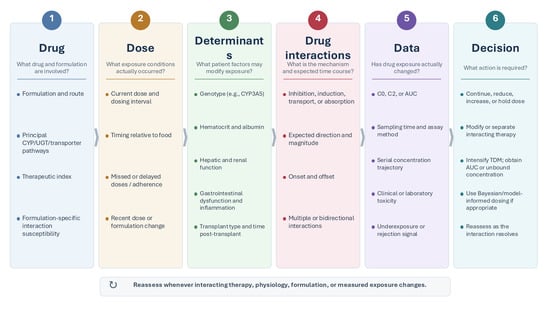
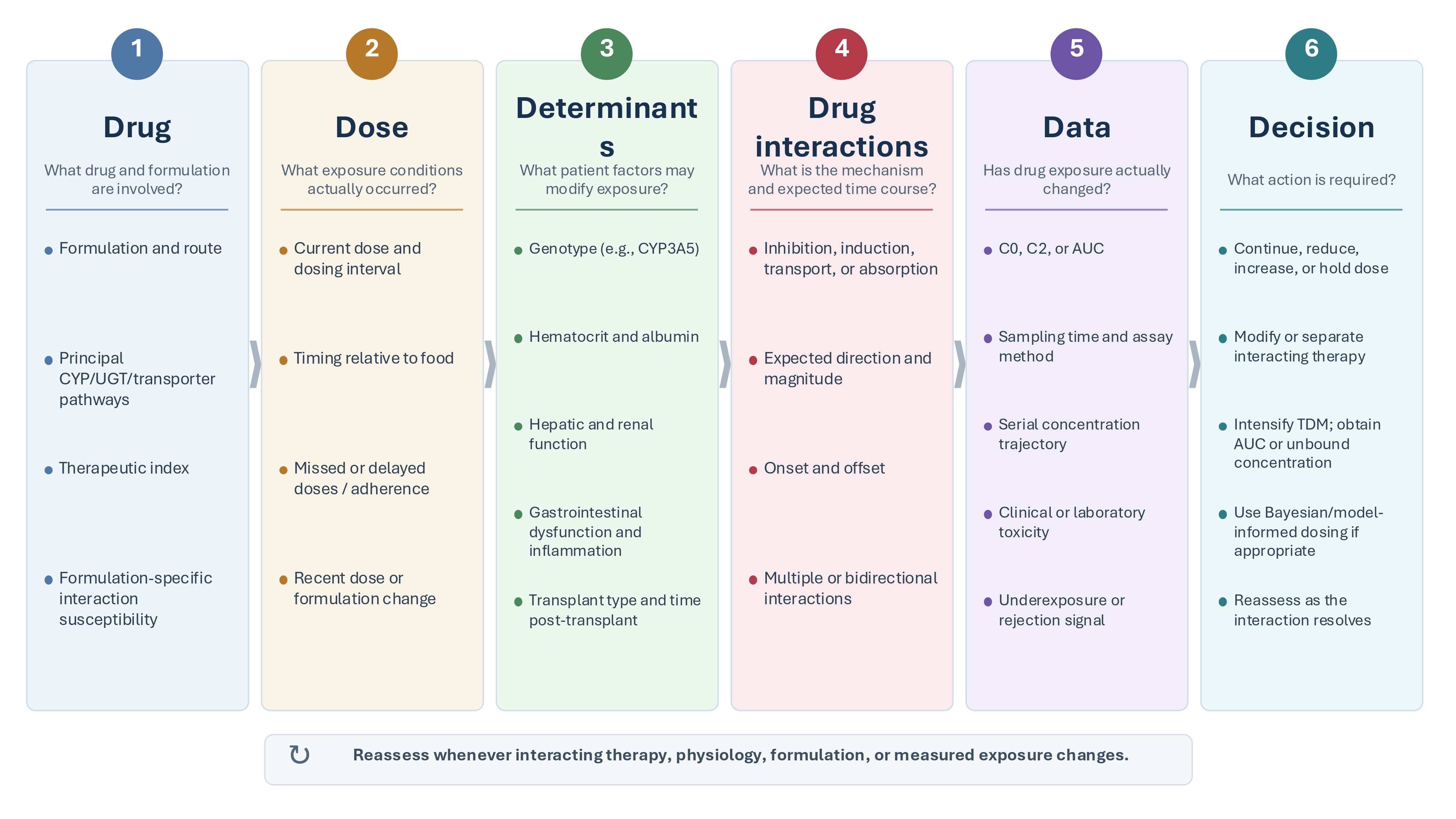

Disclaimer/Publisher’s Note: The statements, opinions and data contained in all publications are solely those of the individual author(s) and contributor(s) and not of MDPI and/or the editor(s). MDPI and/or the editor(s) disclaim responsibility for any injury to people or property resulting from any ideas, methods, instructions or products referred to in the content. |
© 2026 by the author. Licensee MDPI, Basel, Switzerland. This article is an open access article distributed under the terms and conditions of the Creative Commons Attribution (CC BY) license.