


Abstract
Background/Objectives: Glioblastoma multiforme (GBM) is the most aggressive primary tumor of the central nervous system and remains associated with poor prognosis despite current therapies. Magnetic hyperthermia (MHT) mediated by superparamagnetic iron oxide nanoparticles (SPIONs) is a promising therapeutic approach; however, improvements in tumor targeting, colloidal stability, and translational applicability are still needed. This study aimed to develop dopamine-functionalized SPION conjugated with the cyclic cRGD peptide (SPION-DOPA/cRGD) to promote preferential association with GBM cells and evaluate their physicochemical properties, cellular affinity, and therapeutic performance for MHT using a planar coil system. Methods: SPIONs were synthesized by thermal decomposition, functionalized with dopamine, and conjugated with cRGD. Their morphological, structural, magnetic, colloidal, and magnetothermal properties were characterized. Cellular affinity was evaluated in U87MG and HUVECs by Prussian Blue staining and Inductively Coupled Plasma Mass Spectrometry (ICP-MS), and free-cRGD competition was used to assess RGD–integrin-mediated association. Biocompatibility and therapeutic efficacy were assessed by bioluminescence assays in U87MG spheroids following MHT. Results: SPION-DOPA/cRGD exhibited quasi-spherical morphology, cores below 10 nm, superparamagnetic behavior, and relatively consistent hydrodynamic diameters over 120 min in both water and DMEM/F12. The cRGD-containing formulation exhibited enhanced cellular association with U87MG cells while maintaining low interaction with HUVECs; free-cRGD competition reduced nanoparticle association, supporting RGD–integrin interactions. Under a 300 G field, nanoparticles reached 42–45 °C, with a specific absorption rate of 210 ± 19 W g−1. The nanoformulation showed low cytotoxicity from 20 to 5000 μg Fe mL−1, with viability above 96%. SPION-DOPA/cRGD-mediated MHT reduced U87MG spheroid viability to 14.76 ± 1.02%, whereas AMF alone did not affect viability compared with controls. Conclusions: SPION-DOPA/cRGD combines preferential cellular association with U87MG cells, efficient magnetic heating, and a substantial reduction in spheroid viability in a three-dimensional tumor model. Together with a planar coil, these findings support the translational potential of this nanoplatform for preclinical MHT applications in GBM.
1. Introduction
Glioblastoma multiforme (GBM) is the most frequent and aggressive primary malignant tumor of the central nervous system (CNS) in adults [1,2]. It is characterized by diffuse infiltrative growth, cellular, molecular and genomic heterogeneity, rapid proliferation, pseudopalisading necrosis, and intense neovascularization [3]. GBM accounts for approximately 45–50% of all primary malignant brain tumors and has an estimated global incidence of about 3.2 cases per 100,000 inhabitants per year, with a predominance in males and a median age at diagnosis of approximately 64 years. Despite advances in diagnostic and therapeutic approaches, median overall survival remains between 14 and 18 months, the five-year survival rate remains below 10%, and more than virtually all patients experience tumor recurrence. These data highlight the high lethality of this disease and the urgent need for more effective therapeutic strategies [4,5,6,7].
The current standard of care has not changed for over two decades and consists of maximal safe surgical resection followed by temozolomide (TMZ) based radiochemotherapy [7]. However, the diffuse infiltrative nature of GBM prevents complete tumor removal, leading to early recurrence [8,9,10]. Radiotherapy is limited by the radiosensitivity of healthy brain tissue and by intratumoral heterogeneity [11]. The efficacy of TMZ is further compromised by intrinsic resistance mechanisms and by the blood–brain barrier (BBB) [12]. Consequently, despite multimodal treatment, tumor progression typically occurs within a few months [13].
In this context, nanotechnology has emerged as a promising alternative. Nanoparticles exhibit a high surface-to-volume ratio, enhanced reactivity, and the ability to exploit the enhanced permeability and retention (EPR) effect [14,15,16]. Beyond the EPR effect, nanosystems can be engineered to improve colloidal stability, modulate biodistribution, reduce nonspecific uptake, overcome biological barriers, and enhance tumor accumulation through passive and active targeting strategies [16,17,18]. Nanoparticles may be polymeric, lipid-based, metallic, or magnetic [18]. Iron oxide nanoparticles (SPIONs) function as magnetic resonance imaging (MRI) contrast agents and, when functionalized, can serve as theranostic platforms capable of simultaneous diagnosis and therapy [19,20,21]. They also enable controlled drug release and selective recognition of tumor-associated targets [17]. Recent advances in GBM nanomedicine have highlighted SPIONs as versatile platforms for imaging, drug delivery, thermal therapy, magnetochemotherapy, and multimodal therapeutic approaches, particularly when combined with polymeric coatings, polyethylene glycol (PEG), dopamine (DOPA), peptides, or molecular targeting ligands [18,19,21,22,23]. The translational potential of iron oxide nanoparticles is further supported by ferumoxytol, an FDA-approved formulation for iron deficiency anemia that has also been investigated in GBM. Previous studies have demonstrated its applications in evaluating nanoparticle interactions with tumor-associated immune cells and intratumoral distribution [24], characterizing the GBM immune microenvironment through MRI [25], and monitoring nanoparticle accumulation using multimodal imaging [26]. Furthermore, focused ultrasound-mediated blood–brain barrier opening has been shown to enhance ferumoxytol accumulation in experimental GBM [27]. These findings highlight the importance of nanoparticle delivery, tumor-associated interactions, and intratumoral accumulation, supporting the development of surface-functionalized iron oxide nanoplatforms to improve tumor targeting and therapeutic efficacy.
In addition, Fe3O4 nanoparticles possess the ability to generate heat under alternating magnetic fields, which forms the basis of magnetic hyperthermia (MHT) [28].
MHT relies on the conversion of magnetic energy into heat through Néel relaxation, Brownian relaxation, and hysteresis losses, resulting in localized heating (42–45 °C) and tumor cell death by apoptosis and necrosis [29,30,31,32]. Its efficiency is determined by the specific absorption rate (SAR), which depends on the physicochemical properties of the nanoparticles and the parameters of the applied magnetic field [33]. MHT has shown synergistic effects with chemotherapy and radiotherapy by increasing BBB permeability, enhancing drug diffusion, and improving tumor sensitivity to treatment [31,32]. Clinical studies using NanoTherm® have demonstrated the feasibility and safety of this approach [23,34,35]. Nevertheless, important limitations remain, including heterogeneous intratumoral nanoparticle distribution, aggregation, and the need for intralesional administration [22,31,36,37]. Consequently, improving colloidal stability, heating efficiency, biocompatibility, and active tumor targeting has become essential to enhance the translational potential of MHT. Recent developments include hybrid nanoparticles, magnetic nanoclusters, antifouling coatings, PEGylated systems, drug-loaded magnetic structures, and platforms integrated with combination therapies [28,33,38,39,40,41,42,43,44].
Molecular functionalization of SPIONs can further improve tumor selectivity [17,45,46]. Among the most widely explored molecular targets for active tumor targeting are the αvβ3 and αvβ5 integrins, which are highly expressed in angiogenic endothelial cells and in several tumor cell populations, including GBM [47,48,49]. The cyclic Arginine-Glycine-Aspartic acid peptide (cRGD) exhibits high affinity for αvβ3 and αvβ5 integrins, which are overexpressed in GBM cells and tumor-associated vasculature [47,48,49]. Integrin αvβ3 plays a critical role in cell adhesion, migration, invasion, and angiogenesis and has been associated with increased tumor aggressiveness and therapeutic resistance [49,50]. Furthermore, RGD-associated integrins participate in cell–extracellular matrix communication, activation of survival pathways, vascular remodeling, invasive progression, and maintenance of resistant tumor phenotypes, making them attractive targets for molecular imaging, selective drug delivery, and combination therapies [47,48,49,50,51]. The cyclic conformation of cRGD enhances both affinity and stability [50,51]. When conjugated to nanoparticles, cRGD improves cellular internalization and tumor retention, enabling the delivery of therapeutic agents such as TMZ, paclitaxel (PTX), and doxorubicin (DOX) [17,46]. RGD- and cRGD-functionalized nanoparticles have demonstrated enhanced tumor accumulation, greater uptake by αvβ3-positive cells, improved MRI/positron emission tomography (PET)–MRI contrast, reduced nonspecific uptake, and increased therapeutic efficacy in glioma and other tumor models [52,53,54,55,56,57,58,59,60,61,62,63]. DOPA, through the formation of polydopamine (PDA), facilitates adhesion to magnetite surfaces and provides reactive sites for conjugation with ligands such as cRGD [64,65,66]. DOPA/PDA coatings also contribute to colloidal stability, biocompatibility, versatile surface functionalization, and preservation of the magnetic properties of SPIONs [64,65,66,67]. The Fe3O4-DOPA/PDA–cRGD architecture preserves magnetic performance while enhancing active tumor targeting [65]. However, demonstrating the therapeutic potential of these nanoparticles requires experimental models capable of evaluating not only their interaction with tumor cells, but also their distribution, retention, and therapeutic response under conditions that more closely reproduce the structural complexity of the tumor microenvironment.
The low success rate of clinical trials in GBM also highlights the limitations of conventional preclinical models, which often fail to reproduce tumor evolution following surgery, radiotherapy, and chemotherapy [68]. Although two-dimensional (2D) models provide valuable preliminary information, they fail to reproduce the three-dimensional architecture, heterogeneity, and biochemical gradients found in tumors [31,69,70,71,72]. GBM spheroids more accurately mimic three-dimensional organization, cellular plasticity, and tumor invasiveness [73,74]. Biomimetic matrices enable the establishment of physiological gradients and the formation of hypoxic and necrotic regions [75,76]. Organoids represent an intermediate model capable of evaluating nanoparticle distribution, retention, and thermal responses within complex microenvironments [77,78,79]. In this regard, three-dimensional models are particularly relevant for studies involving magnetic nanoparticles and MHT, as they allow investigation of nanoparticle penetration, iron distribution, heating heterogeneity, thermal diffusion, hypoxia-associated resistance, and cellular responses in an architecture that more closely resembles the in vivo tumor microenvironment [70,71,76,77,78,80,81]. Furthermore, future integration with microfluidic systems, tumor-on-a-chip, and blood–brain barrier-on-a-chip platforms may provide a more realistic assessment of the interactions among fluid flow, biological barriers, nanoparticles, and therapeutic responses [82,83,84]. Rather than introducing DOPA functionalization or cRGD targeting as individual novel components, the present study investigates their integration with a planar coil-based magnetic hyperthermia system and their evaluation in a three-dimensional glioblastoma spheroid model. This integrated approach enables nanoparticle physicochemical and magnetic properties, cellular association, magnetothermal performance, and therapeutic response to be examined within the same experimental framework.
This study aimed to develop cRGD-functionalized Fe3O4 nanoparticles as an active targeting platform for GBM and to investigate their cellular association and therapeutic performance in MHT. To this end, physicochemical, colloidal, magnetic, and magnetothermal characterization of the nanoparticles was performed, together with the evaluation of their interaction with tumor cells and the assessment of MHT efficacy in three-dimensional GBM spheroids. Importantly, MHT was performed using a planar coil system, which enables magnetic field application from one side of the target and represents a configuration of particular interest for the development of more localized and accessible MHT applicators. Thus, the study integrates nanoparticle surface functionalization and molecular targeting, three-dimensional tumor modeling, and planar coil-based magnetic field delivery within a single experimental framework, providing a basis for further preclinical investigation and future translational development of MHT strategies for GBM.
2. Materials and Methods
2.1. Materials
Ethylene glycol (ACS reagent), tetrahydrofuran (THF) (ACS reagent), 3,4-dihydroxybenzaldehyde (APC) (98%), dopamine hydrochloride (DOPA) (minimum 98%), N-hydroxysulfosuccinimide (Sulfo-NHS), 1-ethyl-3-(3-dimethylaminopropyl) carbodiimide (EDC), and 2-(N-morpholino)ethanesulfonic acid (MES) were purchased from Sigma-Aldrich. Cyclo(Ala-Arg-Gly-Asp-3-aminomethylbenzoyl) (cRGD; product no. C9357, molecular weight 532.55 g/mol, purity > 96% by HPLC) was purchased from Sigma-Aldrich (St. Louis, MO, USA). Iron(III) acetate was synthesized following a previously reported method [85].
2.2. Synthesis of DOPA-Functionalized SPION
SPIONs were prepared via a thermodecomposition method, utilizing an iron(III) precursor in ethylene glycol as a high-boiling point solvent [86,87]. Briefly, 2.96 g of iron (III) acetate (5 mmol) in ethylene glycol (St. Louis, MO, USA) was heated to 160 °C for 30 min, and then the temperature was increased to 200 °C for 30 min, under vigorous stirring. The dark product formed was precipitated out with THF and isolated using Nd2Fe14B magnets. Purification was carried out by successive cycles of redispersion and magnetic precipitation in small volumes of water and THF, until a colorless supernatant was formed, typically after 3 cycles. Finally, the dark solid was dried and dispersed in water to form a nanofluid composed of 6 nm large monodisperse particles. Then, the nanoparticles were functionalized with DOPA according to the method previously described [86,88]. In brief, 10 mL of SPION nanofluid, corresponding to approximately 1.0 g of SPION (Fe3O4-based) was added to 10 mL of an aqueous solution containing 237 mg (1.25 mmol) of DOPA hydrochloride, and then stirred for 180 min at room temperature. Finally, the functionalized SPION-DOPA was precipitated out magnetically, washed three times with 40 mL of ethanol (St. Louis, MO, USA), and dried in a desiccator under vacuum. Finally, the dark solid was dispersed in a small amount of DIW, and the concentration was adjusted to form a nanofluid with 1 wt% Fe3O4 (titration).
2.3. Physicochemical and Magnetic Characterization of SPION
Transmission electron microscopy (TEM) was employed to evaluate the size and morphology of the nanoparticles. A JEOL JEM 2100 FEG-TEM (JEOL Ltd., Tokyo, Japan) field emission microscope, equipped with a field emission gun (FEG) and operating in ultra-high-resolution (HR-TEM) mode at an acceleration voltage of 200 kV, was utilized. Samples were prepared by drop-casting 2 μL of a 0.1 g L−1 nanoparticle dispersion onto an ultrathin carbon-copper grid (400 mesh) (Ted Pella Inc., Redding, CA, USA) covered with an ultrathin carbon film and Formvar, followed by drying at room temperature. Size distribution histograms were generated by measuring at least 150 individual nanoparticles from TEM images.
The success of the superparamagnetic nanoparticle preparation process was confirmed by measuring the magnetic hysteresis curves using an EG&G Princeton Applied Research Corporation, Model 4500 vibrating sample magnetometer (VSM, Princeton, NJ, USA), coupled with a Walker Scientific model HR8 electromagnet (Walker Scientific, city, state, USA) with a nominal magnetic field varying from −20 to 20 kOe scanning window, and a Lake Shore Model 450 Gaussmeter (Lake Shore Cryotronics Inc., Westerville, OH, USA). The standard protocol involved sweeping the magnetic field from 0 to +20 kOe, then down to −20 kOe, and finally reversing the scan back to +20 kOe to complete a full cycle, while continuously measuring the corresponding sample magnetization.
Fourier transform infrared spectroscopy (FTIR) spectra were recorded using an ALPHA FTIR spectrometer (Bruker Optik GmbH, Ettlingen, Germany) in the range of 4000–400 cm−1, with a spectral resolution of 4 cm−1. Samples were prepared as KBr dispersions containing approximately 0.5 wt% of the nanomaterial.
X-ray diffraction (XRD) measurements were performed using a D2 PHASER X-ray diffractometer (Bruker AXS GmbH, Karlsruhe, Germany) equipped with a Cu source (λ = 1.5418 Å, 30 kV, 15 mA) and a LYNXEYE 1D detector. Powdered nanomaterial samples were analyzed in the 2θ range from 10° to 90°, with a step size of 0.02°.
Hydrodynamic size distribution and zeta potential were measured using a Zetasizer Nano ZS90 (Malvern Panalytical Ltd., Malvern, UK). Measurements were performed using aqueous nanoparticle dispersions at a concentration of 1 mg/mL, within a size range of 0.3–5000 nm. Results were obtained in triplicate, with at least 10 independent measurements per sample (triplicate). To evaluate changes in surface charge during the sequential functionalization process, zeta-potential measurements were performed for SPION, SPION-DOPA, and SPION-DOPA/cRGD at pH 6.0.
2.4. Dopamine-Functionalized Iron Oxide Nanoparticles Conjugated with cRGD Peptide
SPION-DOPA nanoparticles were subjected to surface chemical activation for covalent conjugation with the cRGD peptide, which exhibits selective affinity toward integrin receptors. The conjugation reaction was performed through carbodiimide-mediated coupling using EDC and NHS as activating agents for the sole carboxyl group available in the cRGD structure associated with the aspartic acid residue, thereby enabling its coupling to the amino group of dopamine ligands bound onto the nanoparticle surface through the bidentate catechol end group.
The conjugation reaction was performed by mixing cRGD (50 μL, 1 mg/mL), EDC (50 μL, 0.01 M), Sulfo-NHS (50 μL, 0.02 M), and MES buffer (10 μL, 0.05 M, pH 6.0). These components were gently mixed for 15 min to promote activation of the carboxyl group of cRGD. Subsequently, SPION-DOPA (500 μL, 20 mg Fe/mL) was added to the activated cRGD mixture, and the suspension was maintained under continuous stirring for 1 h at room temperature. Finally, 340 μL of deionized water was added to obtain a final volume of 1 mL. Under these conditions, the activated carboxyl group of cRGD subsequently reacts with the primary amino groups of surface-bound dopamine, resulting in amide bond formation. The EDC/Sulfo-NHS-mediated coupling mechanism used for cRGD conjugation to SPION-DOPA is illustrated in Figure 1.
Figure 1.
EDC/Sulfo-NHS-mediated coupling mechanism for the conjugation of cRGD to SPION-DOPA. The carboxyl group associated with the aspartic acid residue of cRGD is activated by EDC to form an O-acylisourea intermediate, followed by formation of a Sulfo-NHS ester. The activated ester subsequently reacts with primary amino groups of surface-bound dopamine, resulting in the formation of a stable amide bond between cRGD and SPION-DOPA.
At the end of the reaction, a colloidal dispersion of SPION-DOPA/cRGD was obtained, corresponding to the nanoconjugate formed by DOPA-functionalized SPION and the cRGD peptide, intended for application in MHT therapy.
Based on the composition of the 1 mL SPION-DOPA/cRGD preparation, the formulation contained 10 mg of Fe (corresponding to approximately 13.8 mg Fe3O4; 60 μmol Fe3O4), 1.40 mg of dopamine (7.4 μmol, based on the DOPA content estimated by TGA), and 0.05 mg of cRGD (0.09 μmol). Considering near-spherical magnetite nanoparticles with an average TEM diameter of 6.2 nm, the formulation was estimated to contain approximately 2 × 10¹⁶ nanoparticles, corresponding to approximately 240 dopamine molecules per nanoparticle (~2 molecules/nm2). Based on the nominal amount of cRGD used in the conjugation reaction, this corresponds to an estimated ratio of approximately three cRGD molecules per nanoparticle. These values represent theoretical estimates based on nanoparticle dimensions, TGA-derived DOPA content, and the nominal cRGD amount used in the reaction, rather than a direct quantitative determination of cRGD surface loading.
2.5. Time-Dependent Hydrodynamic Size Analysis of SPION-DOPA/cRGD in Cell Culture Medium
The time-dependent hydrodynamic size behavior of SPION-DOPA and SPION-DOPA/cRGD nanoparticles was evaluated by dynamic light scattering (DLS) using a Zetasizer Ultra (Malvern Panalytical Ltd., Malvern, UK). The nanoparticles were prepared at a final concentration of 30 μg Fe/mL and distributed into experimental groups according to the dispersion medium: Milli-Q water and DMEM/F12 culture medium. For each condition, both nanoparticle formulations (SPION-DOPA and SPION-DOPA/cRGD) were analyzed at 0, 60, and 120 min after dispersion. The hydrodynamic diameter (Z-average), polydispersity index (PDI), and intensity-weighted particle size distribution profiles were determined for each experimental condition.
2.6. Magnetic Hyperthermia Setup, Heating Curves, and SAR Determination
MHT assays using a planar coil configuration were performed with a CMA Innovative Heating system (GH Group, Valencia, Spain). The planar coil consisted of four spiral turns, with overall dimensions of approximately 64.73 × 68.18 mm and a copper tube diameter of 5.8 mm. During the MHT experiments, the samples were positioned at the center of the planar coil, in direct contact with its surface. The system enables the generation of alternating magnetic fields ranging from 100 to 300 Gauss, operating at a frequency of 225 kHz.
Temperature variation during magnetic heating was monitored in real time, using a Neoptix T1 fiber-optic temperature sensor (Neoptix Inc., Québec City, QC, Canada) with an accuracy of ±0.2 °C. Thermal stability of the system was maintained using a MiniChiller cooling unit (Mecalor Ltd.a., São Paulo, SP, Brazil).
To evaluate the heating performance of SPION-DOPA/cRGD, 100 μL of an aqueous nanoparticle dispersion at 10 mgFe/mL was used. The experiment was conducted under controlled room temperature conditions at 20 °C to ensure reproducibility throughout the measurements.
Heating curves were recorded as temperature variation over time, starting from 20 °C and reaching approximately 45 °C. The assays were conducted under alternating magnetic fields of 100, 200, and 300 Gauss, at a fixed frequency of 225 kHz. These conditions were used to assess the field-dependent thermal response of SPION-DOPA/cRGD under parameters compatible with MHT applications.
The heating curves were analyzed using Origin 2025 software (OriginLab, Northampton, MA, USA), allowing extraction of the maximum heating rate, (dT/dt)max. The SAR, which expresses the efficiency of magnetic nanoparticles in converting electromagnetic energy into heat, was calculated from experimental heating profiles.
The SAR calculation was performed using the following Equation (1):
where mNP corresponds to the mass of iron (Fe) present in the SPIONs, cNP is the specific heat capacity of magnetite (0.16 kcal/kg°C), ml corresponds to the mass of water, cl is the specific heat capacity of water (1.0 kcal/kg°C), and (dT/dt)max is the maximum temperature variation obtained from the heating curve of SPION-DOPA/cRGD. Therefore, SAR values were normalized to the iron mass and expressed as watts per gram of iron (W g−1).
2.7. Computational Simulation of the Magnetic Field Distribution Generated by the Planar Coil
To evaluate the spatial distribution of the magnetic field generated by the planar coil used in the magnetic hyperthermia experiments, a three-dimensional computational simulation was performed using COMSOL Multiphysics® version 6.4 (COMSOL Inc., Burlington, MA, USA). The simulations were conducted using the AC/DC Module with the Magnetic Fields interface, based on Maxwell’s equations and the electric current density formulation.
The computational model reproduced the geometry of the experimental planar coil described in Section 2.6, consisting of four spiral turns, with overall dimensions of approximately 64.73 × 68.18 mm and a copper tube diameter of 5.8 mm. The coil was modeled as a solid copper conductor with a right-handed spiral configuration, and the electromagnetic simulation was performed at an operating frequency of 225 kHz, consistent with the experimental magnetic hyperthermia conditions.
The spatial distribution of the magnetic flux density was calculated in the computational domain surrounding the coil, with particular attention to the region corresponding to the experimental sample position. Three-dimensional magnetic field distributions, magnetic flux density vectors, and spatial field maps were obtained to characterize the magnetic field generated by the planar coil under the specified operating conditions.
2.8. Cell Culture
For the in vitro experiments, U87MG and Human umbilical vein endothelial cells (HUVECs) cell lines were used. The U87MG cell line, derived from human glioblastoma, was obtained from the Cell Bank of Rio de Janeiro (BCRJ, Rio de Janeiro, Brazil) and was previously transfected with the luciferase gene according to a methodology established by our research group and described elsewhere [89]. HUVECs were kindly donated by Prof. Sandra Helena Poliselli Farsky from the Department of Clinical and Toxicological Analyses, School of Pharmaceutical Sciences, University of São Paulo, São Paulo, Brazil.
U87MG cells were maintained in DMEM/F12 (GIBCO® Invitrogen Corporation, Carlsbad, CA, USA), supplemented with 10% fetal bovine serum (FBS) (GIBCO® Invitrogen Corporation, Carlsbad, CA, USA) and 1% antibiotic-antimycotic solution (Anti-Anti) (GIBCO® Invitrogen Corporation, Carlsbad, CA, USA). Cell culture was performed in 75 cm2 culture flasks (Corning®, Corning, NY, USA) and maintained in a humidified incubator at 37 °C under an atmosphere containing 5% CO2.
For routine subculture, cells were harvested upon reaching approximately 80% confluence. The culture medium was removed, and the cells were washed with phosphate-buffered saline (PBS) (GIBCO® Invitrogen Corporation, Carlsbad, CA, USA). Subsequently, cells were detached using 0.25% trypsin-EDTA (STEMCELL Technologies, Vancouver, BC, Canada) for 3 min at 37 °C. Trypsin activity was neutralized by the addition of complete culture medium supplemented with 10% FBS. The resulting cell suspension was centrifuged at 500 rcf for 5 min, and the cell pellet was resuspended in fresh culture medium for subsequent experiments or routine maintenance.
HUVECs were cultured under the same incubation conditions using RPMI-1640 (GIBCO® Invitrogen Corporation, Carlsbad, CA, USA), supplemented with 10% FBS and 1% antibiotic-antimycotic solution. Culture media were renewed every 48 h throughout the maintenance period.
2.9. Three-Dimensional Spheroid Culture from U87MG Cells
For the establishment of the three-dimensional (spheroid) model, U87MG cells previously cultured under 2D conditions were detached by trypsinization, counted, and resuspended in DMEM culture medium supplemented with 10% (v/v) fetal bovine serum (FBS) and 1% penicillin/streptomycin. The cell suspension was adjusted to a final density of 2 × 10⁴ cells per well and seeded into 96-well U-bottom ultra-low attachment plates (Corning®, Costar®, Corning, NY, USA).
To promote cell self-aggregation and spheroid formation, Matrigel® (Corning® Life Sciences, Oneonta, NY, USA) was added to the culture medium at a final concentration of 2.5% (v/v). Matrigel was maintained under refrigeration during preparation to prevent premature polymerization. The mixture was carefully homogenized on ice prior to cell seeding.
Following seeding, the plates were incubated for 30 min at 37 °C and 5% CO2 under orbital agitation at 250 rpm, allowing uniform cell distribution and initiation of the aggregation process. Subsequently, the plates were maintained under static incubation under the same conditions until the formation of compact spheroidal structures.
Spheroid morphology and stability were monitored by inverted optical microscopy (Nikon®, Tokio, Japan). After 15 days of culture, stable U87MG spheroids were used for SPION-DOPA/cRGD cytotoxicity assays and evaluation of the therapeutic efficacy of MHT.
2.10. Cell Viability Assessment of U87MG Spheroids Treated with SPION-DOPA/cRGD
Cell viability after exposure to SPION-DOPA/cRGD nanoparticles was evaluated using U87MG-derived GBM spheroids cultured for 15 days. For this assay, SPION-DOPA/cRGD nanoparticles were previously dispersed in culture medium and applied to the spheroids at concentrations of 0, 20, 50, 100, 500, 1000 and 5000 µg Fe/mL. Each experimental condition was performed in tubes containing three spheroids, which were incubated for 24 h under standard culture conditions.
After the exposure period, the spheroids were washed three times with serum-free culture medium to remove residual nanoparticles remaining in suspension and minimize their potential influence on the bioluminescence measurements. Cell viability was assessed by bioluminescence imaging (BLI). Briefly, 5 µL of D-luciferin (30 mg/mL; XenoLight, PerkinElmer Inc., Waltham, MA, USA) was added to each sample to detect the luminescent signal generated by the luciferase-catalyzed enzymatic reaction. Images were acquired using an IVIS Lumina LT Series III imaging system (PerkinElmer Inc., Waltham, MA, USA), configured with automatic exposure time, F/Stop 4, binning 4, and a field of view (FOV) of 12.9 cm.
The emitted bioluminescent signal intensity was used as a quantitative indicator of cell viability, with reductions in photon emission interpreted as decreased viable tumor cell activity after nanoparticle exposure. All experiments were performed in quintuplicate.
2.11. Integrin Receptor Blocking by Free cRGD Competition and Quantification of Cell-Associated Iron
To evaluate whether the increased cellular association of SPION-DOPA/cRGD is related to the interaction between nanoparticle-conjugated cRGD and integrin receptors, a competitive receptor-blocking assay using free cRGD was performed. In this assay, cellular receptors capable of recognizing the RGD motif were pre-occupied with free cRGD, thereby reducing the availability of these binding sites for interaction with cRGD conjugated to the nanoparticles. Thus, comparison with cells not subjected to competitive blocking allowed assessment of the contribution of integrin-mediated interactions to the cellular association of the functionalized nanoparticles.
Cells were seeded in 24-well culture plates at a density of 1 × 10⁵ cells per well and maintained under standard culture conditions until approximately 70% confluence was reached. For the competitive blocking condition, cells were pre-incubated with free cRGD (200 µg/mL) for 18 h and subsequently exposed to SPION-DOPA/cRGD (30 µg/mL) for 10, 30, or 120 min. In parallel, cells without free cRGD pre-incubation were exposed to the nanoparticles under the same conditions, while cells not exposed to nanoparticles were used as controls.
At the end of each exposure period, cells were washed to remove non-associated nanoparticles and subjected to Prussian Blue staining for qualitative detection of cell-associated SPION-DOPA/cRGD. Images were acquired using an EVOS M5000 microscope (Thermo Fisher Scientific, Waltham, MA, USA). Nanoparticle cellular association was qualitatively compared between cells pre-incubated with free cRGD and cells without competitive blocking at each incubation time.
Additionally, cell-associated iron was quantified by inductively coupled plasma mass spectrometry (ICP-MS) in parallel samples under the same experimental conditions. Iron quantification was performed according to the procedure described in Section 2.13, allowing quantitative comparison of nanoparticle–cell association between the competitive blocking and non-blocking conditions at each incubation time.
2.12. Evaluation of the Affinity of SPION-DOPA/cRGD Nanoparticles Toward U87MG Glioblastoma Cells
The cellular affinity of SPION-DOPA/cRGD nanoparticles toward U87MG glioblastoma cells was evaluated by comparing their association with U87MG and HUVECs at different incubation times. Both cell lines were seeded in 24-well plates at a density of 5 × 10⁴ cells per well and maintained under standard culture conditions until reaching approximately 60% confluence. U87MG cells were cultured in DMEM/F12 medium, whereas HUVECs were maintained in RPMI-1640 medium, both supplemented with 10% FBS.
Following the initial culture period, the medium was replaced with fresh medium containing SPION-DOPA/cRGD nanoparticles at a concentration of 10 µg Fe/mL. Cells were incubated for 10, 60, and 120 min to evaluate the time-dependent association of the nanoparticles with each cell line. At the end of each incubation period, the wells were washed three times with PBS to remove non-associated or weakly adhered nanoparticles.
Cell-associated iron was qualitatively assessed by Prussian Blue staining according to a protocol previously established by our research group [68]. Briefly, a potassium ferrocyanide solution in 5% hydrochloric acid was added to each well and incubated for 5 min at room temperature. After staining, the cells were washed with PBS to remove excess reagent and maintained in supplemented culture medium.
Samples were analyzed by bright-field optical microscopy using an EVOS digital microscope (Thermo Fisher Scientific, Waltham, MA, USA). Prussian Blue-positive areas were considered indicative of cells’ iron associated with nanoparticles, allowing qualitative comparison of the cellular affinity of SPION-DOPA/cRGD toward U87MG cells, using HUVECs as a comparative reference.
2.13. Quantification of Cell-Associated Iron by ICP-MS
To quantitatively evaluate the cellular association of SPION-DOPA and SPION-DOPA/cRGD, the amount of iron associated with HUVEC and U87MG cells was determined by inductively coupled plasma mass spectrometry (ICP-MS; NexION 350X, PerkinElmer, Waltham, MA, USA) following incubation with 10 μg Fe/mL of nanoparticles. Four experimental groups were evaluated: (i) HUVEC + SPION-DOPA, (ii) HUVEC + SPION-DOPA/cRGD, (iii) U87MG + SPION-DOPA, and (iv) U87MG + SPION-DOPA/cRGD, at incubation times of 0, 10, 60, and 120 min, in addition to untreated control cells.
U87MG and HUVECs treated with SPION-DOPA and SPION-DOPA/RGD were collected by trypsinization, counted, and resuspended in culture medium. Samples containing 105 cells were then subjected to acid digestion for subsequent analysis by ICP-MS. Briefly, 500 µL of each cell suspension was transferred to 1.5 mL polypropylene tubes, followed by the addition of 400 µL of HNO3 and 100 µL of H2O2. Samples were homogenized and incubated at 70 °C under agitation at 450 rpm for 4 h using a ThermoMixer C (Eppendorf®, Hamburg, Germany). Complete digestion was confirmed by the formation of clear solutions, which were stored at −20 °C until ICP-MS analysis.
Iron quantification was performed using dynamic reaction cell mode with methane as the reaction gas to minimize spectral interferences. Analyses were carried out under standardized conditions: radiofrequency power of 1600 W, plasma gas flow of 18 L min−1, nebulizer gas flow of 0.91 L min−1, 50 readings per replicate, and dwell time of 50 ms. The isotope 56Fe was monitored for quantification, and 103Rh+ was used as the internal standard.
Quantification was based on a calibration curve prepared from a certified iron standard solution (PerkinElmer Inc., Waltham, MA, USA; Cat. No. N8145054) ranging from 5 to 40 ppb. Digested samples were diluted tenfold in Milli-Q water containing 2% HNO3, and measurements were performed in quintuplicate to determine the amount of cell-associated iron.
Thus, the measured iron content was used as a quantitative indicator of nanoparticle–cell association, allowing comparison among nanoparticle formulations, incubation times, and cell lines.
2.14. Application of Magneto-Hyperthermia and Evaluation of Therapeutic Response in U87MG Glioblastoma Spheroids
The application of MHT was performed in GBM spheroids on the 15th day of culture, when the spheroids exhibited adequate structural organization for therapeutic assessment. The spheroids were divided into six experimental groups: (i) untreated spheroids (Spheroid); (ii) spheroids incubated with SPION-DOPA without AMF exposure (Spheroid + SPION-DOPA); (iii) spheroids incubated with SPION-DOPA/cRGD without AMF exposure (Spheroid + SPION-DOPA/cRGD); (iv) spheroids exposed to AMF without nanoparticles (Spheroid + AMF); (v) spheroids incubated with SPION-DOPA and subsequently exposed to AMF (Spheroid + SPION-DOPA + AMF); and (vi) spheroids incubated with SPION-DOPA/cRGD and subsequently exposed to AMF (Spheroid + SPION-DOPA/cRGD + AMF).
For groups receiving nanoparticles, SPION-DOPA or SPION-DOPA/cRGD nanoparticles were added to the culture medium at a final concentration of 1 mg Fe/mL. The spheroids were incubated with the nanoparticles for 24 h under standard culture conditions to allow nanoparticle interaction and association with the three-dimensional tumor model. Following this incubation period, three spheroids were transferred to a sterile tube. The samples assigned to AMF exposure were subjected to magnetic hyperthermia treatment by positioning the sample tubes in the planar-coil MHT system to ensure uniform exposure to the alternating magnetic field. Both nanoparticle formulations were evaluated using the same AMF exposure protocol to enable a direct comparison of their therapeutic effects.
The heating protocol was conducted at a fixed frequency of 225 kHz, while the magnetic field intensity was automatically modulated by the equipment control software to maintain the temperature within the therapeutic range of 42–45 °C. Temperature was monitored in real time using a fiber-optic probe inserted directly into the tube containing the spheroids. Each MHT application lasted 30 min per tube and was performed in five independent samples.
After treatment, the spheroids were washed three times with serum-free culture medium to remove residual nanoparticles remaining in suspension. Fresh culture medium was subsequently added, and the spheroids were maintained under standard culture conditions for 72 h before viability assessment. After this period, each sample received 5 µL of D-luciferin, the substrate for luciferase expressed by U87MG cells. Photon emission resulting from D-luciferin oxidation was immediately recorded using a bioluminescence imaging system. Bioluminescence values obtained before treatment were normalized to 100%, and the relative changes in signal among the six experimental groups were used to evaluate the therapeutic response and distinguish the effects of nanoparticle exposure, AMF exposure alone, and MHT mediated by SPION-DOPA and SPION-DOPA/cRGD.
2.15. Statistical Analysis
Data were analyzed using descriptive statistics and are presented as mean ± standard deviation (SD). The normality of data distribution was assessed using the Shapiro–Wilk test. The homogeneity of variances was assessed using Levene’s test. For normally distributed data, comparisons between two groups were performed using Student’s t-test, whereas comparisons among three or more groups were conducted using one-way analysis of variance (ANOVA), followed by Tukey’s post hoc multiple-comparison test when applicable. When normality assumptions were not met, appropriate nonparametric tests were applied. Statistical significance was set at p < 0.05. Statistical analyses and graphical representations were performed using JASP 0.93.3 and Origin 2025 (OriginLab, Northampton, MA, USA).
3. Results
3.1. Morphological, Structural, and Colloidal Characterization of the Nanoparticles
The morphology and size distribution of the SPIONs were investigated using TEM. As depicted in the representative TEM images in Figure 2A,B, the SPIONs exhibit a quasi-spherical shape with diameters less than 10 nm. A log-normal fit of the size distribution histogram (inset Figure 2A) yielded a mean diameter (DTEM) of 6.2 nm with a standard deviation (σTEM) of 1.0 nm.
Figure 2.
Morphological, structural, colloidal, magnetic, and chemical characterization of the iron oxide nanoparticles. (A,B) TEM micrographs of SPION showing their morphology and particle size distribution; the inset in (A) presents the corresponding size histogram. (C) Hydrodynamic diameter distribution of SPION-DOPA measured by DLS. (D) XRD diffractogram of SPION confirming the crystalline magnetite phase (JCPDS No. 19-0629). (E) Magnetization versus applied magnetic field (M–H) curves obtained at 5 K and 300 K, demonstrating the superparamagnetic behavior of the nanoparticles at room temperature. (F) Zero-field-cooled (ZFC) and field-cooled (FC) magnetization curves measured under an applied field of 50 Oe, showing a blocking temperature of approximately 55 K. (G) FTIR spectra of DOPA (red curve), SPION-DOPA (black curve), and SPION (blue curve), with the main characteristic absorption bands and their corresponding vibrational assignments indicated directly on the spectra, supporting the successful functionalization of SPION with DOPA.
The colloidal behavior of SPION-DOPA was evaluated by DLS measurements. As shown in Figure 2C, SPION-DOPA displayed a monomodal hydrodynamic size distribution, with an intensity-weighted average diameter (Z-average) of 16.10 nm and a standard deviation of 9.58 nm.
The crystalline structure of the SPION was evaluated by XRD. As shown in Figure 2D, the diffraction peaks matched the inverse spinel structure of magnetite (Fe3O4), with a face-centered cubic lattice and space group Fd-3m, consistent with JCPDS card No. 19-0629. All observed peaks were indexed to the standard magnetite pattern. The reflection from the (222) plane, expected near 37.4°, was not clearly resolved, likely due to its low relative intensity and overlap with the adjacent broad peak at approximately 35.7°.
The significant broadening of the peaks in the SPION diffractogram is indicative of the nanoparticles’ small crystallite size. Based on the Scherrer equation, the average crystallite size for SPION was calculated to be DXRD = 5.4 ± 0.3 nm from the five most intense peaks. This value is in good agreement with the particle size measured by TEM, suggesting the nanoparticles are highly crystalline.
3.2. Magnetic Properties of SPION
The magnetic properties of the SPION were investigated by vibrating sample magnetometer measurements. The magnetization versus applied magnetic field (M–H) curves recorded at 5 K and 300 K are presented in Figure 2E. Despite the small particle size, the saturation magnetization reached 104 emu g−1 Fe at room temperature, corresponding to approximately 82% of the bulk magnetite value (~127 emu g−1 Fe). In addition, the initial magnetic susceptibility was estimated as χ = 4.1 (SI), slightly higher than the value reported for bulk magnetite (χ = 3.4 SI). The negligible coercivity and remanence observed at 300 K confirm the superparamagnetic behavior of the nanoparticles at room temperature.
The temperature-dependent magnetic behavior of the nanoparticles was further evaluated by zero-field-cooled (ZFC) and field-cooled (FC) measurements using a SQUID magnetometer (Figure 2F). In the ZFC experiment, the sample was initially cooled to 5 K in the absence of an applied magnetic field. Subsequently, a static magnetic field of 50 Oe was applied, and the magnetization was recorded while heating the sample to 300 K. The maximum of the ZFC curve corresponds to the average blocking temperature (TB), which was determined to be approximately 57 K.
Following the ZFC measurement, the sample was cooled back to 5 K under the same applied magnetic field, and the magnetization was recorded during reheating, generating the FC curve. The divergence between the FC and ZFC curves occurs at approximately 90 K, corresponding to the irreversibility temperature (Tirr). Below Tirr, the nanoparticles exhibit irreversible magnetic behavior associated with the presence of coercivity and remanence, whereas above this temperature the system progressively approaches the superparamagnetic regime.
The difference between blocking temperature (TB) and Tirr can be attributed to the particle size distribution and variations in effective magnetic anisotropy. Even with the relatively narrow size distribution observed by TEM (Figure 2A,B), a small fraction of larger nanoparticles may contribute disproportionately to the magnetic energy barrier distribution, resulting in a broader blocking transition. Consequently, the magnetic blocking process is more accurately described by a temperature interval extending from TB to Tirr rather than by a single characteristic temperature.
3.3. FTIR Spectroscopy Analysis
The FTIR spectrum of DOPA (Figure 2G) is characterized by bands at 1616, 1600, 1500, and 1470 cm−1 associated with the vibrational stretching modes of the aromatic ring; 1342 and 1320 cm−1 with bending vibrations of the aliphatic region; 1287 and 1260 cm−1 with C–O stretching; 1206 and 1190 cm−1 with in-plane bending of the phenolic group; 1174, 1146, and 1114 cm−1 with aromatic C–H bending; and 814 cm−1 with C–N stretching.
As shown in Figure 2G, compared with unfunctionalized SPION, SPION-DOPA exhibited new bands at 1482 and 1421 cm−1 attributed to aromatic C–C stretching, 1263 and 1221 cm−1 to C–O stretching, and 1148 and 1120 cm−1 to aromatic C–H bending. The bands assigned to the dihydroxyphenyl group showed pronounced shifts relative to free DOPA, consistent with Fe–O coordination at the nanoparticle surface. In contrast, the positions of the bands related to the stretching mode of the aliphatic C–N group of DOPA showed only slight changes (Figure 2G), suggesting that the amino group remains predominantly available for subsequent conjugation with biomolecules such as cRGD. Additionally, the O–H stretching band at 3414 cm−1 was identified, while the characteristic Fe–O stretching band at 583 cm−1 remained unchanged after functionalization, supporting the preservation of the iron oxide vibrational signature.
Overall, the FTIR results presented in Figure 2G support the successful functionalization of SPION with DOPA, consistent with the coordination of catechol groups to the iron oxide surface while preserving the potential availability of amino groups for subsequent chemical conjugation.
3.4. Zeta-Potential Analysis
Zeta-potential measurements were performed to evaluate changes in surface charge during the sequential functionalization of the nanoparticles. At pH 6.0, SPION exhibited a zeta potential of +19.8 ± 2.1 mV, which increased to +46.3 ± 3.5 mV following DOPA functionalization. After cRGD conjugation, the zeta potential was +44.1 ± 1.8 mV. Thus, DOPA functionalization resulted in a marked increase in positive surface charge, whereas subsequent cRGD conjugation produced only a modest change in zeta potential relative to SPION-DOPA.
3.5. Evaluation of the Hydrodynamic Size Stability of SPION-DOPA/cRGD
The hydrodynamic size stability of SPION-DOPA (black curves) and SPION-DOPA/cRGD (red curves) nanoparticles was assessed by monitoring changes in hydrodynamic size over 120 min in water and DMEM/F12 culture medium (Figure 3A–F). In water, SPION-DOPA exhibited mean hydrodynamic diameters of 33.92, 29.66, and 39.70 nm at 0, 60, and 120 min, respectively (Figure 3A–C), whereas SPION-DOPA/cRGD presented values of 27.93, 27.49, and 22.85 nm at the same time points (Figure 3A–C). These results indicate that both formulations maintained relatively consistent hydrodynamic diameters in water throughout the evaluated period, with SPION-DOPA/cRGD exhibiting smaller mean diameters.
Figure 3.
Time-dependent hydrodynamic size analysis of DOPA-coated iron oxide nanoparticles (SPION-DOPA, black curves) and cRGD-functionalized nanoparticles (SPION-DOPA/cRGD, red curves) by DLS. (A–C) Hydrodynamic diameter distribution of nanoparticles dispersed in water at 0, 60, and 120 min, respectively. (D–F) Hydrodynamic diameter distribution of nanoparticles dispersed in DMEM/F12 at the same incubation times. Intensity-based size profiles (A.U.) were used to assess changes in hydrodynamic size distributions as a function of medium and time.
In DMEM/F12, SPION-DOPA exhibited substantially larger hydrodynamic diameters than those observed in water, reaching 1008.80 nm immediately after dispersion, 621.90 nm after 60 min, and 1041.70 nm after 120 min (Figure 3D–F). The particle size distribution profiles revealed the presence of aggregates in the culture medium. In contrast, SPION-DOPA/cRGD maintained hydrodynamic diameters within the nanometric range, with values of 38.75, 36.87, and 44.89 nm at 0, 60, and 120 min, respectively (Figure 3D–F), demonstrating relatively consistent mean hydrodynamic diameters over time, even in the more complex culture medium. These findings suggest that cRGD functionalization contributed to maintaining a smaller hydrodynamic size compared with SPION-DOPA under the evaluated conditions.
The polydispersity index (PDI) provided additional information regarding the heterogeneity of the nanoparticle size distributions. In water, both formulations exhibited PDI values ranging from 0.32 to 0.61, indicating moderate to high polydispersity despite the relatively consistent hydrodynamic diameters over time. In DMEM/F12, SPION-DOPA presented a PDI of 1.00 at all evaluated time points, indicating a highly heterogeneous system associated with extensive aggregation. Although SPION-DOPA/cRGD also showed elevated PDI values in DMEM/F12 (0.68–1.00), its reported mean hydrodynamic diameter remained below 50 nm throughout the experiment. These elevated PDI values indicate substantial heterogeneity in the dispersions and require caution when interpreting the mean hydrodynamic diameters obtained by DLS. Nevertheless, the relatively consistent hydrodynamic diameters observed for SPION-DOPA/cRGD in both water and DMEM/F12 over 120 min suggest a more favorable hydrodynamic size behavior compared with SPION-DOPA, particularly in the culture medium.
3.6. Characterization of the Magnetic Field Distribution Around the Planar Coil
Characterization of the Magnetic Field Distribution Around the Planar Coil
The spatial distribution of the magnetic field generated by the planar coil was evaluated using three-dimensional finite-element simulations at an operating frequency of 225 kHz. Figure 4 presents the computational geometry, mesh discretization, magnetic field line distributions, and magnetic flux density maps obtained from the simulations.
Figure 4.
Computational characterization of the magnetic field distribution generated by the planar coil. (A) Three-dimensional geometry of the planar coil, showing the spiral configuration and conductor dimensions. (B) Tetrahedral finite-element mesh used for the numerical simulation. (C) Three-dimensional distribution of magnetic field lines around the planar coil. (D) Enlarged view of the central region highlighted in (C), showing the magnetic field line distribution. (E) Top-view representation of the magnetic field lines across the planar coil. (F) Enlarged view of the central region highlighted in (E). (G) Position of the horizontal cut plane located 3 mm above the coil surface, represented by the red plane used to evaluate the spatial distribution of magnetic flux density. (H) Two-dimensional magnetic flux density map obtained at the cut plane, showing the spatial variation in field magnitude, expressed in Gauss (G).
Figure 4A illustrates the three-dimensional geometry of the planar coil, including its four-turn spiral configuration and overall dimensions of approximately 64.73 × 68.18 mm, with a copper tube diameter of 5.8 mm. The computational domain was discretized using a tetrahedral finite-element mesh comprising 368,445 elements, with maximum and mean skewness values of 0.91 and 0.6727, respectively (Figure 4B).
The three-dimensional distribution of magnetic field lines is presented in Figure 4C, showing their spatial organization around the spiral conductor, with a greater concentration toward the central region of the coil. Figure 4D provides an enlarged view of this region, highlighting the local distribution of magnetic field lines and regions associated with higher magnetic flux density.
Figure 4E,F present the magnetic field line distribution from a top-view perspective, allowing visualization of the field organization across the planar coil. The enlarged view in Figure 4F further illustrates the concentration of magnetic field lines toward the central region of the spiral.
To evaluate the spatial distribution of magnetic flux density at a defined distance from the coil, a horizontal cut plane was positioned 3 mm above the coil surface (Figure 4G). The corresponding two-dimensional magnetic flux density map is shown in Figure 4H. The simulated distribution revealed a nonuniform magnetic field, with the highest magnetic flux density, approximately 300 G, concentrated in the central region of the planar coil. This region was spatially aligned with the experimental sample placement area; however, the simulated magnetic flux density map was obtained at a height of 3 mm above the coil, whereas the experimental samples were positioned in direct contact with its surface. Magnetic flux density values progressively decreased toward the peripheral regions, with values ranging from approximately 100 to 300 G across the spiral region and below 100 G in areas farther from the center. These findings demonstrate the spatial nonuniformity of the magnetic field and identify the central region as the area of highest magnetic flux density in the simulated plane, providing a computational characterization of the planar coil used in the MHT experiments.
3.7. Characterization of the Heating Potential of SPION-DOPA/cRGD Under an Alternating Magnetic Field
The heating behavior of SPION-DOPA/cRGD was evaluated under an alternating magnetic field operating at 225 kHz and field amplitudes of 100, 200, and 300 Gauss. As shown in Figure 5, the temperature increase was strongly dependent on the applied magnetic field intensity, demonstrating the magnetic heating capability of the nanoparticles.
Figure 5.
Heating curves of SPION-DOPA/cRGD under an alternating magnetic field operating at 225 kHz and generated by a planar coil. Black, red, and blue curves correspond to field amplitudes of 100, 200, and 300 Gauss, respectively.
At 100 Gauss, a gradual temperature increase was observed, reaching approximately 30 °C after 200 s of exposure (black curve of Figure 5). Increasing the field amplitude to 200 Gauss resulted in improved heating, with temperatures approaching 38 °C after approximately 70 s (red curve of Figure 5). In contrast, the application of 300 Gauss produced a substantially enhanced and faster heating profile, allowing the nanoparticle suspension to reach the therapeutic temperature range commonly associated with MHT (42–45 °C) in less than 50 s (blue curve of Figure 5).
The SAR values exhibited a direct dependence on the magnetic field amplitude. SPION-DOPA/cRGD displayed SAR values of (50 ± 4) W g−1 at 100 Gauss, (59 ± 6) W g−1 at 200 Gauss, and (210 ± 19) W g−1 at 300 Gauss. The approximately four-fold increase in SAR observed at 300 Gauss demonstrates the strong influence of field amplitude on magnetic energy dissipation and heat generation.
The observed heating behavior is consistent with the superparamagnetic nature of the nanoparticles, in which heat generation results from magnetic relaxation mechanisms associated with the dynamic response of the magnetic moments to the alternating magnetic field. The ability of SPION-DOPA/cRGD to rapidly reach therapeutic temperatures highlights the potential of this nanosystem for MHT applications.
3.8. Integrin Receptor Blocking by Free cRGD Reduces SPION-DOPA/cRGD Cellular Association
The contribution of interactions between cRGD and integrin receptors to the cellular association of SPION-DOPA/cRGD was investigated using a free cRGD competition assay. Prussian Blue staining revealed differences in cell-associated iron upon pre-incubation with and without free cRGD, as well as a progressive increase in staining with increasing nanoparticle exposure time (Figure 6A).
Figure 6.
Effect of free cRGD-mediated integrin receptor blocking on SPION-DOPA/cRGD association with U87MG cells. (A) Representative bright-field microscopy images of U87MG cells stained with Prussian Blue after 10, 30, and 120 min of exposure. Experimental conditions included untreated control cells (left column), cells pre-incubated with free cRGD (200 µg/mL) for 18 h before exposure to SPION-DOPA/cRGD (30 µg/mL) (middle column), and cells exposed to SPION-DOPA/cRGD (30 µg/mL) without prior free cRGD pre-incubation (right column). Blue deposits indicate the presence of cell-associated iron. Scale bars = 100 µm. (B) Quantification of cell-associated iron by inductively coupled plasma mass spectrometry (ICP-MS) in U87MG cells under the corresponding experimental conditions. Iron content is expressed as µg Fe per 1 × 10⁵ cells. Boxplots represent the interquartile range, with horizontal lines indicating the median, squares indicating the mean, and circles representing individual measurements (n = 4 per group). Statistical analysis was performed using one-way ANOVA followed by Tukey’s post hoc multiple-comparison test. Significant differences were identified among the seven experimental groups (F(6, 21) = 205.3, p < 0.001), including between blocking and non-blocking conditions at each incubation time (p < 0.001). Reduced cell-associated iron following free cRGD pre-incubation is consistent with competitive inhibition of cRGD-dependent nanoparticle–cell interactions. Scale bar = 100 µm.
After 10 min of exposure, cells directly treated with SPION-DOPA/cRGD already exhibited discrete Prussian Blue-positive deposits, whereas cells pre-incubated with free cRGD showed less evident staining. After 30 min, an increase in iron deposits was observed under both conditions; however, staining was more pronounced in cells exposed to SPION-DOPA/cRGD without prior competition with free cRGD. This difference became more evident after 120 min, when cells directly exposed to SPION-DOPA/cRGD exhibited a much greater amount and intensity of Prussian Blue-positive areas compared to cells pre-incubated with competing free cRGD. Control cells showed no evident blue deposits at the evaluated time points.
Quantitative analysis of cell-associated iron by ICP-MS confirmed the differences observed by Prussian Blue staining (Figure 6B). One-way ANOVA revealed a significant difference among the seven experimental conditions (F(6, 21) = 205.3, p < 0.001; n = 4 per group). At 10 min, cells exposed to SPION-DOPA/cRGD without integrin blocking exhibited an iron content of 0.453 ± 0.048 µg, compared with 0.014 ± 0.001 µg in cells pre-incubated with free cRGD. At 30 min, the corresponding values were 0.561 ± 0.053 µg and 0.056 ± 0.005 µg, respectively. After 120 min, cell-associated iron increased to 1.320 ± 0.158 µg in the non-blocking condition, whereas cells subjected to free cRGD pre-incubation exhibited 0.185 ± 0.022 µg.
Tukey’s post hoc multiple-comparison test revealed statistically significant differences between the blocking and non-blocking conditions at all three incubation times (10, 30, and 120 min; p < 0.001 for each comparison). Relative to the corresponding non-blocking conditions, free cRGD pre-incubation was associated with reductions of approximately 96.9%, 90.0%, and 86.0% in cell-associated iron at 10, 30, and 120 min, respectively. These quantitative findings indicate that competitive blocking markedly reduced nanoparticle association throughout the evaluated incubation period.
The lower nanoparticle association observed upon pre-incubation with free cRGD is consistent with the competitive occupancy of RGD-recognizing binding sites, thereby reducing their availability for interaction with the cRGD-containing nanoparticle formulation. Together, the qualitative and quantitative findings support an important contribution of RGD-dependent interactions to the cellular association of SPION-DOPA/cRGD in U87MG cells. However, the specific integrin receptor subtypes involved cannot be established from these results, as their expression was not directly evaluated in the present study.
3.9. Selective Cellular Affinity of SPION-DOPA/cRGD Toward U87MG Cells
To assess the specificity of the nanoparticles, iron staining in HUVEC and U87MG cells was qualitatively evaluated by Prussian Blue staining following incubation with SPION-DOPA or SPION-DOPA/cRGD at a concentration of 10 µg Fe/mL for 10, 60, and 120 min (Figure 7). This histochemical staining allowed the qualitative visualization of iron-positive areas associated with the different nanoparticles. The same untreated HUVEC control image was used in Figure 7A,E, as both panels represent identical experimental conditions without nanoparticle exposure.
Figure 7.
Qualitative evaluation of iron-positive staining in HUVEC and U87MG cells after incubation with SPION-DOPA and SPION-DOPA/cRGD. Prussian Blue staining was performed after exposure to nanoparticles at 10 µg Fe/mL for 10, 60, and 120 min. (A–D) shows HUVECs incubated with SPION-DOPA, whereas (E–H) shows HUVECs treated with SPION-DOPA/cRGD. (I–L) shows U87MG cells incubated with SPION-DOPA, whereas (M–P) shows U87MG cells treated with SPION-DOPA/cRGD. Scale bar: 400 µm. Panels (A,E) show the same untreated HUVEC control image, as the experimental conditions were identical.
In HUVECs, Figure 7A–D shows the samples incubated with SPION-DOPA, whereas Figure 7E–H shows the cells treated with SPION-DOPA/cRGD. In both conditions, low Prussian Blue positivity was observed throughout the incubation period, with no evident increase over time, although discrete evidence of iron-containing nanoparticles was detected (Figure 7D,H).
In U87MG cells, Figure 7I–L shows the samples incubated with SPION-DOPA, whereas Figure 7M–P shows the cells treated with SPION-DOPA/cRGD. In contrast to HUVECs, U87MG cells exhibited a progressive increase in Prussian Blue positivity over time. This pattern was more evident in cells exposed to SPION-DOPA/cRGD (Figure 7N–P). Comparison between SPION-DOPA and SPION-DOPA/cRGD revealed that the qualitative difference between the two nanoparticles was most pronounced after 10 min of incubation, with stronger Prussian Blue staining observed for SPION-DOPA/cRGD (Figure 7O,P). After 60 min, the visual difference between the two nanoparticles became less evident, and at 120 min, positive staining was observed in both groups, making the qualitative distinction between SPION-DOPA and SPION-DOPA/cRGD less apparent.
Overall, Prussian Blue staining revealed more extensive iron-positive staining in U87MG cells than in HUVECs. Among the U87MG samples, the most evident qualitative difference between the two formulations was observed at the 10 min incubation time, with stronger Prussian Blue staining for SPION-DOPA/cRGD. These qualitative findings suggest a higher presence of iron-containing nanoparticles in U87MG cells exposed to SPION-DOPA/cRGD, supporting the selection of this formulation for subsequent in vitro MHT assays.
3.10. Quantitative Evaluation of Cell-Associated SPION-DOPA and SPION-DOPA/cRGD in HUVEC and U87MG Cells by ICP-MS
The quantitative determination of cell-associated iron by ICP-MS revealed differences dependent on cell line, nanoparticle functionalization, and incubation time. The mean values obtained for each experimental condition are presented in Table 1, whereas the data distribution is shown in Figure 8. Statistical comparisons among experimental groups are detailed in Supplementary Table S1.
Table 1.
Quantitative determination of cell-associated iron by ICP-MS in HUVEC and U87MG cells following incubation with SPION-DOPA and SPION-DOPA/cRGD nanoparticles. Cells were incubated with nanoparticles at a concentration of 10 µg Fe/mL for 10, 60, and 120 min. Untreated cells were used as controls. Values are expressed as mean ± standard deviation (n = 5).
Figure 8.
Boxplot representation of the cell-associated iron content (µg Fe) determined by ICP-MS in HUVEC and U87MG cells following incubation with SPION-DOPA and SPION-DOPA/cRGD (10 µg Fe/mL) for 10, 60, and 120 min. (A) Linear scale showing the distribution of iron associated with each experimental group. (B) Logarithmic scale of the same dataset to facilitate visualization of differences among groups over a wide range of iron contents. Boxplots represent the median, interquartile range, minimum and maximum values, while individual symbols correspond to biological replicates. Statistical significance is indicated as p < 0.01 (**) and p < 0.001 (***).
Initially, homogeneity of variances was assessed using Levene’s test, which indicated significant heterogeneity among the groups (p < 0.001). Therefore, a non-parametric statistical approach was adopted. Comparisons among groups were performed using the Kruskal–Wallis test, which revealed statistically significant differences among the experimental groups (p < 0.001). Subsequently, post hoc pairwise comparisons were performed to identify the specific differences between groups, and the complete statistical results are presented in Supplementary Table S1.
Control HUVEC and U87MG cells, in the absence of nanoparticle exposure, exhibited iron levels close to the background signal of the ICP-MS analysis, corresponding to (5.034 ± 3.519) × 10−6 μg and (5.420 ± 3.519) × 10−6 μg, respectively, with no statistically significant difference between the two cell lines (p = 1.000).
After 10 min of incubation, HUVECs exhibited low levels of cell-associated iron, reaching (4.840 ± 3.134) × 10−4 μg for SPION-DOPA and 0.004 ± 0.002 μg for SPION-DOPA/cRGD, with no statistically significant difference between formulations (p = 1.000). Likewise, no statistically significant differences were observed between HUVEC-SPION-DOPA and U87MG-SPION-DOPA (p = 0.844), or between HUVEC-SPION-DOPA/cRGD and U87MG-SPION-DOPA (p = 0.888). In contrast, U87MG cells treated with SPION-DOPA/cRGD exhibited higher levels of cell-associated iron than HUVECs treated with either nanoparticle formulation, and these differences were statistically significant (p < 0.001). Furthermore, within the U87MG cell line, SPION-DOPA/cRGD exhibited higher cell-associated iron than SPION-DOPA, also with a statistically significant difference (p < 0.001).
After 60 min, HUVECs remained at low levels of cell-associated iron, reaching (5.800 ± 3.701) × 10−4 μg for SPION-DOPA and 0.011 ± 0.004 μg for SPION-DOPA/cRGD, with no statistically significant difference between formulations (p = 1.000). In U87MG cells, the values increased to 0.532 ± 0.061 μg and 0.762 ± 0.057 μg for SPION-DOPA and SPION-DOPA/cRGD, respectively, maintaining a statistically significant difference between the formulations (p < 0.001). Comparisons between HUVEC and U87MG also remained statistically significant for both nanoparticle formulations (p < 0.001), demonstrating greater nanoparticle association with glioblastoma cells. However, no statistically significant difference was observed between U87MG cells treated with SPION-DOPA/cRGD at 10 and 60 min (p = 0.321), indicating that a substantial proportion of nanoparticle association occurred during the initial stage of incubation.
After 120 min, HUVECs exhibited (0.002 ± 8.438 × 10⁻⁴) μg of cell-associated iron for SPION-DOPA and 0.109 ± 0.026 μg for SPION-DOPA/cRGD, with no statistically significant difference between nanoparticles (p = 0.204). In contrast, U87MG cells exhibited 1.048 ± 0.103 μg and 1.210 ± 0.157 μg for SPION-DOPA and SPION-DOPA/cRGD, respectively, maintaining a statistically significant difference between the nanoparticles (p = 0.004). Comparisons between HUVEC and U87MG continued to demonstrate statistically significant differences (p < 0.001), although the magnitude of the difference between SPION-DOPA and SPION-DOPA/cRGD was smaller than that observed at the earlier incubation times.
The data distribution shown in Figure 8A illustrates the progressive increase in cell-associated iron in U87MG cells for both nanoparticles, whereas HUVECs maintained low levels of nanoparticle association throughout the experimental period. The logarithmic representation (Figure 8B) further emphasizes the differences among the experimental groups, particularly those exhibiting lower amounts of cell-associated iron.
The temporal analysis (Supplementary Table S1) showed that HUVECs treated with either SPION-DOPA or SPION-DOPA/cRGD did not exhibit statistically significant differences among 10, 60, and 120 min. In contrast, U87MG cells exhibited a significant increase in cell-associated iron over time for both nanoparticles (p < 0.001). The comparison between SPION-DOPA and SPION-DOPA/cRGD within the U87MG cell line remained statistically significant at all evaluated time points (10 min: p < 0.001; 60 min: p < 0.001; 120 min: p = 0.004). Collectively, these findings demonstrate that cRGD functionalization enhanced nanoparticle association with U87MG cells throughout the experimental period. However, the difference between the two nanoparticles was most pronounced at the initial 10 min incubation time and progressively decreased after 60 and 120 min, indicating that the primary contribution of cRGD was to promote the early association of the nanoparticles with glioblastoma cells.
The direct comparison of the differences in cell-associated iron load determined by ICP-MS provided an additional quantitative assessment of the effect of cRGD functionalization and nanoparticle selectivity between cell lines. Unlike the absolute values presented in Figure 8, this analysis highlights how the magnitude of those parameters evolves over time as a function of nanoparticles’ surface hybridization and cell lines, allowing visualization of the period in which the contribution of cRGD is most pronounced and how this effect changes throughout the incubation period.
Figure 9A compares the difference in cell-associated iron load on U87MG and HUVECs for each nanoparticle functionalization over time. For SPION-DOPA, the difference increased progressively from 0.068 μg Fe at 10 min to 0.532 μg Fe at 60 min, reaching 1.046 μg Fe after 120 min. A similar temporal trend was observed for SPION-DOPA/cRGD, although consistently with larger differences, corresponding to 0.429 μg Fe, 0.751 μg Fe, and 1.101 μg Fe at 10, 60, and 120 min, respectively. The greater loading observed for the cRGD-functionalized nanoparticles at all incubation times indicates enhanced preferential association with U87MG cells relative to HUVECs.
Figure 9.
Differences in cell-associated iron load determined by ICP-MS following incubation of HUVEC and U87MG cells with SPION-DOPA and SPION-DOPA/cRGD. (A) Difference in cell-associated iron load between U87MG and HUVECs for each nanoparticle formulation after 10, 60, and 120 min of incubation. (B) Difference in cell-associated iron load between SPION-DOPA/cRGD and SPION-DOPA within each cell line at the corresponding incubation times. Values were calculated from the mean cell-associated iron content.
Complementarily, Figure 9B compares the difference between SPION-DOPA/cRGD and SPION-DOPA within each cell line, allowing direct evaluation of the contribution of cRGD functionalization. In HUVECs, the differences remained low throughout the incubation period, corresponding to 0.004 μg Fe, 0.011 μg Fe, and 0.107 μg Fe at 10, 60, and 120 min, respectively. In contrast, U87MG cells exhibited substantially larger differences of 0.365 μg Fe, 0.230 μg Fe, and 0.162 μg Fe at the corresponding time points. Notably, the largest difference between the two nanoparticles was observed after 10 min of incubation, followed by a gradual reduction at 60 and 120 min. This behavior indicates that the effect of cRGD functionalization is most pronounced during the initial stage of nanoparticle association, whereas the difference between the functionalized and non-functionalized formulations progressively decreases as the amount of cell-associated iron increases for both formulations over time.
Overall, the comparative analysis presented in Figure 9 demonstrates that the additional benefit provided by cRGD functionalization is enhanced during the early incubation period, when the difference between nanoparticles reaches its maximum. As incubation progresses, both formulations exhibit increased cell-associated iron, resulting in a progressive reduction in the difference between them, while the preferential association of SPION-DOPA/cRGD with U87MG cells remains consistently higher than that observed for SPION-DOPA.
3.11. Cell Viability Evaluation of U87MG Spheroids Following Treatment with SPION-DOPA/cRGD
The viability of U87MG spheroids was evaluated following treatment with SPION-DOPA/cRGD at concentrations of 20, 50, 100, 500, 1000, and 5000 μg Fe/mL using the bioluminescence imaging (BLI) signal as an indicator of cellular metabolic activity (Figure 10). Each experimental group consisted of five independent biological replicates.
Figure 10.
Cell viability of U87MG spheroids after 24 h of exposure to SPION-DOPA/cRGD at increasing concentrations (20, 50, 100, 500, 1000, and 5000 μg Fe/mL), determined by bioluminescence imaging (BLI). Untreated spheroids were used as controls. Individual BLI data points, mean, and median are shown for each experimental group (n = 5). The relative cell viability (%) calculated from the BLI signal is indicated above each experimental group as mean ± standard deviation. Representative BLI images of each experimental group and representative bright-field images of U87MG spheroids are also shown. Scale bar: 200 µm.
The untreated control group exhibited a mean bioluminescence intensity of (3.940 ± 0.011) × 108 photons/s, corresponding to a relative cell viability of 99.66 ± 0.27%. Treatment with SPION-DOPA/cRGD resulted in a progressive concentration-dependent decrease in the BLI signal, with mean intensities of (3.905 ± 0.013) × 108 photons/s at 20 μg Fe/mL, (3.885 ± 0.008) × 108 photons/s at 50 μg Fe/mL, (3.876 ± 0.008) × 108 photons/s at 100 μg Fe/mL, (3.864 ± 0.008) × 108 photons/s at 500 μg Fe/mL, (3.838 ± 0.006) × 108 photons/s at 1000 μg Fe/mL, and (3.808 ± 0.009) × 108 photons/s at 5000 μg Fe/mL. The corresponding relative cell viabilities were 98.78 ± 0.33%, 98.28 ± 0.19%, 98.04 ± 0.21%, 97.76 ± 0.21%, 97.10 ± 0.16%, and 96.32 ± 0.24%, respectively. Thus, even at the highest concentration evaluated (5000 μg Fe/mL), U87MG spheroid viability remained above 96%, indicating a low reduction in metabolic activity following exposure to SPION-DOPA/cRGD.
The assumption of homogeneity of variances was satisfied, as indicated by Levene’s test (F = 0.827, p = 0.559). One-way ANOVA revealed significant differences among the experimental groups (F(6,28) = 106.7, p < 0.001). Tukey’s HSD post hoc test showed that all SPION-DOPA/cRGD concentrations differed significantly from the untreated control (p < 0.001). The 20 μg Fe/mL group also differed significantly from the 50 μg Fe/mL group (p = 0.033) and from all higher concentrations (p < 0.001). No significant differences were observed between 50 and 100 μg Fe/mL (p = 0.679) or between 100 and 500 μg Fe/mL (p = 0.512), whereas 50 and 500 μg Fe/mL differed significantly (p = 0.024). At higher concentrations, significant differences were observed between 500 and 1000 μg Fe/mL (p = 0.002), between 500 and 5000 μg Fe/mL (p < 0.001), and between 1000 and 5000 μg Fe/mL (p < 0.001).
To further characterize the concentration-dependent response, nonlinear regression analysis was performed using a single-exponential decay model. The fitted equation was Int = 3.80978 + 0.09286 exp(−0.001251C), where C represents SPION-DOPA/cRGD concentration (µg Fe/mL) and Int represents BLI intensity (×108 photons/s). The regression yielded a coefficient of determination (R2 = 0.845). Although the fitted model described a concentration-dependent decrease in BLI intensity, the maximum observed reduction was approximately 3.35% relative to the untreated control, indicating a limited change across the investigated concentration range.
Collectively, these results demonstrate a statistically significant concentration-dependent reduction in the viability of U87MG spheroids following exposure to SPION-DOPA/cRGD. However, the magnitude of this reduction was small, with relative cell viability decreasing from 99.66 ± 0.27% in untreated spheroids to 96.32 ± 0.24% at the highest concentration evaluated (5000 μg Fe/mL). Therefore, despite the statistically significant differences observed among the experimental groups, cell viability remained above 96% across the entire concentration range evaluated, demonstrating the low cytotoxicity of SPION-DOPA/cRGD in U87MG spheroids.
3.12. Evaluation of the Therapeutic Efficacy of Magnetic Hyperthermia in U87MG Glioblastoma Spheroids by Bioluminescence
The therapeutic efficacy of MHT was evaluated in U87MG glioblastoma spheroids by quantifying the bioluminescence imaging (BLI) signal under six experimental conditions: untreated spheroids (Spheroid), spheroids treated with SPION-DOPA without AMF (Spheroid + SPION-DOPA), spheroids treated with SPION-DOPA/cRGD without AMF (Spheroid + SPION-DOPA/cRGD), spheroids exposed to AMF without nanoparticles (Spheroid + AMF), spheroids treated with SPION-DOPA and exposed to AMF (Spheroid + SPION-DOPA + AMF), and spheroids treated with SPION-DOPA/cRGD and exposed to AMF (Spheroid + SPION-DOPA/cRGD + AMF). Figure 11 presents representative BLI images together with the corresponding quantitative analysis. The inset in Figure 11 shows the temperature profiles and corresponding magnetic field measurements recorded during MHT treatment of spheroids exposed to AMF in the presence or absence of SPION-DOPA/cRGD. The temperature profile of spheroids treated with SPION-DOPA/cRGD under AMF exposure showed a rapid increase from approximately 20 °C to 43 °C, followed by maintenance of the temperature near 43 °C throughout the treatment period. In contrast, spheroids exposed to AMF alone exhibited only a slight temperature increase, remaining near 21 °C. The corresponding magnetic field profiles are also presented, illustrating the field conditions during both experimental treatments. Statistically significant pairwise comparisons are also indicated directly in Figure 11, with the corresponding p-values provided in Table 2. Each experimental group consisted of five independent biological replicates.
Figure 11.
Evaluation of the therapeutic efficacy of magnetic hyperthermia (MHT) in U87MG glioblastoma spheroids by bioluminescence imaging (BLI). Representative BLI images and quantitative analysis of bioluminescence intensity are shown for six experimental groups: untreated spheroids (Spheroid), spheroids treated with SPION-DOPA without alternating magnetic field (AMF) exposure (Spheroid + SPION-DOPA), spheroids treated with SPION-DOPA/cRGD without AMF exposure (Spheroid + SPION-DOPA/cRGD), spheroids exposed to AMF without nanoparticles (Spheroid + AMF), spheroids treated with SPION-DOPA and exposed to AMF (Spheroid + SPION-DOPA + AMF), and spheroids treated with SPION-DOPA/cRGD and exposed to AMF (Spheroid + SPION-DOPA/cRGD + AMF). The inset shows the temperature and magnetic field profiles recorded during AMF exposure. The red and orange curves represent the temperature and magnetic field profiles, respectively, for spheroids treated with SPION-DOPA/cRGD under AMF exposure, whereas the black and gray curves represent the corresponding profiles for control spheroids exposed to AMF without nanoparticles. Box plots represent the interquartile range, with horizontal lines indicating the median, squares indicating the mean, and open circles representing individual measurements (n = 5 per group). Statistical comparisons were performed using one-way ANOVA followed by Tukey’s post hoc test. Symbols above each group indicate statistically significant differences relative to the following reference groups: @, Spheroid; #, Spheroid + SPION-DOPA; λ, Spheroid + SPION-DOPA/cRGD; ϕ, Spheroid + AMF; and Ω, Spheroid + SPION-DOPA + AMF. Significant differences were observed at p < 0.001, except for comparisons between Spheroid + AMF and Spheroid + SPION-DOPA or Spheroid + SPION-DOPA/cRGD (p = 0.002). The different colors represent the experimental groups in the BLI intensity measurements.
Table 2.
Pairwise comparisons of bioluminescence intensity for the evaluation of magnetic hyperthermia therapeutic efficacy in U87MG glioblastoma spheroids across six experimental groups, determined by one-way ANOVA.
Untreated spheroids exhibited a mean BLI intensity of (3.976 ± 0.007) × 108 photons/s, corresponding to a relative cell viability of 99.74 ± 0.18%. Spheroids treated with SPION-DOPA without AMF exhibited a mean BLI intensity of (3.875 ± 0.008) × 108 photons/s, corresponding to a relative cell viability of 97.21 ± 0.21%. Spheroids treated with SPION-DOPA/cRGD without AMF exhibited a slightly lower BLI intensity of (3.878 ± 0.008) × 108 photons/s, corresponding to a relative cell viability of 97.28 ± 0.19%. Exposure to AMF alone resulted in a mean BLI intensity of (3.968 ± 0.013) × 108 photons/s and a relative cell viability of 99.54 ± 0.32%, demonstrating that AMF exposure alone produced minimal changes in spheroid viability. In contrast, spheroids treated with SPION-DOPA and exposed to AMF exhibited a substantial reduction in BLI intensity to (2.875 ± 0.065) × 108 photons/s, corresponding to a relative cell viability of 72.12 ± 1.64%. The combination of SPION-DOPA/cRGD and AMF resulted in a further reduction in BLI intensity to (0.588 ± 0.041) × 108 photons/s, corresponding to a relative cell viability of 14.76 ± 1.02%.
Statistical analysis revealed significant differences among the six experimental groups (one-way ANOVA; F(5,24) = 8653, p < 0.001). Levene’s test indicated heterogeneity of variances among groups (F(5,24) = 7.129, p < 0.001). Tukey’s HSD post hoc test showed no significant difference between untreated spheroids and spheroids exposed to AMF alone (p = 0.999). No significant difference was observed between spheroids treated with SPION-DOPA and those treated with SPION-DOPA/cRGD in the absence of AMF (p = 1.000). In contrast, spheroids treated with SPION-DOPA/cRGD without AMF exhibited a significant difference compared with untreated spheroids (p < 0.001) and with spheroids exposed to AMF alone (p = 0.002). Similarly, spheroids treated with SPION-DOPA without AMF differed significantly from untreated spheroids (p < 0.001) and from spheroids exposed to AMF alone (p = 0.002). Both MHT-treated groups differed significantly from all groups not receiving combined nanoparticle and AMF treatment (p < 0.001). Importantly, the SPION-DOPA/cRGD + AMF group exhibited significantly lower BLI intensity than the SPION-DOPA + AMF group (p < 0.001). Detailed post hoc comparisons are presented in Table 2.
Collectively, these results demonstrate that the reduction in U87MG spheroid viability was substantially greater when nanoparticle treatment was combined with AMF exposure, particularly for SPION-DOPA/cRGD. AMF alone maintained cell viability at 99.54 ± 0.32%, while SPION-DOPA/cRGD without AMF maintained viability at 97.28 ± 0.19%. Similarly, SPION-DOPA without AMF maintained cell viability at 97.21 ± 0.21%. In contrast, MHT mediated by SPION-DOPA reduced cell viability to 72.12 ± 1.64%, whereas SPION-DOPA/cRGD-mediated MHT resulted in a significantly greater reduction, reaching 14.76 ± 1.02% (p < 0.001). These findings demonstrate that the marked therapeutic effect observed under MHT conditions resulted from the combination of nanoparticles and AMF, with cRGD-functionalized SPION-DOPA exhibiting greater therapeutic efficacy than non-functionalized SPION-DOPA in U87MG glioblastoma spheroids.
4. Discussion
The present study demonstrated that the combination of cRGD-functionalized superparamagnetic SPION and a planar coil-based MHT system represents an integrated strategy to enhance tumor selectivity and improve the translational potential of MHT for glioblastoma treatment. Although DOPA-mediated surface functionalization and cRGD-based targeting are established strategies, the contribution of the present study lies in their integration with a planar coil-based MHT system and their therapeutic evaluation in a three-dimensional GBM spheroid model. This integrated framework enables nanoparticle physicochemical and magnetic properties, cellular association, magnetothermal performance, and therapeutic response to be evaluated within the same experimental platform. Physicochemical and magnetic characterization confirmed the successful synthesis of nanoparticles with properties suitable for biomedical applications, whereas cRGD functionalization was associated with the maintenance of relatively consistent hydrodynamic diameters over time and promoted selective interaction with glioblastoma cells. Furthermore, the use of a planar coil enabled MHT under experimental conditions that are more representative of those required for future translational applications, reinforcing that the effectiveness of this therapeutic approach depends on the integration of nanoparticle engineering, molecular targeting, and appropriately designed magnetic field delivery systems. The following discussion focuses on the main factors that contributed to the performance observed throughout the development and evaluation of this therapeutic platform.
The present study demonstrated the successful development and characterization of DOPA-functionalized superparamagnetic SPION conjugated with the cRGD peptide, with potential application in targeted MHT for glioblastoma (GBM). The thermal decomposition synthesis route produced monodisperse, highly crystalline nanoparticles with an average diameter of 6.2 ± 1.0 nm, a size range that provides superparamagnetic properties ideally suited for biomedical applications [39,40].
TEM and XRD analyses confirmed the formation of pure magnetite with a face-centered cubic crystal structure (space group Fd-3m) and the absence of secondary phases such as hematite or maghemite. The close agreement between the particle sizes determined by TEM and XRD indicates efficient control over the nucleation and crystal growth processes, minimizing size polydispersity, which is an essential factor for ensuring reproducibility in clinical applications [90]. Nanoparticles smaller than 10 nm typically exhibit superparamagnetic behavior, characterized by reversible magnetization and the absence of hysteresis, thereby preventing aggregation after removal of the external magnetic field [42]. This behavior was confirmed by the VSM magnetization (M-H) curves, which showed a saturation magnetization of 104 emu g−1 Fe, a value consistent with highly crystalline magnetic nanoparticles [91].
The ZFC-FC curves revealed a magnetic blocking temperature (TB ≈ 57 K), which is within the range commonly reported for magnetite nanoparticles with diameters between 5 and 8 nm [39]. The blocking temperature depends on the magnetic anisotropy and particle size distribution and is considered an important indicator of magnetic stability. The low coercivity and remanent magnetization observed in the present study further confirmed the superparamagnetic behavior of the nanoparticles, a characteristic that is highly desirable for MHT applications because it maximizes heat generation under an alternating magnetic field while avoiding residual magnetization after field removal [92].
Surface functionalization with DOPA was confirmed by FTIR through the displacement of characteristic catechol absorption bands relative to free DOPA, indicating the formation of stable Fe-O coordination bonds. This chemical anchoring provides surface functional groups for subsequent conjugation with the cRGD peptide through the carboxylate group of the aspartic acid unit. Such surface modification has been associated with enhanced nanoparticle biocompatibility, as previously reported by Arami et al. [93] although its effects on aggregation under biological conditions depend on the dispersion medium and surface characteristics. Furthermore, the catechol moiety of DOPA exhibits a strong affinity for iron oxide surfaces, forming stable Fe-O coordination bonds that contribute to nanoparticle surface functionalization and may influence interactions with the surrounding medium [67].
The direct characterization of cRGD conjugation should also be considered in relation to the relatively low amount of peptide present on the nanoparticle surface. Quantification of ligand density on inorganic nanoparticles remains analytically challenging and is strongly dependent on the nanoparticle core, ligand shell, surface coverage, and sensitivity of the analytical technique employed [94,95]. At low ligand surface concentrations such as in our case, conventional physicochemical techniques may therefore provide insufficient sensitivity for direct detection or reliable quantitative determination. Indeed, low concentrations of surface-immobilized peptides have been reported to remain below the detection limit of XPS [96], while low RGD surface concentrations may not generate sufficiently intense FTIR bands for reliable peptide identification [97]. TGA, although commonly used to estimate organic ligand content, may also present limitations for low coating contents and can be influenced by the other organic components present in much higher concentrations contributing to mass loss [94,95]. These considerations are particularly relevant to the present formulation, in which a small amount of cRGD is introduced onto an already DOPA-functionalized iron oxide nanoparticle. Zeta-potential measurements likewise showed only a modest change between SPION-DOPA (+46.3 ± 3.5 mV) and SPION-DOPA/cRGD (+44.1 ± 1.8 mV); therefore, surface-charge measurements alone do not provide conclusive evidence of cRGD conjugation [95,98]. Consequently, the functional contribution of cRGD was further investigated through biological assays. The free-cRGD competition experiment resulted in reduced cellular association of SPION-DOPA/cRGD, providing functional evidence consistent with the involvement of RGD-recognizing interactions in nanoparticle–cell association. Similar competition/blocking approaches using RGD-functionalized iron oxide nanoparticles have demonstrated reduced nanoparticle association in the presence of free RGD or integrin-blocking conditions, supporting the involvement of RGD-integrin interactions in nanoparticle–cell association [52,99,100,101]. Accordingly, the reduction in cell-associated iron observed after pre-incubation with free cRGD in the present study is consistent with competitive occupancy of RGD-recognizing integrins and indicates that integrin-mediated interactions contribute to the cellular association of SPION-DOPA/cRGD. Nevertheless, the competition assay provides functional evidence of RGD-dependent cellular association and should not be interpreted as a direct quantitative measurement of cRGD surface loading. Accordingly, although the combined physicochemical and biological findings are consistent with the presence of functionally accessible cRGD in the nanoparticle formulation, they do not independently establish covalent conjugation efficiency or the absolute peptide surface density. These parameters remain unquantified in the present study and should be addressed in future investigations using suitable peptide-specific analytical methods.
DLS analysis demonstrated that cRGD functionalization was associated with relatively consistent mean hydrodynamic diameters of SPION-DOPA/cRGD over 120 min in DMEM/F12 culture medium, in contrast to the substantially larger diameters observed for SPION-DOPA. In complex biological media, nanoparticle stability no longer depends solely on the initial surface charge but is instead governed by the balance among attractive van der Waals forces, electrostatic repulsion arising from the electrical double layer, steric repulsion provided by adsorbed molecules, hydration forces, and hydrophobic interactions [102]. The high ionic strength of the culture medium can compress the electrical double layer and promote particle aggregation. Conversely, serum proteins and other biomacromolecules rapidly adsorb onto the nanoparticle surface, forming a protein corona that may act as a steric or electrosteric barrier when a sufficiently homogeneous surface coverage is established [102,103,104].
In this context, the maintenance of the mean hydrodynamic diameter of SPION-DOPA/cRGD below 50 nm over 120 min in DMEM/F12 highlights the distinct dispersion behavior of the cRGD-functionalized formulation compared with SPION-DOPA under the evaluated conditions. Protein corona formation confers a new “biological identity” to nanoparticles by modifying properties such as hydrodynamic size, surface charge, cellular recognition, and uptake pathways [104]. This behavior is particularly relevant for both in vitro and in vivo studies because it closely reproduces physiological conditions, in which plasma proteins rapidly adsorb onto the surface of nanomaterials immediately after their exposure to biological fluids [102,103,104].
The magnetic heating performance was evaluated under alternating magnetic fields of 100, 200, and 300 G, showing a progressive increase in temperature with increasing field intensity. Under 300 G, SPION-DOPA/cRGD reached 45 °C in less than 50 s, corresponding to a SAR value of (210 ± 19) W g−1. This value is within the range reported for superparamagnetic SPION developed for MHT, although direct comparisons should be interpreted with caution because SAR values are strongly influenced by nanoparticle composition, magnetic core size, coating, particle concentration, and the amplitude and frequency of the applied magnetic field [39,92,105]. Nevertheless, the heating efficiency observed in the present study is comparable to that reported for SPION designed for glioblastoma therapy and demonstrates the ability of the developed nanoformulation to rapidly achieve therapeutically relevant temperatures [31,41,72].
Heat generation resulted from Néel and Brownian relaxation losses, which convert magnetic energy into thermal energy under an alternating magnetic field [38]. Consistent with previous reports, increasing the magnetic field intensity promoted greater heat generation, confirming the expected dependence of MHT efficiency on the applied field parameters [72,106].
The temperature range achieved (42–45 °C) is widely recognized as optimal for moderate MHT because it induces apoptosis, protein denaturation, and increased susceptibility of tumor cells to chemotherapy and radiotherapy while minimizing damage to surrounding healthy tissues. Conversely, temperatures above approximately 46 °C are generally associated with irreversible tissue injury and necrotic cell death [31,107,108]. Therefore, the rapid attainment of this therapeutic temperature window further supports the potential application of the developed nanoparticles for glioblastoma treatment.
An additional aspect supporting the translational relevance of the proposed system is the field-frequency product (H·f = 5.4 × 10⁹ A·m−1·s−1), which is close to the classical Atkinson–Brezovich safety criterion (H·f ≤ 5 × 10⁹ A·m−1·s−1) [109]. Although this limit has been revisited in recent years and should not be considered an absolute threshold for clinical application, it remains an important reference for balancing heating efficiency with patient safety during MHT treatment [41,110,111].
In addition to the intrinsic properties of the nanoparticles and the applied magnetic field parameters, coil geometry is an important factor in optimizing MHT, as it directly influences the spatial distribution of the magnetic field and, consequently, the resulting heating profile. Solenoidal and Helmholtz coils are widely employed in experimental MHT studies because they provide a relatively homogeneous magnetic field over their effective working volume, resulting in more uniform heating conditions. However, these configurations generally require larger setups and offer limited flexibility for localized treatment applications [31,41,106]. An additional limitation of conventional solenoidal configurations is that the target must generally be positioned within the coil, which can impose important geometrical constraints when the applicator dimensions are increased to accommodate larger anatomical regions. Previous studies have specifically identified limitations of solenoidal systems for large-animal and potential clinical MHT applications and have investigated planar coils as an alternative configuration [112,113]. In contrast, planar or pancake coils enable the magnetic field to be applied from one side of the target without requiring the anatomical region to be enclosed by the applicator, potentially providing greater flexibility and accessibility for localized MHT applications [113,114]. Consistent with these considerations, Vicentini et al. [114] demonstrated that planar (pancake) coils generate a magnetic field concentrated near the applicator surface, although with a greater spatial field gradient than solenoidal and Helmholtz geometries. Despite this lower field uniformity, planar coils offer relevant characteristics from a translational perspective, including localized magnetic field delivery, close positioning of the applicator to the target region, and easier integration into compact therapeutic devices [114]. In the present study, the planar coil enabled rapid attainment of the therapeutic temperature range (42–45 °C), demonstrating its suitability for efficient heating of the developed nanoformulation under the experimental conditions evaluated. Although further validation in more complex preclinical models is required, these characteristics support continued investigation of planar coil configurations for the future development of more clinically adaptable MHT applicators.
The affinity assessment of SPION-DOPA/cRGD toward U87MG and HUVECs demonstrated that the cRGD-containing formulation exhibited preferential association with U87MG GBM cells, consistent with the potential contribution of RGD-recognizing interactions. Quantitative ICP-MS analysis showed that, after only 10 min of incubation, the cell-associated iron content in U87MG cells treated with SPION-DOPA/cRGD reached 0.3647 µg Fe, a value substantially higher than that observed for the non-cRGD formulation at the same time point, suggesting that RGD-dependent interactions may contribute to the early nanoparticle association phase [84,115,116,117]. Although αvβ3 and αvβ5 integrins are recognized targets of RGD-containing ligands in previously reported studies, their expression was not directly evaluated in the present U87MG and HUVECs models. Therefore, the specific integrin subtypes involved in nanoparticle association cannot be identified from our experimental results. Likewise, receptor-mediated internalization remains a plausible mechanism rather than a directly demonstrated pathway in this study [51,115,118,119].
Several studies have shown that the binding of cRGD to αvβ3 integrins promotes their clustering on the cell surface, a critical event for the activation of receptor-mediated endocytosis, leading cRGD-conjugated nanoparticles to be rapidly trafficked into endosomal and subsequently lysosomal compartments [51,59,115,119,120]. This behavior has been consistently reported in cRGD-targeted systems, in which the enhanced interaction and selective adhesion of nanoparticles to tumor cells are directly associated with the high affinity of the peptide for αvβ3 integrins, which are overexpressed in glioblastoma cells and neoangiogenic vasculature [121]. This active targeting strategy has been shown to significantly increase tumor accumulation while reducing uptake by healthy tissues [49,115], representing a clear advantage over passive targeting approaches for enhancing the therapeutic efficacy of MHT.
In addition, magnetic polymeric vectors such as RGD-functionalized PEG-g-PEI-SPION have demonstrated enhanced tumor suppression through improved intracellular delivery of siRNA, an effect confirmed by competitive inhibition assays using free RGD, which demonstrated the direct involvement of integrin-mediated uptake [122]. Similarly, Fe3O4 nanoparticles functionalized with c(RGDyK) exhibited up to a fivefold higher cellular internalization in U87MG cells than in cells expressing low levels of αvβ3 integrins, resulting in significantly enhanced and more selective magnetic resonance imaging (MRI) contrast [123]. This molecular specificity has also supported the development of multimodal imaging platforms, including PET/MRI and fluorescence/MRI, in which RGD-mediated tumor retention improves imaging sensitivity while reducing the required dose of contrast agents [123]. Likewise, gadolinium-based systems, such as HPMA–Gd–RGD and Gd-DOTA-RGD, have shown superior tumor contrast owing to their preferential binding to angiogenic integrins [123]. Consequently, RGD has become one of the most extensively investigated peptide ligands for active targeting, enhancing cellular internalization, tumor apoptosis, imaging performance, and the translational potential of therapeutic nanoplatforms [123,124]. Collectively, these findings strongly support the selection of SPION-DOPA/cRGD in the present study and reinforce its ability to enhance preferential cellular association, thereby improving the therapeutic potential of MHT.
The contribution of cRGD-integrin interactions was further supported by the competitive blocking assay with free cRGD. Pre-incubation of U87MG cells with free cRGD reduced Prussian Blue staining following exposure to IONP-DOPA/cRGD, and quantitative ICP-MS analysis confirmed significant reductions in cell-associated iron at all evaluated time points (Figure 6). This behavior is consistent with competitive occupancy of RGD-recognizing integrins, reducing the availability of binding sites for interactions with the cRGD-containing nanoparticle formulation. Similar findings have been reported for RGD-functionalized iron oxide nanoparticles in U87MG cells. Xie et al. demonstrated that competitive blocking with free c(RGDyK) reduced the interaction and tumor accumulation of c(RGDyK)-coated Fe3O4 nanoparticles [53], while Li et al. reported markedly reduced uptake of RGD-functionalized ultrafine iron oxide nanoparticles by U87MG cells in the presence of excess free RGD [100]. More recently, competitive pre-blocking of U87-MG cells with free RGD was also shown to substantially decrease the cellular uptake of an RGD-targeted conjugate, providing further evidence that occupation of integrin-binding sites can inhibit RGD-mediated cellular association [53]. These findings, together with previous competitive inhibition studies [125], are consistent with our results and support an RGD–integrin-dependent component in the enhanced cellular association of IONP-DOPA/cRGD. Nevertheless, because the expression of individual integrin subtypes was not experimentally characterized, these findings should be interpreted as functional evidence of RGD-dependent cellular association rather than definitive identification of αvβ3- or αvβ5-mediated recognition.
Studies using RGD-functionalized SPION and hybrid magnetic nanomaterials in glioma and other tumor models have consistently demonstrated greater cellular uptake in αvβ3-positive cells, including U87MG cells characterized in previous investigations, compared with cell lines expressing low levels of this receptor [52,53,54,56,126]. The ICP-MS results obtained in the present study are consistent with these observations, as the most pronounced difference between SPION-DOPA/cRGD and SPION-DOPA was detected during the early incubation period, when RGD-dependent interactions may contribute to nanoparticle association, alongside other cellular and physicochemical processes.
As the incubation time increased, the relative difference between the two formulations gradually decreased, although SPION-DOPA/cRGD still exhibited higher intracellular iron levels after 60 min (0.2304 µg Fe) and 120 min (0.1622 µg Fe). This progressive convergence may reflect, at least in part, the influence of the static in vitro model employed. In the absence of fluid flow or shear forces capable of removing unbound nanoparticles from the vicinity of the cells, gravitational sedimentation may promote particle deposition onto the cell monolayer, increasing nonspecific physical contact and favoring delayed cellular internalization through receptor-independent pathways [100,127,128,129]. This phenomenon has been extensively described for magnetic and polymer-coated nanoparticles and may reduce the relative contribution of RGD-dependent interactions during prolonged incubation. However, the specific contribution of αvβ3 or other integrin subtypes to nanoparticle uptake was not directly determined in the present study [63,127,128].
Another important aspect is that both the surface density of cRGD and the physicochemical properties of the nanoparticle coating, including hydrodynamic behavior, steric repulsion, and antifouling characteristics, influence the balance between specific and nonspecific cellular uptake. Polymeric coatings such as PEG and block copolymers have been shown to reduce protein adsorption and macrophage uptake, thereby preserving the fraction of nanoparticles available for receptor-mediated interactions while prolonging systemic circulation [127,128,130]. When combined with RGD ligands, these coatings not only enhance tumor selectivity but also promote a more homogeneous intratumoral distribution, deeper tissue penetration, and sustained accumulation in glioma and other solid tumor models [53,54,56,62,130]. Although the present study focused on comparing SPION-DOPA and SPION-DOPA/cRGD in a 2D cell culture model, the cell-association profiles observed strongly suggest that active targeting predominantly occurs during the initial phase of cell–nanoparticle interaction and that further surface optimization may enhance this difference under physiological conditions.
The discrepancy between the behavior observed in the static in vitro system and that reported in vivo further highlights the importance of interpreting the ICP-MS data within the context of the experimental model. In animal studies using RGD-functionalized magnetic or polymeric nanoparticles for glioma imaging and therapy, tumor accumulation increases over time because blood flow and systemic clearance continuously remove unbound nanoparticles, whereas particles specifically interacting with αvβ3 integrins remain within the tumor microenvironment and continue to be internalized [52,53,56]. In several of these studies, enhanced RGD-mediated tumor accumulation has been associated with improved therapeutic outcomes following MHT or chemo-hyperthermia, as well as stronger imaging signals, confirming the functional impact of increased intracellular nanoparticle accumulation [53,54,61]. Therefore, the reduced difference observed between SPION-DOPA/cRGD and SPION-DOPA after prolonged incubation in the present static in vitro model should not be interpreted as a loss of targeting specificity but rather as an experimental artifact resulting from nanoparticle sedimentation and increased nonspecific uptake.
Taken together, the ICP-MS results demonstrate enhanced early cellular association of the cRGD-containing formulation with GBM cells, consistent with a contribution from RGD-dependent interactions, as demonstrated by the cell-associated iron content of 0.3647 µg Fe after only 10 min of exposure. This finding is consistent with previous evidence showing that the composition and organization of the ligand shell of SPION strongly modulate nanoparticle–cell interactions and cellular uptake [131]. Although higher intracellular iron levels were still observed for the functionalized formulation at later time points, these results primarily reflect the limitations of the static culture system rather than the actual kinetics of integrin-mediated uptake. Future translation of these findings into more physiologically relevant models will require the implementation of dynamic platforms, such as microfluidic devices with controlled flow, capable of more accurately reproducing the interactions among nanoparticles, tumor cells, and the mechanical forces present within the tumor microenvironment. Such systems are expected to confirm whether, as observed in vivo, RGD-mediated tumor accumulation continues to increase over time when the effects of sedimentation and nonspecific endocytosis are minimized.
Having established that cRGD functionalization promotes selective cellular interaction and enhanced nanoparticle uptake, it is essential to demonstrate that this increased internalization does not compromise cellular safety, thereby supporting its potential therapeutic application. Therefore, the biocompatibility and functional performance of SPION-DOPA/cRGD were subsequently investigated.
The cytotoxicity profile of SPION-DOPA/cRGD was evaluated by bioluminescence assays, demonstrating low cytotoxicity over the extended concentration range evaluated (20–5000 µg Fe mL−1). Although statistically significant differences were observed relative to untreated spheroids, the magnitude of the reduction in viability was small, with cell viability remaining above 96% even at the highest concentration evaluated (5000 µg Fe mL−1). These findings are consistent with previous studies reporting the excellent biocompatibility of DOPA-, polymer-, or protein-coated magnetite nanoparticles [132,133], as well as the favorable in vivo safety profile of functionalized SPION reported by Uchiyama et al. [134].
After confirming the biocompatibility of SPION-DOPA/cRGD under the evaluated conditions, the next step was to determine whether the preferential cellular association and subsequent uptake translated into improved therapeutic efficacy. Accordingly, the ability of these nanoparticles to induce tumor cell death under an alternating magnetic field was investigated, exploring their potential as agents for targeted MHT therapy.
Application of SPION-DOPA/cRGD-mediated MHT to U87MG glioblastoma spheroids resulted in a marked reduction in bioluminescence signal intensity accompanied by a substantial reduction in spheroid viability, demonstrating the therapeutic efficacy of the thermal treatment. Statistical analysis using one-way ANOVA followed by Tukey’s post hoc test confirmed significant differences among the experimental groups (p < 0.001). AMF exposure alone did not significantly affect spheroid viability compared with untreated controls, whereas treatment with either SPION-DOPA or SPION-DOPA/cRGD without AMF produced only small reductions in viability, reaching 97.21 ± 0.21% and 97.28 ± 0.19%, respectively. In contrast, SPION-DOPA-mediated MHT reduced relative spheroid viability to 72.12 ± 1.64%, whereas SPION-DOPA/cRGD-mediated MHT produced a substantially greater reduction, reaching 14.76 ± 1.02%. The difference between these two MHT groups was statistically significant (p < 0.001). These findings demonstrate that the pronounced therapeutic effect resulted from the combination of magnetic nanoparticles and AMF, with cRGD-functionalized SPION-DOPA exhibiting significantly greater therapeutic efficacy than non-functionalized SPION-DOPA in U87MG glioblastoma spheroids [35].
The direct comparison between SPION-DOPA and SPION-DOPA/cRGD under identical MHT conditions provides further evidence of the contribution of cRGD functionalization to the therapeutic response. Although SPION-DOPA nanoparticles were capable of mediating MHT, their therapeutic effect may be associated, at least in part, with nonspecific nanoparticle–cell interactions and cellular internalization under the static in vitro conditions employed in this study. As previously discussed, prolonged nanoparticle incubation in the absence of fluid flow and shear forces may favor sedimentation, cellular contact, and receptor-independent uptake, thereby allowing non-functionalized SPION-DOPA to associate with tumor cells and contribute to heat-mediated therapeutic effects [131,132,133,134]. In contrast, the cRGD-containing formulation may favor additional cellular interactions involving RGD-recognizing integrins, potentially contributing to greater nanoparticle association [51,117,120,121]. However, the specific receptor subtypes and internalization pathways involved were not directly characterized in the present study. This potential contribution of RGD-dependent interactions is consistent with the significantly greater reduction in spheroid viability observed following SPION-DOPA/cRGD-mediated MHT. Nevertheless, the present static model does not reproduce the dynamic transport conditions, biological barriers, and nanoparticle clearance mechanisms encountered in vivo. Under dynamic conditions, nonspecific nanoparticle association may be reduced, potentially increasing the relative advantage of integrin-targeted SPION-DOPA/cRGD [52,53,56]. However, this hypothesis remains to be validated using tumor-on-a-chip platforms incorporating controlled fluid flow or appropriate in vivo tumor models. Therefore, although both nanoparticle formulations demonstrated therapeutic activity under MHT conditions, the present results highlight the potential contribution of cRGD functionalization to improving therapeutic efficacy, while further investigations are necessary to determine the relative contributions of receptor-mediated internalization, nonspecific uptake, and intratumoral nanoparticle distribution.
The reduction in spheroid viability observed after MHT is consistent with the well-established biological effects of hyperthermia within the therapeutic temperature range. Previous studies have shown that heating within approximately 39–45 °C can promote tumor cell death through multiple mechanisms, including DNA damage, activation of apoptotic pathways, mitochondrial dysfunction, oxidative stress and reactive oxygen species (ROS) generation, and heat-stress responses [135]. In glioblastoma models, magnetic hyperthermia has also been associated with apoptosis and increased caspase-3 and caspase-9 activity, with ROS contributing to the cellular response to treatment [136]. Moreover, the therapeutic effects of iron oxide nanoparticle-mediated MHT have previously been demonstrated in glioblastoma models both in vitro and in vivo [43]. However, these specific molecular pathways were not directly investigated in the present study; therefore, the current findings demonstrate the therapeutic response to MHT but do not establish the predominant mechanism of cell death.
In this context, the marked reduction in BLI signal following MHT indicates a substantial decrease in the bioluminescent activity of U87MG spheroids, consistent with an impaired cellular response following thermal treatment. Since BLI depends on cellular metabolic activity and luciferase-mediated light production, these findings should be interpreted as evidence of reduced tumor cell activity rather than direct quantification of cell death [137]. Unlike flow cytometry-based approaches, such as Annexin V/PI staining, which generally require dissociation of three-dimensional spheroids into single-cell suspensions, BLI enables longitudinal assessment while preserving the structural integrity of the spheroids [138]. Furthermore, the use of BLI provides a relevant experimental approach for monitoring treatment responses in three-dimensional tumor models and facilitates future comparisons with in vivo glioblastoma studies employing the same imaging methodology [137,139].
Despite the substantial reduction in cell viability, the therapeutic effect observed was lower than that reported for conventional 2D cell cultures, in which cell viability is often reduced by more than 95% under similar experimental conditions [43,124]. This difference can be attributed to the intrinsic characteristics of three-dimensional spheroids, which more closely reproduce the in vivo tumor microenvironment by establishing gradients of oxygen, nutrients, and cellular metabolism [76,81]. Furthermore, the compact architecture and concentric organization of the cellular layers limit nanoparticle penetration and heat propagation, resulting in heterogeneous thermal distribution that contributes to the survival of cells located within the hypoxic and metabolically less active core regions [140].
The behavior of nanoparticles in three-dimensional tumor models is strongly influenced by transport and penetration barriers that are absent or substantially reduced in conventional monolayer cultures. In the present study, U87MG spheroids were therefore incubated with SPION-DOPA/cRGD for 24 h before MHT application, providing a prolonged period for nanoparticle interaction with it and penetration into the three-dimensional tumor structure before exposure to the alternating magnetic field. This experimental condition is particularly relevant because nanoparticle transport within multicellular spheroids is a time-dependent process influenced by particle size, surface charge, stiffness, surface chemistry, and the structural characteristics of the spheroid [141]. Previous studies have shown that nanoparticles may accumulate preferentially in the peripheral layers of spheroids, while their access to deeper regions remains restricted, resulting in heterogeneous nanoparticle distribution and cellular uptake [142,143]. Thus, although the 24 h incubation period used in our study was intended to favor nanoparticle–spheroid interaction before MHT, it cannot be assumed to result in homogeneous nanoparticle penetration throughout the spheroid. This point may also help explain why the therapeutic response observed in the present 3D model was lower than that commonly reported in 2D cultures. In MHT, heterogeneous nanoparticle distribution is particularly important because spatial differences in nanoparticle concentration may result in heterogeneous heat generation and consequently variable therapeutic responses across the spheroid. The use of a 3D model therefore provides a more stringent and biologically relevant assessment of SPION-DOPA/cRGD-mediated MHT than conventional monolayer cultures [142,143]. Besides these intratumoral barriers, translation to in vivo applications introduces additional limitations, including sequestration and clearance by the mononuclear phagocytic system, which can substantially reduce the fraction of systemically administered nanoparticles reaching the tumor [144].
These findings further emphasize the relevance of three-dimensional spheroid models as more representative experimental platforms, allowing a more realistic evaluation of nanoparticle penetration, thermal distribution, and cellular response to treatment [80]. The magnetic heating performance and enhanced cellular association observed for SPION-DOPA/cRGD support further investigation of this formulation for MHT applications, including its potential integration with chemotherapy or radiotherapy, approaches that have demonstrated synergistic effects in glioblastoma models [41,145]. Overall, the performance of SPION-DOPA/cRGD supports further preclinical investigation of this nanoparticle system as a potential component of combination therapeutic strategies.
5. Limitations and Future Directions
Despite the promising results, several limitations should be acknowledged. The present study was conducted exclusively using static in vitro models, which do not fully reproduce the complexity of the tumor microenvironment, including dynamic flow conditions, interactions with immune system components, and protein corona formation. These factors may influence nanoparticle–cell interactions, intratumoral distribution, and, consequently, the therapeutic response. In addition, the homogeneity of the experimentally generated magnetic field represents another limitation, since, in clinical applications, magnetic field penetration and heat dissipation may vary according to tumor depth and the dielectric properties of the surrounding tissues, thereby affecting the efficiency of MHT [104,108].
Another methodological limitation concerns the use of bioluminescence imaging (BLI) as the primary approach to assess the therapeutic response of U87MG spheroids to MHT. BLI was selected because it allows non-destructive monitoring of tumor cell activity and facilitates future comparisons with in vivo glioblastoma models using the same imaging methodology. Although complementary assays, such as Annexin V/PI staining, could provide additional information regarding cell death, their application to three-dimensional spheroids generally requires cellular dissociation, which may introduce experimental artifacts and influence the detection of apoptotic markers, as reported by Nowak-Terpiłowska et al. [138]. Nevertheless, BLI does not independently confirm cell death, and the absence of complementary viability or cell-death assays represents a limitation of the present study. Future investigations should incorporate additional assays to confirm cell death and characterize the mechanisms underlying the reduction in bioluminescent activity following MHT.
In this context, future studies should validate the performance of SPION-DOPA/cRGD in more complex experimental models. The biosafety assessment should also be extended to non-tumoral neural and neurovascular cell types, including astrocytes, neurons, and pericytes. These cell types could be incorporated into multicellular tumor-on-a-chip or brain-on-a-chip platforms together with tumor and vascular components, allowing the potential effects of SPION-DOPA/cRGD and MHT on the surrounding non-tumoral microenvironment to be investigated. These systems will provide a more physiologically relevant environment by reproducing dynamic conditions such as continuous flow, vascular interfaces, and blood–brain barrier permeability, enabling a more comprehensive evaluation of nanoparticle transport, tumor retention, biodistribution, safety, and therapeutic efficacy [82,83].
Furthermore, the developed platform offers the potential for the incorporation of therapeutic agents, enabling its application in magneto-chemotherapy strategies. Conjugation of chemotherapeutic drugs, such as TMZ or PTX, to SPION-DOPA/cRGD may simultaneously exploit the active molecular targeting provided by cRGD and the localized heating generated by MHT, supporting multimodal therapeutic approaches. Evaluation of these strategies in biomimetic microfluidic models will allow the investigation of nanoparticle transport, tumor penetration, mechanisms of therapeutic resistance, and pharmacodynamic responses under physiologically relevant conditions, thereby contributing to the rational development of nanoplatforms with greater translational potential for glioblastoma treatment.
6. Conclusions
In summary, SPION-DOPA/cRGD exhibited physicochemical, colloidal, magnetic, and magnetothermal properties compatible with targeted MHT applications for glioblastoma. The cRGD-containing formulation exhibited preferential cellular association with U87MG cells, while the marked reduction in cell-associated iron following competitive blocking with free cRGD supported the involvement of RGD-recognizing interactions. Although the conjugation efficiency and peptide surface density were not directly quantified, these findings provide functional evidence consistent with the biological activity of cRGD associated with the nanoparticle formulation. The use of a planar coil enabled efficient heating of the nanoformulation, highlighting the translational potential of this magnetic field configuration for MHT applications. Furthermore, the therapeutic efficacy demonstrated in three-dimensional glioblastoma spheroids supports the use of this platform in a biologically more representative in vitro model than conventional 2D cell cultures. However, these findings should be interpreted as evidence obtained under static in vitro conditions and should not be considered a demonstration of in vivo therapeutic efficacy. Therefore, further validation in dynamic experimental models, including animal models and microfluidic tumor-on-a-chip platforms, will be essential to evaluate nanoparticle transport, biodistribution, tumor retention, therapeutic response, and safety under physiologically relevant conditions.
Supplementary Materials
The following supporting information can be downloaded at: https://www.mdpi.com/article/10.3390/pharmaceutics18111418/s1. Table S1: Pairwise comparisons of cell-associated iron load determined by ICP-MS after incubation of HUVEC and U87MG cells with SPION-DOPA and SPION-DOPA/cRGD.
Author Contributions
Conceptualization: F.A.S.L., J.B.M., K.A. and L.F.G.; methodology: F.A.S.L., J.B.M., N.M.E.V., A.H.A., S.H.T., J.M.M., K.A. and L.F.G.; validation: F.A.S.L., J.B.M., N.M.E.V. and L.F.G.; formal analysis: F.A.S.L., J.B.M., N.M.E.V., A.H.A. and L.F.G.; investigation: F.A.S.L., J.B.M., N.M.E.V., A.H.A., N.D., A.S., M.C., N.M.D., K.F.d.S., M.P.N. and L.F.G.; resources: N.M.E.V., A.H.A., M.P.N., S.H.T., K.A. and L.F.G.; data curation: F.A.S.L., J.B.M., N.M.E.V., A.H.A. and L.F.G.; software: J.M.M.; writing—original draft preparation: F.A.S.L., J.B.M., N.M.E.V., K.F.d.S., N.M.D. and L.F.G.; writing—review and editing: F.A.S.L., J.B.M., N.M.E.V., A.H.A., S.H.T., N.D., A.S., M.C., J.M.M., M.P.N., K.A. and L.F.G.; visualization: F.A.S.L., J.B.M., N.M.E.V. and L.F.G.; supervision: K.A. and L.F.G.; project administration: K.A. and L.F.G.; funding acquisition: K.A. and L.F.G. All authors have read and agreed to the published version of the manuscript.
Funding
This research was supported by CNPq, No. 307318/2023-0, 407003/2025-8, 408769/2024-6, 307063/2025-9 and No. 102035/2024-5; FAPESP, No. 2023/10843-7, 2025/12479-6, 2018/21489-1 and No 2025/26135-7; FINEP 01.23.0237.00; and SisNANO 2.0/MCTIC, No. 442539/2019-3; Hospital Israelita Albert Einstein-Technion (HIAE-Technion) Collaborative Project.
Institutional Review Board Statement
Not applicable.
Informed Consent Statement
Not applicable.
Data Availability Statement
The original contributions presented in the study are included in the article/Supplementary Materials; further inquiries can be directed to the corresponding author.
Acknowledgments
A.S. thanks for the support of the Tamara and Harry Handelsman.
Conflicts of Interest
The authors declare no conflicts of interest.
References
- Louis, D.N.; Perry, A.; Wesseling, P.; Brat, D.J.; Cree, I.A.; Figarella-Branger, D.; Hawkins, C.; Ng, H.K.; Pfister, S.M.; Reifenberger, G.; et al. The 2021 WHO Classification of Tumors of the Central Nervous System: A summary. Neuro-Oncol. 2021, 23, 1231–1251. [Google Scholar] [CrossRef] [Scilit]
- Ostrom, Q.T.; Price, M.; Neff, C.; Cioffi, G.; Waite, K.A.; Kruchko, C.; Barnholtz-Sloan, J.S. CBTRUS Statistical Report: Primary Brain and Other Central Nervous System Tumors Diagnosed in the United States in 2015-2019. Neuro-Oncol. 2022, 24, v1–v95. [Google Scholar] [CrossRef] [Scilit]
- Lan, Z.; Li, X.; Zhang, X. Glioblastoma: An Update in Pathology, Molecular Mechanisms and Biomarkers. Int. J. Mol. Sci. 2024, 25, 3040. [Google Scholar] [CrossRef] [Scilit]
- Obrador, E.; Moreno-Murciano, P.; Oriol-Caballo, M.; López-Blanch, R.; Pineda, B.; Gutiérrez-Arroyo, J.L.; Loras, A.; Gonzalez-Bonet, L.G.; Martinez-Cadenas, C.; Estrela, J.M.; et al. Glioblastoma Therapy: Past, Present and Future. Int. J. Mol. Sci. 2024, 25, 2529. [Google Scholar] [CrossRef] [Scilit]
- Ostrom, Q.T.; Price, M.; Neff, C.; Cioffi, G.; Waite, K.A.; Kruchko, C.; Barnholtz-Sloan, J.S. CBTRUS Statistical Report: Primary Brain and Other Central Nervous System Tumors Diagnosed in the United States in 2016—2020. Neuro-Oncology 2023, 25, iv1–iv99. [Google Scholar] [CrossRef] [Scilit]
- Yalamarty, S.S.K.; Filipczak, N.; Li, X.; Subhan, M.A.; Parveen, F.; Ataide, J.A.; Rajmalani, B.A.; Torchilin, V.P. Mechanisms of Resistance and Current Treatment Options for Glioblastoma Multiforme (GBM). Cancers 2023, 15, 2116. [Google Scholar] [CrossRef] [Scilit]
- Stupp, R.; Mason, W.P.; van den Bent, M.J.; Weller, M.; Fisher, B.; Taphoorn, M.J.; Belanger, K.; Brandes, A.A.; Marosi, C.; Bogdahn, U.; et al. Radiotherapy plus concomitant and adjuvant temozolomide for glioblastoma. N. Engl. J. Med. 2005, 352, 987–996. [Google Scholar] [CrossRef] [Scilit]
- Woo, P.Y.M.; Ho, J.M.K.; Tse, T.P.K.; Lam, S.W.; Mak, C.H.K.; Chan, D.T.M.; Lee, M.W.Y.; Wong, S.T.; Chan, K.Y.; Poon, W.S. Determining a cut-off residual tumor volume threshold for patients with newly diagnosed glioblastoma treated with temozolomide chemoradiotherapy: A multicenter cohort study. J. Clin. Neurosci. Off. J. Neurosurg. Soc. Australas. 2019, 63, 134–141. [Google Scholar] [CrossRef] [Scilit]
- Molinaro, A.M.; Hervey-Jumper, S.; Morshed, R.A.; Young, J.; Han, S.J.; Chunduru, P.; Zhang, Y.; Phillips, J.J.; Shai, A.; Lafontaine, M.; et al. Association of Maximal Extent of Resection of Contrast-Enhanced and Non-Contrast-Enhanced Tumor With Survival Within Molecular Subgroups of Patients With Newly Diagnosed Glioblastoma. JAMA Oncol. 2020, 6, 495–503. [Google Scholar] [CrossRef] [Scilit]
- Spiteri, I.; Caravagna, G.; Cresswell, G.D.; Vatsiou, A.; Nichol, D.; Acar, A.; Ermini, L.; Chkhaidze, K.; Werner, B.; Mair, R.; et al. Evolutionary dynamics of residual disease in human glioblastoma. Ann. Oncol. 2019, 30, 456–463. [Google Scholar] [CrossRef] [Scilit]
- Ali, M.Y.; Oliva, C.R.; Noman, A.S.M.; Allen, B.G.; Goswami, P.C.; Zakharia, Y.; Monga, V.; Spitz, D.R.; Buatti, J.M.; Griguer, C.E. Radioresistance in Glioblastoma and the Development of Radiosensitizers. Cancers 2020, 12, 2511. [Google Scholar] [CrossRef] [Scilit]
- Li, H.; Wu, Y.; Chen, Y.; Lv, J.; Qu, C.; Mei, T.; Zheng, Y.; Ye, C.; Li, F.; Ge, S.; et al. Overcoming temozolomide resistance in glioma: Recent advances and mechanistic insights. Acta Neuropathol. Commun. 2025, 13, 126. [Google Scholar] [CrossRef] [Scilit]
- Sipos, D.; Raposa, B.L.; Freihat, O.; Simon, M.; Mekis, N.; Cornacchione, P.; Kovács, Á. Glioblastoma: Clinical Presentation, Multidisciplinary Management, and Long-Term Outcomes. Cancers 2025, 17, 146. [Google Scholar] [CrossRef] [Scilit]
- Issa, B.; Obaidat, I.M.; Albiss, B.A.; Haik, Y. Magnetic nanoparticles: Surface effects and properties related to biomedicine applications. Int. J. Mol. Sci. 2013, 14, 21266–21305. [Google Scholar] [CrossRef] [Scilit]
- Dolai, J.; Mandal, K.; Jana, N.R. Nanoparticle Size Effects in Biomedical Applications. ACS Appl. Nano Mater. 2021, 4, 6471–6496. [Google Scholar] [CrossRef] [Scilit]
- Rahman, M.A.; Jalouli, M.; Yadab, M.K.; Al-Zharani, M. Progress in Drug Delivery Systems Based on Nanoparticles for Improved Glioblastoma Therapy: Addressing Challenges and Investigating Opportunities. Cancers 2025, 17, 701. [Google Scholar] [CrossRef] [Scilit]
- Blanco, E.; Shen, H.; Ferrari, M. Principles of nanoparticle design for overcoming biological barriers to drug delivery. Nat. Biotechnol. 2015, 33, 941–951. [Google Scholar] [CrossRef] [Scilit]
- Pourmasoumi, P.; Banihashemian, S.A.; Zamani, F.; Rasouli-Nia, A.; Mehrabani, D.; Karimi-Busheri, F. Nanoparticle-Based Approaches in the Diagnosis and Treatment of Brain Tumors. J. Clin. Med. 2024, 13, 7449. [Google Scholar] [CrossRef] [Scilit]
- Janowicz, P.W.; Boele, T.; Maschmeyer, R.T.; Gholami, Y.H.; Kempe, E.G.; Stringer, B.W.; Stoner, S.P.; Zhang, M.; du Toit-Thompson, T.; Williams, F.; et al. Enhanced detection of glioblastoma vasculature with superparamagnetic iron oxide nanoparticles and MRI. Sci. Rep. 2025, 15, 14283. [Google Scholar] [CrossRef] [Scilit]
- Aiswarya Gowri, V.S.; Enoch, K.; Shekhar Pati, S.; Angayarkanni Somasundaram, A. Improved Stability and Hyperthermia Efficiency of PEG-Coated Magnetite Nanoparticles. Results Surf. Interfaces 2025, 21, 100652. [Google Scholar] [CrossRef] [Scilit]
- Qindeel, M.; Irfan, M.; Ullah, S.; Fathi-karkan, S.; Kharaba, Z.; Rahdar, A.; Aliahmad, M.; Aboudzadeh, M.A. Nanotechnology in glioblastoma therapy: Advances in drug delivery systems and diagnostic approaches. J. Drug Deliv. Sci. Technol. 2024, 102, 106322. [Google Scholar] [CrossRef] [Scilit]
- Dhar, D.; Ghosh, S.; Das, S.; Chatterjee, J. A review of recent advances in magnetic nanoparticle-based theranostics of glioblastoma. Nanomedicine 2022, 17, 107–132. [Google Scholar] [CrossRef] [Scilit]
- Grzegorzewski, J.; Michalak, M.; Wołoszczuk, M.; Bulicz, M.; Majchrzak-Celińska, A. Nanotherapy of Glioblastoma-Where Hope Grows. Int. J. Mol. Sci. 2025, 26, 1814. [Google Scholar] [CrossRef] [Scilit]
- Feng, Q.; Xu, X.; Wei, C.; Li, Y.; Wang, M.; Lv, C.; Wu, J.; Dai, Y.; Han, Y.; Lesniak, M.S.; et al. The Dynamic Interactions between Nanoparticles and Macrophages Impact Their Fate in Brain Tumors. Small 2021, 17, e2103600. [Google Scholar] [CrossRef] [Scilit]
- Stoller, J.; Kersch, C.N.; Muldoon, L.L.; Ambady, P.; Harrington, C.A.; Fu, R.; Raslan, A.M.; Dogan, A.; Neuwelt, E.A.; Barajas, R.F., Jr. Deciphering spatially distinct immune microenvironments in glioblastoma using ferumoxytol and gadolinium-enhanced and FLAIR hyperintense MRI phenotypes. Neurooncol. Adv. 2023, 5, vdad148. [Google Scholar] [CrossRef] [Scilit]
- Saladino, G.M.; Mangarova, D.B.; Nernekli, K.; Wang, J.; Annio, G.; Varniab, Z.S.; Khatoon, Z.; Ribeiro Morais, G.; Shi, Y.; Chang, E.; et al. Multimodal imaging approach to track theranostic nanoparticle accumulation in glioblastoma with magnetic resonance imaging and intravital microscopy. Nanoscale 2025, 17, 9986–9995. [Google Scholar] [CrossRef] [Scilit]
- Saladino, G.M.; Martinez, P.J.; Wang, J.; Roudi, R.; Annio, G.; Airan, R.D.; Daldrup-Link, H.E. Transient blood-brain barrier opening by focused ultrasound enhances ferumoxytol accumulation in glioblastoma. Mater. Today Bio 2026, 38, 103280. [Google Scholar] [CrossRef] [Scilit]
- Rego, G.N.A.; Mamani, J.B.; Souza, T.K.F.; Nucci, M.P.; Silva, H.R.D.; Gamarra, L.F. Therapeutic evaluation of magnetic hyperthermia using Fe3O4-aminosilane-coated iron oxide nanoparticles in glioblastoma animal model. Einstein 2019, 17, eAO4786. [Google Scholar] [CrossRef] [Scilit]
- Suriyanto; Ng, E.Y.; Kumar, S.D. Physical mechanism and modeling of heat generation and transfer in magnetic fluid hyperthermia through Néelian and Brownian relaxation: A review. Biomed. Eng. Online 2017, 16, 36. [Google Scholar] [CrossRef] [Scilit]
- Haase, C.; Nowak, U. Role of dipole-dipole interactions for hyperthermia heating of magnetic nanoparticle ensembles. Phys. Rev. B 2012, 85, 045435. [Google Scholar] [CrossRef] [Scilit]
- Manescu Paltanea, V.; Antoniac, I.; Paltanea, G.; Nemoianu, I.V.; Mohan, A.G.; Antoniac, A.; Rau, J.V.; Laptoiu, S.A.; Mihai, P.; Gavrila, H.; et al. Magnetic Hyperthermia in Glioblastoma Multiforme Treatment. Int. J. Mol. Sci. 2024, 25, 10065. [Google Scholar] [CrossRef] [Scilit]
- Rodriguez, B.; Rivera, D.; Zhang, J.Y.; Brown, C.; Young, T.; Williams, T.; Huq, S.; Mattioli, M.; Bouras, A.; Hadjpanayis, C.G. Magnetic Hyperthermia Therapy for High-Grade Glioma: A State-of-the-Art Review. Pharmaceuticals 2024, 17, 300. [Google Scholar] [CrossRef] [Scilit]
- Iacob, N. Pitfalls and Challenges in Specific Absorption Rate Evaluation for Functionalized and Coated Magnetic Nanoparticles Used in Magnetic Fluid Hyperthermia. Coatings 2025, 15, 345. [Google Scholar] [CrossRef] [Scilit]
- Maier-Hauff, K.; Ulrich, F.; Nestler, D.; Niehoff, H.; Wust, P.; Thiesen, B.; Orawa, H.; Budach, V.; Jordan, A. Efficacy and safety of intratumoral thermotherapy using magnetic iron-oxide nanoparticles combined with external beam radiotherapy on patients with recurrent glioblastoma multiforme. J. Neuro-Oncol. 2011, 103, 317–324. [Google Scholar] [CrossRef] [Scilit]
- Johannsen, M.; Gneveckow, U.; Eckelt, L.; Feussner, A.; Waldöfner, N.; Scholz, R.; Deger, S.; Wust, P.; Loening, S.A.; Jordan, A. Clinical hyperthermia of prostate cancer using magnetic nanoparticles: Presentation of a new interstitial technique. Int. J. Hyperth. 2005, 21, 637–647. [Google Scholar] [CrossRef] [Scilit]
- Hilger, I. In vivo applications of magnetic nanoparticle hyperthermia. Int. J. Hyperth. 2013, 29, 828–834. [Google Scholar] [CrossRef] [Scilit]
- Fernández-Afonso, Y.; Asín, L.; Beola, L.; Fratila, R.M.; Gutiérrez, L. Influence of Magnetic Nanoparticle Degradation in the Frame of Magnetic Hyperthermia and Photothermal Treatments. ACS Appl. Nano Mater. 2022, 5, 16220–16230. [Google Scholar] [CrossRef] [Scilit]
- Fortin, J.-P.; Wilhelm, C.; Servais, J.; Ménager, C.; Bacri, J.-C.; Gazeau, F. Size-Sorted Anionic Iron Oxide Nanomagnets as Colloidal Mediators for Magnetic Hyperthermia. J. Am. Chem. Soc. 2007, 129, 2628–2635. [Google Scholar] [CrossRef] [Scilit]
- Guardia, P.; Di Corato, R.; Lartigue, L.; Wilhelm, C.; Espinosa, A.; Garcia-Hernandez, M.; Gazeau, F.; Manna, L.; Pellegrino, T. Water-Soluble Iron Oxide Nanocubes with High Values of Specific Absorption Rate for Cancer Cell Hyperthermia Treatment. ACS Nano 2012, 6, 3080–3091. [Google Scholar] [CrossRef] [Scilit]
- Laurent, S.; Forge, D.; Port, M.; Roch, A.; Robic, C.; Vander Elst, L.; Muller, R.N. Magnetic Iron Oxide Nanoparticles: Synthesis, Stabilization, Vectorization, Physicochemical Characterizations, and Biological Applications. Chem. Rev. 2008, 108, 2064–2110. [Google Scholar] [CrossRef] [Scilit]
- Mahmoudi, K.; Bouras, A.; Bozec, D.; Ivkov, R.; Hadjipanayis, C. Magnetic hyperthermia therapy for the treatment of glioblastoma: A review of the therapy’s history, efficacy and application in humans. Int. J. Hyperth. 2018, 34, 1316–1328. [Google Scholar] [CrossRef] [Scilit]
- Pankhurst, Q.A.; Connolly, J.; Jones, S.K.; Dobson, J. Applications of magnetic nanoparticles in biomedicine. J. Phys. D Appl. Phys. 2003, 36, R167. [Google Scholar] [CrossRef] [Scilit]
- Rego, G.N.A.; Nucci, M.P.; Mamani, J.B.; Oliveira, F.A.; Marti, L.C.; Filgueiras, I.S.; Ferreira, J.M.; Real, C.C.; Faria, D.d.P.; Espinha, P.L.; et al. Therapeutic Efficiency of Multiple Applications of Magnetic Hyperthermia Technique in Glioblastoma Using Aminosilane Coated Iron Oxide Nanoparticles: In Vitro and In Vivo Study. Int. J. Mol. Sci. 2020, 21, 958. [Google Scholar] [CrossRef] [Scilit]
- Sheervalilou, R.; Khoei, S.; Khoee, S.; Shirvaliloo, M.; Sadri, E.; Shirvalilou, S.; Goudarzi, M. Magnetohyperthermia-synergistic glioma cancer therapy enabled by magnetic graphene oxide nanoheaters: Promising nanostructure for in vitro and in vivo applications. Cancer Nanotechnol. 2023, 14, 44. [Google Scholar] [CrossRef] [Scilit]
- Nery de Albuquerque Rego, G.; da Hora Alves, A.; Penteado Nucci, M.; Bustamante Mamani, J.; Anselmo de Oliveira, F.; Gamarra, L.F. Antiangiogenic Targets for Glioblastoma Therapy from a Pre-Clinical Approach, Using Nanoformulations. Int. J. Mol. Sci. 2020, 21, 4490. [Google Scholar] [CrossRef] [Scilit]
- Saraiva, C.; Praça, C.; Ferreira, R.; Santos, T.; Ferreira, L.; Bernardino, L. Nanoparticle-mediated brain drug delivery: Overcoming blood-brain barrier to treat neurodegenerative diseases. J. Control Release 2016, 235, 34–47. [Google Scholar] [CrossRef] [Scilit]
- Kesharwani, P.; Chandra, J.; Karim, S.; Gupta, G.; Karwasra, R.; Sharma, A. αvβ3 integrin targeting RGD peptide-based nanoparticles as an effective strategy for selective drug delivery to tumor microenvironment. J. Drug Deliv. Sci. Technol. 2024, 96, 105663. [Google Scholar] [CrossRef] [Scilit]
- Javid, H.; Oryani, M.A.; Rezagholinejad, N.; Esparham, A.; Tajaldini, M.; Karimi-Shahri, M. RGD peptide in cancer targeting: Benefits, challenges, solutions, and possible integrin-RGD interactions. Cancer Med. 2024, 13, e6800. [Google Scholar] [CrossRef] [Scilit]
- Liu, F.; Wu, Q.; Dong, Z.; Liu, K. Integrins in cancer: Emerging mechanisms and therapeutic opportunities. Phar-Macology Ther. 2023, 247, 108458. [Google Scholar] [CrossRef] [Scilit]
- Hynes, R.O. Integrins: Bidirectional, Allosteric Signaling Machines. Cell 2002, 110, 673–687. [Google Scholar] [CrossRef] [Scilit]
- Kapp, T.G.; Rechenmacher, F.; Neubauer, S.; Maltsev, O.V.; Cavalcanti-Adam, E.A.; Zarka, R.; Reuning, U.; Notni, J.; Wester, H.-J.; Mas-Moruno, C.; et al. A Comprehensive Evaluation of the Activity and Selectivity Profile of Ligands for RGD-binding Integrins. Sci. Rep. 2017, 7, 39805. [Google Scholar] [CrossRef] [Scilit]
- Lee, H.Y.; Li, Z.; Chen, K.; Hsu, A.R.; Xu, C.; Xie, J.; Sun, S.; Chen, X. PET/MRI dual-modality tumor imaging using arginine-glycine-aspartic (RGD)-conjugated radiolabeled iron oxide nanoparticles. J. Nucl. Med. 2008, 49, 1371–1379. [Google Scholar] [CrossRef] [Scilit]
- Xie, J.; Chen, K.; Lee, H.Y.; Xu, C.; Hsu, A.R.; Peng, S.; Chen, X.; Sun, S. Ultrasmall c(RGDyK)-coated Fe3O4 nanoparticles and their specific targeting to integrin alpha(v)beta3-rich tumor cells. J. Am. Chem. Soc. 2008, 130, 7542–7543. [Google Scholar] [CrossRef] [Scilit]
- Zhang, C.; Jugold, M.; Woenne, E.C.; Lammers, T.; Morgenstern, B.; Mueller, M.M.; Zentgraf, H.; Bock, M.; Eisenhut, M.; Semmler, W.; et al. Specific targeting of tumor angiogenesis by RGD-conjugated ultrasmall superparamagnetic iron oxide particles using a clinical 1.5-T magnetic resonance scanner. Cancer Res. 2007, 67, 1555–1562. [Google Scholar] [CrossRef] [Scilit]
- Zhang, J.; Chen, N.; Wang, H.; Gu, W.; Liu, K.; Ai, P.; Yan, C.; Ye, L. Dual-targeting superparamagnetic iron oxide nanoprobes with high and low target density for brain glioma imaging. J. Colloid Interface Sci. 2016, 469, 86–92. [Google Scholar] [CrossRef] [Scilit]
- Zhang, F.; Huang, X.; Zhu, L.; Guo, N.; Niu, G.; Swierczewska, M.; Lee, S.; Xu, H.; Wang, A.Y.; Mohamedali, K.A.; et al. Noninvasive monitoring of orthotopic glioblastoma therapy response using RGD-conjugated iron oxide nanoparticles. Biomaterials 2012, 33, 5414–5422. [Google Scholar] [CrossRef] [Scilit]
- Yu, M.K.; Park, J.; Jeong, Y.Y.; Moon, W.K.; Jon, S. Integrin-targeting thermally cross-linked superparamagnetic iron oxide nanoparticles for combined cancer imaging and drug delivery. Nanotechnology 2010, 21, 415102. [Google Scholar] [CrossRef] [Scilit]
- Echavidre, W.; Picco, V.; Faraggi, M.; Montemagno, C. Integrin-αvβ3 as a Therapeutic Target in Glioblastoma: Back to the Future? Pharmaceutics 2022, 14, 1053. [Google Scholar] [CrossRef] [Scilit]
- Chen, K.; Xie, J.; Xu, H.; Behera, D.; Michalski, M.H.; Biswal, S.; Wang, A.; Chen, X. Triblock copolymer coated iron oxide nanoparticle conjugate for tumor integrin targeting. Biomaterials 2009, 30, 6912–6919. [Google Scholar] [CrossRef] [Scilit]
- Qin, W.; Chandra, J.; Abourehab, M.A.S.; Gupta, N.; Chen, Z.-S.; Kesharwani, P.; Cao, H.-L. New opportunities for RGD-engineered metal nanoparticles in cancer. Mol. Cancer 2023, 22, 87. [Google Scholar] [CrossRef] [Scilit]
- Mansur, A.A.P.; Carvalho, S.M.; Oliveira, L.C.A.; Souza-Fagundes, E.M.; Lobato, Z.I.P.; Leite, M.F.; Mansur, H.S. Bioengineered Carboxymethylcellulose–Peptide Hybrid Nanozyme Cascade for Targeted Intracellular Biocatalytic–Magnetothermal Therapy of Brain Cancer Cells. Pharmaceutics 2022, 14, 2223. [Google Scholar] [CrossRef] [Scilit]
- Senturk, F.; Cakmak, S. Fabrication of curcumin-loaded magnetic PEGylated-PLGA nanocarriers tagged with GRGDS peptide for improving anticancer activity. MethodsX 2023, 10, 102229. [Google Scholar] [CrossRef] [Scilit]
- Fan, X.; Yuan, Z.; Shou, C.; Fan, G.; Wang, H.; Gao, F.; Rui, Y.; Xu, K.; Yin, P. cRGD-Conjugated Fe3O4@PDA-DOX Multifunctional Nanocomposites for MRI and Antitumor Chemo-Photothermal Therapy. Int. J. Nanomed. 2019, 14, 9631–9645. [Google Scholar] [CrossRef] [Scilit]
- Ryu, J.H.; Messersmith, P.B.; Lee, H. Polydopamine Surface Chemistry: A Decade of Discovery. ACS Appl. Mater. Interfaces 2018, 10, 7523–7540. [Google Scholar] [CrossRef] [Scilit]
- Siciliano, G.; Monteduro, A.G.; Turco, A.; Primiceri, E.; Rizzato, S.; Depalo, N.; Curri, M.L.; Maruccio, G. Polydopamine-Coated Magnetic Iron Oxide Nanoparticles: From Design to Applications. Nanomaterials 2022, 12, 1145. [Google Scholar] [CrossRef] [Scilit]
- Lee, H.; Dellatore, S.M.; Miller, W.M.; Messersmith, P.B. Mussel-inspired surface chemistry for multifunctional coatings. Science 2007, 318, 426–430. [Google Scholar] [CrossRef] [Scilit]
- Xu, C.; Xu, K.; Gu, H.; Zheng, R.; Liu, H.; Zhang, X.; Guo, Z.; Xu, B. Dopamine as A Robust Anchor to Immobilize Functional Molecules on the Iron Oxide Shell of Magnetic Nanoparticles. J. Am. Chem. Soc. 2004, 126, 9938–9939. [Google Scholar] [CrossRef] [Scilit]
- Rodgers, L.T.; Villano, J.L.; Hartz, A.M.S.; Bauer, B. Glioblastoma Standard of Care: Effects on Tumor Evolution and Reverse Translation in Preclinical Models. Cancers 2024, 16, 2638. [Google Scholar] [CrossRef] [Scilit]
- Edmondson, R.; Broglie, J.J.; Adcock, A.F.; Yang, L. Three-dimensional cell culture systems and their applications in drug discovery and cell-based biosensors. Assay Drug Dev. Technol. 2014, 12, 207–218. [Google Scholar] [CrossRef] [Scilit]
- Ravi, M.; Paramesh, V.; Kaviya, S.R.; Anuradha, E.; Solomon, F.D. 3D cell culture systems: Advantages and applications. J. Cell. Physiol. 2015, 230, 16–26. [Google Scholar] [CrossRef] [Scilit]
- Costa, E.C.; Moreira, A.F.; de Melo-Diogo, D.; Gaspar, V.M.; Carvalho, M.P.; Correia, I.J. 3D tumor spheroids: An overview on the tools and techniques used for their analysis. Biotechnol. Adv. 2016, 34, 1427–1441. [Google Scholar] [CrossRef] [Scilit]
- Molaei, M.J. Magnetic hyperthermia in cancer therapy, mechanisms, and recent advances: A review. J. Biomater. Appl. 2024, 39, 3–23. [Google Scholar] [CrossRef] [Scilit]
- Lancaster, M.A.; Knoblich, J.A. Organogenesis in a dish: Modeling development and disease using organoid technologies. Science 2014, 345, 1247125. [Google Scholar] [CrossRef] [Scilit]
- Pașca, S.P. The rise of three-dimensional human brain cultures. Nature 2018, 553, 437–445. [Google Scholar] [CrossRef] [Scilit]
- Urbanczyk, M.; Layland, S.L.; Schenke-Layland, K. The role of extracellular matrix in biomechanics and its impact on bioengineering of cells and 3D tissues. Matrix Biol. 2020, 85–86, 1–14. [Google Scholar] [CrossRef] [Scilit]
- Vinci, M.; Gowan, S.; Boxall, F.; Patterson, L.; Zimmermann, M.; Court, W.; Lomas, C.; Mendiola, M.; Hardisson, D.; Eccles, S.A. Advances in establishment and analysis of three-dimensional tumor spheroid-based functional assays for target validation and drug evaluation. BMC Biol. 2012, 10, 29. [Google Scholar] [CrossRef] [Scilit]
- Linkous, A.; Balamatsias, D.; Snuderl, M.; Edwards, L.; Miyaguchi, K.; Milner, T.; Reich, B.; Cohen-Gould, L.; Storaska, A.; Nakayama, Y.; et al. Modeling Patient-Derived Glioblastoma with Cerebral Organoids. Cell Rep. 2019, 26, 3203–3211.e3205. [Google Scholar] [CrossRef] [Scilit]
- Pampaloni, F.; Reynaud, E.G.; Stelzer, E.H. The third dimension bridges the gap between cell culture and live tissue. Nat. Rev. Mol. Cell Biol. 2007, 8, 839–845. [Google Scholar] [CrossRef] [Scilit]
- Qian, X.; Su, Y.; Adam, C.D.; Deutschmann, A.U.; Pather, S.R.; Goldberg, E.M.; Su, K.; Li, S.; Lu, L.; Jacob, F.; et al. Sliced Human Cortical Organoids for Modeling Distinct Cortical Layer Formation. Cell Stem Cell 2020, 26, 766–781.e769. [Google Scholar] [CrossRef] [Scilit]
- Daunys, S.; Janonienė, A.; Januškevičienė, I.; Paškevičiūtė, M.; Petrikaitė, V. 3D Tumor Spheroid Models for In Vitro Therapeutic Screening of Nanoparticles. Adv. Exp. Med. Biol. 2021, 1295, 243–270. [Google Scholar] [CrossRef] [Scilit]
- Fedorova, V.; Pospisilova, V.; Vanova, T.; Amruz Cerna, K.; Abaffy, P.; Sedmik, J.; Raska, J.; Vochyanova, S.; Matusova, Z.; Houserova, J.; et al. Glioblastoma and cerebral organoids: Development and analysis of an in vitro model for glioblastoma migration. Mol. Oncol. 2023, 17, 647–663. [Google Scholar] [CrossRef] [Scilit]
- Bhatia, S.N.; Ingber, D.E. Microfluidic organs-on-chips. Nat. Biotechnol. 2014, 32, 760–772. [Google Scholar] [CrossRef] [Scilit]
- Vetter, J.; Palagi, I.; Waisman, A.; Blaeser, A. Recent advances in blood-brain barrier-on-a-chip models. Acta Biomater. 2025, 197, 1–28. [Google Scholar] [CrossRef] [Scilit]
- Hakim, M.; Kermanshah, L.; Abouali, H.; Hashemi, H.M.; Yari, A.; Khorasheh, F.; Alemzadeh, I.; Vossoughi, M. Unraveling Cancer Metastatic Cascade Using Microfluidics-based Technologies. Biophys. Rev. 2022, 14, 517–543. [Google Scholar] [CrossRef] [Scilit]
- Vrubel, H.; Hasegawa, T.; Oliveira, E.d.; Nunes, F.S. A new facile high yield preparative route for mixed-trinuclear acetate clusters. Inorg. Chem. Commun. 2006, 9, 208–211. [Google Scholar] [CrossRef] [Scilit]
- Toma, S.H.; Santos, J.J.; da Silva, D.G.; Huila, M.F.G.; Toma, H.E.; Araki, K. Improving stability of iron oxide nanofluids for enhanced oil recovery: Exploiting wettability modifications in carbonaceous rocks. J. Pet. Sci. Eng. 2022, 212, 110311. [Google Scholar] [CrossRef] [Scilit]
- Toma, S.H.; Santos, J.J.; Araki, K.; Toma, H.E. Pushing the surface-enhanced Raman scattering analyses sensitivity by magnetic concentration: A simple non core-shell approach. Anal. Chim. Acta 2015, 855, 70–75. [Google Scholar] [CrossRef] [Scilit]
- Peffi Ferreira, L.F.; Mazzi de Oliveira, T.; Toma, S.H.; Toyama, M.M.; Araki, K.; Avanzi, L.H. Superparamagnetic iron oxide nanoparticles (SPIONs) conjugated with lipase Candida antarctica A for biodiesel synthesis. RSC Adv. 2020, 10, 38490–38496. [Google Scholar] [CrossRef] [Scilit]
- Mamani, J.B.; Souza, T.K.F.; Nucci, M.P.; Oliveira, F.A.; Nucci, L.P.; Alves, A.H.; Rego, G.N.A.; Marti, L.; Gamarra, L.F. In Vitro Evaluation of Hyperthermia Magnetic Technique Indicating the Best Strategy for Internalization of Magnetic Nanoparticles Applied in Glioblastoma Tumor Cells. Pharmaceutics 2021, 13, 1219. [Google Scholar] [CrossRef] [Scilit]
- Mahmoudi, M.; Sahraian, M.A.; Shokrgozar, M.A.; Laurent, S. Superparamagnetic Iron Oxide Nanoparticles: Promises for Diagnosis and Treatment of Multiple Sclerosis. ACS Chem. Neurosci. 2011, 2, 118–140. [Google Scholar] [CrossRef] [Scilit]
- Kumar, C.S.S.R.; Mohammad, F. Magnetic nanomaterials for hyperthermia-based therapy and controlled drug delivery. Adv. Drug Deliv. Rev. 2011, 63, 789–808. [Google Scholar] [CrossRef] [Scilit]
- Hergt, R.; Dutz, S. Magnetic particle hyperthermia—Biophysical limitations of a visionary tumour therapy. J. Magn. Magn. Mater. 2007, 311, 187–192. [Google Scholar] [CrossRef] [Scilit]
- Arami, H.; Khandhar, A.; Liggitt, D.; Krishnan, K.M. In vivo delivery, pharmacokinetics, biodistribution and toxicity of iron oxide nanoparticles. Chem. Soc. Rev. 2015, 44, 8576–8607. [Google Scholar] [CrossRef] [Scilit]
- Jayawardena, H.S.N.; Liyanage, S.H.; Rathnayake, K.; Patel, U.; Yan, M. Analytical Methods for Characterization of Nanomaterial Surfaces. Anal. Chem. 2021, 93, 1889–1911. [Google Scholar] [CrossRef] [Scilit]
- Smith, A.M.; Johnston, K.A.; Crawford, S.E.; Marbella, L.E.; Millstone, J.E. Ligand density quantification on colloidal inorganic nanoparticles. Analyst 2017, 142, 11–29. [Google Scholar] [CrossRef] [Scilit]
- Martin, L.J.; Akhavan, B.; Bilek, M.M.M. Electric fields control the orientation of peptides irreversibly immobilized on radical-functionalized surfaces. Nat. Commun. 2018, 9, 357. [Google Scholar] [CrossRef] [Scilit]
- Sivkova, R.; Táborská, J.; Reparaz, A.; de Los Santos Pereira, A.; Kotelnikov, I.; Proks, V.; Kučka, J.; Svoboda, J.; Riedel, T.; Pop-Georgievski, O. Surface Design of Antifouling Vascular Constructs Bearing Biofunctional Peptides for Tissue Regeneration Applications. Int. J. Mol. Sci. 2020, 21, 6800. [Google Scholar] [CrossRef] [Scilit]
- Clogston, J.D.; Vermilya, A. Measuring Zeta Potential of Nanoparticles: Version 1.2. In National Cancer Institute’s Nanotechnology Characterization Laboratory Assay Cascade Protocols; National Cancer Institute (US): Bethesda, MD, USA, 2005. [Google Scholar]
- Kiessling, F.; Huppert, J.; Zhang, C.; Jayapaul, J.; Zwick, S.; Woenne, E.C.; Mueller, M.M.; Zentgraf, H.; Eisenhut, M.; Addadi, Y.; et al. RGD-labeled USPIO inhibits adhesion and endocytotic activity of alpha v beta3-integrin-expressing glioma cells and only accumulates in the vascular tumor compartment. Radiology 2009, 253, 462–469. [Google Scholar] [CrossRef] [Scilit]
- Li, Y.; Xie, M.; Jones, J.B.; Zhang, Z.; Wang, Z.; Dang, T.; Wang, X.; Lipowska, M.; Mao, H. Targeted Delivery of DNA Topoisomerase Inhibitor SN38 to Intracranial Tumors of Glioblastoma Using Sub-5 Ultrafine Iron Oxide Nanoparticles. Adv. Heal. Mater. 2022, 11, e2102816. [Google Scholar] [CrossRef] [Scilit]
- Tsiapa, I.; Efthimiadou, E.K.; Fragogeorgi, E.; Loudos, G.; Varvarigou, A.D.; Bouziotis, P.; Kordas, G.C.; Mihailidis, D.; Nikiforidis, G.C.; Xanthopoulos, S.; et al. (99m)Tc-labeled aminosilane-coated iron oxide nanoparticles for molecular imaging of ανβ3-mediated tumor expression and feasibility for hyperthermia treatment. J. Colloid Interface Sci. 2014, 433, 163–175. [Google Scholar] [CrossRef] [Scilit]
- Moore, T.L.; Rodriguez-Lorenzo, L.; Hirsch, V.; Balog, S.; Urban, D.; Jud, C.; Rothen-Rutishauser, B.; Lattuada, M.; Petri-Fink, A. Nanoparticle colloidal stability in cell culture media and impact on cellular interactions. Chem. Soc. Rev. 2015, 44, 6287–6305. [Google Scholar] [CrossRef] [Scilit]
- Cedervall, T.; Lynch, I.; Lindman, S.; Berggård, T.; Thulin, E.; Nilsson, H.; Dawson, K.A.; Linse, S. Understanding the nanoparticle-protein corona using methods to quantify exchange rates and affinities of proteins for nanoparticles. Proc. Natl. Acad. Sci. USA. 2007, 104, 2050–2055. [Google Scholar] [CrossRef] [Scilit]
- Monopoli, M.P.; Aberg, C.; Salvati, A.; Dawson, K.A. Biomolecular coronas provide the biological identity of nanosized materials. Nat. Nanotechnol. 2012, 7, 779–786. [Google Scholar] [CrossRef] [Scilit]
- Włodarczyk, A.; Gorgoń, S.; Radoń, A.; Bajdak-Rusinek, K. Magnetite Nanoparticles in Magnetic Hyperthermia and Cancer Therapies: Challenges and Perspectives. Nanomaterials 2022, 12, 1807. [Google Scholar] [CrossRef] [Scilit]
- Obaidat, I.M.; Narayanaswamy, V.; Alaabed, S.; Sambasivam, S.; Muralee Gopi, C.V.V. Principles of magnetic hyperthermia: A focus on using multifunctional hybrid magnetic nanoparticles. Magnetochemistry 2019, 5, 67. [Google Scholar] [CrossRef] [Scilit]
- Jordan, A.; Scholz, R.; Maier-Hauff, K.; Johannsen, M.; Wust, P.; Nadobny, J.; Schirra, H.; Schmidt, H.; Deger, S.; Loening, S.; et al. Presentation of a new magnetic field therapy system for the treatment of human solid tumors with magnetic fluid hyperthermia. J. Magn. Magn. Mater. 2001, 225, 118–126. [Google Scholar] [CrossRef] [Scilit]
- Johannsen, M.; Thiesen, B.; Wust, P.; Jordan, A. Magnetic nanoparticle hyperthermia for prostate cancer. Int. J. Hyperth. 2010, 26, 790–795. [Google Scholar] [CrossRef] [Scilit]
- Atkinson, W.J.; Brezovich, I.A.; Chakraborty, D.P. Usable Frequencies in Hyperthermia with Thermal Seeds. IEEE Trans. Biomed. Eng. 1984, BME-31, 70–75. [Google Scholar] [CrossRef] [Scilit]
- Baldea, I.; Iacoviță, C.; Gurgu, R.A.; Vizitiu, A.S.; Râzniceanu, V.; Mitrea, D.R. Magnetic Hyperthermia with Iron Oxide Nanoparticles: From Toxicity Challenges to Cancer Applications. Nanomaterials 2025, 15, 1519. [Google Scholar] [CrossRef] [Scilit]
- Peiravi, M.; Eslami, H.; Ansari, M.; Zare-Zardini, H. Magnetic hyperthermia: Potentials and limitations. J. Indian Chem. Soc. 2022, 99, 100269. [Google Scholar] [CrossRef] [Scilit]
- Castro-Torres, J.L.; Méndez, J.; Torres-Lugo, M.; Juan, E. Development of handheld induction heaters for magnetic fluid hyperthermia applications andin-vitroevaluation on ovarian and prostate cancer cell lines. Biomed. Phys. Eng. Express 2023, 9, 035010. [Google Scholar] [CrossRef] [Scilit]
- Wu, Z.; Zhuo, Z.; Cai, D.; Wu, J.; Wang, J.; Tang, J. An induction heating device using planar coil with high amplitude alternating magnetic fields for magnetic hyperthermia. Technol. Health Care 2015, 23, S203–S209. [Google Scholar] [CrossRef] [Scilit]
- Vicentini, M.; Ferrero, R.; Manzin, A. Influence of coil geometry, supply conditions and nanoparticle heating properties on magnetic hyperthermia in mouse models. Int. J. Therm. Sci. 2024, 203, 109151. [Google Scholar] [CrossRef] [Scilit]
- Danhier, F.; Le Breton, A.; Préat, V. RGD-Based Strategies To Target Alpha(v) Beta(3) Integrin in Cancer Therapy and Diagnosis. Mol. Pharm. 2012, 9, 2961–2973. [Google Scholar] [CrossRef] [Scilit]
- Fang, C.; Veiseh, O.; Kievit, F.; Bhattarai, N.; Wang, F.; Stephen, Z.; Li, C.; Lee, D.; Ellenbogen, R.G.; Zhang, M. Functionalization of iron oxide magnetic nanoparticles with targeting ligands: Their physicochemical properties and in vivo behavior. Nanomedicine 2010, 5, 1357–1369. [Google Scholar] [CrossRef] [Scilit]
- Lin, M.M.; Kim, H.H.; Kim, H.; Dobson, J.; Kim, D.K. Surface activation and targeting strategies of superparamagnetic iron oxide nanoparticles in cancer-oriented diagnosis and therapy. Nanomedicine 2010, 5, 109–133. [Google Scholar] [CrossRef] [Scilit]
- Del Sol-Fernández, S.; Portilla-Tundidor, Y.; Gutiérrez, L.; Odio, O.F.; Reguera, E.; Barber, D.F.; Morales, M.P. Flower-like Mn-Doped Magnetic Nanoparticles Functionalized with αvβ3-Integrin-Ligand to Efficiently Induce Intracellular Heat after Alternating Magnetic Field Exposition, Triggering Glioma Cell Death. ACS Appl. Mater. Interfaces 2019, 11, 26648–26663. [Google Scholar] [CrossRef] [Scilit]
- Zlotver, I.; Sosnik, A. Glucosylated Hybrid TiO2/Polymer Nanomaterials for Actively Targeted Sonodynamic Therapy of Cancer. Small 2024, 20, 2305475. [Google Scholar] [CrossRef] [Scilit]
- Zlotver, I.; Shechtman, N.; Sosnik, A. Hybrid amorphous titanium dioxide/polymeric nano-sonosensitizers towards the actively targeted sonodynamic therapy of brain cancer. J. Colloid Interface Sci. 2025, 694, 137702. [Google Scholar] [CrossRef] [Scilit]
- Goodman, S.L.; Picard, M. Integrins as therapeutic targets. Trends Pharmacol. Sci. 2012, 33, 405–412. [Google Scholar] [CrossRef] [Scilit]
- Wu, C.; Gong, F.; Pang, P.; Shen, M.; Zhu, K.; Cheng, D.; Liu, Z.; Shan, H. An RGD-Modified MRI-Visible Polymeric Vector for Targeted siRNA Delivery to Hepatocellular Carcinoma in Nude Mice. PLoS ONE 2013, 8, e66416. [Google Scholar] [CrossRef] [Scilit]
- Thomas, R.; Park, I.K.; Jeong, Y.Y. Magnetic iron oxide nanoparticles for multimodal imaging and therapy of cancer. Int. J. Mol. Sci. 2013, 14, 15910–15930. [Google Scholar] [CrossRef] [Scilit]
- Li, S.; Li, N.; Yin, Q.; Zhang, Z.; Hu, H.; Hao, L. cRGD-based MRI imaging-enhanced nanoplatform helps DOX target pancreatic cancer. Sci. Rep. 2025, 15, 7217. [Google Scholar] [CrossRef] [Scilit]
- Cui, Y.; Song, X.; Li, S.; He, B.; Yuan, L.; Dai, W.; Zhang, H.; Wang, X.; Yang, B.; Zhang, Q. The impact of receptor recycling on the exocytosis of αvβ3 integrin targeted gold nanoparticles. Oncotarget 2017, 8, 38618–38630. [Google Scholar] [CrossRef] [Scilit]
- Zhang, L.; Zhong, X.; Wang, L.; Chen, H.; Wang, Y.A.; Yeh, J.; Yang, L.; Mao, H. T1-weighted ultrashort echo time method for positive contrast imaging of magnetic nanoparticles and cancer cells bound with the targeted nanoparticles. J. Magn. Reson. Imaging 2011, 33, 194–202. [Google Scholar] [CrossRef] [Scilit]
- Chen, H.; Wang, L.; Yeh, J.; Wu, X.; Cao, Z.; Wang, Y.A.; Zhang, M.; Yang, L.; Mao, H. Reducing non-specific binding and uptake of nanoparticles and improving cell targeting with an antifouling PEO-b-PγMPS copolymer coating. Biomaterials 2010, 31, 5397–5407. [Google Scholar] [CrossRef] [Scilit]
- Li, X.; Wang, Z.; Ma, M.; Chen, Z.; Tang, X.L.; Wang, Z. Self-Assembly Iron Oxide Nanoclusters for Photothermal-Mediated Synergistic Chemo/Chemodynamic Therapy. J. Immunol. Res. 2021, 2021, 9958239. [Google Scholar] [CrossRef] [Scilit]
- Osswald, M.; Jung, E.; Sahm, F.; Solecki, G.; Venkataramani, V.; Blaes, J.; Weil, S.; Horstmann, H.; Wiestler, B.; Syed, M.; et al. Brain tumour cells interconnect to a functional and resistant network. Nature 2015, 528, 93–98. [Google Scholar] [CrossRef] [Scilit]
- Shi, X.; Sun, Y.; Shen, L. Preparation and in vivo imaging of a novel potential αvβ3 targeting PET/MRI dual-modal imaging agent. J. Radioanal. Nucl. Chem. 2022, 331, 3485–3494. [Google Scholar] [CrossRef] [Scilit]
- Cardoso, R.M.; Deda, D.K.; Toma, S.H.; Baptista, M.S.; Araki, K. Beyond electrostatic interactions: Ligand shell modulated uptake of bis-conjugated iron oxide nanoparticles by cells. Colloids Surf. B Biointerfaces 2020, 186, 110717. [Google Scholar] [CrossRef] [Scilit]
- Wu, M.; Zhang, D.; Zeng, Y.; Wu, L.; Liu, X.; Liu, J. Nanocluster of superparamagnetic iron oxide nanoparticles coated with poly (dopamine) for magnetic field-targeting, highly sensitive MRI and photothermal cancer therapy. Nanotechnology 2015, 26, 115102. [Google Scholar] [CrossRef] [Scilit]
- Vakili-Ghartavol, R.; Momtazi-Borojeni, A.A.; Vakili-Ghartavol, Z.; Aiyelabegan, H.T.; Jaafari, M.R.; Rezayat, S.M.; Arbabi Bidgoli, S. Toxicity assessment of superparamagnetic iron oxide nanoparticles in different tissues. Artif. Cells Nanomed. Biotechnol. 2020, 48, 443–451. [Google Scholar] [CrossRef] [Scilit]
- Uchiyama, M.K.; Hebeda, C.B.; Sandri, S.; Paula-Silva, M.d.; Romano, M.; Cardoso, R.M.; Toma, S.H.; Araki, K.; Farsky, S.H.P. In Vivo Evaluation of Toxicity and Anti-Inflammatory Activity of Iron Oxide Nanoparticles Conjugated with Ibuprofen. Nanomedicine 2021, 16, 741–758. [Google Scholar] [CrossRef] [Scilit]
- Szwed, M.; Marczak, A. Application of Nanoparticles for Magnetic Hyperthermia for Cancer Treatment—The Current State of Knowledge. Cancers 2024, 16, 1156. [Google Scholar] [CrossRef] [Scilit]
- Tapeinos, C.; Tomatis, F.; Battaglini, M.; Larrañaga, A.; Marino, A.; Telleria, I.A.; Angelakeris, M.; Debellis, D.; Drago, F.; Brero, F.; et al. Cell Membrane-Coated Magnetic Nanocubes with a Homotypic Targeting Ability Increase Intracellular Temperature due to ROS Scavenging and Act as a Versatile Theranostic System for Glioblastoma Multiforme. Adv. Heal. Mater. 2019, 8, e1900612. [Google Scholar] [CrossRef] [Scilit]
- Klerk, C.P.; Overmeer, R.M.; Niers, T.M.; Versteeg, H.H.; Richel, D.J.; Buckle, T.; Van Noorden, C.J.; van Tellingen, O. Validity of bioluminescence measurements for noninvasive in vivo imaging of tumor load in small animals. BioTechniques 2007, 43, 7–13, 30. [Google Scholar] [CrossRef] [Scilit]
- Nowak-Terpiłowska, A.; Śledziński, P.; Zeyland, J. Impact of cell harvesting methods on detection of cell surface proteins and apoptotic markers. Braz. J. Med. Biol. Res. 2021, 54, e10197. [Google Scholar] [CrossRef] [Scilit]
- de Almeida, P.E.; van Rappard, J.R.; Wu, J.C. In vivo bioluminescence for tracking cell fate and function. Am. J. Physiol. Heart Circ. Physiol. 2011, 301, H663–H671. [Google Scholar] [CrossRef] [Scilit]
- Zanoni, M.; Piccinini, F.; Arienti, C.; Zamagni, A.; Santi, S.; Polico, R.; Bevilacqua, A.; Tesei, A. 3D tumor spheroid models for in vitro therapeutic screening: A systematic approach to enhance the biological relevance of data obtained. Sci. Rep. 2016, 6, 19103. [Google Scholar] [CrossRef] [Scilit]
- Parakhonskiy, B.; Novoselova, M.; Gorin, D.; Abalymov, A. Comprehensive analysis of micro- and nanoparticle internalization in three-dimensional multicellular spheroids. Appl. Mater. Today 2025, 42, 102534. [Google Scholar] [CrossRef] [Scilit]
- Borodina, T.; Gileva, A.; Akasov, R.; Trushina, D.; Burov, S.; Klyachko, N.; González-Alfaro, Y.; Bukreeva, T.; Markvicheva, E. Fabrication and evaluation of nanocontainers for lipophilic anticancer drug delivery in 3D in vitro model. J. Biomed. Mater. Res. B Appl. Biomater. 2021, 109, 527–537. [Google Scholar] [CrossRef] [Scilit]
- Cybulski, P.; Bravo, M.; Chen, J.J.-K.; Van Zundert, I.; Krzyzowska, S.; Taemaitree, F.; Uji-i, H.; Hofkens, J.; Rocha, S.; Fortuni, B. Nanoparticle accumulation and penetration in 3D tumor models: The effect of size, shape, and surface charge. Front. Cell Dev. Biol. 2025, 12, 1520078. [Google Scholar] [CrossRef] [Scilit]
- Mirkasymov, A.B.; Zelepukin, I.V.; Ivanov, I.N.; Belyaev, I.B.; Sh Dzhalilova, D.; Trushina, D.B.; Yaremenko, A.V.; Yu Ivanov, V.; Nikitin, M.P.; Nikitin, P.I.; et al. Macrophage blockade using nature-inspired ferrihydrite for enhanced nanoparticle delivery to tumor. Int. J. Pharm. 2022, 621, 121795. [Google Scholar] [CrossRef] [Scilit]
- Creixell, M.; Bohórquez, A.C.; Torres-Lugo, M.; Rinaldi, C. EGFR-Targeted Magnetic Nanoparticle Heaters Kill Cancer Cells without a Perceptible Temperature Rise. ACS Nano 2011, 5, 7124–7129. [Google Scholar] [CrossRef] [Scilit]
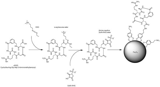
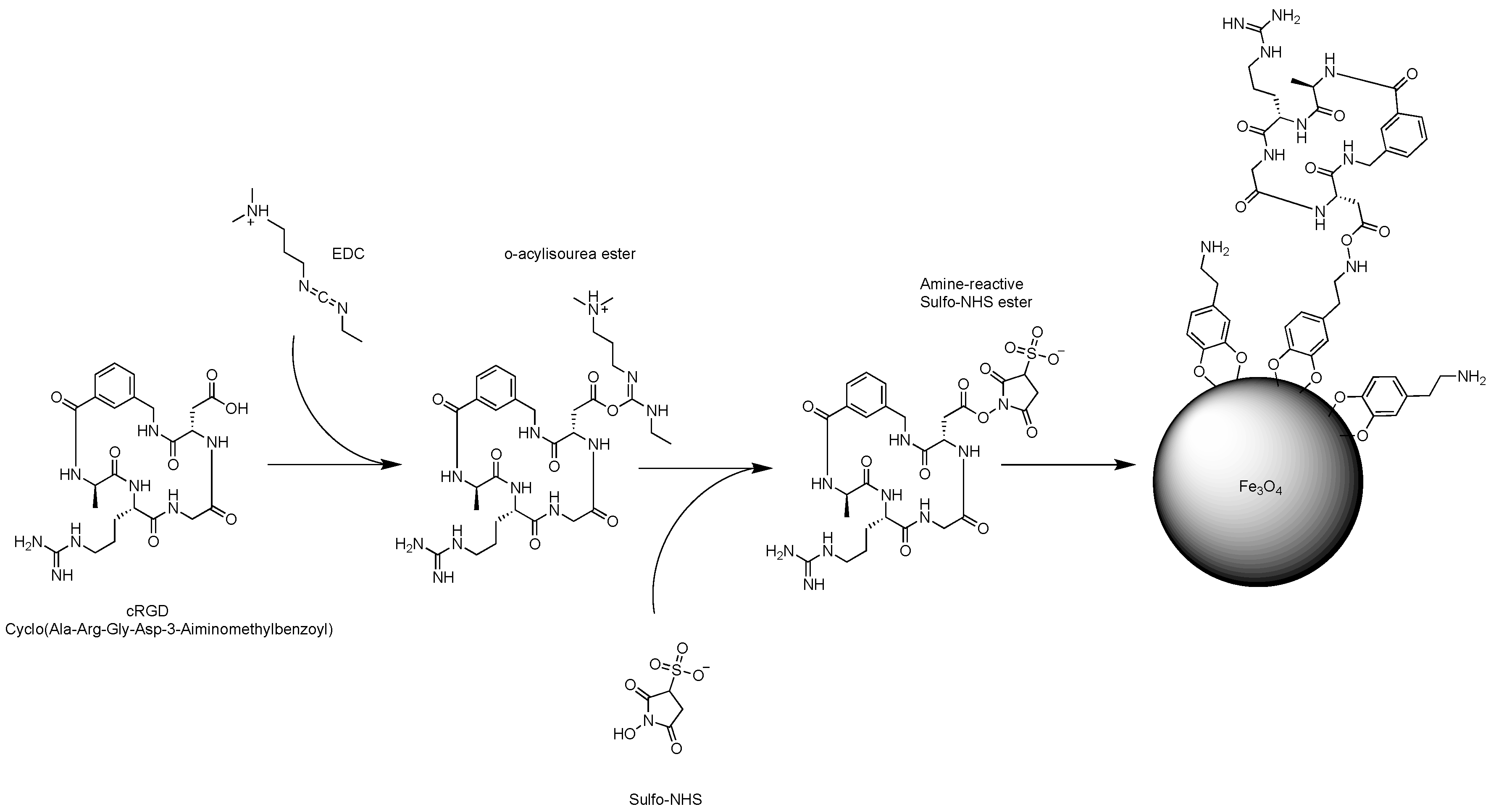
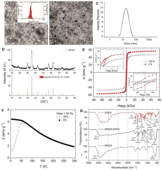
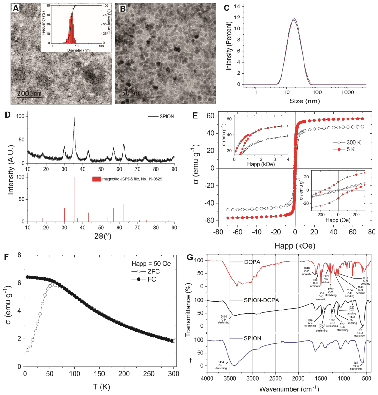
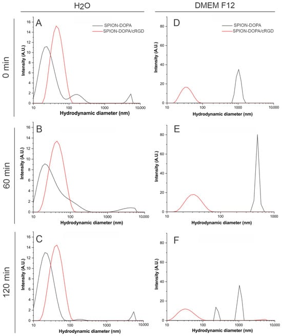
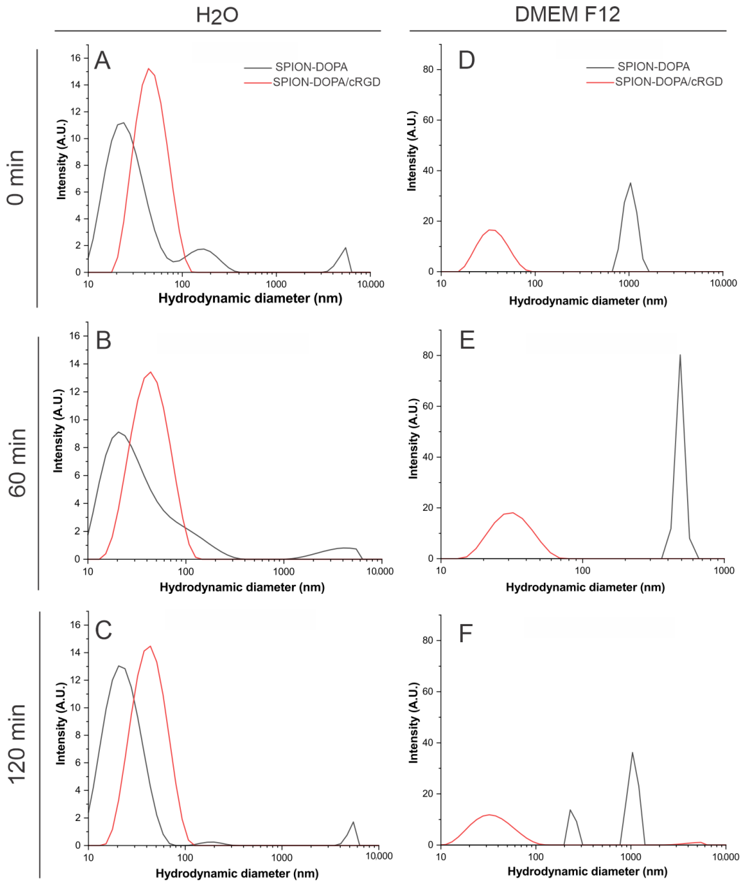
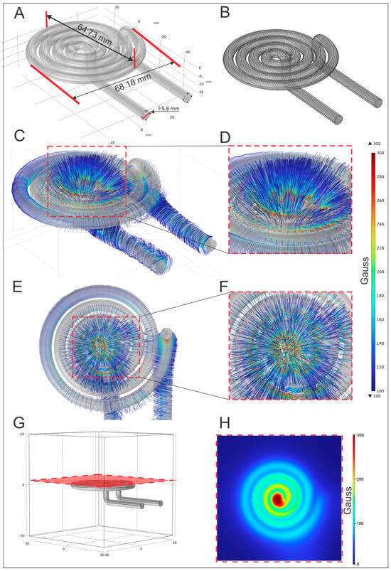
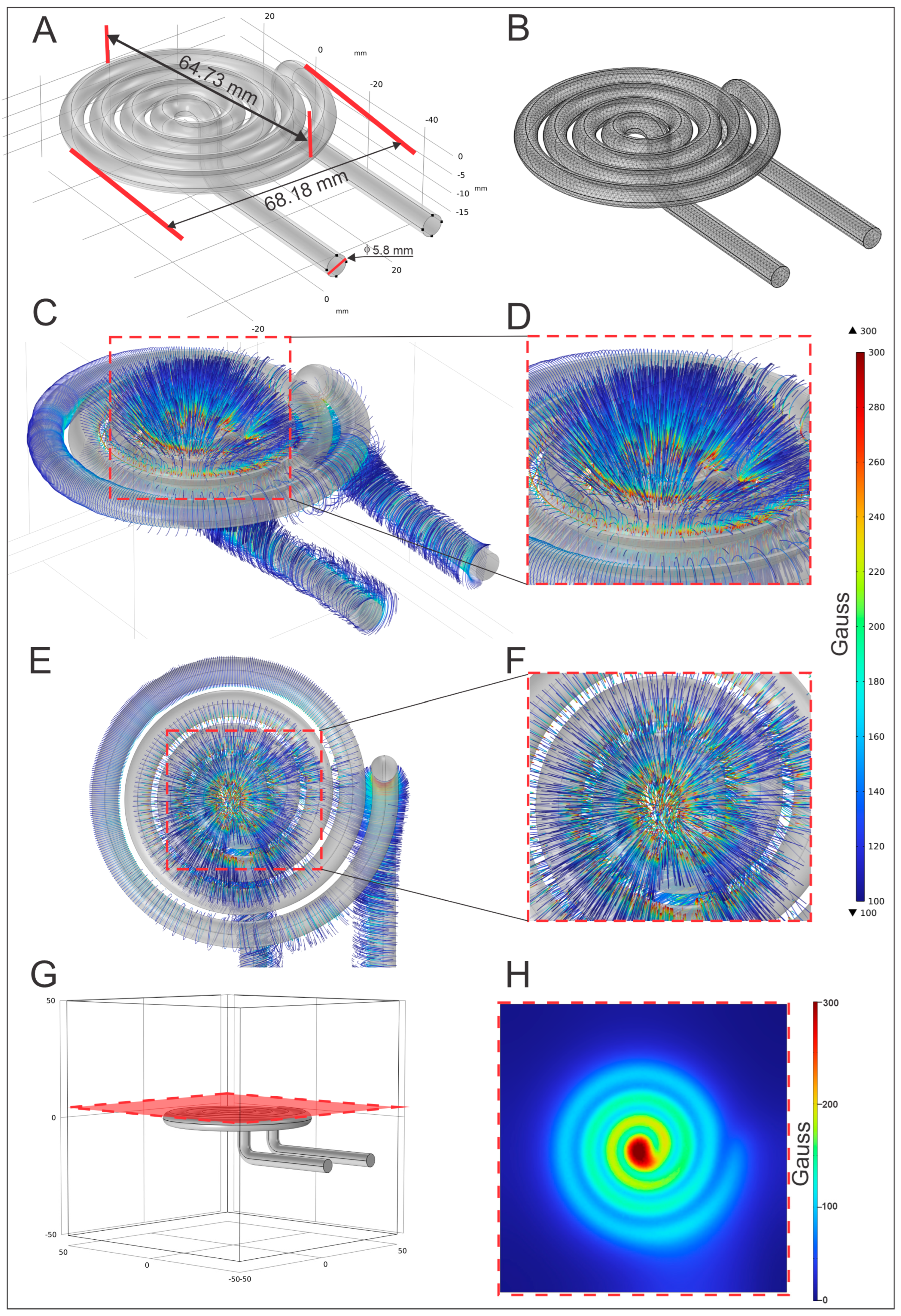
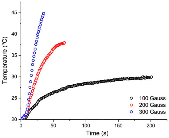
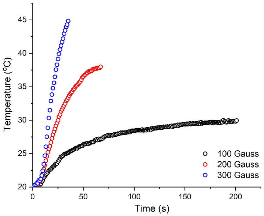
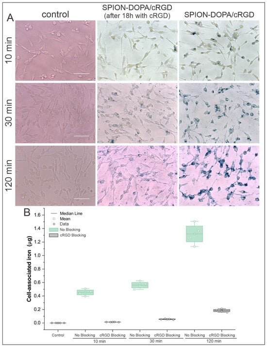
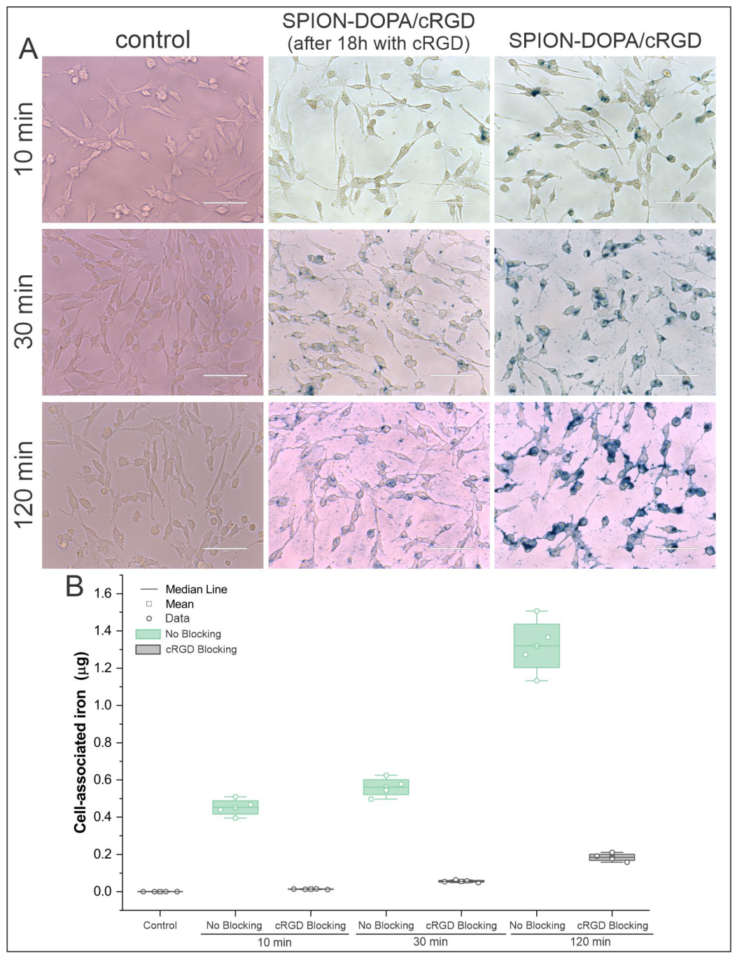
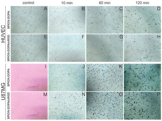
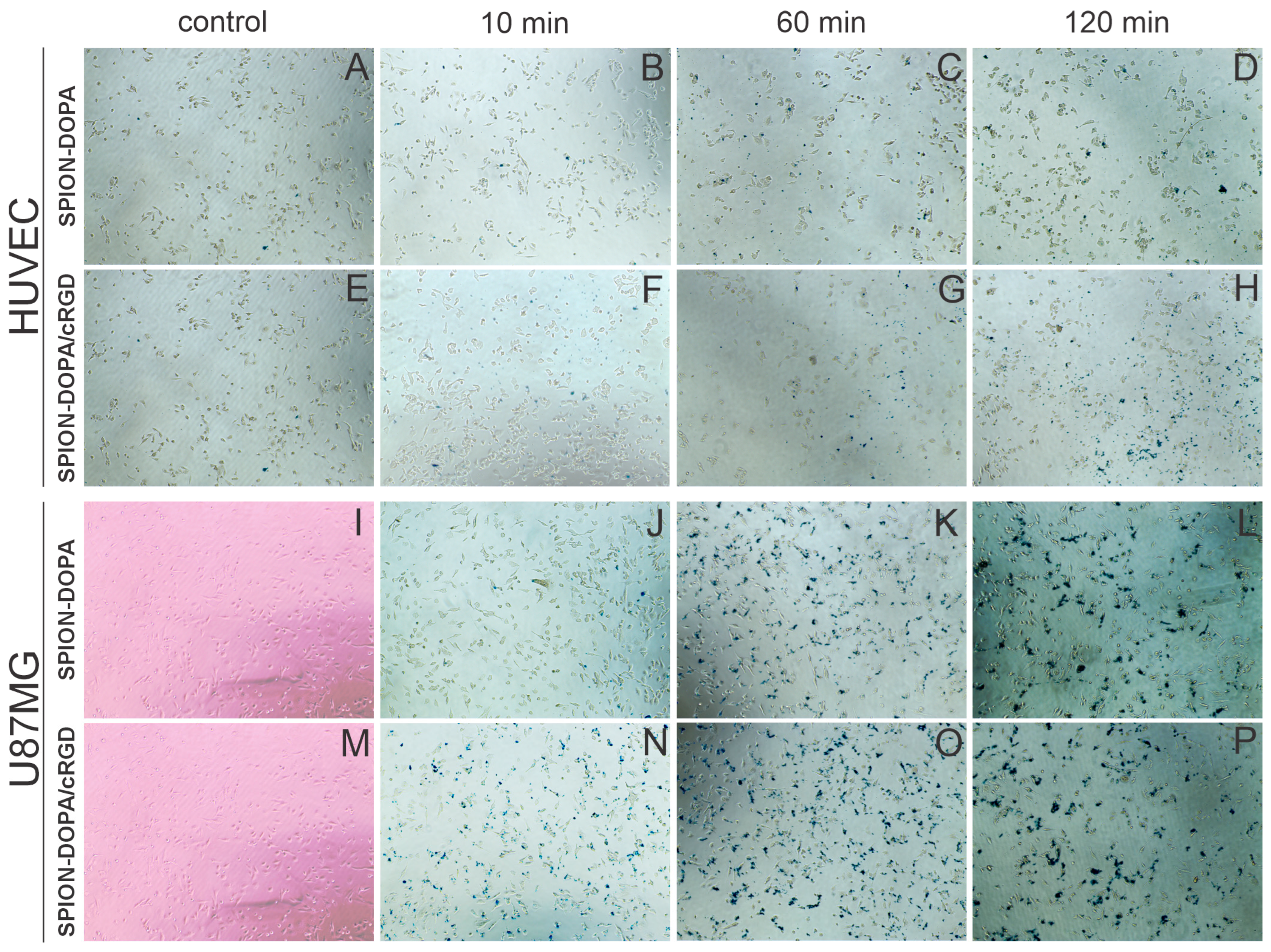
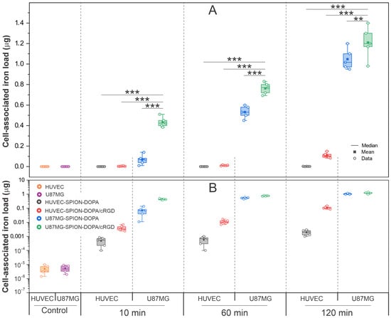
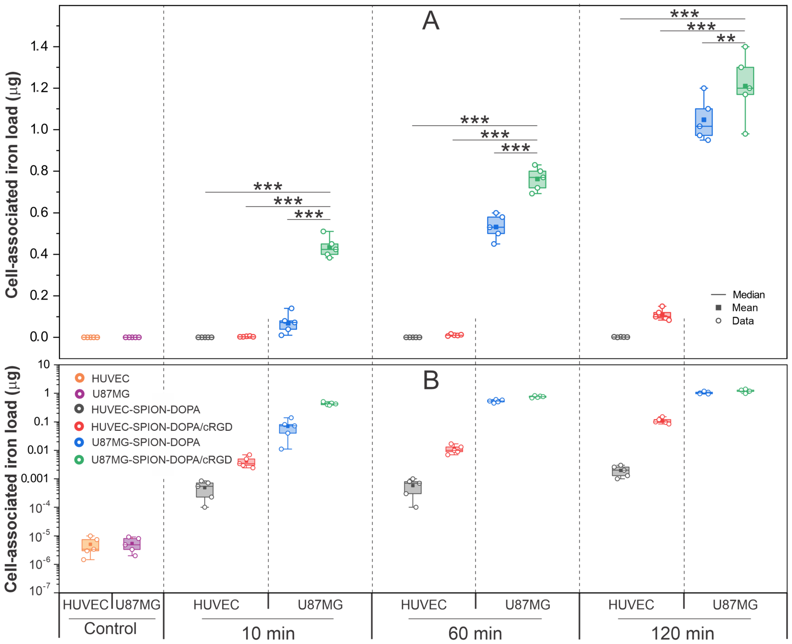
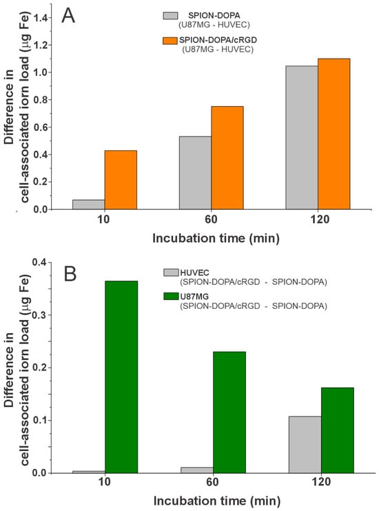
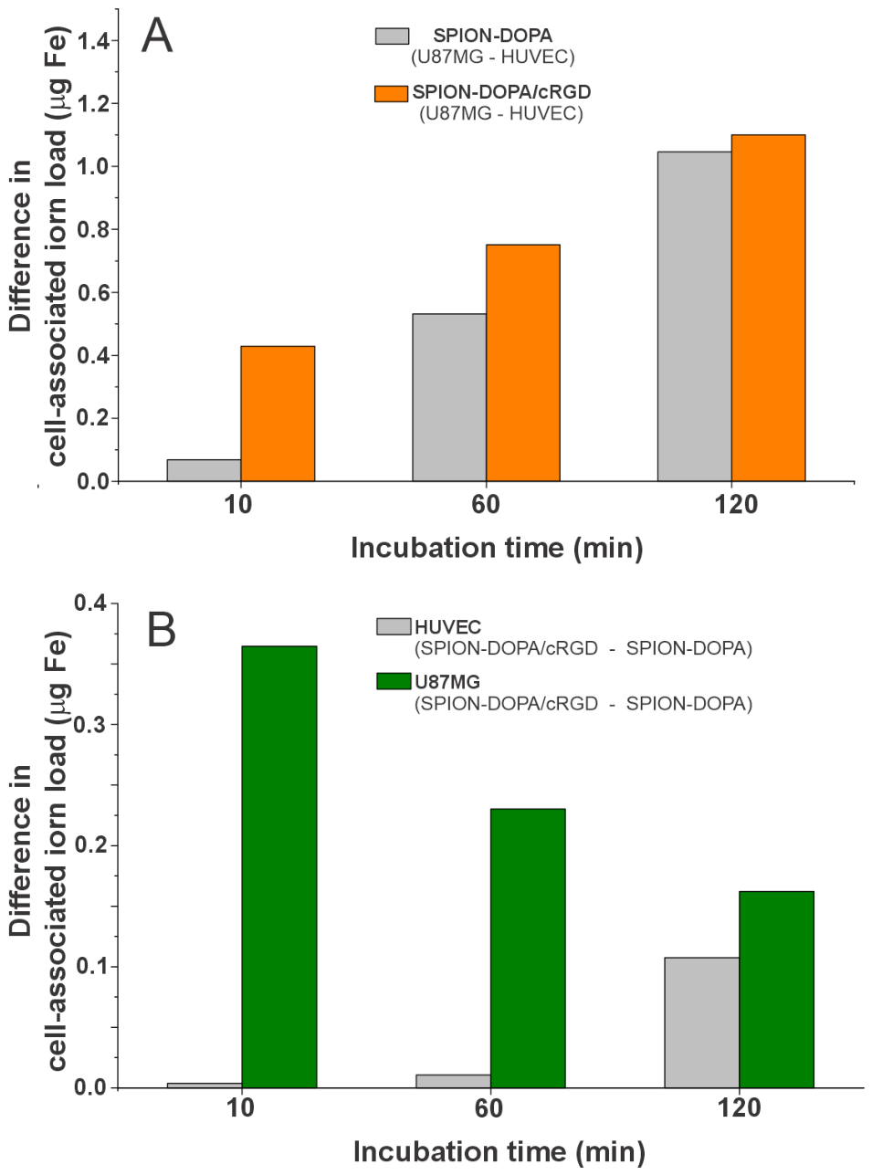
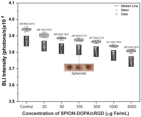
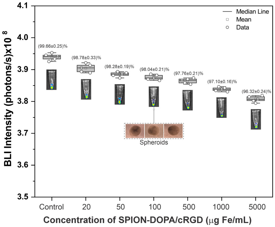
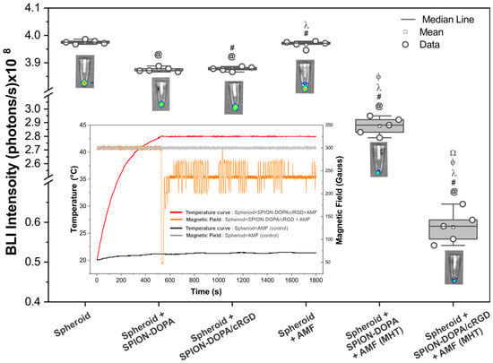
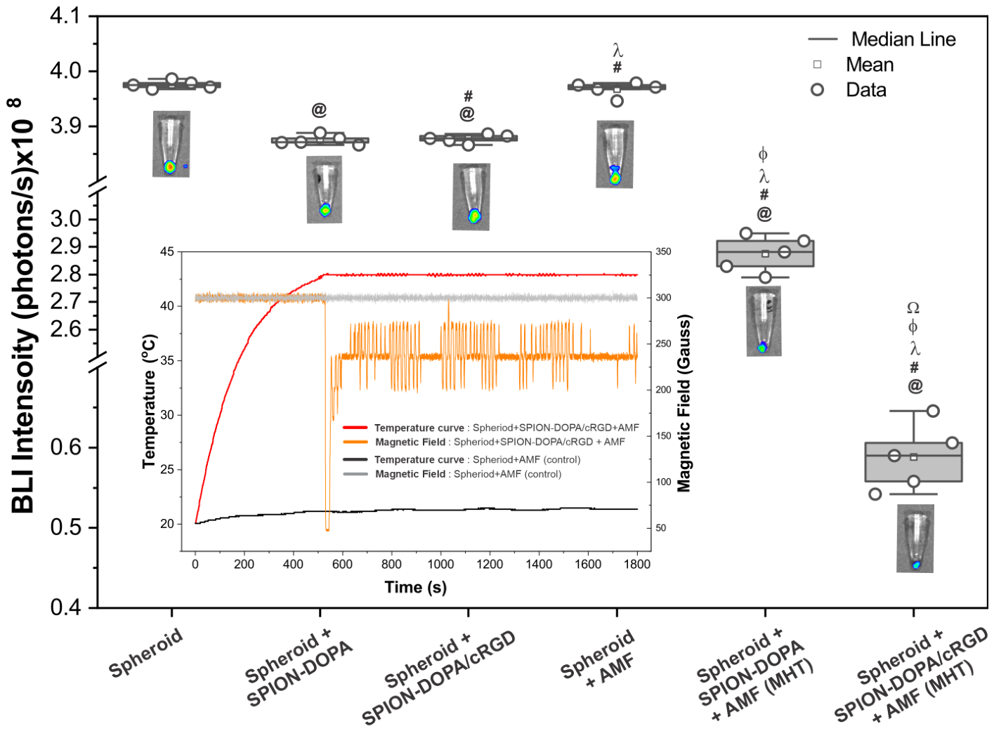

Disclaimer/Publisher’s Note: The statements, opinions and data contained in all publications are solely those of the individual author(s) and contributor(s) and not of MDPI and/or the editor(s). MDPI and/or the editor(s) disclaim responsibility for any injury to people or property resulting from any ideas, methods, instructions or products referred to in the content. |
© 2026 by the authors. Licensee MDPI, Basel, Switzerland. This article is an open access article distributed under the terms and conditions of the Creative Commons Attribution (CC BY) license.