


Abstract
Background/Objectives: Biochanin A belongs to the isoflavone group of phytoestrogens with numerous beneficial effects on human health. Its application in therapy is significantly limited by low aqueous solubility and bioavailability. Cyclodextrin inclusion complexes are used in pharmaceutical formulations to improve the unfavorable properties of the active substance while maintaining its activity. The aim of this work was to examine the influence of complexation with 2-hydroxypropyl-β-cyclodextrin (HPβCD) on the biological activities of biochanin A. The antimicrobial activity of biochanin A and the inclusion complex against Helicobacter pylori was tested using disc diffusion and micro-well dilution assays. Methods: The antioxidant activity of biochanin A and its inclusion complex was examined using radical scavenging (DPPH and ABTS), reducing power (CUPRAC and FRAP), phosphomolybdenum, and metal-chelating assays. The inhibitory activity on enzymes α-amylase, α-glucosidase, tyrosinase, and cholinesterases was also evaluated. Results: The inclusion complex showed stronger antimicrobial activity against H. pylori (MIC = 1.9 mg/mL) than biochanin A itself (MIC = 7.5 mg/mL). Both biochanin A and its inclusion complex showed weak, dose-dependent radical-scavenging activity. The inclusion complex inhibited cholinesterases, whereas free biochanin A did not; the possible contribution of HPβCD itself requires further study. Conclusions: The enhanced activity of biochanin A after complexation may enable the development of more effective formulations.
1. Introduction
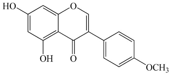
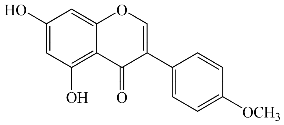
Biochanin A (BCA), 5,7-dihydroxy-4′-methoxyisoflavone, is a secondary plant metabolite from the Fabaceae family. The main source of BCA is red clover (Trifolium pratense) [1], but it is also found in soy, alfalfa, and chickpea. Isoflavones, including BCA (Figure 1), bind to estrogen receptors and show estrogenic activity [2] because they structurally resemble the estrogen hormone. Besides that, BCA shows anti-inflammatory, antioxidant, antimicrobial, antidiabetic, osteoprotective, and anticancer activity by regulating different signaling pathways [3,4,5].
Figure 1.
Structural formula of biochanin A.
Diabetes (diabetes mellitus) is one of the most common chronic diseases today. It occurs when the body does not produce or inadequately uses the pancreatic hormone insulin, which controls the concentration of glucose in the blood and the amount of glucose absorbed by the cells. The late diagnosis and inadequate treatment of diabetes can lead to serious health complications, including dyslipidemia, atherosclerosis, heart attack, and stroke [6]. There are two types of diabetes: type 1, which is treated with insulin, and type 2, which is treated with antidiabetic drugs. In the general population, type 2 diabetes is more frequent (90%) [7]. The use of synthetic antidiabetic drugs is commonly associated with side effects, so safer natural agents are being intensively tested. Numerous studies include isoflavones as potential herbal agents with hypoglycemic, hypolipidemic, and antioxidant activity. The oral application of BCA in diabetic rats decreases the glucose concentration in plasma, increases the insulin concentration, and normalizes the severe hyperlipidemia which is often associated with diabetes [6]. BCA is an agonist of PPARγ (peroxisome proliferator-activated receptor gamma), a nuclear receptor that plays an important role in glucose homeostasis. By activating PPARγ, BCA increases CAP (catabolite activator protein) gene expression and CAP concentration in adipose tissue [6,7,8]. Many studies have shown that BCA possesses antidiabetic activity. However, the exact mechanism of action has not been revealed. The presumed antidiabetic mechanisms of BCA include insulin-mimetic effects, increased glucose uptake into peripheral tissues, pancreatic β-cell stimulation to produce insulin, and regulation of key enzymes in carbohydrate metabolism [9].
Tyrosinase is the key enzyme in melanin biosynthesis, and its inhibition is associated with the prevention of different skin conditions, such as solar melanosis, ephelides, melasma, senile lentigos, and postinflammatory hyperpigmentation [10,11]. Tyrosinase catalyzes the hydroxylation of L-tyrosine into 3,4-dihydroxyphenylalanine (DOPA) and the subsequent oxidation of DOPA into dopaquinone. Melanin is a pigment that determines the phenotypic appearance and has an important role in protecting human skin from the harmful effects of UV radiation. The accumulation of an abnormal amount of melanin in some skin parts leads to hyperpigmentation, which can become a serious aesthetic problem, especially in middle-aged and elderly people. Reduction in tyrosinase activity may improve or prevent conditions associated with skin hyperpigmentation [12,13]. Natural enzyme inhibitors, especially polyphenols and flavonoids, represent promising, less toxic agents with fewer side effects than synthetic inhibitory drugs [11,14,15].
Acetylcholinesterase (AChE) and butyrylcholinesterase (BChE) are enzymes that catalyze the hydrolysis of neurotransmitter acetylcholine to acetate and choline. During neurotransmission, acetylcholine transmits the impulse by binding to its receptors. The enzyme cholinesterase stops the impulse by rapidly hydrolyzing acetylcholine, enabling re-uptake of free choline and re-synthesis of acetylcholine. Various pathological processes affect the synthesis of acetylcholine and thus reduce cholinergic transmission in different parts of the brain, which results in the appearance of neurodegenerative diseases such as Alzheimer’s disease, Parkinson’s disease, and other forms of dementia. By binding to the enzyme, cholinesterase inhibitors stop the degradation of acetylcholine, increase its concentration, and prolong its action in both the central and peripheral nervous system, which can provide symptomatic relief, although they do not halt disease progression. The reversible cholinesterase inhibitors (donepezil, rivastigmine, and galantamine) currently used in therapy exhibit numerous side effects. Therefore, given the incidence of neurodegenerative diseases, it is necessary to find more effective and safer cholinesterase inhibitors [16,17,18,19,20].
Despite numerous beneficial effects, the BCA application in therapy is limited by its low aqueous solubility and bioavailability. One of the ways to correct the unfavorable properties of BCA, while at the same time maintaining its activity, is to make an inclusion complex with 2-hydroxypropyl-β-cyclodextrin [21]. Cyclodextrins are often used as hosts in inclusion complexes because they are natural, non-toxic, stable, easily available compounds, resistant to the action of ultraviolet (UV) and infrared(IR) radiation. Inclusion complexes with cyclodextrins are used in pharmaceutical formulations with the aim of increasing the solubility, dissolution rate, stability, bioavailability, and oral absorption of the drug or to modify the biological activity of the drug [22].
Various formulations of BCA have been presented in the literature with a view to compensating for the shortage of this important biologically active molecule.
Xiaoyan Wu et al. developed BCA-loaded micelles with Pluronic F127 and Plasdone S630 (BCA-FS) in which the BCA solubility was increased at 5.0 mg/mL after encapsulation. In addition, pharmacokinetics evaluation showed a 2.16-fold increase in the relative oral bioavailability of BCA-FS compared with raw BCA, indicating that the mixed micelles may promote absorption in the gastrointestinal tract [23]. Another important contributing factor for the poor bioavailability of flavonoids as well as BCA is their extensive metabolism and participation in enteric and enterohepatic recycling processes, which limit their entry into systemic circulation. The absorbed BCA is efficiently conjugated by different UGT and sulphatase isoforms in enterocytes and hepatocytes, and the conjugates are excreted to the intestinal lumen (directly and indirectly). The conjugates can be further hydrolyzed by intestinal microbes, and BCA and other flavonoids may thus undergo extended recycling instead of reaching the systemic circulation [24]. In order to overcome the issue of first-pass intensive metabolism that BCA undergoes and in order to increase its solubility, Leena Hanski et al. developed buccal formulations based on hydroxypropylcellulose (HPC) and hydroxypropyl methylcellulose (HPMC). The formulations showed an enhanced dissolution rate of BCA compared to powder or a physical mixture, presumably due to the solubilizing effect of hydrophilic additives and the presence of BCA in an amorphous state [25]. The work by Nikolić et al. presents the creation and characterization of a BCA-inclusive complex with 2-hydroxypropyl-β-cyclodextrin. The solubility study has shown that the BCA solubility from the complex increased by 82% [21]. The electrospun polylactide (PLA) based nanofibers as carriers for the controlled release of BCA have also been illustrated in the literature. The rate and time of the release of BCA from PLA fibers are correlated with the amount of the active ingredient in the matrix of the carrier and follow zero-order kinetics [26]. In the paper by Gajic et al., the pH- and thermosensitive copolymer poly(N-isopropylacrylamide-co-acrylic acid), p(NiPAm-co-AA), has been described as a suitable carrier for alternative application routes of BCA with prolonged release. The research has shown that the release of BCA depends on the pH of the environment, with a higher quantity (up to 88.83%) released at pH 7.9 than at pH 4.5 (up to 46.18%) at body temperature [27].
The aim of this work was to examine the influence of complexation with 2-hydroxypropyl-β-cyclodextrin on the biological activities of BCA (antimicrobial activity against H. pylori, antioxidant activity and enzyme-inhibitory activity on α-amylase, α-glucosidase, tyrosinase and cholinesterases).
2. Materials and Methods
2.1. Materials
BCA (purity of 98%), HP-β-CD (purity of 98%; average Mr~1540 g/mol), dimethylsulfoxide (DMSO, 99.8%), and 2,3,5-triphenyltetrazolium chloride (TTC) were purchased from Sigma-Aldrich (Steinheim, Germany). Ethanol (96%, v/v) was purchased from Zorka Pharma (Sabac, Serbia).
2.2. Preparation of the Inclusion Complex
The inclusion complex BCA:HPβCD (1:1 molar ratio) was prepared by co-evaporation, as previously described by Nikolić et al., 2018 [21]. Briefly, a measured amount of HPβCD (211 mg) was dissolved in distilled water (10 cm3), and BCA (39 mg) dissolved in 96% (v/v) ethanol (10 cm3) was added dropwise to the prepared aqueous HPβCD solution. The solution was stirred with a magnetic stirrer (200 rpm) for 48 h. Then, the prepared inclusion complex was evaporated under reduced pressure in a rotary evaporator at 40 °C and dried in a desiccator until a constant mass was obtained.
2.3. Antimicrobial Activity Testing
The antimicrobial activity of BCA and BCA:HPβCD samples was evaluated against the laboratory control strain Helicobacter pylori (Deutsche Sammlung von Mikroorganismen und Zellkulturen; DSMZ 10242) using disc diffusion and micro-well dilution assay. The bacteria were subcultured on Brain Heart Infusion (BHI) agar containing 10% defibrinated sheep blood at 37 °C under microaerophilic conditions (5% O2, 10% CO2, and 85% N2 at 95% humidity) in a moist atmosphere for 3 days. The inoculum of the bacterial strain was prepared from broth cultures, and suspensions were adjusted to 0.5 McFarland standard turbidity (corresponding to 108 CFU/mL). The tested samples, BCA and BCA:HPβCD, were diluted in 100% DMSO (dimethyl sulfoxide) to a concentration of 30 mg/mL.
DMSO is used as a solvent for the tested material in antimicrobial tests since it stabilizes bacterial cells and makes them more resistant to environmental stressors; for this reason, its use in the manufacture of antimicrobial drugs is questionable [28]. Furthermore, unlike other solvents such as ethanol, DMSO may have inhibitory activity on bacterial growth only at maximal concentrations; therefore, it was used as a solvent since it has minimal effects on the antimicrobial activity results of the tested materials.
2.3.1. Disc Diffusion Assay
This method is presented as a consensus standard by the CLSI [29] (Clinical and Laboratory Standards Institute, 2007) with some slight modifications. The antimicrobial tests were carried out by the disc diffusion method using 100 μL of suspension containing 108 CFU/mL spread on a blood agar plate (BA, Torlak Institute, Belgrade, Serbia). The discs (6 mm in diameter, HiMedia Laboratories Pvt. Limited) were impregnated with 20 μL of BCA and BCA:HPβCD solutions and placed on the inoculated BA. The negative control was prepared using the same solvent to dissolve the BCA and BCA:HPβCD (100% DMSO). Antibiotic metronidazole (30 μg/disc) was used as a positive reference standard to determine the sensitivity of H. pylori. The inoculated plates were incubated at 37 °C under microaerophilic conditions in a moist atmosphere. The antimicrobial activity was evaluated by measuring the zone of inhibition of H. pylori. All experiments were performed in five repetitions.
2.3.2. Micro-Well Dilution Assay
A broth microdilution method was used to determine the minimum inhibitory concentration (MIC) and minimum bactericidal concentration (MBC) according to the CLSI (2007) [29]. A serial doubling dilution of the tested samples was prepared in a 96-well microtiter plate over the range of 30.00–0.01 mg/mL in inoculated Brain Heart Infusion broth containing 10% horse serum and 0.25% yeast extract [30]. The final concentration of bacteria in each well was adjusted to 106 CFU/mL. The plate was incubated for 72 h at 37 °C under microaerophilic conditions in a moist atmosphere. Metronidazole served as a positive control over the range of 10.00–0.02 µg/mL. MIC was defined as the lowest concentration of the tested samples at which microorganisms show no visible growth after the addition of TTC (0.5% triphenyl tetrazolium chloride). To determine MBC, a broth was taken from each well and inoculated in BA agar for 72 h at 37 °C under the previously described conditions. MBC is defined as the lowest concentration of the tested samples at which inoculated microorganisms were 99.9% killed. All experiments were performed in triplicate.
2.4. Antioxidant Activity Testing
The antioxidant properties of BCA and the BCA:HPβCD inclusion complex were determined using the methods previously described by Uysal et al. (2017) [10], including ABTS (2,2′-Azino-bis(3-ethylbenzothiazoline-6-sulfonic acid) Radical Cation Decolorization Assay) radical-scavenging, reducing power (CUPRAC (Cupric Ion Reducing Antioxidant Capacity Assay) and FRAP (Ferric Reducing Antioxidant Power Assay)), phosphomolybdenum, and metal-chelating assays.
2.4.1. DPPH (2,2-Diphenyl-1-picrylhydrazyl) Radical Scavenging Assay
The ability of the components to scavenge free DPPH radicals was determined using the DPPH assay. BCA and inclusion complex BCA:HPβCD were dissolved in ethanol, and a series of different concentrations was prepared. The ethanol solution of DPPH radical (1 mL, 300 μmol solution (3 × 10⁻⁴ mol/L)) was added to 2.5 mL of the prepared sample solutions. The absorption was measured at 517 nm immediately after adding the DPPH radical and after 20, 30, 60, and 120 min of incubation with the radical. The absorption at 517 nm was determined for the ethanolic solution of DPPH radical as well, which was diluted in the aforementioned ratio (1 mL of the DPPH radical of the given concentration with 2.5 mL ethanol added). Ethanol was used as a blank. Free-radical-scavenging activity was calculated according to the formula [31]:
- As—Absorption of the “sample” at 517 nm; “sample”—ethanolic solution of the sample (BCA or inclusion complex) treated with DPPH radical solution.
- AB—Absorption of the “blank” at 517 nm; “blank”—ethanolic solution of the sample (BCA or inclusion complex) which is not treated with DPPH radical solution.
- AC—Absorption of the “control” at 517 nm; “control”—ethanolic solution of the DPPH radical.
All absorptions were measured on a UV–Vis VARIAN Cary 100 Conc. Spectrophotometer (Mulgrave, Victoria, Australia).
2.4.2. ABTS (2,2-Azino-bis(3-ethylbenzothiazoline)-6-sulfonic Acid) Radical Scavenging Assay
Briefly, ABTS+ was produced directly by reacting 7 mM ABTS solution with 2.45 mM potassium persulfate and allowing the mixture to stand for 12–16 h in the dark at room temperature. Prior to the beginning of the assay, ABTS solution was diluted with methanol to an absorbance of 0.700 ± 0.02 at 734 nm. The sample solution was added to ABTS solution (2 mL) and mixed. The sample absorbance was read at 734 nm after a 30 min incubation at room temperature. The ABTS-radical-scavenging activity was expressed as milligrams of Trolox equivalents (mg TE/g).
2.4.3. CUPRAC (Cupric Ion Reducing Activity) Assay
The sample solution was added to the premixed reaction mixture containing CuCl2 (1 mL, 10 mM), neocuproine (1 mL, 7.5 mM), and NH4Ac buffer (1 mL, 1 M, pH 7.0). Similarly, a blank was prepared by adding the sample solution (0.5 mL) to the premixed reaction mixture (3 mL) without CuCl2. Then, the sample and the blank absorbances were read at 450 nm after a 30 min incubation at room temperature. The absorbance of the blank was subtracted from that of the sample. CUPRAC activity was expressed as milligrams of Trolox equivalents (mg TE/g).
2.4.4. FRAP (Ferric Reducing Antioxidant Power) Activity Assay
The sample solution was added to the premixed FRAP reagent (2 mL) containing acetate buffer (0.3 M, pH 3.6), 2,4,6-tris(2-pyridyl)-S-triazine (TPTZ) (10 mM) in 40 mM HCl, and ferric chloride (20 mM) in a ratio of 10:1:1 (v/v/v). Then, the sample absorbance was read at 593 nm after a 30 min incubation at room temperature. FRAP activity was expressed as milligrams of Trolox equivalents (mg TE/g).
2.4.5. Phosphomolybdenum Assay
The sample solution was combined with 3 mL of the reagent solution (0.6 M sulfuric acid, 28 mM sodium phosphate, and 4 mM ammonium molybdate). The sample absorbance was read at 695 nm after a 90 min incubation at 95 °C. The total antioxidant capacity was expressed as millimoles of Trolox equivalents (mmol TE/g).
2.4.6. Metal-Chelating Activity Assay
Briefly, the sample solution was added to FeCl2 solution (0.05 mL, 2 mM). The reaction was initiated by the addition of 5 mM ferrozine (0.2 mL). Similarly, a blank was prepared by adding the sample solution (2 mL) to FeCl2 solution (0.05 mL, 2 mM) and water (0.2 mL) without ferrozine. Then, the sample and the blank absorbances were read at 562 nm after a 10 min incubation at room temperature. The absorbance of the blank was subtracted from that of the sample. Metal-chelating activity was expressed as milligrams of EDTA (disodium edetate) equivalents (mg EDTAE/g).
2.5. Enzyme-Inhibitory Activity Testing
Enzyme inhibitory activities (cholinesterase (Elmann’s method), tyrosinase (dopachrome method), α-amylase (iodine/potassium iodide method) and α-glucosidase (p-Nitrophenyl-α-D-glucopyranoside chromogenic (PNPG) method)) of BCA and inclusion complex BCA:HPβCD were determined using the methods previously described by Uysal et al. (2017) [10].
2.5.1. α-Amylase Inhibitory Activity Assay
The sample solution was mixed with an α-amylase solution (ex-porcine pancreas, EC 3.2.1.1, Sigma) (50 μL) in phosphate buffer (pH 6.9 with 6 mM sodium chloride) in a 96-well microplate and incubated for 10 min at 37 °C. After pre-incubation, the reaction was initiated with the addition of starch solution (50 μL, 0.05%). Similarly, a blank was prepared by adding the sample solution to all the reaction reagents without the enzyme (α-amylase) solution. The reaction mixture was incubated for 10 min at 37 °C. The reaction was then stopped with the addition of HCl (25 μL, 1 M). This was followed by the addition of the iodine-potassium iodide solution (100 μL). The sample and blank absorbances were read at 630 nm. The absorbance of the blank was subtracted from that of the sample, and the α-amylase inhibitory activity was expressed as acarbose equivalents (mmol ACAE/g).
2.5.2. α-Glucosidase Inhibitory Activity Assay
The sample solution was mixed with glutathione (50 µL), α-glucosidase solution (from Saccharomyces cerevisiae, EC 3.2.1.20, Sigma-Aldrich, St. Louis, MO, USA) (50 µL) in phosphate buffer (pH 6.8), and PNPG (4-nitrophenyl-α-D-glucopyranoside, Sigma-Aldrich, St. Louis, MO, USA) (50 µL) in a 96-well microplate and incubated for 15 min at 37 °C. Similarly, a blank was prepared by adding the sample solution to all the reaction reagents without the enzyme (α-glucosidase) solution. The reaction was then stopped with the addition of sodium carbonate (50 µL, 0.2 M). The sample and blank absorbances were read at 400 nm. The absorbance of the blank was subtracted from that of the sample, and the α-glucosidase inhibitory activity was expressed as acarbose equivalents (mmol ACAE/g).
2.5.3. Tyrosinase Inhibitory Activity Assay
The sample solution was mixed with the tyrosinase solution (40 μL, Sigma-Aldrich, St. Louis, MO, USA) and phosphate buffer (100 μL, pH 6.8) in a 96-well microplate and incubated for 15 min at 25 °C. The reaction was then initiated with the addition of L-DOPA (40 μL, Sigma-Aldrich, St. Louis, MO, USA). Similarly, a blank was prepared by adding the sample solution to all reaction reagents without enzyme (tyrosinase) solution. The sample and the blank absorbances were read at 492 nm after a 10 min incubation at 25 °C. The absorbance of the blank was subtracted from that of the sample, and the tyrosinase inhibitory activity was expressed as kojic acid equivalents (mg KAE/g).
2.5.4. Cholinesterase (ChE) Inhibitory Activity Assay
The sample solution was mixed with DTNB (5,5-dithiobis(2-nitrobenzoic acid)) (Sigma-Aldrich, St. Louis, MO, USA) (125 µL) and AChE (acetylcholinesterase) (electric eel acetylcholinesterase, Type VI-S, EC 3.1.1.7, Sigma-Aldrich) or BChE (butyrylcholinesterase) (horse serum butyrylcholinesterase, EC 3.1.1.8, Sigma-Aldrich) solution (25 μL) in Tris–HCl buffer (pH 8.0) in a 96-well microplate and incubated for 15 min at 25 °C. The reaction was then initiated with the addition of acetylthiocholine iodide (ATCI, Sigma-Aldrich, St. Louis, MO, USA) or butyrylthiocholine chloride (BTCl, Sigma) (25 μL). Similarly, a blank was prepared by adding the sample solution to all the reaction reagents without the enzyme (AChE or BChE) solution. The sample and the blank absorbances were read at 405 nm after 10 min incubation at 25 °C. The absorbance of the blank was subtracted from that of the sample, and the cholinesterase inhibitory activity was expressed as galantamine equivalents (mg GALAE/g).
2.6. Molecular Modeling
The following protein data bank (PDB) crystal structures prepared in our previous studies on human AChE (PDB ID: 6O52) [32], BChE (PDB ID: 6EQP) [33], and α-amylase (PDB ID: 1B2Y) [34] were retrieved [35,36]. The prepared human models of α-tyrosinase and α-glucosidase using Priestia megaterium tyrosinase (6QXD) [37] and Mus musculus α-glucosidase (7KBJ) [38] as templates were also retrieved. The three-dimensional structures of BCA and HPβCD were retrieved from the PubChem database (https://pubchem.ncbi.nlm.nih.gov/). The BCA–HPβCD inclusion complex was constructed by manually positioning BCA within the hydrophobic cavity of HPβCD [39], followed by energy minimization in UCSF Chimera using the MMFF94 force field, with steepest-descent minimization followed by conjugate-gradient optimization [40]. The minimized structure was inspected for steric clashes and appropriate host–guest geometry.
Docking was performed using AutoDock 4.2.6 (The Scripps Research Institute, La Jolla, CA, USA) with the Lamarckian Genetic Algorithm. The default genetic algorithm settings comprised a population size of 150 individuals, a maximum of 2,500,000 energy evaluations, 27,000 generations, an elitism value of 1, a mutation rate of 0.02, and a crossover rate of 0.80 [41]. Ten independent docking runs were performed for each target–ligand pair. The resulting poses were ranked according to their predicted binding energies and inspected to identify physically plausible binding orientations. Docking performance was validated by redocking the co-crystallized ligand into its original binding site and comparing the predicted pose with the experimentally observed conformation using the root-mean-square deviation (RMSD). Comparison of the predicted poses with their corresponding crystal structures yielded heavy-atom RMSD values of 0.91, 1.11, 1.58, 1.21, and 1.66 Å for AChE, BChE, tyrosinase, amylase, and glucosidase, respectively. Since all RMSD values fell below the widely accepted 2.0 Å benchmark [42], the results demonstrate that the docking protocols reproduced the experimentally observed ligand-binding geometries with acceptable accuracy. The grid-box center coordinates, dimensions, grid spacing used for each docking calculation, and RMSD values of the cocrystal ligands are provided in Table S1. The top-ranked docking poses were selected, and the corresponding protein-ligand complex was prepared by geometry regularization using the Clean Geometry function of BIOVIA Discovery Studio Visualizer v4.5 (Dassault Systèmes BIOVIA, San Diego, CA, USA) [43]. This step corrects bond lengths, bond angles, and steric clashes using built-in rules and does not involve a force field, a charge model, or energy minimization. The structure was therefore not energetically relaxed prior to analysis. Protein–ligand interactions were subsequently visualized using the same software.
2.7. Statistical Analysis of Data
In the antioxidant and enzyme inhibitory assays, the results are expressed as mean ± standard deviation (SD) of three independent experiments. Differences between the tested samples were evaluated using the independent two-sample Student’s t-test, assuming approximately equal variances between groups. Statistical significance was set at p < 0.05. All statistical analyses were performed using XLSTAT software, version 16.0 ((Addinsoft, Paris, France))
3. Results
3.1. Antimicrobial Activity
The antimicrobial activity of BCA and the inclusion complex BCA:HPβCD was previously confirmed against Gram-positive and Gram-negative bacteria and fungi, and it was shown that the antimicrobial activity of BCA was not significantly changed after complexation with 2-hydroxypropyl-β-cyclodextrin [21]. The results of antimicrobial activity testing against H. pylori are shown in Table 1.
Table 1.
Antimicrobial activity of biochanin A and the BCA:HPβCD inclusion complex against H. pylori.
The results of the disc diffusion method showed the inhibition zones of 25 and 52 mm for BCA and the inclusion complex BCA:HPβCD, respectively. This demonstrated significantly higher activity of the inclusion complex, which exhibited very high antimicrobial potential against H. pylori at a concentration of 0.6 mg/disc. Although much lower in comparison to the inclusion complex (when observing inhibition zones), BCA also showed significant activity, since DMSO used as a negative control showed negligible action. DMSO is a strong solvent, and it is highly likely that it did not allow the growth of microorganisms to the extent to which it diffused on the surface of the culture medium. Since all the other parameters on the disc are identical, except for the added active substance, the contribution of the active substance to the expansion of the inhibition zone is visible, thus indicating the effect of BCA and the complex. DMSO is a recommended solvent for the materials tested in antimicrobial screening, as it exhibits antimicrobial activity only at high concentrations. The generated 9 mm inhibition zone is not negligible (the disc being 6 mm). Nevertheless, we think that it does not have any significant effect on the results, i.e., inhibition zones generated for material testing, which are much larger. In any event, it is for these reasons that the disc diffusion method was used for preliminary screening, followed by a more accurate microdilution method, which also yielded good results with regard to the antimicrobial activity of the tested materials against H. pylori bacteria. On the other hand, the antimicrobial testing results obtained by the microdilution method did not show an antimicrobial potential of BCA, since inhibitory action was observed at the concentration where DMSO was inhibitory alone against this bacterium (Table 1, negative control, 12.5% DMSO). In light of this fact, it is not possible to conclude that BCA possesses antimicrobial potency against H. pylori, since this action can be attributed to the solvent. The same testing revealed relatively high antimicrobial potential of the BCA:HPβCD inclusion complex, which was active at a concentration of 1.90 mg/mL. Another important fact observed in the present study is the lack of microbicidal action of both compounds, since MBC was not observed at any tested concentration of either compound.
H. pylori is one of the most resistant human pathogens, and the prevalence of infection is almost 60%. Gastric colonization by H. pylori bacteria increases the risk of developing various gastrointestinal disorders and diseases, including gastritis, ulcers, and cancers [44]. The main drawback of the therapy used for the eradication of H. pylori, which includes the use of proton pump inhibitors and two antibiotics, is the widespread resistance to antibiotics, which is why natural active substances are increasingly being examined. De Mendonça et al. (2020) tested the gastroprotective effects of hydroalcoholic extracts of red propolis (HERP), whose main ingredient is the isoflavone formononetin, in acute ulcer models [45]. HERP showed a dose-dependent gastroprotective effect in all tested concentrations (50, 250, and 500 mg/kg). Besides that, both HERP and formononetin (10 mg/kg) significantly increased gastric mucus secretion while decreasing gastric juice production. Moreover, HERP showed certain antimicrobial activity against H. pylori (inhibition zone of 13 ± 2 mm) at 8 mg/disc. The best antiulcer activity, comparable to omeprazole, was shown by the chloroform extract, in which the most abundant compounds are the isoflavones formononetin and BCA. The gastroprotective effect of BCA can be attributed to the mitochondrial antioxidant cascade and the activation of anti-apoptotic pathways.
Satoh et al. (2010) observed the association of H. pylori infection with the risk of lipid profile imbalance in Japanese males [46]. Significantly elevated LDL (Low-Density Lipoprotein Cholesterol) values and simultaneously decreased HDL (High-Density Lipoprotein Cholesterol) levels, as risk factors for the development of cardiovascular diseases, were detected. Previous research has shown that BCA may play an important role in maintaining normal blood lipid levels [47]. BCA reduces the formation of oxidized LDL by activating the expression of antioxidant enzymes, such as superoxide dismutase, and reducing the content of malondialdehyde, a marker of lipid peroxidation. In addition, BCA inhibits the formation of foam cells by activating cholesterol efflux pathways, maintains the transformation of various lipoproteins by normal regulation of the activity of certain enzymes, and prevents the transport of cholesterol esters between HDL and LDL. Treatment with BCA at a dose of 60 mg/kg per day significantly reduced the levels of LDL (85%) and total cholesterol (39%), increased the activity of lipoprotein lipase (96%) and hepatic triglyceride lipase (78%), and improved the excretion of lipids via feces.
Although the literature reports antimicrobial effects of various flavonoids against H. pylori [48], this is the first time the activity of BCA and its inclusion complex has been demonstrated.
3.2. Antioxidant Activity
The antioxidant capacity of a compound is reflected in its protective potential against free radical attacks, which are considered to contribute to the progression of serious chronic diseases such as cancer, diabetes, or Alzheimer’s. In this sense, antioxidant properties can be considered the first window into pharmacological potential [49]. Flavonoids and isoflavones show strong antioxidant activity in various in vitro tests, whereby the antioxidant activity of isoflavones, although significant, is weaker than the antioxidant activity of flavonols and flavones [50]. The antioxidant activity of isoflavones comes from the presence of hydroxyl groups in the molecule, with a higher number of hydroxyl groups meaning stronger antioxidant activity [51]. The mechanism of BCA antioxidant activity includes increasing the level of antioxidant enzymes, such as superoxide dismutase and catalase, reducing the level of lipid peroxidation markers, and maintaining the total antioxidant status of plasma [47,52].
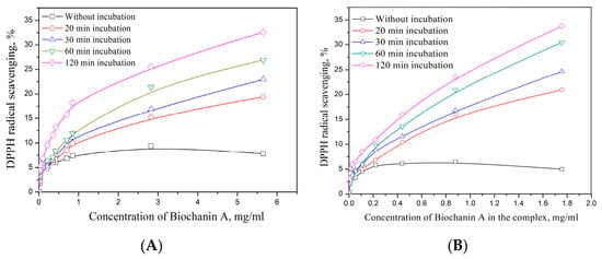
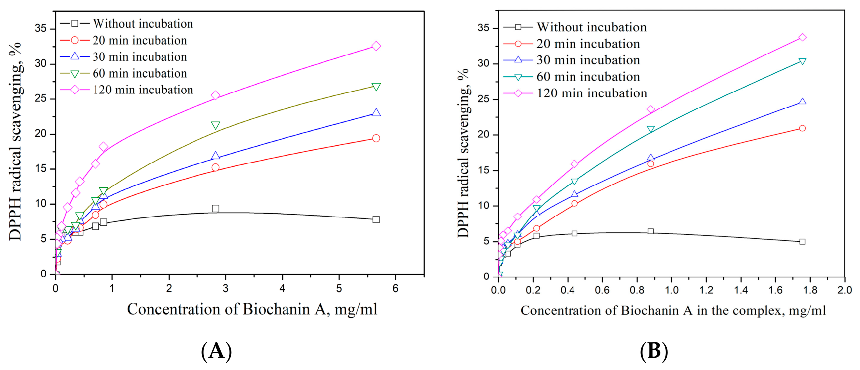
In the current study, the antioxidant properties of BCA and its complex were evaluated by using various chemical assays including free radical scavenging (DPPH and ABTS), reducing power (CUPRAC and FRAP), metal chelating, and phosphomolybdenum assay. The results are given in Figure 2 (DPPH assay) and in Table 2.
Figure 2.
DPPH-radical-scavenging activity of (A) biochanin A and (B) the BCA:HPβCD inclusion complex.
Table 2.
Antioxidant activity of biochanin A and the BCA:HPβCD inclusion complex.
DPPH and ABTS assays are simple and among the most common antioxidant assays [53]. As can be seen from Figure 2, both BCA and its inclusion complex showed weak, dose-dependent radical-scavenging activity, which was significantly improved after incubation with the DPPH radical. Regarding the ABTS assay, BCA showed stronger ability than its complex. Consistent with our results, Magiera et al. (2015) tested certain flavonoids for their DPPH radical scavenging ability, and BCA was reported as the weakest compound among the tested flavonoids [54]. In addition, Hamza Sherif and Gebreyohannes (2018) reported that BCA showed no significant DPPH scavenging ability at 100 µg/mL [55]. In previous reports, it has been stated by some researchers that the number and position of hydroxyl groups in the flavonoid structure play a major role in their antioxidant effects [56]. Reducing power is an important antioxidant mechanism and reflects the electron-donating ability of antioxidant compounds. For this purpose, the reductions of Cu(II) to Cu(I) and Fe(III) to Fe(II) were investigated by CUPRAC and FRAP assays, respectively. In both assays, the reducing effect of BCA was higher than that of its complex. This case could be explained by the fact that some of the hydroxyl groups in the BCA structure are involved in the formation of the complex. Similarly, several researchers reported that the formation of cyclodextrin complexes negatively affected their reducing power [57,58]. The conversion of Mo(VI) to Mo(V) is measured in the phosphomolybdenum assay, which is considered one of the assays for total antioxidant capacity. Like reducing power assays, the inclusion complex (0.12 mmol TE/g) of BCA showed weaker activity than free BCA (1.17 mmol TE/g). The ability of a compound to chelate metals reflects its ability to inhibit the formation of hydroxyl radicals in the Fenton reaction. The chelating ability of BCA (7.36 mg EDTAE/g) was 2.5 times higher than that of its inclusion complex (3.13 mg EDTAE/g). Overall, the formation of the inclusion complex of BCA negatively impacted its antioxidant properties compared to its free form. Therefore, in vitro techniques such as FRAP, ABTS, and DPPH cannot be used independently to determine the antioxidant capacity of an isolated compound/its complex, as they can be affected by different properties of the compound or its complex [59]. With this in mind, further studies, particularly in vivo antioxidant properties of a host/guest complex, should be conducted to understand its full potential.
3.3. Enzyme-Inhibitory Activity
In the pharmaceutical industry, enzymes are of fundamental importance for the development of effective drugs. In particular, enzyme inhibitors are one of the most common drugs on pharmacy shelves and play a crucial role in alleviating the symptoms of global health problems such as obesity, diabetes mellitus, or Alzheimer’s [60]. In this sense, providing novel and potent enzyme inhibitors is gaining interest on the scientific platform. In view of the above facts, we tested the enzyme-inhibitory activity of BCA and its inclusion complex against cholinesterases, amylase, glucosidase, and tyrosinase. The results are given in Table 3.
Table 3.
Enzyme inhibitory properties of BCA and the BCA:HPβCD inclusion complex.
In both AChE and BChE inhibition abilities, the inclusion complex BCA:HPβCD was the only one active against enzymes (AChE: 5.82 mg GALAE/g; BChE: 6.96 mg GALAE/g). Consistent with our results, Orhan et al. (2007) found that BCA had no effect on AChE and BChE at a concentration of 1 mg/mL [61]. The inclusion complex therefore inhibited both cholinesterases under our conditions, although the contribution of HPβCD to this effect remains to be determined. Complexation with HPβCD therefore appears to modify the in vitro enzyme inhibitory profile of BCA. However, these findings were obtained in cell-free enzyme assays only and do not establish any therapeutic efficacy. Any potential relevance of the inclusion complex to neurodegenerative conditions such as Alzheimer’s disease remains speculative and would first require clarification of the contribution of HPβCD, followed by cell-based and in vivo studies, including assessment of blood–brain barrier permeability. Tyrosinase is a key enzyme in the synthesis of melanin, and it is the main target for controlling hyperpigmentation problems. As can be seen from Table 3, tyrosinase inhibition by BCA was not significantly affected by complex formation (p > 0.05). In a previous study by Lin et al. (2011), the anti-melanogenesis ability of BCA was examined using the B16 melanoma cell line, and the compound showed a potent dose-dependent anti-melanogenesis ability [62].
In antidiabetic enzymes (amylase and glucosidase), BCA (amylase: 1.16 mmol ACAE/g; glucosidase: 38.06 mmol ACAE/g) is more effective than its complex (amylase: 0.62 mmol ACAE/g; glucosidase: 32.01 mmol ACAE/g). Similarly, Park et al. (2009) reported antidiabetic effects of some compounds from Pueraria thunbergiana, including BCA [63]. In their study, BCA was the most effective against both amylase and glucosidase. In another study by Mehrabadi et al. (2018), BCA effectively improved diabetic retinopathy by lowering blood sugar levels [64]. In addition, the antidiabetic mechanism of BCA may involve reducing insulin resistance and glycosylated hemoglobin concentrations while improving insulin sensitivity, lipid profile, and liver glycogen storage [65]. On a per-gram basis, free biochanin A was the stronger inhibitor of α-amylase and α-glucosidase, while the complex was the only sample active against cholinesterases. Because the two samples differ greatly in biochanin A content, and HPβCD alone was not tested, these differences should not be taken as a direct comparison of the free and complexed compound.
In the presented study, single BCA displayed stronger amylase and glucosidase inhibitory ability than the inclusion (beta-cyclodextrin complex). This fact can be explained by the involvement of OH groups at positions 5 and 7 of the A ring in the formation of beta-cyclodextrin. In a comprehensive study by Proena et al., the hydroxyl groups are the key players in the inhibition of amylase [28]. Similar results were also reported by Proena et al. for glucosidase inhibition [66].
These results should be read with one limitation in mind. HPβCD alone was not tested in the enzyme assays, so the inhibition measured for BCA:HPβCD cannot be attributed to biochanin A alone. The docking models place the bulky complex at the entrance of the active site of each enzyme, where HPβCD itself may hinder substrate access. In addition, biochanin A makes up only about 16% (w/w) of the complex. Expressed per amount of biochanin A, the complex was more active than the free compound in every assay. Whether this reflects improved solubility of biochanin A, a direct contribution of HPβCD, or both should be established in studies that include HPβCD alone at matched concentrations.
3.4. Molecular Docking
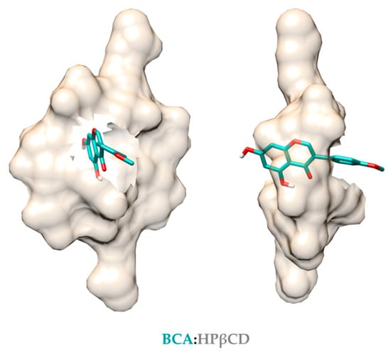
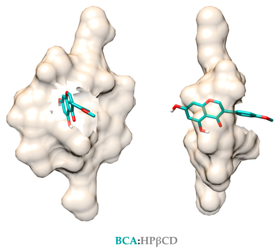
The inclusion complex of BCA:HPβCD was modeled based on a host-guest approach, in which the guest molecule is moved along a vector going through the middle of the cavity [39]. BCA was buried in the hydrophobic cavity of HPβCD via H-bonds, thereby protruding the hydroxyphenyl and methoxyphenyl groups outside the cavity from both sides (Figure 3). The BCA:HPβCD complex and BCA alone were then docked into the active sites of AChE, BChE, tyrosinase, amylase, and glucosidase enzymes. The resulting docking complexes were refined and optimized using the UCSF Chimera program [39] to minimize steric clashes due to the bulkiness of HPβCD. The reported binding energy values should be interpreted cautiously, as subsequent complex refinement and energy minimization may alter the ligand–receptor geometry and the resulting associated interaction energies. Nevertheless, these values appear to be comparable to those of the cocrystal ligands, facilitating quantitative comparison (Table S2). Accordingly, the analysis focused on elucidating the potential binding modes and key intermolecular interactions of BCA and the BCA:HPβCD inclusion complex within the active sites of the target enzymes.
Figure 3.
Model of the BCA:HPβCD inclusion complex.
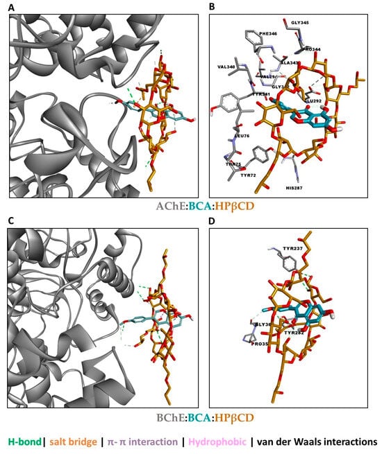
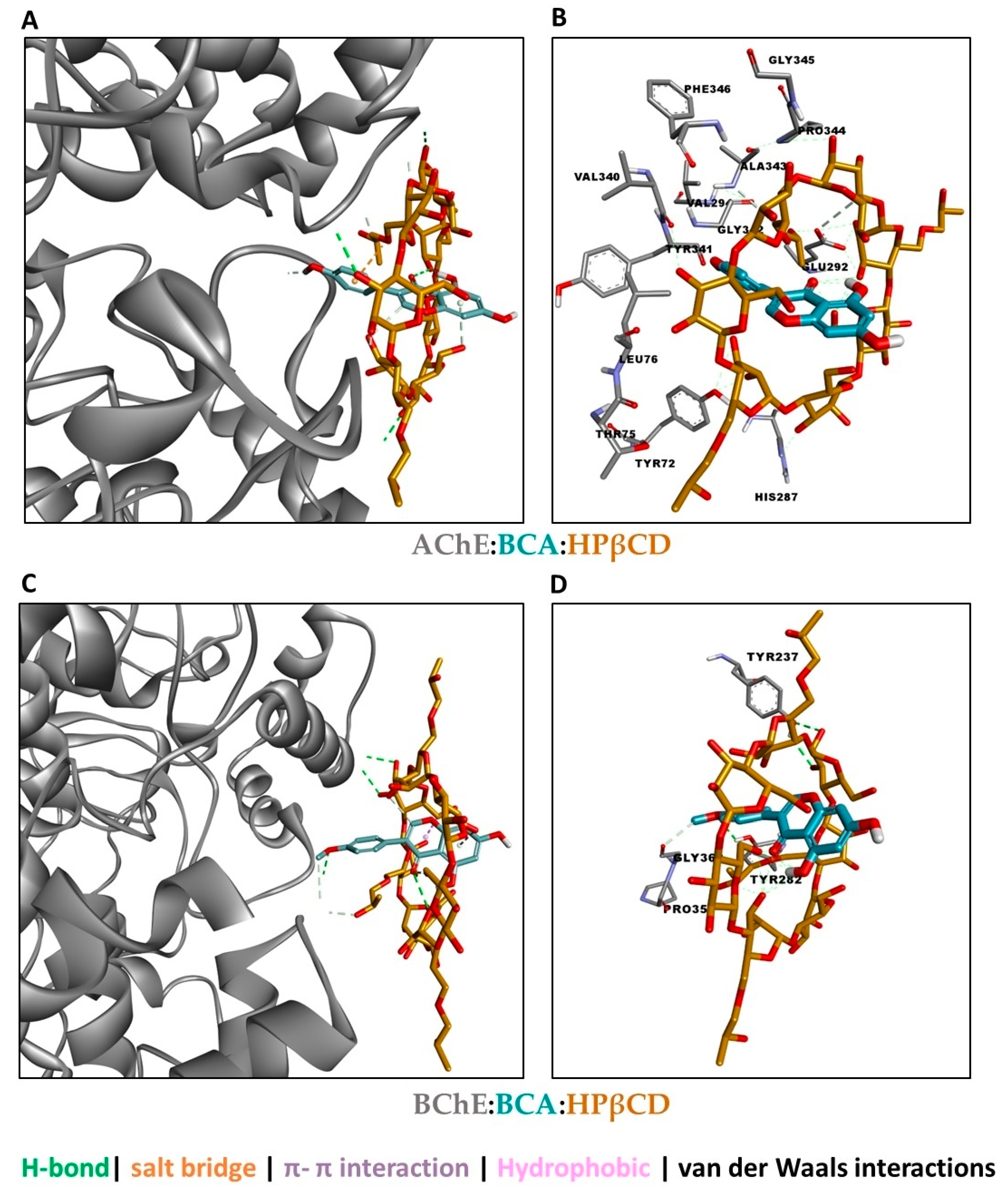
Cholinesterases: In AChE, the complex sits at the gorge entrance, contacting Tyr72, Tyr341, Thr75, Leu76, His287 and Glu292 through hydrogen bonds, hydrophobic contacts and van der Waals contacts (Figure 4A). Tyr72 and Tyr341 belong to the peripheral anionic site, and the catalytic triad lies deeper in a long aromatic-lined gorge [67,68]. The bulky HPβCD cannot enter, so the likely effect is partial or mixed blockage of the gorge entrance. In BChE, the contacts are fewer and on the surface (Pro35, Gly36, Tyr237, Tyr282) (Figure 4B). BChE’s gorge is wider and has fewer aromatic residues than AChE’s, which fits weaker, less specific binding [69].
Figure 4.
(A) Possible binding of the BCA:HPβCD inclusion complex to AChE and (B) interaction. (C) Possible binding of the BCA:HPβCD inclusion complex to BChE and (D) interaction.
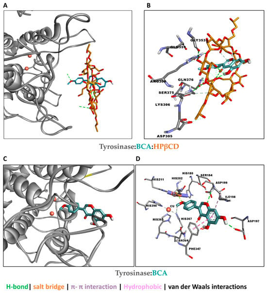
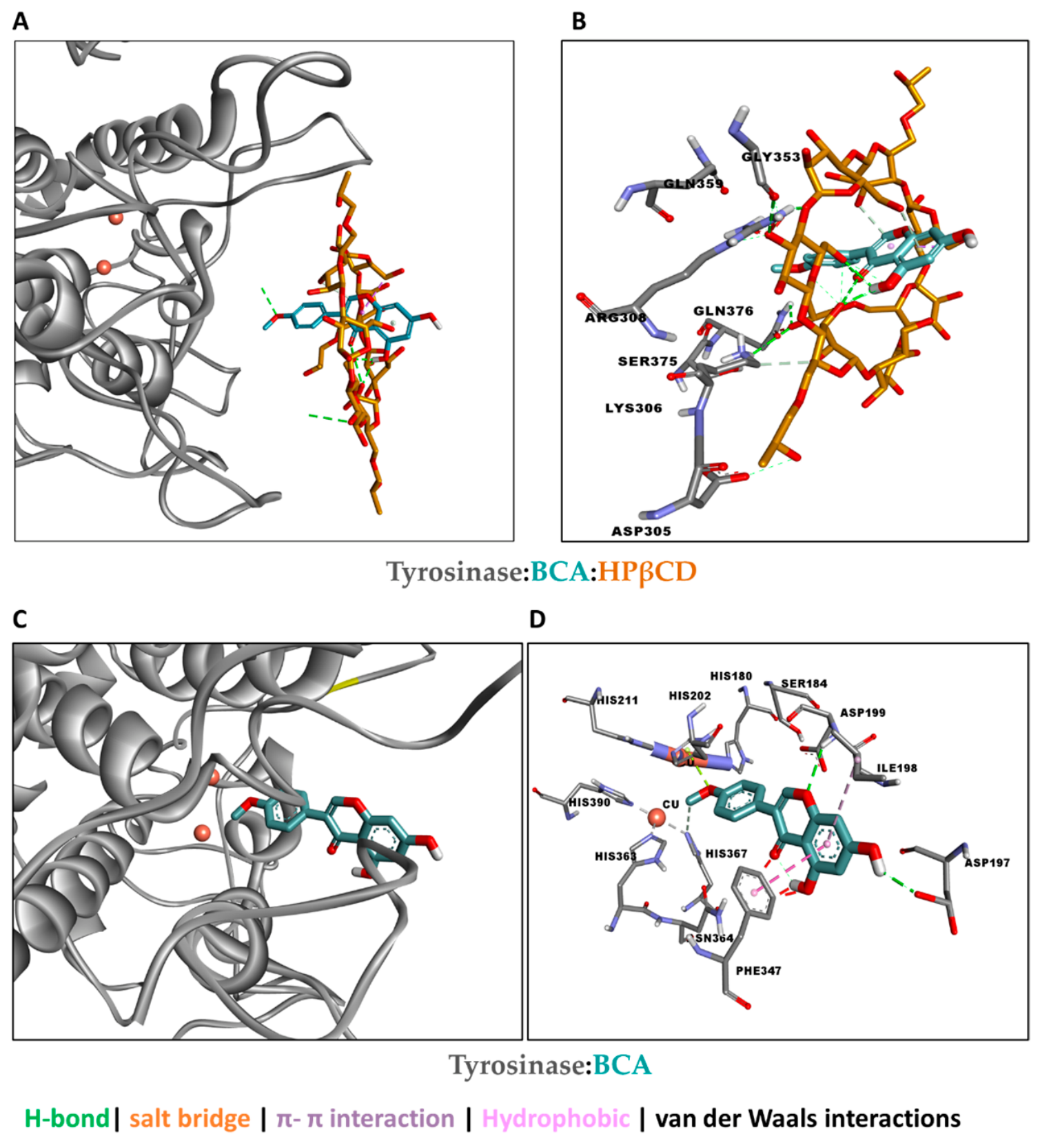
Tyrosinase: The BCA–HPβCD inclusion complex was positioned at the protein surface and interacted with polar residues, including Asp305, Lys306, Arg308, Gln359, Ser375, and Gln376, without direct contact with either copper ion (Figure 5A,B). However, no direct metal–ligand coordination was identified. Free BCA binds close to the dicopper centre, which is held by histidine residues (His202, His363, His367, His390, and others) [70]. It also formed π–π and hydrophobic contacts with Phe347 and hydrogen bonds with Ser184, Asp197, and Asp199 (Figure 5C,D). These predicted binding modes suggest that free BCA may access the catalytic-site region more readily. In contrast, complexation with HPβCD may favor a more peripheral binding orientation.
Figure 5.
(A) Possible binding of the BCA:HPβCD inclusion complex to tyrosinase and (B) interaction. (C) Possible binding of BCA to tyrosinase and (D) interaction.
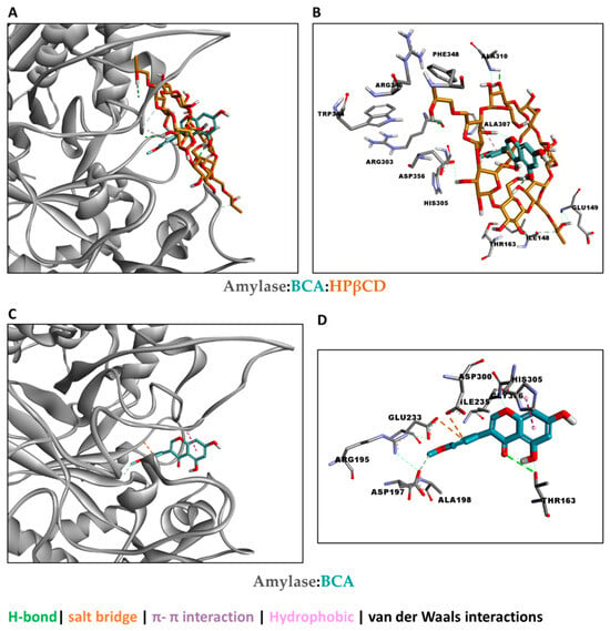
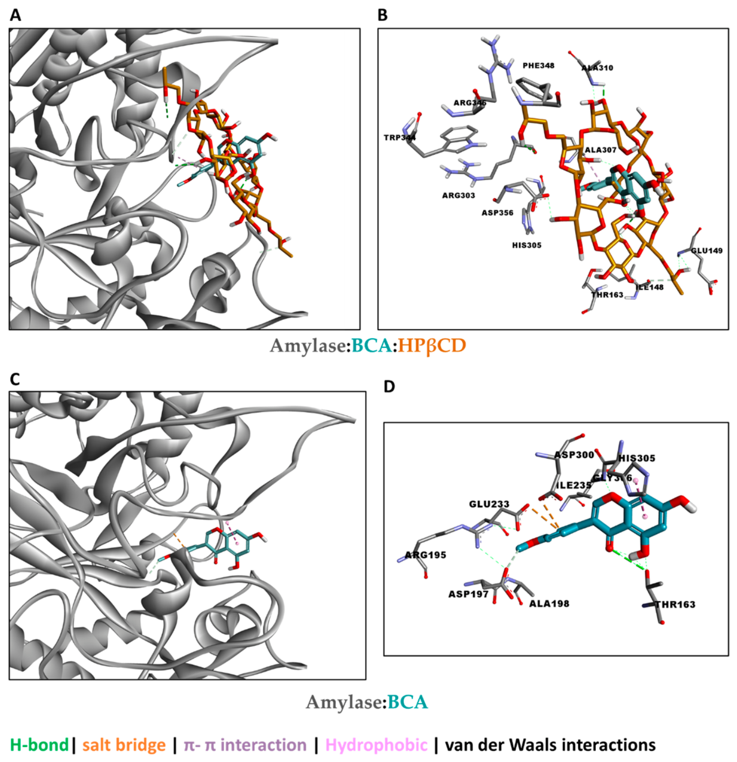
Amylase: The BCA–HPβCD inclusion complex was predicted to associate with the loop region involving Arg303, His305, Ala307, Trp354, and Asp356, with the cyclodextrin hydroxyl groups contributing predominantly hydrogen-bonding interactions (Figure 6A,B). Free BCA interacted with the catalytic-site residues Glu233 and Asp300 through electrostatic contacts, formed hydrogen bonds with Arg195, Asp197, and Thr163, and established a π interaction with His305 (Figure 6C,D). Glu233, Asp197, and Asp300 are established catalytic residues of amylase, whereas His305 lies in a flexible loop bordering the substrate-binding cleft [71]. The involvement of the 303–309 loop is consistent with crystallographic evidence that this region undergoes conformational changes upon ligand binding [72].
Figure 6.
(A) Possible binding of the BCA:HPβCD inclusion complex to amylase and (B) interaction. (C) Possible binding of BCA to amylase and (D) interaction.
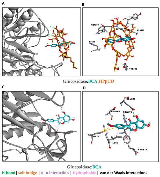
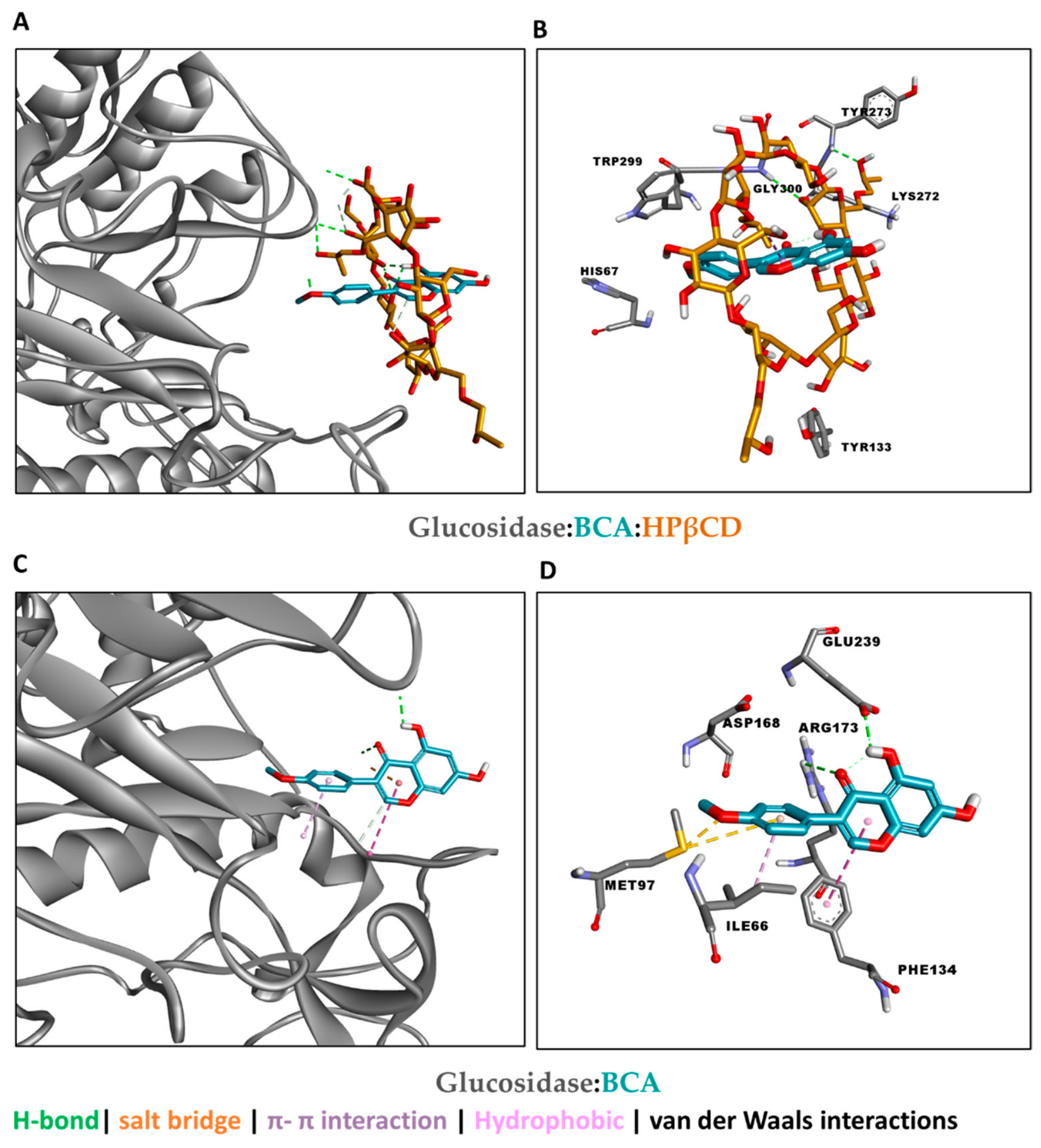
Glucosidase. The BCA–HPβCD inclusion complex was modelled to associate with a more peripheral region involving His67, Tyr133, Trp299, Lys272, and Tyr273 (Figure 7A,B). Free BCA occupied a hydrophobic pocket lined by Ile66, Met97, and Phe134, with hydrogen-bonding interactions involving Arg173 and Glu239 and nearby Asp168 (Figure 7C,D). However, although these residues may contribute to ligand recognition and binding, their essentiality for catalysis has not been established [73].
Figure 7.
(A) Possible binding of the BCA:HPβCD inclusion complex to glucosidase and (B) interaction. (C) Possible binding of BCA to glucosidase and (D) interaction.
Taken together, the free BCA enters the active site and forms directed interactions, while HPβCD sterically limits the complex to peripheral hydrogen bonding. Cyclodextrin complexation mainly improves stability. Interaction types follow the standard classification [74]. These results suggest distinct predicted binding modes for free BCA and the inclusion complex, although docking interactions alone cannot establish their relative inhibitory effects. Importantly, docking a large, flexible host like HPβCD is not so reliable, so these complex poses should be considered qualitative.
The results of our study showed AChE and BChE inhibitory effects for the inclusion complex, not BCA, which is in accordance with the literature. Namely, the results published by Orhan et al. also showed the absence of inhibitory effect of BCA on AChE and BChE. This fact could be explained by two possible mechanisms [61]. In the first mechanism, AChE (PDB code: 1EEA) and BChE (PDB code: 6EQP) have hydrophilic active sites. In this sense, the formation of an inclusion could increase the solubility of the compound, and thus, a stronger bond (especially H-bonds) could be formed with the active site, and eventually, the enzyme could be inhibited. Secondly, if we compare a compound and its inclusion in terms of the diameter, the inclusion will have a larger diameter. In this regard, larger formation could create steric hindrance around the active sites of these enzymes, and if the substrate could not bind to the active sites, then the enzymes would not function and eventually would be inhibited. Despite these possible mechanisms, we strongly recommend further analysis of the inhibitory effect of biological inclusion on the pharmacological enzymes, including AChE and BchE, which will be a part of our further research, together with cell-based and in vivo studies that would be needed before any conclusion on therapeutic relevance could be drawn.
4. Conclusions
This study has shown that the molecular encapsulation of biochanin A in the 2-hydroxypropyl-β-cyclodextrin cavities changes the biological activity of pure biochanin A to some extent. Namely, the antimicrobial activity of biochanin A against Helicobacter pylori was enhanced significantly with its incorporation in the inclusion complex. In addition, the inclusion complex inhibited both cholinesterases, whereas pure biochanin A did not. Since HPβCD alone was not tested, a contribution of the cyclodextrin to this effect cannot be excluded. Moreover, these in vitro results do not demonstrate therapeutic efficacy in Alzheimer’s disease or other neurodegenerative disorders. A slightly better inhibitory effect on tyrosinase was observed through application of the inclusion complex in contrast to pure biochanin A. Conversely, biochanin A displayed a better inhibitory effect on amylase and glucosidase than the inclusion complex. Biochanin A and the inclusion complex showed weak dose-dependent radical-scavenging activity. Based on all of the above, it can be concluded that the inclusion complex of biochanin A with 2-hydroxypropyl-β-cyclodextrin represents a promising step in the development of more efficient pharmaceutical formulations for the treatment of some infections primarily caused by Helicobacter pylori bacteria. The increase in biochanin A activity through complexation with 2-hydroxypropyl-β-cyclodextrin against Helicobacter pylori is thus more significant, as it is known that this bacterium is extremely resistant and therefore sensitive to only a small number of antibiotics.
Supplementary Materials
The following supporting information can be downloaded at https://www.mdpi.com/article/10.3390/pharmaceutics18111404/s1. Table S1: Protein structures and docking grid parameters used to redock the cocrystal ligands, BCA, and BCA:HPβCD complex into the active site of the selected enzymes; Table S2: Estimated docking binding energies of cocrystal ligands and BCA and BCA:HPβCD against target enzymes.
Author Contributions
Conceptualization, I.G., A.C.K., V.N. and L.N.; methodology, I.G., A.C.K., G.Z., A.I.U., V.N. and L.N.; formal analysis, I.G., A.C.K., G.Z., A.I.U., T.M.-K., A.D., M.U., V.N. and L.N.; investigation, I.G., A.C.K., G.Z., T.M.-K., A.D., M.U., V.N. and L.N.; writing—original draft preparation, I.G., A.C.K., V.N. and L.N.; writing—review and editing, I.G., A.C.K., V.N. and L.N. All authors have read and agreed to the published version of the manuscript.
Funding
The authors wish to thank the following sources for financing our scientific research: Ministry of Science, Technological Development and Innovation, Republic of Serbia: (451-03-34/2026-03/200133) and (451-03-33/2026-03/200133) Faculty of Technology in Leskovac, University of Niš ; (451-03-33/2026-03/200134) Faculty of Technology Novi Sad, University of Novi Sad .
Institutional Review Board Statement
Not applicable.
Informed Consent Statement
Not applicable.
Data Availability Statement
The raw data supporting the conclusions of this article will be made available by the authors on request.
Acknowledgments
The authors wish to thank the following sources for financing our scientific research: Republic of Serbia—Ministry of Science, Technological Development and Innovation, Program, for financing scientific research work, number 451-03-34/2026-03/200133 and 451-03-33/2026-03/200133, University of Niš, Faculty of Technology in Leskovac; 451-03-33/2026-03/200134 University of Novi Sad, Faculty of Technology Novi Sad.
Conflicts of Interest
The authors declare no conflicts of interest.
References
- Ramos, G.P.; Dias, P.M.; Morais, C.B.; Fröehlich, P.E.; Dall’Agnol, M.; Zuanazzi, J.A. LC determination of four isoflavone aglycones in red clover (Trifolium pratense L.). Chromatographia 2008, 67, 125–129. [Google Scholar] [CrossRef] [Scilit]
- Schrepfer, S.; Deuse, T.; Münzel, T.; Schäfer, H.; Braendle, W.; Reichenspurner, H. The selective estrogen receptor-β agonist biochanin A shows vasculoprotective effects without uterotrophic activity. Menopause 2006, 13, 489–499. [Google Scholar] [CrossRef] [Scilit] [PubMed]
- Yu, C.; Zhang, P.; Lou, L.; Wang, Y. Perspectives regarding the role of biochanin A in humans. Front. Pharmacol. 2019, 10, 793. [Google Scholar] [CrossRef] [Scilit] [PubMed]
- Yan, J.; Qiu, P.; Zhang, X.; Zhang, Y.; Mi, L.; Peng, C.; Pan, X.; Peng, F. Biochanin A from Chinese Medicine: An Isoflavone with Diverse Pharmacological Properties. Am. J. Chin. Med. 2021, 49, 1623–1643. [Google Scholar] [CrossRef] [Scilit] [PubMed]
- Felix, F.B.; Vago, J.P.; Beltrami, V.A.; Araújo, J.M.D.; Grespan, R.; Teixeira, M.M.; Pinho, V. Biochanin A as a modulator of the inflammatory response: An updated overview and therapeutic potential. Pharmacol. Res. 2022, 180, 106246. [Google Scholar] [CrossRef] [Scilit] [PubMed]
- Harini, R.; Ezhumalai, M.; Pugalendi, K.V. Antihyperglycemic effect of biochanin A, a soy isoflavone, on streptozotocin-diabetic rats. J. Pharm. Res. 2012, 676, 89–94. [Google Scholar] [CrossRef] [Scilit] [PubMed]
- Ghadimi, D.; Goodarzi, M.T.; Ziamajidi, N.; Moradkhani, S. The influence of biochanin a consumption on c-CBL-associated protein level in adipose tissue of streptozotocine-nicotinamide induced diabetic rats. Int. J. Med. Health Sci. 2016, 5, 195–201. [Google Scholar]
- Hammarstedt, A.; Andersson, C.X.; Sopasakis, V.R.; Smith, U. The effect of PPARγ ligands on the adipose tissue in insulin resistance. Prostaglandins Leukot. Essent. Fat. Acids 2005, 73, 65–75. [Google Scholar] [CrossRef] [Scilit] [PubMed]
- Azizi, R.; Goodarzi, M.T.; Salemi, Z. Effect of biochanin a on serum visfatin level of streptozocin-induced diabetic rats. Iran. Red. Crescent Med. J. 2014, 16, e15424. [Google Scholar] [CrossRef] [Scilit] [PubMed]
- Uysal, S.; Zengin, G.; Locatelli, M.; Bahadori, M.B.; Mocan, A.; Bellagamba, G.; Luca, E.D.; Mollica, A.; Aktumsek, A. Cytotoxic and enzyme inhibitory potential of two Potentilla species (P. speciosa L. and P. reptans Willd.) and their chemical composition. Front. Pharmacol. 2017, 8, 290. [Google Scholar] [CrossRef] [Scilit] [PubMed]
- Di Petrillo, A.; González-Paramás, A.M.; Era, B.; Medda, R.; Pintus, F.; Santos-Buelga, C.; Fais, A. Tyrosinase inhibition and antioxidant properties of Asphodelus microcarpus extracts. BMC Complement. Altern. Med. 2016, 16, 453. [Google Scholar] [CrossRef] [Scilit] [PubMed]
- Kumar, C.M.; Sathisha, U.V.; Dharmesh, S.; Rao, A.A.; Singh, S.A. Interaction of sesamol (3, 4-methylenedioxyphenol) with tyrosinase and its effect on melanin synthesis. Biochimie 2011, 93, 562–569. [Google Scholar] [CrossRef] [Scilit] [PubMed]
- Saghaie, L.; Pourfarzam, M.; Fassihi, A.; Sartippour, B. Synthesis and tyrosinase inhibitory properties of some novel derivatives of kojic acid. J. Pharm. Sci. 2013, 8, 233. [Google Scholar]
- Paul, M.K.; Mukhopadhyay, A.K. Tyrosine kinase–role and significance in cancer. Int. J. Med. Sci. 2004, 1, 101. [Google Scholar] [CrossRef] [Scilit] [PubMed]
- Chang, T.S. An updated review of tyrosinase inhibitors. Int. J. Mol. Sci. 2009, 10, 2440–2475. [Google Scholar] [CrossRef] [Scilit] [PubMed]
- Giacobini, E. Cholinesterase inhibitors: New roles and therapeutic alternatives. Pharmacol. Res. 2004, 50, 433–440. [Google Scholar] [CrossRef] [Scilit] [PubMed]
- Soreq, H.; Seidman, S. Acetylcholinesterase—New roles for an old actor. Nat. Rev. Neurosci. 2001, 2, 294–302. [Google Scholar] [CrossRef] [Scilit] [PubMed]
- Čolović, M.B.; Krstić, D.Z.; Lazarević-Pasti, T.D.; Bondžić, A.M.; Vasić, V.M. Acetylcholinesterase inhibitors: Pharmacology and toxicology. Curr. Neuropharmacol. 2013, 11, 315–335. [Google Scholar] [CrossRef] [Scilit] [PubMed]
- Pohanka, M. Cholinesterases, a target of pharmacology and toxicology. Biomed. Pap. Med. Fac. Univ. Palacky. Olomouc Czech. Repub. 2011, 155, 2019–2229. [Google Scholar] [CrossRef] [Scilit] [PubMed]
- Chen, X.; Fang, L.; Liu, J.; Zhan, C.G. Reaction pathway and free energy profile for butyrylcholinesterase-catalyzed hydrolysis of acetylcholine. J. Phys. Chem. 2011, 115, 1315–1322. [Google Scholar] [CrossRef] [Scilit] [PubMed]
- Nikolić, I.; Savić, I.; Popsavin, M.; Rakić, S.; Mihajilov-Krstev, T.; Ristić, I.; Erić, S.; Savić-Gajić, I. Preparation, characterization and antimicrobial activity of inclusion complex of biochanin A with (2-hydroxypropyl)-β-cyclodextrin. J. Pharm. Pharmacol. 2018, 70, 1485–1493. [Google Scholar] [CrossRef] [Scilit] [PubMed]
- Nikolić, V.; Ilić-Stojanović, S.; Petrović, S.; Tačić, A.; Nikolić, L. Administration routes for nano drugs and characterization of nano drug loading. In Characterization and Biology of Nanomaterials for Drug Delivery; Mohapatra, S., Ranjan, S., Dasgupta, N., Mishra, R.K., Thomas, S., Eds.; Elsevier: Amsterdam, The Netherlands, 2019; pp. 587–627. [Google Scholar]
- Wu, X.; Ge, W.; Shao, T.; Wu, W.; Hou, J.; Cui, L.; Wang, J.; Zhang, Z. Enhancing the oral bioavailability of biochanin A by encapsulation in mixed micelles containing Pluronic F127 and Plasdone S630. Int. J. Nanomed. 2017, 12, 1475–1483. [Google Scholar] [CrossRef] [Scilit] [PubMed]
- Jia, X.; Chen, J.; Lin, H.; Hu, M. Disposition of flavonoids via enteric recycling: Enzyme-transporter coupling affects metabolism of biochanin A and formononetin and excretion of their phase II conjugates. J. Pharmacol. Exp. Ther. 2004, 310, 1103–1113. [Google Scholar] [CrossRef] [Scilit] [PubMed]
- Hanski, L.; Genina, N.; Uvell, H.; Malinovskaja, K.; Gylfe, Ǻ.; Laaksonen, T.; Kolaković, R.; Mäkilä, E.; Salonen, J.; Hirvonen, J.; et al. Inhibitory Activity of the Isoflavone Biochanin A on Intracellular Bacteria of Genus Chlamydia and Initial Development of a Buccal Formulation. PLoS ONE 2014, 9, e115115. [Google Scholar] [CrossRef] [Scilit] [PubMed]
- Gajić, I.; Stojanović, S.; Ristić, R.; Ilić-Stojanović, S.; Pilić, B.; Nešić, A.; Najman, S.; Dinić, A.; Stanojević, L.; Urošević, M.; et al. Electrospun Poly(lactide) Fibers as Carriers for Controlled Re-lease of Biochanin A. Pharmaceutics 2022, 14, 528. [Google Scholar] [CrossRef] [Scilit] [PubMed]
- Gajić, I.; Ilić-Stojanović, S.; Dinić, A.; Zdravković, A.; Stanojević, L.; Nikolić, V.; Nikolić, L. Modified Biochanin A Release from Dual pH and Thermo-Responsive Copolymer Hydrogels. Polymers 2021, 13, 426. [Google Scholar] [CrossRef] [Scilit] [PubMed]
- Proença, C.; Freitas, M.; Ribeiro, D.; Tomé, S.M.; Oliveira, E.F.; Viegas, M.F.; Araújo, A.N.; Ramos, M.J.; Silva, A.M.S.; Fernandes, P.; et al. Evaluation of a flavonoids library for inhibition of pancreatic α-amylase towards a structure–activity relationship. J. Enzym. Inhib. Med. Chem. 2019, 34, 577–588. [Google Scholar] [CrossRef] [Scilit] [PubMed]
- CLSI M100-S17; Performance Standards for Antimicrobial Susceptibility Testing. Clinical and Laboratory Standards Institute: Wayne, PA, USA, 2007.
- Hachem, C.Y.; Clarridge, J.E.; Reddy, R.; Flamm, R.; Evans, D.G.; Tanaka, S.K.; Graham, D.Y. Antimicrobial susceptibility testing of Helicobacter pylori comparison of E-test, broth microdilution, and disk diffusion for ampicillin, clarithromycin, and metronidazole. Diagn. Microbiol. Infect. Dis. 1996, 24, 37–41. [Google Scholar] [CrossRef] [Scilit] [PubMed]
- Stanojević, L.; Stanković, M.; Nikolić, V.; Nikolić, L.; Ristić, D.; Čanadanovic-Brunet, J.; Tumbas, V. Antioxidant Activity and Total Phenolic and Flavonoid Contents of Hieracium pilosella L. Extracts. Sensors 2009, 9, 5702–5714. [Google Scholar] [CrossRef] [Scilit] [PubMed]
- Gerlits, O.; Ho, K.-Y.; Cheng, X.; Blumenthal, D.; Taylor, P.; Kovalevsky, A.; Radić, Z. A new crystal form of human acetylcholinesterase for exploratory room-temperature crystallography studies. Chem. Biol. Interact. 2019, 309, 108698. [Google Scholar] [CrossRef] [Scilit] [PubMed]
- Rosenberry, T.; Brazzolotto, X.; Macdonald, I.; Wandhammer, M.; Trovaslet-Leroy, M.; Darvesh, S.; Nachon, F. Comparison of the Binding of Reversible Inhibitors to Human Butyrylcholinesterase and Acetylcholinesterase: A Crystallographic, Kinetic and Calorimetric Study. Molecules 2017, 22, 2098. [Google Scholar] [CrossRef] [Scilit] [PubMed]
- Maurus, R.; Begum, A.; Williams, L.K.; Fredriksen, J.R.; Zhang, R.; Withers, S.G.; Brayer, G.D. Alternative Catalytic Anions Differentially Modulate Human α-Amylase Activity and Specificity. Biochemistry 2008, 47, 3332–3344. [Google Scholar] [CrossRef] [Scilit] [PubMed]
- Fahmy, N.M.; Fayez, S.; Uba, A.I.; Shariati, M.A.; Aljohani, A.S.M.; El-Ashmawy, I.M.; Batiha, G.E.-S.; Eldahshan, O.A.; Singab, A.N.; Zengin, G. Comparative GC-MS Analysis of Fresh and Dried Curcuma Essential Oils with Insights into Their Antioxidant and Enzyme Inhibitory Activities. Plants 2023, 12, 1785. [Google Scholar] [CrossRef] [Scilit] [PubMed]
- Eltayeb, L.M.H.; Yagi, S.; Mohamed, H.M.M.; Zengin, G.; Shariati, M.A.; Rebezov, M.; Uba, A.I.; Lorenzo, J.M. Essential Oils Composition and Biological Activity of Chamaecyparis obtusa, Chrysopogon nigritanus and Lavandula coronopifolia Grown Wild in Sudan. Molecules 2023, 28, 1005. [Google Scholar] [CrossRef] [Scilit] [PubMed]
- Ielo, L.; Deri, B.; Germanò, M.P.; Vittorio, S.; Mirabile, S.; Gitto, R.; Rapisarda, A.; Ronsisvalle, S.; Floris, S.; Pazy, Y.; et al. Exploiting the 1-(4-fluorobenzyl)piperazine fragment for the development of novel tyrosinase inhibitors as anti-melanogenic agents: Design, synthesis, structural insights and biological profile. Eur. J. Med. Chem. 2019, 178, 380–389. [Google Scholar] [CrossRef] [Scilit] [PubMed]
- Karade, S.S.; Hill, M.L.; Kiappes, J.L.; Manne, R.; Aakula, B.; Zitzmann, N.; Warfield, K.L.; Treston, A.M.; Mariuzza, R.A. N-Substituted Valiolamine Derivatives as Potent Inhibitors of Endoplasmic Reticulum α-Glucosidases I and II with Antiviral Activity. J. Med. Chem. 2021, 64, 18010–18024. [Google Scholar] [CrossRef] [Scilit] [PubMed]
- Jursic, B.S.; Zdravkovski, Z.; French, A.D. Molecular modeling methodology of β-cyclodextrin inclusion complexes. J. Mol. Struct. THEOCHEM 1996, 366, 113–117. [Google Scholar] [CrossRef] [Scilit]
- Pettersen, E.F.; Goddard, T.D.; Huang, C.C.; Couch, G.S.; Greenblatt, D.M.; Meng, E.C.; Ferrin, T.E. UCSF Chimera? A visualization system for exploratory research and analysis. J. Comput. Chem. 2004, 25, 1605–1612. [Google Scholar] [CrossRef] [Scilit] [PubMed]
- Morris, G.M.; Huey, R.; Lindstrom, W.; Sanner, M.F.; Belew, R.K.; Goodsell, D.S.; Olson, A.J. AutoDock4 and AutoDockTools4: Automated docking with selective receptor flexibility. J. Comput. Chem. 2009, 30, 2785–2791. [Google Scholar] [CrossRef] [Scilit] [PubMed]
- Castro-Alvarez, A.; Costa, A.M.; Vilarrasa, J. The Performance of Several Docking Programs at Reproducing Protein–Macrolide-Like Crystal Structures. Molecules 2017, 22, 136. [Google Scholar] [CrossRef] [Scilit] [PubMed]
- Dassault Systèmes BIOVIA. Discovery Studio Visualizer, v4.5; Dassault Systèmes: San Diego, CA, USA, 2015. [Google Scholar]
- Dinat, S.; Orchard, A.; Van Vuuren, S. A systematic review of African natural products against gastric ulcers and Helicobacter pylori. J. Ethnopharmacol. 2022, 301, 115698. [Google Scholar] [CrossRef] [Scilit] [PubMed]
- Mendonça, M.A.D.; Ribeiro, A.R.; Lima, A.K.D.; Bezerra, G.B.; Pinheiro, M.S.; Albuquerque-Júnior, R.L.D.; Gomes, M.Z.; Padilha, F.F.; Thomazzi, S.M.; Novellino, E.; et al. Red propolis and its dyslipidemic regulator formononetin: Evaluation of antioxidant activity and gastroprotective effects in rat model of gastric ulcer. Nutrients 2020, 12, 2951. [Google Scholar] [CrossRef] [Scilit] [PubMed]
- Satoh, H.; Saijo, Y.; Yoshioka, E.; Tsutsui, H. Helicobacter Pylori infection is a significant risk for modified lipid profile in Japanese male subjects. J. Atheroscler. Thromb. 2010, 17, 1041–1048. [Google Scholar] [CrossRef] [Scilit] [PubMed]
- Xue, Z.; Zhang, Q.; Yu, W.; Wen, H.; Hou, X.; Li, D.; Kou, X. Potential lipid-lowering mechanisms of biochanin A. J. Agric. Food Chem. 2017, 65, 3842–3850. [Google Scholar] [CrossRef] [Scilit] [PubMed]
- Fukai, T.; Marumo, A.; Kaitou, K.; Kanda, T.; Terada, S.; Nomura, T. Anti-Helicobacter pylori flavonoids from licorice extract. Life Sci. 2002, 71, 1449–1463. [Google Scholar] [CrossRef] [Scilit] [PubMed]
- Mota, J.C.; Almeida, P.P.; Freitas, M.Q.; Stockler-Pinto, M.B.; Guimarães, J.T. Far from being a simple question: The complexity between in vitro and in vivo responses from nutrients and bioactive compounds with antioxidant potential. Food Chem. 2023, 402, 134351. [Google Scholar] [CrossRef] [Scilit] [PubMed]
- Lugasi, A. The role of antioxidant phytonutrients in the prevention of diseases. Acta Biol. Szeged. 2003, 47, 119–125. [Google Scholar]
- Shao, J.; Zhao, T.; Ma, H.P.; Jia, Z.P.; Jing, L.L. Synthesis, characterization, and antioxidant activity of 8-hydroxygenistein. Nat. Prod. Commun. 2020, 15, 1934578X20901399. [Google Scholar] [CrossRef] [Scilit]
- Sadri, H.; Goodarzi, M.T.; Salemi, Z.; Seifi, M. Antioxidant effects of biochanin A in streptozotocin induced diabetic rats. Braz. Arch. Biol. Technol. 2017, 60, e17160741. [Google Scholar] [CrossRef] [Scilit]
- Sadeer, N.B.; Montesano, D.; Albrizio, S.; Zengin, G.; Mahomoodally, M.F. The versatility of antioxidant assays in food science and safety—Chemistry, applications, strengths, and limitations. Antioxidants 2020, 9, 709. [Google Scholar] [CrossRef] [Scilit] [PubMed]
- Magiera, S.; Baranowska, I.; Lautenszleger, A. UHPLC–UV method for the determination of flavonoids in dietary supplements and for evaluation of their antioxidant activities. J. Pharm. Biomed. Anal. 2015, 102, 468–475. [Google Scholar] [CrossRef] [Scilit] [PubMed]
- Hamza Sherif, S.; Gebreyohannes, B. Synthesis, characterization, and antioxidant activities of genistein, biochanin A, and their analogues. J. Chem. 2018, 2018, 4032105. [Google Scholar] [CrossRef] [Scilit]
- Sarian, M.N.; Ahmed, Q.U.; Mat So’ad, S.Z.; Alhassan, A.M.; Murugesu, S.; Perumal, V.; Mohamad, S.N.A.S.; Khatib, A.; Latip, J. Antioxidant and antidiabetic effects of flavonoids: A structure-activity relationship based study. BioMed Res. Int. 2017, 2017, 8386065. [Google Scholar] [CrossRef] [Scilit] [PubMed]
- Siqueira-Lima, P.S.; Brito, R.G.; Araújo-Filho, H.G.; Santos, P.L.; Lucchesi, A.; Araújo, A.A.; Menezes, P.P.; Scotti, L.; Scotti, M.T.; Menezes, I.R.A.; et al. Anti-hyperalgesic effect of Lippia grata leaf essential oil complexed with β-cyclodextrin in a chronic musculoskeletal pain animal model: Complemented with a molecular docking and antioxidant screening. Biomed. Pharmacother. 2017, 91, 739–747. [Google Scholar] [CrossRef] [Scilit] [PubMed]
- Athanasiadis, V.; Palaiogiannis, D.; Bozinou, E.; Lalas, S.I.; Makris, D.P. β-Cyclodextrin-aided aqueous extraction of antioxidant polyphenols from peppermint (Mentha piperita L.). Oxygen 2022, 2, 424–436. [Google Scholar] [CrossRef] [Scilit]
- López-Nicolás, J.M.; Rodríguez-Bonilla, P.; García-Carmona, F. Cyclodextrins and antioxidants. Crit. Rev. Food Sci. Nutr. 2014, 54, 251–276. [Google Scholar] [CrossRef] [Scilit] [PubMed]
- Rauf, A.; Jehan, N. Natural products as a potential enzyme inhibitors from medicinal plants. In Enzyme Inhibitors and Activators; Senturk, M., Ed.; Intech Open: London, UK, 2017; pp. 165–177. [Google Scholar]
- Orhan, I.; Kartal, M.; Tosun, F.; Şener, B. Screening of various phenolic acids and flavonoid derivatives for their anticholinesterase potential. Z. Naturforsch. C J. Biosci. 2007, 62, 829–832. [Google Scholar] [CrossRef] [Scilit] [PubMed]
- Lin, V.C.; Ding, H.Y.; Tsai, P.C.; Wu, J.Y.; Lu, Y.H.; Chang, T.S. In vitro and in vivo melanogenesis inhibition by biochanin A from Trifolium pratense. Biosci. Biotechnol. Biochem. 2011, 75, 914–918. [Google Scholar] [CrossRef] [Scilit] [PubMed]
- Park, J.H.; Baek, M.R.; Lee, B.H.; Yon, G.H.; Ryu, S.Y.; Kim, Y.S.; Park, S.U.; Hong, K.S. α-Glucosidase and α-Amylase Inhibitory Activity of Compounds from Roots Extract of Pueraria thunbergiana. Korean J. Med. Crop Sci. 2009, 17, 357–362. [Google Scholar]
- Mehrabadi, M.E.; Salemi, Z.; Babaie, S.; Panahi, M. Effect of biochanin A on retina levels of vascular endothelial growth factor, tumor necrosis factor-alpha and interleukin-1beta in rats with streptozotocin-induced diabetes. Can. J. Diabetes 2018, 42, 639–644. [Google Scholar] [CrossRef] [Scilit] [PubMed]
- Oza, M.J.; Kulkarni, Y.A. Biochanin A improves insulin sensitivity and controls hyperglycemia in type 2 diabetes. Biomed. Pharmacother. 2018, 107, 1119–1127. [Google Scholar] [CrossRef] [Scilit] [PubMed]
- Proença, C.; Freitas, M.; Ribeiro, D.; Oliveira, E.F.; Sousa, J.L.; Tome, S.M.; Ramos, M.J.; Silva, A.M.S.; Fernandes, P.; Fernandes, E. α-Glucosidase inhibition by flavonoids: An in vitro and in silico structure–activity relationship study. J. Enzym. Inhib. Med. Chem. 2017, 32, 1216–1228. [Google Scholar]
- Colletier, J.P.; Fournier, D.; Greenblatt, H.M.; Stojan, J.; Sussman, J.L.; Zaccai, G.; Silman, I.; Weik, M. Structural insights into substrate traffic and inhibition in acetylcholinesterase. EMBO J. 2006, 25, 2746–2756. [Google Scholar] [CrossRef] [Scilit] [PubMed]
- Sussman, J.L.; Harel, M.; Frolow, F.; Oefner, C.; Goldman, A.; Toker, L.; Silman, I. Atomic structure of acetylcholinesterase from Torpedo californica: A prototypic acetylcholine-binding protein. Science 1991, 253, 872–879. [Google Scholar] [CrossRef] [Scilit] [PubMed]
- Nicolet, Y.; Lockridge, O.; Masson, P.; Fontecilla-Camps, J.C.; Nachon, F. Crystal structure of human butyrylcholinesterase and of its complexes with substrate and products. J. Biol. Chem. 2003, 278, 41141–41147. [Google Scholar] [CrossRef] [Scilit] [PubMed]
- Spritz, R.A.; Ho, L.; Furumura, M.; Hearing, V.J., Jr. Mutational Analysis of Copper Binding by Human Tyrosinase. J. Investig. Dermatol. 1997, 109, 207–212. [Google Scholar] [CrossRef] [Scilit] [PubMed]
- Qin, X.; Ren, L.; Yang, X.; Bai, F.; Wang, L.; Geng, P.; Bai, G.; Shen, Y. Structures of human pancreatic α-amylase in complex with acarviostatins: Implications for drug design against type II diabetes. J. Struct. Biol. 2011, 174, 196–202. [Google Scholar] [CrossRef] [Scilit] [PubMed]
- Nahoum, V.; Roux, G.; Anton, V.; Rougé, P.; Puigserver, A.; Bischoff, H.; Henrissat, B.; Payan, F. Crystal structures of human pancreatic α-amylase in complex with carbohydrate and proteinaceous inhibitors. Biochem. J. 2000, 346, 201–208. [Google Scholar] [CrossRef]
- Okuyama, M.; Saburi, W.; Mori, H.; Kimura, A. α-Glucosidases and α-1,4-glucan lyases: Structures, functions, and physiological actions. Cell. Mol. Life Sci. 2016, 73, 2727–2751. [Google Scholar] [CrossRef] [Scilit] [PubMed]
- Bissantz, C.; Kuhn, B.; Stahl, M. Corrections to A Medicinal Chemist’s Guide to Molecular Interactions. J. Med. Chem. 2010, 53, 6241. [Google Scholar] [CrossRef] [Scilit]

Disclaimer/Publisher’s Note: The statements, opinions and data contained in all publications are solely those of the individual author(s) and contributor(s) and not of MDPI and/or the editor(s). MDPI and/or the editor(s) disclaim responsibility for any injury to people or property resulting from any ideas, methods, instructions or products referred to in the content. |
© 2026 by the authors. Licensee MDPI, Basel, Switzerland. This article is an open access article distributed under the terms and conditions of the Creative Commons Attribution (CC BY) license.