


Abstract
Post-traumatic stress disorder (PTSD) is a severe disorder that is characterized by intrusive thoughts, impaired mood and cognition, hyperarousal, and avoidance in the months and years following a traumatic event. PTSD is comorbid with depression, anxiety, substance abuse, and is challenging for the treatment with psycho- and pharmacotherapy. In light of novel neurobiological findings on the pathogenesis of the disease, innovative approaches to its treatment are investigated. Recently, targeting cholinergic signaling with an antagonist of α7 nicotinic acetylcholine receptors (nAChR), BNC-210, showed a significant promise in clinical trials. This opened up a novel therapeutic strategy that, considering its translational success, calls for a mechanistic understanding. In this review, we dissect the neurobiological role of α7 nAChR in regard of PTSD, and related depression and anxiety states. We discuss the synaptic role of the receptor in fear circuits, immune and endocrine role via the anti-inflammatory cholinergic pathway, and current evidence for the utility of pharmacological modulation of α7 nAChR.
1. Introduction
Post-traumatic stress disorder (PTSD) is a psychiatric disorder that develops in months and years after an exposure to severe traumatic experience. Its characteristic symptoms include re-experiencing of trauma memories, avoidance of cues that remind the individual of the trauma, negative alterations in cognition and mood, intrusive thoughts, and hyperarousal [1,2,3]. The estimated prevalence of PTSD varies; however, the risk groups include refugees and displaced populations [4], combat veterans (with up to 30% occurrence [5]), and victims of assault (up to 20% occurrence [6]).
PTSD is a multifactorial disease, and its development depends on genes [2,7], type of trauma [8], and the sex of the individual [9]. Although it nosologically overlaps with anxiety disorders and depression, The Diagnostic and Statistical Manual of Mental Disorders, Fifth Edition (DSM-5) and the International Classification of Diseases, 11th Revision (ICD-11; World Health Organization) does not classify PTSD as an anxiety disorder [10]; instead, PTSD belongs to the category of trauma- and stressor-related disorders. Given the distinct pathological profile of PTSD, personalized and innovative therapeutic approaches are needed.
First-line treatment for PTSD involves psychotherapy, such as cognitive behavior therapy, while pharmacotherapy is often used in concert [1]. Pharmacotherapy for PTSD is usually aimed at anxiety, arousal, and depression. Currently, the only FDA-approved treatments for PTSD are the serotonin reuptake inhibitors sertraline and paroxetine. A significant proportion of patients receiving psychotherapy and standard drug therapy drop out of or are nonresponsive to therapy [11,12]. Serotonin reuptake inhibitors can be supplemented with other therapeutics for boosting efficacy or targeting comorbid symptoms, e.g., antipsychotics for intrusive thoughts, sedatives for sleep disturbance, or drugs against mood disorders and substance abuse [1,2,13]. Novel pharmacological approaches for PTSD management are searched and investigated. For example, psychedelics, such as MDMA [14,15], α-adrenergic antagonist prazosin [16], and other drugs [17] have been evaluated in clinical studies. Mechanistically, PTSD is linked to endocrine factors, neurotransmitter balance, and distinct fear-related brain circuits [3], thereby various alternative pathways and mechanisms, other than serotoninergic, may be targets for potential drug development [17].
Recently, targeting cholinergic pathway has emerged as a novel, prospective option for the treatment of fear-related pathologies [18]. The cholinergic system in the brain comprises neurons that utilize the neurotransmitter acetylcholine (ACh) for neurotransmission. ACh binds to the two types of membrane receptors: ionotropic nicotinic acetylcholine receptors (nAChR) and metabotropic muscarinic acetylcholine receptors (mAChR). nAChRs are ligand-gated cation channels that mediate fast synaptic transmission and plasticity whereas mAChRs are slow G-protein coupled receptors that modulate overall excitability of neurons. Cholinergic system also includes enzyme and transporter machinery responsible for the synthesis, transport, inactivation, and recycling of ACh [19,20,21,22,23]. This system is largely involved in cognition, learning, and memory with a pronounced role of α7 nAChR subtype [18,24,25,26]. In this review, we discuss the potential of targeting α7 nAChRs for the therapy of PTSD, the neuroimmune, endocrine, and pharmacological prerequisites for this strategy, and its current translational status.
2. Synaptic Transmission and Regulation
2.1. α7 nAChRs as Ionotropic Receptors in Neurons
nAChRs are pentameric channels that display a great receptor heterogeneity resulting from numerous subunit types that can assemble in different stoichiometries [27,28,29,30]. In humans, there are eleven neuronal (α2-7, α9, α10, β2-4) and five muscular nAChR subunits (α1, β1, γ, δ, ε) [31]. α7 subunits mostly form functional homomeric receptors with fast activation and inactivation kinetics. α7-containing heteromers have also been reported [32]. Homomeric α7 receptors demonstrate a high permeability to calcium (PCa/PNa ≈ 6–20) that is comparable to NMDA receptors [33,34,35,36]. Calcium influx in neurons is generally required for neurotransmitter release and synaptic plasticity. However, excessive calcium influx can induce damaging events known as excitotoxicity, which is studied best for ionotropic glutamate receptors and is proposed to underlie several neurodegenerative diseases [37]. In a simplified form, nAChRs can adopt three functional states: closed resting, open, and closed desensitized. The desensitized state arises upon a prolonged exposure to ACh or other activators, including in their presence at subthreshold concentrations, and is unresponsive to further stimulations. The desensitized state is thus protective for the cells against receptor overstimulation and toxic calcium overload [38]. All the three functional states are considered in regard of possible therapeutic significance.
In neurons, α7 receptors can locate at the three key subcellular zones, implementing different mechanisms of regulation [39,40]. The activation of presynaptic α7 nAChRs leads to calcium influx that triggers immediate vesicle exocytosis [41,42,43]. Additionally, they can trigger voltage-gated calcium channels or calcium-induced calcium release from endoplasmic reticulum stores [44,45], which modulate the probability of neurotransmitter release [41,46,47]. Alternatively, α7 receptors can be present at the perisynaptic (synaptic cleft borders at near distance from postsynaptic density) and extrasynaptic zones (outside the synaptic cleft). There, they mediate volume transmission, responding to tonic ACh levels or ACh spillover at high-frequency stimulations [48,49]. Finally, α7 nAChRs located postsynaptically (within postsynaptic density) mediate fast depolarizing currents, activate signaling cascades, and modulate synaptic plasticity [48,50,51]. Localization in astrocytes (non-synaptic) can also contribute to the metabolic support of neurons [52,53,54,55,56].
2.2. α7 nAChRs in Metabotropic Signal Transduction Pathways
Acute physiological or psychological stress and glucocorticoids affect glutamate transmission, including effects on glutamate release, glutamate receptors, glutamate clearance and metabolism. Overstimulation of glutamate receptors (AMPAR and NMDA receptors) triggers excitotoxicity, causing cell death [37]. The α7 nAChR counteracts excitotoxicity through several distinct biological pathways, including lowering neuronal hyperactivation, blocking inflammatory cascades, and neutralizing oxidative cell death [57,58,59].
Currently, it has been extensively shown that α7 nAcRs can mediate metabotropic, or mixed signaling, in addition to their well-recognized ionotropic function [60]. Specifically, the α7 nAChR is linked to PI3K/Akt pathway, which promotes cell survival and neuroprotection, via Src tyrosine kinases. Src/Fyn tyrosine kinase activates PI3K (Phosphoinositide 3-kinase) that restores the surface level of NR2A receptors after tyrosine dephosphorylation [61]. Activation of α7 nAChRs recruits the adaptor protein β-arrestin [62] that modulates Src tyrosine kinase via the exposed SH3 domain for PI3K binding [63,64]. Activated PI3K converts PIP2 into PIP3. The latter recruits AKT to the cell membrane, while PDK1 and mTORC2 phosphorylates it into the active form. AKT next inhibits GSK-3β by phosphorylation, resulting in cell growth and survival, protein synthesis, angiogenesis, and glucose homeostasis [65]. The direct target of GSK-3β is β-catenin, a transcriptional co-activator of the WNT signaling pathway. WNT signaling is linked to the pathogenesis of various diseases such as autoimmune, cardiovascular, myocardial, neurodegenerative, metabolic disorders, and cancer. Phosphorylation of β-catenin by GSK-3β leads to the inhibition of WNT signaling and, consequently, to the enhancing of the NF-κB (nuclear factor kappa-B) pathway, which induces inflammation responses [66,67]. In sum, stimulation of α7 nAChRs triggers a chain of activation events with following inhibition of GSK-3β, that maintains WNT activity and prevents the development of inflammation (α7 nAChRs → Src → PI3K → PIP3 → AKT ⊣ GSK-3β → NF-κB pathway).
The α7 nAChRs are the main players of cholinergic anti-inflammatory pathway that links the brain and the immune system. Endotoxins/LPS, cytokines or injury generate signals traveling via vagus nerve fibers to the celiac-mesenteric ganglion which stimulates splenic nerve, resulting in activation of choline acetyltransferase (ChAT)-expressing T cells and acetylcholine release [68]. In immune cells, activation of α7 nAChRs leads to stimulation of JAK2 (Janus Kinase-2) that phosphorylates STAT3 (signal-transducer and activator of transcription-3). Activated STAT3 dimerizes and translocates to the nucleus in which it prevents the transcription of genes for inflammatory cytokines by interfering with the binding of NF-κB to promotor regions [69]. However, GSK-3β can also activate the transcription factors STAT3 and STAT5 that drive the release of pro-inflammatory IL-6 and lead to pro-inflammation response [66]. Sustained peripheral inflammation breaches the blood–brain barrier and activates resident microglia; thus, systemic immune response translates into central neuroinflammation and excitotoxicity.
As shown, PTSD arises from a disruption of the normal mechanism of fear generation and regulation within the prefrontal cortex, the hippocampus, and the amygdala [70,71]. Acute traumatic experience causes cortisol spikes; however, PTSD patients show paradoxically low baseline cortisol levels and enhanced glucocorticoid receptor (GR) sensitivity. This dysregulation of the hypothalamic–pituitary–adrenal axis, combined with intense stress-induced glutamate release, results in a decrease in BDNF levels in the cortex and hippocampus [72,73,74]. BDNF promotes brain plasticity, adaptation to new experiences, learning, and regeneration [75,76,77,78,79,80,81]. BDNF directly binds to TrkB receptors, activating the MAPK pathway, phosphorylating GRs, and, consequently, promoting healthy synaptic plasticity by enhancing long-term potentiation (LTP). Deficit in BDNF level causes morphological weakening of neurons and severe synaptic depression characterized by impaired LTP. Additionally, BDNF induces TrkB-PI3K-AKT signaling and non-canonical JAK/STAT pathway in neurons, which controls neurogenesis, neuroinflammation, and synaptic plasticity [82,83]. Contrastingly, in amygdala, the level of BDNF increases during the trauma experience, which causes its remodeling and hyperactivation, increasing fear and anxiety [72].
In the prefrontal cortex, α7 nAChRs are present on glutamatergic neurons and GABAergic interneurons [84,85,86]; thus, the receptor activation exerts a bidirectional effect on synaptic plasticity. Selective activation of α7 nAChRs on glutamatergic terminals by endogenous ACh (in the presence of a positive allosteric modulator) leads to glutamate release and spontaneous excitatory postsynaptic currents (EPSCs), thereby inducing synaptic plasticity and normal or facilitated long-term potentiation (LTP) [87]. In contrast, global activation of α7 receptors on GABAergic cell bodies by a combination of a positive allosteric modulator (PNU-120596) and an agonist (PNU-282987) triggers GABA release and spontaneous inhibitory postsynaptic currents (IPSCs), impairing plasticity and reducing LTP [87]. In the hippocampus, α7 nAChRs also regulate excitation and inhibition as in the prefrontal cortex, due to their presence on glutamatergic neurons and GABAergic interneurons. The receptors mediate cholinergic signaling at presynaptic terminals contributing to modulation of glutamate release and synaptic vesicle organization [88]. Thus, the α7 nAChR modulation can restore stress-disrupted signaling from cortex and hippocampus to amygdala [89] and potentially alleviate the behavioral and physiological symptoms of PTSD.
2.3. α7 nAChRs and Fear Circuits of the Brain
The brain regions most consistently associated with PTSD are amygdala complex, hippocampus, and medial prefrontal complex that work in concert for the acquisition and later expression of fear memory [2,90,91,92]. The complementary roles of thalamus and other structures also emerge in recent studies [2]. Conditioned fear stimuli are received by the lateral and basolateral nuclei of the amygdala leading to the fear memory consolidation, which is associated with increased synaptic efficacy and neural remodeling [93,94,95,96,97]. Axons from the centromedial subdivision of the amygdala project to the network of efferent structures that trigger fear and panic responses to traumatic cues [2]. The medial prefrontal cortex provides inhibitory control over amygdala activity, reducing subjective distress [98,99,100], whereas the hippocampus has a role both in the coding of fear memories as well as in the regulation of the amygdala [101,102]. Multiple studies demonstrated a smaller hippocampus volume in PTSD patients compared to healthy individuals, thus establishing hippocampal dysfunction as a pathological marker of PTSD [103,104].
The localization of α7 nAChR shows certain correlation with the neuroanatomical structures involved in PTSD. Extensive data on the nAChR subunit distribution within the mammalian brain is summarized by the “Human protein atlas” project that collects data on protein and RNA profiles over tissue and cell types [105]. According to the single cell RNA-seq data available on that platform, nAChR subunit abundances have distinct mapping patterns within the brain cell clusters (Figure 1).
Figure 1.
Transcriptomic profile of nAChR subunits in the human brain based on single nuclei RNA sequencing (snRNA-seq). Both dot size and color intensity correspond to the mean expression level (nCPM). The plot was generated using R package ggplot2 from the publicly available data (http://www.proteinatlas.org, [105,106]).
CHRNA7 (gene encoding α7 nAChRs) demonstrates a unique expression pattern that is principally different from the other nAChR genes. High (mammillary body cluster) to moderate (CA1-3, 4, dentate gyrus) expression is observed in hippocampal structures (Figure 1), which supports the older report on the α7 transcript distribution within the human hippocampus based on in situ hybridization [107]. This, on the one hand, is consistent with the α7 role in calcium-dependent synaptic plasticity required for fear extinction. Additionally, the dominant expression in the mammillary body cluster might correlate with the receptor involvement in the spatiotemporal context of memory, the impairment of which in PTSD manifests as generalized pathological fear and flashbacks [108,109]. High expression peaks for CHRNA7 are observed in GABAergic inhibitory cell clusters (CGE, midbrain-derived inhibitory interneurons). They do not form anatomically distinct structures but rather designate cell clusters of the similar transcriptome, ontogenesis, and functional tasks. The preference to these clusters suggests that α7 receptors coordinate gamma- and theta-oscillations required for neural synchronization and prevent excessive excitation of fear circuits [110,111,112,113,114]. The prominent presence of CHRNA7 RNA in the amygdala cell cluster integrates into the neural model of pathological fear. CHRNA7 preference to inhibitory cell populations supports the electrophysiological data, indicating the α7 regulatory role in GABAergic inhibition in basolateral amygdala (BLA) [41]. In sum, simultaneous CHRNA7 expression in hippocampal structures and amygdala interneurons provides evidence that the receptor modulates the hippocampus–amygdala axis.
The mRNA of the other nAChR genes (Figure 1), particularly CHRNA4 (encoding α4 subunit) and CHRNB2 (encoding β2 subunit), is less abundant, although this contradicts the reports that showed the major prevalence of α4β2 nAChR receptors in the brain using radioligand- and immunolabeling [115,116,117,118]. Such a discrepancy highlights the importance of complementing transcriptomic data with reliable protein-level evidence. Nevertheless, the patterns of CHRNA4 and CHRNB2 are highly similar, hinting co-expression, which supports the fact that α4 and β2 co-assemble [27,119,120]. Other subunits show highly region-restricted expression. The α3 pattern marks brainstem/midbrain inhibitory pathways. It was reported that α3-containing receptors participate in the pathways underlying nicotine-induced aversion, the mechanism that prevents nicotine intoxication [121,122,123]. The tandem peaks of CHRNA6 (α6) and CHRNB3 (β3) are consistent with the reports that β3 incorporates into α6-containing receptors (α6β2β3) [124,125]. As shown before, their function is prominent in the dopaminergic system [126,127].
2.4. Genetic Variants of α7 nAChRs as Risk Factors
Genetic variants of nAChRs are robustly linked to several diseases. To date, nine human nAChR genes have been associated with Mendelian disorders [31]. The human CHRNA7 gene is located on chromosome 15q13.3, a highly unstable region of the genome prone to DNA rearrangements. Heterozygous deletion of 15q13.3 is associated with a highly variable phenotype: from no to moderate cognitive deficits and epilepsy incidences [128], while patients with homozygous deletions in this region have severe neurodevelopmental problems, with epileptic encephalopathy, hypotonia, and seizures [129]. In contrast, the role and significance of CHRNA7 duplications in the etiology of neuropsychiatric disease is less certain [31]. Duplications spanning the CHRNA7 gene were associated with attention deficit hyperactivity disorder [130]. CHRNA7 duplications were found to be enriched in adolescents with major depressive and anxiety disorders [131]. The identified copy number variants may represent risk factors that interact with other DNA risk variants or environmental factors. CHRNA7 copy increase can affect neuronal development and function. Stem cell-derived neurons obtained from a boy who had autism and CHRNA7 duplications (the affected proband) showed a distinct phenotype, developmental disruptions, and dysregulated gene expression patterns in comparison to the neurons obtained from his unaffected mother with CHRNA7 duplications or unrelated control individuals lacking the duplication. The affected proband and the unaffected mother-derived neurons both had increased action potential firing and elevated cholinergic activity, consistent with increased homomeric CHRNA7 channel activity. However, the mother’s neurons exhibited upregulated gene expression in developmental pathways suggesting that molecular compensation could have contributed to the lack of neurodevelopmental phenotypes in this model [132]. This simultaneously illustrates CHRNA7-associated cell anomalies and phenotypic variance in individuals with 15q13.3 duplications.
A CHRFAM7A gene that was found specifically in humans is a chimeric gene that consists of a partial duplication of CHRNA7 (exons 5–10), forming a hybrid with exons from ULK4 gene (A-C from the original gene and D of unknown provenance) [133]. Copy number of CHRFAM7A is highly variable. It is usually co-expressed with CHRNA7 in several tissues including leukocytes, where its product dup-α7-nAChR is presumed to form hetero-pentamers with α7 nAChR, acting as a dominant negative regulator of the receptor [133,134]. Given that CHRFAM7A is human-restricted and is not accounted for in rodent studies, it might contribute to the translational gap between preclinical and clinical trials [135].
Cognitive deficits in patients with schizophrenia are marked by diminished sensory gating, i.e., ability to filter out unimportant stimuli [136]. Sensory gating can be evaluated using P50 auditory evoked potential, which is a positive electroencephalographic waveform that occurs 50 milliseconds after presentation of an auditory stimulus. In testing conditions, subjects are presented with two consecutive auditory stimuli with a 0.5 s interval. While normal subjects show a reduced P50 to the second stimulus, patients with schizophrenia do not demonstrate the suppression of it [136]. Polymorphisms in the 5′ core promoter region of the CHRNA7 gene were found to be associated with diminished RNA expression and P50 suppression deficits [137]. A major source of P50 wave is hippocampal interneurons. Furthermore, diminished hippocampal function captured by neuroimaging techniques and decreased volume of hippocampal grey matter associated with schizophrenia is interpreted as a deficit of hippocampal inhibitory control [138]. Given that combined studies of smoking behaviors, genetics, receptor distribution and function, animal models and nicotinic effects on schizophrenia symptoms showed significant promise for targeting α7 nAChRs, no such drugs succeeded in larger phase 3 clinical trials [25]. Multiple studies showed impaired sensory gating in individuals with PTSD [139,140,141], as well as deficits in attention, processing speed, executive function, and memory [142]. Taking into account a major α7 presence in hippocampal circuits, CHRNA7 genetic variants may be a pre-existing factor in developing PTSD; however, further studies are required.
3. Cholinergic Receptors in the Neuroimmune and Endocrine Regulation
3.1. Systemic and Central Inflammation
Numerous studies have suggested that inflammation plays a significant role in the pathogenesis of diseases of central nervous system (CNS) (Figure 2) [143,144,145,146]. Infections, tissue injury, or other external stimuli activate components of the innate immune system (macrophages and dendritic cells) that produce pro-inflammatory cytokines (TNF, IL-1, IL-6, and IL-18), leading to the activation of T cells and B cells and thereby amplifying inflammation [147]. Inflammatory cytokines can enter the CNS via cellular, humoral, and neural pathways that bypass the blood–brain barrier (BBB) [147,148]. Consequently, activation of the peripheral immune system can significantly increase pro-inflammatory cytokine levels in the CNS [143,147]. Activation of endothelial cells by inflammatory cytokines (e.g., TNF-α and IL-1β) leads to the prostaglandin and nitric oxide (NO) production [149], activating microglia and astrocytes and initiating neuroinflammation [148]. Microglia, the resident immune cells of the CNS, can generally be divided into basal state, inflammatory state, and anti-inflammatory state microglia [150,151]. Microglia survey the microenvironment in real time for potential signals, including cytokines, chemokines, purines, hormones, and neurotransmitters [151]. Activated microglia and astrocytes produce numerous inflammatory mediators, thereby, promoting neuroinflammation, which can initiate or maintain psychological or neurodegenerative pathology by affecting neurotransmitter synthesis and release, the hypothalamic–pituitary–adrenal (HPA) axis, and the balance of neurotrophic factors in the body [152,153,154]. Removal of microglia or reduction of their activation in neurodegenerative diseases restores synaptic integrity and CNS function [155].
Figure 2.
Schematic representation of the impact of systemic inflammation on the pathogenesis of CNS diseases.
The disruption in the circuits responsible for fear learning and extinction is a result, at least in part, from the interaction of brain structures with systemic inflammation and neuroinflammation [142,156]. PTSD is characterized by significant inflammatory activation and immune dysregulation [157,158]. It includes elevated pro-inflammatory cytokines and markers (TNF-α, IL-1β, IL-2, IL-6, IL17, IFN-γ, C-reactive protein) in both peripheral blood and the central nervous system [156,159], excessive activation of immune cells and microglia [160,161], and disrupted neuroimmune interactions [159,162].
3.2. Hypothalamic–Pituitary–Adrenal (HPA) Axis
The HPA axis is responsible for a slow and prolonged hormonal response to stress (Figure 3) [163]. Sensory input (noxious temperature, injury, inflammation) and psychological stress (fear, anxiety, emotional trauma) activate the HPA axis. The hypothalamus secretes corticotropin-releasing hormone (CRH), activating CRH receptors in the brain, resulting in release of adrenocorticotropic hormone (ACTH) from the pituitary gland [163]. ACTH then triggers the adrenal cortex to produce the stress hormone cortisol. Cortisol binds to glucocorticoid receptors (GRs) exerting widespread immune effects, acting as a negative feedback loop whereby elevated cortisol levels inhibit the hypothalamus and pituitary gland [163,164]. Generally, cortisol is a powerful anti-inflammatory hormone that inhibits the immune system and blocks inflammatory pathways in the body. Short-term elevation of cortisol levels effectively controls acute inflammation. However, long-term high cortisol levels can lead to glucocorticoid resistance of immune cells, enhancing systemic inflammation [165].
Figure 3.
Schematic representation of hypothalamic–pituitary–adrenal axis and cholinergic anti-inflammatory pathway.
Significant alterations in HPA axis function have been found in patients with major depressive disorder (MDD) and post-traumatic stress disorder (PTSD). Approximately 25–30% of individuals suffering from MDD display higher cortisol levels [166,167,168], whereas previously depressed subjects showed no difference in cortisol levels compared with never-depressed individuals [168]. Different alterations in HPA axis function have been observed in PTSD patients, namely, reduced cortisol levels compared with healthy subjects [169]. The hypoactivity of the HPA axis found in PTSD patients is related to enhanced responsiveness and expression of GRs, which in turn facilitates the negative feedback mechanism [170,171]. In addition, when directly exposed to sudden acute stress or traumatic triggers, the HPA axis of PTSD patients can display either an exaggerated, sharp spike in cortisol [172,173,174] or a reduced release of cortisol [175,176], depending on the stress test used in the experiment.
3.3. Cholinergic Anti-Inflammatory Pathway
The cholinergic system plays an important role in regulation of the immune system both in peripheral tissues and in the CNS (Figure 3) [177,178]. ACh signals affect certain immune cells, barrier structures, autonomic efferent pathways, and systemic inflammatory feedback loops [179] with a great involvement of α7 nAChRs [180]. The receptor activation on macrophages, microglia, astrocytes, and other cell types decreases production of pro-inflammatory cytokines and provides anti-inflammatory effects [179]. mAChRs and other nAChR subtypes also contribute to cholinergic neuroimmune communication, but to a lesser extent [181,182]. The cholinergic anti-inflammatory pathway (CAP) is a reflex response of the nervous system to inflammation that limits peripheral immune system activation [183,184]. The brain modulates systemic inflammatory responses through the HPA axis and the cholinergic anti-inflammatory pathway via the parasympathetic nervous system [185]. The afferent fibers of the vagus nerve monitor the presence of inflammatory markers in the body. When the brain detects systemic inflammation, it can activate the HPA axis, prompting the adrenal glands to secrete glucocorticoids into the bloodstream; additionally, the efferent fibers of the vagus nerve release acetylcholine. Acetylcholine can significantly attenuate the release of pro-inflammatory cytokines (tumor necrosis factor (TNF), interleukin (IL)-1β, IL-6, and IL-18) from human macrophages [184]. The vagus nerve classically does not innervate lymphoid organs, and it is generally unclear whether ACh from efferent fibers of the vagus nerve has a significant effect on immune cells. However, vagus nerve fibers can activate the celiac ganglion, which is the starting point for innervation of the spleen by the splenic nerve [186]. Nerve fibers in the spleen utilize norepinephrine as the primary neurotransmitter [187], which is able to activate acetylcholine-producing memory T cells [188]. Activated specific populations of T cells provide sufficient ACh production, which interacts with the α7 nAChR expressed on macrophages in the spleen [188,189], leading to a significant decrease in pro-inflammatory cytokine release and attenuation of systemic inflammatory responses [184,188].
Expression of different components of the cholinergic system in non-neuronal cells of the CNS and in immune system cells underscores the complexity of cholinergic anti-inflammatory pathway regulation. α7 nAChRs are recognized as key players while α9α10 nAChRs are also shown to participate in stress-related responses via cholinergic anti-inflammatory pathway [190,191,192]. The cholinergic system, as well, includes the enzymes responsible for ACh synthesis (choline acetyltransferase, ChAT), transporters (vesicular acetylcholine transporter, VAChT), metabotropic receptors (mAChR), and the enzymes required for ACh degradation (acetylcholinesterase, AChE; butyrylcholinesterase, BChE) [179]. The Protein Atlas database [106] and numerous studies have revealed that expression of ChAT in non-neuronal cells is restricted to certain populations of mononuclear and T cells, whereas AChE and BChE are intensively produced in a variety of cells and organs, providing high levels of ACh-degrading enzymes in the serum [179,193]. The α7 nAChR, important for decreasing inflammation, is present on macrophages, dendritic cells, neutrophils, basophils, NK cells, T and B cells, microglia, and astrocytes. Mononuclear and T cells do not use classical VAChT-driven exocytosis to release ACh to affect the spleen, lymph nodes, or sites of inflammation [193,194]. Most probably, direct cell-to-cell interaction of ACh-producing cells and α7 nAChR-expressing cells is essential to transmit the signal, because of the high activity of AChE and BChE in the serum.
4. Pharmacological Modulation of α7 nAChRs
4.1. Agonists
Substantial overlap exists among PTSD, anxiety disorders, and depression in terms of symptoms, neurobiological mechanisms, current pharmacotherapy, and potential therapeutic targets. Accordingly, in this review we consider pharmacological studies of α7 nAChRs across anxiety disorders, depression, and PTSD, with particular attention to their potential relevance to fear learning and extinction. The data on animal models are summarized in Table 1 and clinical studies on humans are summarized in Table 2.
Table 1.
In vivo effects of α7 nAChR modulators in animal models relevant to anxiety, depression, stress, and fear extinction.
Table 2.
Clinical studies of α7 nAChR modulators in anxiety- and trauma-related disorders.
Studies with different α7 nAChR agonists have provided evidence for anxiolytic- and antidepressant-like effects, although the behavioral consequences of receptor activation are non-linear and highly dependent on the compound used. Some studies have revealed that α7 nAChR agonists were more effective at lower doses, while higher doses had no effect, showing an inverted U-shaped dose–response curve [223,224,225].
The α7-selective full agonist PNU-282987 [226], administered subcutaneously at 3–30 mg/kg in combination with a sub-active dose of the selective serotonin reuptake inhibitor citalopram, produced marked antidepressant-like effects in female NMRI mice in the mouse forced swim test (mFST) [195]. In another study, the mechanisms underlying the antidepressant-like effects of PNU-282987 were examined in a model of unpredictable chronic mild stress (CMS) in male Sprague–Dawley rats. Animals were exposed to CMS for four weeks and subsequently received a single systemic administration of PNU-282987 (1 mg/kg, i.p.). CMS increased immobility in the FST, decreased the number of spontaneously active dopamine neurons in the ventral tegmental area, and increased the firing rate of putative projection neurons in the basolateral amygdala. PNU-282987 reversed these stress-induced behavioral and electrophysiological alterations, suggesting that α7 nAChR activation can modulate interactions between amygdala activity and dopaminergic neurotransmission under chronic stress [196].
However, α7 activation does not invariably produce anxiolytic effects. PNU-282987 administered intraperitoneally at 10 mg/kg to Sprague–Dawley rats increased anxiety-like behavior in the open field test and significantly increased latency to feed in the novelty-suppressed feeding test. The latter effect was significantly reduced by the serotonin 5-HT1A receptor antagonist WAY-100135, whereas the α7 nAChR antagonist methyllycaconitine (MLA) failed to reverse the anxiety-like effects induced by PNU-282987 [197]. These findings emphasize that α7 agonism does not necessarily translate into a uniform anxiolytic phenotype and that downstream serotonergic mechanisms may contribute to some behavioral effects of PNU-282987.
NS9775 represents a pharmacologically distinct approach because, in addition to α7 nAChR agonism (EC50 = 280 nM), it inhibits the reuptake of three monoamines. In male NMRI and C57BL/6 mice, NS9775 (0.3–3.0 mg/kg, p.o.) reversed scopolamine-induced deficits in a modified Y-maze and MK-801-induced learning deficits in the 5-trial inhibitory avoidance test. At 30 mg/kg, NS9775 increased swim distance in the forced swim test, whereas 10 and 30 mg/kg reduced digging behavior in the marble-burying paradigm and increased the number of punished crossings in the four-plate test, indicating pro-cognitive, antidepressant-like, and anxiolytic-like activity [26]. Because NS9775 combines α7 agonism with triple monoamine reuptake inhibition, its behavioral profile cannot be attributed exclusively to α7 nAChR activation.
The selective α7 nAChR agonist DMXBA also demonstrated antidepressant-like effects in a chronic restraint stress (CRS) model. DMXBA (4 mg/kg, i.p.) significantly reduced the duration of immobility in both the FST and tail suspension test (TST) in Kunming mice and attenuated CRS-associated neuroinflammation and neuronal damage. These effects were abolished by the selective α7 nAChR antagonist α-bungarotoxin (1 μg/kg, i.p.), supporting the involvement of α7 nAChRs [199].
Additional evidence comes from genetically modified models. In CaMKIV-null mice, which exhibit deficits in contextual and cued fear-conditioning memory and decreased anxiety-like behavior [227,228] 14-day administration of nicotine (0.003–0.3 mg/kg, s.c.), PNU-282987 (0.5 mg/kg, i.p.), or GTS-21 (1 mg/kg, i.p.) significantly rescued depressive-like behavior in the TST and FST [198]. These findings are particularly relevant to the relationship between α7 nAChRs and stress-related disorders because they indicate that pharmacological stimulation of the receptor can compensate, at least partially, for deficits in intracellular signaling pathways associated with synaptic plasticity.
In kynurenine 3-monooxygenase knockout mice, a model relevant to mechanisms implicated in major depressive disorder [229], nicotine (0.3 mg/kg, s.c.) and galantamine (1 mg/kg, p.o.) increased locomotor activity and prolonged immobility in the FST. These effects were antagonized by MLA (2 mg/kg, s.c.), further implicating α7 nAChRs in the behavioral response [200].
Nicotine and the selective α7 nAChR agonist PHA568487 have also been investigated in stress-related social dysfunction. In male C57BL/6J mice, nicotine (0.3 mg/kg/day, s.c.) or PHA568487 (0.3 mg/kg/day, i.p.), administered once daily for seven consecutive days (days 2–8), attenuated stress-induced impairments in the swim stress and swim tests, social interaction test, and conditioned place preference test. The beneficial effects were blocked by MLA (2 mg/kg, s.c.). At the molecular level, nicotine ameliorated stress-associated reductions in Akt, CaMKII, and ERK phosphorylation and reduced morphological abnormalities in the hippocampus without inducing conditioned place preference, suggesting that hippocampal α7 nAChR signaling contributes to social behavioral deficits after stress [201].
The partial α7 nAChR agonist JN403 [230] (10 and 30 mg/kg, p.o.) also demonstrated anxiolytic-like properties in the social exploration model. In Sprague–Dawley “resident” rats exposed to young Lister Hooded “intruder” rats, JN403 significantly increased the duration of social contacts. This effect persisted after a 6 h pretreatment period and following subchronic administration [202].
The potential relevance of α7 nAChR agonism to PTSD-related somatic symptoms has also been demonstrated in a single prolonged stress (SPS) model in adult Sprague–Dawley rats, an established model of PTSD and its pain comorbidity. Intrathecal administration of the selective α7 nAChR agonist PHA-543613 (EC50 = 65 nM [231]) at 0.3 or 0.6 μg/μL attenuated SPS-induced mechanical allodynia, assessed by paw withdrawal mechanical threshold, in a dose-dependent manner. PHA-543613 also suppressed SPS-induced spinal glial activation and increase in spinal pro-inflammatory cytokines. These effects were abolished by MLA (0.5 μg/μL, i.t.), supporting the involvement of α7 nAChRs [203].
The translational potential of α7 nAChR agonism has also been explored clinically. VQW-765, formerly known as AQW051, is a selective α7 nAChR agonist with an EC50 of 7.5 μM that demonstrated effects on cognitive function in animal models [232]. A randomized, double-blind, placebo-controlled trial included 230 adults (70% women and 30% men; mean age approximately 40 years) with a history of public-speaking anxiety. Participants received a single oral dose of VQW-765 (10 mg; n = 116) or placebo (n = 114) before the Trier Social Stress Test (TSST). Anxiety was assessed using the Subjective Units of Distress Scale (SUDS). Notably, only women showed a statistically significant response to the treatment [218]. VQW-765 is currently in a Phase III clinical trial for social phobia in the USA (oral administration; NCT07221578).
4.2. Positive Allosteric Modulation
An alternative strategy is to enhance endogenous cholinergic signaling rather than directly activate α7 nAChRs. This can be achieved with positive allosteric modulators (PAMs)—compounds that enhance ACh-induced currents but do not activate the receptors when used alone. α7 nAChR PAMs are commonly divided into two classes. Type I PAMs increase α7-mediated currents without substantially altering receptor desensitization kinetics, whereas type II PAMs both potentiate receptor-mediated currents and slow the rate of desensitization [233].
The first evidence for antidepressant activity of an α7 type II PAM was obtained with 3-furan-2-yl-N-p-tolyl-acrylamide (PAM-2). C57BL/6J mice received PAM-2 (1.0 mg/kg, i.p.) daily for three weeks and were evaluated in the FST. PAM-2 produced antidepressant-like effects in both sexes after one week of subchronic treatment and after two weeks of chronic treatment without affecting locomotor activity [205].
A subsequent study compared the effects of the type I PAM NS-1738 (1.0 and 10.0 mg/kg, i.p.) with those of the type II PAMs PNU-120596 (1.0 and 10.0 mg/kg, i.p.) and PAM-2 (0.5 and 1.0 mg/kg, i.p.) in male C57BL/6J mice. All three compounds produced antidepressant-like activity in the FST and TST following subchronic treatment, whereas PAM-2 also remained active following chronic treatment. MLA inhibited these effects, and synergism was observed between PAM-2 and the antidepressant bupropion (10.0 mg/kg). Analysis of signaling pathways in the prefrontal cortex and hippocampus of mice chronically treated with PAM-2 indicated activation of both mTOR and ERK pathways. Importantly, the data suggested that the antidepressant-like effects of α7 PAMs were associated with receptor potentiation rather than with α7 desensitization or neurotransmitter transporter blockade [204].
PAM-2 has also demonstrated anxiolytic-like effects. In naïve male Swiss mice, acute i.p. administration of PAM-2 produced anxiolytic-like activity at 0.5 mg/kg in the elevated plus maze and at 1 mg/kg in the novelty-suppressed feeding test. Following chronic treatment (i.p.), the compound remained active at a lower dose of 0.1 mg/kg in the elevated plus maze. The anxiolytic-like effect of PAM-2 at 0.5 mg/kg was inhibited by MLA (3 mg/kg, i.p.), suggesting α7 nAChR involvement. Furthermore, PAM-2 (0.5 mg/kg) reversed the anxiogenic effects induced by nicotine (0.1 mg/kg) [206].
PNU-120596 has been extensively studied in models of inflammation-associated depression. In male C57BL/6J mice, systemic administration of lipopolysaccharide (LPS; 1 mg/kg, i.p.) increased immobility in the FST and TST and reduced sucrose preference. PNU-120596 (1 or 4 mg/kg, i.p.) dose-dependently prevented these LPS-induced depressive-like changes and reduced the LPS-induced increase in expression of ionized calcium-binding adaptor molecule 1 (Iba-1), a marker of microglial activation, in the hippocampus and prefrontal cortex. Pretreatment with MLA (3 mg/kg, i.p.) significantly attenuated the antidepressant-like effects of PNU-120596 (4 mg/kg, i.p.) [207]. In the same LPS-induced disorder model (LPS 1 mg/kg, i.p.), PNU-120596 (1 or 4 mg/kg) produced anxiolytic-, pro-cognitive-, and antidepressant-like effects in the elevated plus maze, Y-maze, and FST, respectively [208].
The neuroprotective potential of α7 potentiation has also been demonstrated following traumatic brain injury. In adult C57BL/6:129SV mice subjected to cortical contusion injury, PNU-120596 (1 or 3 mg/kg, i.p.), administered for six days, significantly improved neurological motor performance, assessed with a seven-item, four-point neuroscore, as well as motor coordination in the beam-walk, rotarod, and burrowing tests [209]. Although this study did not directly assess fear extinction, it supports the broader concept that α7 nAChR potentiation can influence neural recovery and plasticity following severe stress or brain injury.
Cotinine, the predominant metabolite of nicotine, represents another potentially relevant pharmacological approach. Cotinine has been proposed to act as a positive allosteric modulator of α7 nAChRs, possibly by stabilizing the active receptor conformation [234]. Cotinine reduced anxiety- and depressive-like behavior and facilitated fear extinction in contextual fear conditioning, elevated plus maze, and open field tests [210,235]. Beneficial effects were also reported in restraint, novel object recognition, and FST [211]. Particularly relevant to fear extinction, direct infusion of cotinine into the hippocampus of rats enhanced extinction, and this effect was prevented by the selective α7 nAChR antagonist MLA, providing pharmacological evidence for a role of hippocampal α7 nAChRs in this process [212]. Together, these findings make cotinine one of the more direct experimental links between α7 nAChR modulation and the extinction of conditioned fear.
4.3. Antagonists and Negative Allosteric Modulation
At first glance, the evidence that α7 nAChR agonists and PAMs can improve stress-related phenotypes appears inconsistent with reports that α7 nAChR antagonists or negative allosteric modulators (NAMs) can produce similar behavioral effects. However, these apparently opposing findings may reflect the physiological role of α7 nAChRs as regulators of cholinergic tone rather than a simple linear relationship between receptor activation and therapeutic efficacy. In conditions characterized by excessive or dysregulated cholinergic signaling, reducing α7 nAChR activity may normalize circuit excitability and alter maladaptive stress responses.
Early evidence for this concept came from studies of central nicotinic receptor blockade. Antagonism of central α7 nAChRs produced antidepressant-like effects in mice [213]. In female NMRI mice, the effects of nicotine (0.1–1.0 mg/kg), the α7-selective agonist PNU-282987 (3.0–30.0 mg/kg), the non-selective antagonist mecamylamine (1.0–10.0 mg/kg), the α7-selective antagonist MLA (1.0–10.0 mg/kg), and the non-brain-penetrant non-selective antagonist hexamethonium (3.0–10.0 mg/kg) were examined after subcutaneous administration in the TST and FST. Mecamylamine and MLA produced antidepressant-like effects without confounding motor stimulation, whereas hexamethonium was inactive, supporting the involvement of central rather than peripheral nicotinic receptors. None of the tested nicotinic agonists produced an antidepressant-like profile [213]. These findings are consistent with the cholinergic/adrenergic hypothesis of depression, which proposed that excessive cholinergic activity contributes to depressive states [236].
The role of central nicotinic signaling was further investigated using mecamylamine. In C57BL/6J mice, intraperitoneal administration of mecamylamine 30 min before behavioral testing reduced immobility in the TST at 1.0 mg/kg without affecting baseline locomotor activity. At the same dose, mecamylamine also reduced immobility in the FST. Notably, α7-knockout mice were insensitive to mecamylamine in the FST, suggesting that the antidepressant-like effects of reduced central nicotinic signaling may depend, at least in part, on α7 subunits [214].
More direct evidence for a role of α7 nAChRs in amygdala was obtained in male C57BL/6J mice. Local infusion of mecamylamine into the amygdala at 2 μg/mL, as well as viral-mediated downregulation of the α7 nAChR subunit in the amygdala, produced robust anxiolytic- and antidepressant-like effects in the light–dark exploration box, TST, FST, and social defeat paradigm [215]. These findings are particularly relevant to PTSD, given a central role of amygdala in the fear acquisition, threat appraisal, and extinction.
Sex-dependent effects have also been reported. MLA (5 mg/kg, i.p.) produced significant antidepressant-like effects in male C57BL/6J mice in the TST and FST, whereas the same effect was not observed in female mice. Conversely, the α7 nAChR agonist GTS-21 (17 mg/kg) did not produce antidepressant-like effects in this study [216]. Such sex-dependent responses should be considered when interpreting the apparently divergent behavioral effects of α7 nAChR activation and inhibition.
Interestingly, α7 nAChR inhibition may also contribute to the pharmacological actions of conventional antidepressants. Duloxetine, a prototype dual antidepressant [237], inhibits human α7 nAChR-mediated currents with an IC50 of 0.1 μM [238]. Other antidepressants, including fluoxetine and sertraline, as well as tricyclic antidepressants such as imipramine, amitriptyline, and doxepin, have also been reported to inhibit α7 nAChRs at micromolar concentrations in cellular systems and hippocampal preparations [239,240,241,242]. Thus, α7 nAChR inhibition may represent an underappreciated component of the pharmacological profile of some established antidepressants.
4.4. BNC210: Translational Evidence for α7 nAChR Negative Allosteric Modulation
Among α7 nAChR NAMs, BNC210 provides the strongest translational evidence for therapeutic development in anxiety- and trauma-related disorders. BNC210 was originally developed in a quinolone program by Bionomics Limited (Thebarton, Australia) for the treatment of anxiety, trauma, and stressor-related disorders [243]. It acts as a selective α7 nAChR negative allosteric modulator, with an IC50 of approximately 3 μM for human α7 nAChRs [217].
BNC210 has demonstrated an anxiolytic-like profile across several rodent species and behavioral paradigms. Oral administration to male CD-1 and C57BL/6 mice and Wistar rats reduced anxiety- and stress-related behaviors in the light–dark box (0.1–50 mg/kg), marble-burying test (1–10 mg/kg), conditioned fear extinction (10–100 mg/kg), elevated plus maze under basal and stress-induced conditions (0.1–50 mg/kg), and forced swim test (10–100 mg/kg) [217]. Of particular relevance to the present discussion, BNC210 was active in the conditioned fear extinction paradigm, providing direct preclinical evidence that negative allosteric modulation of α7 nAChRs can facilitate extinction of conditioned fear.
Clinical studies have subsequently provided evidence that BNC210 engages α7 nAChRs in humans. In healthy volunteers, target engagement was demonstrated using a nicotine-shift assay combined with quantitative electroencephalography. In a cholecystokinin tetrapeptide-induced panic model involving 59 participants, BNC210 (2000 mg) significantly reduced both the number and overall intensity of panic symptoms as measured by the Panic Symptom Scale [18].
Functional neuroimaging has provided further evidence for modulation of fear- and anxiety-related circuitry. In a Phase 2 study involving 24 individuals with generalized anxiety disorder (21 women), a low dose of BNC210 administered as an oral suspension (300 mg), but not the higher 2000 mg dose, reduced amygdala responses to fearful faces and decreased task-related anterior cingulate functional connectivity [219]. In the same patient group, low-dose BNC210 significantly reduced Spielberger state anxiety and flight intensity in 21 female participants. BNC210 also altered activity in brain regions involved in anxiety and produced changes in defense-related behavior assessed using the joystick-operated runway task [220].
A spray-dried dispersion tablet formulation was subsequently developed to overcome the solubility and bioavailability limitations of the aqueous suspension. This formulation was used in the Phase 2 social anxiety disorder (SAD) trial (NCT05193409) and the second trial in PTSD patients (NCT02933606) [18]. In the Phase 2 study of SAD (NCT05193409), the efficacy and tolerability of acute BNC210 administration were evaluated using a simulated public-speaking challenge [221]. A total of 151 adults with social anxiety disorder (mean age 36 years; 37% men and 63% women) received a single dose of BNC210 (225 or 675 mg) or placebo before the simulated public-speaking test. Notably, the study did not meet its primary endpoint for the mean change in SUDS scores, although post hoc analysis showed a statistically significant reduction in stress, calculated as the area under the curve (AUC), in the 225 mg group compared with placebo (p = 0.044). BNC210 was well tolerated, supporting further clinical development in the ongoing Phase 3 AFFIRM study (NCT06510504) [221].
Most importantly for PTSD, BNC210 has progressed to clinical testing as a chronic treatment for trauma- and stressor-related disorders. In the 12-week Phase 2b ATTUNE trial (NCT04951076), a new dispersible tablet formulation of BNC210 administered at 900 mg twice daily was evaluated in patients with PTSD. The modified intention-to-treat (ITT) population included 182 participants with a mean age of approximately 42 years, most of whom were women; physical or sexual assault represented the predominant causes of PTSD. At week 12, BNC210 produced a statistically significant and clinically meaningful reduction in overall PTSD symptom severity measured by the Clinician-Administered PTSD Scale for DSM-5 (CAPS-5) compared with placebo. A marked reduction in depressive symptoms was also observed at week 12 according to the Montgomery–Åsberg Depression Rating Scale (MADRS), supporting further development of BNC210 in PTSD [222].
5. Conclusions
Pharmacological modulation of fear-related disorders is generally aimed at restoring or enhancing the synaptic plasticity that underlies adaptive communication between the medial prefrontal cortex, hippocampus, and amygdala. The α7 nAChR is increasingly recognized as one of the cholinergic regulators of these processes. The α7 subtype is generally present in the associated brain regions and cell clusters. Considering its profound role in cognition, learning, and memory, α7 may represent a molecular node for the fine-tuning of fear learning and extinction.
Novel findings position PTSD as a disorder of neuroimmune dysfunction, with important contributions from systemic and central inflammation. The available data suggest that stimulating cholinergic anti-inflammatory pathway may serve as a potential therapeutic strategy for treating fear and anxiety disorders, including PTSD, in the future; however, the current evidence is scarce [156,244]. Given that various components of the cholinergic system are relevant for the neuroendocrine regulation, including, primarily, α7 and α9α10 nAChR receptors, as well as enzyme and transport machineries, targeting them with selective approaches can be investigated.
The pharmacological studies indicate that α7 nAChRs represent a complex and context-dependent target in fear, anxiety, depression, and PTSD. The agonists of α7 represent a straightforward strategy aimed at enhancing cholinergic signaling; however, they produce non-linear effects. Because α7 nAChRs rapidly desensitize following fast activation as well as convert to the desensitized state upon prolonged activation or in the presence of subthreshold activator concentrations, behavioral effects upon the exposure to direct agonist can be complex to interpret or negligible [38]. PAMs represent an interesting alternative to direct agonists, because they enhance the basal level of cholinergic signaling. Both direct agonism and positive allosteric modulation can enhance synaptic plasticity, reduce stress-related behavioral abnormalities, and, in some experimental settings, facilitate fear extinction. Cotinine provides particularly relevant evidence for hippocampal α7-dependent enhancement of extinction. Notably, preclinical animal studies on α7 nAChRs poorly translate to the success of clinical trials, possibly due to massive species-specific differences in cholinergic biology between rodents and humans and insufficient selectivity of the compounds used. Notably, to date, clinical trials of α7 nAChR agonists, partial agonists, and PAMs produced uncertain outcomes, with only a limited number demonstrating durable cognitive improvements in schizophrenia and Alzheimer’s disease patients, despite encouraging preclinical results [225].
At the same time, α7 nAChR antagonism and negative allosteric modulation can also produce anxiolytic- and antidepressant-like effects and may facilitate fear extinction, as exemplified most clearly by BNC210. Rather than indicating a contradiction, these findings might suggest that the therapeutic window of α7 nAChR modulation depends on the basal level of cholinergic signaling, receptor state, brain region, stress history, and behavioral context. The clinical development of BNC210 is especially important because it demonstrates that modulation of α7 nAChRs can possibly be translated into clinical studies of stress-related disorders and PTSD.
Author Contributions
Conceptualization, A.P.K.; writing—original draft preparation, A.P.K., D.I.O., Y.A.L. and Y.A.A.; visualization, A.P.K. and Y.A.A.; resources V.M.T.; writing—review and editing, A.P.K., Y.A.A. and V.M.T. All authors have read and agreed to the published version of the manuscript.
Funding
The study was done with a support of the state assignment of the Ministry of Health of the Russian Federation. (Theme No. 1025040800047-7).
Data Availability Statement
All data available in the text.
Acknowledgments
Figure 2 and Figure 3 were generated with beeGPT (http://beegpt.beeline.ru/) (accessed on 27 October 2026) from the authors’ text and then manually corrected.
Conflicts of Interest
The authors declare no conflicts of interest.
Abbreviations
The following abbreviations are used in this manuscript:
| ACh | acetylcholine |
| AChE | acetylcholinesterase |
| ACTH | adrenocorticotropic hormone |
| BChE | butyrylcholinesterase |
| CAP | cholinergic anti-inflammatory pathway |
| ChAT | choline acetyltransferase |
| CMS | chronic mild stress |
| CRH | corticotropin-releasing hormone |
| CRS | chronic restraint stress |
| GR | glucocorticoid receptor |
| HPA axis | hypothalamic–pituitary–adrenal axis |
| LTP | long-term potentiation |
| mAChR | muscarinic acetylcholine receptors |
| MDD | major depressive disorder |
| mFST | mouse forced swim test |
| MLA | methyllycaconitine |
| nAChR | nicotinic acetylcholine receptors |
| NAM | negative allosteric modulator |
| PAM | positive allosteric modulation |
| PTSD | post-traumatic stress disorder |
| SPS | single prolonged stress |
| TSST | Trier Social Stress Test |
| TST | tail suspension test |
| VAChT | vesicular acetylcholine transporter |
References
- Sartor, Z.; Kelley, L.; Laschober, R. Posttraumatic Stress Disorder: Evaluation and Treatment. Am. Fam. Physician 2023, 107, 273–281. [Google Scholar] [PubMed]
- Ressler, K.J.; Berretta, S.; Bolshakov, V.Y.; Rosso, I.M.; Meloni, E.G.; Rauch, S.L.; Carlezon, W.A. Post-traumatic stress disorder: Clinical and translational neuroscience from cells to circuits. Nat. Rev. Neurol. 2022, 18, 273–288. [Google Scholar] [CrossRef] [Scilit] [PubMed]
- Sherin, J.E.; Nemeroff, C.B. Post-traumatic stress disorder: The neurobiological impact of psychological trauma. Dialogues Clin. Neurosci. 2011, 13, 263–278. [Google Scholar] [CrossRef] [Scilit] [PubMed]
- Mellor, R.; Werner, A.; Moussa, B.; Mohsin, M.; Jayasuriya, R.; Tay, A.K. Prevalence, predictors and associations of complex post-traumatic stress disorder with common mental disorders in refugees and forcibly displaced populations: A systematic review. Eur. J. Psychotraumatol. 2021, 12, 1863579. [Google Scholar] [CrossRef] [Scilit] [PubMed]
- Dedert, E.A.; Green, K.T.; Calhoun, P.S.; Yoash-Gantz, R.; Taber, K.H.; Mumford, M.M.; Tupler, L.A.; Morey, R.A.; Marx, C.E.; Weiner, R.D.; et al. Association of trauma exposure with psychiatric morbidity in military veterans who have served since September 11. J. Psychiatr. Res. 2009, 43, 830–836. [Google Scholar] [CrossRef] [Scilit] [PubMed]
- Scott, K.M.; Koenen, K.C.; King, A.; Petukhova, M.V.; Alonso, J.; Bromet, E.J.; Bruffaerts, R.; Bunting, B.; De Jonge, P.; Haro, J.M.; et al. Post-traumatic stress disorder associated with sexual assault among women in the WHO World Mental Health Surveys. Psychol. Med. 2018, 48, 155–167. [Google Scholar] [CrossRef] [Scilit] [PubMed]
- Klengel, T.; Mehta, D.; Anacker, C.; Rex-Haffner, M.; Pruessner, J.C.; Pariante, C.M.; Pace, T.W.W.; Mercer, K.B.; Mayberg, H.S.; Bradley, B.; et al. Allele-specific FKBP5 DNA demethylation mediates gene-childhood trauma interactions. Nat. Neurosci. 2013, 16, 33–41. [Google Scholar] [CrossRef] [Scilit] [PubMed]
- Reuveni, I.; Bonne, O.; Giesser, R.; Shragai, T.; Lazarovits, G.; Isserles, M.; Schreiber, S.; Bick, A.S.; Levin, N. Anatomical and functional connectivity in the default mode network of post-traumatic stress disorder patients after civilian and military-related trauma. Hum. Brain Mapp. 2016, 37, 589–599. [Google Scholar] [CrossRef] [Scilit] [PubMed]
- Kornfield, S.L.; Hantsoo, L.; Epperson, C.N. What Does Sex Have to Do with It? The Role of Sex as a Biological Variable in the Development of Posttraumatic Stress Disorder. Curr. Psychiatry Rep. 2018, 20, 39. [Google Scholar] [CrossRef] [Scilit] [PubMed]
- American Psychiatric Association. Diagnostic and Statistical Manual of Mental Disorders; American Psychiatric Association: Washington, DC, USA, 2013. [Google Scholar]
- Steenkamp, M.M.; Litz, B.T.; Hoge, C.W.; Marmar, C.R. Psychotherapy for Military-Related PTSD. JAMA 2015, 314, 489. [Google Scholar] [CrossRef] [Scilit] [PubMed]
- Sullivan, G.; Neria, Y. Pharmacotherapy of PTSD: Current Status and Controversies. Psychiatr. Ann. 2009, 39, 342. [Google Scholar] [CrossRef] [Scilit] [PubMed]
- Davis, L.L.; Behl, S.; Lee, D.; Zeng, H.; Skubiak, T.; Weaver, S.; Hefting, N.; Larsen, K.G.; Hobart, M. Brexpiprazole and Sertraline Combination Treatment in Posttraumatic Stress Disorder A Phase 3 Randomized Clinical Trial. JAMA Psychiatry 2025, 82, 218–227. [Google Scholar] [CrossRef] [Scilit] [PubMed]
- Zaretsky, T.G.; Jagodnik, K.M.; Barsic, R.; Antonio, J.H.; Bonanno, P.A.; MacLeod, C.; Pierce, C.; Carney, H.; Morrison, M.T.; Saylor, C.; et al. The Psychedelic Future of Post-Traumatic Stress Disorder Treatment. Curr. Neuropharmacol. 2024, 22, 636–735. [Google Scholar] [CrossRef] [Scilit] [PubMed]
- Shahrour, G.; Sohail, K.; Elrais, S.; Khan, M.H.; Javeid, J.; Samdani, K.; Mansoor, H.; Hussain, S.I.; Sharma, D.; Ehsan, M.; et al. MDMA-assisted psychotherapy for the treatment of PTSD: A systematic review and meta-analysis of randomized controlled trials (RCTs). Neuropsychopharmacol. Rep. 2024, 44, 672–681. [Google Scholar] [CrossRef] [Scilit] [PubMed]
- Raskind, M.A.; Peskind, E.R.; Chow, B.; Harris, C.; Davis-Karim, A.; Holmes, H.A.; Hart, K.L.; McFall, M.; Mellman, T.A.; Reist, C.; et al. Trial of Prazosin for Post-Traumatic Stress Disorder in Military Veterans. N. Engl. J. Med. 2018, 378, 507–517. [Google Scholar] [CrossRef] [Scilit] [PubMed]
- Norred, M.A.; Zuschlag, Z.D.; Hamner, M.B. A Neuroanatomic and Pathophysiologic Framework for Novel Pharmacological Approaches to the Treatment of Post-traumatic Stress Disorder. Drugs 2024, 84, 149–164. [Google Scholar] [CrossRef] [Scilit] [PubMed]
- Rolan, P.; Doolin, E.; Paul, D.; Crossman, J.; Odontiadis, M.; Danjou, P.; Smith, M.; Papapetropoulos, S. The Pathway to Proof-of-Concept for BNC210, a Negative Allosteric Modulator of the Alpha-7 Nicotinic Acetylcholine Receptor (nAChR), for Treatment of Psychiatric Disease. Clin. Pharmacol. Drug Dev. 2025, 15, e1609. [Google Scholar] [CrossRef] [Scilit] [PubMed]
- Hampel, H.; Mesulam, M.M.; Cuello, A.C.; Farlow, M.R.; Giacobini, E.; Grossberg, G.T.; Khachaturian, A.S.; Vergallo, A.; Cavedo, E.; Snyder, P.J.; et al. The cholinergic system in the pathophysiology and treatment of Alzheimer’s disease. Brain 2018, 141, 1917–1933. [Google Scholar] [CrossRef] [Scilit] [PubMed]
- Bohnen, N.I.; Yarnall, A.J.; Weil, R.S.; Moro, E.; Moehle, M.S.; Borghammer, P.; Bedard, M.A.; Albin, R.L. Cholinergic system changes in Parkinson’s disease: Emerging therapeutic approaches. Lancet Neurol. 2022, 21, 381–392. [Google Scholar] [CrossRef] [Scilit] [PubMed]
- Ferreira-Vieira, H.T.; Guimaraes, I.M.; Silva, F.R.; Ribeiro, F.M. Alzheimer’s disease: Targeting the Cholinergic System. Curr. Neuropharmacol. 2016, 14, 101–115. [Google Scholar] [CrossRef] [Scilit] [PubMed]
- Halder, N.; Lal, G. Cholinergic System and Its Therapeutic Importance in Inflammation and Autoimmunity. Front. Immunol. 2021, 12, 660342. [Google Scholar] [CrossRef] [Scilit] [PubMed]
- Jensen, K.P.; DeVito, E.E.; Yip, S.; Carroll, K.M.; Sofuoglu, M. The Cholinergic System as a Treatment Target for Opioid Use Disorder. CNS Drugs 2018, 32, 981–996. [Google Scholar] [CrossRef] [Scilit] [PubMed]
- Singh, S.; Agrawal, N.; Goyal, A. Role of Alpha-7-Nicotinic Acetylcholine Receptor in Alzheimer’s Disease. CNS Neurol. Disord. Drug Targets 2023, 23, 384–394. [Google Scholar] [CrossRef] [Scilit] [PubMed]
- Tregellas, J.R.; Wylie, K.P. Alpha7 nicotinic receptors as therapeutic targets in schizophrenia. Nicotine Tob. Res. 2019, 21, 349–356. [Google Scholar] [CrossRef] [Scilit] [PubMed]
- Andreasen, J.T.; Redrobe, J.P.; Nielsen, E.; Christensen, J.K.; Olsen, G.M.; Peters, D. A combined α7 nicotinic acetylcholine receptor agonist and monoamine reuptake inhibitor, NS9775, represents a novel profile with potential benefits in emotional and cognitive disturbances. Neuropharmacology 2013, 73, 183–191. [Google Scholar] [CrossRef] [Scilit] [PubMed]
- Millar, N.S.; Gotti, C. Diversity of vertebrate nicotinic acetylcholine receptors. Neuropharmacology 2009, 56, 237–246. [Google Scholar] [CrossRef] [Scilit] [PubMed]
- Wu, J.; Lukas, R.J. Naturally-expressed nicotinic acetylcholine receptor subtypes. Biochem. Pharmacol. 2011, 82, 800–807. [Google Scholar] [CrossRef] [Scilit] [PubMed]
- Zoli, M.; Pistillo, F.; Gotti, C. Diversity of native nicotinic receptor subtypes in mammalian brain. Neuropharmacology 2015, 96, 302–311. [Google Scholar] [CrossRef] [Scilit] [PubMed]
- Kalamida, D.; Poulas, K.; Avramopoulou, V.; Fostieri, E.; Lagoumintzis, G.; Lazaridis, K.; Sideri, A.; Zouridakis, M.; Tzartos, S.J. Muscle and neuronal nicotinic acetylcholine receptors: Structure, function and pathogenicity. FEBS J. 2007, 274, 3799–3845. [Google Scholar] [CrossRef] [Scilit] [PubMed]
- Schaaf, C.P. Nicotinic acetylcholine receptors in human genetic disease. Genet. Med. 2014, 16, 649–656. [Google Scholar] [CrossRef] [Scilit] [PubMed]
- Moretti, M.; Zoli, M.; George, A.A.; Lukas, R.J.; Pistillo, F.; Maskos, U.; Whiteaker, P.; Gotti, C. The novel α7β2-nicotinic acetylcholine receptor subtype is expressed in mouse and human basal forebrain: Biochemical and pharmacological characterization. Mol. Pharmacol. 2014, 86, 306–317. [Google Scholar] [CrossRef] [Scilit] [PubMed]
- Séguéla, P.; Wadiche, J.; Dineley-Miller, K.; Dani, J.A.; Patrick, J.W. Molecular cloning, functional properties, and distribution of rat brain α7: A nicotinic cation channel highly permeable to calcium. J. Neurosci. 1993, 13, 596–604. [Google Scholar] [CrossRef] [Scilit] [PubMed]
- Gopalakrishnan, M.; Buisson, B.; Touma, E.; Giordano, T.; Campbell, J.E.; Hu, I.C.; Donnelly-Roberts, D.; Arneric, S.P.; Bertrand, D.; Sullivan, J.P. Stable expression and pharmacological properties of the human α7 nicotinic acetylcholine receptor. Eur. J. Pharmacol. Mol. Pharmacol. 1995, 290, 237–246. [Google Scholar] [CrossRef] [Scilit] [PubMed]
- Castro, N.G.; Albuquerque, E.X. alpha-Bungarotoxin-sensitive hippocampal nicotinic receptor channel has a high calcium permeability. Biophys. J. 1995, 68, 516–524. [Google Scholar] [CrossRef] [Scilit] [PubMed]
- Halevi, S.; Yassin, L.; Eshel, M.; Sala, F.; Sala, S.; Criado, M.; Treinin, M. Conservation within the RIC-3 gene family: Effectors of mammalian nicotinic acetylcholine receptor expression. J. Biol. Chem. 2003, 278, 34411–34417. [Google Scholar] [CrossRef] [Scilit] [PubMed]
- Popoli, M.; Yan, Z.; McEwen, B.S.; Sanacora, G. The stressed synapse: The impact of stress and glucocorticoids on glutamate transmission. Nat. Rev. Neurosci. 2012, 13, 22–37. [Google Scholar] [CrossRef] [Scilit] [PubMed]
- Wang, X.; Bell, I.M.; Uslaner, J.M. Activators of α7 nAChR as Potential Therapeutics for Cognitive Impairment. In Current Topics in Behavioral Neurosciences; Springer International Publishing: Cham, Switzerland, 2020; Volume 45, pp. 209–245. [Google Scholar]
- Jones, I.W.; Wonnacott, S. Precise localization of α7 nicotinic acetylcholine receptors on glutamatergic axon terminals in the rat ventral tegmental area. J. Neurosci. 2004, 24, 11244–11252. [Google Scholar] [CrossRef] [Scilit] [PubMed]
- McKay, B.E.; Placzek, A.N.; Dani, J.A. Regulation of synaptic transmission and plasticity by neuronal nicotinic acetylcholine receptors. Biochem. Pharmacol. 2007, 74, 1120–1133. [Google Scholar] [CrossRef] [Scilit] [PubMed]
- Sizer, S.E.; Parrish, B.C.; McCool, B.A. Chronic Ethanol Exposure Potentiates Cholinergic Neurotransmission in the Basolateral Amygdala. Neuroscience 2021, 455, 165–176. [Google Scholar] [CrossRef] [Scilit] [PubMed]
- Gray, R.; Rajan, A.S.; Radcliffe, K.A.; Yakehiro, M.; Dani, J.A. Hippocampal synaptic transmission enhanced by low concentrations of nicotine. Nature 1996, 383, 713–716. [Google Scholar] [CrossRef] [Scilit] [PubMed]
- McGehee, D.S.; Heath, M.J.S.; Gelber, S.; Devay, P.; Role, L.W. Nicotine Enhancement of Fast Excitatory Synaptic Transmission in CNS by Presynaptic Receptors. Science 1995, 269, 1692–1696. [Google Scholar] [CrossRef] [Scilit] [PubMed]
- Tsuneki, H.; Klink, R.; Léna, C.; Korn, H.; Changeux, J.P. Calcium mobilization elicited by two types of nicotinic acetylcholine receptors in mouse substantia nigra pars compacta. Eur. J. Neurosci. 2000, 12, 2475–2485. [Google Scholar] [CrossRef] [Scilit] [PubMed]
- Brain, K.L.; Trout, S.J.; Jackson, V.M.; Dass, N.; Cunnane, T.C. Nicotine induces calcium spikes in single nerve terminal varicosities: A role for intracellular calcium stores. Neuroscience 2001, 106, 395–403. [Google Scholar] [CrossRef] [Scilit] [PubMed][Green Version]
- Ji, D.; Lape, R.; Dani, J.A. Timing and location of nicotinic activity enhances or depresses hippocampal synaptic plasticity. Neuron 2001, 31, 131–141. [Google Scholar] [CrossRef] [Scilit] [PubMed]
- Grybko, M.J.; Hahm, E.T.; Perrine, W.; Parnes, J.A.; Chick, W.S.; Sharma, G.; Finger, T.E.; Vijayaraghavan, S. A transgenic mouse model reveals fast nicotinic transmission in hippocampal pyramidal neurons. Eur. J. Neurosci. 2011, 33, 1786–1798. [Google Scholar] [CrossRef] [Scilit] [PubMed]
- Fabian-Fine, R.; Skehel, P.; Errington, M.L.; Davies, H.A.; Sher, E.; Stewart, M.G.; Fine, A. Ultrastructural distribution of the α7 nicotinic acetylcholine receptor subunit in rat hippocampus. J. Neurosci. 2001, 21, 7993–8003. [Google Scholar] [CrossRef] [Scilit] [PubMed]
- Bürli, T.; Baer, K.; Ewers, H.; Sidler, C.; Fuhrer, C.; Fritschy, J.M. Single particle tracking of α7 nicotinic AChR in hippocampal neurons reveals regulated confinement at glutamatergic and GABAergic perisynaptic sites. PLoS ONE 2010, 5, 1–14. [Google Scholar] [CrossRef] [Scilit] [PubMed]
- Gu, Z.; Lamb, P.W.; Yakel, J.L. Cholinergic coordination of presynaptic and postsynaptic activity induces timing-dependent hippocampal synaptic plasticity. J. Neurosci. 2012, 32, 12337–12348. [Google Scholar] [CrossRef] [Scilit] [PubMed]
- Kawai, H.; Zago, W.; Berg, D.K. Nicotinic α7 receptor clusters on hippocampal GABAergic neurons: Regulation by synaptic activity and neurotrophins. J. Neurosci. 2002, 22, 7903–7912. [Google Scholar] [CrossRef] [Scilit] [PubMed]
- Sharma, G.; Vijayaraghavan, S. Nicotinic cholinergic signaling in hippocampal astrocytes involves calcium-induced calcium release from intracellular stores. Proc. Natl. Acad. Sci. USA 2001, 98, 4148–4153. [Google Scholar] [CrossRef] [Scilit] [PubMed]
- Teaktong, T.; Graham, A.; Court, J.; Perry, R.; Jaros, E.; Johnson, M.; Hall, R.; Perry, E. Alzheimer’s disease is associated with a selective increase in α7 nicotinic acetylcholine receptor immunoreactivity in astrocytes. Glia 2003, 41, 207–211. [Google Scholar] [CrossRef] [Scilit] [PubMed]
- Yu, W.F.; Guan, Z.Z.; Bogdanovic, N.; Nordberg, A. High selective expression of α7 nicotinic receptors on astrocytes in the brains of patients with sporadic Alzheimer’s disease and patients carrying Swedish APP 670/671 mutation: A possible association with neuritic plaques. Exp. Neurol. 2005, 192, 215–225. [Google Scholar] [CrossRef] [Scilit] [PubMed]
- Li, Y.; Li, L.; Wang, Y.; Li, X.; Ding, X.; Li, L.; Fei, F.; Zheng, Y.; Cheng, L.; Duan, S.; et al. Cholinergic signaling to CA1 astrocytes controls fear extinction. Sci. Adv. 2025, 11, eads7191. [Google Scholar] [CrossRef] [Scilit] [PubMed]
- Zhang, K.; Förster, R.; He, W.; Liao, X.; Li, J.; Yang, C.; Qin, H.; Wang, M.; Ding, R.; Li, R.; et al. Fear learning induces α7-nicotinic acetylcholine receptor-mediated astrocytic responsiveness that is required for memory persistence. Nat. Neurosci. 2021, 24, 1686–1698. [Google Scholar] [CrossRef] [Scilit] [PubMed]
- Shen, H.; Kihara, T.; Hongo, H.; Wu, X.; Kem, W.R.; Shimohama, S.; Akaike, A.; Niidome, T.; Sugimoto, H. Neuroprotection by donepezil against glutamate excitotoxicity involves stimulation of α7 nicotinic receptors and internalization of NMDA receptors. Br. J. Pharmacol. 2010, 161, 127–139. [Google Scholar] [CrossRef] [Scilit] [PubMed]
- Parada, E.; Egea, J.; Buendia, I.; Negredo, P.; Cunha, A.C.; Cardoso, S.; Soares, M.P.; López, M.G. The microglial α7-acetylcholine nicotinic receptor is a key element in promoting neuroprotection by inducing heme oxygenase-1 via nuclear factor erythroid-2-related factor. Antioxid. Redox Signal. 2013, 19, 1135–1148. [Google Scholar] [CrossRef] [Scilit] [PubMed]
- Ferchmin, P.A.; Perez, D.; Eterovic, V.A.; De Vellis, J. Nicotinic receptors differentially regulate N-methyl-daspartate damage in acute hippocampal slices. J. Pharmacol. Exp. Ther. 2003, 305, 1071–1078. [Google Scholar] [CrossRef] [Scilit] [PubMed]
- Sinclair, P.; Kabbani, N. Ionotropic and metabotropic responses by alpha 7 nicotinic acetylcholine receptors. Pharmacol. Res. 2023, 197, 106975. [Google Scholar] [CrossRef] [Scilit] [PubMed]
- Suvarna, N.; Borgland, S.L.; Wang, J.; Phamluong, K.; Auberson, Y.P.; Bonci, A.; Ron, D. Ethanol alters trafficking and functional N-methyl-D-aspartate receptor NR2 subunit ratio via H-Ras. J. Biol. Chem. 2005, 280, 31450–31459. [Google Scholar] [CrossRef] [Scilit] [PubMed]
- Bele, T.; Turk, T.; Križaj, I. Nicotinic acetylcholine receptors in cancer: Limitations and prospects. Biochim. Biophys. Acta—Mol. Basis Dis. 2024, 1870, 166875. [Google Scholar] [CrossRef] [Scilit] [PubMed]
- Pakharukova, N.; Thomas, B.N.; Bansia, H.; Li, L.; Bassford, D.K.; Abzalimov, R.R.; Kim, J.; Kahsai, A.W.; Pani, B.; Xiao, K.; et al. Mechanism of beta-arrestin 1 mediated Src activation via Src SH3 domain revealed by cryo-electron microscopy. Nat. Commun. 2026, 17, 2973. [Google Scholar] [CrossRef] [Scilit] [PubMed]
- Aljedani, S.S.; Sandholu, A.; Aldehaiman, A.; Alharbi, S.; Mak, V.C.Y.; Wu, H.; Wang, P.; Momin, A.A.; Singh, U.; Lugari, A.; et al. SH3 domains selectively activate the PI3 kinase through non-conventional tertiary contacts. Commun. Biol. 2026, 9, 515. [Google Scholar] [CrossRef] [Scilit] [PubMed]
- Shamsan, E.; Almezgagi, M.; Gamah, M.; Khan, N.; Qasem, A.; Chuanchuan, L.; Haining, F. The role of PI3k/AKT signaling pathway in attenuating liver fibrosis: A comprehensive review. Front. Med. 2024, 11, 1389329. [Google Scholar] [CrossRef] [Scilit] [PubMed]
- Shri, S.R.; Manandhar, S.; Nayak, Y.; Pai, K.S.R. Role of GSK-3β Inhibitors: New Promises and Opportunities for Alzheimer’s Disease. Adv. Pharm. Bull. 2023, 13, 688–700. [Google Scholar] [CrossRef] [Scilit] [PubMed]
- Ma, B.; Hottiger, M.O. Crosstalk between wnt/β-catenin and NF-κB signaling pathway during inflammation. Front. Immunol. 2016, 7, 378. [Google Scholar] [CrossRef] [Scilit] [PubMed]
- Pavlov, V.A.; Wang, H.; Czura, C.J.; Friedman, S.G.; Tracey, K.J. The Cholinergic Anti-inflammatory Pathway: A Missing Link in Neuroimmunomodulation. Mol. Med. 2003, 9, 125–134. [Google Scholar] [CrossRef] [Scilit]
- Hone, A.J.; McIntosh, J.M. Nicotinic acetylcholine receptors: Therapeutic targets for novel ligands to treat pain and inflammation. Pharmacol. Res. 2023, 190, 106715. [Google Scholar] [CrossRef] [Scilit] [PubMed]
- Allam, A.K.; Larkin, M.B.; Viswanathan, A.; Sheth, S.A.; Banks, G.P. The neural circuitry of PTSD—An RDOC approach. In Signal Processing Strategies: Advances in Neural Engineering; Elsevier: Amsterdam, The Netherlands, 2024; pp. 33–48. [Google Scholar]
- Lebois, L.A.M.; Wolff, J.D.; Ressler, K.J. Neuroimaging genetic approaches to Posttraumatic Stress Disorder. Exp. Neurol. 2016, 284, 141–152. [Google Scholar] [CrossRef] [Scilit] [PubMed]
- Chakraborty, P.; Chattarji, S.; Jeanneteau, F. A salience hypothesis of stress in PTSD. Eur. J. Neurosci. 2021, 54, 8029–8051. [Google Scholar] [CrossRef] [Scilit] [PubMed]
- Demuyser, T.; Bentea, E.; Deneyer, L.; Albertini, G.; Massie, A.; Smolders, I. Disruption of the HPA-axis through corticosterone-release pellets induces robust depressive-like behavior and reduced BDNF levels in mice. Neurosci. Lett. 2016, 626, 119–125. [Google Scholar] [CrossRef] [Scilit] [PubMed]
- Patel, S.; Kushwaha, R.; Sinha, D.; Soren, K.; Kumar, A.; Chakravarty, S. Vicarious learned helplessness: A translationally relevant novel model of stress contagion elucidating sex-dependent prefrontal cortex pathology. Front. Behav. Neurosci. 2026, 20, 1788847. [Google Scholar] [CrossRef] [Scilit] [PubMed]
- Bathina, S.; Das, U.N. Brain-derived neurotrophic factor and its clinical Implications. Arch. Med. Sci. 2015, 11, 1164–1178. [Google Scholar] [CrossRef] [Scilit] [PubMed]
- Figurov, A.; Pozzo-Miller, L.D.; Olafsson, P.; Wang, T.; Lu, B. Regulation of synaptic responses to high-frequency stimulation and LTP by neurotrophins in the hippocampus. Nature 1996, 381, 706–709. [Google Scholar] [CrossRef] [Scilit] [PubMed]
- Kramár, E.A.; Chen, L.Y.; Lauterborn, J.C.; Simmons, D.A.; Gall, C.M.; Lynch, G. BDNF upregulation rescues synaptic plasticity in middle-aged ovariectomized rats. Neurobiol. Aging 2012, 33, 708–719. [Google Scholar] [CrossRef] [Scilit] [PubMed]
- Walz, C.; Jüngling, K.; Lessmann, V.; Gottmann, K. Presynaptic plasticity in an immature neocortical network requires NMDA receptor activation and BDNF release. J. Neurophysiol. 2006, 96, 3512–3516. [Google Scholar] [CrossRef] [Scilit] [PubMed]
- Benraiss, A.; Chmielnicki, E.; Lerner, K.; Roh, D.; Goldman, S.A. Adenoviral brain-derived neurotrophic factor induces both neostriatal and olfactory neuronal recruitment from endogenous progenitor cells in the adult forebrain. J. Neurosci. 2001, 21, 6718–6731. [Google Scholar] [CrossRef] [Scilit] [PubMed]
- McAllister, A.K.; Lo, D.C.; Katz, L.C. Neurotrophins regulate dendritic growth in developing visual cortex. Neuron 1995, 15, 791–803. [Google Scholar] [CrossRef] [Scilit] [PubMed]
- Barnabé-Heider, F.; Miller, F.D. Endogenously produced neurotrophins regulate survival and differentiation of cortical progenitors via distinct signaling pathways. J. Neurosci. 2003, 23, 5149–5160. [Google Scholar] [CrossRef] [Scilit] [PubMed]
- Hixson, K.M.; Cogswell, M.; Brooks-Kayal, A.R.; Russek, S.J. Evidence for a non-canonical JAK/STAT signaling pathway in the synthesis of the brain’s major ion channels and neurotransmitter receptors. BMC Genomics 2019, 20, 677. Available online: https://doi.org/10.1186/s12864-019-6033-2?urlappend=%3Futm_source%3Dresearchgate.net%26utm_medium%3Darticle. [CrossRef] [Scilit]
- Peng, Y.; Sun, H.; Xu, F.; Ye, H.; Wang, Y.; Zhou, X.; Chang, D.; Wang, N.; Huang, P. Activation of BDNF-TrkB-PI3K-AKT signaling pathway by Tong-Qiao-Huo-Xue Decoction facilitates nerve regeneration and mitigates cerebral ischemia-reperfusion injury. Phytomedicine 2025, 145, 156966. [Google Scholar] [CrossRef] [Scilit] [PubMed]
- Konradsson-Geuken, A.; Gash, C.R.; Alexander, K.; Pomerleau, F.; Huettl, P.; Gerhardt, G.A.; Bruno, J.P. Second-by-second analysis of alpha 7 nicotine receptor regulation of glutamate release in the prefrontal cortex of awake rats. Synapse 2009, 63, 1069–1082. [Google Scholar] [CrossRef] [Scilit] [PubMed]
- Kjaerby, C.; Bundgaard, C.; Fejgin, K.; Kristiansen, U.; Dalby, N.O. Repeated potentiation of the metabotropic glutamate receptor 5 and the alpha 7 nicotinic acetylcholine receptor modulates behavioural and GABAergic deficits induced by early postnatal phencyclidine (PCP) treatment. Neuropharmacology 2013, 72, 157–168. [Google Scholar] [CrossRef] [Scilit] [PubMed]
- Poorthuis, R.B.; Bloem, B.; Schak, B.; Wester, J.; De Kock, C.P.J.; Mansvelder, H.D. Layer-specific modulation of the prefrontal cortex by nicotinic acetylcholine receptors. Cereb. Cortex 2013, 23, 148–161. [Google Scholar] [CrossRef] [Scilit] [PubMed]
- Udakis, M.; Wright, V.L.; Wonnacott, S.; Bailey, C.P. Integration of inhibitory and excitatory effects of α7 nicotinic acetylcholine receptor activation in the prelimbic cortex regulates network activity and plasticity. Neuropharmacology 2016, 105, 618–629. [Google Scholar] [CrossRef] [Scilit] [PubMed]
- Zhong, C.; Akmentin, W.; Role, L.W.; Talmage, D.A. Axonal α7* nicotinic acetylcholine receptors modulate glutamatergic signaling and synaptic vesicle organization in ventral hippocampal projections. Front. Neural Circuits 2022, 16, 978837. [Google Scholar] [CrossRef] [Scilit] [PubMed]
- Wei, J.; Zhong, P.; Qin, L.; Tan, T.; Yan, Z. Chemicogenetic Restoration of the Prefrontal Cortex to Amygdala Pathway Ameliorates Stress-Induced Deficits. Cereb. Cortex 2018, 28, 1980–1990. [Google Scholar] [CrossRef] [Scilit] [PubMed]
- Knight, D.C.; Smith, C.N.; Cheng, D.T.; Stein, E.A.; Helmstetter, F.J. Amygdala and hippocampal activity during acquisition and extinction of human fear conditioning. Cogn. Affect. Behav. Neurosci. 2004, 4, 317–325. [Google Scholar] [CrossRef] [Scilit] [PubMed]
- Fenster, R.J.; Lebois, L.A.M.; Ressler, K.J.; Suh, J. Brain circuit dysfunction in post-traumatic stress disorder: From mouse to man. Nat. Rev. Neurosci. 2018, 19, 535–551. [Google Scholar] [CrossRef] [Scilit] [PubMed]
- Sripada, R.K.; Garfinkel, S.N.; Liberzon, I. Avoidant symptoms in PTSD predict fear circuit activation during multimodal fear extinction. Front. Hum. Neurosci. 2013, 7, 672. [Google Scholar] [CrossRef] [Scilit] [PubMed]
- LaBar, K.S.; Gatenby, J.C.; Gore, J.C.; LeDoux, J.E.; Phelps, E.A. Human amygdala activation during conditioned fear acquisition and extinction: A mixed-trial fMRI study. Neuron 1998, 20, 937–945. [Google Scholar] [CrossRef] [Scilit] [PubMed]
- O’Doherty, D.C.M.; Ryder, W.; Paquola, C.; Tickell, A.; Chan, C.; Hermens, D.F.; Bennett, M.R.; Lagopoulos, J. White matter integrity alterations in post-traumatic stress disorder. Hum. Brain Mapp. 2018, 39, 1327–1338. [Google Scholar] [CrossRef] [Scilit] [PubMed]
- Kitamura, T.; Ogawa, S.K.; Roy, D.S.; Okuyama, T.; Morrissey, M.D.; Smith, L.M.; Redondo, R.L.; Tonegawa, S. Engrams and circuits crucial for systems consolidation of a memory. Science 2017, 356, 73–78. [Google Scholar] [CrossRef] [Scilit] [PubMed]
- Rogan, M.T.; Staubli, U.V.; LeDoux, J.E. Fear conditioning induces associative long-term potentiation in the amygdala. Nature 1997, 390, 604–607. [Google Scholar] [CrossRef] [Scilit] [PubMed]
- Choi, D.I.; Kim, J.; Lee, H.; Kim, J.I.; Sung, Y.; Choi, J.E.; Venkat, S.J.; Park, P.; Jung, H.; Kaang, B.K. Synaptic correlates of associative fear memory in the lateral amygdala. Neuron 2021, 109, 2717–2726.e3. [Google Scholar] [CrossRef] [Scilit] [PubMed]
- Do-Monte, F.H.; Quinõnes-Laracuente, K.; Quirk, G.J. A temporal shift in the circuits mediating retrieval of fear memory. Nature 2015, 519, 460–463. [Google Scholar] [CrossRef] [Scilit] [PubMed]
- Courtin, J.; Chaudun, F.; Rozeske, R.R.; Karalis, N.; Gonzalez-Campo, C.; Wurtz, H.; Abdi, A.; Baufreton, J.; Bienvenu, T.C.M.; Herry, C. Prefrontal parvalbumin interneurons shape neuronal activity to drive fear expression. Nature 2014, 505, 92–96. [Google Scholar] [CrossRef] [Scilit] [PubMed]
- Sierra-Mercado, D.; Padilla-Coreano, N.; Quirk, G.J. Dissociable roles of prelimbic and infralimbic cortices, ventral hippocampus, and basolateral amygdala in the expression and extinction of conditioned fear. Neuropsychopharmacology 2011, 36, 529–538. [Google Scholar] [CrossRef] [Scilit] [PubMed]
- Ji, J.; Maren, S. Electrolytic lesions of the dorsal hippocampus disrupt renewal of conditional fear after extinction. Learn. Mem. 2005, 12, 270–276. [Google Scholar] [CrossRef] [Scilit] [PubMed]
- Heldt, S.A.; Stanek, L.; Chhatwal, J.P.; Ressler, K.J. Hippocampus-specific deletion of BDNF in adult mice impairs spatial memory and extinction of aversive memories. Mol. Psychiatry 2007, 12, 656–670. [Google Scholar] [CrossRef] [Scilit] [PubMed]
- Bremner, J.D.; Randall, P.; Scott, T.M.; Bronen, R.A.; Seibyl, J.P.; Southwick, S.M.; Delaney, R.C.; McCarthy, G.; Charney, D.S.; Innis, R.B. MRI-based measurement of hippocampal volume in patients with combat-related posttraumatic stress disorder. Am. J. Psychiatry 1995, 152, 973–981. [Google Scholar] [CrossRef] [Scilit] [PubMed]
- Logue, M.W.; van Rooij, S.J.H.; Dennis, E.L.; Davis, S.L.; Hayes, J.P.; Stevens, J.S.; Densmore, M.; Haswell, C.C.; Ipser, J.; Koch, S.B.J.; et al. Smaller Hippocampal Volume in Posttraumatic Stress Disorder: A Multisite ENIGMA-PGC Study: Subcortical Volumetry Results from Posttraumatic Stress Disorder Consortia. Biol. Psychiatry 2018, 83, 244–253. [Google Scholar] [CrossRef] [Scilit] [PubMed]
- Uhlén, M.; Fagerberg, L.; Hallström, B.M.; Lindskog, C.; Oksvold, P.; Mardinoglu, A.; Sivertsson, Å.; Kampf, C.; Sjöstedt, E.; Asplund, A.; et al. Tissue-based map of the human proteome. Science 2015, 347, 1260419. [Google Scholar] [CrossRef] [Scilit] [PubMed]
- Karlsson, M.; Zhang, C.; Méar, L.; Zhong, W.; Digre, A.; Katona, B.; Sjöstedt, E.; Butler, L.; Odeberg, J.; Dusart, P.; et al. A single–cell type transcriptomics map of human tissues. Sci. Adv. 2021, 7, eabh2169. [Google Scholar] [CrossRef] [Scilit] [PubMed]
- Rubboli, F.; Court, J.A.; Sala, C.; Morris, C.; Chini, B.; Perry, E.; Clementi, F. Distribution of Nicotinic Receptors in the Human Hippocampus and Thalamus. Eur. J. Neurosci. 1994, 6, 1596–1604. [Google Scholar] [CrossRef] [Scilit] [PubMed]
- Levin, E.D.; Petro, A.; Rezvani, A.H.; Pollard, N.; Christopher, N.C.; Strauss, M.; Avery, J.; Nicholson, J.; Rose, J.E. Nicotinic α7- or β2-containing receptor knockout: Effects on radial-arm maze learning and long-term nicotine consumption in mice. Behav. Brain Res. 2009, 196, 207–213. [Google Scholar] [CrossRef] [Scilit] [PubMed]
- Ren, K.; Thinschmidt, J.; Liu, J.; Ai, L.; Papke, R.L.; King, M.A.; Hughes, J.A.; Meyer, E.M. α7 Nicotinic receptor gene delivery into mouse hippocampal neurons leads to functional receptor expression, improved spatial memory-related performance, and tau hyperphosphorylation. Neuroscience 2007, 145, 314–322. [Google Scholar] [CrossRef] [Scilit] [PubMed]
- Wanaverbecq, N.; Semyanov, A.; Pavlov, I.; Walker, M.C.; Kullmann, D.M. Cholinergic axons modulate GABAergic signaling among hippocampal interneurons via postsynaptic α7 nicotinic receptors. J. Neurosci. 2007, 27, 5683–5693. [Google Scholar] [CrossRef] [Scilit] [PubMed]
- Arnaiz-Cot, J.J.; González, J.C.; Sobrado, M.; Baldelli, P.; Carbone, E.; Gandía, L.; García, A.G.; Hernández-Guijo, J.M. Allosteric modulation of α7 nicotinic receptors selectively depolarizes hippocampal interneurons, enhancing spontaneous GABAergic transmission. Eur. J. Neurosci. 2008, 27, 1097–1110. [Google Scholar] [CrossRef] [Scilit] [PubMed]
- Wang, Y.; Wang, Z.; Wang, J.; Wang, Y.; Henderson, Z.; Wang, X.; Zhang, X.; Song, J.; Lu, C. The modulation of nicotinic acetylcholine receptors on the neuronal network oscillations in rat hippocampal CA3 area. Sci. Rep. 2015, 5, 9493. [Google Scholar] [CrossRef] [Scilit] [PubMed]
- Stoiljkovic, M.; Kelley, C.; Nagy, D.; Hajós, M. Modulation of hippocampal neuronal network oscillations by α7 nACh receptors. Biochem. Pharmacol. 2015, 97, 445–453. [Google Scholar] [CrossRef] [Scilit] [PubMed]
- Gu, Z.; Smith, K.G.; Alexander, G.M.; Guerreiro, I.; Dudek, S.M.; Gutkin, B.; Jensen, P.; Yakel, J.L. Hippocampal Interneuronal α7 nAChRs Modulate Theta Oscillations in Freely Moving Mice. Cell Rep. 2020, 31, 107740. [Google Scholar] [CrossRef] [Scilit] [PubMed]
- Clarke, P.; Schwartz, R.; Paul, S.; Pert, C.; Pert, A. Nicotinic binding in rat brain: Autoradiographic comparison of [3H]acetylcholine, [3H]nicotine, and [125I]-alpha-bungarotoxin. J. Neurosci. 1985, 5, 1307–1315. [Google Scholar] [CrossRef] [Scilit] [PubMed]
- Perry, D.C.; Xiao, Y.; Nguyen, H.N.; Musachio, J.L.; Dávila-García, M.I.; Kellar, K.J.; Dávila-Garcí, M.I.; Kellar, K.J.; Dávila-García, M.I.; Kellar, K.J. Measuring nicotinic receptors with characteristics of α4β2, α3β2 and α3β4 subtypes in rat tissues by autoradiography. J. Neurochem. 2002, 82, 468–481. [Google Scholar] [CrossRef] [Scilit] [PubMed]
- Marks, M.J.; Pauly, J.R.; Gross, S.D.; Deneris, E.S.; Hermans-Borgmeyer, I.; Heinemann, S.F.; Collins, A.C. Nicotine binding and nicotinic receptor subunit RNA after chronic nicotine treatment. J. Neurosci. 1992, 12, 2765–2784. [Google Scholar] [CrossRef] [Scilit] [PubMed]
- Whiteaker, P.; Cooper, J.F.; Salminen, O.; Marks, M.J.; McClure-Begley, T.D.; Brown, R.W.B.; Collins, A.C.; Lindstrom, J.M. Immunolabeling demonstrates the interdependence of mouse brain α4 and β2 nicotinic acetylcholine receptor subunit expression. J. Comp. Neurol. 2006, 499, 1016–1038. [Google Scholar] [CrossRef] [Scilit] [PubMed]
- Flores, C.M.; Rogers, S.W.; Pabreza, L.A.; Wolfe, B.B.; Kellar, K.J. A subtype of nicotinic cholinergic receptor in rat brain is composed of α4 and β2 subunits and is up-regulated by chronic nicotine treatment. Mol. Pharmacol. 1992, 41, 31–37. [Google Scholar] [CrossRef] [Scilit]
- DeDominicis, K.E.; Sahibzada, N.; Olson, T.T.; Xiao, Y.; Wolfe, B.B.; Kellar, K.J.; Yasuda, R.P. The (α4)3(β2)2 stoichiometry of the nicotinic acetylcholine receptor predominates in the rat motor cortex. Mol. Pharmacol. 2017, 92, 327–337. [Google Scholar] [CrossRef] [Scilit] [PubMed]
- Glick, S.D.; Sell, E.M.; McCallum, S.E.; Maisonneuve, I.M. Brain regions mediating α3β4 nicotinic antagonist effects of 18-MC on nicotine self-administration. Eur. J. Pharmacol. 2011, 669, 71–75. [Google Scholar] [CrossRef] [Scilit] [PubMed]
- McCallum, S.E.; Cowe, M.A.; Lewis, S.W.; Glick, S.D. A3Β4 Nicotinic Acetylcholine Receptors in the Medial Habenula Modulate the Mesolimbic Dopaminergic Response To Acute Nicotine in Vivo. Neuropharmacology 2012, 63, 434–440. [Google Scholar] [CrossRef] [Scilit] [PubMed][Green Version]
- Elayouby, K.S.; Ishikawa, M.; Dukes, A.J.; Smith, A.C.W.; Lu, Q.; Fowler, C.D.; Kenny, P.J. a3p Nicotinic acetylcholine receptors in the habenula-interpeduncular nucleus circuit regulate nicotine intake. J. Neurosci. 2021, 41, 1779–1787. [Google Scholar] [CrossRef] [Scilit] [PubMed]
- Gotti, C.; Moretti, M.; Clementi, F.; Riganti, L.; Michael McIntosh, J.; Collins, A.C.; Marks, M.J.; Whiteaker, P. Expression of nigrostriatal α6-containing nicotinic acetylcholine receptors is selectively reduced, but not eliminated, by β3 subunit gene deletion. Mol. Pharmacol. 2005, 67, 2007–2015. [Google Scholar] [CrossRef] [Scilit] [PubMed]
- Tumkosit, P.; Kuryatov, A.; Luo, J.; Lindstrom, J. Β3 Subunits Promote Expression and Nicotine-Induced Up-Regulation of Human Nicotinic A6 Nicotinic Acetylcholine Receptors Expressed in Transfected Cell Lines. Mol. Pharmacol. 2006, 70, 1358–1368. [Google Scholar] [CrossRef] [Scilit] [PubMed]
- Cui, C.; Booker, T.K.; Allen, R.S.; Grady, S.R.; Whiteaker, P.; Marks, M.J.; Salminen, O.; Tritto, T.; Butt, C.M.; Allen, W.R.; et al. The β3 Nicotinic Receptor Subunit: A Component of α-Conotoxin MII-Binding Nicotinic Acetylcholine Receptors that Modulate Dopamine Release and Related Behaviors. J. Neurosci. 2003, 23, 11045–11053. [Google Scholar] [CrossRef] [Scilit] [PubMed]
- Drenan, R.M.; Grady, S.R.; Whiteaker, P.; McClure-Begley, T.; McKinney, S.; Miwa, J.M.; Bupp, S.; Heintz, N.; McIntosh, J.M.; Bencherif, M.; et al. In Vivo Activation of Midbrain Dopamine Neurons via Sensitized, High-Affinity α6* Nicotinic Acetylcholine Receptors. Neuron 2008, 60, 123–136. [Google Scholar] [CrossRef] [Scilit] [PubMed]
- Van Bon, B.W.M.; Mefford, H.C.; Menten, B.; Koolen, D.A.; Sharp, A.J.; Nillesen, W.M.; Innis, J.W.; De Ravel, T.J.L.; Mercer, C.L.; Fichera, M.; et al. Further delineation of the 15q13 microdeletion and duplication syndromes: A clinical spectrum varying from non-pathogenic to a severe outcome. J. Med. Genet. 2009, 46, 511–523. [Google Scholar] [CrossRef] [Scilit] [PubMed]
- Endris, V.; Hackmann, K.; Neuhann, T.M.; Grasshoff, U.; Bonin, M.; Haug, U.; Hahn, G.; Schallner, J.C.; Schröck, E.; Tinschert, S.; et al. Homozygous loss of CHRNA7 on chromosome 15q13.3 causes severe encephalopathy with seizures and hypotonia. Am. J. Med. Genet. Part. A 2010, 152, 2908–2911. [Google Scholar] [CrossRef] [Scilit] [PubMed]
- Williams, N.M.; Franke, B.; Mick, E.; Anney, R.J.L.; Freitag, C.M.; Gill, M.; Thapar, A.; O’Donovan, M.C.; Owen, M.J.; Holmans, P.; et al. Genome-wide analysis of copy number variants in attention deficit hyperactivity disorder: The role of rare variants and duplications at 15q13. Am. J. Psychiatry 2012, 169, 195–204. [Google Scholar] [CrossRef] [Scilit] [PubMed]
- Gillentine, M.A.; Lozoya, R.; Yin, J.; Grochowski, C.M.; White, J.J.; Schaaf, C.P.; Calarge, C.A. CHRNA7 copy number gains are enriched in adolescents with major depressive and anxiety disorders. J. Affect. Disord. 2018, 239, 247–252. [Google Scholar] [CrossRef] [Scilit] [PubMed]
- Meganathan, K.; Prakasam, R.; Baldridge, D.; Gontarz, P.; Zhang, B.; Urano, F.; Bonni, A.; Maloney, S.E.; Turner, T.N.; Huettner, J.E.; et al. Altered neuronal physiology, development, and function associated with a common chromosome 15 duplication involving CHRNA7. BMC Biol. 2021, 19, 147. [Google Scholar] [CrossRef] [Scilit] [PubMed]
- Peng, W.; Mao, L.; Dang, X. The emergence of the uniquely human α7 nicotinic acetylcholine receptor gene and its roles in inflammation. Gene 2022, 842, 146777. [Google Scholar] [CrossRef] [Scilit] [PubMed]
- Araud, T.; Graw, S.; Berger, R.; Lee, M.; Neveu, E.; Bertrand, D.; Leonard, S. The chimeric gene CHRFAM7A, a partial duplication of the CHRNA7 gene, is a dominant negative regulator of α7*nAChR function. Biochem. Pharmacol. 2011, 82, 904–914. [Google Scholar] [CrossRef] [Scilit] [PubMed]
- Ihnatovych, I.; Saddler, R.A.; Sule, N.; Szigeti, K. Translational implications of CHRFAM7A, an elusive human-restricted fusion gene. Mol. Psychiatry 2024, 29, 1020–1032. [Google Scholar] [CrossRef] [Scilit] [PubMed]
- Hashimoto, K. Targeting of α7 Nicotinic Acetylcholine Receptors in the Treatment of Schizophrenia and the Use of Auditory Sensory Gating as a Translational Biomarker. Curr. Pharm. Des. 2015, 21, 3797–3806. [Google Scholar] [CrossRef] [Scilit] [PubMed]
- Leonard, S.; Gault, J.; Hopkins, J.; Logel, J.; Vianzon, R.; Short, M.; Drebing, C.; Berger, R.; Venn, D.; Sirota, P.; et al. Association of promoter variants in the α7 nicotinic acetylcholine receptor subunit gene with an inhibitory deficit found in schizophrenia. Arch. Gen. Psychiatry 2002, 59, 1085–1096. [Google Scholar] [CrossRef] [Scilit] [PubMed]
- Freedman, R.; Olincy, A.; Ross, R.G.; Waldo, M.C.; Stevens, K.E.; Adler, L.E.; Leonard, S. The genetics of sensory gating deficits in schizophrenia. Curr. Psychiatry Rep. 2003, 5, 155–161. [Google Scholar] [CrossRef] [Scilit] [PubMed]
- Neylan, T.C.; Fletcher, D.J.; Lenoci, M.; McCallin, K.; Weiss, D.S.; Schoenfeld, F.B.; Marmar, C.R.; Fein, G. Sensory gating in chronic posttraumatic stress disorder: Reduced auditory p50 suppression in combat veterans. Biol. Psychiatry 1999, 46, 1656–1664. [Google Scholar] [CrossRef] [Scilit] [PubMed]
- Skinner, R.D.; Rasco, L.M.; Fitzgerald, J.; Karson, C.N.; Matthew, M.; Williams, D.K.; Garcia-Rill, E. Reduced sensory gating of the P1 potential in rape victims and combat veterans with posttraumatic stress disorder. Depress. Anxiety 1999, 9, 122–130. [Google Scholar] [CrossRef] [Scilit]
- Gjini, K.; Boutros, N.N.; Haddad, L.; Aikins, D.; Javanbakht, A.; Amirsadri, A.; Tancer, M.E. Evoked potential correlates of Post-Traumatic Stress Disorder in refugees with history of exposure to torture. J. Psychiatr. Res. 2013, 47, 1492–1498. [Google Scholar] [CrossRef] [Scilit] [PubMed]
- Quinones, M.M.; Gallegos, A.M.; Lin, F.V.; Heffner, K. Dysregulation of inflammation, neurobiology, and cognitive function in PTSD: An integrative review. Cogn. Affect. Behav. Neurosci. 2020, 20, 455–480. [Google Scholar] [CrossRef] [Scilit] [PubMed]
- Liu, H.; Zhang, X.; Shi, P.; Yuan, J.; Jia, Q.; Pi, C.; Chen, T.; Xiong, L.; Chen, J.; Tang, J.; et al. α7 Nicotinic acetylcholine receptor: A key receptor in the cholinergic anti-inflammatory pathway exerting an antidepressant effect. J. Neuroinflammation 2023, 20, 84. [Google Scholar] [CrossRef] [Scilit] [PubMed]
- Kiecolt-Glaser, J.K.; Derry, H.M.; Fagundes, C.P. Inflammation: Depression fans the flames and feasts on the heat. Am. J. Psychiatry 2015, 172, 1075–1091. [Google Scholar] [CrossRef] [Scilit] [PubMed]
- Beurel, E.; Toups, M.; Nemeroff, C.B. The Bidirectional Relationship of Depression and Inflammation: Double Trouble. Neuron 2020, 107, 234–256. [Google Scholar] [CrossRef] [Scilit] [PubMed]
- Xu, Z.-Q.; Zhang, W.-J.; Su, D.-F.; Zhang, G.-Q.; Miao, C.-Y. Cellular responses and functions of α7 nicotinic acetylcholine receptor activation in the brain: A narrative review. Ann. Transl. Med. 2021, 9, 509. [Google Scholar] [CrossRef] [Scilit] [PubMed]
- Jones, B.D.M.; Daskalakis, Z.J.; Carvalho, A.F.; Strawbridge, R.; Young, A.H.; Mulsant, B.H.; Husain, M.I. Inflammation as a treatment target in mood disorders: Review. BJPsych Open 2020, 6, e60. [Google Scholar] [CrossRef] [Scilit] [PubMed]
- Brites, D.; Fernandes, A. Neuroinflammation and depression: Microglia activation, extracellular microvesicles and microRNA dysregulation. Front. Cell. Neurosci. 2015, 9, 1–20. [Google Scholar] [CrossRef] [Scilit] [PubMed]
- Schiltz, J.C.; Sawchenko, P.E. Signaling the brain in systemic inflammation: The role of perivascular cells. Front. Biosci. 2003, 8, 1321–1329. [Google Scholar] [CrossRef] [Scilit] [PubMed]
- das Graças Corsi-Zuelli, F.M.; Brognara, F.; da Silva Quirino, G.F.; Hiroki, C.H.; Fais, R.S.; Del-Ben, C.M.; Ulloa, L.; Salgado, H.C.; Kanashiro, A.; Loureiro, C.M. Neuroimmune interactions in schizophrenia: Focus on vagus nerve stimulation and activation of the alpha-7 nicotinic acetylcholine receptor. Front. Immunol. 2017, 8, 259374. [Google Scholar] [CrossRef] [Scilit] [PubMed]
- Gao, C.; Jiang, J.; Tan, Y.; Chen, S. Microglia in neurodegenerative diseases: Mechanism and potential therapeutic targets. Signal Transduct. Target. Ther. 2023, 81, 359. [Google Scholar] [CrossRef] [Scilit] [PubMed]
- Duan, C.M.; Zhang, J.R.; Wan, T.F.; Wang, Y.; Chen, H.S.; Liu, L. SRT2104 attenuates chronic unpredictable mild stress-induced depressive-like behaviors and imbalance between microglial M1 and M2 phenotypes in the mice. Behav. Brain Res. 2020, 378, 112296. [Google Scholar] [CrossRef] [Scilit] [PubMed]
- Jiang, C.; Lin, W.J.; Sadahiro, M.; Labonté, B.; Menard, C.; Pfau, M.L.; Tamminga, C.A.; Turecki, G.; Nestler, E.J.; Russo, S.J.; et al. VGF function in depression and antidepressant efficacy. Mol. Psychiatry 2018, 23, 1632–1642. [Google Scholar] [CrossRef] [Scilit] [PubMed]
- Ancelin, M.L.; Scali, J.; Norton, J.; Ritchie, K.; Dupuy, A.M.; Chaudieu, I.; Ryan, J. Heterogeneity in HPA axis dysregulation and serotonergic vulnerability to depression. Psychoneuroendocrinology 2017, 77, 90–94. [Google Scholar] [CrossRef] [Scilit] [PubMed]
- Wilton, D.K.; Dissing-Olesen, L.; Stevens, B. Neuron-Glia Signaling in Synapse Elimination. Annu. Rev. Neurosci. 2019, 42, 107–127. [Google Scholar] [CrossRef] [Scilit] [PubMed]
- Yang, L.; Lu, J.; Zhao, Z.; Zhang, Z.; Yang, W.; Zhang, G. Research Progress on Inflammation and Immune Dysregulation in PTSD. Brain Behav. 2025, 15, e70633. [Google Scholar] [CrossRef] [Scilit] [PubMed]
- Núñez-Rios, D.L.; Martínez-Magaña, J.J.; Nagamatsu, S.T.; Andrade-Brito, D.E.; Forero, D.A.; Orozco-Castaño, C.A.; Montalvo-Ortiz, J.L. Central and Peripheral Immune Dysregulation in Posttraumatic Stress Disorder: Convergent Multi-Omics Evidence. Biomedicines 2022, 10, 1107. [Google Scholar] [CrossRef] [Scilit] [PubMed]
- Biltz, R.G.; Sawicki, C.M.; Sheridan, J.F.; Godbout, J.P. The neuroimmunology of social-stress-induced sensitization. Nat. Immunol. 2022, 23, 1527–1535. [Google Scholar] [CrossRef] [Scilit] [PubMed]
- Chan, K.L.; Poller, W.C.; Swirski, F.K.; Russo, S.J. Central regulation of stress-evoked peripheral immune responses. Nat. Rev. Neurosci. 2023, 24, 591–604. [Google Scholar] [CrossRef] [Scilit] [PubMed]
- Li, S.; Liao, Y.; Dong, Y.; Li, X.; Li, J.; Cheng, Y.; Cheng, J.; Yuan, Z. Microglial deletion and inhibition alleviate behavior of post-traumatic stress disorder in mice. J. Neuroinflammation 2021, 18, 7. [Google Scholar] [CrossRef] [Scilit] [PubMed]
- Valenza, M.; Facchinetti, R.; Torazza, C.; Ciarla, C.; Bronzuoli, M.R.; Balbi, M.; Bonanno, G.; Popoli, M.; Steardo, L.; Milanese, M.; et al. Molecular signatures of astrocytes and microglia maladaptive responses to acute stress are rescued by a single administration of ketamine in a rodent model of PTSD. Transl. Psychiatry 2024, 14, 209. [Google Scholar] [CrossRef] [Scilit] [PubMed]
- Guo, J.; Chen, S.; Li, F.; Hou, S.; Guo, M.; Yuan, X. CXCL8 delivered by plasma-derived exosomes induces the symptoms of post-traumatic stress disorder through facilitating astrocyte-neuron communication. J. Psychiatr. Res. 2023, 161, 261–272. [Google Scholar] [CrossRef] [Scilit] [PubMed]
- Sheng, J.A.; Bales, N.J.; Myers, S.A.; Bautista, A.I.; Roueinfar, M.; Hale, T.M.; Handa, R.J. The Hypothalamic-Pituitary-Adrenal Axis: Development, Programming Actions of Hormones, and Maternal-Fetal Interactions. Front. Behav. Neurosci. 2021, 14, 601939. [Google Scholar] [CrossRef] [Scilit] [PubMed]
- Raison, C.L.; Miller, A.H. When Not Enough Is Too Much: The Role of Insufficient Glucocorticoid Signaling in the Pathophysiology of Stress-Related Disorders. Am. J. Psychiatry 2003, 160, 1554–1565. [Google Scholar] [CrossRef] [Scilit] [PubMed]
- Yeager, M.P.; Pioli, P.A.; Guyre, P.M. Cortisol exerts bi-phasic regulation of inflammation in humans. Dose Response 2011, 9, 332–347. [Google Scholar] [CrossRef] [Scilit] [PubMed]
- Young, E.A.; Carlson, N.E.; Brown, M.B. Twenty-Four-Hour ACTH and Cortisol Pulsatility in Depressed Women. Neuropsychopharmacology 2001, 252, 267–276. [Google Scholar] [CrossRef] [Scilit] [PubMed]
- Akil, H.; Haskett, R.F.; Young, E.A.; Grunhaus, L.; Kotun, J.; Weinberg, V.; Greden, J.; Watson, S.J. Multiple HPA profiles in endogenous depression: Effect of age and sex on cortisol and beta-endorphin. Biol. Psychiatry 1993, 33, 73–85. [Google Scholar] [CrossRef] [Scilit] [PubMed]
- Høifødt, R.S.; Waterloo, K.; Wang, C.E.A.; Eisemann, M.; Figenschau, Y.; Halvorsen, M. Cortisol levels and cognitive profile in major depression: A comparison of currently and previously depressed patients. Psychoneuroendocrinology 2019, 99, 57–65. [Google Scholar] [CrossRef] [Scilit] [PubMed]
- Pan, X.; Wang, Z.; Wu, X.; Wen, S.W.; Liu, A. Salivary cortisol in post-traumatic stress disorder: A systematic review and meta-analysis. BMC Psychiatry 2018, 18, 324. [Google Scholar] [CrossRef] [Scilit] [PubMed]
- Matić, G.; Milutinović, D.V.; Nestorov, J.; Elaković, I.; Jovanović, S.M.; Perišić, T.; Dunderski, J.; Damjanović, S.; Knežević, G.; Špirić, Ž.; et al. Lymphocyte glucocorticoid receptor expression level and hormone-binding properties differ between war trauma-exposed men with and without PTSD. Prog. Neuro-Psychopharmacol. Biol. Psychiatry 2013, 43, 238–245. [Google Scholar] [CrossRef] [Scilit] [PubMed]
- Yehuda, R.; Golier, J.A.; Yang, R.K.; Tischler, L. Enhanced sensitivity to glucocorticoids in peripheral mononuclear leukocytes in posttraumatic stress disorder. Biol. Psychiatry 2004, 55, 1110–1116. [Google Scholar] [CrossRef] [PubMed]
- Elzinga, B.M.; Schmahl, C.G.; Vermetten, E.; van Dyck, R.; Bremner, J.D. Higher Cortisol Levels Following Exposure to Traumatic Reminders in Abuse-Related PTSD. Neuropsychopharmacology 2003, 28, 1656–1665. [Google Scholar] [CrossRef] [Scilit] [PubMed]
- Gola, H.; Engler, H.; Schauer, M.; Adenauer, H.; Riether, C.; Kolassa, S.; Elbert, T.; Kolassa, I.T. Victims of rape show increased cortisol responses to trauma reminders: A study in individuals with war- and torture-related PTSD. Psychoneuroendocrinology 2012, 37, 213–220. [Google Scholar] [CrossRef] [Scilit] [PubMed]
- Stoppelbein, L.; Greening, L.; Fite, P. The role of cortisol in PTSD among women exposed to a trauma-related stressor. J. Anxiety Disord. 2012, 26, 352–358. [Google Scholar] [CrossRef] [Scilit] [PubMed]
- Wichmann, S.; Kirschbaum, C.; Böhme, C.; Petrowski, K. Cortisol stress response in post-traumatic stress disorder, panic disorder, and major depressive disorder patients. Psychoneuroendocrinology 2017, 83, 135–141. [Google Scholar] [CrossRef] [Scilit] [PubMed]
- Zaba, M.; Kirmeier, T.; Ionescu, I.A.; Wollweber, B.; Buell, D.R.; Gall-Kleebach, D.J.; Schubert, C.F.; Novak, B.; Huber, C.; Köhler, K.; et al. Identification and characterization of HPA-axis reactivity endophenotypes in a cohort of female PTSD patients. Psychoneuroendocrinology 2015, 55, 102–115. [Google Scholar] [CrossRef] [Scilit] [PubMed]
- Ramos-Martínez, I.E.; Rodríguez, M.C.; Cerbón, M.; Ramos-Martínez, J.C.; Ramos-Martínez, E.G. Role of the Cholinergic Anti-Inflammatory Reflex in Central Nervous System Diseases. Int. J. Mol. Sci. 2021, 22, 13427. [Google Scholar] [CrossRef] [Scilit] [PubMed]
- Breit, S.; Kupferberg, A.; Rogler, G.; Hasler, G. Vagus nerve as modulator of the brain-gut axis in psychiatric and inflammatory disorders. Front. Psychiatry 2018, 9, 298797. [Google Scholar] [CrossRef] [Scilit] [PubMed]
- Reale, M.; Costantini, E. Cholinergic Modulation of the Immune System in Neuroinflammatory Diseases. Diseases 2021, 9, 29. [Google Scholar] [CrossRef] [Scilit] [PubMed]
- Pavlov, V.A.; Cui, K.; Addorisio, M.; Williams, D.L.; Yakubenko, V. The α7 nicotinic acetylcholine receptor is an important determinant of monocyte-derived macrophage migration. J. Immunol. 2020, 204, 220.25. [Google Scholar] [CrossRef] [Scilit]
- Wang, K.; Xie, Y.; Chen, X.; Ouyang, X.; Zhao, L.; Chen, H.; Xu, J. The Activation of Muscarinic Acetylcholine Receptors Protects against Neuroinflammation in a Mouse Model through Attenuating Microglial Inflammation. Int. J. Mol. Sci. 2024, 25, 10432. [Google Scholar] [CrossRef] [Scilit] [PubMed]
- Picciotto, M.R.; Higley, M.J.; Mineur, Y.S. Acetylcholine as a Neuromodulator: Cholinergic Signaling Shapes Nervous System Function and Behavior. Neuron 2012, 76, 116–129. [Google Scholar] [CrossRef] [Scilit] [PubMed]
- Pavlov, V.A.; Tracey, K.J. The cholinergic anti-inflammatory pathway. Brain. Behav. Immun. 2005, 19, 493–499. [Google Scholar] [CrossRef] [Scilit] [PubMed]
- Borovikova, L.V.; Ivanova, S.; Zhang, M.; Yang, H.; Botchkina, G.I.; Watkins, L.R.; Wang, H.; Abumrad, N.; Eaton, J.W.; Tracey, K.J. Vagus nerve stimulation attenuates the systemic inflammatory response to endotoxin. Nature 2000, 405, 458–462. [Google Scholar] [CrossRef] [Scilit] [PubMed]
- Guo, H.; Yang, Z.; Cheng, L. Cholinergic regulation of neuroinflammation: Linking microglia, immunometabolism, and neuromodulation. Front. Immunol. 2026, 17, 1837643. [Google Scholar] [CrossRef] [Scilit] [PubMed]
- Berthoud, H.R.; Powley, T.L. Characterization of vagal innervation to the rat celiac, suprarenal and mesenteric ganglia. J. Auton. Nerv. Syst. 1993, 42, 153–169. [Google Scholar] [CrossRef] [Scilit] [PubMed]
- Bellinger, D.L.; Lorton, D.; Hamill, R.W.; Felten, S.Y.; Felten, D.L. Acetylcholinesterase staining and choline acetyltransferase activity in the young adult rat spleen: Lack of evidence for cholinergic innervation. Brain. Behav. Immun. 1993, 7, 191–204. [Google Scholar] [CrossRef] [Scilit] [PubMed]
- Rosas-Ballina, M.; Olofsson, P.S.; Ochani, M.; Valdés-Ferrer, S.I.; Levine, Y.A.; Reardon, C.; Tusche, M.W.; Pavlov, V.A.; Andersson, U.; Chavan, S.; et al. Acetylcholine-synthesizing T cells relay neural signals in a vagus nerve circuit. Science 2011, 334, 98–101. [Google Scholar] [CrossRef] [Scilit] [PubMed]
- Wang, H.; Yu, M.; Ochani, M.; Amelia, C.A.; Tanovic, M.; Susarla, S.; Li, J.H.; Wang, H.; Yang, N.; Ulloa, L.; et al. Nicotinic acetylcholine receptor α7 subunit is an essential regulator of inflammation. Nature 2003, 421, 384–388. [Google Scholar] [CrossRef] [Scilit] [PubMed]
- Mohammadi, S.A.; Burton, T.J.; Christie, M.J. α9-nAChR knockout mice exhibit dysregulation of stress responses, affect and reward-related behaviour. Behav. Brain Res. 2017, 328, 105–114. [Google Scholar] [CrossRef] [Scilit] [PubMed]
- Richter, K.; Herz, S.M.; Stokes, C.; Damaj, M.I.; Grau, V.; Papke, R.L. Pharmacological profiles and anti-inflammatory activity of pCN-diEPP and mCN-diEPP, new alpha9alpha10 nicotinic receptor ligands. Neuropharmacology 2023, 240, 109717. [Google Scholar] [CrossRef] [Scilit] [PubMed]
- Shelukhina, I.; Siniavin, A.; Kasheverov, I.; Ojomoko, L.; Tsetlin, V.; Utkin, Y. α7- and α9-Containing Nicotinic Acetylcholine Receptors in the Functioning of Immune System and in Pain. Int. J. Mol. Sci. 2023, 24, 6524. [Google Scholar] [CrossRef] [Scilit] [PubMed]
- Kawashima, K.; Fujii, T. Extraneuronal cholinergic system in lymphocytes. Pharmacol. Ther. 2000, 86, 29–48. [Google Scholar] [CrossRef] [Scilit] [PubMed]
- Dai, L. Non-neuronal acetylcholine as a spatiotemporal autacoid in the immune and circulatory systems. Int. Immunopharmacol. 2025, 165, 115414. [Google Scholar] [CrossRef] [Scilit] [PubMed]
- Andreasen, J.T.; Redrobe, J.P.; Nielsen, E.O. Combined α7 nicotinic acetylcholine receptor agonism and partial serotonin transporter inhibition produce antidepressant-like effects in the mouse forced swim and tail suspension tests: A comparison of SSR180711 and PNU-282987. Pharmacol. Biochem. Behav. 2012, 100, 624–629. [Google Scholar] [CrossRef] [Scilit] [PubMed]
- Neves, G.A.; Grace, A.A. α7 nicotinic receptor full agonist reverse basolateral amygdala hyperactivity and attenuation of dopaminergic neuron activity in rats exposed to chronic mild stress. Eur. Neuropsychopharmacol. 2019, 29, 1343–1353. [Google Scholar] [CrossRef] [Scilit] [PubMed]
- Pandya, A.A.; Yakel, J.L. Activation of the α7 nicotinic ACh receptor induces anxiogenic effects in rats which is blocked by a 5-HT1a receptor antagonist. Neuropharmacology 2013, 70, 35–42. [Google Scholar] [CrossRef] [Scilit] [PubMed]
- Moriguchi, S.; Inagaki, R.; Yi, L.; Shibata, M.; Sakagami, H.; Fukunaga, K. Nicotine Rescues Depressive-like Behaviors via α7-type Nicotinic Acetylcholine Receptor Activation in CaMKIV Null Mice. Mol. Neurobiol. 2020, 57, 4929–4940. [Google Scholar] [CrossRef] [Scilit] [PubMed]
- Zhao, D.; Xu, X.; Pan, L.; Zhu, W.; Fu, X.; Guo, L.; Lu, Q.; Wang, J. Pharmacologic activation of cholinergic alpha7 nicotinic receptors mitigates depressive-like behavior in a mouse model of chronic stress. J. Neuroinflammation 2017, 14, 234. [Google Scholar] [CrossRef] [Scilit] [PubMed]
- Mori, Y.; Mouri, A.; Kunisawa, K.; Hirakawa, M.; Kubota, H.; Kosuge, A.; Niijima, M.; Hasegawa, M.; Kurahashi, H.; Murakami, R.; et al. Kynurenine 3-monooxygenase deficiency induces depression-like behavior via enhanced antagonism of α7 nicotinic acetylcholine receptors by kynurenic acid. Behav. Brain Res. 2021, 405, 113191. [Google Scholar] [CrossRef] [Scilit] [PubMed]
- Uchida, M.; Kano, M.; Ozaki, N.; Yoshimi, A.; Noda, Y. Involvement of α7 nicotinic acetylcholine receptor-signaling in social behavior impairments in the stressed mice. Neurochem. Int. 2026, 193, 106121. [Google Scholar] [CrossRef] [Scilit] [PubMed]
- Feuerbach, D.; Lingenhoehl, K.; Olpe, H.R.; Vassout, A.; Gentsch, C.; Chaperon, F.; Nozulak, J.; Enz, A.; Bilbe, G.; McAllister, K.; et al. The selective nicotinic acetylcholine receptor α7 agonist JN403 is active in animal models of cognition, sensory gating, epilepsy and pain. Neuropharmacology 2009, 56, 254–263. [Google Scholar] [CrossRef] [Scilit] [PubMed]
- Sun, R.; Zhang, W.; Bo, J.; Zhang, Z.; Lei, Y.; Huo, W.; Liu, Y.; Ma, Z.; Gu, X. Spinal activation of alpha7-nicotinic acetylcholine receptor attenuates posttraumatic stress disorder-related chronic pain via suppression of glial activation. Neuroscience 2017, 344, 243–254. [Google Scholar] [CrossRef] [Scilit] [PubMed]
- Targowska-Duda, K.M.; Budzynska, B.; Michalak, A.; Wnorowski, A.; Loland, C.J.; Maj, M.; Manetti, D.; Romanelli, M.N.; Jozwiak, K.; Biala, G.; et al. Type I and type II positive allosteric modulators of α7 nicotinic acetylcholine receptors induce antidepressant-like activity in mice by a mechanism involving receptor potentiation but not neurotransmitter reuptake inhibition. Correlation with mTOR intrac. Eur. Neuropsychopharmacol. 2021, 52, 31–47. [Google Scholar] [CrossRef] [Scilit] [PubMed]
- Targowska-Duda, K.M.; Feuerbach, D.; Biala, G.; Jozwiak, K.; Arias, H.R. Antidepressant activity in mice elicited by 3-furan-2-yl-N-p-tolyl-acrylamide, a positive allosteric modulator of the α7 nicotinic acetylcholine receptor. Neurosci. Lett. 2014, 569, 126–130. [Google Scholar] [CrossRef] [Scilit] [PubMed]
- Targowska-Duda, K.M.; Budzynska, B.; Michalak, A.; Jozwiak, K.; Biala, G.; Arias, H.R. 3-Furan-2-yl-N-p-tolyl-acrylamide, a highly selective positive allosteric modulator of α7 nicotinic receptors, produces anxiolytic-like activity in mice. J. Psychopharmacol. 2019, 33, 558–567. [Google Scholar] [CrossRef] [Scilit] [PubMed]
- Alzarea, S.; Rahman, S. Effects of alpha-7 nicotinic allosteric modulator PNU 120596 on depressive-like behavior after lipopolysaccharide administration in mice. Prog. Neuro-Psychopharmacol. Biol. Psychiatry 2018, 86, 218–228. [Google Scholar] [CrossRef] [Scilit] [PubMed]
- Alzarea, S.; Rahman, S. Alpha-7 nicotinic receptor allosteric modulator PNU120596 prevents lipopolysaccharide-induced anxiety, cognitive deficit and depression-like behaviors in mice. Behav. Brain Res. 2019, 366, 19–28. [Google Scholar] [CrossRef] [Scilit] [PubMed]
- Winzer-Serhan, U.H.; Ramakrishnan, S.; Li, Y.; Mada, S.; Reddy, D.S. Neuroprotective activity of the α7-nAChR positive allosteric modulator PNU-120596 on short term outcomes after traumatic brain injury. Exp. Neurol. 2026, 404, 115892. [Google Scholar] [CrossRef] [Scilit] [PubMed]
- Zeitlin, R.; Patel, S.; Solomon, R.; Tran, J.; Weeber, E.J.; Echeverria, V. Cotinine enhances the extinction of contextual fear memory and reduces anxiety after fear conditioning. Behav. Brain Res. 2012, 228, 284–293. [Google Scholar] [CrossRef] [Scilit] [PubMed]
- Perez-Urrutia, N.; Mendoza, C.; Alvarez-Ricartes, N.; Oliveros-Matus, P.; Echeverria, F.; Grizzell, J.A.; Barreto, G.E.; Iarkov, A.; Echeverria, V. Intranasal cotinine improves memory, and reduces depressive-like behavior, and GFAP + cells loss induced by restraint stress in mice. Exp. Neurol. 2017, 295, 211–221. [Google Scholar] [CrossRef] [Scilit] [PubMed]
- De Aguiar, R.B.; Parfitt, G.M.; Jaboinski, J.; Barros, D.M. Neuroactive effects of cotinine on the hippocampus: Behavioral and biochemical parameters. Neuropharmacology 2013, 71, 292–298. [Google Scholar] [CrossRef] [Scilit] [PubMed]
- Andreasen, J.; Olsen, G.; Wiborg, O.; Redrobe, J. Antidepressant-like effects of nicotinic acetylcholine receptor antagonists, but not agonists, in the mouse forced swim and mouse tail suspension tests. J. Psychopharmacol. 2009, 23, 797–804. [Google Scholar] [CrossRef] [Scilit] [PubMed]
- Rabenstein, R.L.; Caldarone, B.J.; Picciotto, M.R. The nicotinic antagonist mecamylamine has antidepressant-like effects in wild-type but not β2- or α7-nicotinic acetylcholine receptor subunit knockout mice. Psychopharmacology 2006, 189, 395–401. [Google Scholar] [CrossRef] [Scilit] [PubMed]
- Mineur, Y.S.; Fote, G.M.; Blakeman, S.; Cahuzac, E.L.M.; Newbold, S.A.; Picciotto, M.R. Multiple nicotinic acetylcholine receptor subtypes in the mouse amygdala regulate affective behaviors and response to social stress. Neuropsychopharmacology 2016, 41, 1579–1587. [Google Scholar] [CrossRef] [Scilit] [PubMed]
- Mineur, Y.S.; Mose, T.N.; Blakeman, S.; Picciotto, M.R. Hippocampal α7 nicotinic ACh receptors contribute to modulation of depression-like behaviour in C57BL/6J mice. Br. J. Pharmacol. 2018, 175, 1903–1914. [Google Scholar] [CrossRef] [Scilit] [PubMed]
- O’Connor, S.M.; Sleebs, B.E.; Street, I.P.; Flynn, B.L.; Baell, J.B.; Coles, C.; Quazi, N.; Paul, D.; Poiraud, E.; Huyard, B.; et al. BNC210, a negative allosteric modulator of the alpha 7 nicotinic acetylcholine receptor, demonstrates anxiolytic- and antidepressant-like effects in rodents. Neuropharmacology 2024, 246, 109836. [Google Scholar] [CrossRef] [Scilit] [PubMed]
- He, Y.; Polymeropoulos, C.M.; Mohrman, M.A.; Truslow, S.O.; Xiao, C.; Wu, Y.; Birznieks, G.; Polymeropoulos, M.H. Efficacy and safety of an alpha 7-nicotinic acetylcholine receptor agonist, VQW-765, in subjects with performance anxiety: Randomised, double-blind, placebo-controlled trial. Br. J. Psychiatry 2025, 227, 473–480. [Google Scholar] [CrossRef] [Scilit] [PubMed]
- Wise, T.; Patrick, F.; Meyer, N.; Mazibuko, N.; Oates, A.E.; van der Bijl, A.H.M.; Danjou, P.; O’Connor, S.M.; Doolin, E.; Wooldridge, C.; et al. Cholinergic Modulation of Disorder-Relevant Neural Circuits in Generalized Anxiety Disorder. Biol. Psychiatry 2020, 87, 908–915. [Google Scholar] [CrossRef] [Scilit] [PubMed]
- Perkins, A.; Patrick, F.; Wise, T.; Meyer, N.; Mazibuko, N.; Oates, A.E.; van der Bijl, A.H.M.; Danjou, P.; O’Connor, S.M.; Doolin, E.; et al. Cholinergic modulation of disorder-relevant human defensive behaviour in generalised anxiety disorder. Transl. Psychiatry 2021, 11, 13. [Google Scholar] [CrossRef] [Scilit] [PubMed]
- Papapetropoulos, S.; Doolin, E.; Odontiadis, M.; Paul, D.; Jaros, M.; Rolan, P.; Taylor, C.; Stein, M.B. A phase 2, placebo-controlled study to evaluate the efficacy and safety of BNC210, an alpha-7 nicotinic receptor negative allosteric modulator, for acute, as-needed treatment of social anxiety disorder (SAD)—The PREVAIL study. Psychiatry Res. 2025, 346, 116387. [Google Scholar] [CrossRef] [Scilit] [PubMed]
- Papapetropoulos, S.; Doolin, E.; O’Connor, S.; Paul, D.; Odontiadis, M.; Jaros, M.; Rolan, P.; Stein, M.B. BNC210, an α7 Nicotinic Receptor Modulator, in Post-Traumatic Stress Disorder. NEJM Evid. 2025, 4, EVIDoa2400380. [Google Scholar] [CrossRef] [Scilit] [PubMed]
- Walling, D.; Marder, S.R.; Kane, J.; Fleischhacker, W.W.; Keefe, R.S.E.; Hosford, D.A.; Dvergsten, C.; Segreti, A.C.; Beaver, J.S.; Toler, S.M.; et al. Phase 2 Trial of an Alpha-7 Nicotinic Receptor Agonist (TC-5619) in Negative and Cognitive Symptoms of Schizophrenia. Schizophr. Bull. 2016, 42, 335–343. [Google Scholar] [CrossRef] [Scilit] [PubMed]
- Yang, Y.; Paspalas, C.D.; Jin, L.E.; Picciotto, M.R.; Arnsten, A.F.T.; Wang, M. Nicotinic α7 receptors enhance NMDA cognitive circuits in dorsolateral prefrontal cortex. Proc. Natl. Acad. Sci. USA 2013, 110, 12078–12083. [Google Scholar] [CrossRef] [Scilit] [PubMed]
- Magnussen, J.H. Therapeutic Targeting of the α7 Nicotinic Receptor: Challenges and Prospects for Cognitive Improvement in Alzheimer’s and Schizophrenia. Basic Clin. Pharmacol. Toxicol. 2025, 137, e70061. [Google Scholar] [CrossRef] [Scilit] [PubMed]
- Bodnar, A.L.; Cortes-Burgos, L.A.; Cook, K.K.; Dinh, D.M.; Groppi, V.E.; Hajos, M.; Higdon, N.R.; Hoffmann, W.E.; Hurst, R.S.; Myers, J.K.; et al. Discovery and Structure−Activity Relationship of Quinuclidine Benzamides as Agonists of α7 Nicotinic Acetylcholine Receptors. J. Med. Chem. 2005, 48, 905–908. [Google Scholar] [CrossRef] [Scilit] [PubMed]
- Takao, K.; Tanda, K.; Nakamura, K.; Kasahara, J.; Nakao, K.; Katsuki, M.; Nakanishi, K.; Yamasaki, N.; Toyama, K.; Adachi, M.; et al. Comprehensive Behavioral Analysis of Calcium/Calmodulin-Dependent Protein Kinase IV Knockout Mice. PLoS ONE 2010, 5, e9460. [Google Scholar] [CrossRef] [Scilit] [PubMed]
- Shum, F.W.; Ko, S.W.; Lee, Y.-S.; Kaang, B.-K.; Zhuo, M. Genetic Alteration of Anxiety and Stress-Like Behavior in Mice Lacking CaMKIV. Mol. Pain. 2005, 1, 1744–8069. [Google Scholar] [CrossRef] [Scilit] [PubMed]
- Tashiro, T.; Murakami, Y.; Mouri, A.; Imamura, Y.; Nabeshima, T.; Yamamoto, Y.; Saito, K. Kynurenine 3-monooxygenase is implicated in antidepressants-responsive depressive-like behaviors and monoaminergic dysfunctions. Behav. Brain Res. 2017, 317, 279–285. [Google Scholar] [CrossRef] [Scilit] [PubMed]
- Feuerbach, D.; Nozulak, J.; Lingenhoehl, K.; McAllister, K.; Hoyer, D. JN403, in vitro characterization of a novel nicotinic acetylcholine receptor α7 selective agonist. Neurosci. Lett. 2007, 416, 61–65. [Google Scholar] [CrossRef] [Scilit] [PubMed]
- Wishka, D.G.; Walker, D.P.; Yates, K.M.; Reitz, S.C.; Jia, S.; Myers, J.K.; Olson, K.L.; Jacobsen, E.J.; Wolfe, M.L.; Groppi, V.E.; et al. Discovery of N-[(3R)-1-Azabicyclo [2.2.2]oct-3-yl]furo[2,3-c]pyridine-5-carboxamide, an Agonist of the α7 Nicotinic Acetylcholine Receptor, for the Potential Treatment of Cognitive Deficits in Schizophrenia: Synthesis and Structure−Activity Relationsh. J. Med. Chem. 2006, 49, 4425–4436. [Google Scholar] [CrossRef] [Scilit] [PubMed]
- Feuerbach, D.; Pezous, N.; Weiss, M.; Shakeri-Nejad, K.; Lingenhoehl, K.; Hoyer, D.; Hurth, K.; Bilbe, G.; Pryce, C.R.; McAllister, K.; et al. AQW051, a novel, potent and selective α7 nicotinic ACh receptor partial agonist: Pharmacological characterization and phase I evaluation. Br. J. Pharmacol. 2015, 172, 1292–1304. [Google Scholar] [CrossRef] [Scilit] [PubMed]
- Grønlien, J.H.; Håkerud, M.; Ween, H.; Thorin-Hagene, K.; Briggs, C.A.; Gopalakrishnan, M.; Malysz, J. Distinct profiles of α7 nAChR positive allosteric modulation revealed by structurally diverse chemotypes. Mol. Pharmacol. 2007, 72, 715–724. [Google Scholar] [CrossRef] [Scilit] [PubMed]
- Thomsen, M.S.; Mikkelsen, J.D. Type I and II positive allosteric modulators differentially modulate agonist-induced up-regulation of α7 nicotinic acetylcholine receptors. J. Neurochem. 2012, 123, 73–83. [Google Scholar] [CrossRef] [Scilit] [PubMed]
- Oliveros-Matus, P.; Perez-Urrutia, N.; Alvarez-Ricartes, N.; Echeverria, F.; Barreto, G.E.; Elliott, J.; Iarkov, A.; Echeverria, V. Cotinine Enhances Fear Extinction and Astrocyte Survival by Mechanisms Involving the Nicotinic Acetylcholine Receptors Signaling. Front. Pharmacol. 2020, 11, 303. [Google Scholar] [CrossRef] [Scilit] [PubMed]
- Janowsky, D.; Davis, J.; El-Yousef, M.K.; Sekerke, H.J. A cholinergic-adrenergic hypothesis of mania and depression. Lancet 1972, 300, 632–635. [Google Scholar] [CrossRef] [Scilit] [PubMed]
- Bauer, M.; Möller, H.-J.; Schneider, E. Duloxetine: A new selective and dual-acting antidepressant. Expert. Opin. Pharmacother. 2006, 7, 421–427. [Google Scholar] [CrossRef] [Scilit] [PubMed]
- Nanclares, C.; Gameiro-Ros, I.; Méndez-López, I.; Martínez-Ramírez, C.; Padín-Nogueira, J.F.; Colmena, I.; Baraibar, A.M.; Gandía, L.; García, A.G. Dual Antidepressant Duloxetine Blocks Nicotinic Receptor Currents, Calcium Signals and Exocytosis in Chromaffin Cells Stimulated with Acetylcholine. J. Pharmacol. Exp. Ther. 2018, 367, 28–39. [Google Scholar] [CrossRef] [Scilit] [PubMed]
- Arias, H.R.; Vázquez-Gómez, E.; Hernández-Abrego, A.; Gallino, S.; Feuerbach, D.; Ortells, M.O.; Elgoyhen, A.B.; García-Colunga, J. Tricyclic antidepressants inhibit hippocampal α7* and α9α10 nicotinic acetylcholine receptors by different mechanisms. Int. J. Biochem. Cell Biol. 2018, 100, 1–10. [Google Scholar] [CrossRef] [Scilit] [PubMed]
- Fryer, J.D.; Lukas, R.J. Antidepressants Noncompetitively Inhibit Nicotinic Acetylcholine Receptor Function. J. Neurochem. 1999, 72, 1117–1124. [Google Scholar] [CrossRef] [Scilit] [PubMed]
- Maggi, L.; Palma, E.; Miledi, R.; Eusebi, F. Effects of fluoxetine on wild and mutant neuronal α7 nicotinic receptors. Mol. Psychiatry 1998, 3, 350–355. [Google Scholar] [CrossRef] [Scilit] [PubMed]
- Duarte, Y.; Rojas, M.; Canan, J.; Pérez, E.G.; González-Nilo, F.; García-Colunga, J. Different classes of antidepressants inhibit the rat α7 nicotinic acetylcholine receptor by interacting within the ion channel: A functional and structural study. Molecules 2021, 26, 998. [Google Scholar] [CrossRef] [Scilit] [PubMed]
- O’Connor, S.; Andriambeloson, E.; Huyard, B.; Wagner, S.; Sleebs, B.; Bui, C.; Kremmidiotis, G.; Street, I.; Flynn, B.P. 4.d.001 BNC210 is a novel, fast acting compound with potent anxiolytic activity and no side effects. Eur. Neuropsychopharmacol. 2008, 18, S495. [Google Scholar] [CrossRef] [Scilit]
- Michopoulos, V.; Powers, A.; Gillespie, C.F.; Ressler, K.J.; Jovanovic, T. Inflammation in Fear-and Anxiety-Based Disorders: PTSD, GAD, and beyond. Neuropsychopharmacology 2017, 42, 254–270. [Google Scholar] [CrossRef] [Scilit] [PubMed]
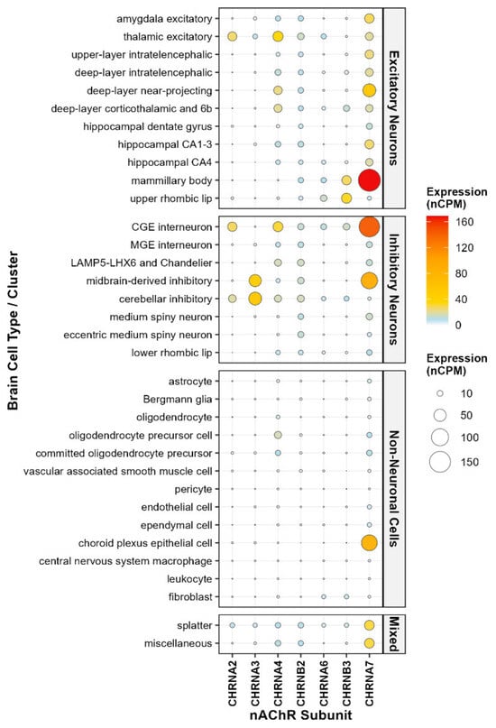
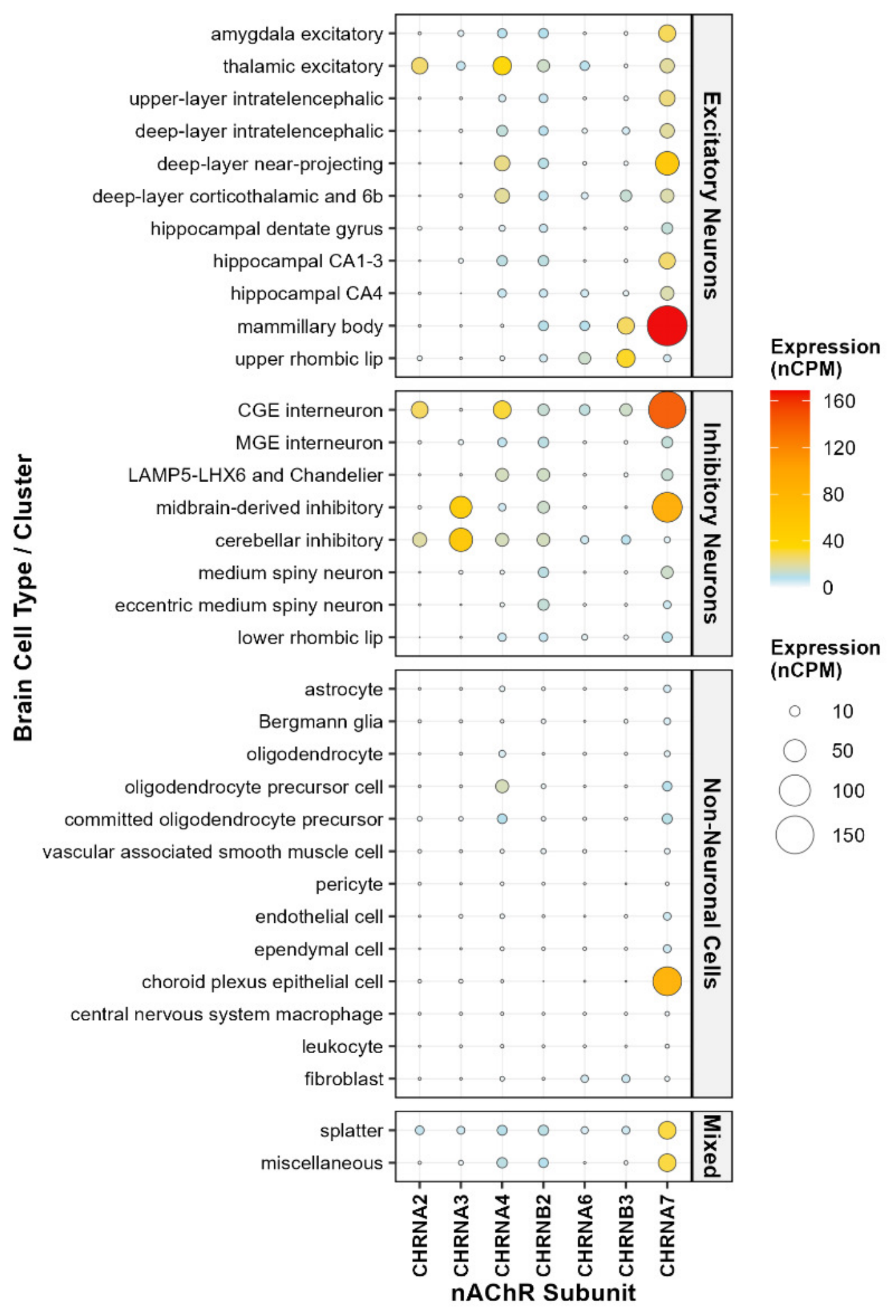
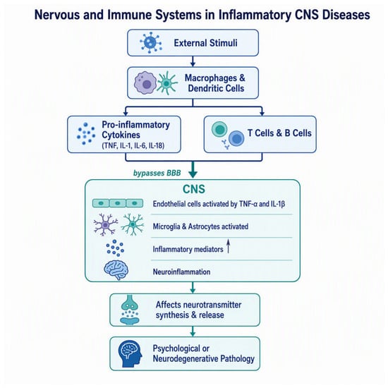
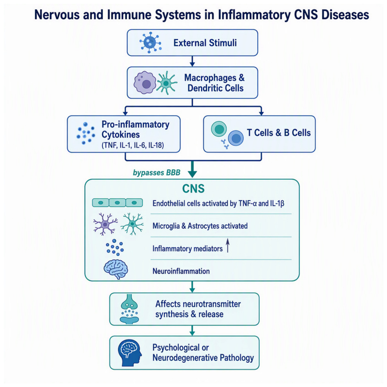
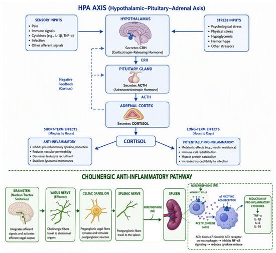
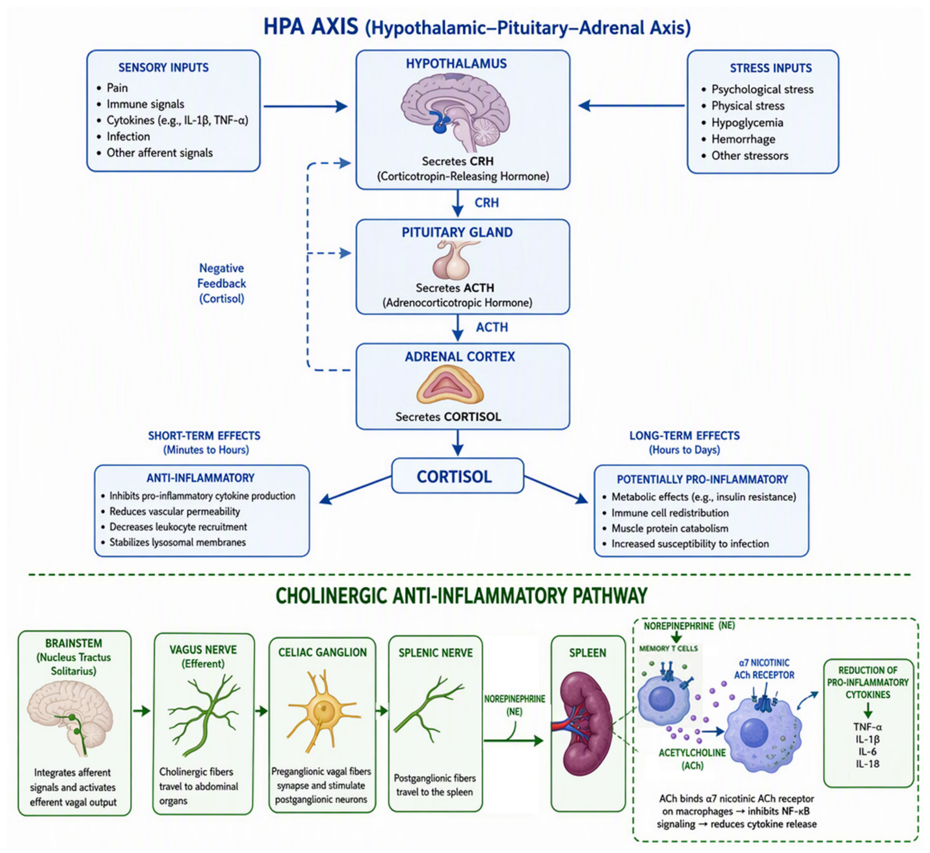

Disclaimer/Publisher’s Note: The statements, opinions and data contained in all publications are solely those of the individual author(s) and contributor(s) and not of MDPI and/or the editor(s). MDPI and/or the editor(s) disclaim responsibility for any injury to people or property resulting from any ideas, methods, instructions or products referred to in the content. |
© 2026 by the authors. Licensee MDPI, Basel, Switzerland. This article is an open access article distributed under the terms and conditions of the Creative Commons Attribution (CC BY) license.